development and application of computational … · jose carlos gómez tamayo mireia dunach masjuan...
TRANSCRIPT

Jose Carlos Gómez Tamayo
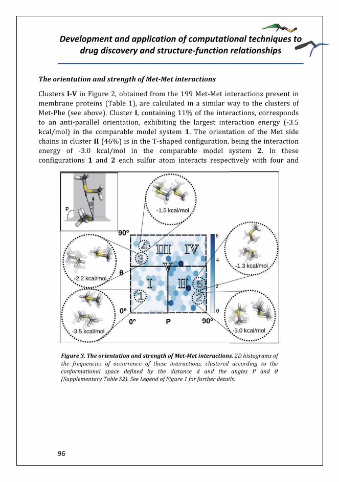
Mireia Dunach MasjuanTutor
Gianluigi Caltabiano Arnau Cordomí Montoya
Director
Tesis doctoral
Departament de Bioquímica i de Biologia Molecular
DEVELOPMENT AND APPLICATION OF COMPUTATIONAL TECHNIQUES TO DRUG DISCOVERY AND STRUCTURE-FUNCTION
RELATIONSHIPS


have permitted to elucidate the crystal structures of many receptors
, which also terms this family of proteins as 7TM receptors


α

αα βγ
β β


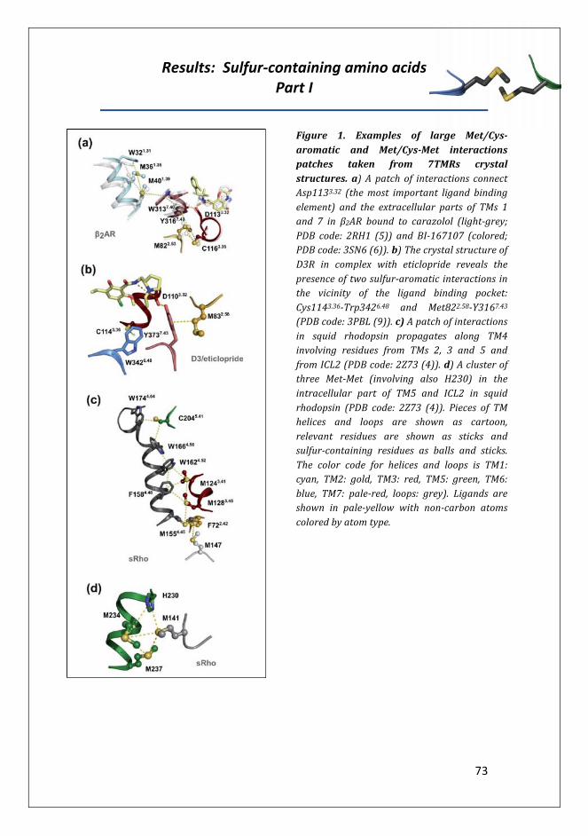
35
36


receptor organism PDB id Met-Aro Met-Met Cys-Aro Cys-Cys Cys-Met totala


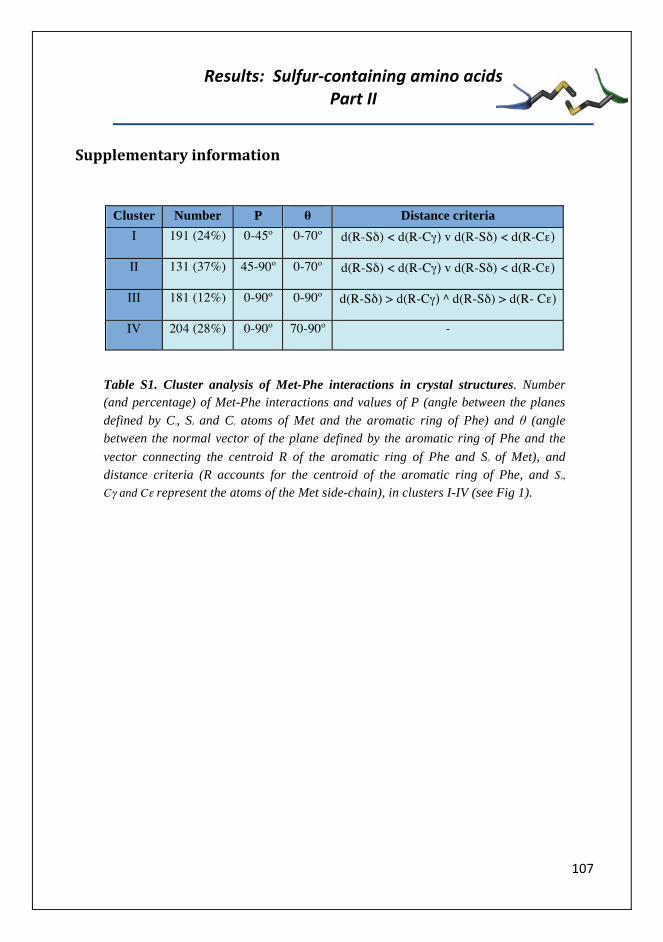
Cluster Number P θ Distance criteria I 191 (24%) 0-45º 0-70º d(R-Sδ) < d(R-Cγ) v d(R-Sδ) < d(R-Cε)
II 131 (37%) 45-90º 0-70º d(R-Sδ) < d(R-Cγ) v d(R-Sδ) < d(R-Cε)
III 181 (12%) 0-90º 0-90º d(R-Sδ) > d(R-Cγ) ^ d(R-Sδ) > d(R- Cε)
IV 204 (28%) 0-90º 70-90º -
Table S1. Cluster analysis of Met-Phe interactions in crystal structures. Number (and percentage) of Met-Phe interactions and values of P (angle between the planes defined by Cγ, Sδ and Cε atoms of Met and the aromatic ring of Phe) and θ (angle between the normal vector of the plane defined by the aromatic ring of Phe and the vector connecting the centroid R of the aromatic ring of Phe and Sδ of Met), and distance criteria (R accounts for the centroid of the aromatic ring of Phe, and Sδ,
Cγ and Cε represent the atoms of the Met side-chain), in clusters I-IV (see Fig 1).

Cluster Number P θ Distance criteria
I 26 (11%) 0-45º 0-60º[d(SδA-SδB) < d(SδA-CγΒ) v d(SδA-SδB) < d(SδA-CεΒ )] ^
[d(SδA-SδB) < d(SδB-CγA v d(SδA-SδB) < d(SδB-CεA )]
II 71 (46%) 45º-90º 0-60º [d(SδA-SδB) < d(SδA-CγΒ) v d(SδA-SδB) < d(SδA-CεΒ )] ^
[d(SδA-SδB) < d(SδB-CγA v d(SδA-SδB) < d(SδB-CεA )]
III 12 (5%) 0-90º 60-90º [d(SδA-SδB) < d(SδA-CγΒ) ^ d(SδA-SδB) < d(SδA-CεΒ )] ^
[d(SδA-SδB) < d(SδB-CγA ^ d(SδA-SδB) < d(SδB-CεA )]
IV 43 (15%) 0-90º 60-90º [d(SδA-SδB) > d(SδA-CγΒ) v d(SδA-SδB) > d(SδA-CεΒ )] ^
[d(SδA-SδB) < d(SδB-CγA v d(SδA-SδB) < d(SδB-CεA )]
V 47 (25%) 0-90º 0-90º [d(SδA-SδB) > d(SδA-CγΒ) ^ d(SδA-SδB) > d(SδA-CεΒ )] ^
[d(SδA-SδB) > d(SδB-CγA v d(SδA-SδB) > d(SδB-CεA )]
Table S2. Cluster analysis of Met-Met interactions in crystal structures. Number (and percentage) of Met-Met interactions and values of P (angle between the planes defined by Cγ, Sδ and Cε atoms of Met) and θ (angle between the normal vector of the plane defined by the Cγ, Sδ and Cε atoms of Met and the vector connecting the Sδ atoms of Met), and distance criteria (sub indexes A and B refer to atoms in distinct side-chains) in clusters I-V (see Fig 2).

Cluster Number P θ Distance criteria
I 26 (11%) 0-45º 0-60º [d(SδA-SδB) < d(SδA-CγΒ) v d(SδA-SδB) < d(SδA-CεΒ )] ^
[d(SδA-SδB) < d(SδB-CγA v d(SδA-SδB) < d(SδB-CεA )]
II 71 (46%) 45º-90º 0-60º [d(SδA-SδB) < d(SδA-CγΒ) v d(SδA-SδB) < d(SδA-CεΒ )] ^
[d(SδA-SδB) < d(SδB-CγA v d(SδA-SδB) < d(SδB-CεA )]
III 12 (5%) 0-90º 60-90º [d(SδA-SδB) < d(SδA-CγΒ) ^ d(SδA-SδB) < d(SδA-CεΒ )] ^
[d(SδA-SδB) < d(SδB-CγA ^ d(SδA-SδB) < d(SδB-CεA )]
IV 43 (15%) 0-90º 60-90º [d(SδA-SδB) > d(SδA-CγΒ) v d(SδA-SδB) > d(SδA-CεΒ )] ^
[d(SδA-SδB) < d(SδB-CγA v d(SδA-SδB) < d(SδB-CεA )]
V 47 (25%) 0-90º 0-90º [d(SδA-SδB) > d(SδA-CγΒ) ^ d(SδA-SδB) > d(SδA-CεΒ )] ^
[d(SδA-SδB) > d(SδB-CγA v d(SδA-SδB) > d(SδB-CεA )]
Table S3. Cluster analysis of Met-Leu interactions in crystal structures. Number (and percentage) of Met-Leu interactions and values of P (angle between the planes defined by the Cγ, Sδ and Cε atoms of Met and the Cδ1, Cγ and Cδ2 atoms of Leu), θ (angle between the normal vector of the plane defined by the Cγ, Sδ and Cε atoms of Met and the vector connecting the Sδ atom of Met and the Cγ atom of Leu) and distance criteria (subindexes A and B refer to atoms in distinct side-chains) in clusters I-V (see Fig 3).

Figure S1. Small-molecule models systems mimicking Met-Phe interactions. Geometry optimized, at the ab-initio MP2/6-31+G(d,p) level of theory, of the interactions between benzene (BNZ, mimicking Phe) and dimethyl sulfide (DMS, mimicking Met), dimethyl ether (DME), and propane (PRP, mimicking Leu). Each optimized structure is designated by an arabic number that corresponds to a roman number of the obtained clusters in crystal structures (see Fig 1). The values of d, P, and θ (see Suppl. Table 1 and Fig 1 for definition), and single point energy calculations at the ab-initio CCSD(T)/6-311+G(3df,2p) level of theory (ECCSD) and by molecular mechanics using the AMBER99 forcefield (EAMBER) are shown.

Figure S2. Small-molecule models systems mimicking Phe-Phe interactions. Geometry optimized models, at the ab-initio MP2/6-31+G(d,p) level of theory, of benzene-benzene (BNZ, mimicking Phe) interactions in the lowest parallel displaced and T-shaped energy configurations. The values of d (calculated as the distance between the centroid R of the aromatic ring of BNZ), P (calculated as the angle between the planes defined by the aromatic rings of BNZ), and θ (angle between the normal vector of the plane defined by the aromatic ring of Phe and the vector connecting the centroids R of the aromatic rings of BNZ), and single point energy calculations at the ab-initio CCSD(T)/6-311+G(3df,2p) level of theory (ECCSD) and by molecular mechanics using the AMBER99 forcefield (EAMBER).

Figure S3. Small-molecule models systems mimicking Met-Met interactions. Geometry optimized models, at the ab-initio MP2/6-31+G(d,p) level of theory, of dimethyl sulfide (DMS, mimicking Met)-DMS and dimethyl ether (DME)-DME interactions. Each energy-minimized structure is designated by an arabic number that corresponds to a roman number of the obtained clusters in crystal structures (see Fig 2). The values of d, P, and θ (see Suppl. Table 2 and Fig 2 for definition), and single point energy calculations at the ab-initio CCSD(T)/6-311+G(3df,2p) level of theory (ECCSD) and by molecular mechanics using the AMBER99 forcefield (EAMBER) are shown.

Figure S4. Small-molecule models systems mimicking Met-Leu interactions. Geometry optimized models, at the ab-initio MP2/6-31+G(d,p) level of theory, of dimethyl sulfide (DMS, mimicking Met) and propane (PRP, mimicking Leu) and PRP-PRP interactions. Each optimized structure is designated by an arabic number that corresponds to a roman number of the obtained clusters in crystal structures (see Fig 3). The values of d, P, and θ (see Suppl. Table 3 and Fig 3 for definition), and single point energy calculations at the ab-initio CCSD(T)/6-311+G(3df,2p) level of theory (ECCSD) and by molecular mechanics using the AMBER99 forcefield (EAMBER) are shown.

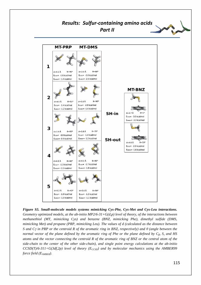
Figure S5. Small-molecule models systems mimicking Cys-Phe, Cys-Met and Cys-Leu interactions. Geometry optimized models, at the ab-initio MP2/6-31+G(d,p) level of theory, of the interactions between methanethiol (MT, mimicking Cys) and benzene (BNZ, mimicking Phe), dimethyl sulfide (DMS, mimicking Met) and propane (PRP, mimicking Leu). The values of d (calculated as the distance between S and Cγ in PRP or the centroid R of the aromatic ring in BNZ, respectively) and θ (angle between the normal vector of the plane defined by the aromatic ring of Phe or the plane defined by C , Sγ and HS atoms and the vector connecting the centroid R of the aromatic ring of BNZ or the central atom of the side-chain to the center of the other side-chain), and single point energy calculations at the ab-initio CCSD(T)/6-311+G(3df,2p) level of theory (ECCSD) and by molecular mechanics using the AMBER99 force field (EAMBER).


117
Development and application of computational techniques to drug discovery and structure-function
relationships
118

119

Development and application of computational techniques to drug discovery and structure-function
relationships
120

121
Development and application of computational techniques to drug discovery and structure-function
relationships
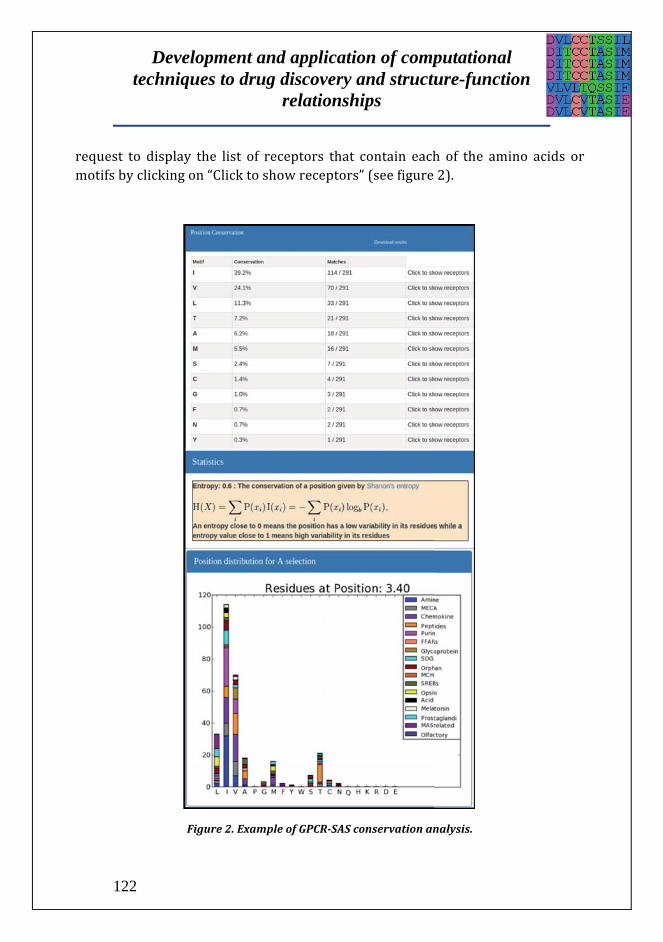
122

123

Development and application of computational techniques to drug discovery and structure-function
relationships
124

125

Development and application of computational techniques to drug discovery and structure-function
relationships
126

127

Development and application of computational techniques to drug discovery and structure-function
relationships
128

129

Development and application of computational techniques to drug discovery and structure-function
relationships
130

131
1. Overington, J. P., Al-Lazikani, B., and Hopkins, A. L. (2006) How many drug targets are there? Nat Rev Drug Discov 5, 993-996
2. Kolakowski, L. F. (1994) GCRDB - A G-protein-coupled receptor database. Receptors & channels 2, 1-7
3. Attwood, T. K., and Findlay, J. B. (1994) Fingerprinting G-protein-coupled receptors. Protein Eng. 7, 195-203
4. Horn, F., Bettler, E., Oliveira, L., Campagne, F., Cohen, F. E., and Vriend, G. (2003) GPCRDB information system for G protein-coupled receptors. Nucleic Acids Res. 31, 294-297
5. Fredriksson, R., Lagerstrom, M. C., Lundin, L. G., and Schioth, H. B. (2003) The G-protein-coupled receptors in the human genome form five main families. Phylogenetic analysis, paralogon groups, and fingerprints. Mol. Pharmacol. 63, 1256-1272
6. Liapakis, G., Cordomi, A., and Pardo, L. (2012) The G-protein coupled receptor family; actors with many faces. Curr Pharm Des. 18, 175-185
7. Katritch, V., Cherezov, V., and Stevens, R. C. (2013) Structure-function of the G protein-coupled receptor superfamily. Annual review of pharmacology and toxicology 53, 531-556
8. Venkatakrishnan, A. J., Deupi, X., Lebon, G., Tate, C. G., Schertler, G. F., and Babu, M. M. (2013) Molecular signatures of G-protein-coupled receptors. Nature 494, 185-194
9. Deupi, X., Dolker, N., Lopez-Rodriguez, M. L., Campillo, M., Ballesteros, J. A., and Pardo, L. (2007) Structural models of class a G protein-coupled receptors as a tool for drug design: insights on transmembrane bundle plasticity. Curr. Top. Med. Chem. 7, 991-998
10. Ballesteros, J. A., and Weinstein, H. (1995) Integrated Methods for Modeling G-Protein Coupled Receptors. Methods Neurosci. 25, 366-428
11. Isberg, V., de Graaf, C., Bortolato, A., Cherezov, V., Katritch, V., Marshall, F. H., Mordalski, S., Pin, J. P., Stevens, R. C., Vriend, G., and Gloriam, D. E. (2015) Generic GPCR residue numbers - aligning topology maps while minding the gaps. Trends in pharmacological sciences 36, 22-31
12. Siu, F. Y., He, M., de Graaf, C., Han, G. W., Yang, D., Zhang, Z., Zhou, C., Xu, Q., Wacker, D., Joseph, J. S., Liu, W., Lau, J., Cherezov, V., Katritch, V., Wang, M. W., and Stevens, R. C. (2013) Structure of the human glucagon class B G-protein-coupled receptor. Nature 499, 444-449
13. Spyridaki, K., Matsoukas, M. T., Cordomi, A., Gkountelias, K., Papadokostaki, M., Mavromoustakos, T., Logothetis, D. E., Margioris, A. N., Pardo, L., and

Development and application of computational techniques to drug discovery and structure-function
relationships
132
Liapakis, G. (2014) Structural-Functional Analysis of the Third Transmembrane Domain of the Corticotropin-releasing Factor Type 1 Receptor: Role In Activation And Allosteric Antagonism. The Journal of biological chemistry 289, 18966-18977
14. Sievers, F., Wilm, A., Dineen, D., Gibson, T. J., Karplus, K., Li, W., Lopez, R., McWilliam, H., Remmert, M., Soding, J., Thompson, J. D., and Higgins, D. G. (2011) Fast, scalable generation of high-quality protein multiple sequence alignments using Clustal Omega. Molecular systems biology 7, 539
15. Gonzalez, A., Cordomi, A., Caltabiano, G., and Pardo, L. (2012) Impact of helix irregularities on sequence alignment and homology modeling of G protein-coupled receptors. Chembiochem : a European journal of chemical biology 13, 1393-1399
16. Becu, J. M., Pele, J., Rodien, P., Abdi, H., and Chabbert, M. (2013) Structural evolution of G-protein-coupled receptors: a sequence space approach. Methods in enzymology 520, 49-66
17. Fredriksson, R., Lagerstrom, M. C., Lundin, L. G., and Schioth, H. B. (2003) The G-protein-coupled receptors in the human genome form five main families. Phylogenetic analysis, paralogon groups, and fingerprints. Molecular Pharmacology 63, 1256-1272
18. Vroling, B., Sanders, M., Baakman, C., Borrmann, A., Verhoeven, S., Klomp, J., Oliveira, L., de Vlieg, J., and Vriend, G. (2011) GPCRDB: information system for G protein-coupled receptors. Nucleic Acids Research 39, D309-319
19. Davies, M. N., Secker, A., Freitas, A. A., Mendao, M., Timmis, J., and Flower, D. R. (2007) On the hierarchical classification of G protein-coupled receptors. Bioinformatics 23, 3113-3118
20. Surgand, J. S., Rodrigo, J., Kellenberger, E., and Rognan, D. (2006) A chemogenomic analysis of the transmembrane binding cavity of human G-protein-coupled receptors. Proteins 62, 509-538
21. Deville, J., Rey, J., and Chabbert, M. (2009) An indel in transmembrane helix 2 helps to trace the molecular evolution of class A G-protein-coupled receptors. Journal of molecular evolution 68, 475-489
22. Pawson, A. J., Sharman, J. L., Benson, H. E., Faccenda, E., Alexander, S. P., Buneman, O. P., Davenport, A. P., McGrath, J. C., Peters, J. A., Southan, C., Spedding, M., Yu, W., Harmar, A. J., and Nc, I. (2014) The IUPHAR/BPS Guide to PHARMACOLOGY: an expert-driven knowledgebase of drug targets and their ligands. Nucleic Acids Res 42, D1098-1106
23. Davies, M., Nowotka, M., Papadatos, G., Dedman, N., Gaulton, A., Atkinson, F., Bellis, L., and Overington, J. P. (2015) ChEMBL web services:

133
streamlining access to drug discovery data and utilities. Nucleic Acids Res 43, W612-620
24. Shannon, C. E. (1948) A Mathematical Theory of Communication. Bell System Technical Journal 27, 379-423
25. Fodor, A. A., and Aldrich, R. W. (2004) On evolutionary conservation of thermodynamic coupling in proteins. J Biol Chem 279, 19046-19050
26. Pele, J., Moreau, M., Abdi, H., Rodien, P., Castel, H., and Chabbert, M. (2014) Comparative analysis of sequence covariation methods to mine evolutionary hubs: examples from selected GPCR families. Proteins 82, 2141-2156
27. Pele, J., Abdi, H., Moreau, M., Thybert, D., and Chabbert, M. (2011) Multidimensional scaling reveals the main evolutionary pathways of class A G-protein-coupled receptors. PloS one 6, e19094
28. Scheerer, P., Park, J. H., Hildebrand, P. W., Kim, Y. J., Krauss, N., Choe, H. W., Hofmann, K. P., and Ernst, O. P. (2008) Crystal structure of opsin in its G-protein-interacting conformation. Nature 455, 497-502
29. Rasmussen, S. G., Devree, B. T., Zou, Y., Kruse, A. C., Chung, K. Y., Kobilka, T. S., Thian, F. S., Chae, P. S., Pardon, E., Calinski, D., Mathiesen, J. M., Shah, S. T., Lyons, J. A., Caffrey, M., Gellman, S. H., Steyaert, J., Skiniotis, G., Weis, W. I., Sunahara, R. K., and Kobilka, B. K. (2011) Crystal structure of the beta(2) adrenergic receptor-Gs protein complex. Nature

Development and application of computational techniques to drug discovery and structure-function
relationships
134

135

136

137

138

139

140

141

142
±±
± ±
± ±
±
±
± ±
± ±
±
±
±±

143
144
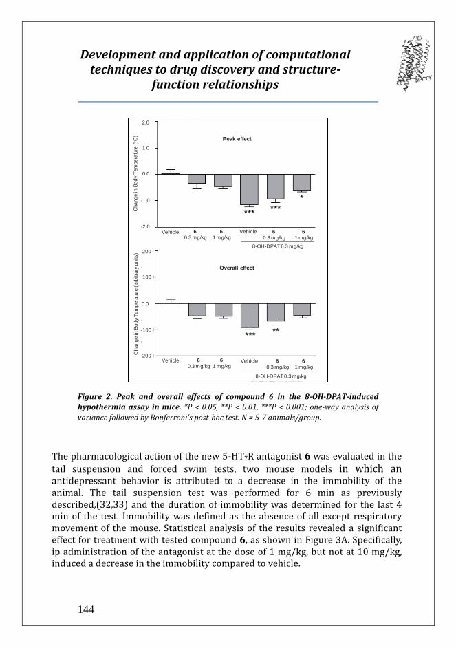
0.0
Cha
nge
in B
ody
Tem
pera
ture
(arb
itrar
y un
its)
-200
200
100
-100
Overall effect
60.3 mg/kg
Vehicle 61 mg/kg
60.3 mg/kg
61 mg/kg
Vehicle
8-OH-DPAT 0.3 mg/kg
Peak effect
2.0
1.0
0.0
-1.0
-2.0
Cha
nge
in B
ody
Tem
pera
ture
(°C
)
60.3 mg/kg
61 mg/kg
Vehicle
8-OH-DPAT 0.3 mg/kg
Vehicle 60.3 mg/kg
61 mg/kg
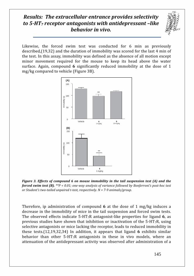
145
61 mg/kg
150
Vehicle
100
50
06
10 mg/kg
Imm
obilit
y (s
)
(A)
150
100
50
06
1 mg/kgVehicle
Imm
obilit
y (s
)
(B)

146

147

148

149

150
151

152
153

154

155

156

157

158