development of cationic niosomes containing novel ... · the graduate school, silpakorn university...
TRANSCRIPT

DEVELOPMENT OF CATIONIC NIOSOMES CONTAINING NOVEL SYNTHESIZED CATIONIC LIPIDS FOR NUCLEIC ACID DELIVERY
By Miss Orapan Paecharoenchai
A Thesis Submitted in Partial Fulfillment of the Requirements for the Degree
Doctor of Philosophy Program in Pharmaceutical Technology
Graduate School, Silpakorn University
Academic Year 2013
Copyright of Graduate School, Silpakorn University

DEVELOPMENT OF CATIONIC NIOSOMES CONTAINING NOVEL SYNTHESIZED CATIONIC LIPIDS FOR NUCLEIC ACID DELIVERY
By Miss Orapan Paecharoenchai
A Thesis Submitted in Partial Fulfillment of the Requirements for the Degree
Doctor of Philosophy Program in Pharmaceutical Technology Graduate School, Silpakorn University
Academic Year 2013
Copyright of Graduate School, Silpakorn University

การพฒนานโอโซมประจบวกทมลพดประจบวกสงเคราะหชนดใหมสาหรบนาสงกรดนวคลอก
โดย นางสาวอรพรรณ แพะเจรญชย
วทยานพนธนเปนสวนหนงของการศกษาตามหลกสตรปรญญาเภสชศาสตรดษฎบณฑต สาขาวชาเทคโนโลยเภสชกรรม
บณฑตวทยาลย มหาวทยาลยศลปากร ปการศกษา 2556
ลขสทธของบณฑตวทยาลย มหาวทยาลยศลปากร

The Graduate School, Silpakorn University has approved and accredited the thesis title of “Development of cationic niosomes containing novel synthesized cationic lipids for nucleic acid delivery” submitted by Miss Orapan Paecharoenchai as a partial fulfillment of the requirements for the degree of Doctor of Philosophy in Pharmaceutical Technology.
……...............................................................................
(Associate Professor Panjai Tantatsanawong, Ph.D.)
Dean of Graduate School
........../..................../.......... The Thesis Advisors
1. Associate Professor Praneet Opanasopit, Ph.D.
2. Associate Professor Auayporn Apirakaramwong, Ph.D.
3. Associate Professor Tanasait Ngawhirunpat, Ph.D. The Thesis Examination Committee
………………………….….…….. Chairman
(Associate Professor Theerasak Rojanarata, Ph.D.)
............../................./............. .................................................... Member
(Assistant Professor Warisada Sila-on, Ph.D.)
............/......................../..............
......................................................... Member
(Associate Professor Praneet Opanasopit, Ph.D.)
............/......................../..............
.................................................... Member
(Associate Professor Auayporn Apirakaramwong, Ph.D.)
............/......................../..............
………………………….….…….. Member
(Associate Professor Tanasait Ngawhirunpat, Ph.D.)
............../................./.............

iv
52354807 : MAJOR : PHARMACEUTICAL TECHNOLOGY
KEY WORD : GENE DELIVERY SYSTEM/SIRNA/NIOSOMES/LIPOSOMES/CATIONIC LIPIDS
ORAPAN PAECHAROENCHAI : DEVELOPMENT OF CATIONIC NIOSOMES CONTAINING NOVEL SYNTHESIZED CATIONIC LIPIDS FOR NUCLEIC ACID DELIVERY. THESIS ADVISORS : ASSOC. PROF. PRANEET OPANASOPIT, Ph.D., ASSOC.PROF. AUAYPORN APIRAKARAMWONG, Ph.D., AND ASSOC. PROF. TANASAIT NGAWHIRUNPAT. 175 pp.
The aim of this study was to formulate the cationic niosomes containing novel synthesized
cationic lipids which can mediate high gene/siRNA transfection, as well as low cytotoxicity in vitro.
In the preliminary study, cationic liposomes were formulated for screening of the structure-
transfection activity relationship of the cationic lipids. The in vitro gene transfection of the cationic
liposomes was performed on a human cervical carcinoma cell line (HeLa cells) using the pDNA
encoding green fluorescent protein (pEGFP-C2). To find out an optimal niosomes formulation
which has similar physical characteristics with liposomes, the niosomes containing non-ionic
surfactants (Tween 20, Tween 80, Span20 and Span 80) and cholesterol were formulated in
various ratios and the turbidity at 400 nm as well as particle size of niosomes were assayed and
compared to liposomes. Six spermine-cationic lipids with differences in hydrophobic part; number
of acyl chain (2 tails and 4 tails) and acyl chain length (C14, C16 and C18) were chosen to prepare
cationic niosomes. The cationic niosomes containing Span20: cholesterol: spermine-cationic lipid
(2.5: 2.5: 1 mM) were successfully prepared and used for gene/siRNA delivery. Gene transfection
mediated by cationic niosomes was affected by cationic lipid structure, as well as weight ratio of
niosomes/DNA. High transfection efficiency observed in HeLa cells was resulted from cationic
niosomes composed of spermine-derivative cationic lipids which are N1,N1-dimyristeroyloxyethyl-
spermine (A10) and Tetra- (N1, N1, N14, N14-steroyloxyethyl)-spermine (A29) at weight ratio of 10
for both lipids. The cationic niosomes also showed low cytotoxicity, as well as haemolytic effect. In
addition, these cationic niosomes could be form complex with siRNA and induced GFP gene
silencing in HeLa cells stably expressing green fluorescent protein (EGFP). The possible cellular
uptake mechanisms of the cationic niosomes were evaluated using inhibitors on various endocytic
pathways. Results from this study implied that the niosomes/DNA complexes might be internalized
predominantly by caveolae- and clathrin-independent pathway. After internalized, the acidification
of endosome-lysosome system is required for pH-dependent endosomal escape, and thus
believed to entry nucleus during mitosis cell division, finally gene expression occurs. The physical
stability study in terms of size and zeta potential measurement revealed that these cationic niosomes were
physically stable at 4 °C for 1 month. Consequently, the cationic niosomes show a promising potential as
gene/siRNA carriers in vitro.
Program of Pharmaceutical Technology Graduate School, Silpakorn University
Student's signature ........................................ Academic Year 2013
Thesis Advisors’ signature 1. .......................... 2. ............................ 3…………………………..

v
52354807 : สาขาวชาเทคโนโลยเภสชกรรม คาสาคญ : ระบบนาสงยน/เอสไออารเอนเอ/นโอโซม/ลโพโซม/ลพดประจบวก อรพรรณ แพะเจรญชย : การพฒนานโอโซมประจบวกทมลพดประจบวกสงเคราะหชนดใหมสาหรบนาสงกรดนวคลอก. อาจารยทปรกษาวทยานพนธ : ภญ.รศ.ดร.ปราณต โอปณะโสภต, ภญ.รศ.ดร.อวยพร อภรกษอรามวง และ ภก.รศ.ดร.ธนะเศรษฐ งาวหรญพฒน. 175 หนา. การศกษาในครงนมวตถประสงคเพอตงตารบนโอโซมประจบวกทมลพดประจบวกสงเคราะหชนดใหม ซงมประสทธภาพสงในการนาสงยน/เอสไออารเอนเอเขาสเซลลเพาะเลยง รวมทงมความเปนพษตอเซลลตา ในการทดสอบเบองตนไดเตรยมตารบลโพโซมประจบวก เพอนามาใชทดสอบหาความสมพนธระหวางโครงสรางทางเคมของลพดประจบวกกบความสามารถในการถายโอนยนเขาสเซลล ในการศกษาการถายโอน ยนเขาสเซลลเพาะเลยงของลโพโซมประจบวกนน ศกษาในเซลลเพาะเลยง human cervical carcinoma cells (HeLa cells) โดยใชพลาสมดดเอนเอทสามารถแปลรหสไดเปน green
fluorescent protein (pEGFP-C2) การศกษาเบองตนเพอหาสตรตารบนโอโซมทเหมาะสมซงมคณสมบตทางกายภาพคลายคลงกบลโพโซม เตรยมตารบนโอโซมซงประกอบดวยสารลดแรงตงผวชนดไมมประจ (Tween 20, Tween 80, Span 20 และ Span
80) และคอเรสเตอรอลทอตราสวนตางๆ โดยทาการวเคราะหคณสมบตทางกายภาพของนโอโซม ไดแก ความขนทความยาวคลน นาโนเมตรและขนาดอนภาคเปรยบเทยบกบลโพโซม จากนนไดคดเลอกลพดประจบวกชนดทเปนอนพนธสเปอรมนจานวน ชนด ทมความแตกตางกนในสวนโครงสรางทไมชอบนา ไดแก จานวนสายไฮโดรคารบอน (2 สาย และ4 สาย) และ ความยาวของสายไฮโดรคารบอน (C14 C16 และC18) เพอนามาเตรยมเปนนโอโซมประจบวก สามารถเตรยมนโอโซมประจบวกไดสาเรจโดยประกอบดวย Span20 คอเรสเตอรอล และลพดประจบวกอนพนธสเปอรมน (อตราสวน 2.5:2.5:1 มลลโมลาร) และใชในการนาสงยน/เอสไออารเอนเอได ศกษาปจจยทมผลตอการถายโอนยนโดยอาศยตวพาทเปนนโอโซมประจบวก ไดแก โครงสรางของลพดประจบวกและอตราสวนของนโอโซมตอดเอนเอ พบวานโอโซมประจบวก ชนด ทประกอบดวยลพดประจบวกอนพนธสเปอรมน N1,N1-dimyristeroyloxyethyl-spermine (A10) และ Tetra- (N1, N1, N14, N14-steroyloxyethyl)-
spermine (A29) มประสทธภาพในการถายโอนยนเขาสเซลล HeLa ดทสด เมอใชอตราสวนนโอโซมตอดเอนเอโดยนาหนกของเทากบ 10 โดยนโอโซมประจบวกดงกลาวยงมความเปนพษตอเซลล และความสามารถในการทาใหเมดเลอดแดงแตกทตา นอกจากน นโอโซมประจบวกทง ชนดนยงสามารถเกดสารประกอบเชงซอนกบเอสไออารเอนเอไดและยงสามารถเหนยวนาใหเกดการยบยงการทางานของยน GFP ในเซลลเพาะเลยง HeLa cells stably expressing green fluorescent protein (EGFP) ไดอกดวย จากการทดสอบกลไกการนาสงยนเขาสเซลลของนโอโซมประจบวก โดยใชสารยบยงการเขาสเซลลโดยผานกระบวนการเอนโดไซโตซสชนดตางๆ พบวา สารประกอบเชงซอนระหวางนโอโซมประจบวกกบดเอนเอ อาจนาเขาสเซลลโดยผานกระบวนการหลกคอ caveolae- และ clathrin-independent เมอเขาสเซลลแลวจะตองเกด acidification of endosome-
lysosome system ซงจาเปนตอการหลบหนออกจากเอนโดโซม จากนนจงเคลอนทเขาสนวเคลยสในระหวางทเซลลเกดการแบงตวแบบไมโตซส และเกดการแสดงออกของยนในทสด การศกษาความคงตวทางกายภาพโดยวดขนาดอนภาคและประจบนพนผวของนโอโซม พบวามความคงตวดเมอเกบทอณหภม 4 องศาเซลเซยสเปนเวลาอยางนอย 1 เดอน ดงนนนโอโซมประจบวกทพฒนาขนนมศกยภาพสาหรบใชในการนาสงยน/เอสไออารเอนเอเขาสเซลลเพาะเลยงได
เทคโนโลยเภสชกรรม บณฑตวทยาลย มหาวทยาลยศลปากรลายมอชอนกศกษา....................................... ปการศกษา 2556 ลายมอชออาจารยทปรกษาวทยานพนธ 1………...................... 2................................ 3. ...................................

vi
ACKNOWLEDGMENTS
This thesis would not have been possible without the help and support of many
people, whom I wish to acknowledge in this section. First of all, I would like to express my
sincere appreciation to my advisor, Assoc. Prof. Dr. Praneet Opanasopit for her supervision,
support and invaluable suggestion throughout my PhD study. My sincere gratitude also goes
to my co-advisors, Assoc. Prof. Dr. Auayporn Apirakaramwong and Assoc. Prof. Dr. Tanasait
Ngawhirunpat for valuable advice and support, as well as my co-advisor at the College of
Pharmacy, Ohio State University, USA, Prof. Dr. Robert J Lee for his valuable support and
suggestion.
Besides my advisors, I would like to thank my thesis committees, Assoc. Prof. Dr.
Theerasak Rojanarata and Asst. Prof. Dr. Warisada Sila-on for giving me insightful
comments and suggestions. I also wish to thank Asst. Prof. Dr. Sunee Techaarpornkul for
helping me cope with siRNA experiment.
I am grateful to acknowledge the Thailand Research Funds through the Golden
Jubilee Ph.D. Program (Grant No. PHD/0092/2551) for the financial support throughout my
study.
I would like to thank my collaborators at the Faculty of Sciences, Ramkhamhaeng
University, Asst. Prof. Dr. Boon-ek Yingyongnarongkul and Miss Nattisa Niyomtham for
helping me synthesized the cationic lipids used in this work.
My special thanks goes to my friends and members of the Pharmaceutical
Development of Green Innovation Group (PDGIG), especially Miss Jintana
Tragulpakseerojn, Miss Areerut Sripattanaporn, Dr. Natthan Charernsriwilaiwat, Dr.
Sureewan Duangjit and Miss Kotchamon Yodkhum for invaluable help, encouragement and
our friendship. I wish to thank the staffs at the Faculty of Pharmacy, Silpakorn University for
their help. I also would like to acknowledge Mr.Worraphan Phisittanachot for helping me
resolve my computer problems.
Finally, my deepest appreciation goes to my beloved family, Mr. Laem
Paecharoenchai, Mrs. Mantana Paecharoenchai, Miss. Wanwimol
Paecharoenchai, Miss Waleeporn Paecharoenchai and Miss Noppawan
Paecharoenchai. Thank you for everything, especially your love, care,
encouragement and support all the time.

vii
TABLE OF CONTENTS
Page
English Abstract ……………………………………………………………… iv
Thai Abstract ….……………………………………………………………… v
Acknowledgements…………………………………………………………… vi
List of Tables………..………………………………………………………… viii
List of Figures ………………….……………………………………………… ix
List of Abbreviations.…………………………….…………………………… xiii
Chapter
1 Introduction….…….………………………………………………….. 1
2 Literature Reviews……………………………………………………. 6
3 Materials and Methods……………………………………………….. 62
4 Results and Discussion……………………………………………….. 96
5 Conclusions…………………………………………………………… 148
References….………………………………………..………………………... 150
Appendix……………………………………………………………………… 163
Biography ……………………………………………………………………... 173

viii
LIST OF TABLES
Table Page
1 Disease targets for gene therapy: monogenic diseases………………. 7
2 Perturbation of endocytosis and intracellular trafficking.……………. 48
3 Commercial reagents for in vitro siRNA transfection in eukaryotic cells 54
4 Pharmaceutical applications of niosomes …………………………… 58
5 Niosomes-based gene/siRNA delivery systems……………………… 61
6 Compositions of QIAGEN Plasmid Midi Kits……………………... 62
7 Diethanolamine-based cationic lipids: A1-A29.……………………... 64
8 Cholesterol derivatives cationic lipids: C1-C72…………………...… 69
9 Sub-classified diethanolamine-based cationic lipids (A1-A29)……… 97
10 Sub-classified cholesterol derivative cationic lipids (C1-C72)……… 98
11 The cationic lipids provided high gene transfection in HeLa cells.…. 104
12 Cytotoxicity of liposomes A1-A29 in HeLa cells.…………………… 105
13 Cytotoxicity of liposomes C1-C72 in HeLa cell……………………… 106
14 Turbidity, particle size and zeta potential of liposomes and niosomes.. 117
15 The particle size and zeta potential of niosomes formulations……..... 119
16 Cytotoxicity of niosomes in HeLa cells……………………………… 120

ix
LIST OF FIGURES
Figure Page
1 The extracellular and intracellular barriers faced by non-viral gene
therapies following systematic delivery……………………….. 8
2 Current systems are invariably taken up into endosome……………. 14
3 The cationic polymers commonly used for gene delivery………….. 26
4 Hypothesis of endosomal escape of polyplexes gene delivery
systems (proton- sponge effect)……….................…………….. 27
5 Basic components of cationic lipids………………………………… 30
6 Structure of current cationic lipids used in gene therapy and the
helper lipid……………………………………………………... 31
7 Schematic representation of the phase structure of cationic lipids as
a function of their packing parameter……………….…………. 32
8 Schematic representation of the different hurdles encountered by a
gene delivery system to enter and traffic into a tumor cell…………. 36
9 Hypothesis of endosomal escape of lipoplexes gene delivery system 37
10 Classification of endocytosis based on endocytosis proteins that are
involved in the initial entry of particles and solutes…………… 38
11 Different mechanisms of endocytosis. ………..……………………. 40
12 Mechanism of RNA interference ……………………………….….. 51
13 Non-ionic surfactant commonly used to prepare niosomes………… 57
14 Restriction Map and Multiple Cloning Site (MCS) of pEGFP-C2…. 82
15 Cultivated HeLa cells and HeLa stable cells ….……………………. 85
16 Transfection efficiency of liposomes (A1-A9)/DNA complexes in
HeLa cells……………………………………………………… 99
17 Transfection efficiency of liposomes (A10-21)/DNA complexes in
HeLa cells…………………………...…………………………. 99

x
LIST OF FIGURES
Figure Page
18 Transfection efficiency of liposomes (A22-A29)/DNA complexes
in HeLa cells…………………………………………………… 100
19 Transfection efficiency of liposomes (C1-C12)/DNA complexes in
HeLa cells…………………………………………………….... 100
20 Transfection efficiency of liposomes (C13-C24)/DNA complexes in
HeLa cells……………………………………………………… 101
21 Transfection efficiency of liposomes (C25-C36)/DNA complexes in
HeLa cells……………………………………………………… 101
22 Transfection efficiency of liposomes (C37-C48)/DNA complexes in
HeLa cells……………………………………………………… 102
23 Transfection efficiency of liposomes (C49-C60)/DNA complexes in
HeLa cells……………………………………………………… 102
24 Transfection efficiency of liposomes (C61-C72)/DNA complexes in
HeLa cells……………………………………………………… 103
25 Cytotoxicity of liposomes (A1-A9)/DNA complexes in HeLa cells…….. 107
26 Cytotoxicity of liposomes (A10-A21)/DNA complexes in HeLa cells…… 108
27 Cytotoxicity of liposomes (A22-A29)/DNA complexes in HeLa cells…… 109
28 Cytotoxicity of liposomes (C1-C16)/DNA complexes in HeLa cells…….. 110
29 Cytotoxicity of liposomes (C17-C32)/DNA complexes in HeLa cells…… 111
30 Cytotoxicity of liposomes (C33-C48)/DNA complexes in HeLa cells….... 112
31 Cytotoxicity of liposomes (C49-C64)/DNA complexes in HeLa cells…... 113
32 Cytotoxicity of liposomes (C65-C72)/DNA complexes in HeLa cells….... 114
33 Turbidity at 400 nm of niosomes formulated with non-ionic
surfactant and cholesterol....................................................................
115
34 Chemical structures of spermine-based cationic lipids...…………… 118

xi
LIST OF FIGURES
Figure Page
35 The particle size of cationic niosomes after storage at 4˚C for 180
days and 25˚C for 30 days……………………………………... 121
36 The zeta potential of cationic niosomes after storage at 4˚C for 180
days and 25˚C for 30 days……………………………………... 122
37 Particle size and Zeta potential at varying weight ratios of
niosomes/DNA complexes…………………………………….. 124
38 Gel retardation assay of niosomes/DNA complexes………………... 126
39 The transmission electron microscope (TEM) images of the cationic
niosomes (A10, A11 and A12) and niosomes/DNA complexes 127
40 The transmission electron microscope (TEM) images of the cationic
niosomes (A27, A28 and A29) and niosomes/DNA complexes 128
41 Green fluorescent proteins expressed in HeLa cells after transfected
with niosomes-A29/DNA complexes at a weight ratio of 10
under inverted microscope…………………………………….. 129
42 In vitro transfection efficiency of niosomes (A10, A11 and
A12)/DNA complexes with weight ratios of 2.5-100 at pH 7.4 in
HeLa cells………………………………………………………….... 130
43 In vitro transfection efficiency of niosomes (A27, A28 and
A29)/DNA complexes with weight ratios of 2.5-100 at pH 7.4
in HeLa cells…………………………………………………… 131
44 In vitro transfection efficiency of niosomes/DNA complexes in
HeLa cells at pH 7.4 in the absence of serum and presence of
10 % serum…………………………………………………….. 134
45 The percentage cell viability of niosomes/DNA complexes with
weight ratios of 2.5-100 at pH 7.4 in HeLa cells………………. 136

xii
LIST OF FIGURES
Figure Page
46 Haemolytic activity of cationic niosomes (A10, A11 and A12) and
niosomes/DNA complexes…………………………………….. 137
47 Haemolytic activity of cationic niosomes (A27, A28 and A29) and
niosomes/DNA complexes…………………………………….. 138
48 Transfection efficiency of niosomes/DNA complexes; A)
niosomes-A10 and B) niosomes-A29………………………….. 141
49 Particle size and zeta potential at varying weight ratios of
niosomes/siRNA complexes; A) niosomes-A10 and B)
niosomes-A29………………………………………………….. 143
50 Gel retardation assay of niosomes/siRNA complexes……………… 144
51 Percentage of EGFP gene silencing at day 4 of transfection by
niosomes/siRNA complexes; A) niosomes-A10 and B)
niosomes-A29 in HeLa cells stably expressing EGFP………… 146
52 Cell viability of niosomes/siRNA complexes; A) niosomes-A10 and
B) niosomes-A29 in HeLa cells stably expressing EGFP……... 147
53 IC50 data of niosomes A10 in HeLa cells…………………………… 169
54 IC50 data of niosomes A11 and A12 in HeLa cells…………………. 169
55 IC50 data of niosomes A27, A28 and A29 in HeLa cells…………… 170
56 Cytotoxicity of various endocytic inhibitors (incubation time 5 h)… 172

xiii
LIST OF ABBREVIATIONS
cm centimeter(s)
cm2 square centimeter
C degree Celsius
DNA deoxyribonucleic acid
etc. et cetera (Latin); for example, such as
et al. and others
Eq. equation
g gram(s)
h hour(s)
i.e. id est (Latin); that is
k kilo(s)
kDa kilodalton(s)
kg kilogram (s)
L liter(s)
mg milligram(s)
min minute(s)
μm micrometer(s)
ml milliliter(s)

xiv
LIST OF ABBREVIATIONS
mm millimeter(s)
mM millimolar(s)
mmol millimole(s)
mV millivolt(s)
MW molecular weight
ng nanogram(s)
nm nanometer(s)
nM nanomolar(s)
nmol nanomole(s)
OD optical density
pDNA plasmid of deoxyribonucleic acid
PBS phosphate -buffered saline
PC phosphatidylcholine
pH potentia hydrogenii (Latin); power of hydrogen, the
negative logarithm of the hydrogen ion concentration
RNA ribonucleic acid
RNAi RNA interference
R2 coefficient of determination

xv
LIST OF ABBREVIATIONS
rpm revolutions per minute or rounds per min
siRNA short interfering ribonucleic acid
SD standard deviation
TEM transmission electron microscopy
UV ultraviolet (spectroscopy)
v/v volume by volume
w/w weight by weight
w/v weight by volume
> more than
< less than
% percentage
%RH percent relative humidity
wavelength

1
CHAPTER 1
INTRODUCTION
1.1 Statement and significance of the research problem
Gene therapy has become a promising approach for treating incurable diseases
such as genetic diseases and non-genetic diseases such as cardiovascular diseases,
autoimmune diseases and cancer by either correcting the genetic defects or
overexpressing therapeutically useful proteins [1]. The prerequisites for successful
gene therapy include not only therapeutically suitable genes but also the development
of safe and efficient gene delivery systems [2]. Gene carriers can be categorized into
two major groups: viral vectors and non-viral vectors. Because of the severe side
effects caused by viral vectors, such as immunogenicity, mutagenesis, and
carcinogenesis, non-viral vectors serve as a viable alternative.
Among the non-viral vectors, cationic liposomes, the vesicular system
prepared from cationic lipids have been widely investigated as potentially efficacious
gene carriers [3-4] due to their ability to form complexes with anionic DNA
molecules and supposed to deliver DNA into the cells via the endosomal pathway [5-
6]. Cationic liposome-mediated gene delivery is affected by numerous factors, and
one of the major factors is the composition of the liposomes. Moreover, the altering
type and amount of cationic lipid composed in the cationic liposomes can enhance the
transfection efficiency [3]. Additionally, several studies have reported that another
vesicular system, non-ionic surfactant vesicles (niosomes), can also be used as an
alternative to liposomes for gene delivery [7-8].
Niosomes are formed from the self-assembly of non-ionic amphiphiles in
aqueous environment resulting in closed bilayer structures [9]. Niosomes are usually
prepared from single chain surfactant molecules such as sorbitan fatty acid esters
(Span™), polyoxyethylenesorbitan fatty acid ester (Tween™), polyoxyethylene alkyl
ethers (Brij™) and polyoxyethylene alkyl esters, often in combination with
cholesterol [10].

2
Cholesterol is known to abolish the gel to liquid phase transition of niosomes
systems resulting in niosomes that are less leaky. Cholesterol is thus generally
included in a 1:1 molar ratio in most niosomes formulations [9, 11]. Niosomes can be
prepared in the same way as liposomes and have similar structures and physical
properties. However, niosomes have several advantages over liposomes including low
cost of production, ease of storing non-ionic surfactants, uniform content, high purity
and greater stability [12].
As a gene carrier, cationic niosomes are usually composed of non-ionic
surfactants (i.e., Tween™ and Span™), cholesterol and cationic lipids [13]. Similar to
cationic liposomes, gene transfection mediated by cationic niosomes is affected by the
niosomes compositions, including the types of surfactants and cationic lipids used [7-
8].
Since first being used for gene therapy in 1987 by Felgner et al., a wide
variety of cationic lipids have been synthesized and investigated as transfection
reagents [14]. Most cationic lipids used as transfection reagents usually contain three
parts: i) hydrophobic group such as alkyl chain, fatty acid chain and cholesteryl
moiety, ii) linker group and iii) positively charged head group which usually contains
amine group that can be protonated at physiological pH. The cationic head group is
essential for binding to nucleic acid phosphate groups, resulting in nucleic acids
condensation [15-16].
One of the most effective cationic lipid head groups is polyamines [17].
Among polyamines, spermine, a well-known polyamine consists of a tetra-amine with
two primary and two secondary amino groups, plays an important role as a gene
carrier [18]. Some commercially available spermine derivatives such as
dioctadecylamidoglycylspermine (DOGS) and dipalmitoylphosphatidylethanolamido-
spermine (DPPES) are available for gene delivery purposes [14, 19, 20]. Furthermore,
the commonly used cationic lipids can be classified by their chemical structure into: i)
monovalent aliphatic lipids, e.g. N[1-(2,3-dioleyloxy) propyl]-N,N,N trimethyl-
ammonium chloride (DOTMA) and 1,2-dioleyl-3-trimethylammonium-propane
(DOTAP), ii) multivalent aliphatic lipids, e.g. dioctadecylamidoglycylspermine

3
(DOGS) and iii) cationic cholesterol derivatives, e.g. 3b-[N-(N0 ,N0-
dimethylaminoethane)-carbamoyl]cholesterol (DC-Chol) [14].
Not only therapeutically suitable genes, but also the specific RNA molecules
can be used in gene therapy. Post-transcriptional gene silencing effect via the RNA
interference (RNAi) mechanism has been widely investigated as a promising new
approach for curing human diseases such as genetic disorders, inflammation and viral
infection, etc [21]. The RNAi mechanism can be triggered by some effective
molecules such as small interfering RNA (siRNA). siRNA are short double-stranded
RNA segments with two nucleotide 3’-overhangs which usually composed of 21–23
nucleotides. Results from siRNA action, a specific mRNA are degraded and the
protein synthesis is inhibited [22]. Consequently, RNAi technology is concerned as a
potential therapeutic agent for diseases of a genetic aetiology. The non-viral delivery
systems of siRNA, including cationic polymers and cationic liposomes, etc., have
been extensively researched [23].
The aim of this study is to formulate the cationic niosomes containing novel
synthesized cationic lipids which can mediate high gene/siRNA transfection, as well
as low cytotoxicity in vitro. The novel synthesized cationic lipids derived from solid
phase synthesis were used in the present study [24].These cationic lipids can be
categorized by their chemical structure into two major groups; diethanolamine-based
cationic lipids (A1-A29) and cationic cholesterol derivatives (C1-C72). In the
preliminary study, the cationic lipids were formulated with phosphatidylcholine (PC)
into cationic liposomes using sonication method [25]. To screen the structure-
transfection activity relationship of the cationic lipids, the in vitro gene transfection of
these cationic liposomes was performed on a human cervical carcinoma cell line
(HeLa cells) using the pDNA encoding green fluorescent protein (pEGFP-C2). To
find out a suitable niosomes formulation, the niosomes containing non-ionic
surfactants (TweenTM 20, TweenTM 80, SpanTM 20 or SpanTM 80) and cholesterol are
formulated and prepared by using sonication method. The physical characteristics of
obtained niosomes are assayed. For example, the turbidity measurement at 400 nm
and particle size analysis comparing to conventional liposomes [11, 26]. An

4
appropriate niosomes formulation which possessed the most similarity physical
characteristics to liposomes is chosen to prepare cationic niosomes.
For gene delivery purpose, the cationic niosomes are formulated by mixing the
selected cationic lipids with the appropriate niosomes formulation. The influences of
various factors, such as the chemical structure of cationic lipids and weight ratios of
carrier to DNA, on the transfection efficiency and cell viability of cationic niosomes
are evaluated. The characterizations of niosomes/DNA complexes in terms of size and
charge measurements as well as gel retardation assays are determined. The
morphology of cationic niosomes and niosomes/DNA complexes is visualized using
transmission electron microscopy (TEM). Additionally, the assessment of haemolytic
activity of cationic niosomes and niosomes/DNA complexes is also performed [27].
The most effective cationic niosomes formulation chosen from in vitro gene
transfection experiment is further investigated as siRNA carriers. The in vitro siRNA
transfection of cationic niosomes is performed on HeLa cells stably expressing green
fluorescent protein (EGFP) using siRNA targeting-GFP [28]. The influences of
various factors, such as weight ratios of niosomes to siRNA, on the transfection
efficiency and cell viability are evaluated, as well as the characterizations of
niosomes/siRNA complexes in terms of size and charge measurements and gel
retardation assays are determined.
Further investigation of cellular internalization mechanism of cationic
niosomes is investigated by using inhibitors specific to various endocytic pathways
[29-32]. Moreover, the physical stability of these cationic niosomes is evaluated by
size and charge measurement.

5
1.2 Objective of this research
1.2.1 To formulate cationic niosomes containing novel-synthesized cationic
lipids as a non-viral gene/siRNA carrier in vitro.
1.2.2 To investigate factors affecting in vitro transfection efficiency and cell
viability of cationic niosomes, such as cationic lipids structure composed in cationic
niosomes as well as weight ratio of niosomes to gene/siRNA.
1.2.3 To investigate the possible cellular internalization mechanisms of
cationic niosomes in HeLa cells which may involve with their role as a gene carrier.
1.3 The research hypothesis
1.3.1 The cationic niosomes are safe and can be potentially used as gene/siRNA
carrier in vitro.
1.3.2 The cationic lipids structure composed in cationic niosomes and weight
ratio of niosomes to gene/siRNA influence on transfection efficiency and cell viability
of cationic niosomes.
1.3.3 The cationic niosomes can be uptake into HeLa cells via clathrin-
dependent endocytosis, clathrin-independent endocytosis or macropinocytosis
pathway.

6
CHAPTER 2
LITERATURE REVIEWS
2.1 Introduction to Gene Therapy
Gene therapy is the use of genes as medicine involving the transfer of a
therapeutic or working copy of a gene into specific cells of an individual in order to
repair a faulty gene copy [33]. Gene therapy may be classified into two types: germ
line gene therapy and somatic gene therapy. In germ line gene therapy, reproductive
cells i.e. sperm and eggs are modified by the introduction of functional genes, which
are integrated into their genome. Therefore, alterations due to therapy would be
heritable and would be passed on to next generation. Theoretically, this approach
should be highly effective for curing genetic diseases and inherited disorders.
However, many jurisdictions prohibit this method for application in human beings, at
least for the present owing to a number of technical difficulties and ethical reasons. In
somatic gene therapy, the therapeutic genes are transferred into the somatic cells of a
patient. Any modifications and effects will be limited to the individual patient only
and will not be heritable to the patient’s offspring or later generation [34].
Consequently, human gene therapy (HGT) may define as the transfer of
nucleic acids to somatic cells of a patient which results in a therapeutic effect, by
either correcting genetic defects or by overexpressing therapeutic useful proteins [2].
Gene therapy offers a new paradigm for treatment of numerous acquired and
inherited human diseases where conventional approaches are less effective. Gene
therapy was originally envisioned for the treatment of genetic disorders, however it is
currently being researched for numerous diseases, including cancer, peripheral
vascular disease, arthritis, neurodegenerative disorders, acquired immunodeficiency
syndrome (AIDS), and other acquired diseases [1]. Some examples of disease targets
for gene therapy are shown in Table 1

7
Table 1 Disease targets for gene therapy: monogenic diseases Disease Gene
Cystic fibrosis
CFTR, α-1-anti-trypsin
Severe combined immuno deficiency (SCID) ADA
Gaucher disease Glucocerebrosidase
Canavan disease Aspartoacylase
Hemophilia A Factor VIII
Hemophilia B Factor IX
Familial hypercholesterolemia LDL-R
Hunter disease Idurinate-2-sulfatase
Muscular dystrophy Sarcoglycan, dystrophin, utrophin
Fanconi anemia Group A gene
Purine nucleoside phosphorylase deficiency PNP
Ornithin transcarbamylase deficiency OTC
Source: Rubanyi, G.M. "The future of human gene therapy." Molecular Aspects of
Medicine, 22,3(2001): 113-142.
The commonly used techniques for gene delivery are ex vivo and in vivo
methods. The ex vivo method involves using either viral or non-viral vector to insert
genes into the cells removed from a patient, followed by re-implantation of these
transduced or transfected cells back into the patient. In spite of the success in clinical
trial, this ex vivo method presents a number of therapeutic limitations. It is a difficult
and expensive process which may restrict both physician and patient’s compliance,
thus this method will probably be preserved for the treatment of severe diseases
having no alternative therapy. The in vivo method, the genes or carriers with the
normal gene are directly injected into patient’s bloodstream to seek out and bind with
target, resulting in the controlled production and distribution of therapeutic proteins
inside the body. This method would serve as an ideal approach for clinical practice
[34, 35].
The prerequisites of successful HGT include therapeutically suitable genes
(with approved role in pathophysiology of the disease), proper gene delivery system,
proof of principle of efficacy and safety in appropriate preclinical models and suitable

8
manufacturing and analytical processes to provide well-defined products for clinical
investigations [2].
2.2 Biological barriers to gene delivery The major limitation for the use of nucleic acids both in vitro and in vivo is the
inability of naked nucleic acids to passively diffuse through cell membrane due to the
electrostatic repulsion of the strong anionic phosphate backbone with the anionic cell
membrane surface. Moreover, cellular entry is further limited by their relatively large
molecular weight. Consequently, multiple delivery methods, both viral and nonviral,
have been developed to bypass these biological problems [36].The various biological
barriers to gene delivery have been identified as the extracellular and intracellular
barriers as shown in Figure 1.
Figure 1 The extracellular and intracellular barriers faced by non-viral gene therapies
following systematic delivery. Source: McCrudden, C.M. and H.O. McCarthy. “Cancer Gene Therapy – Key
Biological Concepts in the Design of Multifunctional Non-Viral Delivery Systems.”
in Gene Therapy - Tools and Potential Applications, F.M. Molina, Editor (2013).
Intech. p. 213-247.

9
The lack of correlation between in vitro and in vivo outcomes is one of the
obstacles encountered by many scientists, thus carrier characteristics promoting
efficient transfection in vitro may be ineffective in vivo, resulting in difficulties to
identify features which overcome both intracellular and extracellular barriers.
Moreover, systematic comparisons of the extensively used non-viral gene delivery
systems are not always carried out. A systematic study of both the extracellular and
intracellular barriers to gene transfer confronted by polyplexes, lipoplexes,
lipopolyplexes and polymeric vesicles is urgently required. Systemic barriers such as
the difficulty in targeting specific organs, tissues or cell types as well as the
intracellular barriers such as the crossing of the endosomal and nuclear membrane
may all pose different levels of challenge to these various delivery systems [37].
2.2.1 Extracellular barriers to gene delivery [38]
Needle-administered systemic therapeutics can bypass the skin, but confront
extracellular barriers before reaching their site of action. Local administrations such
as direct injection of the carriers into the target site or topical administration are not
encountered with the circulation problems, however, still confronted with barriers
such as extracellular matrix or inflammatory and immune responses. The various
administration routes exhibit their own unique hindrances to nucleic acid delivery.
Intravenous [39] and intramuscularly-administered [40] therapies are subject to
nuclease degradation from the point of entry. In contrast, naked uncomplexed anti-
respiratory syncytial virus (RSV) siRNA was almost as effective as that complexed
with TransIT-TKO transfection reagent when nasally-administered in mice [41],
supporting that nasally-administered gene therapy may not be likely to nuclease
degradation. The pDNA complexed with poly(d,l-lactic-co-glycolic) acid (PLGA) and
dimethyldioctadecylammonium bromide (DDAB) generated nanoparticles capable for
traversing one of the most unkind barriers, the gastric mucus [42].
For intravenous administration, the complex of DNA into lipo- or polyplex
nanoparticles in non-viral delivery can efficiently protect the pDNA from nuclease
degradation [43]. In the circulation, however, non-viral carriers can be subject to non-
specific binding with serum proteins due to the interaction between the positive

10
charges of the nanoparticles and the negative charge of circulatory proteins, resulting
in aggregation or dissociation of nanoparticles [44].
Positive charge is required to assure interaction of the nanoparticle with its
target cells, however the foreign hydrophobic particles can be eliminated from the
circulation by the mononuclear phagocytic system (MPS) through opsonization [45].
The MPS was neutralized in mice by pre-treatment with polyinosinic acid (a synthetic
nucleic acid strand) before therapeutic measles virus treatment; this led to competitive
inhibition of the scavenging of particles by macrophages, and improved virus delivery
to and efficacy at SKOV3 xenografts [46]. The embolization of microvessels and
nondelivery of the therapeutic to target site can be resulted from aggregation of
nanoparticles [44].
The colloidal instability due to the differential in ionicity between gene
carriers and the extracellular space is another limitation for nanoparticles [43]. The
investigation of non-specific interaction between nanoparticles and plasma proteins
has been performed by the coupling of hydrophilic molecules to the nanoparticles.
The most generally used candidate is poly(ethylene glycol) (PEG), whose anionicity
has led to reduced aggregation and improved transfection ability [47]. Particulate gene
delivery systems are also subject to entrapment by the mononuclear phagocyte system
(reticuloendothelial system - RES), when they are captured and held in the spleen or
liver [48], which was responsible for the inactivation of adenoviral vectors that have
been used as viral gene therapeutics [49]. Avoidance of non-specific interaction,
referred to as ‘stealth’, is essential for successful gene delivery. The circulation time
of nanocarriers can be prolonged by functionalization of non-viral gene vectors with
agents such as PEG (PEGylated), resulting in RES avoidance [44].
Assumption that a gene delivery system remains in active form in the
circulation and the target tissue is reached, extravasation from the circulation is
crucial. The transport of macromolecules out of the circulation is restricted by the
architecture of normal vasculature. One characteristic of tumor vasculature that can be
applied in gene therapy is its propensity to leakiness. The leaky vessel phenomenon,
known as the enhanced permeability and retention (EPR) effect, has been utilized to
enable the delivery of pDNA-containing particles in various malignancies [50].

11
The presence of blood-brain barrier, where tight junctions between endothelial
cells of the capillaries restrict the passage of molecules much more than at other
capillary sites in the body, is a major vascular obstacle that a gene therapy can
encounter. One of the interesting methods available for gene therapy is the use of
ultrasound-targeted microbubble destruction (UTMD); nucleic acid contained within a
gas-filled microbubble is administered, before exposure to ultrasonic waves at a
frequency that exceeds the resonance frequency of the microbubbles, causing their
destruction and increasing in capillary and cell membrane permeability [51]. This
technique was reported to deliver pDNA for the green fluorescent protein (GFP)
reporter gene across the mouse blood-brain barrier, thus provides novel possibilities
for overwhelming this circulatory barrier [52].
2.2.2 Intracellular barriers to gene delivery [38]
2.2.2.1 The cell membrane and endocytosis
The target site to which delivery is required can be determined
by the nucleic acid cargo of nanocarrier. The plasmid DNA must be transported to the
nucleus to enable transcription, while siRNA require only reach the cytoplasm to
interfere with translation [53]. The lipid bilayer membrane of animal cells is a major
hindrance to cellular entry of all cargoes. The cell membrane can be disrupted using
physical methods in certain conditions to allow delivery of naked pDNA. The
techniques include electroporation (local destabilization of the cell membrane using
an electric pulse), sonoporation (membrane destabilization using ultrasound) or laser
irradiation (introduction of transient pores in cell membrane using a lens-focussed
laser beam). However, the application of these methods is limited by inaccessibility to
most tissues [54].
The two fundamental properties of pDNA which restrict its
cellular entry are its large size and high negative charge. These limitations can be
abolished by condensation and neutralization of nucleic acids into nanoparticles [44].
The aggregation of excessive sized particles can cause embolization of narrow
capillaries, thus nanoparticulate gene delivery systems tend to be smaller than 200
nm. Nevertheless, in vivo delivery of fluorescent labelled liposomes of up to 400 nm

12
diameter has been reported [55]. Particle size also seems to determine the
internalization pathway of complexes; 200 nm particles have entered cells by clathrin-
dependent routes, 300 nm particles by caveolae-mediated pathways [45].
Optimization of the net charge (or zeta potential) of carrier/nucleic acid complex of
lipid/polymer/peptide and pDNA complexes is completed by the electrostatic
interaction between the negatively charged phosphate residues present in the pDNA
and the positively charged nitrogen in carrier. The net charge of the obtained particle
can be increased by increasing the carrier (nitrogen) to pDNA (phosphate) ratio
(known as N:P ratio) [44]. The therapeutic potential of nanoparticles can be hindered
by negative charge of serum proteins. This problem can be overcome by increasing
the N:P ratio of complexes above that adequate to condense the pDNA. The alteration
of the net charge of the nanoparticles can significantly change the plasma proteins
level that interact with the particles. Similar liposomal particles with charges of -9.0, -
11.4 and -27.4 were incubated with human plasma, and the interacting proteins were
identified; 117 proteins were bound to particles of all three charges, while 12, 6 and
15 plasma proteins interacted specifically with the three particle charge types
respectively [56]. The report of Kim et al. stated that hyperbranched polysiloxysilane
nanoparticles with a moderate positive charge (46 mV) were more efficient gene
delivery vehicles than analogous particles with a high positive charge (64 mV) [57].
Consequently, the particle size and charge of nanoparticles are the important
parameters affecting the cellular internalization of the systems.
Electrostatic interaction between the cationic nanoparticle
and the anionic cell membrane which facilitate association between the cell and
nanoparticle was supposed to result in endocytosis of nanoparticle, although the
internalization appears to be material and cell type-dependent. The cellular entry of
nanoparticles through endocytosis is generally by pinocytosis in the majority of cases,
rather than by phagocytosis [9]. Payne et al. investigated a mechanism of endocytosis
by observing the intracellular trafficking of PEI- and Lipofectamine™-complexed
nucleic acids in mammalian cells. The results revealed that endocytosis depended on
cell surface heparin sulphate proteoglycans (HSPG) and was associated with
dynamin- and flotillin, rather than clathrin- and caveolin-dependent mechanisms [59].

13
2.2.2.2 Endosomal escape
Following the cellular entry via endocytosis, the cargo is
entrapped in endosome (Figure 2). Endosomes are a range of membrane-bound
organelles which include early, late and recycling endosomes that are responsible for
the short-term storage and sorting of endocytosed materials, including
macromolecules as well as pathogens (including viruses). After cargo is endocytosed,
it is either removed from the cell by the recycling endosome, or the complex process
of endosome maturation, late endosomes fuse with lysosomes and then active
degradation of endosome cargoes occurs [60]. Macromolecules which are unable to
escape from the endosome are subject to lysosomal degradation.
The mechanism of endosomal escape by non-viral vectors
depends on the complexing vehicle used. Cationic lipids seem to interact with the
anionic endosomal membrane, resulting in ion pair formation and consequent
transformation to inverted hexagonal phase (HII), resulting in disruption of the
endosomal membrane. Alternatively, an inversion of the endosomal membrane as a
result of electrostatic interactions has been proposed, which would trigger the release
of nucleic acid cargo in the cytoplasm. Whereas, cationic polymer, i.e.
polyethylenimine (PEI) can mediate endosome disruption by acting as proton sponges
[45].

14
Figure 2 Current systems are invariably taken up into endosomes where they would
eventually be degraded. A mechanism that allows endosomal escape, e.g.
by disruption after osmotic swelling (proton sponge) is required. After
escaping into the cytoplasm the nucleic acid (plasmid DNA) needs to gain
entry into the nucleus to be able to utilize the nuclear transcription
machinery and initiate gene expression. Access to the nuclear machinery
can in principle occur during cell division when the nuclear envelope
disappears through the nuclear pores which allow shuffling of suitable
molecules between nucleus and cytoplasm.
Source: Dufes, C., I.F. Uchegbu, and A.G. Schatzlein. "Dendrimers in gene delivery."
Advanced Drug Delivery Reviews, 57,15(2005): 2177-2202.
2.2.2.3 Nuclear envelope penetration
The final barrier confronted by pDNA gene therapy is the
nuclear envelope, a barrier punctuated with nuclear pores impermeable to molecules
larger than 70 kDa, or approximately 10 nm in diameter [61]. The fusion of liposome
with the nuclear membrane which directly facilitates cargo transfer from carrier to
nucleus has been reported [62]. The nuclear membrane temporarily disappears during
the mitotic division, which can allow pDNA transgene entry [58]. The nuclear pores

15
can be actively targeted for penetration by the use of nuclear localization signaling
(NLS) peptides or DNA targeting sequences (DTS). NLSs are short clusters of basic
amino acids (such as lysine) that bind to importins, receptors which facilitate
cytoplasm-nuclear transport [63]. The enlargement of the pores to approximately 30
nm in diameter results from the active transport of macromolecules through nuclear
pore complexes [64]. The nuclear localization peptide used to guide transgene
delivery to the nucleus are such as SV40 from Simian virus 40, NLSs from
adenovirus E1a, the transcription factor c-myc, mouse FGF3, and the DNA repair
protein PARP, etc [ 63, 65].
2.2.3 Evading the immune system [37]
Although viruses are very efficient nucleic acid delivery vehicles, alternative
delivery vehicles are being researched to avoid the problems associated with viral
systems. Basically, viruses remain foreign pathogens, agents that the human body has
evolved to defend itself from. Among the generally used viral vectors, adenoviral,
adenovirus-associated vectors and lentivirus vectors all produce immune responses in
mice and humans, with antibodies frequently being produced against both the
packaging vector as well as the transgene product. The adaptive immune response can
be triggered after exposure to viral particles. Pinocytosis of viral particles by
immature dendritic cells elicits maturation of the dendritic cells into mature antigen-
presenting cells which present antigens in major histocompatability complexes
(MHCs). Activation of T cells by antigen presentation leads to both the destruction of
the antigen-presenting cells, and the recruitment and activation of B cells, responsible
for antibody production.
Attempts to avoid inducement of immunologic responses have been made by
the viral gene therapists that include deletion or nullification of viral coding genes and
elimination of pathogenic genes, or use of targeting delivery to assure avoidance of
the immune cells. Moreover, pharmacological immunosuppression has been
extensively employed to avoid the neutralization of various viral gene therapies.

16
2.2.3.1 DNA-mediated immune responses
Non-viral gene therapy strategies show fewer immune responses than
the viral system, although certain aspects of non-viral complexes mark them as targets
for immune system intervention. An early report into immune responses induced by
non-viral gene therapy revealed cytokine induction (TNFα and IL-1β) by PEI/DNA
complexes; the degree of immune induction was determined by the route of delivery,
aerosol proving less harmful than intravenous. In mice, lipoplex administration
evoked complement activation and induction of IFN-γ, TNF-α, IL-6, and IL-12. These
effects were independent of N:P ratio or the cationic lipid complexed with the pDNA.
Although observed immune responses tend to be dose-dependent, dose reduction to
avoid immune induction consequently also decreases the transfection efficiency of the
complexes, emphasizing the narrow therapeutic index of non-viral gene therapies.
A number of procedures have been investigated to abolish immune responses
upon non-viral gene therapy administration. The encapsulation of various anti-
inflammatory agents; dexamethasone, prednisone, indomethacin, tetrandrine and
gliotoxin into DOTAP/pLuciferase liposomes, inhibited TNFα expression compared
to that seen in the absence of anti-inflammatory. Significantly, the complexing of
dexamethasone into the complex did not influence on the complex’s ability to deliver
its pDNA cargo. Moreover, the immune response can be avoided by the removal of
unnecessary bacterial DNA that contains the immunological CpG motifs, bacterial
origin of replication, as well as genes for plasmid antibiotic resistance which are not
essential for transgene expression.
2.2.3.2 Carrier-mediated immune responses
The immune responses to non-viral gene therapy result from not only
the bacterial CpG motifs, but also the delivery vehicles. The research from Kyoto
University stated the immune responses that can be generated by liposomes. Using
CpG-free pDNA in lipoplexes, the researchers demonstrated activation of IFNβ,
TNFα and IL-6 in macrophages from TLR9 knockout mice. The degree of the
immune response (as determined by in vitro cytokine induction) was dependent on the
cationic lipid content of the complex. The response elicited by the cationic lipids can

17
be concluded as Lipofectamine 2000 > Lipofectamine Plus > DOTMA/DOPE >
DOTMA/cholesterol. The inertness of DOTMA/cholesterol as delivery vehicle was
supported further in vivo, when CpG-free pDNA lipoplexes provoked no IL-6 or IFNβ
induction after intravenous injection in mice. The targeting of nucleic acid to specific
cells/tissues could also abolish the immune response by preventing the transfection of
non-target cells, especially the antigen-presenting cells.
2.2.4 Targeting in non-viral systems [37]
The optimal gene carrier should protect its cargo from degradation in
the circulation, enable extravasation from the bloodstream, cellular internalize,
facilitate endosomal escape to deliver the cargo to either the cytoplasm, or if
necessary, transport to the nucleus. In addition, the optimal vector should also be non-
immunogenic. In cancer researches, the nucleic acid cargo to be delivered is a
therapeutic designed to over-express a protein or knockdown a gene to show an anti-
cancer effect. Targeting has become an essential in the search for a suitable carrier to
avoid normal tissue damage, widespread toxicity, as well as to achieve a clinically
viable therapeutic product.
2.2.4.1 Enhanced permeation and retention (EPR) effect
Exploitation of the tumor microenvironment demonstrates an
apparent alternative in the targeting strategies employed by various delivery systems.
The enhanced permeation and retention effect (EPR) is a phenomenon by which there
is defective architecture in blood vessels, extended angiogenesis, increased vascular
permeability and an impaired function of the mononuclear phagocytic system. The
result of these tumor-specific physiological alterations is that macromolecules larger
than 40 kDa selectively ‘leak’ out of the blood vessels and extravasate into the
interstitial tumor tissue.
An important factor for utilizing the EPR effect is particle size.
Several studies have revealed that nanoparticles up to 400 nm in diameter can
permeate across tumor vessels. Nevertheless, circulation times also play an important
role in successful tumor transduction, with a minimum of 6 h required for the EPR
18
effect to occur. Consequently, the EPR effect has been utilized not only in
chemotherapy drug design but also in gene delivery.
To increase circulation time, the nanoparticles are PEGylated to
avoid clearance by the reticuloendothelial system (RES) and evade an immune
response. As stated above, if the EPR effect is to be exploited in solid tumors, a long
circulation time is required and PEG shows a possible resolution. However, the
physiochemical properties of numerous delivery systems are changed when PEG is
introduced, especially in the case of the nucleic acid cargoes such as siRNA or DNA.
The nucleic acids need to be delivered to the correct intracellular destination.
However, the PEG not only reduces the overall charge of the nanoparticles, which
may decrease the cellular uptake, but also impairs disruption of the endosome.
Therefore if PEGylation is required for the purpose of EPR targeting, the novel
delivery systems should be developed to overcome the intracellular barriers and
effective deliver of nucleic acid.
2.2.4.2 Targeting ligands
The incorporation of targeting ligands which specifically bind to
cell-surface receptors presents an interesting approach of targeting. This method is
dependent upon possession of the knowledge of which receptor or combinations of
receptors are hyperactivated on the cancer cell surface. One example is the
asialoglycoprotein receptor (ASGPr) which, although present on the surface of normal
hepatocytes, is overexpressed in hepatocarcinoma cells. In the treatment of
hepatocellular carcinoma, the ligand asialofetuin has been attached to a novel
lipopolymeric nanoparticle to deliver the immunostimulatory IL-12 cytokine.
Following intratumoral administration of the targeted nanoparticles, the researchers
showed survival in 75% of mice treated with targeted nanoparticles compared to 38%
in the non-targeted nanoparticles. The result indicates that the presence of the ASGPr
targeting ligand enhances intracellular internalization via receptor-mediated
endocytosis. The systemic delivery of either nanoparticle type, luciferase expression
in the liver and lungs was evaluated. Luciferase expression was 10-fold grater in the
livers of those mice which received targeted nanoparticles. However, there was also

19
gene expression in the lung with no significant differences between targeted and non-
targeted nanoparticles which indicates that further formulations may be required. To
ensure appropriate targeting, the evaluation of gene expression in all organs is
essential. Another targeting ligand useful for cancer gene therapy is transferrin.
Transferrin receptor is overexpressed in numerous malignancies including bladder,
breast and lung. The differential expression of the transferrin receptor and its location
make it an ideal target for systemic targeting.
2.3 Methods for enhancing gene delivery
2.3.1 Physical methods
2.3.1.1 Electroporation
The plasmid uptake through the hydrophobic pore of the cell
membrane is restricted because the plasmid size is much larger than the size of the
average pore (the hydrophobic pore size is approximately 1.10 nm). Therefore, the
cellular entry of numerous macromolecules hindered by the cell membrane has been
possible by using electroporation. Electroporation or electropermeabilization involves
the use of an electric field to open up pores in the cell membrane. The DNA can enter
the cell directly into the cytoplasm and eventually into the nucleus during the time
that the pores are open. This process is a physical process, not dependent on special
characteristics of the cell, and thus can be practically used in any cell type. The
optimal amplitude and length of pulse will vary for each cell type, so the suitable
condition of the process should be optimized for each cell type. Increasing the
voltage would increase the amount of DNA per cell owing to the induction of a larger
pore size; however, the toxicity would also be higher. This method has been used to
transfer genes in vivo [66, 67] and in vitro [68].
2.3.1.2 Sonoporation
Sonoporation (or ultrasound) applies the ultrasonic
frequencies to disrupt cell membrane for facilitating the uptake of plasmid. The
cavitation resulting from the ultrasonic waves increases the permeability of the cells,
allowing for efficient gene delivery. Ultrasonic energy essentially is delivered as a
sound wave. Like any wave, transmission through a heterogeneous medium results in

20
the loss of intensity due to absorption and deflection or refraction within the medium
or tissue. Absorption of the wave itself varies with the protein, fat and water content
and the acoustic impedance of the tissue. Proteins absorb ultrasound well but fat and
skin do not. The interfaces formed with other media such as air or bloods reflect the
change of refraction of the wave. The ultrasound can be emphasized on a target organ
or region [34].
2.3.1.3 Gene gun
Gene delivery can be carried out using a gene gun (gene-
mediated particle bombardment). DNA is loaded onto microscopic gold beads and
shot into the cells using a helium-powered gun. The advantages of gene gun in gene
delivery are that it is easy to produce the gold beads, genes can be delivered to both
dividing and non-dividing cells, and the method is nonpathogenic. Moreover, gene
transfer mediated by gene gun can also be applied to various tissue cells and cancer
cells in vivo with relatively high efficiency. Disadvantages of this particle-
bombardment-mediated gene transfer are that it requires specific equipment and that
only the surface portion of organs can be transfected [1].
2.3.1.4 Direct DNA injection and microinjection
The direct method to introduce DNA into the cells is
microinjection, either into the cytoplasm or into the nucleus. This is a microsurgical
procedure performed on a single cell, using a glass needle, a precision positioning
device to control the movement of the micropipette and a microinjector. The
advantages of this process are, such as DNA is applied directly to the site of action,
culture environment can be controlled, the amount of DNA reaching the target site
can be controlled and high efficiency. However, the disadvantages of this method are
special equipment required, technically demanding, thus not reproducible all the time
and can be performed only in a single cell [69].

21
2.3.2 Viral vectors [1, 34]
One of the most promising vectors currently employed is harmless
viruses. Viruses have evolved a mean of encapsulating and delivering their genes to
human cells in a pathogenic manner. Scientists have attempted to take advantage of
the viral competency and manipulate the viral genome and replace them with working
human gene. The modified virus can be used to smuggle genes into cells with great
efficiency. Some viruses insert their genes into the host genome, but do not actually
enter the cell. Others penetrate the cell membrane concealed as protein molecule and
enter the cell. When the transplanted gene is ‘switched on’ in the right location inside
the cell of an infected person, it can then issue instructions essential for the cell to
produce the protein which was previously missed or changed. The commonly used
viral vectors that have been tested in phases I/II clinical trials are retrovirus and
adenovirus. However, other viral vectors researched in the laboratory have shown
promising results in preclinical studies.
2.3.2.1 Retrovirus
A retrovirus is a single-stranded enveloped RNA virus which
encodes reverse transcriptase (a RNA-dependent DNA polymerase) as an integral
component of its life cycle. The preferably used retrovirus is the Moloney murine
leukemia virus (MMLV). Retroviruses preferentially infect the dividing cells, thus
targeting actively replicating cells while sparing non-dividing cells. The restriction on
the size of the insert (nonviral sequence) is a major disadvantage of utilizing a
retrovirus as a vector. Other limitations include difficulty in large-scale production of
the virus, as well as the inability to infect non-dividing cells.
2.3.2.2 Adenovirus
Adenoviruses are double-stranded DNA-containing viruses.
The common cold virus is included in this family of viruses. Adenoviral vectors
currently employed in both laboratory and clinic are replication incapable.
Consequently, viral replication (production of infectious viruses) is prevented in
patients who obtain this vector system. The several advantages of using adenoviral

22
vectors are such as facile, large-scale ability to infect both dividing and nondividing
cells and expression of therapeutic proteins at a high level. Despite the advantages,
significant obstacles still exist. However, a major hindrance that has limited the use of
this vector system is the induction of humoral and cellular immunity against the virus.
Therefore, administering repeated treatments to maintain expression of the desired
gene has been restricted. Nevertheless, methods of avoiding the development of an
immune response against the virus have been investigated using an animal model. In
humans, repeated administration via the intratumoral route has been demonstrated to
provide transgene expression and antitumor effects, even in the presence of high
levels of antiadenoviral antibodies.
2.3.2.3 Adeno-associated virus (AAV)
AAV is a single-stranded DNA virus capable of infecting both
dividing and nondividing cells. Preclinical studies using AAVs have shown promising
outcomes based on which phase I clinical trials for treatment of cancer, cystic fibrosis
(CF), and hemophilia are proposed. However, as with retrovirus and adenovirus,
AAV has its restrictions. These limitations include difficulties with large-scale
production, loss of ability of the AAV vector to integrate into the host genome, as
well as limitations in insert size.
2.3.2.4 Herpes simplex virus (HSV)
HSV is a large double-stranded DNA virus. HSV is usually
present in more than 90% of the human population, however it remains latent in its
activity. When active, it is able to infect neuronal tissue. Because of its ability to
infect neurons, HSV is being tested as a vector for the treatment of neuronal disorders
including cancer of neuronal origin.
2.3.2.5 Vaccinia virus
Vaccinia virus is a large double-stranded DNA virus. The
vaccinia virus can be an ideal vector for transporting large genetic inserts because of

23
its large DNA size. However, the use of vaccinia viruses is still in its beginning;
results of several preclinical studies using this vector have shown promise.
2.3.3 Non-Viral vectors
In general, viruses are very efficient gene vectors, however, severe
limitations about safety are hampered their use. Viral vectors may provoke
mutagenesis and carcinogenesis. Repeated administration of a viral vector induces an
immunoresponse which abolishes the transgene expression. Considering these
limitations, non-viral vectors offer an attractive alternative [65].
The death of a patient in a gene therapy trial using viral vectors has
accelerated the research on non-viral vectors. Additionally, the advantages of non-
viral vectors such as no limitation in DNA size for packaging and the possibility of
modification with ligands for tissue- or cell-specific targeting, also support the use of
this system. Non-viral gene carriers generally used are polymer-based [66] and
cationic lipid-based delivery system [67, 68]. By using non-viral vectors, such as
cationic liposomes or polymers, the limitations resulting from viral vectors can be
avoided. The complexes they form with DNA are defined as “lipoplexes” or
“polyplexes, respectively, and establish the most promising alternative to the use of
viral vectors for gene therapy [69].
The use of cationic lipids and cationic polymers for gene transfer was
introduced by Felgner et al. (1987) [70] and Wu and Wu (1987) [71], respectively.
Their use has moved forward speedily from transfection of cell cultures to clinical
gene therapy applications. Among about 60 synthetic transfection agents which are
available commercially, more than 50% are based on the use of cationic reagents [69].
2.3.3.1 Polymer-based delivery system
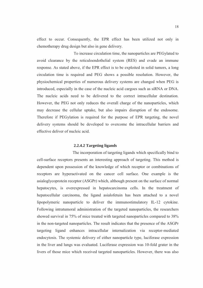
The most vastly used polymers as a gene carrier are
cationic polymers such as polyethylenimine (PEI), poly-L-lysine (PLL), chitosan,
spermine and polyamidoamines (PAMAM) dendrimers, polypeptides, etc [72, 73]
(Figure 3). Cationic polymers can be combined with DNA to form a particulate
complex, and can condense DNA molecules to a relatively small size, compared to

24
cationic liposomes. This can be essential for gene transfer, as small particle size may
be preferable for improving transfection efficacy, especially in vivo [69].
Polyethylenimine (PEI)
One of the most preferable used cationic polymers
for gene delivery is PEI due to its superior transfection efficiency and consistency in
transfection in a wide variety of cell types. PEI has primary (25%), secondary (50%)
and tertiary amines (25%), of which two-thirds of the amines are protonated in a
physiological environment. The unprotonated amines with different pKa values confer
a buffering capacity over a wide range of pH. The buffering property provides PEI an
opportunity to escape from the endosome owing to the mechanism known as “proton
sponge effect”. The hypothesis presumes that at physiological pH only 1–6 nitrogen
atoms are protonated. Upon lowering the pH (i.e., in endosomes) the proportion of
protonated nitrogen increases, and creates a charge gradient which induces a Cl−
influx. The increase in Cl− concentration induces a water influx, following by
endosome swelling and rupture [69]. This may prevent the degradation of DNA in the
endosomal compartment during the maturation of the endosome to lysosome,
facilitating intracellular trafficking of DNA. High cationic density of PEI also
contributes to the formation of highly condensed particles by interacting with DNA.
However, this property may correlate with significant cytotoxicity. An investigation
of linear PEI with different cationic densities, prepared by controlled hydrolysis of
poly(2-ethyl-2-oxazolin), revealed that cell cytotoxicity and transfection efficiency
were dependent not only on cationic density but also on molecular weight of the
polymer. Studies with linear PEIs showed even higher transfection efficiency and
lower cytotoxicity compared to branched PEI [66, 74]. PEI exists in a number of
molecular weight (0.42-800 kDa) and transfection efficiency is highest at a molecular
weight of between 12 to 70 kDa and the most commonly used PEI molecular weight
is 22-25 kDa [75]. PEI of 25 kDa or above has shown efficient transfection efficiency
in various cell lines. However, high molecular weight PEI is toxic to the cells and
PEI/pDNA complexes tend to aggregation [17].

25
The hypothesis of endosomal escape of PEI through proton sponge effect is
illustrated in Figure 4.
Poly-L-lysine (PLL)
One of the firstly employed cationic polymers for
gene delivery is PLL. Since the formation of polyelectrolyte complexes between PLL
and DNA was identified, PLL has been extensively used as a non-viral gene carrier.
The primary amine groups of lysine in PLL, which are protonated in a physiological
pH, electrostatically interact with anionic phosphate groups of DNA to form
nanoparticulate polyelectrolyte complexes. PLL having a molecular weight of less
than 3000 could not form stable complexes with DNA, suggesting that the number of
primary amine in the PLL backbone is necessary for the complex formation. Although
PLLs with high molecular weight have some properties suitable for gene delivery, the
PLL/DNA complexes showed a relatively high cytotoxicity and a trend to aggregate
and precipitate depending on the ionic strength. An advantage of PLL for in vivo
applications is its biodegradable nature. However, PLL polyplexes are rapidly bound
to plasma proteins and then eliminated from the circulation. Co-transfection with
endosomolytic agents is required for successful transfection with first-generation
cationic polymers. Therefore, PLL has poor transfection efficiency when applied
alone or without modifications, this may due to a lack of amino groups allowing
endosomolysis [66, 69, 74].
26
Figure 3 The cationic polymers commonly used for gene delivery.
Source: Wong, S.Y., J.M. Pelet, and D. Putnam. "Polymer systems for gene Delivery-
Past, present, and future." Progress in Polymer Science, 32,8-9(2007): 799-837.

27
Figure 4 Hypothesis of endosomal escape of polyplexes gene delivery systems
(proton- sponge effect).
Source: Morille, M. et al. "Progress in developing cationic vectors for non- viral
systemic gene therapy against cancer." Biomaterials, 29,24-25(2008): 3477-3496.
Chitosan and modified chitosan derivatives
Chitosan, obtained from deacetylation of chitin, is a
biodegradable polysaccharide composed of two subunits, D-glucosamine and N-
acetyl-D-glucosamine, which are linked by a (1 4) glycosidic linkage. Chitosan are
practically non-toxic polymers, can be degraded by lysozymes into common amino
sugars. The primary amino groups of N-deacetylated subunits confer a high cationic
density at acidic and neutral pH. Whereas high molecular weight chitosan (above 100
kDa) is dissolved only in dilute acid solution, low molecular weight chitosan
(LMWC, 22kDa) is highly soluble in physiological buffer solutions. Chitosan is a
polycation, thus having a strong affinity for DNA and can spontaneously form
microspheric particles through complex coacervation [20]. The transfection efficiency
of chitosan/DNA complexes is dependent on several factors, including the degree of

28
deacetylation and molecular weight of the chitosan, plasmid concentration, charge
ratio of amine (chitosan) to phosphate (DNA), serum concentration, pH, and cell type
[76]. LMWC could form complexes with a plasmid DNA and efficiently protect the
DNA from enzymatic degradation by DNase I. LMWC showed grater transfection
efficiency than PLL, while the cytotoxicity of LMWC was lower than PLL.
Hydrophobically modified chitosan was synthesized by conjugating with deoxycholic
acid. By interacting with DNA, the modified chitosan formed spherical self-
aggregates of an average diameter of 160 nm and exhibited efficient transfection for
COS-7 cells. Lactosylated chitosan was also prepared by reductive amidation and
evaluated as a carrier for asialoglycoprotein receptor-targeted gene delivery. The
onset of gene expression with the chitosan/DNA complexes was delayed compared to
PEI/DNA complexes. This may be due to the retardation of endosomal escape of
chitosan/DNA complexes [66].
Dendrimers
Dendrimers are the globular nanoscaled
macromolecules with a unique architecture composed of three distinct domains: (i) a
central core that is either a single atom or a group having at least two identical
chemical functionalities, (ii) branches emanating from the core, consisted of repeat
units having at least one junction of branching, whose repetition is organized in a
geometric progression which results in a series of radially concentric layers called
generations (G), and (iii) many identical terminal functional groups, usually located in
the exterior of the macromolecule, which play an important role in the gene-
complexing or drug entrapping ability. Because of their molecular architecture,
dendrimers exhibit some unique physical and chemical properties, which make them
particularly interesting for drug and gene delivery applications. The presence of
numerous terminal groups facilitates multiple simultaneous interactions with solvents,
surfaces, or other molecules. As a result, dendrimers tend to show high solubility,
reactivity, and binding. The multiple interactions between surface amines of
dendrimers and nucleic acid phosphates are also necessary for the formation of
dendrimer/DNA complexes.

29
Dendrimers interact with all forms of nucleic
acids via electrostatic interaction to form complexes, which condense/compact the
nucleic acid. During the complex formation, the extended configuration of the nucleic
acid is altered and a more compact configuration results, with the cationic dendrimer
amines and the anionic nucleic acid phosphates reaching the local charge
neutralization resulting in the formation of dendrimer–nucleic acid complexes
“dendriplexes”. The dendrimers commercially available for gene delivery are
polyamidoamine (PAMAM) and polypropylenimine (PPI) [72, 77]. Dendrimers have
primary amine groups on their surface and tertiary amine groups inside. The primary
amine groups responsible for DNA binding, compact it into nanoscaled particles and
promote its cellular uptake, while the embedded tertiary amino groups act as a proton
sponge in endosomes and promote the release of DNA into the cytoplasm [74].
2.3.3.2 Cationic lipid-based system
Since the first synthetic cationic lipid, N-[1-(2,3-
dioleyloxy)propyl]-N,N,N-trimethylammonium chloride (DOTMA) was used for
gene delivery in 1987 by Felgner et al. [70], a number of cationic lipids have been
synthesized and researched as transfection reagents [74].
The cationic lipids usually contain three parts: i)
hydrophobic group such as alkyl chain, fatty acid chain and cholesteryl moiety, ii)
linker group and iii) positively charged head group which usually contains amine
group that can be protonated at physical pH [17, 74]. The basic structure of cationic
lipids is presented in Figure 5. The cationic head group is necessary for the binding of
nucleic acids phosphate groups, resulting in nucleic acids condensation. All cationic
lipids are positively charged amphiphile systems. The cationic lipids are generally
prepared and used in formulation of liposomes. The complexes between nucleic acids
and liposomes are call lipoplexes [36, 69].

30
Figure 5 Basic components of cationic lipids 3-h[N(NV,NV-dimethylaminoethane)-
carbamoyl] cholesterol (DC-Chol) and N-[1-(2,3-dioleyloxy)propyl]-N,N,N
trimethylammonium chloride (DOTMA). (a) Hydrophobic lipid group; (b)
linker group; (c) cationic headgroup
Source: Mahato, R.I. "Water insoluble and soluble lipids for gene delivery."
Advanced Drug Delivery Reviews, 57,5(2005): 699-712.
The cationic lipids can be classified into various subgroups base on their basic
structural characteristics (Figure 6) as follow [74]:
1) Monovalent aliphatic lipids characterized by a single amine function in the
headgroup e.g. N [1-(2,3-dioleyloxy) propyl]-N,N,N-trimethylammonium chloride
(DOTMA), 1,2-dioleyl-3-trimethylammonium-propane (DOTAP), N-(2-
hydroxyethyl)-N,N-dimethyl-2,3-bis(tetradecyloxy-1-propanaminium bromide)
(DMRIE),
2) Multivalent aliphatic lipids which contain several amine functions in the
polar headgroups such as the spermine group e.g. dioctadecylamidoglycylspermine
(DOGS), dipalmitoylphosphatidylethanolamidospermine (DPPES),
3) Cationic cholesterol derivatives e.g. 3β-[N-(N',N'-dimethylaminoethane)-
carbamoyl]cholesterol (DC-Chol), bis-guanidium-tren-cholesterol (BGTC).

31
Figure 6: Structure of current cationic lipids used in gene therapy and the helper lipid
DOPE. DOTMA: N [1-(2,3-dioleyloxy) propyl]-N,N,N-trymethylamonium
chloride, DOTAP: 1,2-dioleyl-3-trimethylamonium-propane, DMRIE: N-
(2-hydroxyethyl)-N,N-dimethyl-2,3-bis(tetradecyloxy-1-propananium
bromide), DOTIM : 1-[2-(oleyloxy)ethyl]-2-oleyl-3-(2-hydroxyethyl)
imidazolinium chloride, DOGS: dioctadecylamidoglycylspermine, DC-
Chol: [N-(N',N'-dimethylaminoethane)-carbamoyl] cholesterol, BGTC: bis-
guanidium-tren-cholesterol, DOPE: 1,2-dyoleyl-sn-glycerol-3-
phosphoethanolamine
Source: Morille, M. et al. "Progress in developing cationic vectors for non-viral
systemic gene therapy against cancer." Biomaterials, 29,24-25(2008): 3477-3496.
Cationic lipids are amphiphilic molecules, implying that they consist
of both hydrophilic and hydrophobic part. An important characteristic of the
amphiphile with regard to its application as a gene carrier is its geometry. Like any
amphiphile, when suspended in an aqueous environment, cationic lipids can adopt
various structures, including the micellar, lamellar, cubic and inverted hexagonal

32
phase (Figure 7). The type of structure can be predicted by a factor known as the
packing parameter, P. This packing parameter, P=v/alc, is defined as the ratio of the
hydrocarbon volume, v, and the product of the effective head group area, a, and the
critical length of the lipid tail, lc. Thus, when P exceeds the value of 1, i.e. the area
occupied by the hydrocarbon chains is much larger than that of the head group, the
lipid tends to adopt the inverted hexagonal phase, which in essence is a bilayer
destabilizing structure, adoption of this phase seems to be an important step in the
mechanism by which genes or oligonucleotides (ODNs) are delivered into the cytosol,
following lipoplex internalization [67].
Figure 7 Schematic representation of the phase structure of cationic lipids as a
function of their packing parameter. The geometry of the amphiphile,
defined by the packing parameter P, determines the organization of self-
assembled cationic lipids. For P<1/2 it is predicted that a cone-like shaped
monomer will assemble into structures with positive curvature
corresponding to a micellar phase (spherical micelles, wormlike or
cylindrical micelles or non-inverted hexagonal HI phase). When P is
between 1/2 and 1 the curvature of the self-assembled amphiphiles is close
to 0 leading to the lamellar phase (bilayers). For P>1 the structures formed
display a negative curvature leading to inverted phases (inverted micelles or
inverted hexagonal HII phases).
Source: Wasungu, L. and D. Hoekstra. "Cationic lipids, lipoplexes and intracellular
delivery of genes." Journal of Controlled Release, 116,2(2006): 255-264.

33
Commercially available cationic lipids used as liposome formulations
or alone for gene delivery include DOTMA, 2,3-dioleyloxy-[2(sperminecarboxamido)
ethyl]-N,N-dimethyl-1-propanammonium trifluoroacetate (DOSPA)(Lipofectamine®),
and dioctadecylamido-glycylspermine (DOGS) (Transfectam®), etc [17].
The liposomes generally contain at least two constituents: a cationic
lipid and a neutral lipid, which is called helper lipid. To improve transfection
efficiency, cationic lipids are often combined with helper lipids such as DOPE (1, 2-
dioleoyl-sn-glycero-3-phosphoethanolamine) and cholesterol [36, 69, 74].
DOPE and cholesterol are frequently used as neutral lipids. On its own,
DOPE forms inverted hexagonal HII (non-bilayer) phase structures at neutral pH and
physiologic temperatures. When combined with a cationic lipid, however, it can
participate in bilayer formation. When the cationic lipid is laterally phase-separated
by interaction with negatively charged molecules or macromolecules, the DOPE may
form non-bilayer structures. The latter may facilitate the destabilization of the cellular
membranes which the cationic lipid interacts, possibly promoting the cytoplasmic
delivery of nucleic acids [69].
Cholesterol is a naturally occurring lipid and is metabolized in the
body. The early success of DC-Chol lipid-based gene delivery provoked the
development of novel cholesterol-based cationic lipids. Cationic lipids in T-shape
headgroups tend to be more efficient than linear counterparts. The levels of gene
expression obtained with spermine cholesteryl carbamate and spermidine cholesteryl
carbamate in T-shape orientation was 50-100 folds higher both in vitro and in vivo
than that observed with DC-Chol, which has only a single protonatable amine.
Cholesterol-containing cationic liposomes were found to be more structurally stable in
physiologic media, thus enabling the lipoplexes to reach their target site of action,
therefore protecting the DNA from degradation, and ultimately facilitating
transfection [17, 69].

34
Structure of cationic lipids is one of the major factors affecting
transfection efficiency. Based on the structures of their cationic headgroups, cationic
lipids can be classified into quaternary ammonium lipids, lipopolyamines, cationic
lipids bearing both quaternary ammonium and polyamine moieties, amidinium and
guanidinium salt lipids and heterocyclic cationic lipids. A quaternary ammonium
headgroup is vastly used in a wide variety of the established cationic lipids like
DOTAP, DOTMA and DMRIE. There are fewer lipids containing a primary,
secondary or tertiary amino headgroup.
Hydrophobic group lipid anchors can be either a cholesterol derivative or
a double chain hydrocarbon. The double chain hydrocarbons are generally 12–18
carbon units in length, can be either completely saturated or contain double bonds
(e.g. oleoyl group) and are not necessarily symmetrical [17]. Cationic lipids with
various types of hydrocarbon chains have been investigated thoroughly. The common
types of chain lengths are C8:0 to C18:1. The use of mono-unsaturated fatty acid
chains have resulted in higher transfection, possibly owing to their influence on
increasing membrane fluidity [69]. Even small structural alterations to these domains
are known to influence on transfection efficiency.
The linker group is an important component, which determines the
chemical stability and biodegradability of the lipid. The linker groups should be
biodegradable yet strong sufficient to survive in a biological environment. The
commonly employed linker groups are such as ethers and esters, glycerol-type
moiety, a phosphate or phosphonate linker, amino acids, peptides or aromatic rings,
although amides and carbamates can also be used. The chemistry of the actual linker
has most frequently been of the carbamate or amide variety, both of which are
chemically stable and biodegradable. Alternatively, they are a carbonate or an ester,
which are not stable chemically, but are biodegradable (e.g. DOTAP), or are
chemically stable but non-biodegradable (e.g. DOTMA). The connecting moieties can
be designed to be tunable, thus they are stable during formulation, storage,
administration and initial circulation, but can be degraded rapidly at the target site
(e.g. in an endosome) [17, 69].

35
2.4 Basic mechanisms of transfection by cationic lipids and polymers
The general mechanisms of lipoplex- and polyplex-mediated gene
transfection involve the following steps [69, 74]:
i) Nano-sized lipoplex or polyplex particles are formed from the
combination of cationic liposomes or polymers with nucleic acids in buffered aqueous
solution.
ii) The particles bind to the cell surface by non-specific, electrostatic
interaction between the positively charged complexes and the negatively charged cell
surface
iii) Cellular entry via endocytosis or endocytosis-like mechanisms.
iv) Endosomal escape of uptaken particles
When the pH of the endosome compartments drops from pH 7 to 5.5
and part of the bound nucleic acids should escape from early endosomes into the
cytosol. The mechanisms of endosomal escape are fundamentally different for
lipoplexes and polyplexes. Cytoplasmic transport of endosomes plays a key role in
bringing the transfecting material near the perinuclear region.
v) Nucleus entry:
The lipoplexes or polyplexes then dissociate and the released plasmid
DNA enters the nucleus by either of two hypothesized pathways:
a) Passive DNA entry into the nucleus during the mitotic cell
division when the nuclear membrane disintegrates temporarily or
b) Active transport of the DNA through nuclear pores.
These two mechanisms are not mutually exclusive in a given cell type.
The overall mechanism of gene transfection into tumor cell mediated by non-viral
gene carrier is illustrated in (Figure 8).

36
Figure 8 Schematic representation of the different hurdles encountered by a gene
delivery system to enter and traffic into a tumor cell.
Source: Morille, M. et al. "Progress in developing cationic vectors for non-viral
systemic gene therapy against cancer." Biomaterials, 29,24-25(2008): 3477-3496.
Nuclear pore was found to be a crucial barrier to gene delivery. The initial
release of DNA into the cytoplasm is possibly mediated by the destabilization of the
endosome membrane by the cationic lipids. This belief is supported by the
observation that the fusogenic lipid DOPE is usually required for effective
transfection. Transbilayer flip–flop of negatively charged phospholipids from the
cytoplasmic leaflet to the luminal leaflet of the endosome has been hypothesized to
cause the formation of charge-neutral ion pairs, thus releasing the DNA from the

37
complex (Figure 9). This would result in a weakening of the electrostatic interactions
between DNA and cationic lipids, and eventually the release of the DNA from the
lipoplexes. Detergent-like destabilization of the endosomal membrane is another
possible hypothesis of endosomal escape of lipoplexes. Cationic polymers are devoid
of a hydrophobic domain, thus cannot fuse with or destabilize the endosome by direct
interaction with the endosomal membrane as in the case of cationic lipids. The
possible mechanism of endosomal escape of lipoplexes is proton sponge effect as
mention above.
Figure 9 Hypothesis of endosomal escape of lipoplexes gene delivery system.
Source: Morille, M. et al. "Progress in developing cationic vectors for non-viral
systemic gene therapy against cancer." Biomaterials, 29,24-25(2008): 3477-3496.
2.5 Cellular Uptake Pathways for Nonviral Gene Delivery
Most nonviral gene carriers cannot directly cross the cell membrane owing to
their large size and hydrophilic nature. Endocytosis which is the vesicular uptake of
extracellular macromolecules has been established as a major cellular internalization
mechanism of nonviral carriers. Multiple mechanisms for endocytosis have been

38
described to date (Figure. 10). After endocytosis, the internalized molecules tend to be
trapped in the intracellular vesicles and eventually fuse with lysosomes where they are
degraded. Consequently, the barrier of gene delivery involves not only the cellular
uptake of genes, but also their intracellular availability at the target sites. After
endocytosis-mediated uptake, nonviral carriers such as liposomes or peptides which
can enhance cytosolic release of internalized molecules are necessary for achieving
successful gene delivery. Moreover, other nonendocytic methods which can bypass
endocytosis such as microinjection, permeabilization, electroporation, fusion and
penetration also can be used for gene delivery [78].
Figure 10 Classification of endocytosis based on endocytosis proteins that are
involved in the initial entry of particles and solutes.
Source: Sahay, G., D.Y. Alakhova, and A.V. Kabanov. "Endocytosis of -
nanomedicines." Journal of Controlled Release, 145,3(2010): 182-195.
2.5.1 Endocytic Uptake Pathways [78-80]
Endocytosis refers to the cellular uptake of macromolecules and solutes into
membrane-bound vesicles derived by the invagination and pinching off of parts of the
cell membrane. Three modes of endocytosis can be kinetically defined: fluid-phase,
adsorptive and receptor-mediated endocytosis. Fluid-phase endocytosis refers to the

39
bulk uptake of solutes in the exact proportion to their concentration in the
extracellular fluid. This is a nonspecific and low-efficiency process. On the contrary,
in adsorptive and receptor-mediated endocytosis, macromolecules are bound to the
cell surface and concentrated before internalization. In adsorptive endocytosis,
molecules favorably interact with complementary binding sites (e.g., by lectin or
charged interaction). After that, the bound molecules will follow the fate of cell
membrane. In receptor-mediated endocytosis, certain ligands can specifically bind to
their receptors on the cell surface and become concentrated before internalization. The
capability of receptor mediated endocytosis reflects both the affinity of the ligand-
receptor interaction and the concentration of the complexes in clathrin-coated pits.
Endocytosis can also be classified into two broad categories, phagocytosis or
cell eating (the uptake of large particles) and pinocytosis or cell drinking (the uptake
of fluid and solutes).
Different classifications of pinocytosis have been proposed. The recent
approach is based on the proteins involved in different endocytic pathways (Figure
11). In this approach pinocytosis is classified as clathrin-dependent endocytosis (also
known as clathrin-mediated endocytosis, CME) and clathrin-independent endocytosis.
The clathrin-independent pathways are further classified as 1) caveolae-
mediated endocytosis, 2) clathrin- and caveolae-independent endocytosis and 3)
macropinocytosis.
Clathrin- and caveolae-independent pathways are sub-classified as Arf6-
dependent, flotillin-dependent, Cdc42-dependent and RhoA-dependent endocytosis.

40
Figure 11 Different mechanisms of endocytosis. There are multiple pathways for
cellular entry of particles and solutes. The picture of endocytosis trafficking
is actively researched and evolving. In all cases the initial stage of
endocytosis proceeds from the plasma membrane portals of cellular entry
and involves engulfment of cargo into intracellular vesicles. The second
stage often involves sorting of the cargo through endosomes. It is followed
by the final stage during which the cargo is delivered to its final destination,
recycled to extracellular milieu or delivered across cells (not shown). The
figure is a simplified representation of complex trafficking mechanisms and
their cross-talks. Abbreviations are: CCV, clathrin coated vesicles, CLIC,
clathrin-independent carriers; GEEC, GPI-anchored protein-enriched
compartment; GPI, glycophosphatidylinositol, MVB, multivesicular body.
Source: Sahay, G., D.Y. Alakhova, and A.V. Kabanov. "Endocytosis of-
nanomedicines." Journal of Controlled Release, 145,3(2010): 182-195.
2.5.1.1 Phagocytosis
Phagocytosis is defined as the engulfment of cells, bacteria and
large solid particles. It is typically restricted to specialized professional phagocytes
such as macrophages, monocytes, neutrophils and dendritic cells. Basically,

41
phagocytosis constitutes a defence mechanism against foreign substances with broad
size ranges (25 nm to several μm's in diameter). Phagocytosis by macrophages usually
occurs after opsonisation of particles by IgG antibodies or complement proteins in the
bloodstream. The opsonized particles are then recognized by the Fc-receptor, which
binds antibodies, or by complement receptors, which bind complement factors.
Additionally, phagocytosis by macrophages can be triggered by scavenger receptors
which bind negatively charged lipoproteins, ormannose/fructose receptors that bind
lectins. The phagosomes fuse intracellularly after 30–60 min with late endosomes and
lysosomes, accumulating microbicidal peroxidases, nitrogen reduction products,
esterases and cathepsins for degradation of the cargo in the so-called phagolysosome.
Other cells like fibroblasts, endothelial and epithelial cells may also show some
phagocytic activities, e.g. they can internalize pathogens such as Yersinia, Shigella,
Salmonella or Listeria. Phagocytosis of these pathogens by non-professional
phagocytes is mediated by fibronectin or laminin receptors or by heparan sulfates
displayed on the surface of cell membrane. Moreover, non-viral nanocarriers for
nucleic acid delivery have been reported to be internalized by epithelial cells via
phagocytosis, after their association with heparan sulfate proteoglycans on the cell
surface [79].
2.5.1.2 Macropinocytosis
Macropinocytosis is defined as a transient bulk internalization
process that is typically characterized by the formation of membrane ruffles and the
engulfment of large volumes of fluid into large uncoated vacuoles of 0.2–10 μm,
known as macropinosomes. In contrast to phagocytosis, macropinocytosis can also be
active in the absence of particles. Macropinosomes form spontaneously or in response
to growth factors or cell penetrating peptides. Macropinosomes have no coat and do
not concentrate receptors. In addition, macropinosomes vary in size, sometimes being
as large as 5μm in diameter. Macropinocytosis is an efficient route for the
nonselective endocytosis of solute macromolecules due to their relatively large size.
Macropinocytosis fulfills various functions, particularly when massive fluid-phase
endocytosis is essential. This route facilitates the bulk uptake of soluble antigens by

42
immature dendritic cells. Moreover, some pathogens (e.g. Shigella, Salmonella,
adenovirus, HIV type I) trigger macropinocytosis to facilitate their own uptake. Their
formation can be also induced by apoptotic bodies and necrotic cells. Some cell types,
such as immature dendritic cells and macrophages, exhibit a non-selective constitutive
macropinocytosis process by which they are constantly sampling their environment
for foreign substances. The molecular mechanism of macropinocytosis is still
unclarified and seems to show only a few distinct molecular regulators, when
compared to phagocytosis. However, some specific proteins were identified in recent
studies. One of them is brefeldin A-ADP ribosylated substrate (BARS), which was
reported to play a key role in macropinosome closure. BARS is a substrate of p21-
activated kinase (PAK1), the latter is present on macropinosomes and mediates the
regulation of cytoskeletal dynamics during macropinocytosis [78-79].
2.5.1.3 Clathrin-mediated endocytosis (CME)
The major and best-characterized endocytic pathway to date is
CME, which is characterized by the formation of clathrin-coated pits. CME is the
“classical route” of cellular entry, which is present and inherently active in all
mammalian cells. This pathway is responsible for uptake of essential nutrients like
cholesterol carried into cells by low density lipoprotein (LDL) via the LDL receptor,
or iron carried by transferrin (Tf) via the Tf receptor. These proteins are now
generally used as markers of CME. Other functions of CME include down-regulation
of cell signaling by internalization and degradation of receptors and maintaining
cellular homeostasis, for example, by trafficking ion pumps. Mechanistically, CME
involves engulfment of receptors associated with their ligands to a coated pit. The
coated pit forms owing to polymerization of a cytosolic protein called clathrin-1,
which also requires assembly proteins like AP180 and AP-2. The coated pit is formed
in a lattice of clathrin. The clathrin pits typically have a diameter of 120 nm and are
pinched of from the cell membrane with the help of dynamin. Dynamin is a small
GTPase that polymerizes around the neck of the invaginated membrane in a coil
which contracts in an energy-dependent way. After formation of the clathrin-coated
vesicle, the clathrin lattice is rapidly depolymerized, so that the vesicles can fuse with

43
each other and with early or sorting endosomes. For gene delivery, CME can be
targeted by using certain ligands, such as transferrin, which can specifically recognize
certain receptors on the cell surface. This results in an increase in the internalization
of the particles and offers the possibility of targeting specific cells that substantially
overexpress the receptors. However, genes which are internalized through CME are
generally trapped in endosomes followed by enzymatic degradation in lysosomes, and
the final result is that genes have little or almost no access to their target sites.
Therefore, entrapment and degradation can be considered as two separate barriers,
because preventing lysosomal degradation results in an accumulation of genes in
intracellular vesicles without enhancing cytosolic release. Consequently, genes must
avoid degradation in lysosomes to reach the nucleus, and need to be released from
intracellular vesicles into the cytosol [78-80].
2.5.1.4 Caveolae-Mediated Endocytosis (CavME)
The first clathrin-independent pathway described was CavME.
It is thought to originate from lipid rafts of 60–80 nm, enriched in caveolin-1 and
stabilized by cavins, referred to as caveolae. Caveolae are small, hydrophobic
membrane microdomains which are rich in cholesterol and glycosphingolipids.
Classically, caveolae were defined as flask-shaped invaginations of the plasma
membrane, but they can also be flat, tubular, or detached vesicles. Caveolae are
abundant in muscle, endothelial cells, fibroblasts and adipocytes and absent in
neurons and leukocytes. They are a subset of lipid rafts, the cholesterol-rich plasma
membrane regions which cluster endocytosis and signal transduction functionalities.
The definitive characteristic of caveolae is the presence of the hairpin-like membrane
protein, caveolin-1, which is essential for biogenesis of caveolae. Due to this protein
caveolae assume their hallmark flaskshaped structure (60–80 nm) and can engulf
cargo molecules, which bind to caveolae surface. In addition to caveolin-1, which
presents in most cells, there are other isoforms like caveolin-2 or caveolin-3 (specific
for muscle). Other components of the caveolae endocytic machinery include proteins
like cavin, which induces membrane curvature, dynamin, which enables vesicle
scission, as well as vesicle-associated membrane protein (VAMP2) and synaptosome-
associated protein (SNAP), which mediate subsequent vesicle fusion, etc. After

44
budding of the plasma membrane the caveolae vesicles transport and fuse with
caveosomes or MVBs that have neutral pH.
This pathway seems to be slower compared to the CME in vitro
but at least in some cases it can bypass lysosomes. Therefore, several pathogens
including viruses and bacteria exploit this pathway to prevent lysosomal degradation.
For the same reason this pathway is believed to be beneficial for cellular delivery of
proteins and DNA. The typical molecules to undergo the caveolae-mediated
endocytosis are CTB and Shiga toxin. These molecules interact with the
glycosphingolpids (GSL) residing in caveolae, such as GM-1 (CTB) and Gb3 (Shiga
Toxin). Both CTB and Shiga Toxin are sometimes used as markers for caveolae.
However, it should be noted that they are not restricted to caveolae and can also enter
through clathrin- and caveolae-independent pathways. Caveolae association with a
family of cholesterol-binding proteins called caveolins, which function to create
and/or mediate these structures. The mechanisms of caveolar internalization have
been elucidated by visualizing the trafficking of the SV40 that uses caveolae to gain
entry into the cells. Caveolin-1 is a very promiscuous protein, which binds and
promotes ordering of multiple molecules including lipids (cholesterol, GSL, etc.),
fatty acids, and membrane proteins. Therefore, caveolae sequester multiple ligands
responsible for cellular signaling and their downstream signaling components in close
proximity for efficient signal activation and transduction. Some examples include
heterotrimeric G proteins, nonreceptor tyrosine kinases, insulin receptor, platelet
derived growth-factor receptor, and endothelial nitric-oxide synthase (eNOS).
The caveolar uptake is a nonacidic and nondigestive route of
internalization. Caveolae do not suffer a drop in pH, and most pathogens that are
internalized by caveolae can be directly transported to the Golgi and/or endoplasmic
reticulum, thus avoiding normal lysosomal degradation.
Another term associated with caveolae is lipid rafts. Markers for
lipid rafts are frequently found within caveolae. In general, caveolin-containing rafts
are referred to as caveolae, whereas caveolin-devoid rafts are denoted by a variety of
names such as glycolipid-enriched membranes and caveolae-like domains.

45
Cholesterol is essential for caveolar uptake and drugs which
specifically bind to cholesterol perturb internalization through the caveolae. Caveolae
also depend on the actin cytoskeleton, and drugs which cause the depolymerization of
the actin cytoskeleton such as cytochalasin D can inhibit caveolae uptake without
affecting clathrin-mediated endocytosis. Genestein, a tyrosine kinase inhibitor, can
also inhibit caveolae. It is generally believed that caveolar uptake does not lead to
lysosomal degradation.
Therefore, this pathway seems to be advantageous in terms of
DNA delivery. Evidence supporting the existence of a role of caveolae in the uptake
of cationic polymer-DNA complexes and the class of protein transduction domains
(PTDs), such as the TAT peptide, have appeared. Another report suggested that large
particles (500 nm) are preferentially taken up through caveolae where they do not
suffer lysosomal degradation. However, caveolae are slowly internalized and small in
size, and their fluid phase volume is small. Thus, it is unlikely that they contribute
significantly to constitutive endocytosis, although the situation is different in
endothelial cells in which caveolae constitute 10 to 20% of the cell surface.
Caveolae-mediated endocytosis is still a promising route for
gene delivery, particularly if the internalization can be increased, possibly through the
use of specific receptors for caveolae. Furthermore, the caveolae-mediated
endocytosis is the most prominent transendothelial pathway and thus this route may
be employed for trans-vascular delivery of nanomaterials [78-80].
2.5.1.5 Clathrin- and caveolae-independent pathways
Flotillin-mediated endocytosis (FlotME) was described for the
first time as a distinct endocytic pathway in 2006, and considered to be indispensable
for the internalization of certain glycosylphosphatidylinositol (GPI)-anchored proteins
like CD59, amyloid precursor protein and others. Flotillins are ubiquitous membrane-
bound proteins which are reported to be involved in several processes such as insulin
signaling, T-cell activation, membrane trafficking, phagocytosis, EGFR signaling and
cell motility. Their overexpression leads to filopodia-like protrusions in various cell
lines, suggesting their role in signaling in actin-dependent processes. Flotillins were

46
found to be associated with both dynamin independent as well as dynamin-dependent
endocytic processes. The endocytic event originates in the co-assembly of flotillin-1
and flotillin-2 into hetero-oligomers. These flotillins display similar topologies as
caveolin-1, and generate after activation by tyrosine kinases a scaffold that stabilizes
specialized plasma membrane microdomains, spatially distinct from caveolae and
clathrin-coated pits. Despite their necessity for the endocytosis of aforementioned
ligands, the exact role of the flotillins remains to be elucidated. It is possible that
flotillins facilitate clustering of lipid-associated receptors and certain lipid species,
inducing membrane budding and vesicle formation. Glycosphingolipids, bacterial
toxins (e.g. cholera toxin, VacA toxin) and several GPI-anchored proteins were
reported to be internalized via clathrin-independent, tubular invaginations of the
plasma membrane, called CLICs. The formation of the CLICs is independent of
dynamin. They are devoid of caveolin1 and flotillin1, but are associated with the
activity of the small GTPases Cdc42 and Arf1. Cdc42-dependent endocytosis has
been reported to be responsible for the largest fraction of CIE fluid uptake. The GPI-
linked proteins, which are internalized via CLIC vesicles, are trafficked to GPI-
enriched early endosomal compartments or GEECs. GRAF1 was recently identified
as a key protein for the CLIC-GEEC endocytic pathway.
Another pathway is RhoA-dependent endocytosis. This
constitutive endocytic pathway was reported to be involved in the internalization of
interleukin-2 receptor. Also Arf6-dependent endocytosis is considered to be a distinct
pathway, involved in the uptake of major histocompatibility complex class I proteins
and integrins, often independent of dynamin. Many of these clathrin-independent
gateways are currently intensively investigated and characterized which leads to a
continuous output of new insights and discoveries on these endocytic mechanisms
[79].

47
2.5.2 Chemical inhibitors of Endocytic pathways
The chemical inhibitors of many endocytic pathways have been currently
employed to study the endocytosis of nanomedicines. One way to perform exclusion
studies is the use of chemical inhibitors like chlorpromazine, filipin, genistein, and
others (Table 2). However, the effects of these inhibitors are poorly characterized,
non-specific and cell type dependent. To obtain reliable results, the applied
concentrations and incubation times should be optimized for cytotoxic side-effects
which may be dependent on the cell type. Furthermore, it should be noted that the
chemical inhibitors can affect several endocytic pathways depending on the cell type.
Therefore, the effect of these chemical inhibitors on the endocytic mechanism should
be characterized in the relevant cell type. This characterization requires the use of
fluorescent endocytic markers which should be internalized via one specific endocytic
route. Only then the effect of the inhibitor treatment on these endocytic portals can be
reliably evaluated, generally by flow cytometry. Some endocytic ligands which have
often been employed as endocytic markers are transferrin (hTf), lactosylceramid
(LacCer), cholera toxin subunit B (CTXB), dextran, fluorescent microspheres or
bacteria. Unfortunately, a reliable assessment of their cellular processing is sometimes
lacking for these endocytic markers. For example, CTXB is known to be internalized
by several endocytic pathways in the same cell type. Additionally, the endocytic
mechanism by which these endocytic markers are internalized might also depend on
the cell type. Consequently, it is clear that careful characterization of the endocytic
inhibitors on a particular cell type is crucial to obtain reliable conclusions on the
endocytic internalization of nanomedicines [78].

48
Table 2 Perturbation of endocytosis and intracellular trafficking.
Treatment Effect Mechanism
Low temperature
General inhibitor of endocytosis
Energy depletion
Metabolic inhibitors General inhibitor of endocytosis Energy depletion
Potassium depletion Specific inhibitor of CME Dissociation of clathrin lattice
Cytosol acidification Specific inhibitor of CME Dissociation of clathrin lattice
Hypertonic medium Specific inhibitor of CME Dissociation of clathrin lattice
Chlorpromazine Specific inhibitor of CME Dissociation of clathrin lattice
Filipin Specific inhibitor of caveolae Cholesterol binding
Nystatin Inhibitor of caveolae Sequester cholesterol
Methyl-β-cyclodextrin Inhibitor of caveolae Deplete cholesterol
Genestein Inhibitor of caveolae Tyrosine kinase inhibitor
Cytochalasins Inhibitors of caveolae and macropinocytosis
Actin depolymerization
Amiloride Specific inhibitor of macropinocytosis
Inhibits the Na+/H+ exchange protein
Phorbol esters Specific stimulators of macropinocytosis
Protein kinase C activators
Wortmannin Inhibitor of macropinocytosis Phosphatidyl inositol-3-phosphate inhibitor
Monensin Inhibitor of endosome maturation
Prevents endosome acidification
Chloroquine
Disrupting endosomes and lysosomes
Prevents endosome acidification and causes swelling to endosomes and lysosomes
Ammonium chloride Disrupting endosomes and lysosomes acidification
Increases pH of late endosomes and lysosomes
Nocodazole Inhibit vesicular transport Depolymerises microtubules
Source: Khalil, I.A. et al. "Uptake pathways and subsequent intracellular trafficking in
nonviral gene delivery" Pharmacological Reviews, 58,1(2006): 32-45.
Vercauteren, D. et al. "On the cellular processing of non-viral nanomedicines
for nucleic acid delivery: mechanisms and methods." Journal of Controlled Release,
161,2(2012): 566-581.

49
2.6 Introduction to RNAi Therapeutics
The early study on gene therapy focused on the delivery of DNA. However,
in 1998, the role of double-stranded RNA in gene therapy through RNA interference
(RNAi) was discovered [81], with Craig C. Mello and Andrew Fire being awarded the
2006 Nobel Prize in Physiology or Medicine. RNA interference (RNAi) based gene
silencing provides a great potential tool in treatment of numerous gene-related
diseases such as genetic disorder, cancer, inflammation and viral infection, etc [21,
82]. Post-transcriptional gene silencing through RNAi mechanism can be triggered by
some effector molecules such as small interfering RNA (siRNA), resulting in specific
mRNA degradation, then inhibition of abnormal protein synthesis [83-85]. However,
the prerequisites for successful RNAi therapeutics require not only therapeutically
siRNA, but also the safe and efficacious siRNA delivery system. [22].
RNAi is a naturally biological process occurs in cytoplasm. This process is
highly conserved among multicellular organisms diverse as plants, worms, yeast, and
human. It refers to a two-part intracellular pathway in which precursor double-
stranded RNA molecules present in the cell are first processed by the dicer
endonuclease into short 21-23 nucleotide fragments containing 2 nucleotide single-
stranded 3’overhangs on each strand. These effector RNAs, called short interfering
RNAs (siRNAs), then become incorporated into an RNA-inducing silencing protein
complex (RISC) in which one strand of the unwound siRNA acts as a guide sequence
to target the cleavage of homologous RNAs.
In plants, RNAi plays an important role in cellular defense, protecting the cell
from inappropriate expression of repetitive sequences, transposable elements, and
virus infections. In addition to evidence that similar defense functions may still be
active in mammalian cells, an increasing number of short RNA molecules are being
found encoded in the mammalian genome. These endogenous RNAs, or microRNAs
(miRNAs), are also processed by dicer into siRNA effectors, and are proving to
regulate the expression of genes involved in a variety of cellular processes such as
proliferation, apoptosis and differentiation [83].

50
2.6.1 History of RNA interference
The RNAi phenomenon accidently discovered when the anti-sense strand was
injected to block expression of the par-1 gene in the nematode C. elegans. The
expression was interfered, however, upon performing their controls the research found
that the sense strand also reduced the expression of that gene. Even earlier biologists
had unknowingly witnessed the process of RNA interference when performing
experiments on petunias and found that when they introduced a pigment-producing
gene under the control of a promoter into the flowers they did not get expected results.
Instead of getting the expected deep purple color, the flowers were diversified or they
were completely white. In 1998, when Fire and Mello first injected double stranded
RNA into C. elegans and were rewarded with a much more effective gene silencing
effect. Until now the mystery was unrevealed, except it was found that the initiator of
this post-transcriptional gene silencing was dsRNA, but how it happens, was still a
question [85].
2.6.2 The mechanism of RNA interference
In a many cell types long dsRNA are processed by a host RNase III, termed
Dicer, to form siRNA, a short dsRNA molecule usually contain 21–23 nucleotides
with two nucleotide 3’-overhangs. Dicer-processed siRNAs and synthetic siRNAs
trigger post-transcriptional gene silencing through RNAi mechanism by undergoing
an ATP-dependent unwinding step, then incorporated into a high-molecular-weight
protein complex termed RISC (RNA-induced silencing complex) which contains
single-stranded siRNA. However, it is unclear whether the unwinding step takes place
before or immediately after the incorporation of the siRNA duplex into RISC.
Thereafter, the RISC is remodeled into its active form RISCp, which contains the
proteins essential for cleaving the target mRNA at the site where the guide antisense
siRNA binds. Following cleavage, the RISCp is released to cleave additional mRNA
molecules, whereas the cleaved mRNA is degraded by cellular ribonucleases (Figure
12). Consequently, siRNA is an attractive tool for knocking down specific gene of
interest. The concept that siRNA can be developed as potential therapeutic agents has
already inspired the research on RNAi [83].

51
Figure 12 Mechanism of RNA interference: silencing of viral genes through
activation of RISC. siRNA combined with RISC binds to mRNA causing
cleavage of its sense strand.
Source: Rahman, M et al. “RNA interference: The story of gene silencing in plants
and humans." Biotechnology Advances, 26(2008): 202–209.
To use siRNA as a therapeutic agent, it is crucial that siRNA must not cause
any effects other than those related to the knockdown of the target gene. This issue is
particularly important in therapeutic applications
The delivery of siRNAs can be achieved by exogenous application of
synthetic siRNAs or via a gene therapy approach which relies on the endogenous
expression of siRNA. Initially, siRNAs corresponding to the targeted genes were
chemically synthesized in vitro and then delivered to cells. This delivery mechanism
offers the possibility of developing pharmaceutical siRNAs that are expected to be

52
useful in pathological situations where immediate and/or short-term effects are
required [84].
2.6.3 Therapeutic applications of RNAi [83]
Some examples of diseases target to RNAi therapeutics are listed below.
Ocular disease
Angiogenesis play a key role in several ocular diseases including age-
related macular degeneration (AMD), herpetic stromal keratitis, and diabetic
retinopathy. VEGF has been found to be directly involved in the destructive
vascularization associated with these ocular diseases. The ability of VEGF-
specific siRNAs to reduce VEGF-dependent vascular invasion of the eye has not
only been demonstrated in animal models but phase I AMD clinical trials in
patients is ongoing as well.
Neurological disease
Parkinson’s disease, Huntington’s disease (HD), fragile X syndrome,
amyotrophic lateral sclerosis (ALS), and spinobulbar muscular atrophy, are just some
of the prominent neurological diseases for which RNAi-based therapies might prove
useful. The relevant commonality among these neurodegenerative disorders is that
they result from dominant mutations in a single allele. Therefore, in all cases, the
sequence specificity offered by RNAi may provide methods of inhibiting expression
of the mutant target gene while allowing the essential wild type allele to be expressed.
Oncology The potential utility of RNAi in inhibiting cancer cell proliferation has
recently been demonstrated in many in vitro as well as in vivo studies.
Direct targeting of oncogenes:
The ability of siRNAs to discriminate between mutant and wild type
alleles displays the same advantages in the field of oncology. Multiple oncogenic
gene fusions formed by chromosomal translocation in leukemia and lymphoma have
been readily inhibited by RNAi approaches in vitro.

53
Viral infection
One of the earliest proposed therapeutic uses of RNAi was to inhibit viral
infection, and several companies developing RNAi-based therapies have ongoing
programs focused on viral targets i.e. Hepatitis C Virus, Hepatitis B Virus and HIV,
etc.
Targeting viral RNAs:
Many reports have shown that pretreatment or co-treatment with virus-
specific siRNAs can be used to inhibit the expression and/or replication of numerous
viruses in vitro and in vivo. In particular, investigations into the potential antiviral
benefits of RNAi have focused heavily on HIV and Hepatitis C Virus.
Limitation of siRNA delivery
Although gene knockdown through RNAi mechanism is more specific and
high potential compared to the other antisense technologies. Due to the large
molecular weight ( 13 kDa) and high negative charge, siRNA cannot freely pass
across the cell membrane. Moreover, siRNA are susceptible to nuclease degradation
results in poor stability. Therefore, the successful RNAi therapeutics requires an
effective siRNA delivery system which can deliver siRNA to theirs site of action in
the cytosol as well as protect siRNA form enzymatic degradation, thus lead to
efficient gene silencing [86-89].
A field of research on nanocarriers for siRNA delivery has been widely
developed and progressed very rapidly [22, 90]. Non-viral delivery systems of siRNA
such as polymer-and liposomes-based system have been extensively investigated [91-
94]. A non-ionic surfactant vesicular system (niosomes) which poses similar
properties with liposomes also shows a promising potential for gene [7-8] and siRNA
delivery [95-96].The transfection reagents commercially available for in vitro siRNA
delivery are presented in Table 3

54
Table 3 Commercial reagents for in vitro siRNA transfection in eukaryotic cells.
Sources: Akhtar, S. and I. Benter. "Toxicogenomics of non-viral drug delivery
systems for RNAi: potential impact on siRNA-mediated gene silencing activity and
specificity" Advanced Drug Delivery Reviews, 59,2-3(2007): 164-182.
Zhang, S. et al. "Cationic lipids and polymers mediated vectors for delivery of
siRNA" Journal of Controlled Release, 123,1(2007): 1-10.
2.7 Non-ionic surfactant vesicles (Niosomes)
Niosomes are non-ionic surfactant vesicles formed by self-assembly of non-
ionic amphiphiles in aqueous environment into bilayer structure [9]. Similarly with
liposomes in their structure and physical properties, niosomes containing both
aqueous and lipid compartments, thus can be used for hydrophilic and lipophilic
Trade name Delivery system
siPORTTM Amine (Ambion®)
SuperFect® (Qiagen)
PolyFect® (Qiagen)
LipofectamineTM2000 (Invitrogen©)
Lipofectin (Invitrogen©)
LipofectamineTM RNAiMAX (Invitrogen©)
OligofectamineTM (Invitrogen©)
siPORT™ NeoFX™ (Ambion®)
RNAiFect (Qiagen)
DOTAP (Roche)
DMRIE-C (Invitrogen©)
DOSPER (Roche)
Cationic polymers (polyamine mixture)
Dendrimers
Dendrimers
Cationic liposomes
Cationic liposomes
Cationic liposomes
Cationic liposomes
Cationic liposomes
Cationic liposomes
Cationic liposomes
Cationic liposomes
Cationic liposomes

55
compounds delivery. A major component of niosomes is non-ionic surfactant
providing niosomes several advantages over liposomes, including low cost of
formulation, ease of non-ionic surfactants storage, uniform content, high purity and
greater stability [12]. Non-ionic surfactants are preferable used for preparing vesicles
due to their abilities to form stable formulation, as well as their benefits such as less
toxic, less hemolytic, less irritating to cell membrane and tend to maintain pH up to
physiological pH compared to the other type of surfactants. Non-ionic surfactants can
also act as wetting agent, solubilizer, emulsifier and permeability enhancer which are
beneficial for preparing niosomes formulation [97]. Therefore, niosomes can enhance
bioavailability of poor water-soluble drugs, as well as improve the stability of
encapsulated drugs by sustained release manner [98].
2.7.1 Basic components of niosomes
The structural components of niosomes mainly include nonionic surfactants,
cholesterol and hydration medium [98]. The ability to form bilayer vesicles depends
on the hydrophilic-lipophilic balance (HLB) of surfactant, the critical packing
parameter (CPP) as well as chemical structure of other components. A CPP in the
range of 0.5 to 1 indicates that surfactant is likely to form the spherical vesicles [9,
99]. The HLB value of surfactant can impact the formation of niosomes. A surfactant
with high HLB value (between 14 to17) is not suitable to prepare niosomes, wheareas
the niosomes provide highest entrapment efficiency yielded from a surfactant with
HLB value of 8.6. Decreasing of entrapment efficiency observed from a surfactant
with HLB value lower than 8.6 (between1.7 to 8.6), moreover a combination of
surfactant with cholesterol is required when using a surfactant with low HLB value
(>6 or lower) [100-103].

56
2.7.1.1 Non-ionic surfactants
Non-ionic surfactants preferably used to prepare niosomes can be
classified by chemical structure as provided below [9, 97, 98, 104] (Figure 13).
i) alkyl ethers and alkyl glyceryl ethers such as polyoxyethylene 4 lauryl
ether (Brij 30), polyoxyethylene cetyl ethers (Brij58) and Polyoxyethylene
stearyl ethers (Brij72 and Brij76)
ii) Sorbitan fatty acid esters such as Span20, Span40, Span60 and Span80
iii) Polyoxyethylene fatty acid esters (polysorbate) such as Tween20,
Tween60 and Tween80
iv) Gemini surfactants
v) Bola Surfactants
2.7.1.2 Cholesterol
Cholesterol is usually combined with non-ionic surfactant for preparing
niosomes [10, 105]. Gel to liquid phase transition of niosomes can be abolished by
adding cholesterol resulting in niosomes which are less leaky [9]. The niosomes
formulations which contain cholesterol are more rigid. Moreover, a 1:1 ratio between
the cholesterol and non-ionic surfactants in the formulation shows an optimum ratio
for producing physically stable niosomes [106]. An increase in mechanical stiffness of
niosomal membranes may be caused by the interactions between surfactant and
cholesterol [107]. Cholesterol is thus generally composed in a 1:1 molar ratio in most
niosomes formulations [83].
2.7.1.3 Hydration medium
The most commonly used hydration medium for preparing niosomes is
phosphate buffer at various pH. The exact pH of the hydration medium depends on
the solubility of the drug being encapsulated in niosomes [97-98].

57
Bola A-16
Bola C-16
Bola D-16
Brij 30
Brij 58
Brij 70
Gemini surfactant 1
Gemini surfactant 2
Tween 20
Tween 80
Tween 60
Span 20
Span 40
Span 60
Span 80
Figure 13 Non-ionic surfactant commonly used to prepare niosomes. Structures of alkyl
ethers and alkyl glycerylethers (Brij®; Sigma Aldrich, MO, USA), sorbitan
fatty acid esters (Span®; Sigma Aldrich), polyoxyethylene fatty acid esters
(Tween®; Sigma Aldrich), Gemini surfactants 1 and 2, and Bola surfactants.
Source: Paecharoenchai, O. et al. "Nonionic surfactant vesicles for delivery of RNAi
therapeutics" Nanomedicine 8,11(2013): 1865-1873.

58
2.7.1.4 Charge inducers
Niosomes are often stabilized by adding some charge molecules to the
bilayer. The stability of vesicles can be improved by induction of surface charge of
vesicles. Charge inducer acts by preventing the fusion of vesicles due to repulsive
forces of the same charge and provide higher values of zeta potential. Most
commonly used negative charge inducers are dicetyl phosphate, dihexadecyl
phosphate and lipoamine acid, whereas positive charge inducers are sterylamine and
cetyl pyridinium chloride [9, 104].
2.7.2 Applications of niosomes
Niosomes can be vastly used in pharmaceutical fields as described in Table 4
Table 4 Pharmaceutical applications of niosomes.
Fields of application
Type of encapsulated drugs/agents
Drug delivery
NSAIDs, anticancer, antibacterial, anti-fungal,
anti-viral, steroids, anti-glaucoma, anti-diabetics,
local anesthetics, muscle relaxants, diasnostic agents,
contraceptives, hormones, vitamins
Immunization DNA vaccine, vaccine adjuvants
Protein/peptide drugs Insulin,vasopressin
Nucleic acid delivery Plasmid DNA, oligonucleotides,
siRNA
NSAIDs: Non-steroidal anti-inflammatory drugs.
Source: Paecharoenchai, O. et al. "Nonionic surfactant vesicles for delivery of RNAi
therapeutics" Nanomedicine 8,11(2013): 1865-1873.

59
2.8 Niosomes as a gene/siRNA carrier
Similarly with liposomes, cationic niosomes can also be used for nucleic acids
delivery. The basic components of cationic niosomes as a gene carrier are non-ionic
surfactants, cholesterol and cationic lipids [8].
As a gene carrier, niosomes have been applied in various fields such as topical
gene delivery onto the skin [104] immunization by delivery of DNA vaccine, etc [12,
108]. The successful in vivo gene transfection mediated by niosomes into rat skin,
resulted in high level beta-galactosidase and luciferase reporter genes expression was
reported by Raghavachari et al. [109]. The in vitro study of Manosroi et al.
demonstrated a potential application of elastic cationic niosomes composed of tween
61, cholesterol and DDAB as a topical tyrosinase gene carrier in vitiligo treatment
[110]. Manosroi et al. reported that the enhancing transdermal absorption of luciferase
plasmid (pLuc) loaded in elastic cationic niosomes was higher than liposomes, when
combined with iontophoresis or the stratum corneum (SC) stripping method. It might
be due to structural changing of the SC resulting from the solubilization properties of
surfactant composed in niosomes [111]. Moreover, this study also showed that pLuc
was more stable when loaded in elastic niosomes. To improve the stability and
cellular delivery of oligonucleotides (OND), Huang et al. prepared PEGylated
cationic niosomes by modifying cationic niosomes with polyethylene glycol (PEG).
Complexes of PEGylated cationic niosomes and OND showed a neutral zeta potential
with particle size about 300 nm. The PEG-modification significantly decreased the
binding of serum protein and prevented particle aggregation in serum. The loaded
OND showed more resistance to serum nuclease as well as PEGylated cationic
niosomes showed a higher efficiency of OND cellular uptake in serum compared to
cationic niosomes. PEGylated cationic niosomes are promising carrier for improved
OND potency in vivo due to their stable properties. [7].
For immunization purpose, Vyas et al. have developed niosomes-based system
for DNA-vaccine delivery in order to reduce cost and avoid instability problems of
liposomes-based system. DNA encoding hepatitis-B surface antigen (HBsAg) was
encapsulated in niosomes composed of Span 85 and cholesterol. The results showed
that immunization using topical niosomes can elicit a comparable serum antibody titer

60
and endogenous cytokines levels as compared to intramuscular recombinant HBsAg
and topical liposomes. The study implied the potential of niosomes-based DNA
vaccine for effective topical immunization [12]. Gene transfection mediated by
cationic niosomes is influenced by the niosomes compositions, including the types of
surfactants and cationic lipids used [7-8, 13]. In addition, the successful in vitro
siRNA delivery using Span 80-niosomes was recently report by Zhou et al [95-96].
The niosomes-based gene/siRNA delivery systems are listed in Table 5.

61
Table 5 Niosomes-based gene/siRNA delivery systems.
Source: Paecharoenchai, O. et al. "Nonionic surfactant vesicles for delivery of RNAi
therapeutics" Nanomedicine 8,11(2013): 1865-1873.
Niosomes
DNA / siRNA
Model
Results
Tween61:Chol:DDAB (1:1:0.5 M ratio)
Luciferase plasmid
(pLuc)
Rat skin
Enhancing transdermal absorption of pLuc
GDL:Cho:POE-10 β-galactosidase reporter Rat skin
Intense staining of follicular and epidermal cells
Span85:Cho (7:3 M ratio)
HBsAg
BALB/c mice
High serum antibody titer and endogenous cytokines
Span40:DC-Chol (1:1 M ratio) with PEG2000-DSPE (5mol %)
Oligodeoxynucleotides
COS-7 cell line PEGylated cationic niosomes showed a higher efficiency of oligodeoxynucleotides cellular uptake and decreased the binding of serum protein
DOTAP/Span 80/TPGS (50:49:1/50:45:5 M ratio) “SPANosomes”
siGFP, siArom
MDA-MB-231 cells with/without stably transfected GFP, aromatase-expressing cell line SK-Br-3
Transfection efficiency greater than cationic liposome-based reagent, 66% GFP gene silencing, 77% aromatase gene- silencing
DOTAP/Span 80/TPGS (50:49:1 molar ratio) “SPANosomes”
FAM-siRNA, Cy3-MB, Silencer® renilla luciferase siRNA (Invitrogen, CA, USA)
SK Hep-1 cells with stable luciferase expression, (SK Hep-1 Luc) and flow cytometry
Divergent cellular pharmacokinetic profiles of the niosomes and liposomes were associated with different cellular entry pathways
Chol: Cholesterol; DC-Chol: 1-cholesteryl 3-N-(dimethylaminoethyl) carbamate; DDAB: Dimethyl dioctadecyl ammonium bromide; DOTAP: 1,2-dioleoyl-3-trimethylammonium-propane; FAM: Carboxyfluorescein; GDL: Glyceryl dilaurate; GFP: Green fluorescent protein; HBsAg: Hepatitis B surface antigen; PEG2000-DSPE: 1,2-distearoyl-sn glycero-3-phosphoethanolamine-N-(PEG)-2000; POE-10: Polyoxyethylene-10 stearyl ether; siArom: siRNA targeting aromatase; siGFP: Silencerฎ enhanced GFP siRNA; TPGS: d-a-tocopheryl PEG-1000 succinate.

62
CHAPTER 3
MATERIALS AND METHODS
3.1 Materials
3.1.1 DNA purification kit, QIAGEN Plasmid Midi Kit (Qiagen, Santa
Clarita, CA, USA) is presented in Table 3.1
Table 6 Compositions of QIAGEN Plasmid Midi Kits.
Buffer Compositions
Buffer P1 (Resuspension buffer)
50 mM Tris.Cl, pH 8.0; 10 mM EDTA; 100 g/ml RNase A
Buffer P2 (Lysis buffer)
200 mM NaOH, 1% SDS (w/v)
Buffer P3 (Neutralization buffer) 3.0 M potassium acetate, pH 5.5
Buffer QBT (Equilibration buffer) 750 mM NaCl; 50 mM MOPS, pH 7.0; 15% isopropanol (v/v) ; 0.15% Triton X-100 (v/v)
Buffer QC (Wash buffer)
1.0 M NaCl ; 50 mM MOPS, pH 7.0; 15% isopropanol (v/v)
Buffer QF (Elution buffer) 1.25 M NaCl ; 50 mM Tris.Cl, pH 8.5; 15% isopropanol (v/v)
3.1.2 Silencer™ siRNA Construction Kit
siRNA-EGFP(+) and siRNA-EGFP(−) were synthesized using
Ambion’s Silencer™ siRNA Construction Kit (Ambion, USA)
3.1.3 Tissue culture reagents
Tissue culture reagents were purchased from GIBCO™ (Grand Island, NY,
USA).

63
- MEM Medium
- Fetal bovine serum EU Approved origin
- L-Glutamine GIBCO™
- MEM non-essential amino acids 100x w/o
-
3.1.4 The human leukocyte poor packed red cells (LPRC)
LPRC preserved with SAGM red cell preservation solution (0.9 g
dextrose monohydrate, 0.877 g sodium chloride, 0.525 g mannitol and 0.0169
g adenine dissolved in 100 mL water for injection) was donated from National
Blood Centre Thai Red Cross Society, Bangkok, Thailand.
3.1.5 Reagents for preparing liposomes and niosomes
- Pure phosphatidylcholine stabilized with 0.1% ascorbyl palmitate, PC
(Phospholipon® 90G, Phospholipid GmbH, Nordrhein-Westfalen, Germany)
- Polyoxyethylene sorbitan monolaurate, Tween 20 (Loba Chemie PVT.
Ltd., Mumbai, India.)
- Polyoxyethylene sorbitan monooleate, Tween 80 (Sigma-Aldrich®, MO,
USA)
- Sorbitan monolaurate, Span 20 (Sigma-Aldrich®, MO, USA)
- Sorbitan monooleate, Span 80 (Sigma-Aldrich®, MO, USA)
- Cholesterol (Carlo Erba Reagenti, MI, Italy)
- Novel synthesized cationic lipids as illustrated in Table 7-8 were
obtained from the Faculty of Sciences, Ramkhamhaeng University. The
synthesis and characterization procedure of these synthetic compounds were
described in the previous report [24]

64
Table 7 Diethanolamine-based cationic lipids: A1-A29.
No Sample Code Structure M.W.
A1 BY-PO 1 H2N N O
O
O
O
568
A2 BY-PO 2 H2N N O
O
O
O
625
A3 BY-PO 3 H2N N O
O
O
O
680
A4 BY-PO 4 N O
O
O
O
H2N
582
A5 BY-PO 5 N O
O
O
O
H2N
638
A6 BY-PO 6 N O
O
O
O
H2N
694
A7 BY-PO 7 NH
N O
O
O
O
H2N
611
A8 BY-PO 8 NH
N O
O
O
O
H2N
667
A9 BY-PO 9 NH
N O
O
O
O
H2N
723

65
No Sample code Structure MW
A10 BY-PO 10 N O
O
O
O
HNN
HH2N
710
A11 BY-PO 11 N O
O
O
O
HNN
HH2N
766
A12 BY-PO 12 N O
O
O
O
HNN
HH2N
822
A13 BY-PO 13 O N O
O
O
O
OH2N
656
A14 BY-PO 14 O N O
O
O
O
OH2N
712
A15 BY-PO 15 O N O
O
O
O
OH2N
768

66
No Sample code Structure MW
A16 BY-PO 16 N O
O
O
O
OOH2N
712
A17 BY-PO 17 N O
O
O
O
OOH2N
768
A18 BY-PO 18
N O
O
O
O
OOH2N
824
A19 BY-PO 19 N O
O
O
O
OOOH2N
728
A20 BY-PO 20 N O
O
O
O
OOOH2N
784
A21 BY-PO 21 N O
O
O
O
OOOH2N
840

67
No Sample code Structure MW
A22
BY-PO 23
N
O
OO
O
HN
N O
O
O
O
1260
A23 BY-PO 24
N
O
OO
O
HN
N O
O
O
O
1372
A24 BY-PO 25
N
O
OO
O
N O
O
O
O
HN
1162
A25 BY-PO 26
N
O
OO
O
N O
O
O
O
HN
1274
A26 BY-PO 27
N
O
OO
O
N O
O
O
O
HN
1386

68
No Sample Code Structure MW
A27 BY-PO 28
HN
HNN O
N
O
O
O
O
O
O
O
1233
A28 BY-PO 29
HN
HNN O
N
O
O
O
O
O
O
O
1245
A29
BY-PO 22
HN
HNN O
N
O
O
O
O
O
O
O
1443

69
Table 8 Cholesterol derivatives cationic lipids: C1-C72.
Sample Code Structure MW
C1 BY-PO-30 H2N
HN N O
O
O
OO
H2N
660.6
C2 BY-PO-31 H2NHN N O
O
O
+
OON+
703.6
C3 BY-PO-32 H2NHN N O
O
O
OON
HO
HO
748.6
C4 BY-PO-33 H2NHN
N OO
O
OOHNH2N
NH
702.5
C5 BY-PO-34 H2NHN N O
O
O
OOH2N
715.6
C6 BY-PO-35 H2NHN N O
O
O
OO
+
N+
760.6
C7 BY-PO-36 H2NHN N O
O
O
OON
HO
HO
804.6
C8 BY-PO-37 H2N
HN N O
O
O
OOHNH2N
NH
758.6

70
No. Sample Code Structure MW
C9 BY-PO-38 H2NHN N O
O
O
OOOH2N
732.6
C10 BY-PO-39 H2NHN N O
O
O
OOON+
775.6
C11 BY-PO-40 H2NHN N O
O
O
OOON
HO
HO
820.6
C12 BY-PO-41 H2NHN N O
O
O
OOONH
H2N
NH
774.6
C13 BY-PO-42 HN N O
O
OH2N
OOH2N
674.5
C14 BY-PO-43 HN N O
O
OH2N
OON+
717.6
C15 BY-PO-44 HN N O
O
OH2N
OON
HO
HO
762.6
C16 BY-PO-45
HN N O
O
OH2N
OOHNH2N
NH
716.6

71
No. Sample Code Structure MW
C17 BY-PO-46 HN N O
O
O
OO
H2N
H2N
730.6
C18 BY-PO-47 HN N O
O
O
OO
H2N
N+
773.7
C19 BY-PO-48 HN N O
O
O
OO
H2N
N
HO
HO
818.7
C20 BY-PO-49 HN N O
O
O
OO
H2N
HNH2N
NH
772.6
C21 BY-PO-50 HN N O
O
O
OOOH2N
H2N
746.6
C22 BY-PO-51 HN N O
O
O
OOON+
H2N
789.6
C23 BY-PO-52
834.6
C24 BY-PO-53
HN N O
O
O
OOONH
H2N
NH
H2N
788.6
HN N O
O
O
OOON
HO
HO
H2N

72
No Sample Code
Structure MW
C25 BY-PO-54 HN N O
O
O
H2N
OOH2N
688.6
C26 BY-PO-55 HN N O
O
O
H2N
OON+
731.6
C27 BY-PO-56 HN N O
O
O
H2N
OONHO
HO
776.6
C28 BY-PO-57 HN N O
O
O
H2N
OOHNH2N
NH
730.6
C29 BY-PO-58 HN N O
O
O
OO
H2N
H2N
744.6
C30 BY-PO-59 HN N O
O
O
OO
H2N
N+
787.7
C31 BY-PO-60 HN N O
O
O
OO
H2N
N
HO
HO
832.7
C32 BY-PO-61
786.6 HN N O
O
O
OO
HN
H2N
H2N
NH

73
No Sample Code Structure MW
C33 BY-PO-62 HN N O
O
O
OO
OH2N
H2N
760.6
C34 BY-PO-63 HN N O
O
O
OOON+
H2N
803.7
C35 BY-PO-64 HN N O
O
O
OO
ON
HO
HO
H2N
848.7
C36 BY-PO-65 HN N O
O
O
OOONH
H2N
NH
H2N
802.6
C37 BY-PO-66 H2N N O
O
NHOOH2N
O
660.5
C38 BY-PO-67 N N O
O
NHOOH2N
O
+
703.6
C39 BY-PO-68 N N O
O
NHOOH2N
O
HO
HO
748.6
C40 BY-PO-69
720.5 NHN O
O
NHO
OH2N
O
H2N
NH

74
No Sample Code Structure MW
C41 BY-PO-70 N O
O
NHOOH2N
OH2N
674.5
C42 BY-PO-71 N O
O
NHOOH2N
ON+
717.6
C43 BY-PO-72 N O
O
NHOOH2N
ON
HO
HO
762.6
C44 BY-PO-73 N O
O
NHOOH2N
ONH
H2N
NH
716.6
C45 BY-PO-74 N O
O
NHOOH2N
O
H2N
688.5
C46 BY-PO-75 N O
O
NHOOH2N
O
N+
713.6
C47 BY-PO-76 N O
O
NHOOH2N
O
N
OH
HO
776.6
C48 BY-PO-77 N O
O
NHOOH2N
O
NHH2N
NH
730.6

75
No Sample Code Structure MW
C49 BY-PO-78 H2N N O
O
NHOOH2N
O
716.6
C50 BY-PO-79 N N O
O
NHOOH2N
O
+
759.6
C51 BY-PO-80 N N O
O
NHOOH2N
O
HO
HO
805.6
C52 BY-PO-81 NH N O
O
NHOOH2N
O
H2N
NH
758.6
C53 BY-PO-82 N O
O
NHOO
OH2N
H2N
730.6
C54 BY-PO-83 N O
O
NHOO
ON
H2N
+
773.6
C55 BY-PO-84 N O
O
NHOO
ON
H2N
HO
HO
818.6
C56 BY-PO-85 N O
O
NHOO
ONH
H2N
H2N
NH
772.6
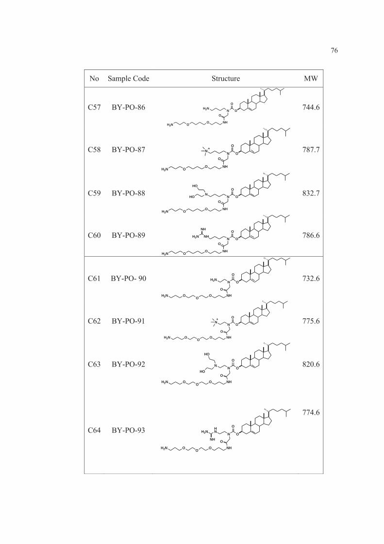
76
No Sample Code Structure MW
C57 BY-PO-86 N O
O
NHOO
O
H2N
H2N
744.6
C58 BY-PO-87 N O
O
NHOO
O
N
H2N
+
787.7
C59 BY-PO-88 N O
O
NHOO
O
N
H2N
HO
HO
832.7
C60 BY-PO-89 N O
O
NHOO
O
NH
H2N
H2N
NH
786.6
C61 BY-PO- 90 H2N N O
O
NHOOOO
H2N
732.6
C62 BY-PO-91 N N O
O
NHOOOO
H2N
+
775.6
C63 BY-PO-92 N N O
O
NHOOOO
H2N
HO
HO
820.6
C64 BY-PO-93 HN N O
O
NHOOOO
H2N
H2N
NH
774.6

77
No Sample Code Structure MW
C65 BY-PO-94 N O
O
NHOO
OH2N
OH2N
746.6
C66 BY-PO-95 N O
O
NHOO
ON
OH2N
+
789.6
C67 BY-PO-96 N O
O
NHOO
ON
OH2N
HO
HO
834.6
C68 BY-PO-97 N O
O
NHOO
ONH
OH2N
H2N
NH
788.6
C69 BY-PO-98 N O
O
NHOO
O
H2N
OH2N
760.6
C70 BY-PO-99 N O
O
NHOO
O
N
OH2N
+
803.7
C71 BY-PO-100 N O
O
NHOO
O
N
OH2N
HO
HO
848.7
C72 BY-PO-101 N O
O
NHOO
O
HN
OH2N
H2N
NH
804.6

78
3.1.6 All other chemicals
- Lipofectamine2000TM
(Invitrogen, NY, USA)
- Inhibitors of endocytic pathways
o Filipin complex, from Streptomyces filipinensis (Sigma-
Aldrich ®, St Louis, MO, USA)
o Geneticin selective antibiotic (G418 Sulfate) 50mg/ml
solution (GIBCO™(
o Nocodazole (Sigma-Aldrich ®, St Louis, MO, USA)
o Methyl-β-cyclodextrin (Sigma-Aldrich ®, St Louis, MO, USA)
o Wortmannin (Sigma-Aldrich ®, St Louis, MO, USA)
o Chlorpromazine hydrochloride (Sigma-Aldrich ®, St Louis,
MO, USA)
o Genistein (Sigma-Aldrich ®, St Louis, MO, USA)
o Ammonium chloride (Sigma-Aldrich®, St Louis, MO, USA)
- Gene Pure LE Agarose 500g (ISC BioExpress , USA)
- Lambda DNA / Hind III Markers (Promega, Madison, MI, USA)
- Blue/Orange 6x loading dye (Promega, Madison, MI, USA)
- Sodium chloride (UNIVAR Ajax Finechem; analytical reagent grade)
- Casein enzyme hydrolysate, type-1; Tryptone (HIMEDIA Himedia
Laboratories Pvt. Ltd.)
- Yeast extract powder (HIMEDIA Himedia Laboratories Pvt. Ltd.)
- Sterile water for irrigation (General Hospital Products Public Co., Ltd.)
- Sodium bicarbonate (Analar BDH ;VWR International Ltd.)
- Dimethyl sulfoxide (Fisher Scientific; analytical reagent grade)
0.25% Trypsin-EDTA (GIBCO ™)
- Trypan blue stain 0.4% (GIBCO™)
- Branched polyethylenimine, PEI (Sigma-Aldrich Chemie. GmbH,
Germany)
- 3-(4,5-dimethylthiazol-2-yl)-2,5-diphenyl-tetrazolium bromide, MTT
(Sigma-Aldrich®, St Louis, MO, USA)
- Ethanol absolute (Scharlau ET0016 Scharlau Chemie SPAIN

79
analytical reagent grade)
- Tris(hydroxymethyl) aminomethane, Pacific Science, Thailand,
molecular biology grade)
- Chloroform (VWR International Ltd. England analytical reagent grade)
3.2 Equipments
1. 1.5 ml, 2 ml Eppendrof® tube
2. 10 ml test tube
3. 10 ml glass bottle
4. 15 ml, 50 ml centrifuge tubes-sterile (Biologic research company)
5. 24-well tissue culture test plate (TPP®; Switzerland)
6. 25 cm2 and 75 cm2 cell culture flask (Corning®; Corning Incorporated)
7. 96-well cell culture cluster (Costar®; Corning Invorporated)
8. Analytical balance (Satorius CP224S, Scientific promotion Co., Ltd.)
9. Desiccator
10. N2 gas in laminar hood
11. Bath sonicator
12. Probe sonicator (Sonics Vibra CellTM, Newtown, CT, USA)
13. Centrifuge (Sorvall® Biofuge Stratos, Thermo Fisher Scientific Inc., USA)
14. Automatic autoclave (Model: LS-2D; Scientific promotion CO., Ltd.)
15. Bacterial incubator (Contherm; Lab Focus CO., Ltd)
16. Cellulose acetate filter 0.2 μm (Sartorius AG. 37070 Goettingen, Germany)
17. Centrifuge (Hermle Z300K; Labnet®; Lab Focus CO., Ltd.)
18. CO2 incubator (HERA Cell 240 Heraeus)
19. UV/Visible spectrophotometer (NanoVue Plus™, GE Healthcare, USA.)
20. UV/Visible spectrophotometer (Agilent 8453 UV-Visible spectrophotometer,
Agilent Technologies, Germany)
21. Fluorescence microscope (Model: GFP-B, wavelengths: excitation filter
480/40 and emission filter 535/50)
22. Fusion universal microplate analyzer (Model No: AOPUS01 and A153601; A
Packard bioscience company)

80
23. GeneRay UV-Photometer (Biometra® λ260/280 nm)
24. Inverted microscope (Eclipse TE 2000-U; Model: T-DH Nikon® Japan)
25. Laminar air flow (BIO-II-A)
26. Magnetic stirrer and magnetic bar
27. Measuring pipettes (1, 2, 5, 10 ml)
28. Micropipette 0.1-2 μl, 2-20 μl, 10-100 μl, 20-100 μl, 100-1000 μl
(Masterpette®; Bio-Active Co., Ltd.) and micropipette tip
29. pH meter (HORIBA compact pH meter B-212)
30. Pipette aid (Powerpette Plus; Bio-Active Co., Ltd.)
31. Protein and nucleic acid electrophoresis (MyRUN intelligent electrophoresis
unit; Cosmobio CO., Ltd., Japan)
32. Sartorius® filter set (Sartorius BORO 3.3 Goettingen, Germany)
33. Shaking incubator (GFL 3031, UK)
34. Water bath (Hetofrig CB60; Heto High Technology)
35. Vortex mixer
36. Zetasizer Nano ZS (Malvern instruments Ltd., Malvern, UK)
37. Transmission electron microscope (JEOL JEM-1230, JEOL, Tokyo, Japan)
38. Refrigerator 4°C, -20°C and -80

81
3.3 Methods
3.3.1 Preparation of liposomes and niosomes
3.3.1.1 Preparation of non-ionic liposomes/niosomes
Liposomes and niosomes used in this study were prepared using a
sonication method. Briefly, PC was dissolved in a chloroform: methanol
mixture (2:1 v/v) to obtain a 5mM PC solution used to prepare liposomes.
Similarly, non-ionic surfactants (Tween 20, Tween 80, Span 20 or Span 80)
and cholesterol were dissolved in an ethanol: chloroform mixture (1:1 v/v) to
obtain a 2.5:2.5 mM solution used to prepare the niosomes. The total lipids
amount of liposomes and niosomes was fixed at 5 mM or 0.015 mmol. The
solvents were evaporated under N2 gas flow to generate a thin film at the
bottom of the test tubes. The thin film was then left in a desiccator overnight
to remove the remaining organic solvents, and thin film hydration was
performed using a Tris-buffer (20 mM Tris and 150 mM NaCl, pH 7.4). After
hydration, the dispersion is sonicated using a bath sonicator for 30 minutes
followed by a probe sonicator (Vibra-CellTM Ultrasonic Processor) each for 30
minutes in two cycles.
3.3.1.2 Preparation of cationic liposomes/niosomes
The cationic niosomes and cationic liposomes were prepared in
the same way as the non-ionic niosomes, except that cationic lipid dissolved in
a chloroform: methanol mixture (2:1 v/v) was added to PC solution for
liposomes and a solution of surfactant and cholesterol for niosomes. Finally, a
solution of PC: cationic lipid at a molar ratio of 5:1, as well as a solution of
surfactant: cholesterol: cationic lipid at a molar ratio of 2.5: 2.5: 1 were used
to prepare the thin film. The total lipids amount of liposomes and niosomes
was fixed at 6 mM or 0.018 mmol.
3.3.2 Plasmid DNA preparation
The DNA encoding green fluorescence protein (pEGFP-C2) was used to
observe gene expression (Figure 14). The plasmid DNA (pDNA) was
purchased from Invitrogen. To amplify the pDNA, it was transformed into

82
Escherichia coli DH5- (E. Coli). After amplification of the E.Coli, the
plasmids were isolated by using QIAGEN Plasmid Midi Kits.
Figure 14 Restriction Map and Multiple Cloning Site (MCS) of pEGFP-C2. pEGFP-
C2 encodes a red-shifted variant of wild-type GFP which has been
optimized for brighter fluorescence and higher expression in mammalian
cells. (Excitation maximum = 488 nm; emission maximum = 507 nm.)
pEGFP-C2 encodes the GFPmut1 variant which contains the double-amino-
acid substitution of Phe-64 to Leu and Ser-65 to Thr. The coding sequence
of the EGFP gene contains more than 190 silent base changes which
correspond to human codon-usage preferences. Sequences flanking EGFP
have been converted to a Kozak consensus translation initiation site to
further increase the translation efficiency in eukaryotic cells. The MCS in
EGFP-C2 is between the EGFP coding sequences and the SV40 poly A.
Genes cloned into the MCS will be expressed as fusions to the C terminus
of EGFP if they are in the same reading frame as EGFP and there are no
intervening stop codons. SV40 polyadenylation signals downstream of the

83
EGFP gene direct proper processing of the 3' end of the EGFP mRNA. The
vector backbone also contains an SV40 origin for replication in mammalian
cells expressing the SV40 T-antigen. A neomycin-resistance cassette
(Neor), consisting of the SV40 early promoter, the neomycin/kanamycin
resistance gene of Tn5, and polyadenylation signals from the Herpes
simplex virus thymidine kinase (HSV TK) gene, allows stably transfected
eukaryotic cells to be selected using G418. A bacterial promoter upstream
of this cassette expresses kanamycin resistance in E. coli. The pEGFP-C2
backbone also provides a pUC origin of replication for propagation in E.
coli and an f1 origin for single-stranded DNA production.
Source: CLONTECH Laboratories, Inc. Catalog #6083-1
Available from: http://www.ebiotrade.com/buyf/productsf/Clontech/632316.htm
3.3.2.1 Isolation and purification of pDNA
The DNA was isolated using QIAGEN Plasmid Midi Kits
according to the manufacturer’s protocol. The purification process of pDNA is shown
as follows;
1. Pick a single colony from a freshly streaked selective plate and inoculated in
100 ml of LB medium. Grow at 37 C for 12-16 h with vigorous shaking
(approx. 200 rpm).
2. Harvest the bacterial cells by centrifugation at 6000 x g for 15 min at 4 C.
3. Resuspend the bacterial pellet in 4 ml Buffer P1.
4. Add 4 ml Buffer P2, mix thoroughly by vigorously inverting the sealed tube
4-6 times, and incubate at room temperature (15 – 25 C) for 5 min.
5. Add 4 ml of chilled Buffer P3, mix immediately and thoroughly by
vigorously inverting 4-6 times, and incubate on ice for 15 min.
6. Centrifuge at 20,000 x g for 30 min at 4 C. Remove supernatant containing
plasmid DNA promptly.
7. Centrifuge the supernatant again at 20,000 x g for 15 min at 4 C. Remove
supernatant containing plasmid DNA promptly.

84
8. Equilibrate a QIAGEN-tip 100 by applying 5 ml Buffer QBT, and allow the
column to empty by gravity flow.
9. Apply the supernatant from step 7 to the QIAGEN-tip and allow it to enter the
resin by gravity flow.
10. Wash the QIAGEN-tip with 2 x 10 ml Buffer QC.
11. Elute DNA with 5 ml Buffer QF.
12. Precipitate DNA by adding 3.5 ml (0.7 volumes) room-temperature
isopropanol to the eluted DNA. Mix and centrifuge immediately at 15,000
x g for 30 min at 4 C. Carefully decant the supernatant.
13. Wash DNA pellet with 2 ml of room-temperature 70% ethanol, and centrifuge
at 15,000 x g for 10 min at 4 C. Carefully decant the supernatant without
disturbing the pellet.
14. Air-dry the pellet for 5-10 min, and re-dissolve the DNA in a suitable volume
of TE buffer, pH 8.0.
3.3.2.2 Quantification of pDNA concentration
The concentration of pDNA was determined by GeneRay UV
Photometer (Biometra 260/280 nm). The quantification process of pDNA is shown
as follows: A 35 l of the pure plasmid was diluted into 665 l of distilled water and
gently mixed. Then, the diluted plasmid was measured at 260 nm and 280 nm with
GeneRay UV Photometer (Biometra 260/280 nm). The concentration of pDNA
was calculated using follow equation:
Plasmid concentration = 50 g/ml x OD260nm x DF Eq.1
- A solution of 50 g/ml of an average double-stranded DNA has an OD260nm of 1.
- OD260nm is the optical density from the absorbance reading.
- DF is the dilution factor (as above is 20).
DNA maximally absorbs ultraviolet light at a wavelength of about
260 nm. It is the bases that are principally responsible for this absorption, while

85
absorption at 280 nm indicates protein contamination. Often a ratio of 1.8 A260/A280 is
given as means of determining purity [112].
3.3.3 Cell culture, routine maintenance
The human cervical carcinoma cell line (HeLa cells) was used as a
model for the transfection and cytotoxicity study of DNA. Whereas, HeLa
Cells Stably Expressing Green Fluorescent Protein (HeLa stable cells) was
used as a model for the transfection and cytotoxicity study of siRNA. The cell
lines observed under an inverted microscope were illustrated in Figure 15.
Figure 15 Cultivated HeLa cells and HeLa stable cells on 75 cm2 tissue culture flask,
image 10× objective using an inverted microscope.
3.3.3.1 Establishing HeLa stable cells
HeLa cells with stable constitutive expression of EGFP were
generated. Briefly, HeLa cells were transfected with complexes of 0.8μg of
pEGFP-C2 plasmids (Clontech, USA) and 2.0 μl Lipofectamine™ 2000
(Invitrogen, USA) according to the manufacturer’s protocol. Briefly, the day
before transfection, cells were seeded 1×10⁵ cells into each of one well of a
24-well plate and incubated at 37˚C with 5% CO2. Prior to transfection,
medium was removed and washed one time with PBS then added with a
mixture of 100 μl of complexes and 500 μl of serum free medium, then
incubated at 37˚C with 5% CO2 for 12 h. After that, the medium was removed
and fresh medium was added, then further incubation until cells growed 100%
confluent. To generate clones of stable cell lines, the cells were trypsinized
and aliquot into a 100 mm cell culture dishes and added with selective
HeLa cells HeLa stable cells

86
medium, a MEM supplemented with 10% FBS and 0.5 mg/ml G418
(Geneticin® Invitrogen, USA) which is used to select and maintain EGFP
gene expression. Individual clones were isolated and expanded in modified
Eagle’s medium (MEM), supplemented with 10% fetal bovine serum (FBS)
and treated with 0.1 mg/ml G418 every 3–4 weeks to maintain the EGFP gene
expression [28].
3.3.3.2 Cultivation of HeLa and HeLa stable cells
HeLa cells and HeLa stable cells were grown in 250ml tissue culture
flask containing complete growth medium (MEM) in a humidified air
atmosphere (5% CO2, 95% RH, 37˚C). Cultivated cells were visualized using
an inverted microscope to detect cross-contamination or visible microbial
contamination. The cells were passage every 3-5 days as spit ratio 1:5 to 1:10.
3.3.3.3 Preparation of culture media
The process of preparation of 1X complete culture medium is shown as
follows: fetal bovine serum (FBS) was heat inactivated by incubating for 30
min in a 56 °C water bath. MEM Medium powder was dissolved in 5% less
sterile water than desired total volume of medium with gentle stirring and 2.2
or 2.0 g of NaHCO3 per L of medium was added for adjust pH to 0.2-0.3
below desired final working pH (pH 7.4). The solution was sterilized
immediately by membrane filtration. Then the solution was supplemented
with: 1% v/v Glutamax-I or L-Glutamine, 1% v/v Non-essential Amino Acids,
10% v/v heat inactivated FBS.
3.3.3.4 Thawing frozen cells
The cells were thawed quickly in a 37 °C water bath and one vial of
cells was diluted into 10 ml of complete growth medium, gently mixed. It was
centrifuged at 750 rpm 5 min 25 C for removing DMSO containing cell
culture freezing medium that can be toxic to cell. The medium above the
pellet was removed and the cells in complete growth medium were
resuspended. The cell suspension was diluted to appropriate concentration,
and transferred into a tissue culture flask.

87
3.3.3.5 Subculturing
Cultivated HeLa and HeLa stable cells were visualized using an
inverted microscope to 70-80% confluency in the TC flasks. The media was
aspirated and rinsed one time with PBS. 0.25% trypsin/EDTA solution was
added, the cells were incubated at 37 °C for approximately 1 min or until the
cells detached and floated. This can be confirmed by periodic visual
inspection of the flasks or observing cells under an inverted microscope.
Trypsin/EDTA was inactivated by adding excess serum-containing medium.
Cell suspension was removed to a conical tube and cell was pelleted by
centrifugation at 750 rpm for 5 min. Complete culture medium was added to
detached cells. The cells were resuspended by using pipette up and down.
The aliquots of the cell suspension were added to new TC flasks.
Subcultivation ratio was 1:5. The complete culture medium was added for a
total of 15 ml per 75 cm2 TC flasks. The cell culture was then placed in
incubator.
3.3.3.6 Freezing cells
Cells to be frozen should be in late log phase growth. After the cell
culture subculturing, pellet of cells was diluted in complete growth medium.
For one 75 cm2 cell culture flask, resuspended in 5 ml of complete culture
medium. The cell suspension tube and 5 labeled cryotubes were pre-cooled
on ice for 5 minutes before adding 250 μl of DMSO into the suspension cell
and mixing gently. 1ml of cell suspension was transferred into cryoprotective
tubes. The cryostocks were stored at -20 C, -80 C overnight, respectively.
For long term storage, the cells were stored in liquid nitrogen.
3.3.4 Characterization of liposomes and niosomes
3.3.4.1 Turbidity measurement
The turbidity of liposomes and niosomes was determined in triplicate by
measuring the absorbance at 400 nm using a UV/Visible spectrophotometer
(NanoVue Plus™, GE Healthcare, USA). Further characterization of the
niosomes formation was investigated by increasing molar ratio of non-ionic

88
surfactant to cholesterol and measuring of the alteration in turbidity at 400 nm.
using UV/Visible spectrophotometer (Agilent 8453 UV-Visible
spectrophotometer, Agilent Technologies, Germany).The drop of turbidity is
related to the structural change from niosomes to micelles.
3.3.4.2 Size and zeta potential measurements
The particle size and surface charge of the liposomes and niosomes
were determined by photon correlation spectroscopy using a Zetasizer Nano
ZS (Malvern Instruments Ltd, Malvern, UK). The liposomes and niosomes
were diluted 1:10 with distilled water that filtered through a 0.22 μm
membrane filter to obtain a volume required for each measurement (about 1
ml). All samples were measured in triplicate at room temperature.
3.3.5 Cytotoxicity test of cationic liposomes and cationic niosomes
The cell viability was investigated using the MTT assay. HeLa cells
were seeded into a 96-well plate at a density of 8x10³ cells/well in 100 μl of
complete medium and were incubated at 37 °C and 5 % CO2 overnight. Prior
to the cytotoxicity study, the medium was removed, and the cells were treated
with variable concentrations of carriers. The day after treatment, the cells were
rinsed with PBS and incubated in 100 μl MTT-containing medium (1 mg/ml)
for 4 hours. The formazan crystals formed in living cells were dissolved in 100
μl dimethyl sulfoxide (DMSO) per well. Relative cell viability (%) was
calculated based on the absorbance observed at 550 nm using a microplate
reader (Universal Microplate Analyzer, Models AOPUS01 and AI53601,
Packard BioScience, CT, USA) and compared with non-treated cells. The
viability of non-treated cells is defined as 100 %.
3.3.6 In vitro gene transfection experiment
3.3.6.1 Preparation and characterization of carrier/DNA
complexes
The carrier/DNA complexes were prepared at variable weight ratios of
carrier to DNA by adding the carrier solution to a DNA solution (the amount
of DNA was fixed at 0.5 μg). The mixture was gently mixed by pipetting up

89
and down for 3-5 seconds to initiate complex formation. Then, the mixture
was left at room temperature for 30 minutes to complete the complex
formation process. Agarose gel electrophoresis was performed to assure
complex formation using 1 % agarose gels (1 g agarose in 100 ml TAE buffer
containing ethidium bromide 0.5μg/ml) and TAE as a running buffer. The
electrophoresis was carried out for 45 minutes at 100 V. Ten microliters of
carrier/DNA complexes containing 0.5 μg of DNA were loaded per well.
3.3.6.2 Size and zeta potential measurements of carrier/DNA
complexes
The particle size and surface charge of the carrier/DNA complexes
were determined by photon correlation spectroscopy using a Zetasizer Nano
ZS (Malvern Instruments Ltd, Malvern, UK). The liposomes, niosomes and
the niosomes/DNA complexes at varying weight ratio were diluted with
distilled water that filtered through a 0.22 μm membrane filter to obtain a
volume required for each measurement (about 1 ml). All samples were
measured in triplicate at room temperature.
3.3.6.3 In vitro transfection of carrier/DNA complexes into HeLa
cells
The day before transfection, HeLa cells were seeded into a 48-well
plate at a density of 2x10⁴ cells/well in 0.25 ml of complete medium (MEM
containing 10 % fetal bovine serum and supplemented with 1 % L-glutamine,
1 % non-essential amino acids solution and 100 U/ml penicillin and 100 μg/ml
streptomycin) and were incubated at 37 °C and 5 % CO2 overnight. Prior to
transfection, the medium was removed, and the cells were incubated with 0.25
ml of the carrier/DNA complexes in serum-free medium at variable weight
ratios containing 0.5 g of pEGFP-C2 for 24 hours at 37 °C and 5 % CO2.
Cells transfected with naked pEGFP-C2 were used as negative controls, and
cells transfected with LipofectamineTM 2000 complexed with DNA at a weight
ratio of 2 were used as positive controls. The day after transfection, the cells
were washed with pH 7.4 phosphate-buffered saline (PBS) and replaced with

90
complete medium. Green fluorescent protein (GFP) expression was directly
observed under a fluorescent microscope (Model: GFP-B, wavelengths:
excitation filter 480/40 and emission filter 535/50). All transfection
experiments were performed in triplicate. The complete medium containing 10
% fetal bovine serum was used to investigate the serum effect on transfection
efficiency.
3.3.7 Cytotoxicity test of carrier/DNA complexes
The cell viability was investigated using the MTT assay. HeLa cells
were seeded into a 96-well plate at a density of 8x10³ cells/well in 100 μl of
complete medium and were incubated at 37 °C and 5 % CO2 overnight. Prior
to the cytotoxicity study, the medium was removed, and the cells were treated
with the carrier/DNA complexes under the same conditions as in the
transfection experiment. The day after treatment, the cells were rinsed with
PBS and incubated in 100 μl MTT-containing medium (1 mg/ml) for 4 hours.
The formazan crystals formed in living cells were dissolved in 100 μl
dimethyl sulfoxide (DMSO) per well. Relative cell viability (%) was
calculated based on the absorbance observed at 550 nm using a microplate
reader (Universal Microplate Analyzer, Models AOPUS01 and AI53601,
Packard BioScience, CT, USA) and compared with non-treated cells. The
viability of non-treated cells is defined as 100 %.
3.3.8 Haemolytic assay of cationic niosomes and niosomes/DNA
complexes
The haemolytic activity was evaluated using the human leukocyte-poor
packed red cells (LPRC). Briefly, LPRC was centrifuged at 3800 rpm for 5
minutes to remove the plasma and additive solution, after that the red blood
cells (RBCs) were washed 3 times with PBS. The RBCs were distributed in
96-well plate (100 μl/well) and an equal volume of cationic niosomes or
niosomes/DNA complexes under the same concentration as in the transfection
experiment (both diluted with PBS) was added. After incubation at 37 ºC for 1
h, the plates were centrifuged at 800xg for 10 minutes and the supernatant was
collected. The absorbance of the supernatant was measured at 550 nm using a

91
microplate reader (Universal Microplate Analyzer, Models AOPUS01 and
AI53601, Packard BioScience, CT, USA). The RBCs treated with PBS were
used as negative control (normal haemolysis), whereas the RBCs treated with
sterile water were used as positive control (100% haemolysis). In each plate,
positive and negative control wells were added. The percentage of haemolytic
activity of each sample was estimated from the absorbance at 550 nm using
the following equation; [(A-A0)/(Amax-A0)] × 100, where A0 is an
absorbance of negative control and Amax is an absorbance of positive control
which corresponds to 100% haemolysis. The percentages of haemolysis (Y
axis) were plotted against log niosomes concentration (X axis) and the HC50
value of niosomes (niosomes concentration that lyses 50% RBCs) was
estimated.
3.3.9 Transmission Electron Microscopy
The morphological analysis of cationic niosomes and niosomes/DNA
complexes was carried out using a transmission electron microscope (JEOL
JEM-1230, JEOL, Tokyo, Japan). A small drop of sample was placed on a 200
mesh carbon-coated copper grid. The excess sample was removed using filter
paper, after that the sample was air-dried and viewed at 80kV.
3.3.10 Effect of various inhibitors on in vitro transfection in HeLa cells
HeLa cells were seeded into 48-well plates at a density of 1×10⁴
cells/well in 0.2 ml of MEM complete medium and were incubated at 37 °C
and 5 % CO2 overnight. Prior to transfection, the medium was removed and
cells were rinsed with PBS, pH 7.4.The cells were pre-incubated for 30 min
with various inhibitors separately at concentrations which were not toxic to the
cells. Then, cells were replenished with 0.2 ml in serum-free medium
containing niosomes/DNA complexes at weight ratios provided highest
transfection (DNA was fixed at 0.5 μg/well) and incubated for 4.5 h. Cells
transfected with naked pEGFP-C2 were used as negative controls, and cells
transfected with LipofectamineTM 2000 complexed with DNA at a weight ratio
of 2 were used as positive controls. After transfection, the cells were washed

92
with pH 7.4 phosphate-buffered saline (PBS) and grown in complete medium
for 24h to allow for GFP expression. Green fluorescent protein (GFP)
expression was directly observed under a fluorescent microscope (Model:
GFP-B, wavelengths: excitation filter 480/40 and emission filter 535/50). All
transfection experiments were performed in triplicate.
3.3.11 Investigation of cationic niosomes as siRNA carrier
3.3.11.1 Preparation of siRNA
The EGFP targeted siRNA, siRNA-EGFP(+), and the mismatch
siRNA, siRNA-EGFP(−) were synthesized using Ambion’s Silencer™ siRNA
Construction Kit (Ambion, USA). The duplex siRNA-EGFP(+) contains sense
5′-gcu gac ccu gaa guu cau cuu-3′ and antisense 5′-gau gaa cuu cag ggu cag
cuu-3′. The siRNA-EGFP(−), contains sense 5′-gca ccg cuu acg uga uac uuu-
3′, and antisense 5′-agu auc acg uaa
3.3.11.2 Preparation and characterization of the
niosomes/siRNA complexes
The niosomes/siRNA complexes were prepared at variable
weight ratios of niosomes to siRNA by adding the niosomes solution (diluted
with DEPC-treated water) to a siRNA solution (the amount of siRNA was
fixed at 7.5 pmol or 0.105 μg). The mixture was gently mixed by pipetting up
and down for 3-5 seconds to initiate complex formation. Then, the mixture
was left at room temperature for 30 minutes to complete the complex
formation process. Complex formation was assured by agarose gel
electrophoresis using 1 % agarose gels (1 g agarose in 100 ml Trisborate-
EDTA (TBE) buffer containing ethidium bromide 0.5 μg/ml) The
electrophoresis was carried out for 20 min at 100 V in TBE running buffer pH
8.3. Ten microliters of niosomes/siRNA complexes were loaded per well.
3.3.11.3 In vitro siRNA transfection of cationic niosomes
Briefly, HeLa stable cells were seeded at the density of 8,000
cells/well in black clear-bottom, 96-well plates with MEM, supplemented with

93
10% FBS in a humidified atmosphere (5% CO2, 95% air, 37°C) 24 h prior to
transfection. Measurement of EGFP was performed using a fluorescence
microplate reader (Universal Microplate Analyzer) with excitation/emission at
485/530 nm. The seeding variation as measured by the fluorescence intensity
of each well before transfection was less than 10%. The niosomes/siRNA-
EGFP(+) and the niosomes/siRNA-EGFP(−) complexes at various weight
ratios were formed as described in the complex formation (the amount of
siRNA per well was fixed at 15 pmol or 0.210 μg). Prior to transfection, the
cells were washed with PBS and added with 100 μl culture medium containing
niosomes/siRNA complexes. The cells were incubated for 6 h before changing
culture medium. Plates were incubated for a further 4 days at 37°C and 5%
CO2. The culture medium was replaced every day and the fluorescence
intensity was measured daily for 4 days. To take into account the variation of
fluorescent intensity due to unequal initial number of cells in each well, %
seeding variation was first calculated (Eq. 1) and then used the values to adjust
the fluorescent intensity obtained from each well by Eq. 2. The percentage of
EGFP gene silencing was calculated by Eq. 3.

94
Where Iav,d0 is the average fluorescent intensity of all wells prior to
transfection (day 0), In,d0 is the fluorescent intensity of individual well at day
0, In,d1−4 is the fluorescent intensity at days one through four of each well, Iad is
adjusted fluorescent intensity, Iad,siRNA(+) is adjusted fluorescent intensity of
individual well with niosomes/siRNA(+) complexes, and Iad,siRNA(−) is the
average value of adjusted fluorescent intensity of three wells with
niosomes/siRNA(−) complexes.
3.3.11.4 Cytotoxicity test of niosomes/siRNA complexes
The investigation of cell viability was performed using MTT
assay. After transfection experiment (day 4), the plates containing HeLa stable
cells which were transfected with niosomes/siRNA were used to evaluate
cytotoxicity. The cells were added with 100 μl MTT-containing medium (1
mg/ml) for 4 hours. The formazan crystals formed in living cells were
dissolved in 100 μl dimethyl sulfoxide (DMSO) per well. Relative cell
viability (%) was calculated based on the absorbance observed at 550 nm
using a microplate reader (Universal Microplate Analyzer, Models AOPUS01
and AI53601, Packard BioScience, CT, USA) and compared with non-treated
cells. The viability of non-treated cells is defined as 100 %.
3.3.11.5 Size and zeta potential measurements of
niosomes /siRNA complexes
The particle size and surface charge of the niosomes/siRNA
complexes were determined by photon correlation spectroscopy using a
Zetasizer Nano ZS (Malvern Instruments Ltd, Malvern, UK). The
niosomes/siRNA complexes at varying weight ratio were diluted with distilled
water that filtered through a 0.22 μm membrane filter to obtain a volume
required for each measurement (about 1 ml). All samples were measured in
triplicate at room temperature.

95
3.3.12 Physical stability study
The physical stability of the cationic niosomes at 4°C and 25°C was
determined by monitoring the particle size and zeta potential every month for
at least 6 months.
3.3.13 Statistical analysis
The significant differences in transfection efficiency and cell viability
were statistically tested by using one-way analysis of variance (ANOVA)
followed by LSD post hoc test for analyzing transfection efficiency and Tukey
post hoc test for determining cell viability. The significance level was set at
p<0.05.

96
CHAPTER 4
RESULTS AND DISCUSSION
4.1 Preliminary study
4.1.1 Screening of cationic lipids by in vitro transfection efficiency and
cytotoxicity
4.1.2 Screening of niosomes formulations
4.2 Cationic niosomes formulations
4.2.1 Particle size and zeta potential
4.2.2 Cytotoxicity of cationic niosomes
4.2.3 Physical stability of cationic niosomes
4.3 Cationic niosomes as gene carrier
4.3.1 Characterization of cationic niosomes/DNA complexes
4.3.1.1 Particle size and zeta potential
4.3.1.2 Gel retardation assay
4.3.1.3 Morphological analysis
4.3.2 In vitro gene transfection
4.3.3 Effect of serum on in vitro gene transfection
4.3.4 Cytotoxicity of niosomes/DNA complexes
4.3.5 Haemolytic assay
4.3.6 Investigation of cellular internalization of niosomes/DNA complex
4.4 Cationic niosomes as siRNA carrier
4.4.1 Characterization of cationic niosomes/siRNA complexes
4.4.1.1 Particle size and zeta potential
4.4.1.2 Gel retardation assay
4.4.2 In vitro siRNA transfection
4.4.3 Cytotoxicity of niosomes/siRNA complexes

97
4.1 Pre-liminary study
4.1.1 Screening of in vitro transfection efficiency of cationic lipids
The structure of cationic lipid used as transfection reagent is one of the major
factors influence on gene transfection. To screen the in vitro transfection efficiency of
cationic lipids, the cationic liposomes composed of PC: cationic lipid in a molar ratio
of 5:1 were transfected into HeLa cells.
The cationic lipids can be categorized base on cationic lipids structure into two
major groups; diethanolamine-based cationic lipids (A1-A29) and cholesterol
derivative cationic lipids (C1-C72). In addition, these cationic lipids can also be sub-
classified by the differences in hydrophilic head group as presented in Table 9-10.
Table 9 Sub-classified diethanolamine-based cationic lipids (A1-A29).
Cationic lipid Head group Linker group Hydrophobic group
A1, A2, A3 Same Same C14, C16, C18, respectively
A4, A5, A6 Same Same C14, C16, C18, respectively
A7, A8, A9 Same Same C14, C16, C18, respectively
A10, A11, A12 Same Same C14, C16, C18, respectively
A13, A14, A15 Same Same C14, C16, C18, respectively
A16, A17, A18 Same Same C14, C16, C18, respectively
A19, A20, A21 Same Same C14, C16, C18, respectively
A22, A23 Same Same C14, C16, respectively
A24, A25, A26 Same Same C14, C16, C18, respectively
A27, A28, A29 Same Same C14, C16, C18, respectively
For diethanolamine-based cationic lipids (A1-A29), the cationic lipids in each
group have same hydrophilic head group and linker group, but different hydrophobic
group (acyl chain length: C14, C16 and C18) (Table 9). For cholesterol derivative
cationic lipids (C1-C72), the cationic lipids in each group have same linker group and
hydrophobic group, but different hydrophilic head group (different amine head group:
primary amine, quaternary ammonium, amino alcohol and guanidine) (Table 10).
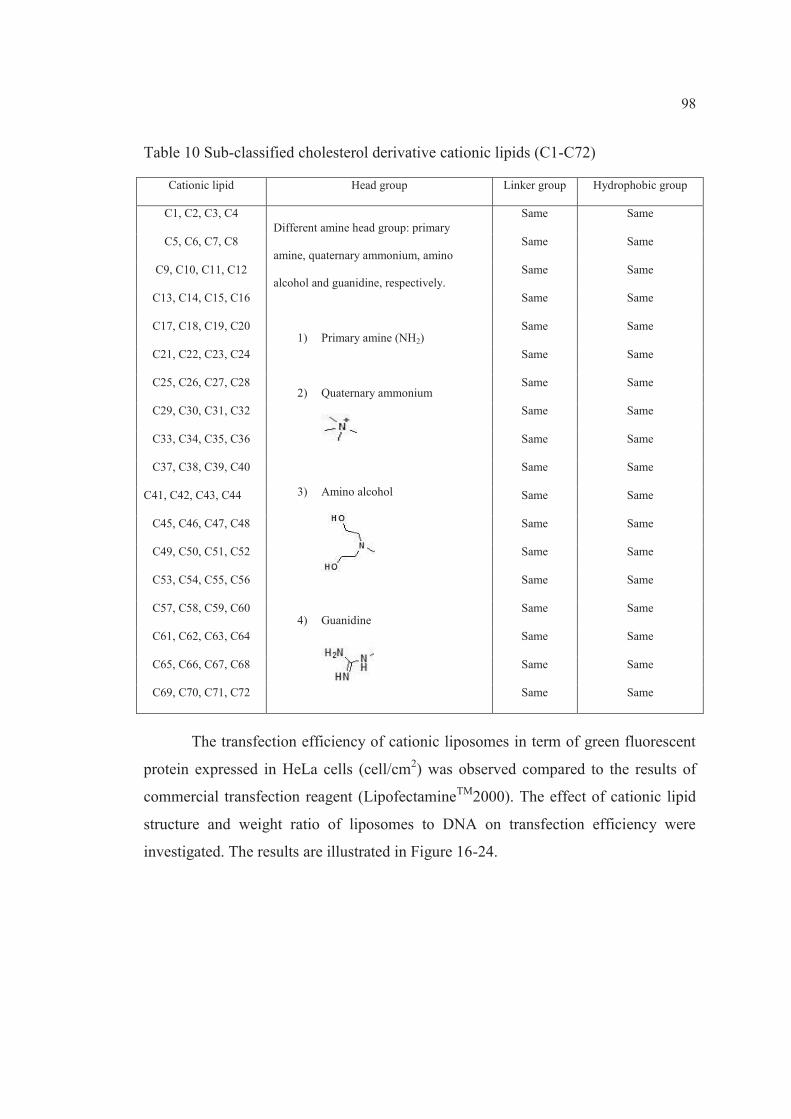
98
Table 10 Sub-classified cholesterol derivative cationic lipids (C1-C72)
Cationic lipid Head group Linker group Hydrophobic group
C1, C2, C3, C4 Different amine head group: primary
amine, quaternary ammonium, amino
alcohol and guanidine, respectively.
1) Primary amine (NH2)
2) Quaternary ammonium
3) Amino alcohol
4) Guanidine
Same Same
C5, C6, C7, C8 Same Same
C9, C10, C11, C12 Same Same
C13, C14, C15, C16 Same Same
C17, C18, C19, C20 Same Same
C21, C22, C23, C24 Same Same
C25, C26, C27, C28 Same Same
C29, C30, C31, C32 Same Same
C33, C34, C35, C36 Same Same
C37, C38, C39, C40 Same Same
C41, C42, C43, C44 Same Same
C45, C46, C47, C48 Same Same
C49, C50, C51, C52 Same Same
C53, C54, C55, C56 Same Same
C57, C58, C59, C60 Same Same
C61, C62, C63, C64 Same Same
C65, C66, C67, C68 Same Same
C69, C70, C71, C72 Same Same
The transfection efficiency of cationic liposomes in term of green fluorescent
protein expressed in HeLa cells (cell/cm2) was observed compared to the results of
commercial transfection reagent (LipofectamineTM2000). The effect of cationic lipid
structure and weight ratio of liposomes to DNA on transfection efficiency were
investigated. The results are illustrated in Figure 16-24.

99
Figure 16 Transfection efficiency of liposomes (A1-A9)/DNA complexes in HeLa cells
Figure 17 Transfection efficiency of liposomes (A10-A21)/DNA complexes in HeLa
cells

100
Figure 18 Transfection efficiency of liposomes (A22-A29)/DNA complexes in HeLa
cells Figure 19 Transfection efficiency of liposomes (C1-C12)/DNA complexes in HeLa
cells

101
Figure 20 Transfection efficiency of liposomes (C13-C24)/DNA complexes in HeLa
cells Figure 21 Transfection efficiency of liposomes (C25-C36)/DNA complexes in HeLa
cells

102
Figure 22 Transfection efficiency of liposomes (C37-C48)/DNA complexes in HeLa
cells Figure 23 Transfection efficiency of liposomes (C49-C60)/DNA complexes in HeLa
cells

103
Figure 24 Transfection efficiency of liposomes (C61-C72)/DNA complexes in HeLa
cells
From the transfection results, the gene transfection mediated by cationic
liposomes was mainly affected by the structure of cationic lipid composed in
liposomes [7-8]. In comparison with the result of LipofectamineTM2000 transfection
at weight ratio of 2 (about 4,126 cell/cm2), the high level gene transfection
(approximately 3,000 – 4,000 cell/cm2) were observed from liposomes formulated
with cationic lipid A10 and A29 (Figure 17-18) which are diethanolamine-based
cationic lipid and C7 (Figure 19) which is cholesterol derivative cationic lipid. The
structure of these cationic lipids yielded high gene transfection is shown in Table 11.
Among diethanolamine-based cationic lipids, the effective cationic head group
provided high gene transfection is spermine as presents in the structure of cationic
lipid A10 and A29. Whereas, the efficacious head group of cholesterol derivative
cationic lipid is amino alcohol as presents in the structure of cationic lipid C7.

104
Table 11 The cationic lipids provided high gene transfection in HeLa cells.
Cationic lipid Structure Maximum transfection efficiency (Cell/cm2)
A10 N O
O
O
O
HNN
HH2N
3,480
A29 N
N O
O
O
O
O
O
O
O
HN
NH
3,497
C7 H2NHN N O
O
O
OON
HO
HO
4,217
Not only the cationic lipid structure, but also the weight ratio of liposomes to
DNA mainly affected gene transfection mediated by cationic liposomes. The
transfection efficiency gradually increased as the weight ratio increased to a
maximum value, then decreased. The pattern of gene transfection with weight ratio is
a bell-shape like. This finding was in accordance with the previous report [25].
Moreover, the maximum transfection efficiency yielded from various cationic lipids
occurred at a different weight ratio.
To screen the cytotoxicity of cationic lipids, the cytotoxicity value of cationic
liposomes in term of 50% Inhibitory Concentration (IC50) was evaluated in HeLa
cells. The IC50 of cationic liposomes are presented in Table 12-13.

105
Table 12 Cytotoxicity of liposomes A1-A29 in HeLa cells.
Liposomes approximate IC50 (μg/ml)
A1 517.60 A2 410.35 A3 412.85 A4 375.90 A5 412.81 A6 328.97 A7 409.32 A8 15.23 A9 69.05
A10 12.99 A11 26.13 A12 7.81 A13 532.84 A14 288.29 A15 415.28 A16 326.85 A17 500.82 A18 397.75 A19 333.86 A20 260.34 A21 396.59 A22 39.67 A23 108.85 A24 178.83 A25 181.18 A26 355.45 A27 17.01 A28 328.42 A29 9.86

106
Table 13 Cytotoxicity of liposomes C1-C72 in HeLa cells.
Liposomes approximate IC50 (μg/ml)
Liposomes approximate IC50 (μg/ml)
C1 50.52 C37 10.73 C2 32.99 C38 11.36 C3 12.42 C39 2.48 C4 29.38 C40 11.50 C5 3.75 C41 0.94 C6 11.22 C42 6.21 C7 32.43 C43 1.12 C8 12.84 C44 25.43 C9 7.81 C45 2.31
C10 49.67 C46 15.83 C11 57.35 C47 12.84 C12 16.81 C48 3.73 C13 33.87 C49 14.15 C14 27.36 C50 1.63 C15 33.70 C51 7.45 C16 5.95 C52 586.46 C17 12.41 C53 25.41 C18 16.74 C54 1.31 C19 17.09 C55 6.65 C20 16.69 C56 0.51 C21 3.58 C57 9.36 C22 22.56 C58 0.11 C23 4.08 C59 1.03 C24 22.60 C60 11.34 C25 8.90 C61 0.18 C26 23.46 C62 10.95 C27 7.42 C63 1.73 C28 42.55 C64 8.00 C29 6.27 C65 4.31 C30 25.05 C66 4.99 C31 7.95 C67 4.92 C32 12.04 C68 47.26 C33 7.09 C69 13.53 C34 18.47 C70 15.62 C35 2.42 C71 1.27 C36 28.94 C72 1.53
Additionally, the cytotoxicity of liposome/DNA complexes at various weight
ratios which covered the weight ratio used for transfection, was also investigated in
HeLa cells as illustrated in Figure 25-32.

107
Figure 25 Cytotoxicity of liposomes (A1-A9)/DNA complexes in HeLa cells

108
Figure 26 Cytotoxicity of liposomes (A10-A21)/DNA complexes in HeLa cells

109
Figure 27 Cytotoxicity of liposomes (A22-A29)/DNA complexes in HeLa cells

110
Figure 28 Cytotoxicity of liposomes (C1-C16)/DNA complexes in HeLa cells

111
Figure 29 Cytotoxicity of liposomes (C17-C32)/DNA complexes in HeLa cells

112
Figure 30 Cytotoxicity of liposomes (C33-C48)/DNA complexes in HeLa cells

113
Figure 31 Cytotoxicity of liposomes (C49-C64)/DNA complexes in HeLa cells

114
Figure 32 Cytotoxicity of liposomes (C65-C72)/DNA complexes in HeLa cells
The percentage cell viability of liposomes A10 and A29 at the range of weight
ratio the highest transfection yielded was closer to control (non-treated cells) than
liposomes C7. Therefore, consideration in term of safety, the diethanolamine-based
cationic lipid A10 and A29 seem to be more safety than the cholesterol derivative
cationic lipid C7.
As the results above, two sub-groups of diethanolamine-based cationic lipids
were chosen to prepare cationic niosomes for further investigation as gene carriers.
The chosen cationic lipids are listed below:
i) A10, A11 and A12
ii) A27, A28 and A29

115
4.1.2 Screening of niosomes formulations
The micelle-to-vesicle phase transition of surfactant can be detected by
general techniques such as UV–Vis spectroscopy, dynamic light scattering and cryo-
TEM [113].
The optimal molar ratio of non-ionic surfactant (Tween 20, Tween 80, Span 20
or Span 80) and cholesterol for preparing niosomes was screened by measurement of
the turbidity at 400 nm [11] using UV-Visible spectrophotometer (Agilent 8453,
Agilent Technologies, Germany). The turbidity at 400 nm of niosomes formulated
with non-ionic surfactant and cholesterol is presented in Figure 33.
Figure 33 Turbidity at 400 nm of niosomes formulated with non-ionic surfactant; A)
Tween 20 and Tween 80, B) Span 20 and Span 80 and cholesterol.
Turbidity quantifies the degree to which light traveling through a sample is
scattered by the suspended particles. Turbidity intrinsically depends on the amount of
0
0.1
0.2
0.3
0.4
Turb
idity
at 4
00 n
m
Molar ratio of surfactant to cholesterol
Tween20
Tween 80
A
0
1
2
3
4
Turb
idity
at 4
00 n
m
Molar ratio of surfactant to cholesterol
Span 20
Span 80
B

116
colloidal particles suspended in liquid, the difference in refractive index between the
particles and the medium and the particle size distribution [26].
In both Span formulations, the increasing turbidity was observed when molar
ratio of Span: cholesterol increased from 0.01:1 to 10:1. The rapid increasing of
turbidity was found at a molar ratio of 0.5:1 in both Span formulations (Figure 33B).
It can be assume that the micelle-to-vesicle phase transition occurred at this molar
ratio [113]. In contrast, the drop of turbidity was found in both Tween formulations
when the molar ratio of Tween: cholesterol increased from 0.01:1 to 10:1. These
findings might be involved with HLB value of surfactant which used to prepare
niosomes. The ability to form bilayer vesicles depends on the hydrophilic-lipophilic
balance (HLB) of surfactant, the critical packing parameter (CPP) as well as chemical
structure of other components. A CPP in the range of 0.5 to 1 indicates that surfactant
is likely to form the spherical vesicles [9, 99]. The HLB value of surfactant can
impact the formation of niosomes. A surfactant with high HLB value (between 14
to17) is not suitable to prepare niosomes. The combination of surfactant with
cholesterol plays a fundamental role in vesicle formation when using a surfactant with
low HLB value (> 6 or lower) [100-103, 114] The combination of cholesterol and
non-ionic surfactants in a 1:1 molar ratio shows an optimum ratio for producing
physically stable niosomes [106]. Cholesterol is thus generally composed in a 1:1
molar ratio in most niosomes formulations [83]. Therefore, a 1:1 molar ratio of
surfactant: cholesterol was selected to prepared niosomes.
To find out the suitable niosomes formulation possessed similar physical
properties with the liposomes, further investigation of niosomes in terms of turbidity
at 400 nm (NanoVue Plus™, GE Healthcare, USA) and particle size were evaluated.
The niosomes formulated with surfactant (Tween 20, Tween 80, Span 20 or Span 80)
and cholesterol in an equal molar ratio (2.5:2.5mM) was investigated in comparison
with liposomes formulated from PC (5mM). The total lipids amount in niosomes and
liposomes formulation was fixed at 0.015 mmol. As mentioned above, Span 20 (HLB
= 8.6) and Span 80 (HLB = 4.3) seem to be more suitable for preparing niosomes than
Tween 20 (HLB = 16.7) and Tween 80 (HLB = 15.0). Additionally, the surfactant
with high HLB value like Tween can also act as solubilizer.

117
Table 14 shows the turbidity of liposomes and niosomes. The turbidity at 400
nm of liposomes was 0.039 whereas niosomes was ranged from 0.019 to 0.214
depended on the non-ionic surfactants (Tween 20, Tween 80, Span 20 and Span 80).
The niosomes formulation containing Span 20 and cholesterol in a molar ratio of
2.5:2.5 showed the most similarity turbidity to liposomes.
Table 14 Turbidity, particle size and zeta potential of liposomes and niosomes.
Total lipid concentration was fixed at 5mM. Each value represents the mean ±
standard deviation (SD) from three independent experiments.
The particles sizes of the vesicles were determined. The results revealed that
the niosomes particle size ranged from 164 to 236 nm, whereas liposomes were
approximately 87 nm (Table 14). Larger niosomes particle size correlated with higher
turbidity. Considering both turbidity and particle size, the niosomes formulation
containing Span 20 and cholesterol in a molar ratio of 2.5:2.5 showed the most
similarity to liposomes. Therefore, this formulation was chosen to prepare the cationic
niosomes that were used in gene transfections.
Formulations Turbidity at 400 nm Particle size
(nm±SD)
Zeta potential
(mV±SD)
Liposomes (PC = 5 mM) 0.039±0.001 87 ±2 -4±1
Niosomes (Tween 20: Cho = 2.5:2.5 mM) 0.019±0.001 173±19 -16±3
Niosomes (Tween 80: Cho = 2.5:2.5 mM ) 0.022±0.007 164 ±7 -11±1
Niosomes (Span 20: Cho = 2.5:2.5 mM) 0.051±0.001 165 ±1 -46±1
Niosomes (Span 80: Cho = 2.5:2.5 mM ) 0.214±0.013 236 ±6 -51±2

118
4.2 Cationic niosomes formulations
Cationic niosomes are formulated from non-ionic surfactant, cholesterol and
chosen spermine-cationic lipids in a molar ratio of 2.5: 2.5: 1. The total lipids amount
was fixed at 0.018 mmol (equal with cationic liposomes).The chemical structures of
cationic lipids used for preparing cationic niosomes are presented in Figure 34
Figure 34 Chemical structures of spermine-based cationic lipids; 2tail-spermine lipids
(A10, A11 and A12) and 4tail-spermine lipids (A27, A28 and A29).
A10) N1,N1-dimyristeroyloxyethyl-spermine (spermine2t-C14),
A11) N1,N1-palmitoyloxyethyl-spermine (spermine2t-C16),
A12) N1,N1-steroyloxyethyl-spermine (spermine2t-C18),
A27) Tetra- (N1, N1, N14, N14-myristeroyloxyethyl)-spermine (spermine4t-C14)
A28) Tetra- (N1, N1, N14, N14-palmitoyloxyethyl)-spermine (spermine4t-C16)
A29) Tetra- (N1, N1, N14, N14-steroyloxyethyl)-spermine (spermine4t-C18)
N O
O
O
O
HNN
HH2N
A10
N O
O
O
O
HNN
HH2N
A11
A12
N O
O
O
O
HNN
HH2N
N
N O
O
O
O
O
O
O
O
HN
NH
O
O
A27
A28
N
N O
O
O
O
O
O
O
O
HN
NH
A29
N
N O
O
O
O
O
O
O
O
HN
NH

119
4.2.1 Particle size and zeta potential of cationic niosomes
The particle size and zeta potential of niosomes formulations are presented in
Table 15. The particle size of the cationic niosomes ranged from 213 to 487 nm for
niosomes-spermine-2tail and 385 to 876 nm for niosomes-spermine-4tail. The particle
size compared to the blank niosomes as follows: niosomes-spermine-4tail (A27 > A28
> A29) > niosomes-spermine-2tail (A12 > A11 > A10) > blank niosomes. The larger
hydrophobic part of cationic lipid used, the larger particle size observed.
Table 15 The particle size and zeta potential of niosomes formulations. Each
value represents the mean ± SD from three independent experiments.
Formulations Particle size
(nm±SD)
Zeta potential
(mV±SD)
Niosomes (Span 20: Cholesterol)
165 ±1 -46±1
Niosomes-A10 (Span 20: Cholesterol: spermine2t-C14)
213 ±2 +70±3
Niosomes-A11 (Span 20: Cholesterol: spermine2t-C16)
315 ±13 +57±6
Niosomes-A12 (Span 20: Cholesterol: spermine2t-C18)
487 ±12 +76±3
Niosomes-A27 (Span 20: Cholesterol: spermine4t-C14)
876 ±25
+42±7
Niosomes-A28 (Span 20: Cholesterol: spermine4t-C16)
462 ±10
+49±1
Niosomes-A29 (Span 20: Cholesterol: spermine4t-C18)
385 ±12 +54±1

120
The zeta potential of the cationic niosomes ranged from 57 to 76 mV for
niosomes-spermine-2tail and 42 to 54 mV for niosomes-spermine-4tail. The zeta
potential was in the following order: niosomes-spermine-2tail > niosomes-spermine-
4tail. The larger hydrophobic part of cationic lipid used, the lower zeta potential
observed.
4.2.2 Cytotoxicity of cationic niosomes
To assure of safety as a gene carrier, the cytotoxicity of cationic niosomes was
determined using MTT assay. The cationic niosomes in the concentration of 0.1, 1,
10, 100 and 1,000 μg/ml were applied to the cells for 24h. The cytotoxicity values of
carriers were presented as 50% Inhibitory Concentration (IC50). The IC50 were
calculated from graph plotted between % cell viability vs concentration of carrier (for
an example, see appendix B). The results revealed that cytotoxicity of cationic
niosomes were concentration-dependent. The IC50 values of the cationic niosomes
were presented in Table 16. The IC50 values of cationic niosomes were in the
following order: A27 > A 28 > A12 > A29 > A11 > A10. Thus, the cytotoxicity of the
niosomes was in the following order: A10 > A11> A29 > A12 > A28 > A27.
Table 16 Cytotoxicity of niosomes in HeLa cells.
Niosomes approximate IC50 (μg/ml)
A10 49.28
A11 190.89
A12 999.92
A27 2,487.79
A28 1,300.63
A29 752.28

121
4.2.3 Physical stability of cationic niosomes
One of the important indicators of the stability response of colloids is
particle size. Increases in particle size generally cause by the aggregation and fusion
of colloidal vesicles, related with an unstable colloidal system. After storage of the
cationic niosomes at 4 °C and 25 °C for 30 days, the particle size and zeta potential of
the niosomes were determined (Figure 35-36).
Figure 35 The particle size of cationic niosomes; niosomes-A10, A11, A12, A27, A28
and A29 after storage at A) 4˚C for 180 days (d0-d180) and B) 25 ˚C for 30
days (d0-d30)
0
500
1000
1500
2000
2500
3000
d0 d30
Part
icle
size
(nm
)
Storage time (Days)
A10A11A12A27A28A29
25°B
0
500
1000
1500
2000
2500
3000
d0 d30 d45 d60 d90 d120 d50 d180
Part
icle
size
(nm
)
Storage time (Days)
A10A11A12A27A28A29
4°C
A

122
Figure 36 The zeta potential of cationic niosomes; niosomes-A10, A11, A12, A27,
A28 and A29 after storage at A) 4˚C for 180 days (d0-d180) and B) 25 ˚C for
30 days (d0-d30)
-40
-20
0
20
40
60
80
100
Zeta
Pot
entia
l (m
V)
Storage time (Days)
A10A11A12A27A28A29
4°C
-20
0
20
40
60
80
100
d0 d30
Zeta
pot
entia
l (m
V)
Storage time (Days)
A10
A11
A12
A27
A28
A29
25°C B
A

123
The particle size of all niosome formulations stored at 25 °C obviously
increased compared with the initial value (day 0), whereas no alterations in particle
size were observed at 4 °C for any formulation. The zeta potential of all niosome
formulations stored at 25 °C decreased markedly compared with the initial value,
whereas almost no changes in zeta potential were observed at 4 °C for any
formulation. It can be implied from this finding that the cationic niosomes are
physically unstable after being kept at 25 °C.
Further investigation on particle size and zeta potential alteration after
storage at 4 °C was determined monthly for 180 days (d0-d180). After being kept later
than 30 days, the dramatically increasing of particle size of all niosome formulations
was observed, whereas the zeta potential of all niosome formulations obviously
decreased. Therefore, the temperature and storage age are the important factors
affecting the physical stability of the niosome formulations.
To prevent aggregation, the surface charge of vesicles must high enough to
provide suitable electrostatic repulsion [13]. Cationic niosomes only display good
physicochemical stability after refrigeration (4 °C). This finding implies that the
stability of cationic niosomes is primarily influenced by temperature. The alterations
in the crystalline structure of lipids can be induced by temperature which is correlated
with energy input. This finding was in agreement with a previous report [115] that a
decrease in the zeta potential of the SLNs associated with rapid growth of solid lipid
nanoparticles (SLNs) stored at 50 C. Moreover, high temperatures can induce
destabilization owing to changes in zeta potential [26]. Therefore, the recommended
storage condition for these niosome formulations was 4 °C for 1 month.

124
4.3 Cationic niosomes as gene carrier
4.3.1 Characterization of niosomes/DNA complexes
4.3.1.1 Particle size and zeta potential
The particle size and zeta potential of the niosomes/DNA complexes
were determined at weight ratios of 2.5, 10, 20, 30, 40 and 100. The particle size and
zeta potential were plotted against the weight ratios of the niosomes/DNA complexes
(Figure 37).
Figure 37 Particle size ( ) and Zeta potential ( ) at varying weight ratios of
niosomes/DNA complexes; niosomes-A10, A11, A12, A27, A28 and A29.
Each value represents the mean ± standard deviation of three measurements.
The particle size of all formulations of niosomes-spermine-2tail (A10, A11
and A12)/DNA complexes was decreased when increasing weight ratio from 2.5 to
20, while the particle size was markedly increased when increasing weight ratio from
30 to 100. It could be assumed from this finding that the optimal weight ratio for the
niosomes/DNA complexes was between 2.5 and 20, the range in which reductions in
the size of the complexes associated with increased weight ratios. The result implies
that these cationic niosomes could condense DNA into a compact size [116]. The zeta
potential of niosomes/DNA complexes was initially observed to be a negative value at

125
a weight ratio of 2.5 in all niosomes formulations. As the weight ratio increased to 30,
the zeta potential moved from negative to neutral and then the zeta potential went to
positive as the weight ratio increased from 40 to 100. The positive correlation of zeta
potential and weight ratio revealed that the cationic niosomes were able to bind and
neutralize the negative charge of DNA. Whereas, the particle size of all formulations
of niosomes-spermine-4tail (A27, A28 and A29)/DNA complexes did not obviously
change when weight ratio increased from 2.5 to 30, whereas an increase in particle
size resulted from further increasing the weight ratio from 30 to 100. A negative value
of the zeta potential of niosomes/DNA complexes in all niosomes formulations was
observed for weight ratios ranging from 2.5 to 40. The zeta potential went from
negative to neutral as the weight ratio increased from 40 to 100. These findings imply
that the lack of alteration in particle size and the negative value of the zeta potential
observed with the increasing weight ratios cited might be owing to the adsorption of
negatively charged DNA molecules at the surface of the niosomes via electrostatic
interactions.
4.3.1.2 Gel retardation assay
Optimal conditions for complex formation were evaluated by gel
retardation assay to determine the degree of binding between cationic niosomes and
DNA at varying weight ratios. The weight of positively charged niosomes (due to
amine groups) to negatively charged DNA (due to the phosphate groups) ratios, which
were referred to as weight ratios of the particle formulations were varied by altering
the concentration of cationic niosomes and fixing the DNA concentration. Generally,
the DNA migration will be retarded if the carrier can form complexes with DNA.
Moreover, if complex formation completely occurs, the DNA migration will
disappear [117].
The complex formation was visualized by agarose gel electrophoresis as
shown in Figure 38. The gel retardation assay illustrated that niosomes-spermine-2tail
(A10, A11 and A12) achieved complete complex formation at weight ratios above 15,
20 and 25, respectively. Whereas, niosomes-spermine-4tail (A27, A28 and A29)

126
achieved complete complex formation at weight ratios above 130, 100 and 60,
respectively. It can be concluded that the cationic niosomes containing spermine-
cationic lipids can condense DNA at different binding affinities. Niosome-spermine-
2tail which provided higher zeta potential had better DNA binding affinity than
niosomes-spermine-4tail.
Figure 38 Gel retardation assay of niosomes/DNA complexes; niosomes A10, A11,
A12, A27, A28 and A29. Lane 1, Hind III DNA marker; lane 2 pEGFP-
C2; lane 3–8, niosomes/DNA complexes at weight ratios of 5, 10, 15, 20,
25 and 30 for niosomes A10, A11 and A12, weight ratios of 80, 90, 100,
110, 120 and 130 for niosomes-A27, weight ratios of 50, 60, 70, 80, 90 and
100 for niosomes-A28 and weight ratios of 35, 40, 45, 50, 55, 60 for
niosomes-A29.

127
4.3.1.3 Morphological analysis
The cationic niosomes and niosomes/DNA complexes were
morphological characterized using transmission electron microscopy. The TEM
images illustrated that cationic niosomes had a spherical shape, a distinct border with
a smooth surface. The TEM images of niosomes-A10, A11 and A12 are shown in
Figure 39A-C, whereas the TEM images of niosomes-A27, A28 and A29 are shown
in Figure 40A-C. The morphology of the niosomes/DNA complex at the weight ratio
that provided the highest gene transfection was illustrated in Figure 39D-F for
niosomes-A10, A11 and A12 and Figure 40D-F for niosomes-A27, A28 and A29. The
niosomes/DNA complex had a spherical shape with a rough surface and a larger
particle size. This might be due to the adsorption of anionic DNA molecules at the
cationic surface of the niosomes via electrostatic interactions.
Figure 39 The transmission electron microscope (TEM) images at magnification
×100k of the cationic niosomes; A) niosomes-A10, B) niosomes-A11, (c)
niosomes-A12 and niosomes/DNA complexes; D) niosomes-A10/DNA
complexes at weight ratio of 10, E) niosomes-A11/DNA complexes at
weight ratio of 10 and F) niosomes-A12/DNA complexes at weight ratio of
5.

128
Figure 40 The transmission electron microscope (TEM) images at magnification ×50k
of the cationic niosomes; A) niosomes-A27, B) niosomes-A28, (c)
niosomes-A29 and niosomes/DNA complexes; D) niosomes-A27/DNA
complexes at weight ratio of 20, E) niosomes-A28/DNA complexes at
weight ratio of 30 and F) niosomes-A29/DNA complexes at weight ratio of
10.
4.3.2 In vitro gene transfection
Any efficacious gene carrier should provide high transfection efficiency. The
complexes between cationic niosomes and pEGFP-C2 were transfected into a human
cervical carcinoma cell line (HeLa cells). The transfection efficiency was observed in
term of green fluorescent proteins expressed in HeLa cells (cell/cm2) as shown in
Figure 41. Cationic niosomes-mediated gene transfection is influenced by numerous
factors including the types of surfactants and cationic lipids which compose in the
niosomes [7-8]. In this study, effects of multiple factors including number of
hydrophobic chain (2tails and 4tails), acyl chain length (C14, C16 and C18) of
cationic lipids and niosomes/DNA weight ratio on transfection efficiency were
determined in comparison with LipofectamineTM 2000 at a weight ratio of 2 (positive
control) and naked pDNA (negative control).

129
Figure 41 Green fluorescent proteins expressed in HeLa cells after transfected with
niosomes-A29/DNA complexes at a weight ratio of 10 under inverted
microscope (10× Objective); A) optical image B) fluorescent image and C)
merged image.
For niosomes-spermine-2tail which are niosomes-A10 (C14), niosomes-A11
(C16) and niosomes-A12 (C18), the transfection efficiency of the niosomes-
A10/DNA complexes at any weight ratio was significantly different from the naked
DNA transfection. However, the transfection efficiency of niosomes-A11/DNA and
niosomes-A12/DNA complexes was only significantly different from the negative
control at weight ratios of 2.5-40 and 2.5-15, respectively (Figure 42). Generally,
higher transfection efficiencies correlated with higher weight ratios. However,
increasing the weight ratio above the highest transfection efficiency resulted in a
decrease in transfection efficiency. This finding was in agreement with our previous
report [118]. The highest transfection efficiency observed from the niosomes-A10,
A11 and niosomes-A12 was at weight ratios of 10, 10 and 5, respectively. The
transfection efficiency decreased as follows: niosomes-A10 (7,556±92 cells/cm2) >
niosomes-A11 (6,897±292 cells/cm2) > niosomes-A12 (5,453±36 cells/cm2). Among
the niosomes, the highest transfection efficiency was observed with the niosomes-C14
formulation at a weight ratio of 10. In addition, there was no significant difference
between the transfection efficiency of the niosomes-C14 formulation and the
LipofectamineTM 2000 positive control (p > 0.05) (the data did not show). Therefore,
the niosomes-A10 (composed of spermine-2t-C14 cationic lipid) at a weight ratio of
10 was an efficacious gene carrier in vitro.

130
Figure 42 In vitro transfection efficiency of niosomes (A10, A11 and A12)/DNA
complexes with weight ratios of 2.5-100 at pH 7.4 in HeLa cells. Each
value represents the mean S.D. of triplicate experiments (* indicates p <
0.05 compared with naked DNA).
For niosomes-spermine-4tail which are niosomes-A27 (C14), niosomes-A28
(C16) and niosomes-A29 (C18), the niosomes-A28/DNA complexes and niosomes-
A29/DNA complexes showed significantly higher transfection efficiency compared
with naked DNA transfection at any weight ratio, whereas the transfection efficiency
of niosomes-A27/DNA complexes was only significantly higher than the negative
control at weight ratios of 10-40 (Figure 43). In general, higher weight ratios were
correlated with higher transfection efficiencies. Nevertheless, a decrease in
transfection efficiency was observed if the weight ratio was increased above that
yielding the highest transfection efficiency. This finding is in agreement with our

131
previous reports [25, 119]. The highest transfection efficiency observed for the
niosomes-A27, niosome-A28 and niosomes-A29 was at weight ratios of 20, 30 and
10, respectively. The transfection efficiency decreased as follows: niosomes-A29
(7,993±94 cells/cm2) > niosomes-A28 (5,958±197 cells/cm2) > niosomes-A27
(2,082±63 cells/cm2).
Figure 43 In vitro transfection efficiency of niosomes (A27, A28 and A29)/DNA
complexes with weight ratios of 2.5-100 at pH 7.4 in HeLa cells. Each
value represents the mean S.D. of triplicate experiments (* indicates p <
0.05 compared with naked DNA).
Among the niosomes, the highest transfection efficiency was observed in the
niosomes-A29 formulation at a weight ratio of 10. In addition, no significant
difference was observed between the transfection efficiency of the niosomes-A29
formulation at weight ratios from 10 to 20 and that of the LipofectamineTM 2000
positive control (p > 0.05) (data not shown). These findings demonstrated that not

132
only the weight ratio but also the alkyl chain length of the spermine-cationic lipids
influenced on the transfection efficiency. Increasing the alkyl chain length increased
the transfection efficiency. The high efficiency of gene transfection of niosomes-C18
might be attributed to the small particle size (385 ±12 nm), high zeta potential (+54±1
mV) and complete DNA complex formation at lower weight ratios (above 60) relative
to niosomes-C16 (above 100) and niosomes-C14 (above 130). Accordingly, the
niosomes-A29 (composed of spermine-4t-C18 cationic lipid) at a weight ratio of 10
serves as an effective gene carrier in vitro.
However, niosomes-A29 achieved complete complex formation at weight
ratios above 60. Therefore, at this weight ratio of 10, where the highest transfection
efficiency was yielded, complex formation was not complete. This finding is in
accordance with niosomes-A10 (composed of spermine-2t-C14 cationic lipid) that at
the complex formation was not completed at weight ratio where the highest
transfection efficiency was yielded (weight ratio of 10). These results were different
from our previous study on cationic liposomes formulated with phosphatidylcholine
(PC): spermine-C14 cationic lipid (liposome-C14) [119]. The highest transfection
efficiency was observed at a weight ratio of 25 (above complete complex). This might
be due to the different cellular internalization mechanism or intracellular processing
between liposomes and niosomes.
Numerous factors such as hydrophobicity, electrostatic interaction,
amphiphilic structure, etc. are involved in the cellular internalization mediated by
non-virus carriers. The study of Huang et al. demonstrated that the cellular uptake of
oligonucleotides via the endocytosis-dependent route can be enhanced by surface
modifications of cationic liposomes with non-ionic surfactants (Span 85, 80, 40 and
20). The authors explained that suitable hydrophobicity of cationic liposomes could
promote cellular uptake. Too strong hydrophobicity may hamper the carrier approach
the cell surface, which was coated with a hydration layer and hydrophilic
glycoproteins. However, if the hydrophobicity reduces to some level, it is
disadvantageous to adhesion and phagocytosis. Moreover, Span may disturb the liquid
crystal structures of lipid bilayers, and destabilization of cell membranes could
promote the cellular entry of xenobiotics [120]. The mechanism of cellular uptake

133
process of surfactant vesicles has also been evaluated [29]. The study reported that
surfactant vesicles were mainly internalized into the cells via the cholesterol-
dependent clathrin-mediated endocytosis pathway. Reasonably, surfactant vesicles
bind nonspecifically to cellular receptors which are clustered in coated regions and
then internalized. After cellular internalization, effective gene carriers must enable the
endosomal escape. The mechanism of cellular internalization of niosomes-siRNA
complexes using cationic niosomes (SPANosome) formulated with DOTAP: Span 80:
TPGS in a molar ratio of 50:49:1 was evaluated by Zhou et al. They reported that the
niosomes-siRNA complexes were internalized by tumor cells predominantly through
the caveolae-mediated pathway, which is not subject to lysosomal delivery and is thus
less degradative. On the contrary, the cellular uptake pathway of Lipofectamine–
siRNA was primarily clathrin-mediated endocytosis [95].
Lysosomotropic detergents such as lipophilic amine can be protonated at
intralysosomal acidic pH levels, resulted in enhanced endosomal escape [121].
Spermine, a well-known polyamine consists of a tetra-amine with two primary and
two secondary amino groups, play an essential role as gene carrier [18]. Polyamines
such as spermine and spermidine play an important role in living organisms, where
they act as metabolic regulators, in cell proliferation and differentiation, and as
stabilizing agents of nucleic acid structure and conformation. Polyamines interactions
with nucleic acids can enhance the stability of double and triple stranded DNA.
Moreover, polyamines are also known to protect DNA from oxidative stress,
damaging agents, ionizing radiation, and endonuclease digestion, etc [122-125]
As this study investigated cationic niosomes containing Span 20 niosomes and
spermine-cationic lipids, the high gene transfection observed in this work might be
due to the ability of the cationic lipids to stabilize DNA and be protonated at
intralysosomal acidic pH, resulting in endosomal membrane destabilization and the
cytosolic release of the carried DNA, eventually, gene transcription and translation.
Thus, the role of cationic niosomes in gene delivery as well as the mechanism
of cellular internalization and intracellular process will be further investigated.

134
4.3.3 Serum effect on in vitro gene transfection
The ability to function in serum-supplemented cell culture medium is a major
consideration for transfection reagents. A major obstacle hindering the clinical
success of cationic transfection lipids is their serum incompatibility. The in vitro gene
transfer efficacies of cationic amphiphiles usually get adversely affected in the
presence of serum. In general, however, cationic liposomes-mediated gene
transfection is inhibited by serum [126]. The serum-incompatibility of cationic
transfection lipids is believed to initiate via adsorption of negatively charged serum
proteins onto the positively charged cationic liposome surfaces preventing their
efficient interaction with cell surface and/or internalization [127].
The effect of serum on the transfection efficiency of cationic niosomes
(niosomes-A10, A11, A12, A27, A28 and A29) at weight ratios which provided the
highest gene transfection was determined (Figure 44). The results showed that there
was no significant differences in transfection efficiency of all niosomes formulation in
the presence (10 % FBS) and absence of serum (p > 0.05). Therefore, this is another
advantage of the cationic niosomes system as gene carriers.
Figure 44 In vitro transfection efficiency of niosomes/DNA complexes in HeLa cells
at pH 7.4 in the absence of serum (white bars) and presence of 10 % serum
(black bars); A) niosomes A10, A11 and A12 (weight ratios of 10, 10 and
5), B) niosomes-A27, 28 and 29 (weight ratios of 20, 30 and 10). Each
value represents the mean S.D. of triplicate experiments.

135
4.3.4 Cytotoxicity of niosomes/DNA complexes
Suitable gene delivery systems require not only high transfection efficiency
but also safety. The MTT assay was performed in HeLa cells to ensure the safety of
the gene carriers. The cell viability of the niosomes/DNA complex-treated cells was
compared with the non-treated cells (controls) as shown in Figure 45. The cell
viabilities of the safe formulations should be similar to those of untreated cells.
For niosomes-spermine-2tail; niosomes-A10, A11 and A12, it was found that
niosomes-A10 was safe to be used at the weight ratio of 2.5-30, whereas niosomes-
A11 and niosomes-A12 were safe at weight ratios of 2.5-15 and 2.5-5, respectively.
These niosomes formulations had low toxicity at the concentrations with the highest
transfection efficiency (weight ratios of 10, 10 and 5 for niosome-A10, A11 and A12,
respectively).
For niosomes-spermine-4tail; niosomes-A27, A28 and A29, the results
showed that niosomes-A27 was safe for use at a weight ratio of 2.5-25, whereas
niosomes-A28 and niosomes-A29 were safe at weight ratios of 2.5-30 and 2.5-15,
respectively. These cationic niosomes had low toxicity at the weight ratios which
provided the highest transfection efficiency (weight ratios of 20, 30 and 10 for
niosome-A27, A28 and A29, respectively). Cytotoxicity has been correlated with the
increasing weight ratios of carrier to DNA [116, 118]. Therefore, it can be assumed
that these cationic niosomes are safe to be used in vitro.

136
Figure 45 The percentage cell viability of niosomes/DNA complexes with weight
ratios of 2.5-100 at pH 7.4 in HeLa cells; niosomes-A10, A11, A12, A27,
A28 and A29. Each value represents the mean S.D. of triplicate
experiments (* indicates p < 0.05 compared with control).

137
4.3.5 Haemolytic assay
The red blood cells (RBCs), a marker of general membrane toxicity, were used
to investigate the haemolytic activity of cationic niosomes and niosomes/DNA
complexes [27]. To determine the haemolytic activity of cationic niosomes in terms of
HC50, the percentage of hemolysis was plotted against the log niosomes concentration
(Figure 46-47).
Figure 46 Haemolytic activity of A) cationic niosomes and B) niosomes/DNA
complexes; niosomes-A10 ( ), niosomes-A11 ( ), niosomes-A12 ( ). Each
value represents the mean ± standard deviation of three wells. * Statistically
significant (P<0.05).

138
Figure 47 Haemolytic activity of A) cationic niosomes and B) niosomes/DNA
complexes; niosomes-A27 ( ), niosomes-A28 ( ), niosomes-A29 ( ). Each
value represents the mean ± standard deviation of three wells. * Statistically
significant (P<0.05).
All niosome formulations in the concentrations of 0.2 to 2,000 μg/ml used in
this study show a low haemolytic effect, and the estimated HC50 of all formulations
were above 2,000 μg/ml (the concentration of niosomes prepared in this study was
approximately 2,000 μg/ml) (Figure. 46A-47A). Moreover, the niosomes/DNA
complexes at the weight ratios used in the transfection experiment (weight ratios of
2.5 to 100) also demonstrated a low haemolytic effect (Figure 46B-47B). Therefore, it

139
might be concluded that these niosomes formulations provided as a promising
alternative to be used in vivo. In consideration of safety, the internal use of Span is restricted due to its
surfactant properties. In the present study, however, the concentration of Span 20 in
the niosome formulation which provided the highest transfection efficiency
demonstrated low haemolytic activity. Moreover, niosomes composed of Span 85 and
cholesterol encapsulating DNA encoding hepatitis B surface antigen (HBsAg) has
been successfully reported for topical genetic immunization against hepatitis B in
mice [12]. Therefore, it might be assumed from this finding that that Span-niosomes
serve as a promising alternative for in vivo application.
4.3.6 Investigation of cellular internalization of niosomes/DNA complex
From the transfection experiment, it was found that cationic niosomes
formulated from spermine-cationic lipid A10 and A29 (niosomes-A10 and A29) can
mediated high gene transfection in HeLa cells. However, the uptake mechanisms of
these systems remain unclear.
Effect of endocytic inhibitors on transfection efficiency of niosomes/DNA
complexes was evaluated to determine the possible cellular internalization of these
niosomes systems in HeLa cells which may involve with their role as gene carriers.
Endocytosis, the vesicular uptake of extracellular macromolecules, has been
established as the major mechanism for nonviral vectors internalization into the cells.
[78]. After endocytosis, the internalized molecules tend to be trapped in the
intracellular vesicles and finally fuse with lysosomes where they are degraded.
Consequently, the obstacle of gene delivery involves not only the cellular uptake of
genes, but also their intracellular availability at the target sites [78-80].
In this study, the cells were treated with the following inhibitors separately at
concentrations which were not toxic to the cells, 20 nM wortmannin (inhibitor of
macropinocytosis), 0.5 mM methyl-β-cyclodextrin (inhibitor of clathrin- and
caveolae-mediated endocytosis), 5 μM chlorpromazine (specific inhibitor of clathrin-
mediated endocytosis), 50 μM genistein (inhibitor of caveolae-mediated endocytosis)
and 1 μM filipin (specific inhibitor of caveolae-mediated endocytosis) [79], 20 mM

140
ammonium chloride (inhibitor of endosome-lysosome system acidification) [128] and
10 μM nocodazole (inhibitor of microtubules-dependent endocytosis) [32].
It was found that the GFP expression mediated by the niosomes/pEGFP
complexes was inhibited by all endocytic inhibitors (p<0.05) at the different degree of
transfection inhibition (Figure 48). For niosomes-A10, the degree of transfection
inhibition was in the following order: nocodazole (30%) > genistein (33%) >
ammonium chloride (50%) > filipin (84%) > wortmannin and chlorpromazine (88%)
> methyl-β-cyclodextrin (90%). Whereas, the degree of transfection inhibition of
niosomes-A29 was in the following order: nocodazole (15%) > genistein (37%) >
ammonium chloride (39%) > methyl-β-cyclodextrin (58%) > filipin (82%) >
chlorpromazine (88%) > wortmannin (94%).
Due to the highest degree of transfection inhibition of both niosomes was
resulted from nocodazole, it can be assumed that cellular internalization of the
niosomes was involve with cytoskeletal transport i.e. microtubule enhancement [129].
Genistein, a non-specific caveolae-mediated endocytosis, also play a major role in
blocking of niosomes transfection. However, the investigation of caveolae-mediated
pathway using filipin, a cholesterol binding agent serve as specific inhibitor of
caveolae-mediated endocytosis was performed. Filipin showed an intermediate effect
on transfection inhibition of niosomes, thus the cellular entry of these niosomes might
involve with the clathrin- and caveolae-independent endocytosis.
For niosomes-A29, methyl-β-cyclodextrin which can inhibit both clathrin- and
caveolae- dependent pathway show high inhibition effect, however chlorpromazine
(specific inhibitor of clathrin-mediated endocytosis) show a little effect. This finding
can be implied that niosomes-A29 can be uptake through clathrin-pathway, but not
primarily. Wortmannin, an inhibitor of macropinocytosis also showed little effect on
gene transfection of both niosomes. In contrast, the high gene transfection inhibition
of both niosomes was mainly resulted from ammonium chloride, a lysosomotropic
agent used for inhibition of endosome-lysosome system acidification.

141
A
B
Figure 48 Transfection efficiency of niosomes/DNA complexes; A) niosomes-A10
and B) niosomes-A29. Each value represent the means ± SD (n = 3). Stars
represent significant difference from cells transfected with niosomes/DNA
complexes. The statistical significance was set at *P < 0.05.

142
It can be concluded from these findings that these niosomes composed of
Span20: cholesterol: spermine-cationic lipid (2.5:2.5:1 mM) can mediated high gene
transfection through internalization predominantly by caveolae- and clathrin-
independent pathway. After internalized, the acidification of endosome-lysosome
system is required for pH-dependent endosomal escape [129], finally believed to be
passive transported to nucleus during mitosis-cell division, then transcription and
translation occurs [69, 74].
Additional study to proof that the niosomes/DNA complexes were entrapped
in endosome after internalization by using endosomal escape agents may be
necessary. Facilitating the endosomal escape is an important strategy in delivery of
the particles or nanocarriers entering the cells via the endocytic pathway and
subsequently entrapped in endosome. The chemical endosomal escape agents which
can disrupt endosome by the proton sponge effect are such as polyethylenimine (PEI)
and poly(amidoamine)s (PAAs), etc. If the pathway involves with endosomal uptake,
the increase in transfection efficiency of the carrier/DNA complex will be observed
when co-incubation with endosomal escape agent [130].

143
4.4 Cationic niosomes as siRNA carrier
Cationic niosomes which provided high gene transfection in HeLa cells which
are niosomes-A10 and niosome-A29 were further investigated for siRNA delivery in
HeLa stable cells.
4.4.1Characterization of the niosomes/siRNA complexes
4.4.1.1 Size and zeta potential measurement of niosomes/siRNA
complexes
The particle size and zeta potential of niosomes/siRNA complexes
were performed at weight ratios of 25, 50, 75, 100, 150 and 100. The particle size and
zeta potential were plotted against weight ratios of niosomes/siRNA as shown in
Figure 49.
Figure 49 Particle size ( ) and zeta potential ( ) at varying weight ratios of
niosomes/siRNA complexes; A) niosomes-A10 and B) niosomes-A29. Each
value represents the mean ± S.D. of three measurements.
The results revealed that the particle size of niosomes/siRNA complexes in
both formulations was decreased when increasing weight ratio from 25 to 150. The
particle size of niosomes-A10/siRNA complexes (203 - 420 nm) was smaller than
A B

144
niosomes-A29/siRNA complexes (345 - 891 nm). It could be implied from this
finding that these cationic niosomes were able to condense siRNA into a compact size
through an electrostatic interaction.
An initial negative value of the zeta potential of niosomes/siRNA complexes
was observed in niosomes-A29 formulation (weight ratio of 25). The alteration of zeta
potential from negative value to positive value was found when weight ratio increased
from 25 to 150. Whereas, the zeta potential of niosomes-A10/siRNA complexes was
found to be highly positive at any weight ratio and slightly increased when increasing
weight ratio. The increasing of zeta potential when increasing weight ratio of
niosomes/siRNA demonstrated that the cationic niosomes was able to bind and
neutralize the negative charge of siRNA.
4.4.1.2 Gel retardation assay
The complex formation between cationic niosomes and siRNA
at varying weight ratio of niosomes/siRNA was assured by agarose gel
electrophoresis as visualized in Figure 50
Figure 50 Gel retardation assay of niosomes/siRNA complexes. Lane 1, siRNA; lane
2–8, niosomes/siRNA complexes at weight ratios of 2.5, 5, 7.5, 10, 12.5, 15
and 30 for A) niosomes-A10 and weight ratios of 10, 50, 75, 100, 150, 200,
250 for B) niosomes-A29.

145
The gel retardation assay illustrated that the complexes formation of
niosomes-A10 and niosomes-A29 completely occurred (no siRNA band observed) at
weight ratios above 12.5 and 150, respectively. It can be assumed that niosomes
containing different spermine-cationic lipids had the ability to condense DNA at
different binding ability and niosomes-A10 show superior siRNA binding ability than
niosomes-A29.
4.4.2 In vitro transfection of niosomes/siRNA complexes
The HeLa cells that stably expresses EGFP was used to investigate the
gene-silencing efficiency of niosomes/siRNA complexes. LipofectamineTM 2000
(weight ratio of 2.5) was used as a positive control. siRNA concentration was fixed at
15 pmol/well (210 ng/well) and the weight ratio of niosomes/siRNA was varied.
Transfection of EGFP stable cells with niosomes/siRNA(−) complexes containing
EGFP-mismatch siRNA and that with niosomes/siRNA(+) complexes were
performed in parallel. The results of EGFP gene silencing of niosomes/siRNA
complexes are presented in Figure 51
To obtain specific EGFP gene silencing, the mean fluorescent intensity of
wells treated with niosomes/siRNA(+) complexes was subtracted from that of wells
treated with niosomes/siRNA(−) complexes of the same weight ratio [28]. The naked
siRNA showed non-significant gene-silencing effect. The positive control of
Lipofectamine/siRNA complexes at a weight ratio of 2.5 showed the maximum gene-
silencing efficiency of 39.0±0.5%. It was found that niosomes-A10/siRNA complexes
at a weight ratio of 50 showed the maximum gene silencing of 27.8±2.5% (Figure
51A), whereas niosomes-A29/siRNA complexes at a weight ratio of 75 showed the
maximum gene silencing of 19.4±2.9% (Figure 51B). The level of gene silencing
gradually increased when increasing weight ratio. Although the EGFP gene silencing
of both niosomes were significantly lower than those of Lipofectamine (data did not
show), the gene silencing of both niosomes were significantly higher than those of
naked siRNA (+) transfection at the weight ratio ranging from 25 to 75. This finding
shows a promising possibility of niosomes for siRNA delivery.

146
Figure 51 Percentage of EGFP gene silencing at day 4 of transfection by
niosomes/siRNA complexes; A) niosomes-A10 and B) niosomes-A29 in
HeLa cells stably expressing EGFP. Each value represents the mean ± SD
of three wells. *Statistically testing P<0.05 compared to naked siRNA(+)
transfection.
A
B

147
4.4.1 Cytotoxicity of niosomes/siRNA complexes
To assure of safety, the cytotoxicity of niosomes/siRNA complexes were
determined using MTT assay. At the weight ratio which not toxic to the cells, the cell
viability of carrier/siRNA will not differ from control (non-treated cells). The results
reveal that the niosomes/siRNA complexes were safe to be used at weight ratio
ranging from 25-50 for niosomes-A10 (Figure 52A) and 25-75 for niosomes-A29
(Figure 52B). Therefore, the niosomes-A10 and niosomes-A29 were safely to be used
at the weight ratio which the highest gene silencing was yielded.
Figure 52 Cell viability of niosomes/siRNA complexes; A) niosomes-A10 and B)
niosomes-A29 in HeLa cells stably expressing EGFP. Each value represents
the mean ± SD of three wells. Differences values * were statistically
significant (P<0.05).

148
CHAPTER 5
CONCLUSIONS
In the present study, the cationic niosomes composed of Span20: cholesterol:
novel synthesized cationic lipid (molar ratio of 2.5: 2.5: 1) were successfully
formulated for gene/siRNA delivery in vitro. Factors affecting transfection efficiency
and cell viability of cationic niosomes transfection, such as the chemical structure of
cationic lipids and weight ratios of niosomes to nucleic acid (gene/siRNA) were
evaluated. The investigation of the cationic niosomes in terms of cytotoxicity and
physical stability were performed. Additionally, the possible cellular internalization
mechanisms of the cationic niosomes in HeLa cells which may involve with their role
as a gene carrier were also clarified. It can be concluded as follows:
5.1 Factors affecting gene transfection mediated by cationic niosomes
5.1.1 Structure of cationic lipids
The structure of cationic lipids used for preparing cationic niosomes mainly
influences on transfection efficiency. High transfection efficiency was resulted from
spermine-derivative cationic lipids which are N1,N1-dimyristeroyloxyethyl-spermine
(A10) and Tetra- (N1, N1, N14, N14-steroyloxyethyl)-spermine (A29).
5.5.2 Weight ratio of carriers/DNA
The weight ratios of niosomes to nucleic acids (gene/siRNA) also markedly
affect the transfection efficiency as well as safety of this system. The higher
transfection efficiency correlated with the higher weight ratio used. However,
increasing the weight ratio above the highest transfection efficiency resulted in a
decrease in transfection efficiency. In addition, the higher cytotoxicity also related
with the higher weight ratio. Therefore, the weight ratio of carriers/DNA needed to be
optimized to find out the optimal ratio which provides high transfection efficiency, as
well as safety.

149
5.2 Cytotoxicity and physical stability of cationic niosomes
The cytotoxicity of cationic niosomes was concentration-dependent and
related with cationic lipid structure composed in niosomes. The evaluation of the
cationic niosomes in terms of particle size and zeta potential measurement revealed
that the cationic niosomes were physically stable at 4 °C for 1 month.
5.3 Internalization mechanisms of cationic niosomes/DNA complex
The gene expression mediated by the niosomes/DNA complexes was inhibited
by various endocytic inhibitors (p<0.05) at the different degree of transfection
inhibition. The results from these findings imply that the cationic niosomes
composed of Span20: cholesterol: spermine-cationic lipid (2.5: 2.5: 1 mM) can
mediate high gene transfection through internalization predominantly by caveolae-
and clathrin-independent pathway. After internalized, the acidification of endosome-
lysosome system is required for pH-dependent endosomal escape, finally believed to
be passive transported to nucleus during mitosis-cell division, then transcription and
translation occurs.
5.4 Applications of niosomes for gene delivery
Although, the size of niosomes/DNA complexes which provided the highest in
vitro gene transfection was relatively large (approximately 500 nm), this niosomes
system might be used for topical DNA delivery. Several studies reported that niosome
formulations successfully delivered genes to the skin and provided excellent
transfection efficiency. This shows another possibility of niosomes for cutaneous gene
therapy. In addition, some studies reported that niosomes can also facilitate siRNA
delivery in vitro. The current study demonstrates that niosomes can potentially be
used as a gene carrier in vitro and also supports that Span 20 niosomes exhibit a low
haemolytic effect and might serve as a hopeful alternative in vivo. However, further
investigations of the in vivo efficacy as well as safety of this system are still required.

150
REFERENCES
[1] Mhashilkar, A. et al. (2001). "Gene therapy: Therapeutic approaches and
implications." Biotechnology Advances, 19(4): 279-297.
[2] Rubanyi, G.M. (2001). "The future of human gene therapy." Molecular
Aspects of Medicine, 22(3): 113-142.
[3] Maitani, Y. et al. (2007). "Cationic liposome (DC-Chol/DOPE=1:2) and a
modified ethanol injection method to prepare liposomes, increased gene
expression." International Journal of Pharmaceutics, 342(1–2): 33-39.
[4] Serikawa, T. et al. (2000). "A new cationic liposome for efficient gene
delivery with serum into cultured human cells: a quantitative analysis using
two independent fluorescent probes." Biochimica et Biophysica Acta (BBA),
1467(2): 419-430.
[5] Noguchi, A. et al. (1998). "Membrane fusion plays an important role in gene
transfection mediated by cationic liposomes." FEBS Letters, 433(1–2): 169-
173.
[6] Xu, Y. and F.C. Szoka. (1996). "Mechanism of DNA Release from Cationic
Liposome/DNA Complexes Used in Cell Transfection." Biochemistry,
35(18): 5616-5623.
[7] Huang, Y. et al. (2008). "PEGylated synthetic surfactant vesicles (Niosomes):
novel carriers for oligonucleotides." Journal of Materials Science: Materials
in Medicine, 19(2): 607-614.
[8] Manosroi, A. et al. (2008). "Stability of luciferase plasmid entrapped in
cationic bilayer vesicles." International Journal of Pharmaceutics, 356(1-
2): 291-299.
[9] Uchegbu, I.F. and S.P. Vyas. (1998). "Non-ionic surfactant based vesicles
(niosomes) in drug delivery." International Journal of Pharmaceutics,
172(1–2): 33-70.
[10] Geusens, B. et al. (2011). "Lipid-mediated gene delivery to the skin."
European Journal of Pharmaceutical Sciences, 43(4): 199-211.

151
[11] Fang, J.Y. et al. (2001). "Effect of liposomes and niosomes on skin
permeation of enoxacin." International Journal of Pharmaceutics, 219(1-2):
61-72.
[12] Vyas, S.P. et al. (2005). "Non-ionic surfactant based vesicles (niosomes) for
non-invasive topical genetic immunization against hepatitis B." International
Journal of Pharmaceutics, 296(1-2): 80-86.
[13] Huang, Y. et al. (2011). "Polysorbate cationic synthetic vesicle for gene
delivery." Journal of Biomedical Materials Research, 96(3): 513-519.
[14] Morille, M. et al. (2008). "Progress in developing cationic vectors for non-
viral systemic gene therapy against cancer." Biomaterials, 29(24-25): 3477-
3496.
[15] Brown, M.D., A.G. Schatzlein, and I.F. Uchegbu. (2001). "Gene delivery with
synthetic (non viral) carriers." International Journal of Pharmaceutics,
229(1-2): 1-21.
[16] Tros de Ilarduya, C., Y. Sun, and N. Duzgunes. (2010). "Gene delivery by
lipoplexes and polyplexes." European Journal of Pharmaceutical Sciences,
40(3): 159-170.
[17] Mahato, R.I. (2005). "Water insoluble and soluble lipids for gene delivery."
Advanced Drug Delivery Reviews, 57(5): 699-712.
[18] Azzam, T. et al. (2004). "Hydrophobized dextran-spermine conjugate as
potential vector for in vitro gene transfection." Journal of Controlled
Release, 96(2): 309-323.
[19] Gaucheron, J., C. Santaella, and P. Vierling. (2002). "Transfection with
fluorinated lipoplexes based on fluorinated analogues of DOTMA, DMRIE
and DPPES." Biochimica et Biophysica Acta (BBA), 1564(2): 349-358.
[20] Behr, J.P. et al. (1989). "Efficient gene transfer into mammalian primary
endocrine cells with lipopolyamine-coated DNA." Proceedings of the
National Academy of Sciences of the United States of America, 86(18):
6982-6986.

152
[21] Lares, M.R., J.J. Rossi, and D.L. Ouellet. (2010). "RNAi and small interfering
RNAs in human disease therapeutic applications." Trends in Biotechnology,
28(11): 570-579.
[22] David, S. et al. (2010). "Non-viral nanosystems for systemic siRNA
delivery." Pharmacological Research, 62(2): 100-114.
[23] Akhtar, S. and I. Benter. (2007). "Toxicogenomics of non-viral drug delivery
systems for RNAi: potential impact on siRNA-mediated gene silencing
activity and specificity." Advanced Drug Delivery Reviews, 59(2-3): 164-
182.
[24] Yingyongnarongkul, B.E. et al. (2009). "High transfection efficiency and low
toxicity cationic lipids with aminoglycerol-diamine conjugate." Bioorganic &
Medicinal Chemistry, 17(1): 176-188.
[25] Paecharoenchai, O. et al. (2012). "Effect of Acyl Chain Length of Spermine
Derivatives on Transfection Efficiency." Advanced Materials Research, 506:
445-448.
[26] Heurtault, B. et al. (2003). "Physico-chemical stability of colloidal lipid
particles." Biomaterials, 24(23): 4283-4300.
[27] Lopes, R.M. et al. (2012). "Formulation of oryzalin (ORZ) liposomes: in vitro
studies and in vivo fate." European Journal of Pharmaceutics and
Biopharmaceutics, 82(2): 281-290.
[28] Techaarpornkul, S. et al. (2010). "Chitosan-mediated siRNA delivery in vitro:
effect of polymer molecular weight, concentration and salt forms." AAPS
PharmSciTech, 11(1): 64-72.
[29] Di Marzio, L. et al. (2008). "pH-sensitive non-phospholipid vesicle and
macrophage-like cells: binding, uptake and endocytotic pathway." Biochimica
et Biophysica Acta (BBA), 1778(12): 2749-2756.
[30] Huth, U.S., R. Schubert, and R. Peschka-Suss. (2006). "Investigating the
uptake and intracellular fate of pH-sensitive liposomes by flow cytometry and
spectral bio-imaging." Journal of Controlled Release, 110(3): 490-504.

153
[31] Liu, J. et al. (2010). "Endocytic uptake of a large array of HPMA copolymers:
Elucidation into the dependence on the physicochemical characteristics."
Journal of Controlled Release, 143(1): 71-79.
[32] Apirakaramwong, A. et al. (2012). "Mechanisms of Cellular Uptake with
Chitosan/DNA Complex in Hepatoma Cell Line." Advanced Materials
Research, 506: 485-488.
[33] Candotti, F. (2001). "Gene therapy for immunodeficiency." Current Allergy
and Asthma Reports, 1(5): 407-415.
[34] Misra, S. (2013). "Human Gene Therapy: A Brief Overview of the Genetic
Revolution." Journal of the Association of Physicians of India, 61: 41-46.
[35] Tomlinson, E. and A.P. Rolland. (1996). "Controllable gene therapy
pharmaceutics of non-viral gene delivery systems." Journal of Controlled
Release, 39: 357-372.
[36] Brown, M.D., A.G. Schatzlein, and I.F. Uchegbu. (2001). "Gene delivery with
synthetic (non viral) carriers." International Journal of Pharmaceutics.
229(1-2): 1-21.
[37] McCrudden, C.M. and H.O. McCarthy. (2013). “Cancer Gene Therapy – Key
Biological Concepts in the Design of Multifunctional Non-Viral Delivery
Systems.” in Gene Therapy - Tools and Potential Applications, F.M.
Molina, Editor. Intech. 213-247.
[38] Kawabata, K., Y. Takakura and M. Hashida. (1995). “The fate of plasmid
DNA after intravenous injection in mice: involvement of scavenger receptors
in its hepatic uptake.” Pharmaceutical Research, 12(6): 825-830.
[39] Mumper, R.J. et al. (1996). “Polyvinyl derivatives as novel interactive
polymers for controlled gene delivery to muscle.” Pharmaceutical Research,
13(5): 701-709.
[40] Bitko, V. et al. (2005). “Inhibition of respiratory viruses by nasally
administered siRNA.” Nature Medicine, 11(1): 50-55.
[41] Dawson, M. et al. (2004).”Transport of polymeric nanoparticle gene carriers in
gastric mucus.” Biotechnology Progress, 20(3): 851-857.

154
[42] Wiethoff, C.M. and C.R. Middaugh. (2003). “Barriers to nonviral gene
delivery.” Journal of Pharmaceutical Sciences, 92(2): 203-217.
[43] Miyata, K., N. Nishiyama and K. Kataoka. (2012). “Rational design of smart
supramolecular assemblies for gene delivery: chemical challenges in the
creation of artificial viruses.” Chemical Society Reviews, 41(7): 2562-2574.
[44] Wang, T., J.R. Upponi and V.P. Torchilin. (2012). “Design of multifunctional
non-viral gene vectors to overcome physiological barriers: dilemmas and
strategies.” International Journal of Pharmaceutics, 427(1): 3-20.
[45] Liu, Y.P. et al. (2012). “Polyinosinic acid decreases sequestration and
improves systemic therapy of measles virus.” Cancer Gene Therapy, 19(3):
202-211.
[46] Finsinger, D. et al. (2000). “Protective copolymers for nonviral gene vectors:
synthesis, vector characterization and application in gene delivery.” Gene
Therapy, 7(14): 1183-1192.
[47] Xu, Z. et al. (2010). “Induction of shock after intravenous injection of
adenovirus vectors: a critical role for platelet-activating factor.” Molecular
Therapy, 18(3): 609-616.
[48] Stone, D. et al. (2007). “Adenovirus-platelet interaction in blood causes virus
sequestration to the reticuloendothelial system of the liver.” Journal of
Virology, 81(9): 4866-4871.
[49] Acharya, S. and S.K. Sahoo. (2011). “PLGA nanoparticles containing various
anticancer agents and tumour delivery by EPR effect.” Advanced Drug
Delivery Reviews, 63(3): 170-183.
[50] Geis, N.A., H.A. Katus, R. Bekeredjian. (2012). “Microbubbles as a vehicle
for gene and drug delivery: current clinical implications and future
perspectives.” Current Pharmaceutical Design, 18(15): 2166-2183
[51] Huang, Q. et al. (2012). “Effective gene transfer into central nervous system
following ultrasound-microbubbles-induced opening of the blood-brain
barrier.” Ultrasound in Medicine and Biology, 38(7): 1234-1243.

155
[52] Gary, D.J., N. Puri and Y.Y. Won. (2007). “Polymer-based siRNA delivery:
perspectives on the fundamental and phenomenological distinctions from
polymer-based DNA delivery.” Journal of Controlled Release, 121(1-2): 64-
73.
[53] Kawakami, S., Y. Higuchi and M. Hashida. (2008). “Nonviral approaches for
targeted delivery of plasmid DNA and oligonucleotide.” Journal of
Pharmaceutical Sciences, 97(2): 726-745.
[54] Yuan, F. et al. (1995). “Vascular permeability in a human tumor xenograft:
molecular size dependence and cutoff size.” Cancer Research, 55(17): 3752-
3756.
[55] Capriotti, A.L et al. (2012). “Do plasma proteins distinguish between
liposomes of varying charge density?” Journal of Proteomics, 75(6): 1924-
1932.
[56] Kim, W.J. et al. (2009). “Hyperbranched polysiloxysilane nanoparticles:
surface charge control of nonviral gene delivery vectors and nanoprobes.”
International Journal of Pharmaceutics, 376(1-2): 141-152.
[57] Khalil, I.A., et al., (2006). "Uptake pathways and subsequent intracellular
trafficking in nonviral gene delivery." Pharmacological Reviews. 58(1): 32-
45.
[58] Payne, C.K. et al. (2007). “Internalization and trafficking of cell surface
proteoglycans and proteoglycan-binding ligands.” Traffic, 8(4): 389-401.
[59] Mercer, J., M. Schelhaas and A. Helenius. (2010).“Virus entry by
endocytosis.” Annual Review of Biochemistry, 79: 803-833.
[60] Melchior, F. and L. Gerace. (1995). “Mechanisms of nuclear protein import.”
Current Opinion in Cell Biology, 7(3): 310-318.
[61] Kamiya, H. et al. (2002). “Visualization of intracellular trafficking of
exogenous DNA delivered by cationic liposomes.” Biochemical and
Biophysical Research Communications, 298(4): 591-597.
[62] Martin, M.E. and K.G. Rice. (2007). “Peptide-guided gene delivery.” AAPS
Journal, 9(1): 18-29

156
[63] Dworetzky, S.I., R.E. Lanford and C.M. Feldherr. (1988). “The effects of
variations in the number and sequence of targeting signals on nuclear uptake.”
Journal of Cell Biology, 107(4):1279-1287.
[64] Aronsohn, A.I. and J.A. Hughes. (1998). “Nuclear localization signal peptides
enhance cationic liposome-mediated gene therapy.” Journal of Drug
Targeting, 5(3): 163-169.
[65] Liu, F. and L. Huang. (2002). "Development of non-viral vectors for systemic
gene delivery." Journal of Controlled Release, 78(1-3): 259-266.
[66] Park, T.G., J.H. Jeong, and S.W. Kim. (2006). "Current status of polymeric
gene delivery systems." Advanced Drug Delivery Reviews, 58(4): 467-486.
[67] Wasungu, L. and D. Hoekstra. (2006). "Cationic lipids, lipoplexes and
intracellular delivery of genes." Journal of Controlled Release, 116(2): 255-
264.
[68] Mahato, R.I. (2005). "Water insoluble and soluble lipids for gene delivery."
Advanced Drug Delivery Reviews, 57(5): 699-712.
[69] Tros de Ilarduya, C., Y. Sun, and N. Duzgunes. (2010). "Gene delivery by
lipoplexes and polyplexes." European Journal of Pharmaceutical Sciences,
40(3): 159-170.
[70] Felgner, P.L. et al. (1987). "Lipofection: a highly efficient, lipid-mediated
DNA-transfection procedure." Proceedings of the National Academy of
Sciences of the United States of America, 84(21): 7413-7417.
[71] Wu, G.Y. and C.H. Wu. (1987). "Receptor-mediated in vitro gene
transformation by a soluble DNA carrier system." Journal of Biological
Chemistry, 262(10): 4429-4432.
[72] Dutta, T. et al. (2010). "Dendrimer nanocarriers as versatile vectors in gene
delivery." Nanomedicine: Nanotechnology, Biology, and Medicine, 6(1):
25-34.
[73] Wong, S.Y., J.M. Pelet, and D. Putnam. (2007). "Polymer systems for gene
delivery—Past, present, and future." Progress in Polymer Science, 32(8–9):
799-837.

157
[74] Morille, M. et al. (2008). "Progress in developing cationic vectors for non-
viral systemic gene therapy against cancer." Biomaterials, 29(24-25): 3477-
3496.
[75] Uchegbu, I.F. et al. (2009). “Polymers and dendrimers for gene delivery in
gene therapy.” in Gene and cell therapy N. S. Templeton Editor. USA: CRC
Press LLC, 321-340.
[76] Dang, J.M. and K.W. Leong. (2006). "Natural polymers for gene delivery and
tissue engineering." Advanced Drug Delivery Reviews, 58(4): 487-499.
[77] Dufes, C., I.F. Uchegbu, and A.G. Schatzlein. (2005). "Dendrimers in gene
delivery." Advanced Drug Delivery Reviews, 57(15): 2177-2202.
[78] Khalil, I.A. et al. (2006). "Uptake pathways and subsequent intracellular
trafficking in nonviral gene delivery." Pharmacological Reviews, 58(1): 32-
45.
[79] Vercauteren, D. et al. (2012). "On the cellular processing of non-viral
nanomedicines for nucleic acid delivery: mechanisms and methods." Journal
of Controlled Release, 161(2): 566-581.
[80] Sahay, G., D.Y. Alakhova, and A.V. Kabanov. (2010). "Endocytosis of
nanomedicines." Journal of Controlled Release, 145(3): 182-195.
[81] Fire, A et al. (1998). "Potent and specific genetic interference by double
stranded RNA in Caenorhabditis elegans." Nature, 391: 806-811.
[82] Leung, R.K.M. and P.A. Whittaker. (2005). "RNA interference: From gene
silencing to gene-specific therapeutics." Pharmacology & Therapeutics,
107(2): 222-239.
[83] Uprichard, S.L. (2005). "The therapeutic potential of RNA interference."
FEBS Letters, 579(26): 5996-6007.
[84] Sioud, M. (2004). "Therapeutic siRNAs." Trends in Pharmacological
Sciences, 25(1): 22-28.
[85] Mahmood ur, R. et al. (2008). "RNA interference: The story of gene silencing
in plants and humans." Biotechnology Advances, 26(3): 202-209.
[86] Aagaard, L. and J.J. Rossi. (2007). "RNAi therapeutics: Principles, prospects
and challenges." Advanced Drug Delivery Reviews, 59(2–3): 75-86.

158
[87] Reischl, D and A. Zimmer. (2009). “Drug delivery of siRNA therapeutics:
potentials and limits of nanosystems.” Nanomedicine, 5: 8–20.
[88] Oh, Y.-K. and T.G. Park. (2009)."siRNA delivery systems for cancer
treatment." Advanced Drug Delivery Reviews, 61(10): 850-862.
[89] Wang, J. et al. (2010). "Delivery of siRNA therapeutics: barriers and
carriers." AAPS Journal, 12(4): 492-503.
[90] de Martimprey, H. et al. (2009). "Polymer nanocarriers for the delivery of
small fragments of nucleic acids: Oligonucleotides and siRNA." European
Journal of Pharmaceutics and Biopharmaceutics, 71(3): 490-504.
[91] Zhang, S. et al. (2007). "Cationic lipids and polymers mediated vectors for
delivery of siRNA." Journal of Controlled Release, 123(1): 1-10.
[92] Patil, Y. and J. Panyam. (2009). "Polymeric nanoparticles for siRNA delivery
and gene silencing." International Journal of Pharmaceutics, 367(1–2):
195-203.
[93] Zhang, C. et al. (2006). "siRNA-containing liposomes modified with
polyarginine effectively silence the targeted gene." Journal of Controlled
Release, 112(2): 229-239.
[94] Gary, D.J., N. Puri, and Y.-Y. Won. (2007). "Polymer-based siRNA delivery:
Perspectives on the fundamental and phenomenological distinctions from
polymer-based DNA delivery." Journal of Controlled Release, 121(1–2): 64-
73.
[95] Zhou, C. et al. (2012). "SPANosomes as delivery vehicles for small
interfering RNA (siRNA)." Molecular Pharmaceutics, 9(2): 201-210.
[96] Zhou, C. et al. (2013). "Comparative cellular pharmacokinetics and
Pharmacodynamics of siRNA delivery by SPANosomes and by cationic
liposomes." Nanomedicine: Nanotechnology, Biology and Medicine, 9(4):
504-513.
[97] Kumar, G.P. and P. Rajeshwarrao. (2011). "Nonionic surfactant vesicular
systems for effective drug delivery—an overview." Acta Pharmaceutica
Sinica B, 1(4): 208-219.

159
[98] Mahale, N.B. et al. (2012). "Niosomes: Novel sustained release nonionic
stable vesicular systems — An overview." Advances in Colloid and
Interface Science, 183–184(0): 46-54.
[99] Šegota, S. and D.u.i. Težak. (2006). "Spontaneous formation of vesicles."
Advances in Colloid and Interface Science, 121(1–3): 51-75.
[100] Biswal, S. et al. (2008) “Vesicles of non ionic surfactants (niosomes) and drug
delivery potential.” International Journal of Pharmaceutical Sciences and
Nanotechnology, 1: 1-8.
[101] Shahiwala, A. and A. Misra. (2002). "Studies in topical application of
niosomally entrapped Nimesulide." Journal of Pharmacy & Pharmaceutical
Sciences, 5(3): 220-225.
[102] Yoshioka, T., B Stermberg and A.T. Florence. (1994). “Preparation and
properties of vesicles (niosomes) of sorbitan monoesters (Span 20, 40, 60, and
80) and a sorbitan trimester (Span 85).” International Journal of
Pharmaceutics,105: 1-6.
[103] Lawrence, M.J. et al. (1996). “The formation, characterization and stability of
non-ionic surfactant vesicles.” STP Pharma Sci, 1: 49-60.
[104] Sankhyan, A. and P. Pawar. (2012). “Recent Trends in Niosome as Vesicular
Drug Delivery System.” Journal of Applied Pharmaceutical Science, 2(6):
20-32
[ 105] Manosroi, A. et al. (2003). "Characterization of vesicles prepared with various
non-ionic surfactants mixed with cholesterol." Colloids and Surfaces B:
Biointerfaces, 30(1–2): 129-138.
[106] Uchegbu, I.F. and A.T. Florence. (1995). “Non-ionic surfactant vesicles
(Niosomes): physical and pharmaceutical chemistry.” Advances in Colloid
and Interface Science, 58(1): 1–55.
[107] Nasseri, B. (2005). "Effect of cholesterol and temperature on the elastic
properties of niosomal membranes." International Journal of
Pharmaceutics, 300(1-2): 95-101.

160
[108] Perrie, Y. et al. (2004). "Surfactant vesicle-mediated delivery of DNA
vaccines via the subcutaneous route." International Journal of
Pharmaceutics, 284(1-2): 31-41.
[109] Raghavachari, N. and W.E. Fahl. (2002). "Targeted gene delivery to skin cells
in vivo: a comparative study of liposomes and polymers as delivery vehicles."
Journal of Pharmaceutical Sciences, 91(3): 615-622.
[110] Manosroi, J. et al. (2010). "Enhancement of transdermal absorption, gene
expression and stability of tyrosinase plasmid (pMEL34)-loaded elastic
cationic niosomes: potential application in vitiligo treatment." Journal of
Pharmaceutical Sciences, 99(8): 3533-3541.
[111] Manosroi, A. et al. (2009). "Transdermal enhancement through rat skin of
luciferase plasmid DNA loaded in elastic nanovesicles." Journal of Liposome
Research, 19(2): 91-98.
[112] Sullivan, S.M. (2003). “Introduction to gene therapy and guidelines to
pharmaceutical development.” in Pharmaceutical Gene Delivery Systems,
A. Rolland, and S. Sullivan, Editors USA: Marcel Dekker
[113] Cano-Sarabia, M. et al. (2010). "Cholesterol induced CTAB micelle-to-
vesicle phase transitions." Journal of Colloid and Interface Science, 350(1):
10-15.
[114] Di Marzio, L. et al. (2011). "Novel pH-sensitive non-ionic surfactant vesicles:
comparison between Tween 21 and Tween 20." Colloids and Surfaces B:
Biointerfaces, 82(1): 18-24.
[115] Freitas, C. and R.H. Müller. (1998). "Effect of light and temperature on zeta
potential and physical stability in solid lipid nanoparticle (SLN™)
dispersions." International Journal of Pharmaceutics, 168(2): 221-229.
[116] Chen, J.L. et al. (2007). "Liposomes modified with polycation used for gene
delivery: preparation, characterization and transfection in vitro."
International Journal of Pharmaceutics, 343(1-2): 255-261.
[117] Weecharangsan, W. et al. (2008). "Evaluation of chitosan salts as non-viral
gene vectors in CHO-K1 cells." International Journal of Pharmaceutics,
348(1–2): 161-168.

161
[118] Opanasopit, P. et al. (2011). "Type and composition of surfactants mediating
gene transfection of polyethylenimine-coated liposomes." International
Journal of Nanomedicine, 6: 975-983.
[119] Paecharoenchai, O. et al. (2012). "Structure relationship of cationic lipids on
gene transfection mediated by cationic liposomes." AAPS PharmSciTech,
13(4): 1302-1308.
[120] Huang, Y.Z. et al. (2006). "Cationic liposomes modified with non-ionic
surfactants as effective non-viral carrier for gene transfer." Colloids and
Surfaces B: Biointerfaces, 49(2): 158-164.
[121] Liang, E. and J. Hughes. (1998). "Characterization of a pH-sensitive
surfactant, dodecyl-2-(1'-imidazolyl) propionate (DIP), and preliminary
studies in liposome mediated gene transfer." Biochimica et Biophysica Acta,
1369(1): 39-50.
[122] Ha, H.C. et al. (1998). “The natural polyamine spermine functions directly as
a free radical scavenger.” Proceedings of the National Academy of Sciences
of the United States of America, 95: 11140–11145.
[123] Ha, H.C. et al. (1998). “Structural specificity of polyamines and polyamine
analogues in the protection of DNA from strand breaks induced by reactive
oxygen species.” Biochemical and Biophysical Research Communications,
244: 298–303.
[124] Rider, J.E. et al. (2007). “Spermine and spermidine mediate protection against
oxidative damage caused by hydrogen peroxide.” Amino Acids, 2: 231–240.
[125] Nayvelt, I. et al. (2010). “DNA condensation by chiral α-methylated polyamine
analogues and protection of cellular DNA from oxidative damage.”
Biomacromolecules, 11: 97–105.
[126] Sato, T., T. Ishii, and Y. Okahata. (2001). "In vitro gene delivery mediated by
chitosan. effect of pH, serum, and molecular mass of chitosan on the
transfection efficiency." Biomaterials, 22(15): 2075-2080.
[127] Li, S. et al. (1999). "Dynamic changes in the characteristics of cationic lipidic
vectors after exposure to mouse serum: implications for intravenous
lipofection." Gene Therapy, 6(4): 585-594.

162
[128] Asokan, A. and M.J. Cho. (2005). "Cytosolic delivery of macromolecules 4.
Head group-dependent membrane permeabilization by pH-sensitive
detergents." Journal of Controlled Release, 106(1-2): 146-153.
[129] Ashok, A. and W.J. Atwood. (2003). "Contrasting roles of endosomal pH and
the cytoskeleton in infection of human glial cells by JC virus and simian virus
40." Journal of Virology, 77(2): 1347-1356.
[130] Varkouhi, A. K. et al. (2011). “Endosomal escape pathways for delivery of
biologicals.” Journal of Controlled Release, 151: 220-228.

163
APPENDIX

164
APPENDIX A

165
Calculations
Calculation of total lipids concentration in liposomes/niosomes formulation
Preparation of cationic liposomes
Cationic liposomes (PC: cationic lipid = 5:1 mM), thus the total lipids amount
for 3 ml liposomes is 0.018 mmol (PC: cationic lipid = 0.015:0.003 mmol).
Stock solutions
300 mM PC (MW of PC = 773 g/mole):
300 mmol/1000 ml = 0.3 mmol/ml = 0.3 × 10-3 × 773 g/mol = 231.9 mg/ml.
PC was dissolved in a chloroform:methanol mixture (2:2 v/v).
3 mM Cationic lipid (A1-A29 and C1-C72) :
3 mmol/1000 ml = 0.003 mmol/ml = 0.3 × 10⁻³ × MW g/ml = 0.3*MW mg/ml.
Cationic lipids were dissolved in a chloroform:methanol mixture (2:2 v/v).
Calculation
Preparation of 3 ml liposomes containing PC: cationic lipid = 5:1 mM
C1V1 = C2V2
Cstock Vstock = Cfinal Vfinal
PC (5mM): 300 mM × Vstock ml = 5 mM × 3 ml
Vstock = 0.05 ml = 50μl
Cationic lipid (1mM) : 3 mM × Vstock ml = 1 mM × 3 ml
Vstock = 1 ml
The mixture of PC stock solution (50μl) and cationic lipid stock solution (1
ml) was used for preparing thin film. After that, the thin film was hydrated by adding
3 ml Tris buffer pH 7.4.
Total lipid concentration in 3 ml liposomes = PC + cationic lipid:
PC (0.3 mmol/ml × 0.05 ml = 0.015 mmol × 773 g/mol = 11.6 mg)
Cationic lipid (0.003 mmol/ml × 1 ml = 0.003 mmol × MW = 0.003MW mg).

166
Preparation of cationic niosomes
Cationic niosomes (surfactant: cholesterol: cationic lipid = 2.5:2.5:1 mM),
thus the total lipids amount for 3 ml niosomes is 0.018 mmol (surfactant: cholesterol:
cationic lipid = 0.0075:0.0075:0.003 mmol).
Stock solutions
200 mM Surfactant (Tween20, Tween80, Span20 or Span80):
200 mmol/1000 ml = 0.2 mmol/ml = 0.2 × 10⁻³ × MW g/ml = 0.2MW mg/ml
MW of Tween20 = 1227.72 g/mol
MW of Tween80 = 1308.90 g/mol
MW of Span20 = 346.47 g/mol
MW of Span80 = 428.62 g/mol
Surfactant and cholesterol were dissolved in an ethanol: chloroform mixture
(1:1 v/v).
200 mM Cholesterol (MW of cholesterol = 386.65 g/mol)
200 mmol/1000 ml = 0.2 mmol/ml = 0.2 × 10-3 × 386.65 g/ml = 77.33 mg/ml
3 mM Cationic lipid (A1-A29 and C1-C72) :
3 mmol/1000 ml = 0.003 mmol/ml = 0.3 × 10⁻³ × = 0.3MW mg/ml.
Cationic lipids were dissolved in a chloroform:methanol mixture (2:2 v/v).
Calculation
Preparation of 3 ml niosomes containing surfactant: cholesterol: cationic lipid =
2.5:2.5:1 mM.
C1V1 = C2V2
Cstock Vstock = Cfinal Vfinal
Surfactant (2.5mM): 200 mM × Vstock ml = 2.5 mM × 3 ml
Vstock = 0.0375 ml = 37.5 μl
Cholesterol (2.5mM): 200 mM × Vstock ml = 2.5 mM × 3 ml
Vstock = 0.0375 ml = 37.5 μl
Cationic lipid (1mM) : 3 mM × Vstock ml = 1 mM × 3 ml
Vstock = 1 ml

167
The mixture of surfactant stock solution (37.5 μl), cholesterol stock solution (37.5 μl)
and cationic lipid stock solution (1 ml) was used for preparing thin film. After that,
the thin film was hydrated by adding 3 ml Tris buffer pH 7.4.
Total lipids concentration in 3 ml niosomes = surfactant + cholesterol +
cationic lipid:
Surfactant (0.2 mmol/ml × 0.0375 ml = 0.0075 mmol × MW = 0.0075MW mg)
Cholesterol (0.2 mmol/ml × 0.0375 ml = 0.0075 mmol × 386.65 g/mol = 2.90 mg)
Cationic lipid (0.003 mmol/ml × 1 ml = 0.003 mmol × MW = 0.003MW mg).
siRNA Quantification
14 μg of RNA in 1 nmol of an average 21-mer dsRNA
= 21 nucleotides (nt) × 2 strands = 42 nt × 0.333 μg/nmol for each nucleotide
= 14 μg/nmol = 14 ng/pmol
Examples of calculation for siRNA amount (pmol to μg):
For gel retardation assay, siRNA amount was fixed at 7.5 pmol/well.
siRNA 7.5 pmol = 7.5 pmol × 14 ng/pmol = 105 ng = 0.105 siRNA 7.5 pmol = 7.5
pmol × 14 ng/pmol = 105 ng = 0.1 μg
For siRNA transfection experiment, siRNA amount was fixed at 15 pmol/well.
siRNA 15 pmol = 15 pmol × 14 ng/pmol = 210 ng = 0.201 μg
Thus, weight ratio of niosomes to siRNA used in this study can be calculated and
varied.

168
APPENDIX B

169
y = -7.862ln(x) + 91.289 R² = 0.8766
0.0020.0040.0060.0080.00
100.00120.00
0 200 400 600 800 1000
%Ce
ll vi
abili
ty
Concentration of niosomes (μg/ml)
Niosome A11
y = -6.033ln(x) + 91.674 R² = 0.8422
0.0020.0040.0060.0080.00
100.00120.00
0 200 400 600 800 1000
%Ce
ll Vi
abili
ty
Concentration of niosomes (μg/ml)
Niosome A12
y = -9.518ln(x) + 87.097 R² = 0.9339
0.0020.0040.0060.0080.00
100.00120.00
0 200 400 600 800 1000
%Ce
ll Vi
abili
ty
Concentration of niosome (μg/ml)
Niosome A10
Calculation of IC50 of carriers
The cytotoxicity values of carriers were presented as 50% Inhibitory
Concentration (IC50). The IC50 were calculated from graph plotted between % cell
viability vs concentration of carrier as the following example:
IC50 of niosomes
Figure 53 IC50 data of niosomes A10 in HeLa cells.
y = -9.518ln(x) + 87.097 ; y = 50
x = 49.28 μg/ml
IC50 of niosome A10 = 49.28 μg/ml
IC50 = 190.89 μg/ml IC50 = 999.92 μg/ml
Figure 54 IC50 data of niosomes A11 and A12 in HeLa cells.

170
y = -0.0122x + 80.351 R² = 0.9264
0.0020.0040.0060.0080.00
100.00120.00
0 500 1000 1500 2000
%Ce
ll Vi
abili
ty
Concentration of niosome (μg/ml)
Niosomes A27
y = -0.0191x + 74.842 R² = 0.854
0.0020.0040.0060.0080.00
100.00120.00
0 500 1000 1500 2000
%Ce
ll Vi
abili
ty
Concentration of niosome (μg/ml)
Niosomes A28
y = -0.0631x + 97.469 R² = 0.9921
0.0020.0040.0060.0080.00
100.00120.00
0 200 400 600 800 1000
%Ce
ll Vi
abili
ty
Concentration of niosome (μg/ml)
Niosomes A29
IC50 = 2487.79 μg/ml
IC50 = 1300.63 μg/ml IC50 = 752.28 μg/ml
Figure 55 IC50 data of niosomes A27, A28 and A29 in HeLa cells.

171
APPENDIX C

172
Cytotoxicity of inhibitors on endocytic pathways
Figure 56 Cytotoxicity of various endocytic inhibitors (incubation time 5 h).

173
BIOGRAPHY
Name Orapan Paecharoenchai, Ms.
Date of Birth April 8, 1986
Place of Birth Bangkok, Thailand
Nationality/Religion Thai/Buddhism
E-mail address [email protected]
Education
2009 - Present Doctor of Philosophy, Ph.D. in Pharmaceutical Technology
Silpakorn University, Thailand
2004 -2008 Bachelor of Pharmacy (2st Class Honors)
Silpakorn University, Thailand
Scholarship
2009-2013 Thailand Research Funds through the Golden Jubilee Ph.D.
Program (Grant No. PHD/0092/2551)
2013 Visiting scholar at College of Pharmacy, The Ohio State
University, OH, USA.
Presentation
Poster
1. Orapan Paecharoenchai, Praneet Opanasopit, Tanasait Ngawhirunpat,
Auayporn Apirakaramwong and Theerasak Rojanarata. “Intestinal
Permeability Enhancement of Chitosan Salts Using Caco-2 Cells Monolayer”,
The 27th Annual Research Conference in Pharmaceutical Sciences,
Chulalongkorn University, Bangkok, Thailand; December 3, 2010.
2. Khanitha Roongwitwathunyoo, Orapan Paecharoenchai, Praneet
Opanasopit, Apichart Suksamrarn and Boon-ek Yingyongnarongkul.
“Synthesis and of Cationic Lipids with Ether Spacer between Cationic Head
and Linker for DNA Delivery”, The 7th PERCH-CIC PERDO, Pattaya,
Thailand; May 4-7, 2011.

174
3. Orapan Paecharoenchai, Tittaya Suksamran, Tanasait Ngawhirunpat,
Theerasak Rojanarata and Praneet Opanasopit. “Development of Chitosan
Nanoparticles for Gene Delivery Using Electrohydrodynamic Spraying
Techniques”, The 2nd International conference Manufacturing Science and
Engineering (ICMSE2011), Guilin, China; April 9-11, 2011.
4. Orapan Paecharoenchai, Nattisa Niyomtham, Auayporn Apirakaramwong,
Boon-Ek Yingyongnarongkul and Praneet Opanasopit. “Effect of Acyl Chain
Length of Spermine Derivatives on Transfection Efficiency”, Chiang Mai
International Conference on Biomaterials and Applications 2011
(CMICBA2011), Chiang Mai, Thailand; August 9-10, 2011.
5. Orapan Paecharoenchai1, Nattisa Niyomtham, Tanasait Ngawhirunpat,
Theerasak Rojanarata, Boon-ek Yingyongnarongkul and Praneet Opanasopit.
“Non-ionic surfactant vesicles composed of novel spermine-derivative cationic
lipids as an effective gene carrier in vitro”, Drug Discovery & Therapy World
Congress 2013 (DDTWC2013), Boston, MA, USA; June 3-5, 2013.
Oral
1. Orapan Paecharoenchai, Nattisa Niyomtham, Auayporn Apirakaramwong,
Tanasait Ngawhirunpat,1 Theerasak Rojanarata, Boon-ek Yingyongnarongkul,
and Praneet Opanasopit “Effect of cationic lipids structure on gene
transfection mediated by cationic liposomes”, The 2nd Current Drug
Development International Conference, Phuket, Thailand; May, 2-4, 2012.
Scientific Publication
Original Research Article
1. Orapan Paecharoenchai, Tittaya Suksamran, Tanasait Ngawhirunpat,
Theerasak Rojanarata and Praneet Opanasopit. “Development of Chitosan
Nanoparticles for Gene Delivery Using Electrohydrodynamic Spraying
Techniques”. Advanced Materials Research. 2011; 194-196: 541-544.
2. Praneet Opanasopit, Orapan Paecharoenchai, Theerasak Rojanarata,
Tanasait Ngawhirunpat and Uracha Ruktanonchai. “Type and composition of

175
surfactants mediating gene transfection of polyethylenimine-coated
liposomes”. International Journal of Nanomedicine. 2011; 6: 975–983.
3. Orapan Paecharoenchai, Nattisa Niyomtham, Auayporn Apirakaramwong,
Boom-Ek Yingyongnarongkul and Praneet Opanasopit. “Effect of Acyl Chain
Length of Spermine Derivatives on Transfection Efficiency”. Advanced
Materials Research. 2012; 506: 445-448.
4. Orapan Paecharoenchai, Nattisa Niyomtham, Tanasait Ngawhirunpat,
Theerasak Rojanarata, Boon-Ek Yingyongnarongkul, Praneet Opanasopit.
“Cationic niosomes composed of spermine-based cationic lipids mediate high
gene transfection efficiency”. Journal of Drug Targeting.2012; 20(9): 783-792.
5. Orapan Paecharoenchai, Nattisa Niyomtham, Auayporn
Apirakaramwong,Tanasait Ngawhirunpat, Theerasak Rojanarata, Boon-ek
Yingyongnarongkul and Praneet Opanasopit. “Structure Relationship of
Cationic Lipids on Gene Transfection Mediated by Cationic Liposomes”.
AAPS PharmSciTech.2012; 13(4): 1302-1308.
6. Orapan Paecharoenchai, Nattisa Niyomtham, Lalita Leksantikul, Tanasait
Ngawhirunpat, Theerasak Rojanarata, Boon-ek Yingyongnarongkul and
Praneet Opanasopit. “Non-ionic surfactant vesicles composed of novel
spermine-derivative cationic lipids as an effective gene carrier in vitro”. AAPS
PharmSciTech.: ARTICLE IN PRESS.
Review Article
1. Orapan Paecharoenchai , Lesheng L Teng , Bryant C Yung, Lirong Teng ,
Opanasopit P and Robert J Lee. “Nonionic surfactant vesicles for delivery of
RNAi therapeutics”. Nanomedicine. 2013; 8(11): 1865-1873.
E-Book Chapter
1. Orapan Paecharoenchai, Bryant C Yung, Praneet Opanasopit and Robert J.
Lee. “Synthesis of oligonucleotide nanoparticles by microfluidic method”. e-
Book Chapter: www.future-science.com: ARTICLE IN PRESS.