development/plasticity/repair shank3ispartofazinc ... · pdf filedevelopment/plasticity/repair...
TRANSCRIPT
Development/Plasticity/Repair
Shank3 Is Part of a Zinc-Sensitive Signaling System ThatRegulates Excitatory Synaptic Strength
Magali H. Arons,1* X Kevin Lee,2* Charlotte J. Thynne,2 X Sally A. Kim,1 Claudia Schob,3 Stefan Kindler,3
X Johanna M. Montgomery,2* and Craig C. Garner1,4*1Department of Psychiatry and Behavioral Sciences, Stanford University, Stanford, California 94305, 2Department of Physiology, University of Auckland,Auckland 92019, New Zealand, 3Institute for Human Genetics, University Medical Center Hamburg-Eppendorf, 20246 Hamburg, Germany, and 4GermanCenters for Neurodegenerative Disorders, Charite-Universitatsmedizin Berlin, 10117 Berlin, Germany
Shank3 is a multidomain scaffold protein localized to the postsynaptic density of excitatory synapses. Functional studies in vivo and invitro support the concept that Shank3 is critical for synaptic plasticity and the trans-synaptic coupling between the reliability of presyn-aptic neurotransmitter release and postsynaptic responsiveness. However, how Shank3 regulates synaptic strength remains unclear. TheC terminus of Shank3 contains a sterile alpha motif (SAM) domain that is essential for its postsynaptic localization and also binds zinc,thus raising the possibility that changing zinc levels modulate Shank3 function in dendritic spines. In support of this hypothesis, we findthat zinc is a potent regulator of Shank3 activation and dynamics in rat hippocampal neurons. Moreover, we show that zinc modulationof synaptic transmission is Shank3 dependent. Interestingly, an autism spectrum disorder (ASD)-associated variant of Shank3(Shank3R87C) retains its zinc sensitivity and supports zinc-dependent activation of AMPAR-mediated synaptic transmission. However,elevated zinc was unable to rescue defects in trans-synaptic signaling caused by the R87C mutation, implying that trans-synaptic in-creases in neurotransmitter release are not necessary for the postsynaptic effects of zinc. Together, these data suggest that Shank3 is a keycomponent of a zinc-sensitive signaling system, regulating synaptic strength that may be impaired in ASD.
Key words: hippocampus; postsynaptic density; Shank3; zinc
IntroductionThe Shank family of proteins (Shank1/ProSAP3, Shank2/Pro-SAP1, and Shank3/ProSAP2) are highly enriched within the
postsynaptic density (PSD) of excitatory synapses in the hip-pocampus and cortex (Kreienkamp, 2008). Shank proteins ex-hibit differential temporal expression patterns, with Shank2expression occurring early, followed by Shank3 and then Shank1(Grabrucker et al., 2011b). They are considered central regulatorsof postsynaptic function, interacting with a large number ofpostsynaptic molecules including glutamate receptors, structuralproteins, and the actin cytoskeleton (Boeckers et al., 2002;Kreienkamp, 2008). Shank3 also interacts with the C-terminaltails of Neuroligin 1 and 3 (Meyer et al., 2004) and thus coupleschanges in postsynaptic reception with presynaptic neurotrans-mitter release via the trans-synaptic Neurexin/Neuroligin com-plex (Arons et al., 2012). Both knock-down of endogenousShank3 and overexpression of autism spectrum disorder (ASD)-
Received Jan. 8, 2016; revised July 6, 2016; accepted July 8, 2016.Author contributions: J.M.M. and C.C.G. designed research; M.H.A., K.L., C.J.T., S.A.K., C.S., and S.K. performed
research; M.H.A., K.L., J.M.M., and C.C.G. analyzed data; J.M.M. and C.C.G. wrote the paper.This work was funded by the Marsden Fund (Royal Society of New Zealand), the German Academic Exchange
(DAAD Research Scholarship to J.M.M.), the Neurological Foundation of New Zealand (J.M.M.), the National Insti-tutes of Health (Grant MH100717), United States/Israel Binational Science Foundation (2007425), Phelan McDermidSyndrome Foundation, NeuroCure, the Federal Government of Germany (Grant SFB665) and the German Center forNeurodegenerative Disorders (C.C.G.). We thank members of the Montgomery and Garner laboratories for helpfuldiscussion and Sergio Leal-Ortiz and Noam Ziv for assistance with FRAP analysis.
The authors declare no competing financial interests.*M.H.A. and K.L. contributed equally to this work. J.M.M. and C.C.G. share senior co-authorship.Correspondence should be addressed to either of the following: Craig C. Garner, DZNE-Berlin c/o Charite-
Universitatsmedizin Berlin, Virchowweg 6, 10117 Berlin, Germany, E-mail: [email protected]; or JohannaM. Montgomery, Department of Physiology and Centre for Brain Research, University of Auckland, Private Bag92019, New Zealand. E-mail: [email protected].
DOI:10.1523/JNEUROSCI.0116-16.2016Copyright © 2016 the authors 0270-6474/16/369124-11$15.00/0
Significance Statement
Shank3 is a postsynaptic protein associated with neurodevelopmental disorders such as autism and schizophrenia. In this study,we show that Shank3 is a key component of a zinc-sensitive signaling system that regulates excitatory synaptic transmission.Intriguingly, an autism-associated mutation in Shank3 partially impairs this signaling system. Therefore, perturbation of zinchomeostasis may impair, not only synaptic functionality and plasticity, but also may lead to cognitive and behavioral abnormal-ities seen in patients with psychiatric disorders.
9124 • The Journal of Neuroscience, August 31, 2016 • 36(35):9124 –9134
associated Shank3 variants interfere with trans-synaptic signal-ing, indicating that presynaptic and postsynaptic coupling arelikely targeted by ASD mutations (Kumar and Christian, 2009;Miles, 2011; Arons et al., 2012). Shank3-deficient mice show def-icits in glutamatergic excitatory synaptic transmission, decreasedLTP expression, and altered social interaction and anxiety-related behaviors (Bozdagi et al., 2010; Wang et al., 2011; Kouseret al., 2013; Duffney et al., 2015; Lee et al., 2015; Speed et al.,2015). Together, these data indicate that Shank3 is critical forlong lasting forms of synaptic plasticity that underpin behavior.
The C-terminal sterile alpha motif (SAM) domain in Shank3(Boeckers et al., 2005) binds zinc and this binding affects Shank3’soligomerization state in vitro (Baron et al., 2006). In primary neu-rons, modulation of zinc levels alters the thickness of the PSD and thesynaptic localization of Shank3 and other synaptic proteins such asHomer1, but only in young neurons, which express higher levels ofzinc-sensitive Shanks (Shank2 and Shank3; Grabrucker et al.,2011b). The later expression of Shank1, the zinc-insensitive Shank,decreases the zinc responsiveness of synaptic proteins in more ma-ture neurons (Grabrucker et al., 2011b). Intriguingly, the overex-pression of Shank1 can drive young neurons to become zincinsensitive, with no loss of synapses or effect on synaptic proteinexpression occurring with zinc chelation (Grabrucker et al., 2011b).
At synapses, zinc is stored in glutamatergic synaptic vesiclesand coreleased with glutamate (Assaf and Chung, 1984; Howell etal., 1984; Westbrook and Mayer, 1987; Mayer and Vyklicky, 1989;Smart et al., 1994). Vesicular zinc release is required for the in-duction of presynaptic mossy fiber LTP by changing Pr (Lu et al.,2000; Li et al., 2001; Pan et al., 2011) and has also been shown toenhance postsynaptic LTP in area CA1 of the hippocampus in aconcentration-dependent manner by subunit-specific modula-tion of the NMDA and P2X receptors (Izumi et al., 2006; Takedaet al., 2010; Lorca et al., 2011; Takeda et al., 2012). Zinc enters thepostsynaptic neuron via different channels, including calcium-permeable AMPA receptors, NMDA receptors, voltage-gated cal-cium channels, and TRPM7 channels (Manzerra et al., 2001; Kimet al., 2002; Huang et al., 2008; Paoletti et al., 2009; Inoue et al.,2010; Watt et al., 2013), where it is highly enriched within den-dritic spines (Li et al., 2001; Grabrucker et al., 2011b) and mod-ulates synaptic transmission and plasticity (Mayer and Vyklicky,1989; Izumi et al., 2006; Takeda et al., 2010; Lorca et al., 2011; Panet al., 2011) through mechanisms that are poorly understood.Once in the spine, buffers such as metallothionein III likely facil-itate the sequestration of free zinc (Ebadi et al., 1990; Masters etal., 1994; Grabrucker et al., 2014).
Here, we investigated whether zinc-dependent regulation ofsynaptic function involves Shank3. In particular, we investigatedwhether zinc modulates the real-time dynamics and trans-synaptic signaling of Shank3. We also studied the role of Shank3in zinc-dependent modulation of synaptic transmission. Ourstudies reveal that Shank3 dynamics and activation are indeedregulated by zinc. Moreover, we find that Shank3 is essentialfor zinc-dependent modulation of AMPAR-mediated synaptictransmission. Intriguingly, an ASD-associated Shank3 variant(Shank3R87C) that impairs synaptic signaling (Arons et al., 2012)remains responsive to zinc and, while still supporting zinc-dependent increases in synaptic transmission, it fails to mediatetrans-synaptic signals that modulate the reliability of presynapticneurotransmitter release. These data support the concept thatShank3 is a crucial regulator of zinc-dependent signaling at excit-atory synapses.
Materials and MethodsNeuronal cultures. Primary dissociated hippocampal neuron cultureswere prepared from embryonic day 18 (E18) rats of either sex as de-scribed previously (Goslin et al., 1988). All animal experiments wereperformed in compliance with the guidelines for the welfare of experi-mental animals issued by the National Institutes of Health. All experi-ments were conducted in strict compliance with Administrative Panel onLaboratory Animal (APLAC)-approved animal protocols from StanfordUniversity (protocol #14607) and The University of Auckland.
Expression constructs and transfection. To examine zinc-dependent ef-fects on Shank3 synaptic signaling, Shank3 activation, and synapsefunction specifically, hippocampal neurons were transfected at 9 d invitro (DIV9) with wild-type-Shank3 (WT-Shank3), a Shank3 ASD pointmutant (Shank3R87C), or shRNA-Shank3 using Lipofectamine 2000(Thermo Fisher Scientific) or calcium phosphate precipitation (Waites etal., 2009). cDNA fragments encoding full-length WT-Shank3 andShank3R87C from rat were cloned into pDEST53 (Thermo Fisher Scien-tific) and pEGFP C1 (Clontech) vectors, respectively (Arons et al., 2012).An shRNA specifically targeting rat Shank3 mRNAs was expressed usingpZOff (Leal-Ortiz et al., 2008; Arons et al., 2012).
Immunostaining. At DIV14, cells were placed in one of four Tyrode’ssolution conditions for 30 min: normal Tyrode’s solution, Tyrode’s so-lution containing zinc as 10 �M ZnCl2 or 30 �M ZnCl2, or Tyrode’ssolution with zinc depletion (10 �M TPEN). Neurons were fixed in 4%paraformaldehyde with sucrose, followed by ice-cold methanol, for anadditional 15 min. Cells were washed, permeabilized, and incubated inblocking solution (2% BSA, 2% glycine, 0.2% gelatin in 50 mM NH4Cl),followed by primary antibody incubation (Homer1, Synaptic Systems;VGLUT1, NeuroMab) for 1 h at room temperature (RT). Cells werewashed in PBS, incubated for 1 h at RT with secondary antibodies,washed again, and mounted on slides (Vector Laboratories).
Synaptic measurements, analysis, and statistics. Images of synapsesfrom immunostained hippocampal neurons were taken on a spinningdisk confocal microscope (Zeiss) with MetaMorph image acquisitionsoftware as described previously (Arons et al., 2012). Puncta-by-punctaanalysis was performed using Openview, which individually boxespuncta and measures puncta intensity values in each of the channels.Puncta intensity values in transfected neurons were normalized to valuesin untransfected neurons and the data plotted as ratios of transfectedversus untransfected cells, as in our previous work (Arons et al., 2012).Because zinc does not induce changes in synaptic protein levels in un-transfected mature neurons that express Shank1 (Grabrucker et al.,2011b), this ratio analysis reveals the absolute change in Shank3-transfected neurons. The normalization of fluorescent intensities be-tween transfected and nontransfected cells on the same coverslip alsoreduces variability between experimental preparations and provides adirect measure of differences in expression levels under identical condi-tions. Statistical analysis was performed using Microsoft Excel and datawere tested for significance using two-tailed Student’s t test and ANOVA.p-values �0.05 were considered significant.
Fluorescence recovery after photobleaching (FRAP) and live imaging. Alllive imaging experiments were performed on a custom-built (by C.C.G.)scanning confocal microscope (Leal-Ortiz et al., 2008). FRAP was per-formed as described previously (Tsuriel et al., 2006; Waites et al., 2009).Neurons were live mounted in a perfusion system with Tyrode’s mediumcontaining baseline zinc (normal Tyrode’s), added zinc (10 or 30 �M
ZnCl2), or zinc depletion (10 �M TPEN). FRAP was performed 20 minafter the addition of zinc or TPEN. Three baseline images were acquiredbefore FRAP was performed, selecting five to six puncta and bleachingthem to �10 –20% of their initial fluorescence value by multiple scan-ning passes (15–20) of a high-intensity laser beam (488 nm wavelength).The recovery of fluorescence at the bleached puncta was imaged for 45min, every 30 s for the first 7.5 min and then every 2.5 min thereafter.
For each time point t, FRAP intensity values of all puncta were ex-pressed relative to baseline fluorescence before bleaching (Ipre). To con-trol for the continuous photobleaching during image acquisition, theintensity of bleached puncta (It/Ipre) was normalized against that of non-
Arons, Lee et al. • Shank3 Is a Postsynaptic Zinc Sensor J. Neurosci., August 31, 2016 • 36(35):9124 –9134 • 9125
bleached puncta (Inb,t/Inb,pre) at the same time point and in the same fieldof view according to the following equation:
Ft �It � Inb,pre
Ipre � Inb,t
To allow us to calculate the means of the recovery traces for each condi-tion, the FRAP data were further normalized to the first value after pho-tobleaching (t � 0, set to zero) as follows:
Fnorm �Ft � F0
Fpre � F0
where Fnorm is the normalized relative fluorescent intensity, Ft the inten-sity at time t, F0 at time t � 0, and Fpre � 1 the intensity before photo-bleaching. Curves were fitted to the mean experimental data according tothe following equation, as described previously (Tsuriel et al., 2006):
f�t� � a � � 1 � e�t
�1� � �1 � a�� 1 � e�t
�2�where �1 and �2 are two recovery time constants and a and (1 � a) are therelative fraction of fluorescence in the two components. The theoreticalparameters a, �1, and �2 were extracted through minimizing the sum ofsquared residuals using a macro written in Excel (by Noam Ziv).
Whole-cell recordings. Paired whole-cell patch-clamp recordings wereperformed from cultured hippocampal neurons as described previously(Waites et al., 2009; Li et al., 2011; Arons et al., 2012). Briefly, neuronswere perfused at room temperature with artificial CSF (ACSF) contain-ing the following (in mM): 119 NaCl, 2.5 KCl, 1.3 MgSO4, 2.5 CaCl2, 1Na2HPO4, 26.2 NaHCO3, and 11 glucose. For paired recordings, presyn-aptic neurons were held in current clamp and induced to fire actionpotentials by brief injection (20 ms) of depolarizing current. Postsynap-tic neurons were held in voltage-clamp mode at �65 mV (Multiclamp;Axon). Internal solution consisted of the following (in mM): 120 potas-sium gluconate, 40 HEPES, 5 MgCl2, 0.3 NaGTP, 2 NaATP, and 5 QX314(postsynaptic cell only), pH 7.2. ZnCl2 (10 �M) or TPEN (10 �M) werebath applied in ACSF to cultured neurons. No changes in cell health,morphology, or cell membrane integrity were detected electrophysi-ologically by bath application of ZnCl2 or TPEN. Series resistance (Rs)was monitored throughout the duration of all recordings and data wereexcluded if Rs increased �20%.
Online data acquisition and offline analysis was performed withpClamp (Clampex version 9.2). Failures rates of synaptic transmissionwere calculated as the percentage of trials throughout the duration ofeach paired recording in which no postsynaptic current was identifiedwithin 10 ms of the presynaptic action potential peak; that is, it wasindistinguishable from baseline, as described in our previous studies(Montgomery et al., 2001; Arons et al., 2012). Due to the inherent vari-ability in AMPAR EPSC amplitude between pairs of neurons (Pavlidisand Madison, 1999; Pavlidis et al., 2000; Montgomery et al., 2001; Waiteset al., 2009; Li et al., 2011), AMPAR EPSC amplitudes before, during, andafter zinc/TPEN application were normalized to baseline amplitude foreach paired recording. Statistical significance was determined usingtwo-tailed Student’s t test and ANOVA, with the level of significance setat p � 0.05.
ResultsShank3 synaptic signaling is zinc sensitiveShank3 plays a major role at excitatory synapses, where it hasbeen shown to increase Homer levels and AMPA receptor-mediated postsynaptic responses and also to alter presynapticVGluT1 levels and transmitter release probability via a trans-synaptic signaling role (Arons et al., 2012). To assess the contri-bution of wild-type and ASD-mutant Shank3 to zinc-sensitivesignaling at excitatory synapses specifically, we overexpressedShank3 variants to create synapses with a higher percentage ofShank3 compared with Shank1 or Shank2. We applied physio-logically relevant levels of zinc (10 –30 �M; note that �100 �M
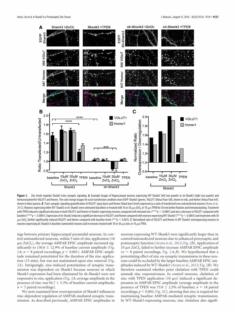
zinc concentrations are toxic to neurons; Sensi et al., 2011) andexamined the activation and dynamic properties of Shank3within dendritic spines. As a measure of Shank3 activation, wefirst used a trans-synaptic signaling assay that monitors changesin the levels of presynaptic and postsynaptic proteins at Shank3-containing excitatory synapses (Arons et al., 2012; Fig. 1A–C).Quantifying the synaptic levels of VGLUT1 and Homer1b at syn-apses with control (untransfected) or elevated levels of Shank3(EGFP-Shank3 transfected) revealed an �160% increase in bothVGLUT1 and Homer1b at EGFP-Shank3-containing synapses(Fig. 1A,B), supporting the hypothesis that Shank3 mediatestrans-synaptic changes in synaptic protein levels (Arons et al.,2012).
To assess for a possible role for zinc in Shank3-mediated trans-synaptic signaling, neuronal cultures expressing EGFP-Shank3were exposed to exogenous zinc (10 or 30 �M ZnCl2) or TPEN(10 �M), a cell-permeant zinc chelator, for 20 min. Elevatingextracellular zinc from 0.67 �M (present in our Neurobasalmedium) to 10 or 30 �M maintained, but did not further incre-ase, the Shank3-induced enhancement of both VGLUT1 andHomer1b at EGFP-Shank3 positive synapses. However, zinc che-lation with TPEN significantly decreased the levels of both pro-teins compared with VGLUT1 and Homer1b levels in elevatedzinc (p � 0.001; Fig. 1A,B). Moreover, VGLUT1 was also signif-icantly reduced compared with baseline levels. These data showthat changes in zinc levels can alter the ability of Shank3 to in-crease presynaptic and postsynaptic proteins, supporting the hy-pothesis that Shank3 synaptic signaling is zinc sensitive.
This hypothesis was tested directly by transfecting hippocam-pal neurons with a short-hairpin RNA (shRNA) that eliminatesthe expression of Shank3 selectively (sh-Shank3; Arons et al.,2012). As reported previously, knock-down of Shank3 had noeffect on the levels of either VGLUT1 or Homer1b in untreatedcells (Fig. 1B; Arons et al., 2012). In the absence of Shank3, 10 �M
TPEN did not affect the synaptic levels of these proteins. Theabsence of a response was not surprising because these synapseslacked zinc-responsive Shank3 but still expressed zinc-insensitiveShank1 (Grabrucker et al., 2011b). However, the addition of thehigher zinc concentration (30 �M) caused a decrease in VGLUT1and Homer1b in Shank3-depleted versus Shank3-untransfectedcells (Fig. 1B). In the absence of Shank3, we hypothesize thatother synaptic proteins that respond to higher zinc concentra-tions may contribute to the regulation of VGluT and Homerlevels (e.g., Shank2). Alternatively, our data suggest that higherzinc levels normally activate Shank3 and maintain synaptic pro-tein levels and, in the absence of Shank3, high zinc is not able tomaintain synaptic protein expression at normal levels.
Because control (untransfected) neurons express both zinc-responsive (Shank2 and Shank3) and zinc-unresponsive (Shank1)proteins, we also compared directly zinc-induced changes inVGLUT/Homer1b in neurons with Shank3 gain of function withShank3 loss of function (Fig. 1C). A normalized ratio of VGLUT1and Homer1b in EGFP-Shank3-overexpressing neurons to neuronsexpressing sh-Shank3 was therefore calculated for each of the treat-ment conditions (Fig. 1C). This analysis indicates that most of theincrease in trans-synaptic signaling caused by the overexpression ofEGFP-Shank3 is regulated by zinc.
Zinc-dependent regulation of excitatory synaptictransmission
To determine whether the above zinc-induced changes in Shank3synaptic signaling also induce functional changes in excitatorysynaptic transmission, we performed paired whole-cell record-
9126 • J. Neurosci., August 31, 2016 • 36(35):9124 –9134 Arons, Lee et al. • Shank3 Is a Postsynaptic Zinc Sensor
ings between primary hippocampal pyramidal neurons. In con-trol untransfected neurons, within 5 min of zinc application (10�M ZnCl2), the average AMPAR EPSC amplitude increased sig-nificantly to 130.0 12.9% of baseline current amplitude (Fig.2A; n � 8 paired recordings; p � 0.001). AMPAR EPSC ampli-tude remained potentiated for the duration of the zinc applica-tion (15 min), but was not maintained upon zinc removal (Fig.2A). Intriguingly, zinc-induced potentiation of synaptic trans-mission was dependent on Shank3 because neurons in whichShank3 expression had been eliminated by sh-Shank3 were notresponsive to zinc application (Fig. 2A; average amplitude in thepresence of zinc was 94.7 5.3% of baseline current amplitude;n � 7 paired recordings).
We next examined how overexpression of Shank3 influenceszinc-dependent regulation of AMPAR-mediated synaptic trans-mission. As described previously, AMPAR EPSC amplitudes in
neurons expressing WT-Shank3 were significantly larger than incontrol/untransfected neurons due to enhanced presynaptic andpostsynaptic function (Arons et al., 2012; Fig. 2B). Application of10 �M ZnCl2 failed to further increase AMPAR EPSC amplitude(n � 8 paired recordings, Fig. 2A,B). We hypothesized that apotentiating effect of zinc on synaptic transmission in these neu-rons could be occluded by the larger baseline AMPAR EPSC am-plitudes induced by WT-Shank3 (Arons et al., 2012; Fig. 2B). Wetherefore examined whether prior chelation with TPEN couldunmask zinc responsiveness. In control neurons, chelation ofzinc with TPEN application (10 �M) induced a significant de-pression in AMPAR EPSC amplitude (average amplitude in thepresence of TPEN was 73.8 2.5% of baseline; n � 18 pairedrecordings; p � 0.001; Fig. 2C), showing that zinc is required formaintaining baseline AMPAR-mediated synaptic transmission.In WT-Shank3-expressing neurons, zinc chelation also signifi-
Figure 1. Zinc levels regulate Shank3 trans-synaptic signaling. A, Example images of hippocampal neurons expressing WT-Shank3 (left two panels) or sh-Shank3 (right two panels) andimmunostained for VGLUT1 and Homer. The color merge images for each transfection condition show EGFP-Shank3 (green), VGLUT1 (Alexa Fluor 568, shown in red), and Homer (Alexa Fluor 647,shown in blue) puncta. B, Trans-synaptic signaling quantification of VGLUT1 (gray bars) and Homer (black bars) levels expressed as a ratio of transfected over untransfected neurons (Arons et al.,2012). Neurons expressing either WT-Shank3 or sh-Shank3 were untreated (baseline) or treated with 10 or 30 �M ZnCl2 or 10 �M TPEN for 30 min before fixation and immunostaining. Treatmentwith TPEN induced a significant decrease in both VGLUT1 and Homer in Shank3-expressing neurons compared with elevated zinc (****p � 0.0001) and also a decrease in VGLUT1 compared withbaseline (****p � 0.0001). Expression of sh-Shank3 induced a significant decrease in VGLUT1 and Homer compared with neurons expressing WT-Shank3 (****p � 0.0001) and treatment with 30�M ZnCl2 further significantly reduced VGLUT1 and Homer compared with baseline levels (***p � 0.001). C, Normalized ratio of VGLUT1 and Homer in WT-Shank3-overexpressing neurons toneurons expressing sh-Shank3 in baseline (untreated) neurons and in neurons treated with 10 or 30 �M zinc or 10 �M TPEN.
Arons, Lee et al. • Shank3 Is a Postsynaptic Zinc Sensor J. Neurosci., August 31, 2016 • 36(35):9124 –9134 • 9127
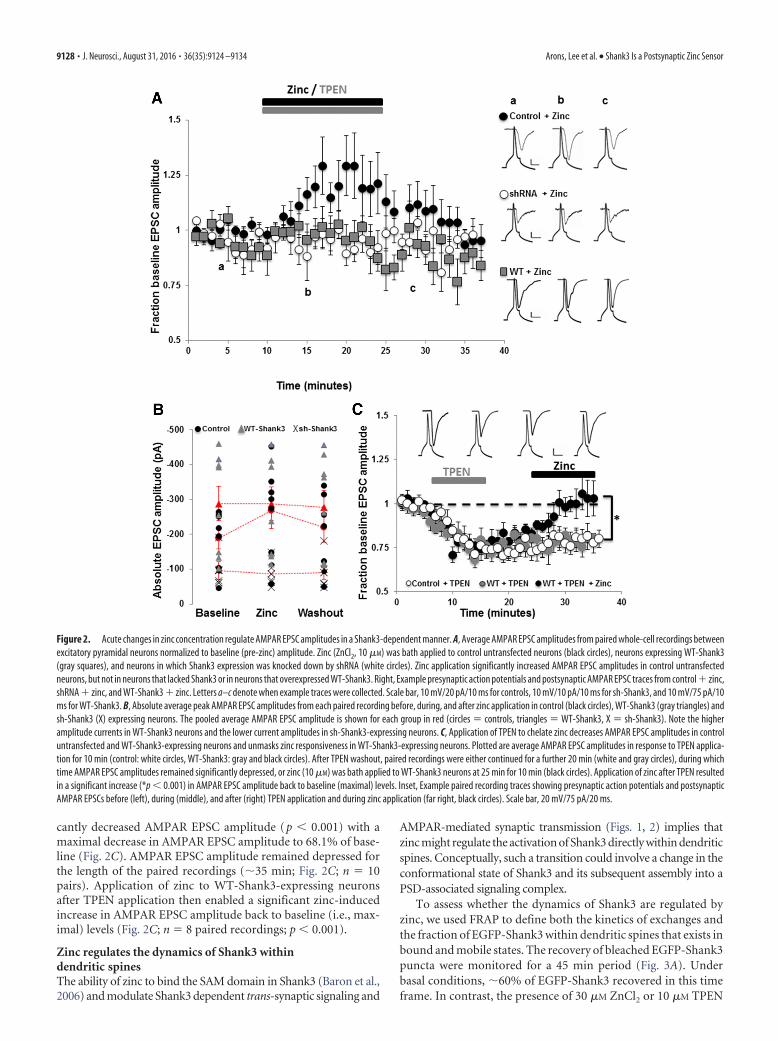
cantly decreased AMPAR EPSC amplitude (p � 0.001) with amaximal decrease in AMPAR EPSC amplitude to 68.1% of base-line (Fig. 2C). AMPAR EPSC amplitude remained depressed forthe length of the paired recordings (�35 min; Fig. 2C; n � 10pairs). Application of zinc to WT-Shank3-expressing neuronsafter TPEN application then enabled a significant zinc-inducedincrease in AMPAR EPSC amplitude back to baseline (i.e., max-imal) levels (Fig. 2C; n � 8 paired recordings; p � 0.001).
Zinc regulates the dynamics of Shank3 withindendritic spinesThe ability of zinc to bind the SAM domain in Shank3 (Baron et al.,2006) and modulate Shank3 dependent trans-synaptic signaling and
AMPAR-mediated synaptic transmission (Figs. 1, 2) implies thatzinc might regulate the activation of Shank3 directly within dendriticspines. Conceptually, such a transition could involve a change in theconformational state of Shank3 and its subsequent assembly into aPSD-associated signaling complex.
To assess whether the dynamics of Shank3 are regulated byzinc, we used FRAP to define both the kinetics of exchanges andthe fraction of EGFP-Shank3 within dendritic spines that exists inbound and mobile states. The recovery of bleached EGFP-Shank3puncta were monitored for a 45 min period (Fig. 3A). Underbasal conditions, �60% of EGFP-Shank3 recovered in this timeframe. In contrast, the presence of 30 �M ZnCl2 or 10 �M TPEN
Figure 2. Acute changes in zinc concentration regulate AMPAR EPSC amplitudes in a Shank3-dependent manner. A, Average AMPAR EPSC amplitudes from paired whole-cell recordings betweenexcitatory pyramidal neurons normalized to baseline (pre-zinc) amplitude. Zinc (ZnCl2, 10 �M) was bath applied to control untransfected neurons (black circles), neurons expressing WT-Shank3(gray squares), and neurons in which Shank3 expression was knocked down by shRNA (white circles). Zinc application significantly increased AMPAR EPSC amplitudes in control untransfectedneurons, but not in neurons that lacked Shank3 or in neurons that overexpressed WT-Shank3. Right, Example presynaptic action potentials and postsynaptic AMPAR EPSC traces from control zinc,shRNA zinc, and WT-Shank3 zinc. Letters a–c denote when example traces were collected. Scale bar, 10 mV/20 pA/10 ms for controls, 10 mV/10 pA/10 ms for sh-Shank3, and 10 mV/75 pA/10ms for WT-Shank3. B, Absolute average peak AMPAR EPSC amplitudes from each paired recording before, during, and after zinc application in control (black circles), WT-Shank3 (gray triangles) andsh-Shank3 (X) expressing neurons. The pooled average AMPAR EPSC amplitude is shown for each group in red (circles � controls, triangles � WT-Shank3, X � sh-Shank3). Note the higheramplitude currents in WT-Shank3 neurons and the lower current amplitudes in sh-Shank3-expressing neurons. C, Application of TPEN to chelate zinc decreases AMPAR EPSC amplitudes in controluntransfected and WT-Shank3-expressing neurons and unmasks zinc responsiveness in WT-Shank3-expressing neurons. Plotted are average AMPAR EPSC amplitudes in response to TPEN applica-tion for 10 min (control: white circles, WT-Shank3: gray and black circles). After TPEN washout, paired recordings were either continued for a further 20 min (white and gray circles), during whichtime AMPAR EPSC amplitudes remained significantly depressed, or zinc (10 �M) was bath applied to WT-Shank3 neurons at 25 min for 10 min (black circles). Application of zinc after TPEN resultedin a significant increase (*p � 0.001) in AMPAR EPSC amplitude back to baseline (maximal) levels. Inset, Example paired recording traces showing presynaptic action potentials and postsynapticAMPAR EPSCs before (left), during (middle), and after (right) TPEN application and during zinc application (far right, black circles). Scale bar, 20 mV/75 pA/20 ms.
9128 • J. Neurosci., August 31, 2016 • 36(35):9124 –9134 Arons, Lee et al. • Shank3 Is a Postsynaptic Zinc Sensor
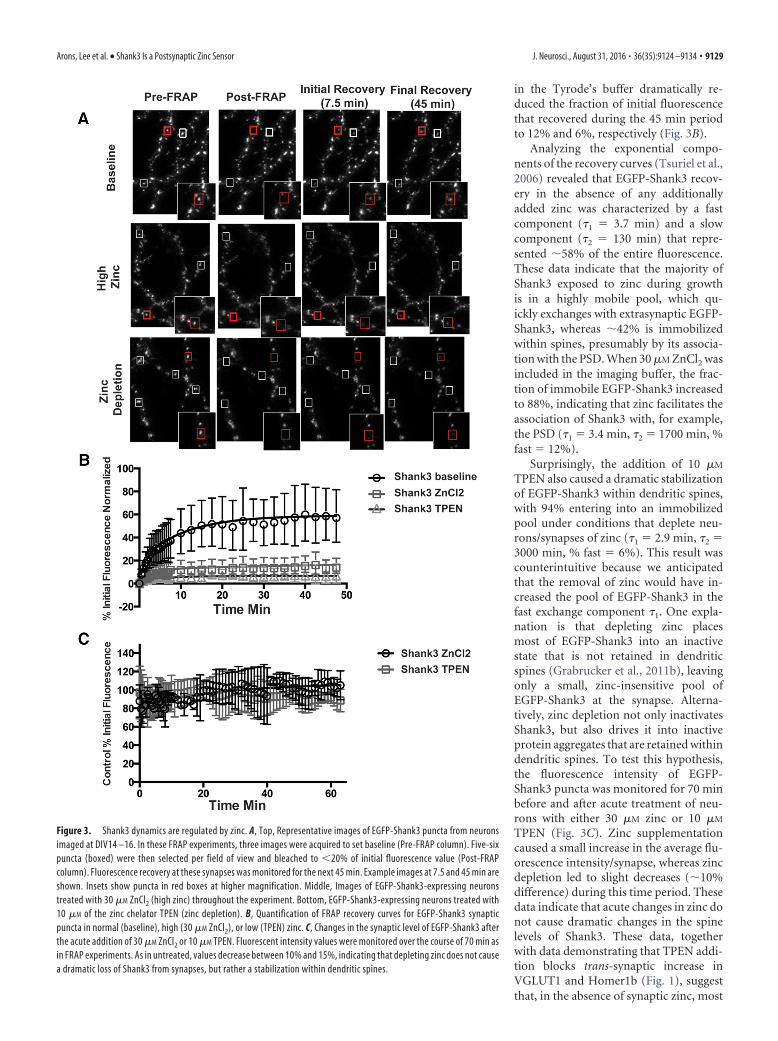
in the Tyrode’s buffer dramatically re-duced the fraction of initial fluorescencethat recovered during the 45 min periodto 12% and 6%, respectively (Fig. 3B).
Analyzing the exponential compo-nents of the recovery curves (Tsuriel et al.,2006) revealed that EGFP-Shank3 recov-ery in the absence of any additionallyadded zinc was characterized by a fastcomponent (�1 � 3.7 min) and a slowcomponent (�2 � 130 min) that repre-sented �58% of the entire fluorescence.These data indicate that the majority ofShank3 exposed to zinc during growthis in a highly mobile pool, which qu-ickly exchanges with extrasynaptic EGFP-Shank3, whereas �42% is immobilizedwithin spines, presumably by its associa-tion with the PSD. When 30 �M ZnCl2 wasincluded in the imaging buffer, the frac-tion of immobile EGFP-Shank3 increasedto 88%, indicating that zinc facilitates theassociation of Shank3 with, for example,the PSD (�1 � 3.4 min, �2 � 1700 min, %fast � 12%).
Surprisingly, the addition of 10 �M
TPEN also caused a dramatic stabilizationof EGFP-Shank3 within dendritic spines,with 94% entering into an immobilizedpool under conditions that deplete neu-rons/synapses of zinc (�1 � 2.9 min, �2 �3000 min, % fast � 6%). This result wascounterintuitive because we anticipatedthat the removal of zinc would have in-creased the pool of EGFP-Shank3 in thefast exchange component �1. One expla-nation is that depleting zinc placesmost of EGFP-Shank3 into an inactivestate that is not retained in dendriticspines (Grabrucker et al., 2011b), leavingonly a small, zinc-insensitive pool ofEGFP-Shank3 at the synapse. Alterna-tively, zinc depletion not only inactivatesShank3, but also drives it into inactiveprotein aggregates that are retained withindendritic spines. To test this hypothesis,the fluorescence intensity of EGFP-Shank3 puncta was monitored for 70 minbefore and after acute treatment of neu-rons with either 30 �M zinc or 10 �M
TPEN (Fig. 3C). Zinc supplementationcaused a small increase in the average flu-orescence intensity/synapse, whereas zincdepletion led to slight decreases (�10%difference) during this time period. Thesedata indicate that acute changes in zinc donot cause dramatic changes in the spinelevels of Shank3. These data, togetherwith data demonstrating that TPEN addi-tion blocks trans-synaptic increase inVGLUT1 and Homer1b (Fig. 1), suggestthat, in the absence of synaptic zinc, most
Figure 3. Shank3 dynamics are regulated by zinc. A, Top, Representative images of EGFP-Shank3 puncta from neuronsimaged at DIV14 –16. In these FRAP experiments, three images were acquired to set baseline (Pre-FRAP column). Five-sixpuncta (boxed) were then selected per field of view and bleached to �20% of initial fluorescence value (Post-FRAPcolumn). Fluorescence recovery at these synapses was monitored for the next 45 min. Example images at 7.5 and 45 min areshown. Insets show puncta in red boxes at higher magnification. Middle, Images of EGFP-Shank3-expressing neuronstreated with 30 �M ZnCl2 (high zinc) throughout the experiment. Bottom, EGFP-Shank3-expressing neurons treated with10 �M of the zinc chelator TPEN (zinc depletion). B, Quantification of FRAP recovery curves for EGFP-Shank3 synapticpuncta in normal (baseline), high (30 �M ZnCl2), or low (TPEN) zinc. C, Changes in the synaptic level of EGFP-Shank3 afterthe acute addition of 30 �M ZnCl2 or 10 �M TPEN. Fluorescent intensity values were monitored over the course of 70 min asin FRAP experiments. As in untreated, values decrease between 10% and 15%, indicating that depleting zinc does not causea dramatic loss of Shank3 from synapses, but rather a stabilization within dendritic spines.
Arons, Lee et al. • Shank3 Is a Postsynaptic Zinc Sensor J. Neurosci., August 31, 2016 • 36(35):9124 –9134 • 9129

of Shank3 enters into a stabilized inactivestate within spines (see model in Fig. 6).
ASD-associated Shank3R87C remainszinc sensitiveASD-associated mutations in Shank3 im-pair synaptic transmission and, more spe-cifically, presynaptic/postsynaptic couplingthrough Neurexin/Neuroligin (Arons et al.,2012). Shank3 variants containing aminoacid substitutions within the N-terminalAnkyrin repeats localize properly at syn-apses (Durand et al., 2007; Arons et al.,2012), suggesting that the mechanism of in-activation is independent of synaptic local-ization. Given the prominent role for zinc inthe activation of Shank3 (Figs. 1, 3), we con-sidered whether Shank3 forms harboringsingle amino acid substitutions are func-tionally dead due to their inability to re-spond to zinc.
As an initial test of this hypothesis, weused FRAP to determine whether the zinc-sensitive dynamics of one of these mutantproteins, Shank3R87C, was altered (Fig. 4A).In normal Tyrode’s solution, the extent ofrecovery for synaptic EGFP-Shank3R87C re-vealed that less than half of the protein(26%) is in a mobile pool and the majority isin an immobile pool (�1 � 3.9 min, �2 �60.9 min). Zinc induced a dramatic stabili-zation of EGFP-Shank3R87C similar to wild-type (Figs. 3B, 4A), with 30 �M zinc shifting96% of the molecules into an immobile state(�1 � 3.8 min, �2 � 3400 min). Zinc deple-tion with TPEN increased the fraction of theR87C mutant in the immobile pool (96%for R87C; �1 � 1.5 min, �2 � 510 min).Overall, these experiments reveal thatShank3R87C remains zinc responsive.
Zinc activation of Shank3R87C promotes functionalpostsynaptic but not presynaptic changesConceptually, we anticipated that, although the dynamics are quali-tatively different, as in wild-type Shank3, elevating zinc shouldpush Shank3R87C into an open/active conformation that becomesincorporated into the PSD, whereas zinc depletion would push asignificant fraction into an immobile inactive state. To explorethis possibility, we investigated whether the addition of zincpromotes the recruitment of the Shank3-binding partnerHomer1b into dendritic spines and triggers a trans-synapticincrease in VGLUT1 (Fig. 4B–D). As reported previously, innormal Tyrode’s solution (no added zinc), the Shank3R87C
mutant appeared to be functionally dead because the levels ofHomer1b and VGLUT1 at synapses expressing this moleculeare no different from untransfected neurons (Fig. 4C). After20 min in 10 or 30 �M ZnCl2, the postsynaptic levels ofHomer1b were rescued to wild-type EGFP-Shank3 levels (Fig.4C), suggesting that zinc can indeed activate Shank3R87C, al-lowing it to recruit its postsynaptic binding partners into den-dritic spines. However, zinc treatment had no effect on thepresynaptic levels of VGLUT1, indicating that it was still un-able to elicit a trans-synaptic signal.
Because Shank3R87C retains postsynaptic zinc responsiveness,we next investigated whether AMPAR-mediated synaptic trans-mission could be enhanced by zinc in neurons expressing EGFP-Shank3R87C (Fig. 5). Specifically, we observed that zinc was ableto induce a significant increase in AMPAR EPSC amplitudes thatwas of the same magnitude in EGFP-Shank3R87C-expressing neu-rons as was observed in control untransfected neurons (130.0 8.6%, n � 10 paired recordings, p � 0.001; Fig. 5A). The poten-tiation was again not sustained after zinc removal (Fig. 5A).
The fact that trans-synaptic signaling could not be rescued byzinc (Fig. 4) suggests that the zinc-induced increase in AMPAR-mediated synaptic transmission in Shank3R87C-expressing neu-rons is localized to the postsynapse. To test this hypothesis, wemeasured failure rates of AMPAR synaptic transmission (Fig.5B). Neurons expressing Shank3R87C showed a significantlyhigher failure rate (21.7 4.37%) compared with control un-transfected (2.63 2.22%), Shank3 WT (0%), or shRNA-Shank3-expressing neurons (9.0 2.40%; p � 0.05 in all cases).However, no significant change in failure rate occurred in the pres-ence of zinc (or TPEN) in any group (Fig. 5B; in zinc: Shank3R87C
24.57 6.56%; control 2.63 2.12%; Shank3 WT 0%; sh-Shank313.75 3.1%; p � 0.05 in all cases), supporting the hypothesis that
Figure 4. Shank3R87C retains zinc responsiveness. A, Plotted rates of FRAP of EGFP-Shank3R87C puncta in control conditions(baseline) and in the presence of 10 �M ZnCl2 or 10 �M TPEN. Similar to WT-Shank3, both zinc and TPEN increased the pool ofimmobile Shank3R87C, indicating that it is still zinc responsive. B, Example images of hippocampal neurons transfected withShank3R87C and immunostained for VGLUT1 and Homer. The colored merge shows EGFP-Shank3R87C, VGLUT1 (Alexa Fluor 568,shown in red), and Homer (Alexa Fluor 647, shown in blue) colocalized puncta. C, Trans-synaptic signaling quantification of VGLUT1(gray bars) and Homer (black bars) levels expressed as a ratio of transfected over untransfected neurons (Arons et al., 2012) inneurons expressing Shank3R87C. The expression levels of VGLUT1 and Homer were significantly decreased in Shank3R87C-expressing neurons compared with WT-Shank3-expressing neurons ( p � 0.01). Both 10 and 30 �M zinc led to significant in-creases in Homer levels in Shank3R87C-expressing neurons (****p � 0.0001), but no significant increases were induced in VGLUT1( p � 0.05). TPEN application reversed the increase in Homer levels in Shank3R87C-expressing neurons (****p � 0.0001).
9130 • J. Neurosci., August 31, 2016 • 36(35):9124 –9134 Arons, Lee et al. • Shank3 Is a Postsynaptic Zinc Sensor
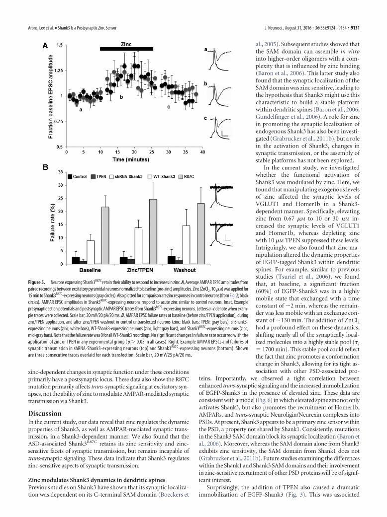
zinc-dependent changes in synaptic function under these conditionsprimarily have a postsynaptic locus. These data also show the R87Cmutation primarily affects trans-synaptic signaling at excitatory syn-apses, not the ability of zinc to modulate AMPAR-mediated synaptictransmission via Shank3.
DiscussionIn the current study, our data reveal that zinc regulates the dynamicproperties of Shank3, as well as AMPAR-mediated synaptic trans-mission, in a Shank3-dependent manner. We also found that theASD-associated Shank3R87C retains its zinc sensitivity and zinc-sensitive facets of synaptic transmission, but remains incapable oftrans-synaptic signaling. These data indicate that Shank3 regulateszinc-sensitive aspects of synaptic transmission.
Zinc modulates Shank3 dynamics in dendritic spinesPrevious studies on Shank3 have shown that its synaptic localiza-tion was dependent on its C-terminal SAM domain (Boeckers et
al., 2005). Subsequent studies showed thatthe SAM domain can assemble in vitrointo higher-order oligomers with a com-plexity that is influenced by zinc binding(Baron et al., 2006). This latter study alsofound that the synaptic localization of theSAM domain was zinc sensitive, leading tothe hypothesis that Shank3 might use thischaracteristic to build a stable platformwithin dendritic spines (Baron et al., 2006;Gundelfinger et al., 2006). A role for zincin promoting the synaptic localization ofendogenous Shank3 has also been investi-gated (Grabrucker et al., 2011b), but a rolein the activation of Shank3, changes insynaptic transmission, or the assembly ofstable platforms has not been explored.
In the current study, we investigatedwhether the functional activation ofShank3 was modulated by zinc. Here, wefound that manipulating exogenous levelsof zinc affected the synaptic levels ofVGLUT1 and Homer1b in a Shank3-dependent manner. Specifically, elevatingzinc from 0.67 �M to 10 or 30 �M in-creased the synaptic levels of VGLUT1and Homer1b, whereas depleting zincwith 10 �M TPEN suppressed these levels.Intriguingly, we also found that zinc ma-nipulation altered the dynamic propertiesof EGFP-tagged Shank3 within dendriticspines. For example, similar to previousstudies (Tsuriel et al., 2006), we foundthat, at baseline, a significant fraction(60%) of EGFP-Shank3 was in a highlymobile state that exchanged with a timeconstant of �2 min, whereas the remain-der was less mobile with an exchange con-stant of �130 min. The addition of ZnCl2had a profound effect on these dynamics,shifting nearly all of the synaptically local-ized molecules into a highly stable pool (�2
� 1700 min). This stable pool could reflectthe fact that zinc promotes a conformationchange in Shank3, allowing for its tight as-sociation with other PSD-associated pro-
teins. Importantly, we observed a tight correlation betweenenhanced trans-synaptic signaling and the increased immobilizationof EGFP-Shank3 in the presence of elevated zinc. These data areconsistent with a model (Fig. 6) in which elevated spine zinc not onlyactivates Shank3, but also promotes the recruitment of Homer1b,AMPARs, and trans-synaptic Neuroligin/Neurexin complexes intoPSDs. At present, Shank3 appears to be a primary zinc sensor withinthe PSD, a property not shared by Shank1. Consistently, mutationsin the Shank3 SAM domain block its synaptic localization (Baron etal., 2006). Moreover, whereas the SAM domain alone from Shank3exhibits zinc sensitivity, the SAM domain from Shank1 does not(Grabrucker et al., 2011b). Future studies examining the differenceswithin the Shank1 and Shank3 SAM domains and their involvementin zinc-sensitive recruitment of other PSD proteins will be of signif-icant interest.
Surprisingly, the addition of TPEN also caused a dramaticimmobilization of EGFP-Shank3 (Fig. 3). This was associated
Figure 5. Neurons expressing Shank3R87C retain their ability to respond to increases in zinc. A, Average AMPAR EPSC amplitudes frompaired recordings between excitatory pyramidal neurons normalized to baseline (pre-zinc) amplitudes. Zinc (ZnCl2, 10�M) was applied for15 min to Shank3R87C-expressing neurons (gray circles). Also plotted for comparison are zinc responses in control neurons (fromFig. 2; blackcircles). AMPAR EPSC amplitudes in Shank3R87C-expressing neurons respond to acute zinc similar to control neurons. Inset, Examplepresynaptic action potentials and postsynaptic AMPAR EPSC traces from Shank3R87C-expressing neurons. Letters a–c denote when exam-ple traces were collected. Scale bar, 20 mV/20 pA/20 ms. B, AMPAR EPSC failure rates at baseline (before zinc/TPEN application), duringzinc/TPEN application, and after zinc/TPEN washout in control untransfected neurons (zinc: black bars; TPEN: gray bars), shShank3-expressing neurons (zinc, white bars), WT-Shank3-expressing neurons (zinc, light gray bars), and Shank3R87C-expressing neurons (zinc,mid-gray bars). Note that the failure rate was 0 for all WT-Shank3 recordings. No significant changes in failure rate occurred with theapplication of zinc or TPEN in any experimental group ( p � 0.05 in all cases). Right, Example AMPAR EPSCs and failures ofsynaptic transmission in shRNA-Shank3-expressing neurons (top) and Shank3R87C-expressing neurons (bottom). Shownare three consecutive traces overlaid for each transfection. Scale bar, 20 mV/25 pA/20 ms.
Arons, Lee et al. • Shank3 Is a Postsynaptic Zinc Sensor J. Neurosci., August 31, 2016 • 36(35):9124 –9134 • 9131
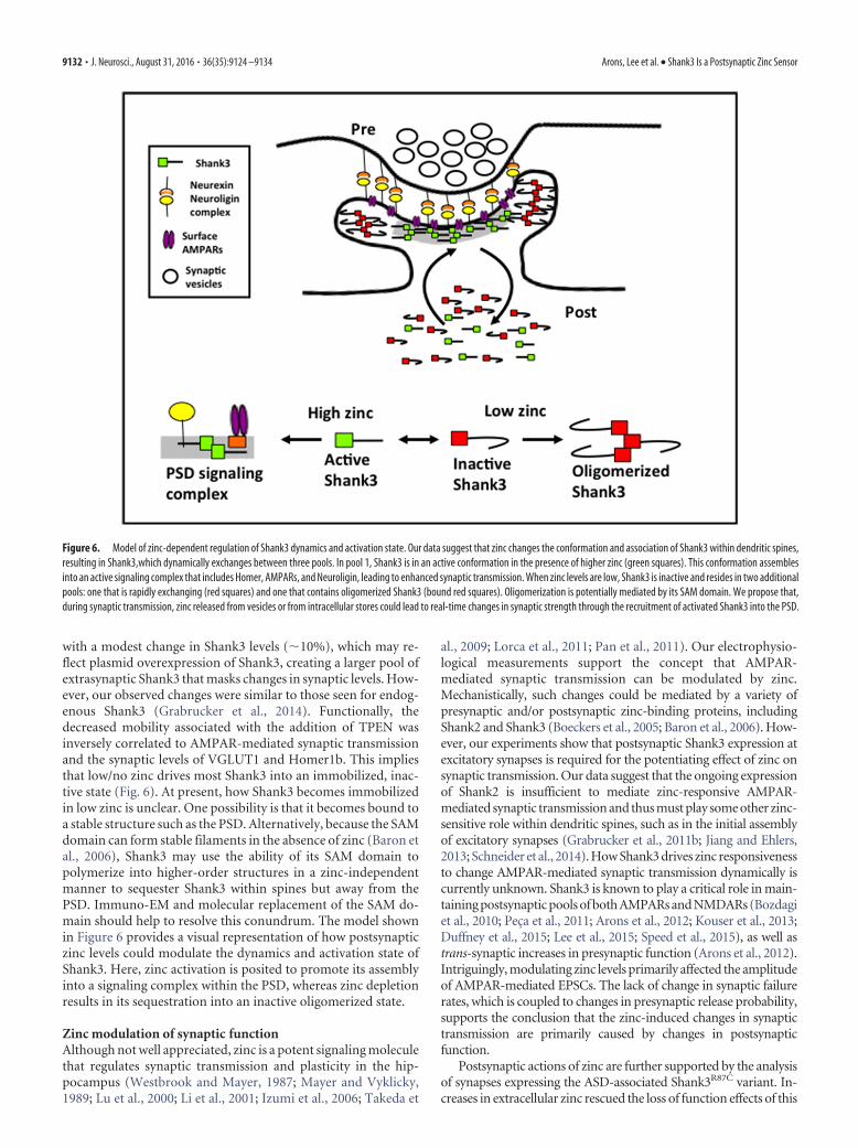
with a modest change in Shank3 levels (�10%), which may re-flect plasmid overexpression of Shank3, creating a larger pool ofextrasynaptic Shank3 that masks changes in synaptic levels. How-ever, our observed changes were similar to those seen for endog-enous Shank3 (Grabrucker et al., 2014). Functionally, thedecreased mobility associated with the addition of TPEN wasinversely correlated to AMPAR-mediated synaptic transmissionand the synaptic levels of VGLUT1 and Homer1b. This impliesthat low/no zinc drives most Shank3 into an immobilized, inac-tive state (Fig. 6). At present, how Shank3 becomes immobilizedin low zinc is unclear. One possibility is that it becomes bound toa stable structure such as the PSD. Alternatively, because the SAMdomain can form stable filaments in the absence of zinc (Baron etal., 2006), Shank3 may use the ability of its SAM domain topolymerize into higher-order structures in a zinc-independentmanner to sequester Shank3 within spines but away from thePSD. Immuno-EM and molecular replacement of the SAM do-main should help to resolve this conundrum. The model shownin Figure 6 provides a visual representation of how postsynapticzinc levels could modulate the dynamics and activation state ofShank3. Here, zinc activation is posited to promote its assemblyinto a signaling complex within the PSD, whereas zinc depletionresults in its sequestration into an inactive oligomerized state.
Zinc modulation of synaptic functionAlthough not well appreciated, zinc is a potent signaling moleculethat regulates synaptic transmission and plasticity in the hip-pocampus (Westbrook and Mayer, 1987; Mayer and Vyklicky,1989; Lu et al., 2000; Li et al., 2001; Izumi et al., 2006; Takeda et
al., 2009; Lorca et al., 2011; Pan et al., 2011). Our electrophysio-logical measurements support the concept that AMPAR-mediated synaptic transmission can be modulated by zinc.Mechanistically, such changes could be mediated by a variety ofpresynaptic and/or postsynaptic zinc-binding proteins, includingShank2 and Shank3 (Boeckers et al., 2005; Baron et al., 2006). How-ever, our experiments show that postsynaptic Shank3 expression atexcitatory synapses is required for the potentiating effect of zinc onsynaptic transmission. Our data suggest that the ongoing expressionof Shank2 is insufficient to mediate zinc-responsive AMPAR-mediated synaptic transmission and thus must play some other zinc-sensitive role within dendritic spines, such as in the initial assemblyof excitatory synapses (Grabrucker et al., 2011b; Jiang and Ehlers,2013; Schneider et al., 2014). How Shank3 drives zinc responsivenessto change AMPAR-mediated synaptic transmission dynamically iscurrently unknown. Shank3 is known to play a critical role in main-taining postsynaptic pools of both AMPARs and NMDARs (Bozdagiet al., 2010; Peca et al., 2011; Arons et al., 2012; Kouser et al., 2013;Duffney et al., 2015; Lee et al., 2015; Speed et al., 2015), as well astrans-synaptic increases in presynaptic function (Arons et al., 2012).Intriguingly, modulating zinc levels primarily affected the amplitudeof AMPAR-mediated EPSCs. The lack of change in synaptic failurerates, which is coupled to changes in presynaptic release probability,supports the conclusion that the zinc-induced changes in synaptictransmission are primarily caused by changes in postsynapticfunction.
Postsynaptic actions of zinc are further supported by the analysisof synapses expressing the ASD-associated Shank3R87C variant. In-creases in extracellular zinc rescued the loss of function effects of this
Figure 6. Model of zinc-dependent regulation of Shank3 dynamics and activation state. Our data suggest that zinc changes the conformation and association of Shank3 within dendritic spines,resulting in Shank3,which dynamically exchanges between three pools. In pool 1, Shank3 is in an active conformation in the presence of higher zinc (green squares). This conformation assemblesinto an active signaling complex that includes Homer, AMPARs, and Neuroligin, leading to enhanced synaptic transmission. When zinc levels are low, Shank3 is inactive and resides in two additionalpools: one that is rapidly exchanging (red squares) and one that contains oligomerized Shank3 (bound red squares). Oligomerization is potentially mediated by its SAM domain. We propose that,during synaptic transmission, zinc released from vesicles or from intracellular stores could lead to real-time changes in synaptic strength through the recruitment of activated Shank3 into the PSD.
9132 • J. Neurosci., August 31, 2016 • 36(35):9124 –9134 Arons, Lee et al. • Shank3 Is a Postsynaptic Zinc Sensor
altered protein, enabling zinc-mediated increases in AMPAR-mediated synaptic transmission. However, because elevated zinc lev-els did not enhance the levels of VGLUT1 in presynaptic boutonsformed onto neurons expressing GFP-Shank3R87C, nor did they re-duce the failure rates of synaptic transmission, this mutation doesimpair its ability to signal trans-synaptically even with enhanced zinclevels. Moreover, acute addition/depletion of zinc causes a dramaticchange in the fraction in mobile and immobile pools of Shank3R87C
(Fig. 4). Like wild-type Shank3, increasing zinc levels from 0.6 to 10�M activates Shank3R87C and promotes the recruitment of the post-synaptic Shank3-binding partner Homer1b into dendritic spines.This amino acid exchange therefore does not appear to affect the zincsensitivity of Shank3.
An important unresolved question is what role Shank3 andzinc play in synaptic transmission. Studies of synaptic plasticity atCA1 synapses have found that zinc has a profound effect on theinduction of LTP, but not its maintenance (Izumi et al., 2006;Takeda et al., 2010; Lorca et al., 2011). Moreover, synapses lack-ing Shank3 fail to undergo LTP (Bozdagi et al., 2010; Wang et al.,2011; Kouser et al., 2013; Speed et al., 2015), suggesting that zincand Shank3 may operate in concert to regulate this facet of syn-aptic plasticity. Although this is a provocative concept, our dataindicate that acute zinc alone is insufficient to induce long-lastingforms of plasticity because AMPAR-mediated currents returnedto baseline within minutes of its removal. A likely partner is thecoactivation of NMDARs, known to be critical for the inductionof LTP (Herron et al., 1986; Wigstrom and Gustafsson, 1986).Zinc-dependent activation of Shank3 could therefore promoteincreased AMPAR recruitment to synapses, similar to what isknown to occur with actin polymerization (Allison et al., 1998;Durand et al., 2012; Duffney et al., 2015) and the expression ofLTP (Shi et al., 1999; Oh et al., 2006; Jaskolski et al., 2009; Makinoand Malinow, 2009; Jurado et al., 2013; Zheng et al., 2015). How-ever, because zinc influences the strength of synaptic transmis-sion and Shank3 dynamics in a rapid and reversible manner, theunderlying mechanisms may differ from those induced duringpersistent changes in synaptic strength and structure.
In summary, our studies reveal that Shank3 not only senseschanges in postsynaptic zinc, but also is a key component of a zinc-sensitive signaling pathway at excitatory synapses. Importantly, zinchomeostasis is disrupted in neuropsychiatric disorders includingASD (Curtis and Patel, 2008; Grabrucker et al., 2011a; Russo andDevito, 2011; Yasuda et al., 2011). Elevation of zinc has been shownto rescue normal social interaction via Src and NMDAR activation inShank2 and Tbr1 ASD mouse models (Lee et al., 2015), whereaschronic zinc deficiency induces the loss of Shank2/3 and increasesthe incidence of ASD-related behaviors (Grabrucker et al., 2014).Together with our results, these data suggest that environmental/dietary factors such as changes in zinc levels could alter the Shank3-signaling system and reduce the optimal performance of Shank3-dependent excitatory synaptic function. Therefore, strategies toactivate this zinc-sensitive pathway could potentially restore thefunctionality of these synapses.
ReferencesAllison DW, Gelfand VI, Spector I, Craig AM (1998) Role of actin in an-
choring postsynaptic receptors in cultured hippocampal neurons: differ-ential attachment of NMDA versus AMPA receptors. J Neuroscience 18:2423–2436. Medline
Arons MH, Thynne CJ, Grabrucker AM, Li D, Schoen M, Cheyne JE, Boeck-ers TM, Montgomery JM, Garner CC (2012) Autism associatedmutations in ProSAP2/Shank3 impair synaptic transmission andNeurexin-Neuroligin mediated transsynaptic signaling J Neurosci 32:14966 –14978. CrossRef
Assaf SY, Chung SH (1984) Release of endogenous Zn2 from brain tissueduring activity. Nature 308:734 –736. CrossRef Medline
Baron MK, Boeckers TM, Vaida B, Faham S, Gingery M, Sawaya MR, SalyerD, Gundelfinger ED, Bowie JU (2006) An architectural framework thatmay lie at the core of the postsynaptic density. Science 311:531–535.CrossRef Medline
Boeckers TM, Bockmann J, Kreutz MR, Gundelfinger ED (2002) ProSAP/Shank proteins: a family of higher order organizing molecules of thepostsynaptic density with an emerging role in human neurological dis-ease. J Neurochem 81:903–910. CrossRef Medline
Boeckers TM, Liedtke T, Spilker C, Dresbach T, Bockmann J, Kreutz MR,Gundelfinger ED (2005) C-terminal synaptic targeting elements forpostsynaptic density proteins ProSAP1/Shank2 and ProSAP2/Shank3.J Neurochem 92:519 –524. CrossRef Medline
Bozdagi O, Sakurai T, Papapetrou D, Wang X, Dickstein DL, Takahashi N,Kajiwara Y, Yang M, Katz AM, Scattoni ML, Harris MJ, Saxena R, Silver-man JL, Crawley JN, Zhou Q, Hof PR, Buxbaum JD (2010) Haploinsuf-ficiency of the autism-associated Shank3 gene leads to deficits in synapticfunction, social interaction, and social communication. Mol Autism 1:15.CrossRef Medline
Curtis LT, Patel K (2008) Nutritional and environmental approaches topreventing and treating autism and attention deficit hyperactivity disor-der (ADHD): a review. J Altern Complement Med 14:79 – 85. CrossRefMedline
Duffney LJ, Zhong P, Wei J, Matas E, Cheng J, Qin L, Ma K, Dietz DM,Kajiwara Y, Buxbaum JD, Yan Z (2015) Autism-like deficits in Shank3-deficient mice are rescued by targeting actin regulators. Cell Rep 11:1400 –1413. CrossRef Medline
Durand CM et al. (2007) Mutations in the gene encoding the synaptic scaf-folding protein SHANK3 are associated with autism spectrum disorders.Nat Genet 39:25–27. CrossRef Medline
Durand CM, Perroy J, Loll F, Perrais D, Fagni L, Bourgeron T, MontcouquiolM, Sans N (2012) SHANK3 mutations identified in autism lead to mod-ification of dendritic spine morphology via an actin-dependent mecha-nism. Mol Psychiatry 17:71– 84. CrossRef Medline
Ebadi M, Murrin LC, Pfeiffer RF (1990) Hippocampal zinc thionein andpyridoxal phosphate modulate synaptic functions. Ann N Y Acad Sci 585:189 –201. CrossRef Medline
Goslin K, Schreyer DJ, Skene JH, Banker G (1988) Development of neuro-nal polarity: GAP-43 distinguishes axonal from dendritic growth cones.Nature 336:672– 674. CrossRef Medline
Grabrucker AM, Rowan M, Garner CC (2011a) Brain-delivery of zinc-ionsas potential treatment for neurological diseases: mini review. Drug DelivLett 1:13–23. Medline
Grabrucker AM, Knight MJ, Proepper C, Bockmann J, Joubert M, Rowan M,Nienhaus GU, Garner CC, Bowie JU, Kreutz MR, Gundelfinger ED,Boeckers TM (2011b) Concerted action of zinc and ProSAP/Shank insynaptogenesis and synapse maturation. EMBO J 30:569 –581. CrossRefMedline
Grabrucker S, Jannetti L, Eckert M, Gaub S, Chhabra R, Pfaender S, MangusK, Reddy PP, Rankovic V, Schmeisser MJ, Kreutz MR, Ehret G, BoeckersTM, Grabrucker AM (2014) Zinc deficiency dysregulates the synapticProSAP/Shank scaffold and might contribute to autism spectrum disor-ders. Brain 137:137–152. CrossRef Medline
Gundelfinger ED, Boeckers TM, Baron MK, Bowie JU (2006) A role for zincin postsynaptic density asSAMbly and plasticity? Trends Biochem Sci31:366 –373. CrossRef Medline
Herron CE, Lester RA, Coan EJ, Collingridge GL (1986) Frequency-dependent involvement of NMDA receptors in the hippocampus: a novelsynaptic mechanism. Nature 322:265–268. CrossRef Medline
Howell GA, Welch MG, Frederickson CJ (1984) Stimulation-induced up-take and release of zinc in hippocampal slices. Nature 308:736 –738.CrossRef Medline
Huang YZ, Pan E, Xiong ZQ, McNamara JO (2008) Zinc-mediated trans-activation of TrkB potentiates the hippocampal mossy fiber-CA3 pyramidsynapse. Neuron 57:546 –558. CrossRef Medline
Inoue K, Branigan D, Xiong ZG (2010) Zinc-induced neurotoxicity medi-ated by transient receptor potential melastatin 7 channels. J Biol Chem285:7430 –7439. CrossRef Medline
Izumi Y, Auberson YP, Zorumski CF (2006) Zinc modulates bidirectionalhippocampal plasticity by effects on NMDA receptors. J Neurosci 26:7181–7188. CrossRef Medline
Arons, Lee et al. • Shank3 Is a Postsynaptic Zinc Sensor J. Neurosci., August 31, 2016 • 36(35):9124 –9134 • 9133

Jaskolski F, Mayo-Martin B, Jane D, Henley JM (2009) Dynamin-dependent membrane drift recruits AMPA receptors to dendritic spines.J Biol Chem 284:12491-12503. CrossRef Medline
Jiang YH, Ehlers MD (2013) Modeling autism by SHANK gene mutations inmice. Neuron 78:8 –27. CrossRef Medline
Jurado S, Goswami D, Zhang Y, Molina AJ, Sudhof TC, Malenka RC (2013)LTP requires a unique postsynaptic SNARE fusion machinery. Neuron77:542–558. CrossRef Medline
Kim TY, Hwang JJ, Yun SH, Jung MW, Koh JY (2002) Augmentation byzinc of NMDA receptor-mediated synaptic responses in CA1 of rat hip-pocampal slices: mediation by Src family tyrosine kinases. Synapse 46:49 –56. CrossRef Medline
Kouser M, Speed HE, Dewey CM, Reimers JM, Widman AJ, Gupta N, Liu S,Jaramillo TC, Bangash M, Xiao B, Worley PF, Powell CM (2013) Loss ofpredominant Shank3 isoforms results in hippocampus-dependent im-pairments in behavior and synaptic transmission. J Neurosci 33:18448 –18468. CrossRef Medline
Kreienkamp HJ (2008) Scaffolding proteins at the postsynaptic density:shank as the architectural framework. Handb Exp Pharmacol 186:365–380. CrossRef Medline
Kumar RA, Christian SL (2009) Genetics of autism spectrum disorders.Curr Neurol Neurosci Rep 9:188 –197. CrossRef Medline
Leal-Ortiz S, Waites CL, Terry-Lorenzo R, Zamorano P, Gundelfinger ED,Garner CC (2008) Piccolo modulation of Synapsin1a dynamics regu-lates synaptic vesicle exocytosis. J Cell Biol 181:831– 846. CrossRefMedline
Lee EJ, Choi SY, Kim E (2015) NMDA receptor dysfunction in autism spec-trum disorders. Curr Opin Pharmacol 20:8 –13. CrossRef Medline
Li D, Specht CG, Waites CL, Butler-Munro C, Leal-Ortiz S, Foote JW, Ge-noux D, Garner CC, Montgomery JM (2011) SAP97 directs NMDA re-ceptor spine targeting and synaptic plasticity. J Physiol 589:4491– 4510.CrossRef Medline
Li Y, Hough CJ, Suh SW, Sarvey JM, Frederickson CJ (2001) Rapid translo-cation of Zn(2) from presynaptic terminals into postsynaptic hip-pocampal neurons after physiological stimulation. J Neurophysiol 86:2597–2604. Medline
Lorca RA, Rozas C, Loyola S, Moreira-Ramos S, Zeise ML, Kirkwood A,Huidobro-Toro JP, Morales B (2011) Zinc enhances long-term poten-tiation through P2X receptor modulation in the hippocampal CA1 re-gion. Eur J Neurosci 33:1175–1185. CrossRef Medline
Lu YM, Taverna FA, Tu R, Ackerley CA, Wang YT, Roder J (2000) Endog-enous Zn(2) is required for the induction of long-term potentiationat rat hippocampal mossy fiber-CA3 synapses. Synapse 38:187–197.CrossRef Medline
Makino H, Malinow R (2009) AMPA receptor incorporation into synapsesduring LTP: the role of lateral movement and exocytosis. Neuron 64:381–390. CrossRef Medline
Manzerra P, Behrens MM, Canzoniero LM, Wang XQ, Heidinger V, IchinoseT, Yu SP, Choi DW (2001) Zinc induces a Src family kinase-mediatedup-regulation of NMDA receptor activity and excitotoxicity. Proc NatlAcad Sci U S A 98:11055–11061. CrossRef Medline
Masters BA, Quaife CJ, Erickson JC, Kelly EJ, Froelick GJ, Zambrowicz BP,Brinster RL, Palmiter RD (1994) Metallothionein III is expressed inneurons that sequester zinc in synaptic vesicles. J Neurosci 14:5844 –5857.Medline
Mayer ML, Vyklicky L Jr (1989) The action of zinc on synaptic transmissionand neuronal excitability in cultures of mouse hippocampus. J Physiol415:351–365. CrossRef Medline
Meyer G, Varoqueaux F, Neeb A, Oschlies M, Brose N (2004) The complex-ity of PDZ domain-mediated interactions at glutamatergic synapses: acase study on Neuroligin. Neuropharmacology 47:724 –733. CrossRefMedline
Miles JH (2011) Autism spectrum disorders: a genetics review. Genet Med13:278 –294. CrossRef Medline
Montgomery JM, Pavlidis P, Madison DV (2001) Pair recordings reveal all-silent synaptic connections and the postsynaptic expression of long-termpotentiation. Neuron 29:691–701. CrossRef Medline
Oh MC, Derkach VA, Guire ES, Soderling TR (2006) Extrasynaptic mem-brane trafficking regulated by GluR1 serine 845 phosphorylation primesAMPA receptors for long-term potentiation. J Biol Chem 281:752–758.CrossRef Medline
Pan E, Zhang XA, Huang Z, Krezel A, Zhao M, Tinberg CE, Lippard SJ,
McNamara JO (2011) Vesicular zinc promotes presynaptic and inhibitspostsynaptic long-term potentiation of mossy fiber-CA3 synapse. Neuron71:1116 –1126. CrossRef Medline
Paoletti P, Vergnano AM, Barbour B, Casado M (2009) Zinc at glutamater-gic synapses. Neuroscience 158:126 –136. CrossRef Medline
Pavlidis P, Madison DV (1999) Synaptic transmission in pair recordingsfrom CA3 pyramidal cells in organotypic culture. J Neurophysiol 81:2787–2797. Medline
Pavlidis P, Montgomery J, Madison DV (2000) Presynaptic protein kinaseactivity supports long-term potentiation at synapses between individualhippocampal neurons. J Neurosci 20:4497– 4505. Medline
PecaJ,FelicianoC,TingJT,WangW,WellsMF,VenkatramanTN,LascolaCD,FuZ,Feng G (2011) Shank3 mutant mice display autistic-like behaviours and striataldysfunction. Nature 472:437–442. CrossRef Medline
Russo AJ, Devito R (2011) Analysis of copper and zinc plasma concentra-tion and the efficacy of zinc therapy in individuals with Asperger’s syn-drome, pervasive developmental disorder not otherwise specified(PDD-NOS) and autism. Biomark Insights 6:127–133. CrossRef Medline
Schneider K, Seemann E, Liebmann L, Ahuja R, Koch D, Westermann M,Hubner CA, Kessels MM, Qualmann B (2014) ProSAP1 and membranenanodomain-associated syndapin I promote postsynapse formation andfunction. J Cell Biol 205:197–215. CrossRef Medline
Sensi SL, Paoletti P, Koh JY, Aizenman E, Bush AI, Hershfinkel M (2011)The neurophysiology and pathology of brain zinc. J Neurosci 31:16076 –16085. CrossRef Medline
Shi SH, Hayashi Y, Petralia RS, Zaman SH, Wenthold RJ, Svoboda K, Mali-now R (1999) Rapid spine delivery and redistribution of AMPA recep-tors after synaptic NMDA receptor activation. Science 284:1811–1816.CrossRef Medline
Smart TG, Xie X, Krishek BJ (1994) Modulation of inhibitory and excitatoryamino acid receptor ion channels by zinc. Prog Neurobiol 42:393– 441.CrossRef Medline
Speed HE, Kouser M, Xuan Z, Reimers JM, Ochoa CF, Gupta N, Liu S, PowellCM (2015) Autism-associated insertion mutation (InsG) of Shank3exon 21 causes impaired synaptic transmission and behavioral deficits.J Neurosci 35:9648 –9665. CrossRef Medline
Takeda A, Fuke S, Ando M, Oku N (2009) Positive modulation of long-termpotentiation at hippocampal CA1 synapses by low micromolar concen-trations of zinc. Neuroscience 158:585–591. CrossRef Medline
Takeda A, Iwaki H, Ando M, Itagaki K, Suzuki M, Oku N (2010) Zinc dif-ferentially acts on components of long-term potentiation at hippocampalCA1 synapses. Brain Res 1323:59 – 64. CrossRef Medline
Takeda A, Itagaki K, Ando M, Oku N (2012) Involvement of N-methyl-D-aspartate receptor subunits in zinc-mediated modification of CA1 long-term potentiation in the developing hippocampus. J Neurosci Res 90:551–558. CrossRef Medline
Tsuriel S, Geva R, Zamorano P, Dresbach T, Boeckers T, Gundelfinger ED,Garner CC, Ziv NE (2006) Local sharing as a predominant determinantof synaptic matrix molecular dynamics. PLoS Biol 4:e271. CrossRefMedline
Waites CL, Specht CG, Hartel K, Leal-Ortiz S, Genoux D, Li D, Drisdel RC,Jeyifous O, Cheyne JE, Green WN, Montgomery JM, Garner CC (2009)Synaptic SAP97 isoforms regulate AMPA receptor dynamics and access topresynaptic glutamate. J Neurosci 29:4332– 4345. CrossRef Medline
Wang X, McCoy PA, Rodriguiz RM, Pan Y, Je HS, Roberts AC, Kim CJ,Berrios J, Colvin JS, Bousquet-Moore D, Lorenzo I, Wu G, Weinberg RJ,Ehlers MD, Philpot BD, Beaudet AL, Wetsel WC, Jiang YH (2011) Syn-aptic dysfunction and abnormal behaviors in mice lacking major isoformsof Shank3. Hum Mol Genet 20:3093–3108. CrossRef Medline
Watt NT, Griffiths HH, Hooper NM (2013) Neuronal zinc regulation andthe prion protein. Prion 7:203–208. CrossRef Medline
Westbrook GL, Mayer ML (1987) Micromolar concentrations of Zn2 an-tagonize NMDA and GABA responses of hippocampal neurons. Nature328:640 – 643. CrossRef Medline
Wigstrom H, Gustafsson B (1986) Postsynaptic control of hippocampallong-term potentation. J Physiol 81:228 –236. Medline
Yasuda H, Yoshida K, Yasuda Y, Tsutsui T (2011) Infantile zinc deficiency:association with autism spectrum disorders. Sci Rep 1:129. CrossRefMedline
Zheng N, Jeyifous O, Munro C, Montgomery JM, Green WN (2015) Syn-aptic activity regulates AMPA receptor trafficking through different recy-cling pathways. Elife 4. CrossRef Medline
9134 • J. Neurosci., August 31, 2016 • 36(35):9124 –9134 Arons, Lee et al. • Shank3 Is a Postsynaptic Zinc Sensor