development/plasticity/repair hypoxia
TRANSCRIPT

Development/Plasticity/Repair
Hypoxia-Induced Developmental Delays of InhibitoryInterneurons Are Reversed by Environmental Enrichment inthe Postnatal Mouse Forebrain
Mila Komitova,1 Dionysios Xenos,1* Natalina Salmaso,1* Kathy May Tran,1 Theresa Brand,1 Michael L. Schwartz,2
Laura Ment,3 and Flora M. Vaccarino1,2
1Child Study Center, 2Department of Neurobiology, and 3Department of Pediatrics, Yale University School of Medicine, New Haven, Connecticut 06520
Infants born premature experience hypoxic episodes due to immaturity of their respiratory and central nervous systems. This profoundlyaffects brain development and results in cognitive impairments. We used a mouse model to examine the impact of hypoxic rearing(9.5–10.5% O2 ) from postnatal day 3 to 11 (P3–P11) on GABAergic interneurons and the potential for environmental enrichment toameliorate these developmental abnormalities. At P15 the numbers of cortical interneurons expressing immunohistochemically detect-able levels of parvalbumin (PV), somatostatin (SST), and vasoactive intestinal peptide were decreased in hypoxic-reared mice by 59%,32%, and 38%, respectively, compared with normoxic controls. Hypoxia also decreased total GABA content in frontal neocortex by 31%.However, GAD67-EGFP knock-in mice reared under hypoxic conditions showed no changes in total number of GAD67-EGFP � cells andno evidence of increased interneuron death, suggesting that the total number of interneurons was not decreased, but rather, thathypoxic-rearing decreased interneuron marker expression in these cells. In adulthood, PV and SST expression levels were decreased inhypoxic-reared mice. In contrast, intensity of reelin (RLN) expression was significantly increased in adult hypoxic-reared mice comparedwith normoxic controls. Housing mice in an enriched environment from P21 until adulthood normalized phenotypic interneuron markerexpression without affecting total interneuron numbers or leading to increased neurogenesis. Our data show that (1) hypoxia decreasesPV and SST and increases RLN expression in cortical interneurons during postnatal cortical development and (2) enriched environmenthas the capacity to normalize the interneuron abnormalities in cortex.
IntroductionThe effects of premature birth on the developing brain are notwell understood. Commonly used models of perinatal brain in-jury based on hypoxia-ischemia cause focal infarct damage thatmimics only the most extreme cases of intraventricular hemor-rhage and porencephalic lesions (for review, see Scafidi et al.,2009). To recapitulate the forebrain volume loss, enlargement ofventricles and gradual structural and functional improvementthat are seen in the great majority of preterm children, we devel-oped a rodent model of chronic early postnatal hypoxia
(Schwartz et al., 2004; Fagel et al., 2006). Mice subjected tochronic postnatal hypoxia suffer acute decreases of the totalnumbers of excitatory neurons in the cerebral cortex, followed bya recovery in excitatory neuron numbers �1 month after theinsult, in part through increased neurogenesis from glial fibrillaryacidic protein (GFAP)-expressing neural stem cells (Bi et al.,2011); in contrast, inhibitory interneuron subtypes that are im-munoreactive for the calcium-binding protein parvalbumin(PV) or calretinin (CR) remain chronically decreased (Fagel etal., 2006, 2009). Thus, interneuron loss could be responsible inpart for the persistent behavioral impairments in spatial andworking memory in this model (Li et al., 2009).
In this study, we characterized the short- and long-term ef-fects of hypoxia on interneurons in the cerebral cortex and ex-plored the mechanisms of the hypoxia-induced perturbations.Interneurons were phenotyped with markers such as PV, soma-tostatin (SST), reelin (RLN), and vasoactive intestinal polypep-tide (VIP), thus identifying virtually all cortical interneuronsubtypes (Wonders and Anderson, 2006; Gelman and Marín,2010; Rudy et al., 2011; Fig. 1D). Our data strongly suggest thathypoxic injury inhibits maturation of the neurochemical proper-ties of PV�, SST�, and RLN� interneurons. To further investi-gate the ability of known therapeutic interventions to impactthese changes in cortical interneurons, we compared mice rearedin enriched environment to those reared in standard housing.Enriched environment is a powerful intervention paradigm that
Received Nov. 13, 2012; revised June 14, 2013; accepted July 8, 2013.Author contributions: M.K., L.M., and F.V. designed research; M.K., D.X., N.S., K.M.T., T.B., and M.L.S. performed
research; M.K., D.X., N.S., K.M.T., and F.V. analyzed data; M.K. and F.V. wrote the paper.This work was supported by P01 NS062686-04 and R01 NS060750 from the National Institute of Neurological
Disorders and Stroke. M.K. was supported by a fellowship from the Swedish Brain Foundation (Hjarnfonden) andN.S. by a fellowship from the Fonds de Recherche en Sante du Quebec (2009 –2012) and the Canadian Institute ofHealth Research (2012–present). We thank Drs. Charles Stiles and John Alberta at Dana Farber Cancer Institute,Boston, for their gift of Olig2 antibody and Dr. Kazuaki Yoshikawa at Osaka University, Japan, for his gift of Dlx-2antibody. We thank Dr. Hanna Stevens and Dr. Karen Muller-Smith for useful discussions. Dr. George Anderson, YaleChild Study Center, is greatly acknowledged for technical advice about HPLC. We acknowledge Dr. Simone Tomasi,Elise Cheng, Suzannah Luft, Devon Fagel, and Dr. Teresa Sandoval-Minero for useful discussions and technical help.
The authors declare no competing financial interests.*D.X. and N.S. contributed equally to this work.Correspondence should be addressed to Flora Vaccarino, Child Study Center, 230 South Frontage Road, New
Haven, CT 06520. E-mail: [email protected]:10.1523/JNEUROSCI.5286-12.2013
Copyright © 2013 the authors 0270-6474/13/3313375-13$15.00/0
The Journal of Neuroscience, August 14, 2013 • 33(33):13375–13387 • 13375

has been shown to be beneficial during recovery from variousbrain lesions (Nithianantharajah and Hannan, 2006). Moreover,in a recent study we reported that post hypoxia environmentalenrichment enhanced the generation of excitatory neurons in thehippocampal dentate gyrus and improved performance inhippocampal-mediated spatial memory (Salmaso et al., 2012).Interestingly, in the current study, we found that enriched envi-ronment is able to reverse virtually all the GABAergic interneu-ron perturbations caused by hypoxia.
Materials and MethodsRearing in hypoxia and enrichment. An experimental scheme of the studyis depicted in Figure 1A. There were four experimental groups: normoxiareared in standard laboratory conditions, i.e., nonenriched environment(NX-NE); hypoxia reared in standard environment (HX-NE); normoxiareared in enriched environment (NX-EN); and hypoxia reared in en-riched environment (HX-EN). Dams and litters in the hypoxic condi-tions were housed in standard laboratory cages placed inside a speciallyconstructed airtight Plexiglas chamber with a continuous flow of air withO2 displaced by N2 to reach an overall concentration of O2 in the cham-ber of 9.5–10.5%. Oxygen levels were monitored from minute to minuteby sensors coupled to a computer. The period of hypoxia began at post-natal day 3 (P3) and continued for 8 d until P11. Hypoxic chambers wereinspected twice daily to ensure appropriate oxygen levels.
The enriched environment period began at P21, day of weaning, andcontinued until P35 or P47. After weaning and gender separation, nomore than eight pups were housed per cage. Mice in the enriched envi-ronment were housed in large Plexiglas cages measuring 24 cm W � 20cm H � 46 cm L (Fig. 1B). A running wheel; a series of clear and coloredplastic “habit-trails” of different configurations; and several small plastic,or hard rubber, or wooden balls and objects of different shapes werescattered on the cage floor. Additionally, metal link chains or smallwooden blocks were suspended from the cage roof. Every 3 d, enrich-
ment cage objects were changed, cleaned, disinfected, and rearranged toensure novelty. After weaning, mice in the nonenriched environmentgroups were housed two to three per cage in standard rack mount Plexi-glas cages measuring 18 cm W � 13 cm H � 29 cm L (Fig. 1C). Thebottoms of all enriched and non enriched cages were lined with corncobbedding. All mice were exposed to a 12 h light/dark cycle and were pro-vided with water and food ad libitum. For 1 week after weaning, food wasprovided in moistened form on the cage floor in addition to the regularwire bar cage top food hopper. Cages were changed weekly. All proce-dures were approved by the Yale Animal Resources Center and Institu-tional Animal Care and Use Committee.
Knock-in and transgenic mice. All mice used in this study were back-crossed to C57BL/6J mice for at least 10 generations. We used GAD67-EGFP knock-in mice, carrying the enhanced green fluorescent protein(EGFP) gene in the endogenous Gad1 locus, which encodes the GABAsynthetic enzyme glutamate decarboxylase isoform 67 (GAD67; Tama-maki et al., 2003). In this line, virtually all inhibitory GABA interneuronsare fully labeled with EGFP from embryogenesis onward. The GFAP-CreER T2 (GCE) mice were generated as previously described (Ganat etal., 2006). In these mice, the Cre recombinase-estrogen receptor type 2fusion protein (CreER T2) is expressed under the control of the humanGFAP promoter (Gf2 fragment) (Brenner et al., 1994). PCR for genotyp-ing was performed using the following primers spanning parts of theGFAP promoter and the Cre gene (5-GCAACGAGTGATGAGGTTCGCAAG-3) (forward) and (5-TCCGCCGCATAACCAGTGAAACAG-3)(reverse).
GCE mice were bred with CAG-CAT-EGFP (Nakamura et al., 2006) orR26R LacZ Cre reporter mice (Soriano, 1999) to produce double trans-genic mice in which reporter expression was inducible in GFAP lineagecells by tamoxifen treatment. The CAG-CAT-EGFP reporter mice weregenotyped using primers to the EGFP gene (5-AAGTTCATCTGCACCACCG-3) (forward) and (5-TGCTCAGGTAGTGGTTGTCG-3) (re-verse). The R26R LacZ reporter mice were genotyped using the specificprimers: (5-AAAGTCGCTCTGAGTTGTTAT-3), (5-GCGAAGAGTTTGTCCTCAACC-3), and (5�-GGAGCGGGAGAAATGGATATG-3�).
The numbers of animals used in experiments ranged between threeand six per group from at least two different litters for most experimentsto avoid litter effects. Each group contained both male and female mice.
Tamoxifen administration. GCE;CAG-CAT-EGFP, or GCE;R26R micewere injected with tamoxifen to induce reporter expression in cells wherethe GFAP promoter was active. Tamoxifen, dissolved in sunflower seedoil, was administered daily (60 mg/kg) by intraperitoneal injections fromP12 to P14. Fate mapping of reporter-positive GFAP-lineage cells wasperformed at P35 and at P47.
Thymidine analog administration. To study the effects of enriched en-vironment on neurogenesis, mice from the four experimental groupswere given the thymidine analog 5-bromo-2-deoxyuridine (BrdU;Roche), which labels proliferating cells in the S-phase of the cell cycle andtheir resulting progeny. The BrdU tracer was given as intraperitonealinjections (50 mg/kg body weight), spaced 12 h apart and starting on P21,upon weaning into enriched or nonenriched environment conditions,for 7 d. Mice were perfused at P47.
HPLC. Normoxic and hypoxic-reared mice were instantly killed bycervical dislocation. Brains were quickly harvested and sectioned into 1mm sections that were flash frozen until processing. Tissue punches wereobtained of the hippocampus; striatum; and occipital, frontal, and pari-etal cortices using a 500 �m diameter punch and the samples wereweighed using a microbalance. Content of gamma-aminobutyric acid inthe brain was determined after precolumn orthophthaldehyde-3-mercaptopropionic acid (OPT-MPA) derivatization as previously de-scribed (Durkin et al., 1988) with the following modifications. Tissuesamples were sonicated in 20 volumes (v/w) of homogenizing solution,separation was on 25 � 0.46 cm Phenomenex Ultremex 5 �m C18 HPLCcolumn, and a Shimadzu 10Axl fluorometric detector was used. GABAwas determined using an (unmodified) mobile phase of 50% 0.20 M
sodium acetate, pH 3.8, containing 100 mg/L sodium EDTA/50% aceto-nitrile (0.9 ml/min) and determined with an intra-assay coefficient ofvariation of 3.1%. GABA content was expressed as nanogram GABA permilligram wet tissue weight.
Figure 1. A, Experimental design of the study. Mice were housed in normoxic (NX) or hypoxic(HX) conditions from P3 to P11. Mice were perfused and analyzed (An) at P12, P15, P35, andP47. Upon weaning at P21, mice were either introduced into enriched environment (EN; B) orhoused in standard, nonenriched (NE) conditions (C). An, time of analysis. D, Schematic of thedifferent interneuron subpopulations identified by the different markers used in the study(adapted from Gelman and Marín, 2010). NPY, neuropeptide Y.
13376 • J. Neurosci., August 14, 2013 • 33(33):13375–13387 Komitova et al. • Cortical Interneurons in Hypoxic-Reared Mice

Tissue processing. Mice were deeply anesthetized with an intraperito-neal injection of ketamine-xylazine and perfused transcardially with PBSfollowed by 4% paraformaldehyde (PFA). Brains were dissected from theskull, postfixed in the same fixative for 24 h, and subsequently placed ina 30% sucrose solution for 24 – 48 h until equilibration whereafter thebrains were frozen. They were then embedded in optimal cutting tem-perature compound. Serial sagittal 20 �m sections were cut with a LeicaCM1900 cryostat at �20°C. Sections were placed on SuperFrost plusglass slides and stored at �80°C until processing. Alternatively, followingpostfixation brains were sectioned coronally at 50 �m thickness with aLeica VT 1000S vibratome at room temperature. Free-floating sectionswere stored at �4°C in a 0.04% sodium azide (NaN3/PBS) solution untilsubsequent staining.
Immunohistochemistry. Sections were washed in PBS and blocked for1 h in 0.3% Triton-X in PBS containing 5% normal goat serum. Theywere then incubated overnight in blocking solution containing primaryantibodies at �4°C and then washed three times with PBS. Afterward,sections were incubated for 1 h in secondary antibodies in blocking so-lution at room temperature. They were washed again in PBS, mountedwhere applicable, and coverslipped using Vectashield mounting mediumcontaining DAPI where applicable (Vector Laboratories).
For BrdU and EGFP colabeling experiments, sequential staining wasperformed starting with labeling for EGFP with primary and secondaryantibody, followed by fixation in 4% PFA for 15 min. Subsequently,sections were pretreated by incubating in 2N HCl for 40 min at 37°C todenature DNA to allow for BrdU detection, followed by several washeswith PBS before incubating with primary anti-BrdU antibody followedby incubation in a fluorescently tagged secondary antibody.
For GABA or GAD67 colabeling with other markers, sequential stain-ing was also performed, starting with GABA or GAD67 labeling withprimary and secondary antibody without any detergent since this greatlydiminished staining, followed by fixation in 4% PFA for 15 min andstaining for a second marker in blocking solution containing detergent.Use of detergent permeabilization has been reported to decrease theintensity of staining for GABA (Onteniente et al., 1986) and is not rou-tinely used for GAD67 immunostaining; see, for example, Freichel et al.(2006). We confirmed that staining with anti-GAD67 while using deter-gent permeabilization resulted in much weaker stained cell bodies andstrongly increased punctate labeling that tended to obscure the cell bod-ies, perhaps having to do with conformation change in epitope inducedby detergent. The mild permeabilization achieved by freezing/thawingthe tissue, as happens with staining of cryosections, was enough for asatisfactory staining with anti-GABA and anti-GAD67 antibodies.
The following primary antibodies were used for immunohistochemistry:chicken anti-� galactosidase (1:1000; Abcam), rat anti-BrdU (1:250; Accu-rate Chemical), rabbit anti-activated caspase-3 (1:500; Cell Signaling Tech-nology), guinea pig anti-Dlx-2 (1:9000; kindly provided by Dr. KazuakiYoshikawa, Osaka University, Japan), guinea pig anti-GABA (1:1000; Ab-cam), mouse anti-GAD67 (1:2000; Millipore), chicken anti-GFP (1:1000;Abcam), rabbit anti-Ki67 (1:500; Vector Laboratories), mouse anti-NeuN(1:100; Millipore), rabbit anti-Olig2 (1:20,000; kindly provided by Drs.Charles Stiles and John Alberta, Dana Farber Cancer Institute); rabbitanti-PV (1:500; SWANT), mouse anti-PV (1:500; Sigma), mouse anti-RLN(1:800; Millipore), rat anti-STT (1:200; Millipore), and rabbit anti-VIP (1:800; Immunostar). The following secondary antibodies were used:species-specific goat Alexa Fluor 350, 488, and 594 IgG (1:1000; In-vitrogen) as well as DyLight 649-conjugated AffiniPure Donkey Anti-Rat IgG (H�L) and CY3-conjugated AffiniPure Donkey Anti-MouseIgG (H�L) (1:250; The Jackson Laboratory).
Stereological cell quantification. Cells labeled with the different markerswere quantified in 6 serial sagittal or 12 serial coronal sections spanningthe entire brain hemisphere in the sagittal or coronal plain, spaced 600�m or 500 �m apart, respectively. Cells were identified as positive for amarker if they expressed immunoreactivity visually deemed to be abovebackground, even if it was very weak. This means that cells exhibitingvarying levels of immunolabeling, from very weakly to very stronglystained, were all identified as marker positive. Unbiased stereological cellcounting technique was applied using a Zeiss Axioskop 2 Mot Plus fluo-rescent microscope (Carl Zeiss) connected to a motorized stage con-
trolled by StereoInvestigator software (MicroBrightfield) as previouslydescribed (Bi et al., 2011). Contours of the cerebral cortex (excluding thepiriform and prelimbic cortices) and hippocampus (dentate gyrus andcornu ammonis) were delineated at 2.5 or 10� magnification usingDAPI stain to delineate reference points. The StereoInvestigator softwareallowed for systematic and random sampling of cell counts using theoptical fractionator method. Cells were counted at 40� magnificationusing a 3D counting frame in a sampling grid as specified in Table 1. Thecoefficient of error (Gundersen), m � 1, and the average cell count persampling site are described for each marker and region in Table 1.
Quantification of intensity and distribution of expression of interneuronmarker. Quantification of the intensity levels of immunolabeling withPV, RLN, and SST antibodies in individual cells was performed on im-ages of RLN/SST and PV/RLN double immunostained brain sectionsfrom GAD67-EGFP normoxic and hypoxic-reared P47 mice. Serial im-ages spanning 8 �m in the z-plane were taken of the whole span of theprimary motor (M1) and primary somatosensory cortex, hindlimb re-gion (S1HL) 1.32 mm lateral to bregma with a 20� objective on a ZeissObserver.Z1 microscope equipped with an ApoTome2. Exposure timewas kept the same for all the images. A maximum projection image wascreated from each z-stack using Zeiss Axiovision 4.8 software.
Photo composites were created for each region aligning the images usingAdobe Photoshop software, to represent the full extent of the cortex. DAPInuclear labeling was used to determine cortical layer localization as uppercortical layers (L1–L4) or lower cortical layers (L5–L6). Interneurons wereidentified as GAD67-EGFP� cells. All interneurons expressing PV, SST, andRLN above background were numbered in the Adobe Photoshop software.The intensity of marker expression was determined using ImageJ 1.44c soft-ware (National Institutes of Health). The intensity of immunolabeling cor-rected for background signal was determined using the following formula:Corrected Total Cell Fluorescence � Integrated Density � (Area of selectedcell � Mean fluorescence of background).
Statistical analysis. Statistical analyses were performed using StatView4.51 and SPSS software. We used Student’s t test to test significance whenonly two groups were compared (normoxia vs hypoxia), two-wayANOVA when four groups were compared (normoxia vs hypoxia andstandard vs enriched environment), or a factorial ANOVA for each PV,SST, and RLN immunolabeling intensity measurements in individualcells. Values were considered significant when p � 0.05. Fisher’s LSD posthoc analyses were conducted when p values reached significance. Data arepresented as mean � SEM.
ResultsLong-lasting decrease in PV expression in the neocortex afterhypoxia and normalization with environmental enrichmentTo examine the more immediate impact of hypoxia on inhibitoryinterneurons, we quantified the numbers of neurons exhibitingPV immunoreactivity (PV�) 4 d after the exposure to chronic sub-
Table 1. Parameters for stereological cell counts in the cortex as well as thecoefficient of error and the average cell count per sampling site for each markerand region
Counting frame Sampling grid size
Coefficientof error(Gundersen),m � 1
Average cellcounts/samplingsite
CTXPV 100 � 100 � 5 �m 700 � 700 �m 0.14 0.47RLN 100 � 100 � 5 �m 700 � 700 �m 0.16 0.64SST 100 � 100 � 5 �m 700 � 700 �m 0.19 0.42VIP 100 � 100 � 5 �m 700 � 700 �m 0.27 0.11GAD67 EGFP 100 � 100 � 5 �m 700 � 700 �m 0.08 1.87GABA 100 � 100 � 5 �m 700 � 700 �m 0.14 1.22Dlx-2 100 � 100 � 5 �m 700 � 700 �m 0.14 0.87BrdU 100 � 100 � 5 �m 700 � 700 �m 0.17 0.30
Hp PV 284 � 182 � 5 �m 452 � 356 �m 0.14 0.67
CTX, cortex; Hp, hippocampus.
Komitova et al. • Cortical Interneurons in Hypoxic-Reared Mice J. Neurosci., August 14, 2013 • 33(33):13375–13387 • 13377

lethal hypoxia between P3 and P11. In theneocortex of mice perfused at P15, hypoxiainduced an over 2-fold decrease in the num-ber of interneurons that expressed immu-nohistochemically detectable levels of PV ascompared with mice reared in room air(normoxic controls; p � 0.01) (Fig. 2A,C).Cortical volume was also 23% lower inhypoxic-reared mice, or 49.7 � 3 mm3
compared with 64.8 � 3.7 mm3 in nor-moxic controls (p � 0.01). The overall den-sity of PV-expressing interneurons in theneocortex was 4450 � 647 cells/mm3 and8246 � 892 cells/mm3 in hypoxic and nor-moxic mice, respectively, a 46% decrease inhypoxic-reared mice as compared with nor-moxic controls (p � 0.01).
Because it has been well documentedthat PV cells show ongoing maturationduring the first postnatal month (del Ríoet al., 1994; de Lecea et al., 1995), we ex-amined whether hypoxia-induced defi-ciencies in PV� cells would change acrossdevelopmental periods. Thus, we quanti-fied PV-immunoreactive interneurons inthe neocortex of normoxic- and hypoxic-reared mice perfused at either P35 or inearly adulthood, at P47. The number ofinterneurons that expressed immunohis-tochemically detectable levels of PV wassignificantly decreased in the neocortex ofhypoxic-reared mice at P35 (p � 0.01; Fig.2D), although the magnitude of this de-crease was 32% and thus smaller (p � 0.01)than the 59% decrease observed at P15.However, contrary to what was seen at P15,there were no significant differences in cor-tical volume between hypoxic-reared miceand normoxic controls at P35. Analysis atP47 also revealed a 39% decrease in PV�
cells in the neocortex of hypoxic-rearedmice (Fig. 2B,E; p � 0.01) and no signifi-cant effect of hypoxia on cortical volume.Qualitative assessment indicated that gener-ally, the PV� interneurons in the neocortexof hypoxic-reared mice appeared smallerand less strongly stained when comparedwith normoxic controls (Fig. 2B). For aquantitative assessment of staining intensityand cell size, see below.
Since we observed such a pronounceddeficit in PV expression within the neo-cortex, we wanted to examine whether other brain areas wereaffected. An over 2-fold decrease in the number of immuno-histochemically detected PV � cells, similar to that observed inthe neocortex, was found in the hippocampus at P15 (Fig.2 F, G; p � 0.01), with the density of hippocampal PV-expressing interneurons also decreasing to 1482 � 278 cellsper mm 3 in hypoxic mice as compared with 2607 � 149 cellsper mm 3 in normoxic controls ( p � 0.05). Hippocampal vol-ume was also decreased in hypoxic-reared mice comparedwith normoxic controls (9.5 � 0.2 mm 3 vs 13.7 � 1.1 mm 3;p � 0.05). However, in contrast to the cortex, at P35 and P47
the numbers of PV-expressing cells in the hippocampus didnot differ significantly between normoxic and hypoxic-rearedmice; in fact, the number of PV � hippocampal neurons de-tected immunohistochemically actually showed a trend to-ward an increase in hypoxic-reared mice at P47 ( p � 0.08; Fig.2 H, I ). Thus, it would seem that the neocortex is uniquelyaffected with regards to PV expression, and even though therewas a degree of spontaneous recovery, the number of PV-expressing interneurons remained significantly decreased inthe neocortex of hypoxic-reared mice even at early adulthood,several weeks following the cessation of hypoxia exposure.
Figure 2. Effects of hypoxic rearing and environmental enrichment on PV-expressing interneurons. A, B, PV immuno-histochemistry in the neocortex of normoxic (NX) and hypoxic (HX)-reared mice at P15 (A) and P47 (B); P47 groups of NXand HX mice were reared in standard, nonenriched (NE) or enriched (EN) environments from P21 until analysis. Stereologi-cal quantification of PV � cells in P15 (C), P35 (D), and P47 (E) cortex (Ctx) for all four experimental groups. Rearing micein HX decreased PV interneuron numbers in neocortex and subsequent EN normalized PV � cell numbers (P35: main effectof hypoxia, p � 0.05; P47: main effect of hypoxia and enrichment, both p � 0.001). F–I, Hippocampal (Hp) PV immuno-labeling (F ) and stereological quantification of PV � cells at P15 (G), 35 (H ), and 47 (I ). Rearing mice in HX decreased PV �
interneurons in the hippocampus at P15 but PV � cells recovered spontaneously. Scale bars: 20 �m. Asterisks denotesignificant differences from all other groups at p � 0.05.
13378 • J. Neurosci., August 14, 2013 • 33(33):13375–13387 Komitova et al. • Cortical Interneurons in Hypoxic-Reared Mice
The hypoxia-induced deficits in PV interneuron immunore-activity coincide with a developmental period during which thisneuronal population undergoes key maturation based in part onexcitatory neuronal input (Patz et al., 2004; Sugiyama et al.,2008). To test the hypothesis of whether the negative effects ofhypoxia on neocortical interneurons could be mitigated by an
activity-rich environment, we exposedmice that had been previously reared innormoxic or hypoxic conditions to an en-riched environment from weaning at P21until perfusion. The number of PV-expressing interneurons in the neocortexof these animals was then compared withthe numbers in mice that had beenhoused in standard laboratory cage con-ditions. Environmental enrichment wasconstructed by housing mice in spaciousgroup cages equipped with a running wheelas well as various toys, thus providing micewith opportunities for voluntary physicalactivity as well as novelty exploration andlearning situations (Fig. 1B; Salmaso et al.,2012). We found that at both P35 and P47,environmental enrichment following hyp-oxia significantly increased the number ofPV-immunoreactive interneurons in neo-cortex (Fig. 2B,D,E; P35:main effect of hyp-oxia, p � 0.05; P47: main effect of hypoxia,p � 0.001). In contrast to standard housedmice, hypoxic-reared mice that were ex-posed to enriched environment no longerexhibited a significant deficit in PV-immunoreactive interneurons, such thatthey were no different from normoxic con-trols. Interestingly, environmental enrich-ment also increased PV expression in thecortex of normoxic mice, but only at P47(Fig. 2E; P47: main effect of enrichment, p�0.001).
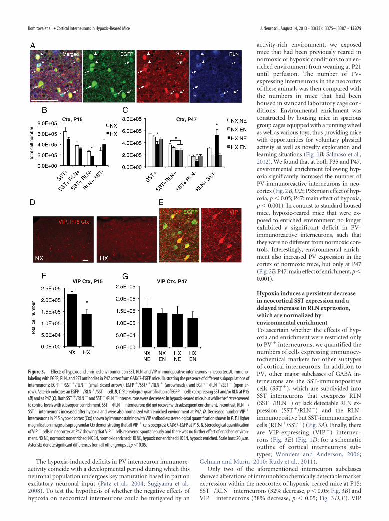
Hypoxia induces a persistent decreasein neocortical SST expression and adelayed increase in RLN expression,which are normalized byenvironmental enrichmentTo ascertain whether the effects of hyp-oxia and enrichment were restricted onlyto PV � interneurons, we quantified thenumbers of cells expressing immunocy-tochemical markers for other subtypesof cortical interneurons. In addition toPV, other major subclasses of GABA in-terneurons are the SST-immunopositivecells (SST�), which are subdivided intoSST interneurons that coexpress RLN(SST�/RLN�) or lack detectable RLN ex-pression (SST�/RLN�) and the RLN-immunopositive but SST-immunonegativecells (RLN�/SST�) (Fig. 3A). Finally, thereare VIP-expressing (VIP �) interneu-rons (Fig. 3E) (Fig. 1D; for a schematicoutline of cortical interneurons sub-types; Wonders and Anderson, 2006;
Gelman and Marín, 2010; Rudy et al., 2011).Only two of the aforementioned interneuron subclasses
showed alterations of immunohistochemically detectable markerexpression within the neocortex of hypoxic-reared mice at P15:SST�/RLN� interneurons (32% decrease, p � 0.05; Fig. 3B) andVIP� interneurons (38% decrease, p � 0.05; Fig. 3D,F). VIP
Figure 3. Effects of hypoxic and enriched environment on SST, RLN, and VIP-immunopositive interneurons in neocortex. A, Immuno-labeling with EGFP, RLN, and SST antibodies in P47 cortex from GAD67-EGFP mice, illustrating the presence of different subpopulations ofinterneurons: EGFP �/SST �/RLN � (small closed arrows), EGFP �/SST/ �/RLN � (arrowheads), and EGFP �/RLN �/SST � (open ar-row). Asterisk indicates an EGFP �/RLN �/SST � cell. B, C, Stereological quantification of EGFP � cells coexpressing SST and/or RLN at P15(B) and at P47 (C). Both SST �/RLN �and SST �/RLN � interneurons were decreased in hypoxic-reared mice, but while the first recoveredto control levels with subsequent enrichment, SST �/RLN � interneurons did not recover with subsequent enrichment. In contrast, RLN �/SST � interneurons increased after hypoxia and were also normalized with enriched environment at P47. D, Decreased number VIP �
interneurons in P15 hypoxic cortex (Ctx) shown by immunostaining with VIP antibodies; stereological quantification shown in F. E, Highermagnification image of supragranular Ctx demonstrating that all VIP � cells coexpress GAD67-EGFP at P15. G, Stereological quantificationof VIP � cells in neocortex at P47 showing that VIP � cells recovered spontaneously and there was no further effect of enriched environ-ment. NX NE, normoxic nonenriched; NX EN, normoxic enriched; HX NE, hypoxic nonenriched; HX EN, hypoxic enriched. Scale bars: 20�m.Asterisks denote significant differences from all other groups at p � 0.05.
Komitova et al. • Cortical Interneurons in Hypoxic-Reared Mice J. Neurosci., August 14, 2013 • 33(33):13375–13387 • 13379

Figure 4. Intensity and distribution of expression of interneuron marker expression in upper (L1–L4) and lower (L5–L6) layers of primary motor or primary somatosensory cortex (hindlimb) ofnormoxic and hypoxic mice at P47. Quantification of average expression and numbers of cells with distinct expression levels, respectively, with regards to PV (A, B), (Figure legend continues.)
13380 • J. Neurosci., August 14, 2013 • 33(33):13375–13387 Komitova et al. • Cortical Interneurons in Hypoxic-Reared Mice

expression recovered spontaneously at P47 and was unaffected byenrichment status (Fig. 3G). In contrast, at P47, both overallSST-expressing cells as well as those that colocalized RLN orshowed no RLN immunolabeling were significantly decreased byhypoxia (p � 0.05; Fig. 3C). Introduction of environmental en-richment upon weaning at P21, i.e., 10 d after the end of chronichypoxia, was sufficient to correct the above-mentioned hypoxia-induced decrease in SST� and SST�/RLN� cells (Fig. 3C; SST�:main effect of hypoxia p � 0.05; SST�/RLN� interaction be-tween normoxia/hypoxia and standard/enrichment: p � 0.054).In contrast, the number of SST-immunoreactive cells that coex-press RLN was not modified by enrichment status (Fig. 3C). In-triguingly, the RLN�/SST� interneuron subpopulation showeda remarkable 73% increase at P47 in hypoxic-reared mice (Fig.3C), which was totally reversed by enrichment (Fig. 3C; interac-tion between normoxia/hypoxia and standard/enrichment p �0.05). Notably, these counts were performed in GAD67-EGFPmice, hence we only counted the EGFP�/RLN� GABAergic cells,which is important as RLN has been shown to also be present insome projection neurons in the cortex (Deguchi et al., 2003).
Quantification of intensity and distribution of expression ofinterneuron marker expression in cortex of P47 normoxicand hypoxic miceDuring our stereological cell counts it was evident that marker-positive cells exhibited varying levels of immunolabeling. To gaina more detailed understanding of the effects of hypoxia on in-terneuron marker expression, we quantified the intensity levels oflabeling with PV, SST, and RLN antibodies in individual in-terneurons (GAD67-EGFP� cells) as well as counted marker-positive cells using micrographs of RLN/SST and PV/RLNdouble-immunostained sections from P47 GAD67-EGFP brains.We evaluated potential regional effects within the cortex by map-ping the distribution of marker-positive cells of varying labelingintensities in two areas along the rostrocaudal axis, primary mo-tor (M1) and primary somatosensory cortex, hindlimb region(S1HL), and in the upper (L1–L4) and lower (L5, L6) corticallayers.
The intensity of PV immunostaining was significantly de-creased by 28 and 21%, respectively, in upper cortical layers ofmotor and somatosensory cortex in hypoxic mice (interactionbetween hypoxia and cortical layer distribution, p � 0.01; Fig.4A). However, hypoxia did not significantly affect the amount ofPV expression in deep cortical layers of either cortical region.When we evaluated the distribution of PV� cell numbers amongsix different intensity categories of PV expression, hypoxia de-creased the numbers of PV� cells across all intensity ranges inboth motor and somatosensory cortex (Fig. 4B; interaction be-tween number and intensity ranges not significant). When con-sidering all intensities together, numbers of PV-expressing cellswere significantly decreased by 20% in hypoxic mice without anyinteractions by cortical region or layer localization (main effect ofhypoxia p � 0.05). We also found that PV� cells exhibited asignificantly decreased cell size but only in upper cortical layers ofmotor cortex (15.1 � 0.4 and 13.5 � 0.5 �m in normoxic and
hypoxic mice, respectively; interaction between hypoxia andupper/lower cortical layer distribution, p � 0.001).
Next, we quantified levels of SST immunoreactivity in indi-vidual cells in upper and lower layers of motor and somatosen-sory cortex. Expression of SST was significantly decreased byhypoxia in upper as well as in lower cortical layers of somatosen-sory cortex (54 and 39% decrease, respectively) but was not sig-nificantly affected in motor cortex (interaction between hypoxiaand cortical layer distribution, p � 0.05; Fig. 4C). When we eval-uated the distribution of SST� cell numbers among six differentintensity categories of SST expression, hypoxia seemed to de-crease SST� cells in the medium intensity ranges, but had a vari-able effect in the low intensity range, such that when consideringall intensities together, the total number of SST-immunoreactivecells was not significantly changed by hypoxia (Fig. 4D). How-ever, the subset of SST�/RLN� cells was decreased by hypoxia insomatosensory cortex by 81 and 53% in upper and lower corticallayers, respectively (interaction between hypoxia and motor/so-matosensory cortex distribution, p � 0.01; interaction betweenhypoxia and upper/lower cortical layer distribution, p � 0.05;data not shown). The size of SST� cells was not affected by hyp-oxia (data not shown).
Finally, we quantified the levels of RLN immunostaining inindividual cells in upper and lower layers of motor and somato-sensory cortex. We found that hypoxia increased the intensity ofRLN immunostaining in all regions and layers examined (Fig.4E), with a trend toward a stronger effect in somatosensory cortex(main effect of hypoxia p � 0.0001; interaction between hypoxiaand motor/somatosensory cortex distribution, p � 0.068; Fig.4E). Furthermore, hypoxia increased the numbers of RLN� cellsin the medium to high intensity ranges, but did not affect thenumbers of RLN� cells in the low intensity categories, whichwere the most numerous. When considering all intensitiestogether, the numbers of RLN � cells were unchanged by hyp-oxia in both regions (Fig. 4F ). Hypoxia exhibited a complexeffect on RLN � cell size. Hypoxia decreased RLN � cell size inupper layers of motor cortex (12.1 � 0.3 �m vs 10.8 � 0.3 �min normoxic and hypoxic mice, respectively) and increasedRLN � cell size in lower layers of somatosensory cortex (11.6 �0.3 �m vs 12.9 � 0.3 �m in normoxic and hypoxic mice,respectively; interaction between hypoxia and cortical regiondistribution, p � 0.001; interaction between hypoxia and cor-tical layer distribution, p � 0.05).
Last, we evaluated any potential relationship between RLNand SST expression or RLN and PV expression in sections doublestained for RLN and SST or PV and RLN. There was no signifi-cant correlation (negative or otherwise) between RLN and SSTintensity across double-labeled cells. When we evaluated colabel-ing between PV and RLN, we found that very few PV� cellscoexpressed RLN. Interestingly, they were increased in hypoxiccortex, specifically in upper layers. However, since they were sofew (on average 0.2–2.1 cells per section) we could not perform ameaningful correlation analysis of PV and RLN expression.
In conclusion, combining measurements of immunostainingintensities of marker-positive individual cells with cell counts inmotor and somatosensory cortex suggests that hypoxia decreasesthe number of PV� cells across all intensity ranges and in allregions and layers examined, decreases the number of SST� cellsin the medium intensity range in the somatosensory cortex, andincreases RLN immunoreactivity in the medium to high intensityranges with no overall change in RLN� cell number.
4
(Figure legend continued.) SST (C, D), and RLN (E, F). Asterisks denote significant differences( p � 0.05) as determined by factorial ANOVA conducted for each marker. Images depict rep-resentative examples of single cells expressing low or high levels of a given marker. The num-bers signify the background corrected intensity of cell fluorescence for each cell and marker. NX,normoxia; HX, hypoxia; M, motor cortex; SS, somatosensory cortex; L1– 4, cortical layers 1– 4;L5– 6, cortical layers 5 and 6; ctx, cortex.
Komitova et al. • Cortical Interneurons in Hypoxic-Reared Mice J. Neurosci., August 14, 2013 • 33(33):13375–13387 • 13381

Hypoxia does not alter the totalnumber of interneurons incerebral cortexNext, we examined whether the hypoxia-induced decreases in immunostaining forinterneuron markers were due to an actualloss of interneurons. To this end, we esti-mated the total numbers of GAD67-EGFP�
GABA interneurons in the same cohorts ofmice in which we had observed decreasedstaining of interneuron marker� cells. Weused the GAD67-EGFP line wherein virtu-ally all inhibitory GABA interneurons arefully labeled with EGFP reporter from em-bryogenesis onward (Tamamaki et al.,2003). The EGFP reporter protein in thesemice does not change with maturation orplasticity of GABA interneurons through-out early postnatal development (Fig. 5B,C)likely due to stability of the EGFP protein,allowing an estimation of their number in-dependently from their maturation stage(Jiao et al., 2006). In this new cohort of ani-mals, we still observed persistent decreasesin numbers of immunohistochemically de-tected PV� cells with a subsequent recoveryafter environmental enrichment, as well asan increase in PV� cells in normoxic-enriched mice (Fig. 5C). However, thenumbers of EGFP reporter-positive in-terneurons in neocortex did not differ sig-nificantly between normoxic and hypoxicmice, neither at P15 nor at P47 (Fig. 5A–C).The total numbers of GAD67-EGFPreporter-positive cells per cortex were2,626,693 � 136,824 in normoxic mice ver-sus 2,259,182 � 136,303 in hypoxic-rearedmice at P15. At P47, the total numbersof reporter-positive interneurons were2,448,017 � 185,196 and 2,598,123 �171,593 in normoxic and hypoxic mice, re-spectively. Furthermore, we found no sig-nificant differences in total (EGFP�)inhibitory interneuron numbers betweenmice housed in standard laboratory envi-ronment and environmentally enrichedmice at P47 (Fig. 5C).
To directly examine whether cell deathwas responsible for the pronounced lossof PV interneuron marker-positive cells atP15, we colabeled cells with EGFP and theapoptotic marker activated caspase-3 inbrain sections from GAD67-EGFP mice.Very few apoptotic cells were identifiedper neocortex with no significant differ-ences in the numbers of activatedcaspase-3� cells between normoxic con-trols and hypoxic-reared mice. Moreover,there was no overlap between activatedcaspase-3 and EGFP (Fig. 5D). To under-stand whether GABA interneuron pro-genitor cells were dying, we analyzedbrain sections from mice perfused imme-
Figure 5. Analysis of interneuron lineage cells in the neocortex. A, Costaining of PV (red) and GAD67-EGFP reporter (green) inP15 cortex (Ctx). B, C, Stereological quantification of GAD67 EGFP � cells in the cortex (Ctx) at P15 (B) and P47 (C). EGFP � cellnumbers do not change across conditions, whereas PV � cells counted in the same sections are decreased by hypoxia (HX) andincreased by enriched environment (EN). D–F, P12–P15 neocortex from HX-reared mice labeled with antibodies to activatedcaspase-3 (red) and GAD67-EGFP (green; D), Dlx-2 (red), and Olig2 (blue; E), and activated caspase-3 (green) and Dlx-2 (red; F).Interneurons do not undergo noticeable increases in cell death in response to rearing in HX. G, H, Stereological quantification ofDlx-2 � cells in P15 (G) and P47 (H) neocortex. There are no significant changes in Dlx-2 � cell numbers in response to HX or EN. I,The numbers of all GABA � cells and GABA �Dlx-2 � colabeled cells in P15 cortex were decreased in HX-reared mice. J, HPLCmeasurement of GABA content in different brain regions showed that HX rearing significantly decreased GABA in frontal cortex.Scale bars: A, E, 20 �m; D, 10 �m. Asterisks denote significant differences from normoxic controls at p � 0.05. NX NE, normoxicnonenriched; NX EN, normoxic enriched; HX NE, hypoxic nonenriched; HX EN, hypoxic enriched; Fr, frontal; Par, parietal; Occ,occipital, Hipp, hippocampus; Str, striatum.
13382 • J. Neurosci., August 14, 2013 • 33(33):13375–13387 Komitova et al. • Cortical Interneurons in Hypoxic-Reared Mice

diately after hypoxic rearing (P12) that were colabeled with acti-vated caspase-3 and the transcription factor Dlx-2, which isexpressed in GABAergic progenitors since embryogenesis (Bul-fone et al., 1993). The Dlx-2 protein showed complete overlapwith inhibitory neuron lineage markers such as GAD67-EGFPand was also occasionally coexpressed with mature interneuronmarkers such as PV; Dlx-2 was not expressed in oligodendrocytelineage cells, labeled by the transcription factor Olig2 (Fig. 5E).Again, there was no difference in numbers of caspase-3� cellsbetween normoxia and hypoxia-reared mice and almost all of theactivated caspase-3� cells identified in neocortex lacked Dlx-2expression (Fig. 5F). Consistent with the idea that there was nodeath of interneuron progenitors or immature GABA neurons inhypoxic-reared mice, neither hypoxia nor environmental enrich-ment had any significant effects on the numbers of Dlx-2-positivecells in P15 or P47 brains (Fig. 5G,H). Together, our data show-ing unchanged numbers of total interneurons and interneuronprogenitors and lack of cell death strongly suggest that the pro-nounced and significant decreases in PV and SST cortical in-terneuron immunoreactivity (Figs. 2, 3) were not due to a defacto loss of interneurons.
Hypoxia decreases GABA content in the neocortexThe fact that the numbers of immunohistochemically detectedPV� and SST�/RLN� interneurons, which together comprisethe majority of cortical inhibitory cells, were significantly de-creased by hypoxia and increased by enrichment, but the totalnumber of inhibitory neurons (marked by EGFP) and their pro-genitors did not differ between these conditions, suggests thathypoxia and enrichment might somehow influence the expres-sion of mature interneuron markers. It is well established thatPV� neurons undergo their final differentiation in the first threepostnatal weeks and therefore their phenotypic maturation couldbe susceptible to environmental manipulations during this time.Unlike the EGFP reporter driven by the gad1 promoter, the expres-sion of GAD67 mRNA and protein undergo a steady upregulationwith increased maturation of interneurons during the early postna-tal period (Huang and Akbarian, 2007) and are affected by sensorydeprivation (Jiao et al., 2006). We thus considered levels of GABAneurotransmitter expression in the brain as a marker of interneuronmaturity and function.
To test this hypothesis, we first evaluated colocalization be-tween the inhibitory neurotransmitter GABA and GAD67 indouble-immunolabeled sections from normoxic adolescent miceand found that there was a large overlap between the two markers(85% of strongly GAD67� cells were also GABA�), thus allowingus to use these phenotypes interchangeably. We chose to useGABA as a marker since the positive staining was mainly periso-matic and positive cells could be unequivocally identified incontrast to GAD67, where positive cells not only exhibited stain-ing of the cell body but also strong punctuate staining of pro-cesses, which made cell counts more difficult. To obtain amolecular index of maturation of the Dlx-2-positive interneuronlineage cells in neocortex, we costained sections from P15 micewith antibodies for Dlx-2 and GABA. We found that the totalnumbers of GABA� cells (p � 0.001) as well as GABA�/Dlx-2�
(p � 0.001) cells were significantly and comparably decreased inthe cortex of hypoxia-reared mice as compared with normoxiccontrols at P15 (Fig. 5I). To confirm the changes we observed inGABA immunolabeling, we measured GABA content in differentparts of the cortex and in other brain areas such as basal gan-glia and hippocampus using HPLC. Hypoxic-reared mice ex-hibited lower GABA content in general, with the most
pronounced difference in frontal cortex, confirming our cellcounts (Fig. 5J; p � 0.05).
The decrease in the fraction of Dlx2� cells expressing detect-able GABA levels at P15 by immunohistochemistry, togetherwith absence of changes in total numbers of GAD67-EGFP� cellsor Dlx-2� interneuron progenitors, suggest that hypoxia is likelyto delay interneuron maturation in the cortex. The data also sug-gest that there is some degree of catch-up in hypoxic mice, asPV� interneurons are relatively less depleted at P47, 5 weeks afterhypoxia, as compared with P15. Hence, we were interested toassess whether GABA immunoreactivity also exhibited an im-provement at several weeks after hypoxia. In the cortex ofhypoxic-reared mice at P47, the numbers of GABA-immunoreactive cells were not significantly different from nor-moxic mice (1,018,842 � 180,272 and 936,433 � 25,288 GABA�
cells in hypoxic and normoxic mice, respectively).
Recovery of interneuron subpopulations after hypoxia andenvironmental enrichment is not due to neurogenesisSince it has previously been shown that GABAergic GAD67�
interneuron progenitors in cortical layer 1 can be induced toproliferate and replenish interneuron populations after ahypoxic-ischemic insult (Ohira et al., 2010), we examined P15cortical sections for colabeling of GABA with the proliferationmarker Ki67. However, we found no evidence of Ki67� cells thatcoexpressed GABA in either normoxic or hypoxic-reared mice(Fig. 6A,B). Moreover, we immunolabeled sections from P15mice with Ki67 and Dlx-2 as an additional assessment of whetherinhibitory interneuron progenitors were induced to proliferate inresponse to hypoxia. Dlx-2� cells were almost exclusively Ki67negative (Fig. 6C) and thus, likely to be postmitotic in both nor-moxic and hypoxic mice. Last, we did not find any overlap be-tween GAD67-EGFP and Ki67 at P15 (Fig. 6D). We thereforeconcluded that there was no evidence of proliferation of GABAinterneuron lineage cells in P15 cortex in any of the conditions.This suggests that the partial recovery of interneuron populationsobserved at later time points after hypoxia are likely not due toneurogenesis from interneuron progenitor cells.
To further assess whether enriched environment led to genera-tion of inhibitory interneurons in the neocortex, we administeredthe thymidine analog BrdU to GAD67-EGFP hypoxic-reared miceand normoxic controls between P21–P27, immediately upon wean-ing and onset of enriched or standard cage conditions, and perfusedthe mice 4 weeks later. The numbers of BrdU� cells were signifi-cantly increased in the cortex of hypoxic-reared nonenriched miceversus normoxic controls (251,931 � 24,684 BrdU� cells vs128,565 � 12,846 BrdU� cells; p � 0.05). However, environmentalenrichment had no effect on the number of BrdU-labeled cells.BrdU� GAD67-EGFP colabeled cells were rarely found and if so,they were only present in normoxic but not in hypoxic mice, regard-less of enriched housing status. Most of the BrdU� cells did notcoexpress GAD67-EGFP (Fig. 6E,F); not surprisingly, as we havepreviously reported that newborn cells labeled after hypoxia mostlyexpress oligodendrocyte and astroglial markers (Fagel et al., 2006).
We next examined whether GABAergic neurons could be gen-erated in enriched animals from a non-GABAergic lineage. Usinggenetic fate mapping, we recently demonstrated that hypoxia in-creases the genesis of cortical excitatory neurons from GFAP�
cells in juvenile mice (Bi et al., 2011). To examine whetherGFAP� cells were able to generate new GABAergic neurons inresponse to environmental enrichment, tamoxifen-inducibleGCE mice carrying the CAG-CAT-EGFP or the R26R LacZ re-porter were reared in hypoxic or normoxic conditions, injected
Komitova et al. • Cortical Interneurons in Hypoxic-Reared Mice J. Neurosci., August 14, 2013 • 33(33):13375–13387 • 13383

with tamoxifen daily from P12 to P14 andthen subjected to enrichment startingfrom P21 until P35 or P47. When we an-alyzed coexpression between reporter andinhibitory interneuron markers in theneocortex at P35, there were no reporter�
cells that coexpressed GABA in normoxicor hypoxic nonenriched mice. However,in 1 of 3 normoxic-reared enriched miceand in 2 of 4 hypoxic-reared enrichedmice, reporter� cells that coexpressedGABA were observed. These double-labeled putative inhibitory interneuronswere, however, very rare. They consti-tuted 1% among reporter� cells or 0.3%among GABA� cells in the normoxic en-riched mouse, and 2% among reporter�
cells or 0.9% among GABA� cells in thetwo hypoxic-reared enriched mice. Theserare reporter � cells that coexpressedGABA were NeuN negative and exhibitedan immature morphology with a small cellbody and very few processes. We then an-alyzed reporter-tagged GFAP-lineage cellsin the neocortex at P47. As previously de-scribed (Bi et al., 2011), we observed mul-tiple reporter-positive cells with neuronalmorphology in the cortex of mice from allfour groups. These reporter� cells, how-ever, exhibited pyramidal cell morphol-ogy and none of them coexpressed PV(Fig. 6G–L) or CR, an interneuronmarker expressed in a subpopulation ofSST and VIP-positive cells (data notshown). In contrast, many reporter� cellsexpressed the excitatory neuron tran-scription factor Tbr1 and exhibitedcomplex dendritic architecture in en-riched environment mice (Fig. 6M–O).Together, our data suggest that the re-covery of interneurons after hypoxiaand environmental enrichment is, forthe most part, not due to generation ofnew neurons.
DiscussionThis study presents compelling evidencethat our model of hypoxic brain injury,which is clinically relevant to prematurelyborn infants, results in a long-lasting de-crease perturbation in the differentiationof PV� and SST� interneuron subtypeswithin the neocortex, coupled with a tran-sient decrease in overall GABA neurotransmitter content, mostnotably in the frontal cortex. Introducing hypoxic-reared miceinto an enriched environment for �1 month largely normalizedthe neurochemical profile of inhibitory interneurons.
Our analysis showed that rather than affecting the total numberof interneurons, hypoxia decreased the expression of RLN and SSTand increased the expression of RLN within interneurons, resultingin decreased numbers of PV� and SST/RLN-interneurons in themature cerebral cortex. Indeed, a previous study using our hypoxiamodel reported gene expression changes in cortex and hippocampus
consistent with disrupted synaptic maturation after hypoxia (Cur-ristin et al., 2002). Using GAD67-EGFP transgenic mice in which theEGFP reporter labels GABA interneurons since embryogenesis, re-gardless of gad1 mRNA levels, we show that hypoxia did not cause aloss of interneurons, as total GAD67-EGFP� cell number was un-changed by hypoxic rearing and there was no activation of caspase-3in these cells. Furthermore, hypoxia neither changed the number ofDlx2� GABAergic progenitors in the cortex, nor caused their death.Immunolabeling intensity measurements directly demonstrated de-creased PV and SST expression and increased RLN expression inindividual cortical interneurons.
Figure 6. Assessment of cell proliferation and neurogenesis in cortex. Boxed areas on a sagittal and coronal cresyl violet-stainedsection depict where the images were taken. A, Costaining of P15 cortex from hypoxic (HX)-reared mice with Ki67 (blue), GABA(red), and NeuN (green) showing a Ki67 � cell next to a GABA � NeuN � double-labeled cell (yellow when colocalized). B, Samestaining with the NeuN channel extinguished, showing the GABA-positive cell (red) next to and distinct from the Ki67-positive cell(red). C, D, Immunolabeling of P15 cortex from HX-reared mice with Ki67 (blue) and Dlx-2 (red; C), and GAD67-EGFP (green) andKi67 (red; D showing lack of proliferation in interneuron lineage cells. E, F, GAD67-EGFP (green) and BrdU (red) colabeling of P47cortex from normoxic nonenriched (NX NE; E) and hypoxic-reared enriched mice (HX EN; F) showing lack of interneuron generation.G–L, Immunolabeling of P47 cortex with EGFP (green) and PV (red) in P47 cortical sections from GCE mice, in which GFAP � cellshad been permanently tagged with reporter by administering tamoxifen at P12–P14. Note EGFP reporter � cells with neuronalmorphology that lack PV expression and the presence of EGFP reporter � bushy morphology typical of astroglia. M–O, Immuno-labeling of P47 cortex with EGFP (green), Tbr1 (red), and NeuN (blue) in P47 cortical sections from GCE mice, showing genesis ofTbr1� neurons from GFAP � cells (arrows). Arrowheads point to astrocytes. Scale bars: A, B, M–O, 10 �m; in all otherparts, 20 �m.
13384 • J. Neurosci., August 14, 2013 • 33(33):13375–13387 Komitova et al. • Cortical Interneurons in Hypoxic-Reared Mice

Environmental enrichment restored PV and SST expressionbut increased neither GAD67-EGFP� postmitotic GABA neu-rons nor Dlx-2� GABA precursors, suggesting that enrichmentdid not cause an overall increase in GABAergic cells or theirprogenitors, but rather normalized the expression of matureinterneuron markers. However, it is possible that low-grade gen-eration of GABAergic neurons might take place from a Dlx-2-negative glial source. For example, we recently demonstrated thatpostnatal GFAP� cells generate cortical excitatory neurons (Bi etal., 2011) and that GFAP� cells generate GABAergic interneu-rons, including PV� cells, in the early postnatal cerebellum (Sil-bereis et al., 2009). Thus, we examined whether GFAP� cells wereable to generate new GABAergic neurons in response to environ-mental enrichment. After 2 weeks of enrichment, we found pu-tative GFAP� cell-derived interneurons. However they were veryrare and appeared quite immature. After a total of 4 weeks inenriched environment, we did not find any astroglia-derived in-hibitory interneurons, suggesting that these cells do not surviveinto maturity.
A retroviral fate-mapping study previously demonstrated thatthere was an increase in proliferating cells expressing GAD67 incortical layer 1 after a hypoxic ischemic insult in adult rats, andthat these cells generated GABAergic interneurons, but not PV�
cells (Ohira et al., 2010). However, we did not see any colabelingof Ki67 with GABA or GAD67-EGFP reporter in early postnatalmice irrespectively of hypoxic rearing, nor did we observe anyappreciable levels of Ki67 expression in Dlx-2� cells. Moreover,based on our BrdU fate-mapping experiments, we failed to seeany generation of inhibitory interneurons in the cortex at latertime points after hypoxia regardless of enrichment status. We didfind newborn BrdU-labeled interneurons in normoxic enrichedmice. These cells were, however, rare and unlikely to account forthe increase in PV� cells found in normoxic, enriched mice.Together, these data led us to conclude that generation of newinterneurons is not the reason for the partial recovery of in-terneuron marker expression after early postnatal hypoxia or thefull recovery achieved through environmental enrichment.
The regional effect of variations in oxygen level on inhibitoryinterneurons has been examined in various models. Decreases inGABA, GAD65/67, and PV-immunoreactive cells, mostly tran-sient, have been reported in the hippocampus in perinatal ratanoxia models (Dell’Anna et al., 1996; Wang et al., 2011) and inthe cortex in a prenatal hypoxia model (Louzoun-Kaplan et al.,2008) as well as in a perinatal hypoxia-ischemia model (Failor etal., 2010). Our findings are consistent with these studies. In ad-dition, we found that chronic postnatal hypoxia elicited a persis-tent decrease in SST immunoreactivity and an increase in RLNimmunoreactivity within inhibitory cortical neurons in adult-hood. RLN is an extracellular matrix protein that regulates cellmigration and is secreted by Cajal–Retzius cells during brain de-velopment (Caviness and Sidman, 1973; Zhao and Frotscher,2010). However, in the postnatal brain, RLN is primarily pro-duced by GABAergic interneurons (Ramos-Moreno et al., 2006)and is believed to be involved in synaptic function possiblythrough modulation of the actin cytoskeleton (Herz and Chen,2006). It remains to be tested whether the generally increasedRLN expression we measured in the cortex of hypoxic-rearedmice might have adaptive or maladaptive functions in modulat-ing synaptic plasticity and interneuron function.
Perturbed interneuron differentiation and thus decreasedGABAergic signaling in postnatal development could have detri-mental effects on cortical function and lasting repercussionsfor behavior. At birth, all interneuron populations have been
formed, have migrated from the ganglionic eminences, and havelargely incorporated into the cortex (Batista-Brito and Fishell,2009). However, early postnatal neocortical connectivity is stillimmature and undergoes a pronounced process of network or-ganization. During embryonic brain development and in theearly postnatal period GABA acts as an excitatory neurotransmit-ter and becomes inhibitory only later (Ben-Ari, 2002). The ex-pression of PV, a calcium-binding protein that modulatesintracellular Ca 2� transients in fast-spiking GABAergic cells(Caillard et al., 2000; Collin et al., 2005), correlates with the func-tional maturation of fast-spiking GABAergic interneurons (Plot-kin et al., 2005). The maturation of PV� inhibitory neurons, andGABA neurons in general, causes the termination of the criticalperiod of synaptic plasticity, which is the period when the corticalsynaptic network can be reorganized in response to differences insensory inputs (Hensch et al., 1998; Hensch and Stryker, 2004).The delays in PV and SST interneuron maturation caused byhypoxia may thus confer prolonged and exaggerated plasticity tothe cerebral cortex, an intriguing hypothesis since neuroimagingdata suggest that preterm subjects engage alternative pathwaysfor language processing (Myers et al., 2010; Scheinost et al.,2012). In adulthood, PV� and SST� interneurons form a denseinhibitory network of synapses onto adjacent pyramidal cells(Fino and Yuste, 2011; Packer and Yuste, 2011) and are keyplayers in the regulation of cortical excitability. The firing of PV�
neurons generates gamma oscillations, which improve the effi-ciency of cortical processing and are disrupted in several neuro-psychiatric disorders (Bartos et al., 2007; Cardin et al., 2009;Sohal et al., 2009). In mice, decreased numbers of PV� interneu-rons correlate with the degree of deficit in spatial memory in theMorris water maze task (Stevens et al., 2012). Hence, the lastingdecrease in PV and SST expression and therefore dampened in-hibitory neurotransmission may be responsible for the increasedhyperactivity and deficits in learning and spatial memory ob-served after hypoxic insults (Chahboune et al., 2009; Li et al.,2009). Conversely, the behavioral improvement observed afterenvironmental enrichment (Salmaso et al., 2012) might in part bemediated by normalization of interneuron neurochemical prop-erties. Indeed, we found that enriched environment completelyreversed the hypoxia-induced decrease in PV and SST expressionin cerebral cortex. Enriched housing conditions in adulthoodwere previously found to influence PV� cell numbers in the hip-pocampus and cortex, in one case decreasing PV� cells in ratsthat had undergone neonatal anoxia (Iuvone et al., 1996), and, inanother case, increasing the number of PV� cells in perilesionalcortex of an adult rat stroke model (Inacio et al., 2011). Con-versely, social isolation in rats was shown to decrease the numberof PV� interneurons in the hippocampus (Harte et al., 2007) andPV expression in prefrontal cortex (Schiavone et al., 2009). Earlypostnatal environmental enrichment has been shown to increaseGABAergic neurotransmission and developmental synaptic mat-uration (He et al., 2010). However it is not known whether en-richment increases GABA neurotransmission by inducing PVand SST expression, as shown here, or whether it affects otherbasic properties of interneurons.
PV and SST expression is contingent upon excitatory neuro-nal input and neurotrophin signaling (Patz et al., 2004; Close etal., 2012; Denaxa et al., 2012). Treatment with the NMDA recep-tor antagonist ketamine results in decreased PV immunoreactiv-ity with maintained interneuron numbers (Behrens et al., 2007;Powell et al., 2012). Whether hypoxia acts to directly influenceinterneurons or indirectly affect their maturation by first nega-tively affecting excitatory neurons (Fagel et al., 2006, 2009) re-
Komitova et al. • Cortical Interneurons in Hypoxic-Reared Mice J. Neurosci., August 14, 2013 • 33(33):13375–13387 • 13385
mains to be shown. It also remains to be determined whetherenriched environment acts to increase PV and SST expression byincreasing excitatory drive or secretion of neurotrophic factors,or both.
ReferencesBartos M, Vida I, Jonas P (2007) Synaptic mechanisms of synchronized
gamma oscillations in inhibitory interneuron networks. Nat Rev Neuro-sci 8:45–56. CrossRef Medline
Batista-Brito R, Fishell G (2009) The developmental integration of corticalinterneurons into a functional network. Curr Top Dev Biol 87:81–118.CrossRef Medline
Behrens MM, Ali SS, Dao DN, Lucero J, Shekhtman G, Quick KL, Dugan LL(2007) Ketamine-induced loss of phenotype of fast-spiking interneuronsis mediated by NADPH-oxidase. Science 318:1645–1647. CrossRefMedline
Ben-Ari Y (2002) Excitatory actions of GABA during development: the na-ture of the nurture. Nat Rev Neurosci 3:728 –739. CrossRef Medline
Bi B, Salmaso N, Komitova M, Simonini MV, Silbereis J, Cheng E, Kim J, LuftS, Ment LR, Horvath TL, Schwartz ML, Vaccarino FM (2011) Corticalglial fibrillary acidic protein-positive cells generate neurons after perinatalhypoxic injury. J Neurosci 31:9205–9221. CrossRef Medline
Brenner M, Kisseberth WC, Su Y, Besnard F, Messing A (1994) GFAP pro-moter directs astrocyte-specific expression in transgenic mice. J Neurosci14:1030 –1037. Medline
Bulfone A, Kim HJ, Puelles L, Porteus MH, Grippo JF, Rubenstein JL (1993)The mouse Dlx-2 (Tes-1) gene is expressed in spatially restricted domainsof the forebrain, face and limbs in midgestation mouse embryos [pub-lished erratum appears in Mech Dev 1993 Aug;42:187]. Mech Dev 40:129 –140. CrossRef Medline
Caillard O, Moreno H, Schwaller B, Llano I, Celio MR, Marty A (2000) Roleof the calcium-binding protein parvalbumin in short-term synaptic plas-ticity. Proc Natl Acad Sci U S A 97:13372–13377. CrossRef Medline
Cardin JA, Carlen M, Meletis K, Knoblich U, Zhang F, Deisseroth K, Tsai LH,Moore CI (2009) Driving fast-spiking cells induces gamma rhythm andcontrols sensory responses. Nature 459:663– 667. CrossRef Medline
Caviness VS Jr, Sidman RL (1973) Time of origin or corresponding cellclasses in the cerebral cortex of normal and reeler mutant mice: an auto-radiographic analysis. J Comp Neurol 148:141–151. CrossRef Medline
Chahboune H, Ment LR, Stewart WB, Rothman DL, Vaccarino FM, Hyder F,Schwartz ML (2009) Hypoxic injury during neonatal development inmurine brain: correlation between in vivo DTI findings and behavioralassessment. Cereb Cortex 19:2891–2901. CrossRef Medline
Close J, Xu H, De Marco García N, Batista-Brito R, Rossignol E, Rudy B,Fishell G (2012) Satb1 is an activity-modulated transcription factorrequired for the terminal differentiation and connectivity of medial gan-glionic eminence-derived cortical interneurons. J Neurosci 32:17690 –17705. CrossRef Medline
Collin T, Chat M, Lucas MG, Moreno H, Racay P, Schwaller B, Marty A, LlanoI (2005) Developmental changes in parvalbumin regulate presynapticCa2� signaling. J Neurosci 25:96 –107. CrossRef Medline
Curristin SM, Cao A, Stewart WB, Zhang H, Madri JA, Morrow JS, Ment LR(2002) Disrupted synaptic development in the hypoxic newborn brain.Proc Natl Acad Sci U S A 99:15729 –15734. CrossRef Medline
Deguchi K, Inoue K, Avila WE, Lopez-Terrada D, Antalffy BA, QuattrocchiCC, Sheldon M, Mikoshiba K, D’Arcangelo G, Armstrong DL (2003)Reelin and disabled-1 expression in developing and mature human cor-tical neurons. J Neuropathol Exp Neurol 62:676 – 684. Medline
del Río JA, de Lecea L, Ferrer I, Soriano E (1994) The development ofparvalbumin-immunoreactivity in the neocortex of the mouse. Brain ResDev Brain Res 81:247–259. CrossRef Medline
de Lecea L, del Río JA, Soriano E (1995) Developmental expression of parv-albumin mRNA in the cerebral cortex and hippocampus of the rat. BrainRes Mol Brain Res 32:1–13. CrossRef Medline
Dell’Anna E, Geloso MC, Magarelli M, Molinari M (1996) Development ofGABA and calcium binding proteins immunoreactivity in the rat hip-pocampus following neonatal anoxia. Neurosci Lett 211:93–96. CrossRefMedline
Denaxa M, Kalaitzidou M, Garefalaki A, Achimastou A, Lasrado R, Maes T,Pachnis V (2012) Maturation-promoting activity of SATB1 in MGE-derived cortical interneurons. Cell Rep 2:1351–1362. CrossRef Medline
Durkin TA, Anderson GM, Cohen DJ (1988) High-performance liquid
chromatographic analysis of neurotransmitter amino acids in brain.J Chromatogr 428:9 –15. Medline
Fagel DM, Ganat Y, Silbereis J, Ebbitt T, Stewart W, Zhang H, Ment LR,Vaccarino FM (2006) Cortical neurogenesis enhanced by chronic peri-natal hypoxia. Exp Neurol 199:77–91. CrossRef Medline
Fagel DM, Ganat Y, Cheng E, Silbereis J, Ohkubo Y, Ment LR, Vaccarino FM(2009) Fgfr1 is required for cortical regeneration and repair after perina-tal hypoxia. J Neurosci 29:1202–1211. CrossRef Medline
Failor S, Nguyen V, Darcy DP, Cang J, Wendland MF, Stryker MP, McQuillenPS (2010) Neonatal cerebral hypoxia-ischemia impairs plasticity in ratvisual cortex. J Neurosci 30:81–92. CrossRef Medline
Fino E, Yuste R (2011) Dense inhibitory connectivity in neocortex. Neuron69:1188 –1203. CrossRef Medline
Freichel C, Potschka H, Ebert U, Brandt C, Löscher W (2006) Acute changesin the neuronal expression of GABA and glutamate decarboxylase iso-forms in the rat piriform cortex following status epilepticus. Neurosci-ence 141:2177–2194. Medline
Ganat YM, Silbereis J, Cave C, Ngu H, Anderson GM, Ohkubo Y, Ment LR,Vaccarino FM (2006) Early postnatal astroglial cells produce multilin-eage precursors and neural stem cells in vivo. J Neurosci 26:8609 – 8621.CrossRef Medline
Gelman DM, Marín O (2010) Generation of interneuron diversity in themouse cerebral cortex. Eur J Neurosci 31:2136 –2141. CrossRef Medline
Harte MK, Powell SB, Swerdlow NR, Geyer MA, Reynolds GP (2007) Defi-cits in parvalbumin and calbindin immunoreactive cells in the hippocam-pus of isolation reared rats. J Neural Transm 114:893– 898. CrossRefMedline
He S, Ma J, Liu N, Yu X (2010) Early enriched environment promotes neo-natal GABAergic neurotransmission and accelerates synapse maturation.J Neurosci 30:7910 –7916. CrossRef Medline
Hensch TK, Stryker MP (2004) Columnar architecture sculpted by GABAcircuits in developing cat visual cortex. Science 303:1678 –1681. CrossRefMedline
Hensch TK, Fagiolini M, Mataga N, Stryker MP, Baekkeskov S, Kash SF(1998) Local GABA circuit control of experience-dependent plasticity indeveloping visual cortex. Science 282:1504 –1508. CrossRef Medline
Herz J, Chen Y (2006) Reelin, lipoprotein receptors and synaptic plasticity.Nat Rev Neurosci 7:850 – 859. CrossRef Medline
Huang HS, Akbarian S (2007) GAD1 mRNA expression and DNA methyl-ation in prefrontal cortex of subjects with schizophrenia. PLoS One2:e809. CrossRef Medline
Inacio AR, Ruscher K, Wieloch T (2011) Enriched environment downregu-lates macrophage migration inhibitory factor and increases parvalbuminin the brain following experimental stroke. Neurobiol Dis 41:270 –278.CrossRef Medline
Iuvone L, Geloso MC, Dell’Anna E (1996) Changes in open field behavior,spatial memory, and hippocampal parvalbumin immunoreactivity fol-lowing enrichment in rats exposed to neonatal anoxia. Exp Neurol 139:25–33. CrossRef Medline
Jiao Y, Zhang C, Yanagawa Y, Sun QQ (2006) Major effects of sensory ex-periences on the neocortical inhibitory circuits. J Neurosci 26:8691– 8701.CrossRef Medline
Li Q, Liu J, Michaud M, Schwartz ML, Madri JA (2009) Strain differences inbehavioral and cellular responses to perinatal hypoxia and relationshipsto neural stem cell survival and self-renewal: modeling the neurovascularniche. Am J Pathol 175:2133–2146. CrossRef Medline
Louzoun-Kaplan V, Zuckerman M, Perez-Polo JR, Golan HM (2008) Pre-natal hypoxia down regulates the GABA pathway in newborn mice cere-bral cortex; partial protection by MgSO4. Int J Dev Neurosci 26:77– 85.CrossRef Medline
Myers EH, Hampson M, Vohr B, Lacadie C, Frost SJ, Pugh KR, Katz KH,Schneider KC, Makuch RW, Constable RT, Ment LR (2010) Functionalconnectivity to a right hemisphere language center in prematurely bornadolescents. Neuroimage 51:1445–1452. CrossRef Medline
Nakamura T, Colbert MC, Robbins J (2006) Neural crest cells retain multi-potential characteristics in the developing valves and label the cardiacconduction system. Circ Res 98:1547–1554. CrossRef Medline
Nithianantharajah J, Hannan AJ (2006) Enriched environments, experience-dependent plasticity and disorders of the nervous system. Nat Rev Neurosci7:697–709. CrossRef Medline
Ohira K, Furuta T, Hioki H, Nakamura KC, Kuramoto E, Tanaka Y, FunatsuN, Shimizu K, Oishi T, Hayashi M, Miyakawa T, Kaneko T, Nakamura S
13386 • J. Neurosci., August 14, 2013 • 33(33):13375–13387 Komitova et al. • Cortical Interneurons in Hypoxic-Reared Mice

(2010) Ischemia-induced neurogenesis of neocortical layer 1 progenitorcells. Nat Neurosci 13:173–179. CrossRef Medline
Onteniente B, Tago H, Kimura H, Maeda T (1986) Distribution of gamma-aminobutyric acid-immunoreactive neurons in the septal region of the ratbrain. J Comp Neurol 248:422– 430. CrossRef Medline
Packer AM, Yuste R (2011) Dense, unspecific connectivity of neocorticalparvalbumin-positive interneurons: a canonical microcircuit for inhibi-tion? J Neurosci 31:13260 –13271. CrossRef Medline
Patz S, Grabert J, Gorba T, Wirth MJ, Wahle P (2004) Parvalbumin expres-sion in visual cortical interneurons depends on neuronal activity andTrkB ligands during an Early period of postnatal development. CerebCortex 14:342–351. CrossRef Medline
Plotkin JL, Wu N, Chesselet MF, Levine MS (2005) Functional and molec-ular development of striatal fast-spiking GABAergic interneurons andtheir cortical inputs. Eur J Neurosci 22:1097–1108. CrossRef Medline
Powell SB, Sejnowski TJ, Behrens MM (2012) Behavioral and neurochemi-cal consequences of cortical oxidative stress on parvalbumin-interneuronmaturation in rodent models of schizophrenia. Neuropharmacology 62:1322–1331. Medline
Ramos-Moreno T, Galazo MJ, Porrero C, Martínez-Cerdeno V, Clasca F(2006) Extracellular matrix molecules and synaptic plasticity: immuno-mapping of intracellular and secreted Reelin in the adult rat brain. EurJ Neurosci 23:401– 422. CrossRef Medline
Rudy B, Fishell G, Lee S, Hjerling-Leffler J (2011) Three groups of interneu-rons account for nearly 100% of neocortical GABAergic neurons. DevNeurobiol 71:45– 61. Medline
Salmaso N, Silbereis J, Komitova M, Mitchell P, Chapman K, Ment LR,Schwartz ML, Vaccarino FM (2012) Environmental enrichment in-creases the GFAP� stem cell pool and reverses hypoxia-induced cognitivedeficits in juvenile mice. J Neurosci 32:8930 – 8939. CrossRef Medline
Scafidi J, Fagel DM, Ment LR, Vaccarino FM (2009) Modeling prematurebrain injury and recovery. Int J Dev Neurosci 27:863– 871. CrossRefMedline
Scheinost D, Benjamin J, Lacadie CM, Vohr B, Schneider KC, Ment LR,Papademetris X, Constable RT (2012) The intrinsic connectivity distri-bution: a novel contrast measure reflecting voxel level functional connec-tivity. Neuroimage 62:1510 –1519. CrossRef Medline
Schiavone S, Sorce S, Dubois-Dauphin M, Jaquet V, Colaianna M, Zotti M,Cuomo V, Trabace L, Krause KH (2009) Involvement of NOX2 in thedevelopment of behavioral and pathologic alterations in isolated rats. BiolPsychiatry 66:384 –392. CrossRef Medline
Schwartz ML, Vaccarino F, Chacon M, Yan WL, Ment LR, Stewart WB(2004) Chronic neonatal hypoxia leads to long term decreases in thevolume and cell number of the rat cerebral cortex. Semin Perinatol 28:379 –388. CrossRef Medline
Silbereis J, Cheng E, Ganat YM, Ment LR, Vaccarino FM (2009) Precur-sors with glial fibrillary acidic protein promoter activity transientlygenerate GABA interneurons in the postnatal cerebellum. Stem Cells27:1152–1163. CrossRef Medline
Sohal VS, Zhang F, Yizhar O, Deisseroth K (2009) Parvalbumin neuronsand gamma rhythms enhance cortical circuit performance. Nature 459:698 –702. CrossRef Medline
Soriano P (1999) Generalized lacZ expression with the ROSA26 Cre re-porter strain. Nat Genet 21:70 –71. CrossRef Medline
Stevens HE, Jiang GY, Schwartz ML, Vaccarino FM (2012) Learning andmemory depend on fibroblast growth factor receptor 2 functioning inhippocampus. Biol Psychiatry 71:1090 –1098. CrossRef Medline
Sugiyama S, Di Nardo AA, Aizawa S, Matsuo I, Volovitch M, Prochiantz A,Hensch TK (2008) Experience-dependent transfer of Otx2 homeopro-tein into the visual cortex activates postnatal plasticity. Cell 134:508 –520.CrossRef Medline
Tamamaki N, Yanagawa Y, Tomioka R, Miyazaki J, Obata K, Kaneko T(2003) Green fluorescent protein expression and colocalization with cal-retinin, parvalbumin, and somatostatin in the GAD67-GFP knock-inmouse. J Comp Neurol 467:60 –79. CrossRef Medline
Wang Y, Zhan L, Zeng W, Li K, Sun W, Xu ZC, Xu E (2011) Downregulationof hippocampal GABA after hypoxia-induced seizures in neonatal rats.Neurochem Res 36:2409 –2416. CrossRef Medline
Wonders CP, Anderson SA (2006) The origin and specification of corticalinterneurons. Nat Rev Neurosci 7:687– 696. CrossRef Medline
Zhao S, Frotscher M (2010) Go or stop? Divergent roles of Reelin in radialneuronal migration. Neuroscientist 16:421– 434. CrossRef Medline
Komitova et al. • Cortical Interneurons in Hypoxic-Reared Mice J. Neurosci., August 14, 2013 • 33(33):13375–13387 • 13387