development/plasticity/repair ...€¦ · development/plasticity/repair...
TRANSCRIPT

Development/Plasticity/Repair
Differential Subcellular Targeting of Glutamate ReceptorSubtypes during Homeostatic Synaptic Plasticity
Cary Soares,1,2* Kevin F. H. Lee,1,2* Wissam Nassrallah,2 and Jean-Claude Beïque2,3,4
1Neuroscience Graduate Program, 2Department of Cellular and Molecular Medicine, 3Centre for Stroke Recovery, and 4Centre for Neural Dynamics, Facultyof Medicine, University of Ottawa, Ottawa, Ontario K1H 8M5, Canada
Homeostatic processes are believed to contribute to the stability of neuronal networks that are perpetually influenced by Hebbian formsof synaptic plasticity. Whereas the rules governing the targeting and trafficking of AMPA and NMDA subtypes of glutamate receptorsduring rapid Hebbian LTP have been extensively studied, those that are operant during homeostatic forms of synaptic strengthening areless well understood. Here, we used biochemical, biophysical, and pharmacological approaches to investigate glutamate receptor regu-lation during homeostatic synaptic plasticity. We show in rat organotypic hippocampal slices that prolonged network silencing induceda robust surface upregulation of GluA2-lacking AMPARs, not only at synapses, but also at extrasynaptic dendritic and somatic regions ofCA1 pyramidal neurons. We also detected a shift in NMDAR subunit composition that, in contrast to the cell-wide surface delivery ofGluA2-lacking AMPARs, occurred exclusively at synapses. The subunit composition and subcellular distribution of AMPARs andNMDARs are therefore distinctly regulated during homeostatic synaptic plasticity. Thus, because subunit composition dictates keychannel properties, such as agonist affinity, gating kinetics, and calcium permeability, the homeostatic synaptic process transcends thesimple modulation of synaptic strength by also regulating the signaling and integrative properties of central synapses.
IntroductionHebbian forms of synaptic plasticity, i.e., long-term potentiation(LTP) and long-term depression (LTD), exhibit features that areconsistent with a synaptic encoding of information and, as such,have come to dominate our understanding of how memories arestored in the brain (Kessels and Malinow, 2009). However, neuralnetwork models that implement solely Hebbian plasticity are in-herently unstable due to the positive-feedback nature of LTP andLTD (Turrigiano, 2008; Lee et al., 2013). The discovery of ho-meostatic plasticity has been received with great interest in partbecause it provides a biologically plausible means to stabilize per-petually active and plastic neural networks (Turrigiano, 2008;Lazar et al., 2009; Lee et al., 2013). Homeostatic synaptic plasticity(HSP) enables neurons to adapt to sustained alterations in overallcellular activity by bidirectionally regulating the strength of ex-citatory and inhibitory synaptic transmission. For example, neu-rons respond to prolonged inactivity by a cell-wide enhancement
of excitatory synaptic strength, and adapt to sustained hyperac-tivity by a global synaptic depression of excitatory synapses (Tur-rigiano et al., 1998).
Despite enacting fundamentally different roles in neuronalfunction, Hebbian and homeostatic synaptic plasticity sharecommon synaptic loci of expression. Indeed, both forms of plas-ticity can manifest through the regulation of postsynaptic gluta-mate receptor number and/or function. Some, but not all, studieshave reported that synaptic strengthening during HSP is medi-ated by the insertion of GluA2-lacking AMPARs (Ju et al., 2004;Thiagarajan et al., 2005; Sutton et al., 2006; Aoto et al., 2008;Groth et al., 2011), a calcium-permeable subtype of AMPAR ex-pressed at very low levels under baseline conditions in pyramidalneurons (Beïque and Huganir, 2009; Lu et al., 2009). Interest-ingly, this AMPAR subtype has also been implicated in LTP ex-pression (Plant et al., 2006; Guire et al., 2008), although thisremains controversial (Adesnik and Nicoll, 2007; Gray et al.,2007). Furthermore, the subunit composition of synapticNMDARs is also dynamically regulated. Recent work indicatesthat synaptic NMDAR subunit composition is highly regulatedduring LTP, with subunit switching occurring as rapidly as syn-aptic potentiation per se (Bellone and Nicoll, 2007). It remainsunclear, however, whether such mechanisms also operate duringhomeostatic synaptic strengthening.
Here, we show that prolonged network inactivity in organo-typic hippocampal slices leads to a cell-wide surface delivery ofcalcium-permeable GluA2-lacking AMPARs that populate bothsynaptic and extrasynaptic sites. Synaptic NMDARs, but not theirextrasynaptic counterparts, undergo a switch from predomi-nantly GluN2B-containing to GluN2A-containing NMDARs inresponse to prolonged inactivity. These results, therefore, expand
Received May 3, 2013; revised July 12, 2013; accepted July 16, 2013.Author contributions: C.S., K.F.H.L., and J.-C.B. designed research; C.S., K.F.H.L., and W.N. performed research;
C.S. and K.F.H.L. contributed unpublished reagents/analytic tools; C.S. and K.F.H.L. analyzed data; C.S., K.F.H.L., andJ.-C.B. wrote the paper.
This research was funded by the Heart and Stroke Foundation Center for Stroke Recovery, the Canadian Instituteof Health Research, the Natural Sciences and Engineering Research Council of Canada, and the Canadian Foundationfor Innovation (to J.C.B.). We thank Nina Ahlskog for her valuable contribution in setting up the surface biotinylationassay; Riadh Haj-Dahmen for his assistance in the early phases of the project; and members of the Beïque andBergeron laboratories for helpful discussions.
*C.S. and K.F.H.L. contributed equally to this work.The authors declare no competing financial interests.Correspondence should be addressed to Jean-Claude Beïque, 451 Smyth Road, RGN 3501N, Ottawa, ON K1H 8M5,
Canada. E-mail: [email protected]:10.1523/JNEUROSCI.1873-13.2013
Copyright © 2013 the authors 0270-6474/13/3313547-13$15.00/0
The Journal of Neuroscience, August 14, 2013 • 33(33):13547–13559 • 13547
the known repertoire of the cellular processes involved in thehomeostatic regulation of excitability of CA1 pyramidal neurons.The synaptic homeostatic response is thus not merely limited tothe regulation of synaptic strength as a means to control excit-ability, but encompasses broader alterations that fundamentallyinfluence key features of excitatory synaptic transmission.
Materials and MethodsOrganotypic slice culture. Organotypic slice cultures were prepared usinga modified method of the original interface technique (Stoppini et al.,1991). Male and female Sprague Dawley rats (Charles River Laboratories;6 – 8 d old) were anesthetized by isofluorane (Baxter Corporation) inha-lation and decapitated according to procedures approved by the Univer-sity of Ottawa Animal Care Committee. Individual hippocampi wereremoved in ice-cold cutting solution containing the following (in mM):119 choline chloride, 2.5 KCl, 4.3 MgSO4, 1.0 CaCl2, 1.0 NaH2PO4–H2O,1.3 Na-ascorbate, 11 glucose, 1 kynurenic acid, 26.2 NaHCO3, saturatedwith 95% O2 and 5% CO2, pH 7.3 (295–310 mOsm/L). Hippocampalslices (400 �m) were obtained using an MX-TS Tissue Slicer (Siskyou).Individual hippocampal slices were transferred to membrane inserts(catalog #PICM03050, Millipore) and maintained in six-well plates at34°C in 95% O2 and 5% CO2 containing Neurobasal-A-based culturemedia. Slice culture media were exchanged at 1 d in vitro (DIV) and thenevery 2–3 d thereafter. After 6 – 8 DIV, tetrodotoxin (TTX; 1 �M; TocrisBioscience) was added to the treatment group.
Whole-cell electrophysiology. Whole-cell recordings were performed onCA1 pyramidal neurons from control (CTL) slices or slices incubated for3– 4 d with TTX. For recording, one slice was removed from a cultureinsert and placed in a recording chamber, and cells in stratum pyramidaleof the CA1 subfield were visualized under differential interference con-trast using a BX61WI upright microscope [with a 40�/0.8 numericalaperture (NA) or 60�/1.0 NA objective; Olympus] or a Zeiss Axio Ex-aminer D1 upright microscope (40�/0.75 NA objective). All experi-ments were performed at room temperature in Ringer’s solutioncontaining the following (in mM): 119 NaCl, 2.5 KCl, 1.3 MgSO4, 2.5CaCl2, 1.0 NaH2PO4, 11 glucose, and 26.2 NaHCO3 (or low Mg 2� Ring-er’s solution containing 0.1 MgSO4, 3.0 CaCl2) saturated with 95% O2
and 5% CO2, pH 7.3 (295–310 mOsm/L). Additional drugs were addedto the Ringer’s solution, as indicated in the text (in mM): 0.001 TTX, 0.1picrotoxin, 0.01 NBQX, 0.05– 0.1 DL-APV, 0.003 ifenprodil, and 0.021-naphthyl acetyl sperminetrihydrochloride (NASPM; all purchasedfrom Tocris Bioscience). Whole-cell recordings were performed using anAxon Multiclamp 700B amplifier, filtered at 2 kHz, sampled at 10 kHz,and digitized with an Axon Digidata 1440A digitizer. Borosilicate glassrecording electrodes (World Precision Instruments) were pulled using aNarashige PC-10 vertical puller and had resistances ranging from 3 to 5M�. For voltage-clamp recordings, electrodes were filled with an inter-nal solution containing the following (in mM): 115 cesium methane-sulfonate, 0.4 EGTA, 5 tetraethylammonium-chloride, 2.8 NaCl, 20HEPES, 3 ATP-Mg, 0.5 GTP, 10 Na-phosphocreatine (all purchasedfrom Life Technologies), and 5 QX-314 (purchased from Abcam), pH7.2–7.3 (280 –290 mOsm/L). For all voltage-clamp recordings, accessresistance was continuously monitored during the experiment by deliv-ering a 5 mV hyperpolarizing step at the onset of every electrophysiolog-ical sweep. All recordings were analyzed using Clampfit 10.2 (MolecularDevices) and Origin 8 analysis software (OriginLab). All voltages wereleft uncompensated. All error bars represent SEM.
For AMPAR current–voltage ( I–V) curves, 0.1 mM spermine (TocrisBioscience) was included in the internal solution. To calculate a rectifi-cation index for AMPAR I–V relationships, the slope was calculated forboth the inward (�70 to 0 mV) and outward (0 to �40 mV) portions ofthe curve, and the ratio of the outward slope over the inward slope wascomputed (Beïque et al., 2011; Granger et al., 2013). The amplitudes ofAMPA and NMDA currents for AMPA/NMDA ratios were estimatedusing the EPSC recorded at �40 mV, based on their respective timecourses, as previously described (Beïque et al., 2006). For decay analysisof NMDAR-mediated EPSCs [both evoked EPSCs (eEPSCs) at �40 mVand uncaging EPSCs at �60 mV], a biexponential fit was used to calcu-
late a weighted � value (�w; Vicini et al., 1998). To ensure that all evokedcurrents were monosynaptic, extreme care has been taken to minimizepolysynaptic activity. A glass-patch electrode was used to electricallystimulate glutamate release from axons and was positioned close to theproximal apical dendritic arbor of the recorded neurons. Stimulationintensity was kept low, eliciting eEPSCs of small amplitude (�100 pA,typically �50 pA). In some cases, 10 –30 nM TTX was included in theRinger’s solution to dampen polysynaptic activity.
Peak-scaled nonstationary noise analysis of AMPAR-miniature EPSCs.For estimates of mean AMPAR channel conductance (�) and the numberof channels exposed to glutamate ( N), peak scaled noise analysis ofAMPAR-miniature EPSCs (mEPSCs) was performed using Mini Analy-sis software (Synaptosoft). All recordings with �50 events were dis-carded from the analysis. An average mEPSC waveform from eachrecording was scaled to the peak of each mEPSC in the recording, and thevariance of current fluctuations around the mean for each point in timewas calculated. The average current variance relationship was thenbinned into 30 time points (independent of the amplitude of the averagemEPSC), and the data were fit with the following parabolic equation:
�2 � il � l2/N � b, (1)
where � 2 � variance, I � mean current, i � single-channel current, N �number of open channels at peak current, and b � background variance.From this equation, � was calculated by dividing i by the driving force(�80 mV). Recordings were discarded if the parabolic fits of the currentvariance plots had R 2 � 0.5. Since R 2 values of the parabolic fits weregenerally �0.75, our estimate of N (i.e., number of channels) was notaffected by a skewed variance versus mean relationship (Traynelis et al.,1993; Hartveit and Veruki, 2007).
Two-photon imaging and uncaging. Simultaneous two-photon (2P)imaging and glutamate uncaging was performed using two Ti:Sapphirepulsed lasers (MaiTai-DeepSee; Spectra Physics) coupled to an OlympusMPE-1000 galvanometer scanning system. Before uncaging experiments,lasers were aligned to one another using fluorescent beads. One Ti:Sap-phire laser was tuned to 810 nm to visualize morphology (Alexa Fluor594), and the second laser was tuned to 720 nm for MNI-glutamate(MNI-Glu) uncaging. Synchronization of electrophysiological and opti-cal equipment was accomplished using a Master-8 pulse generator(A.M.P.I.).
Glutamate uncaging experiments were performed with 2.5 mM MNI-glutamate trifluoroacetate (Femtonics) and tetrodotoxin supplementedto the Ringer’s solution, while 0.03 mM Alexa Fluor 594 hydrazide (Na-salt; Invitrogen) was included in the internal recording solution. Whole-cell electrophysiological recordings were performed as described above.Neurons were allowed to fill with the dye for a minimum of 10 min beforethe onset of uncaging experiments. All two-photon uncaging EPSCs weregenerated from proximal secondary and tertiary apical dendrites to min-imize issues of space clamp. The intensity of each laser was independentlycontrolled using two independent acousto-optic modulators. The inten-sity of the uncaging laser was tuned to generate a 10 –20 pA AMPAR-mediated 2P-EPSC at a holding potential of �70 mV for AMPAR–I–Vcurves. For NMDAR uncaging experiments, laser intensity was set togenerate a NMDAR-mediated 2P-EPSC �25 pA when recorded in low-magnesium Ringer’s solution (0.1 mM Mg 2�, 3 mM Ca 2�) at �60 mV inthe presence of 0.01 mM NBQX (Tocris Bioscience) and 0.01 mM glycine.Because no particular care was taken to assure constant uncaging laserpower between experiments in different neurons or slices (by normaliz-ing for uneven light scattering at different tissue depth), we have notdirectly compared absolute amplitudes of 2P-EPSCs between experi-ments. Rather, we have compared metrics that are largely independent oflaser power (i.e., rectification properties and decay kinetics: see Results).Uncaging laser power was, however, kept constant when uncaging wasperformed on neighboring spines and shaft regions of the same dendriticsegment.
Image analysis. For spine density and spine volume measurements,two-photon image stacks of proximal apical dendrites were obtainedafter a minimum of 30 min of dye filling following whole-cell access.Image stacks were gathered in optical sections of 0.5– 0.7 �m, with an
13548 • J. Neurosci., August 14, 2013 • 33(33):13547–13559 Soares, Lee et al. • AMPA and NMDAR Changes in Homeostatic Plasticity

X–Y resolution between 0.05 and 0.1 �m/pixel. Spine density was calcu-lated after manually sectioning apical dendritic reconstructions into 10�m segments. Spine volume measurements were calculated using anintensity-based method, as previously described (Matsuzaki et al., 2001;Beïque et al., 2006). In cases where spine diameters (i.e., FWHM) areshown (see Figs. 7, 8), the intensity-based method of determining spinevolume was not applicable due to limitations in the images gathered (i.e.,images of spines from uncaging experiments were not gathered for vol-ume analysis, which necessitates a high-resolution image containingmany large and resolvable spines for calibrating the intensity-basedmethods). All measurements for spine volume and spine density weregenerated from unprocessed images.
Analysis of dendritic complexity was achieved using Neuron Studioanalysis software (Computational Neurobiology and Imaging Center,Mount Sinai School of Medicine, New York, NY). Dendritic arbors ofCA1 pyramidal neurons were modeled in Neuron Studio software, anddendritic complexity measurements were extracted. The dendritic com-plexity metric reported is an underestimate of the total dendritic com-plexity, as only apical regions were included in the analysis. Complexityof Alexa Fluor 594-filled CA1 pyramidal neurons from control- andTTX-treated slices [postnatal day 7 (P7) to P8 and 9 –10 DIV] werecompared with age-matched neurons from acute slices (P16 –P18).
Surface biotinylation assay. Organotypic slices were removed from theincubator and rapidly placed in ice-cold TBS� (in mM; 20 Tris, 0.5 KCl,13.7 NaCl, 20 MgCl2, 20 CaCl2, pH 7.4) containing 0.5 mg/ml Sulfo-NHS-SS-Biotin; Pierce/Thermo Scientific) for 20 min. Unbound biotinwas removed by washing slices with ice-cold TBS�. Slices were lysed inlysis buffer [in mM; 150 NaCl, 20 HEPES, 2 EDTA, and (as percentages)0.1 SDS and 1 Triton X-100] using a Dounce homogenizer, and sonicated(2 � 10 s, 25% on a Vibra-Cell, VCX130; Sonics). The cleared superna-tant was incubated with equilibrated NeutrAvidin beads (Pierce/Thermo
Scientific) at 4°C for 1.5 h. Beads were washed six times in a TBS solutionwith 0.05% SDS, and bound protein was eluted with elution buffer [inmM; 50 Tris-HCl, 1 DTT, and (as percentage) 2 SDS] and boiled at 100°Cfor 10 min.
Equal concentrations of internal and surface proteins were loaded onSDS-PAGE, and Western blot was performed with the following an-tibodies: anti-GluN1 (1:3000, mouse monoclonal) and anti-glycinereceptor (GlyR; 1:1000, mouse monoclonal) from Synaptic Systems; anti-GluN2A (1:1500, rabbit polyclonal), anti-GluN2B (1:1500, rabbit poly-clonal), anti-GluA1 (1:1000 rabbit monoclonal) from Millipore; anti-GluA2(1:2000, rabbit polyclonal) from Pierce Antibodies; and anti-�-actin (1:6000, mouse monoclonal) from Genscript. The chemiluminescent intensi-ties were recorded using an Odyssey Fc Imaging System (LI-COR).Quantitative analyses were performed by determining the intensity of eachband with Image Studio software (LI-COR).
ResultsEnhanced AMPAR conductance during multiplicativehomeostatic synaptic strengthening in CA1pyramidal neuronsWe first sought to recapitulate key features of HSP in an organo-typic hippocampal slice preparation. Consistent with previousstudies, CA1 pyramidal neurons from hippocampal slices incu-bated for 3– 4 d with TTX (1 �M) exhibited a robust increase inthe amplitude (CTL: 16.51 0.98 pA, n � 35 cells; TTX: 22.50 0.74 pA, n � 33 cells; p � 0.01; Tyler and Pozzo-Miller, 2003; Kimand Tsien, 2008; Arendt et al., 2013) and frequency of AMPAR-mediated mEPSCs (CTL: 0.20 0.03 Hz; TTX: 0.36 0.06 Hz;p � 0.01) as determined by whole-cell electrophysiological re-
Figure 1. Multiplicative scaling of CA1 pyramidal neurons in response to prolonged TTX treatment. A, Current traces (membrane potential: Vm � �70 mV) of AMPAR-mediated mEPSCs(AMPAR-mEPSCs) from CA1 pyramidal neurons in control and TTX-treated hippocampal slices. B, Top, Average AMPAR-mEPSC amplitudes (for each cell) and a cumulative distribution of allAMPAR-mEPSC amplitudes (cells pooled) from control and TTX-treated neurons. Bottom, Average frequency of AMPAR-mEPSCs (for each cell) and a cumulative distribution of all AMPAR-mEPSCinter-event intervals (cells pooled) from control and TTX-treated neurons. C, Top, Rank-ordered plot of pooled AMPAR-mEPSC amplitudes (n � 3380 each, random subset of TTX event amplitudes)was fit through the origin with a linear function (orange line) where the slope (1.169) represents the scaling factor. Bottom, The distribution of AMPAR-mEPSC amplitudes of TTX-treated neuronswas scaled iteratively by increasing scaling factors from 0.5 to 1.5 (shown in bins of 0.01; only p values flanking the highest p value are shown; K–S test was used to calculate p values). D, Top,Amplitude distributions of AMPAR-mEPSCs recorded in control neurons (gray) and TTX-treated neurons (blue; p � 0.01, K–S test). Bottom, Amplitude distributions of AMPAR-mEPSCs recorded incontrol neurons (gray) and a scaled TTX-treated distribution (orange) using the scaling factor derived in C ( p � 0.34, K–S test). E, Cumulative distribution of AMPAR-mEPSC amplitudes (as in B) withan additional “TTX-scaled” distribution (i.e., TTX-treated AMPAR-mEPSC amplitudes divided by the scaling factor).
Soares, Lee et al. • AMPA and NMDAR Changes in Homeostatic Plasticity J. Neurosci., August 14, 2013 • 33(33):13547–13559 • 13549

cordings (Fig. 1A,B). A key feature of homeostatic synapticstrengthening is that the amplitude distribution of AMPAR-mEPSCs is scaled by a single common factor, likely reflectingcell-wide synaptic changes whereby each synapse scales propor-tionally to its original weight (Turrigiano et al., 1998; Lee et al.,2013). To establish whether such multiplicative scaling occurs inour experimental paradigm, we derived a scaling factor using twoindependent analytical methods (Turrigiano et al., 1998; Kim etal., 2012). First, we plotted the rank-order relationship of 3380randomly selected AMPAR-mediated mEPSC amplitudes fromboth control and TTX-treated neurons (Fig. 1C). As true multi-plicativity necessitates the exclusion of additive components(Kim et al., 2012), the scaling factor was obtained from the slopeof the linear relationship through the origin of the rank-orderedplot. Consistent with multiplicative HSP, the distribution ofmEPSC amplitudes from TTX-treated neurons overlapped wellwith the control distribution when divided (or “scaled”) by thisscaling factor [scaling factor � 1.169; p � 0.34, Kolmogorov–
Smirnov (K–S) test; Fig. 1D,E]. Using a separate approach, weiteratively tested 1000 scaling factors ranging from 0.5 to 1.5 (Fig.1C; see Materials and Methods; Kim et al., 2012). We foundperfect agreement in the scaling factors obtained from these dif-ferent tests, indicating that prolonged TTX treatment inducedmultiplicative scaling of synaptic AMPAR function in our exper-imental paradigm.
We then performed a peak-scaled nonstationary noise analy-sis of AMPAR-mediated mEPSCs to determine whether pro-longed network silencing alters the unitary properties ofAMPAR-mediated synaptic transmission (Fig. 2A; Hartveit andVeruki, 2007). Whereas the average number of synaptic AMPARswas unchanged (CTL: 12.44 1.31, n � 17 cells; TTX: 12.72 0.72, n � 18 cells; p � 0.33; Fig. 2B), we found that the meanAMPAR channel conductance was enhanced in TTX-treatedneurons (CTL: 12.01 1.12 pS; TTX: 17.65 1.05 pS; p � 0.01;Fig. 2C). These changes in postsynaptic AMPAR function werenot accompanied by alterations in the volume (CTL: 0.24 0.01
Figure 2. Recruitment of higher-conductance synaptic AMPARs following prolonged TTX treatment. A, Top, Traces of AMPAR-mEPSC from a single voltage-clamp recording (Vm ��70 mV) ofa control and TTX-treated CA1 pyramidal neuron. Bottom, Current-variance plots from peak-scaled nonstationary noise analysis of AMPAR-mEPSCs (see Materials and Methods). Inset, Initial slopeof the parabolic fit was used to calculate the mean AMPAR channel conductance (�). B, Number of channels ( N) open at the peak of cell-averaged AMPAR-mEPSC. Cumulative distribution of N isplotted for each condition. C, Mean AMPAR � for AMPAR-mEPSCs recorded from CTL and TTX-treated neurons. Cumulative distribution of � is plotted for each condition. D, 2P images of CA1pyramidal neurons filled with Alexa Fluor 594 (30 �M). All images were taken �20 min after gaining whole-cell access to allow adequate intracellular dye loading. Scale bar, 2 �m. E, Volumes ofdendritic spines on apical dendrites of control and TTX-treated neurons calculated using an intensity-based method (see Materials and Methods). Cumulative distribution of spine volumes for eachcondition is plotted. F, Average density of dendritic spines on apical dendrites of filled CA1 pyramidal neurons. Cumulative distribution of spine densities sampled in 10 �m dendritic segments.
13550 • J. Neurosci., August 14, 2013 • 33(33):13547–13559 Soares, Lee et al. • AMPA and NMDAR Changes in Homeostatic Plasticity

�m 3, n � 227 spines, 8 cells; TTX: 0.26 0.01 �m 3, n � 271spines, 8 cells; p � 0.07, K–S test; Fig. 2E) or density of den-dritic spines (CTL: 6.41 0.27 spines/10 �m; TTX: 6.18 0.24spines/10 �m; p � 0.98, K–S test; Fig. 2F), as determined bytwo-photon imaging of neurons filled with Alexa Fluor 594. Inprinciple, this increased AMPAR conductance could be mediatedby post-translational modifications to AMPARs, or by changes inthe pore-forming subunit composition of AMPARs. Althoughthere is support for the latter possibility in dissociated neuronalcultures (Thiagarajan et al., 2005; Sutton et al., 2006; Groth et al.,2011), it is unclear whether the subunit composition of AMPARsis homeostatically regulated in CA1 pyramidal neurons from or-ganotypic slices. We therefore sought to determine whether thesubunit composition of synaptic AMPARs at CA1 synapses isaltered following prolonged TTX treatment.
Homeostatic upregulation of synapticGluA2-lacking AMPARsWestern blot analyses of organotypic hippocampal slice lysateswere performed to first determine whether changes in AMPARsubunit expression occurred in response to prolonged inactivity.TTX-induced inactivity caused an increase in the expression ofGluA1, but not GluA2, AMPAR subunits (TTX/CTL ratio:GluA1, 2.62 0.30, n � 3; GluA2, 0.96 0.07, n � 4; Fig. 3A).Moreover, surface biotinylation experiments from these slicesrevealed a robust homeostatic enhancement of surface GluA1with no change in the amount of surface GluA2 or glycine recep-tor subunits (TTX/CTL ratio: GluA1, 2.22 0.26, n � 9; GluA2,1.03 0.07, n � 10; GlyR, 1.01 0.10, n � 7; Fig. 3B). Thesebiochemical findings, combined with the increased AMPAR con-ductance detected by nonstationary noise analysis (Fig. 2C), raise
the possibility that prolonged inactivity triggers a specific up-regulation of GluA2-lacking AMPARs. Since this hypothesis isconsistent with some (Ju et al., 2004; Thiagarajan et al., 2005;Sutton et al., 2006; Aoto et al., 2008; Groth et al., 2011) but not allpreviously reported evidence (O’Brien et al., 1998; Gainey et al.,2009; Anggono et al., 2011) using broadly analogous manipula-tions in dissociated neuronal cultures, we further investigatedthis possibility in our hippocampal slice paradigm using biophys-ical and pharmacological methods.
AMPARs containing GluA2 subunits exhibit linear I–V rela-tionships, while those lacking GluA2 display inward rectificationdue to a pore block by intracellular polyamines at depolarizedpotentials (Bowie and Mayer, 1995). We thus used this biophys-ical signature to test the hypothesis that HSP involves an upregu-lation of GluA2-lacking AMPARs. We calculated rectificationindices for I–V curves constructed from pharmacologically iso-lated AMPAR-mediated eEPSCs at Schaffer collateral (SC) syn-apses in the presence of intracellular spermine (100 �M). Asexpected, control AMPAR-eEPSCs displayed linear I–V relation-ships (rectification index: 1.01 0.09, n � 13 cells; Fig. 3C,D),consistent with the presence of predominantly GluA2-containingAMPARs at these synapses (Beïque and Huganir, 2009; Lu et al.,2009). In contrast, we detected a strong voltage-dependent blockof AMPAR-eEPSCs in TTX-treated neurons (rectification index:TTX, 0.64 0.11, n � 8 cells; Fig. 3C,D), supporting the presenceof synaptic GluA2-lacking AMPARs in response to inactivity.
We then measured the sensitivity of AMPAR-eEPSCs to theselective antagonist of GluA2-lacking AMPARs, NASPM (20�M). Bath administration of NASPM robustly reduced theamplitude of AMPAR-eEPSCs recorded from TTX-treatedneurons, whereas AMPAR-eEPSCs from control slices were
Figure 3. Increased surface expression of GluA1 and the emergence of inwardly rectifying AMPARs at SC synapses following prolonged TTX treatment. A, Representative Western blots andquantification for the change in AMPA receptor subunit expression, plotted as a TTX/CTL ratio of band intensity, in hippocampal lysates from control and TTX-treated slices. All bands were normalizedto �-actin before calculating the TTX/CTL ratio. B, Representative Western blots of biotinylated (surface) and nonbiotinylated (internal) fractions from control and TTX-treated hippocampal slices.Relative surface expression of AMPA and GlyR subunits from control and TTX-treated slices, plotted as a TTX/CTL ratio of band intensity. C, I–V relationship of evoked AMPAR-EPSCs. Left, Currenttraces at different holding potentials (�70 to �40 mV; with 100 �M intracellular spermine). Middle, Corresponding I–V curves from individual control and TTX-treated neurons. Right, Average I–Vrelationship of evoked AMPAR-EPSCs from control and TTX-treated neurons. D, Rectification indices for control and TTX-treated neurons in B, computed as the ratio of the slope (m) of the outwardportion of the I–V curve over that of the inward portion ( p � 0.01, unpaired Student’s t test).
Soares, Lee et al. • AMPA and NMDAR Changes in Homeostatic Plasticity J. Neurosci., August 14, 2013 • 33(33):13547–13559 • 13551
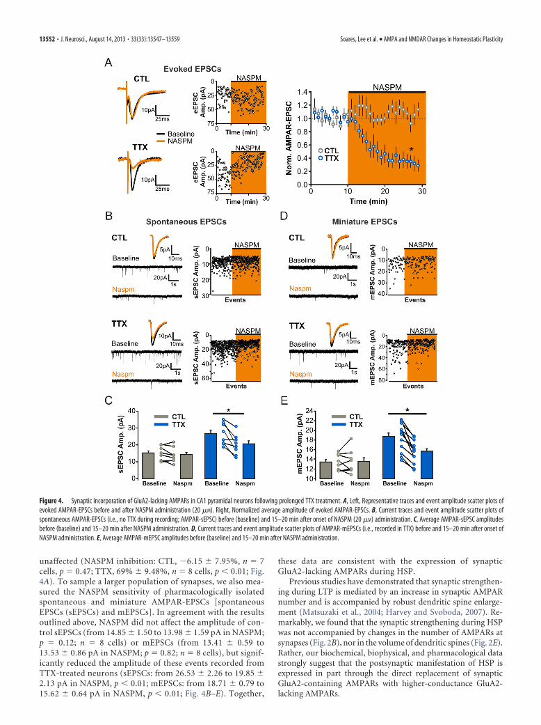
unaffected (NASPM inhibition: CTL, �6.15 7.95%, n � 7cells, p � 0.47; TTX, 69% 9.48%, n � 8 cells, p � 0.01; Fig.4A). To sample a larger population of synapses, we also mea-sured the NASPM sensitivity of pharmacologically isolatedspontaneous and miniature AMPAR-EPSCs [spontaneousEPSCs (sEPSCs) and mEPSCs]. In agreement with the resultsoutlined above, NASPM did not affect the amplitude of con-trol sEPSCs (from 14.85 1.50 to 13.98 1.59 pA in NASPM;p � 0.12; n � 8 cells) or mEPSCs (from 13.41 0.59 to13.53 0.86 pA in NASPM; p � 0.82; n � 8 cells), but signif-icantly reduced the amplitude of these events recorded fromTTX-treated neurons (sEPSCs: from 26.53 2.26 to 19.85 2.13 pA in NASPM, p � 0.01; mEPSCs: from 18.71 0.79 to15.62 0.64 pA in NASPM, p � 0.01; Fig. 4B–E). Together,
these data are consistent with the expression of synapticGluA2-lacking AMPARs during HSP.
Previous studies have demonstrated that synaptic strengthen-ing during LTP is mediated by an increase in synaptic AMPARnumber and is accompanied by robust dendritic spine enlarge-ment (Matsuzaki et al., 2004; Harvey and Svoboda, 2007). Re-markably, we found that the synaptic strengthening during HSPwas not accompanied by changes in the number of AMPARs atsynapses (Fig. 2B), nor in the volume of dendritic spines (Fig. 2E).Rather, our biochemical, biophysical, and pharmacological datastrongly suggest that the postsynaptic manifestation of HSP isexpressed in part through the direct replacement of synapticGluA2-containing AMPARs with higher-conductance GluA2-lacking AMPARs.
Figure 4. Synaptic incorporation of GluA2-lacking AMPARs in CA1 pyramidal neurons following prolonged TTX treatment. A, Left, Representative traces and event amplitude scatter plots ofevoked AMPAR-EPSCs before and after NASPM administration (20 �M). Right, Normalized average amplitude of evoked AMPAR-EPSCs. B, Current traces and event amplitude scatter plots ofspontaneous AMPAR-EPSCs (i.e., no TTX during recording; AMPAR-sEPSC) before (baseline) and 15–20 min after onset of NASPM (20 �M) administration. C, Average AMPAR-sEPSC amplitudesbefore (baseline) and 15–20 min after NASPM administration. D, Current traces and event amplitude scatter plots of AMPAR-mEPSCs (i.e., recorded in TTX) before and 15–20 min after onset ofNASPM administration. E, Average AMPAR-mEPSC amplitudes before (baseline) and 15–20 min after NASPM administration.
13552 • J. Neurosci., August 14, 2013 • 33(33):13547–13559 Soares, Lee et al. • AMPA and NMDAR Changes in Homeostatic Plasticity

Homeostatic switch in synaptic NMDARsubunit compositionThe dynamic nature of NMDAR trafficking and targeting behav-ior at rest, during postnatal development, and during Hebbianplasticity has gained considerable appreciation over the past 2decades (Lau and Zukin, 2007). In part because HSP has over-whelmingly been studied in dissociated neuronal cultures, apreparation that does not lend itself with ease to the study ofNMDAR function, the homeostatic regulation of NMDARs hasbeen far less extensively studied than that of AMPARs (Perez-Otano and Ehlers, 2005). To determine whether alterations inNMDAR function accompanied the homeostatic enhancementin AMPAR function outlined above, we evoked EPSCs whileholding neurons at �70 and �40 mV to compute the ratio of
AMPAR and NMDAR contributions to SC synapse function (seeMaterials and Methods). We found that the ratio of AMPA toNMDA receptor components of eEPSCs was not altered by pro-longed inactivity (CTL: 0.87 0.13, n � 13 cells; TTX: 0.87 0.12, n � 12 cells; p � 0.98; Fig. 5A), consistent with previousfindings in both neuronal cultures and organotypic slicesyounger than those used here (Watt et al., 2000; Arendt et al.,2013). Since AMPAR function was significantly enhanced duringHSP in our experimental conditions (Fig. 1B), this finding sug-gests a concomitant upregulation of both synaptic NMDA andAMPAR function during HSP. In line with this notion, Westernblot analysis of hippocampal slice lysates (Fig. 5B) and surfacebiotinylation experiments (Fig. 5C) revealed an increase in theexpression and surface delivery of all three major NMDAR subunits
Figure 5. Homeostatic upregulation of surface NMDARs in CA1 pyramidal neurons in response to prolonged TTX treatment. A, AMPA/NMDA ratio of evoked EPSCs (see Materials and Methods).B, Representative Western blots and quantification of changes in NMDAR subunit expression, plotted as a TTX/CTL ratio of band intensity, in hippocampal lysates of control and TTX-treated slices.All bands were normalized to �-actin before calculating the TTX/CTL ratio. C, Representative Western blots of biotinylated (surface) and nonbiotinylated (internal) fractions from control andTTX-treated slices. Relative surface expression of NMDA receptor subunits between control and TTX-treated slices plotted as a TTX/CTL ratio of band intensity. D, Change in holding current inducedby bath administration of 50 �M DL-APV while holding the neurons at �40 mV ( p � 0.05, unpaired Student’s t test). These experiments were performed with NBQX, picrotoxin, and TTX in theRinger’s solution. E, Amplitude of the inward current induced by bath administration of NMDA (5 �M; Vm ��60 mV in 0.1 mM Mg 2�). All NMDA bath administration experiments were performedwith NBQX, picrotoxin, and TTX in the Ringer’s solution. F, 2P images of filled CA1 pyramidal neurons were reconstructed using Neuron Studio software (see Materials and Methods). Dendritic length(in micrometers) is plotted for CA1 pyramidal neurons from TTX-treated or control organotypic slices (P7 and 9 –10 DIV) and from age-matched neurons from acute slices (P16 –P17 animals; n �10, 12, and 9 neurons for control, TTX-treated, and acute slices, respectively).
Soares, Lee et al. • AMPA and NMDAR Changes in Homeostatic Plasticity J. Neurosci., August 14, 2013 • 33(33):13547–13559 • 13553
found in the hippocampus in TTX-treatedslices (TTX/CTL ratio for hippocampal ly-sates: GluN1, 1.43 0.09, n � 4; GluN2A,1.52 0.16, n � 6; GluN2B, 1.85 0.13,n � 3; Fig. 5B; TTX/CTL ratio for biotinyl-ated samples: GluN1, 1.31 0.10, n � 10;GluN2A, 1.86 0.35, n � 8; GluN2B,1.69 0.24, n � 15; Fig. 5C).
To further establish whether NMDARfunction is homeostatically regulated inCA1 pyramidal neurons, we gatheredtwo complementary electrophysiologicalreadouts of NMDAR function. First, wereasoned that an upregulation of surfaceNMDARs could be revealed by examiningthe degree of tonic activation of these re-ceptors by ambient levels of extracellularglutamate (Sah et al., 1989). To this end,we monitored changes in whole-cell cur-rent of CA1 pyramidal neurons inducedby bath administration of the NMDARantagonist DL-APV (50 �M; see Materialsand Methods). NMDAR blockade in con-trol neurons induced a small but highlyreproducible change in holding current(15.48 10.86 pA, n � 5 cells), thus re-vealing the presence of an ambient gluta-mate tone in organotypic hippocampalslices (Fig. 5D). Interestingly, the magni-tude of this tonic current was more thanthree times greater in TTX-treated slices(56.15 9.51 pA, n � 7 cells) comparedwith that seen in controls. In principle,this difference could reflect an upregula-tion of surface NMDARs, an alteration inthe regulation of ambient extracellularglutamate concentration, or a combinationof both. To directly measure NMDAR func-tion, we next monitored the whole-cell response to bath administra-tion of NMDA (5 �M for 3 min) and found that NMDA inducedsignificantly larger whole-cell currents in TTX-treated neuronscompared with control (CTL: 128.69 15.44 pA, n � 16 cells; TTX:198.98 21.42 pA, n � 16 cells; p � 0.05; Fig. 5E). This enhance-ment was likely not due to an overall greater membrane surface areain TTX-treated neurons, since the dendritic arborization betweencontrol and TTX-treated neurons was not different (CTL: 732.36 86.43 �m, n � 10 cells; TTX: 787.48 48.56 �m, n � 12 cells; p �0.56; Fig. 5F). These functional and morphological measurementsaligned well with our biochemical data (Fig. 5B,C), and togetherthey demonstrate that prolonged inactivity induced a robustupregulation of surface NMDAR expression in CA1 pyramidalneurons.
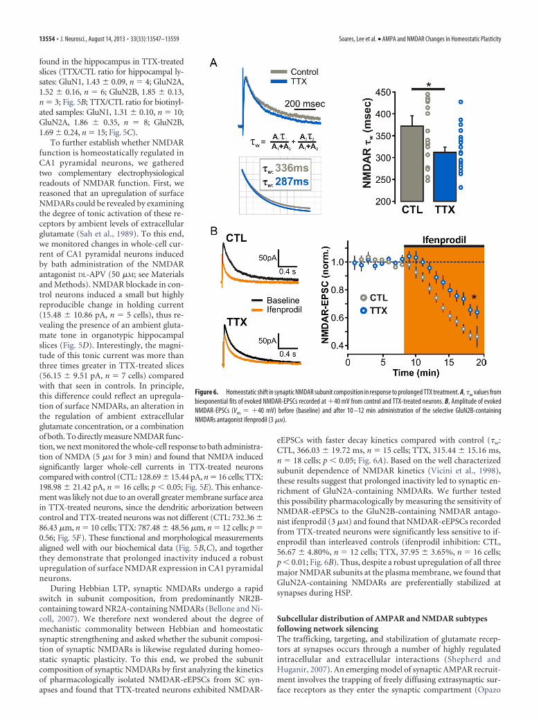
During Hebbian LTP, synaptic NMDARs undergo a rapidswitch in subunit composition, from predominantly NR2B-containing toward NR2A-containing NMDARs (Bellone and Ni-coll, 2007). We therefore next wondered about the degree ofmechanistic commonality between Hebbian and homeostaticsynaptic strengthening and asked whether the subunit composi-tion of synaptic NMDARs is likewise regulated during homeo-static synaptic plasticity. To this end, we probed the subunitcomposition of synaptic NMDARs by first analyzing the kineticsof pharmacologically isolated NMDAR-eEPSCs from SC syn-apses and found that TTX-treated neurons exhibited NMDAR-
eEPSCs with faster decay kinetics compared with control (�w:CTL, 366.03 19.72 ms, n � 15 cells; TTX, 315.44 15.16 ms,n � 18 cells; p � 0.05; Fig. 6A). Based on the well characterizedsubunit dependence of NMDAR kinetics (Vicini et al., 1998),these results suggest that prolonged inactivity led to synaptic en-richment of GluN2A-containing NMDARs. We further testedthis possibility pharmacologically by measuring the sensitivity ofNMDAR-eEPSCs to the GluN2B-containing NMDAR antago-nist ifenprodil (3 �M) and found that NMDAR-eEPSCs recordedfrom TTX-treated neurons were significantly less sensitive to if-enprodil than interleaved controls (ifenprodil inhibition: CTL,56.67 4.80%, n � 12 cells; TTX, 37.95 3.65%, n � 16 cells;p � 0.01; Fig. 6B). Thus, despite a robust upregulation of all threemajor NMDAR subunits at the plasma membrane, we found thatGluN2A-containing NMDARs are preferentially stabilized atsynapses during HSP.
Subcellular distribution of AMPAR and NMDAR subtypesfollowing network silencingThe trafficking, targeting, and stabilization of glutamate recep-tors at synapses occurs through a number of highly regulatedintracellular and extracellular interactions (Shepherd andHuganir, 2007). An emerging model of synaptic AMPAR recruit-ment involves the trapping of freely diffusing extrasynaptic sur-face receptors as they enter the synaptic compartment (Opazo
Figure 6. Homeostatic shift in synaptic NMDAR subunit composition in response to prolonged TTX treatment. A, �w values frombiexponential fits of evoked NMDAR-EPSCs recorded at �40 mV from control and TTX-treated neurons. B, Amplitude of evokedNMDAR-EPSCs (Vm � �40 mV) before (baseline) and after 10 –12 min administration of the selective GluN2B-containingNMDARs antagonist ifenprodil (3 �M).
13554 • J. Neurosci., August 14, 2013 • 33(33):13547–13559 Soares, Lee et al. • AMPA and NMDAR Changes in Homeostatic Plasticity

and Choquet, 2011), and an analogous diffusional trappingmechanism has also been described for NMDARs (Groc et al.,2006; Bard et al., 2010). Moreover, the functional enhancementof AMPAR transmission during LTP is highly dependent on this
reserve pool of nonsynaptic receptors (Makino and Malinow,2009; Granger et al., 2013). Although our electrophysiologicaldata outlined above clearly demonstrate that the subunit compo-sition of synaptic AMPARs (Figs. 3, 4) and NMDARs (Fig. 6) are
Figure 7. Cell-wide homeostatic upregulation of GluA2-lacking AMPARs in response to prolonged TTX treatment. A, Left, 2P image of a control CA1 pyramidal neuron filled with Alexa Fluor 594to visualize dendritic morphology. Scale bar, 15 �m. Right, Enlarged view of an apical dendritic segment with red crosshairs illustrating the site of 2P glutamate uncaging (1 ms at 720 nm). Scalebar, 2 �M. At �70 mV, glutamate uncaging elicits a postsynaptic AMPAR-mediated response, whereas at �40 mV uncaging of glutamate also activates longer-decaying NMDAR EPSCs. B, Top,AMPAR-2P-EPSCs can be generated to match the amplitude of AMPAR-mEPSCs from the same recording. Bottom, Peak scaling of the average traces of AMPAR-2P-EPSCs and AMPAR-mEPSCs revealsa similar rise and decay time course. C, A set of control experiments whereby three uncaging pulses (separated by 500 ms) were elicited at each of the three points illustrated with red crosshairs. Inexperiment b, the uncaging positions of sites 2 and 3 were brought closer to the dendrite to elicit a response mediated by extrasynaptic receptors. Scale bars, 1 �m. D, I–V relationship ofAMPAR-2P-EPSCs generated at distinct subcellular locations. Top, 2P images of secondary apical dendritic segment show sites of glutamate uncaging (red crosshairs). Scale bars: dendrite images,1 �m; soma images, 5 �m. Bottom, AMPAR-2P-EPSCs at different holding potentials (�70 to �40 mV; with 100 �M intracellular spermine) with red arrow depicts the timing of the 1 ms uncagingpulse. E, Average I–V curves of 2P-EPSCs from each subcellular location in both control and TTX conditions. F, Rectification indices for all spine, dendritic, and somatic I–V curves presented in E ( p �0.01; unpaired Student’s t test). G, Rectification indices of 2P-EPSCs generated from pairs of spine and neighboring (�5 �m) extrasynaptic shaft regions. H, Diameters (FWHM) of all dendritic spinesprobed for AMPAR-2P-EPSC I–V relationships.
Soares, Lee et al. • AMPA and NMDAR Changes in Homeostatic Plasticity J. Neurosci., August 14, 2013 • 33(33):13547–13559 • 13555
altered during HSP, it is unclear whetherthese changes reflect synapse-specific regu-lation or rather diffuse, cell-wide, changes insurface glutamate receptor expression. Tospecifically address this issue, we took ad-vantage of subunit-specific biophysical sig-natures of AMPAR and NMDAR subtypesin combination with the ability afforded by2P-uncaging of MNI-Glu to activate gluta-mate receptors at defined subcellular com-partments (Fig. 7A–C).
To determine the spatial extent ofGluA2-lacking AMPAR surface expres-sion, we analyzed the I–V relationship of2P glutamate uncaging-evoked AMPAR-mediated EPSCs (AMPAR-2P-EPSCs) atdendritic spines and nearby (�5 �m) ex-trasynaptic shaft regions of secondaryand tertiary proximal apical dendrites. Inagreement with a previous study in neuro-nal cultures (Beïque et al., 2011), uncagingof MNI-glutamate onto spines and ontoextrasynaptic shaft and somatic regions ofcontrol neurons yielded AMPAR-2P-EPSCs exhibiting linear I–V relationships,although rectifying currents were occasion-ally encountered (rectification index: spine,1.10 0.06, n � 16; dendrite, 1.01 0.08,n � 9; soma, 0.98 0.04, n � 6; Fig. 7D–F).Thus, GluA2-containing AMPARs appearto dominate both synaptic and extrasynap-tic regions of control CA1 neurons. Consis-tent with the upregulation of synapticGluA2-lacking AMPARs (i.e., synapticallyevoked EPSCs; Figs. 3, 4) in response to pro-longed inactivity, we observed strong in-wardly rectifying AMPAR-2P-EPSCs whenuncaging pulses were directed onto the tipsof dendritic spines in TTX-treated neurons(rectification index: 0.76 0.09, n � 12spines; Fig. 7D–F). The changes in the rectifying properties of 2P-EPSCs from spines between control and TTX-treated neurons couldnot be accounted for by an experimental bias toward morphologi-cally dissimilar spines in the treatment groups (p � 0.47; Fig. 7H).Interestingly, inwardly rectifying AMPAR-2P-EPSCs were also de-tected when glutamate was uncaged onto dendritic shafts and so-matic regions of TTX-treated neurons (rectification index: dendrite,0.61 0.11, n � 10; soma, 0.30 0.04, n � 4; Fig. 7D–F). Together,our data suggest that prolonged inactivity drives a robust cell-wideexpression of GluA2-lacking AMPARs at both synaptic and extra-synaptic regions of CA1 pyramidal neurons.
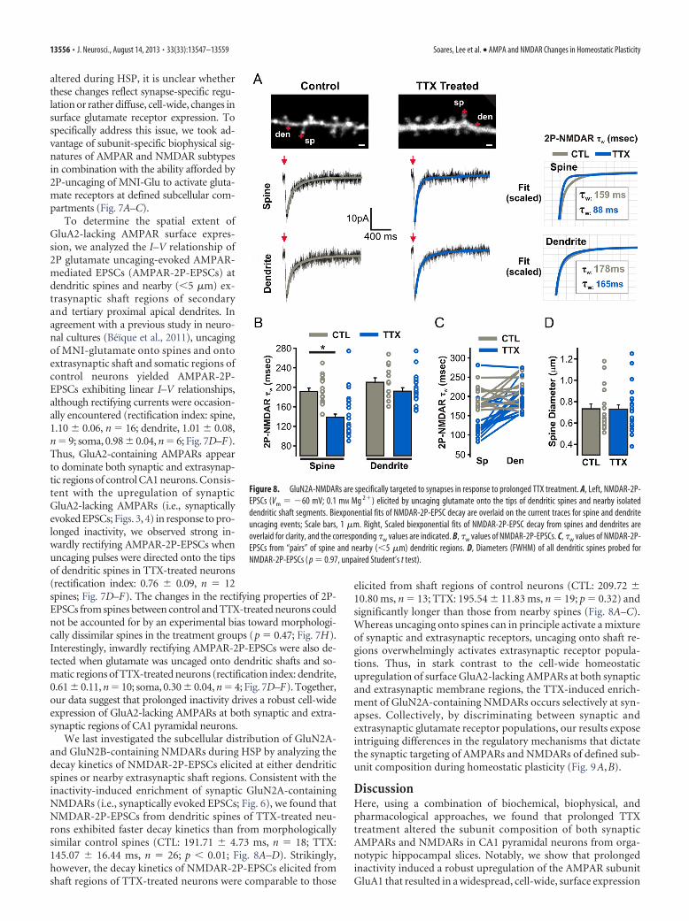
We last investigated the subcellular distribution of GluN2A-and GluN2B-containing NMDARs during HSP by analyzing thedecay kinetics of NMDAR-2P-EPSCs elicited at either dendriticspines or nearby extrasynaptic shaft regions. Consistent with theinactivity-induced enrichment of synaptic GluN2A-containingNMDARs (i.e., synaptically evoked EPSCs; Fig. 6), we found thatNMDAR-2P-EPSCs from dendritic spines of TTX-treated neu-rons exhibited faster decay kinetics than from morphologicallysimilar control spines (CTL: 191.71 4.73 ms, n � 18; TTX:145.07 16.44 ms, n � 26; p � 0.01; Fig. 8A–D). Strikingly,however, the decay kinetics of NMDAR-2P-EPSCs elicited fromshaft regions of TTX-treated neurons were comparable to those
elicited from shaft regions of control neurons (CTL: 209.72 10.80 ms, n � 13; TTX: 195.54 11.83 ms, n � 19; p � 0.32) andsignificantly longer than those from nearby spines (Fig. 8A–C).Whereas uncaging onto spines can in principle activate a mixtureof synaptic and extrasynaptic receptors, uncaging onto shaft re-gions overwhelmingly activates extrasynaptic receptor popula-tions. Thus, in stark contrast to the cell-wide homeostaticupregulation of surface GluA2-lacking AMPARs at both synapticand extrasynaptic membrane regions, the TTX-induced enrich-ment of GluN2A-containing NMDARs occurs selectively at syn-apses. Collectively, by discriminating between synaptic andextrasynaptic glutamate receptor populations, our results exposeintriguing differences in the regulatory mechanisms that dictatethe synaptic targeting of AMPARs and NMDARs of defined sub-unit composition during homeostatic plasticity (Fig. 9A,B).
DiscussionHere, using a combination of biochemical, biophysical, andpharmacological approaches, we found that prolonged TTXtreatment altered the subunit composition of both synapticAMPARs and NMDARs in CA1 pyramidal neurons from orga-notypic hippocampal slices. Notably, we show that prolongedinactivity induced a robust upregulation of the AMPAR subunitGluA1 that resulted in a widespread, cell-wide, surface expression
Figure 8. GluN2A-NMDARs are specifically targeted to synapses in response to prolonged TTX treatment. A, Left, NMDAR-2P-EPSCs (Vm � �60 mV; 0.1 mM Mg 2�) elicited by uncaging glutamate onto the tips of dendritic spines and nearby isolateddendritic shaft segments. Biexponential fits of NMDAR-2P-EPSC decay are overlaid on the current traces for spine and dendriteuncaging events; Scale bars, 1 �m. Right, Scaled biexponential fits of NMDAR-2P-EPSC decay from spines and dendrites areoverlaid for clarity, and the corresponding �w values are indicated. B, �w values of NMDAR-2P-EPSCs. C, �w values of NMDAR-2P-EPSCs from “pairs” of spine and nearby (�5 �m) dendritic regions. D, Diameters (FWHM) of all dendritic spines probed forNMDAR-2P-EPSCs ( p � 0.97, unpaired Student’s t test).
13556 • J. Neurosci., August 14, 2013 • 33(33):13547–13559 Soares, Lee et al. • AMPA and NMDAR Changes in Homeostatic Plasticity

of GluA2-lacking AMPARs. Remarkably, despite inducing a ro-bust and generalized upregulation of the three major hippocam-pal NMDAR subunits, network silencing triggered a switch inthe subunit composition of solely the synaptic population ofNMDARs, leaving unaltered the composition of their extrasyn-aptic counterparts. Altogether, these findings highlight the no-tion that the homeostatic mechanisms used by neurons to adjusttheir excitability levels regulate synapse function in ways beyondsolely modifying synaptic strength per se.
A number of previous studies have reported conflicting evi-dence regarding the subunit composition of AMPARs involved inhomeostatic synaptic potentiation. A recent review (Lee, 2012a)attempted to reconcile these discrepancies by documenting dif-ferences in the pharmacological paradigms used to induce HSP.Specifically, it was highlighted that the selective regulation ofGluA1 occurred after prolonged blockade of both network activ-ity (i.e., TTX) and NMDARs (Ju et al., 2004; Sutton et al., 2006;Aoto et al., 2008), whereas both GluA1 and GluA2 expressionwere affected when neurons were treated with TTX alone(O’Brien et al., 1998; Gainey et al., 2009; Anggono et al., 2011). Incontrast to this unifying picture, we provide here a number ofcomplementary and converging lines of evidence indicating thatTTX treatment alone led to a robust and selective upregulation ofGluA1 expression and formation of GluA2-lacking AMPARs inCA1 pyramidal neurons in an organotypic slice preparation. Thedirect replacement of GluA2-containing AMPARs with higher-conductance GluA2-lacking AMPARs during homeostatic plas-ticity offers an effective means to enhance synaptic strengthwithout the need to increase receptor number or to increase spinevolume. This scenario is consistent with homeostatic plasticityoccurring at single synapses (Beïque et al., 2011) and is in linewith manifestations of homeostatic synaptic plasticity in vivo (Heet al., 2012).
It is pertinent to compare and contrast the mechanistic under-pinnings of Hebbian and homeostatic synaptic strengthening,including those involving subunit composition of glutamate re-ceptors. While a transient insertion of GluA2-lacking AMPARshas been observed following LTP induction (Plant et al., 2006;Guire et al., 2008), the role of this particular subtype of AMPARs
in LTP is controversial (Adesnik and Nicoll, 2007; Gray et al.,2007). Likewise, the implication of GluA2-lacking AMPARs inhomeostatic synaptic strengthening is also debated, as outlinedabove. These divergences may reflect the presence of distinct syn-aptic plasticity mechanisms that are heavily dependent on subtle-ties in experimental conditions and paradigms. Nevertheless, thehomeostatic switch in NMDAR subunit composition we reporthere is highly analogous to that previously shown to occur duringHebbian LTP (Bellone and Nicoll, 2007). Indeed, LTP was shownto be accompanied by a GluN2B-containing toward GluN2A-containing NMDAR subunit switch that exhibited a time coursehighly similar to that of the synaptic delivery of AMPARs.Although our results provide limited insights into the precisetime course of the inactivity-induced subunit switches forboth AMPARs and NMDARs, these homeostatic adaptive mech-anisms might be occurring simultaneously. It is thus tempting tospeculate that both Hebbian and homeostatic synaptic strength-ening use common mechanisms involving the concerted upregu-lation of AMPARs and GluN2A-containing NMDARs. Futurestudies will be required to substantiate this possibility and furtherestablish the extent of the molecular commonalities betweenHebbian and homeostatic synaptic plasticity.
The homeostatic adjustments reported here for both AMPARand NMDAR subunit composition likely influence Hebbian plas-ticity rules. Indeed, in vivo visual deprivation paradigms that leadto homeostatic upregulation of synapse function in visual cortex(Goel and Lee, 2007; Gao et al., 2010) impart a metaplastic influ-ence that modifies the stimulus threshold for inducing LTP andLTD (Philpot et al., 2001, 2003), and spike-timing-dependentsynaptic plasticity (Guo et al., 2012). These changes appear tobe caused by an increased proportion of GluN2B-containingNMDARs at synapses, in an apparent contrast to what we reporthere. Whereas the various in vivo visual deprivation paradigmsreduce thalamic synaptic input into visual cortex, it is unclear towhat extent they reduce overall network excitability in the visualcortex. Thus, it is possible that the GluN2A enrichment we reporthere following prolonged TTX treatment represents a homeo-static response to prolonged neuronal silencing, whereas theGluN2B enrichment observed following visual deprivation re-
Figure 9. Differential subcellular targeting of glutamate receptor subtypes during HSP. A, Top, AMPARs containing the GluA2 subunit predominate at both synaptic and extrasynaptic regions ofCA1 pyramidal neurons. Bottom, When network activity is silenced by prolonged TTX treatment, there is a homeostatic upregulation of GluA2-lacking AMPARs in both synaptic and extrasynapticcompartments of the neuronal membrane. B, Top, CA1 pyramidal neurons display a mixed population of NMDARs containing both GluN2A and GluN2B subunits. Bottom, When network activity issilenced by prolonged TTX treatment, there is an indiscriminate increase in surface NMDARs subunits; however, GluN2A-containing NMDARs are preferentially localized/stabilized at synapses.
Soares, Lee et al. • AMPA and NMDAR Changes in Homeostatic Plasticity J. Neurosci., August 14, 2013 • 33(33):13547–13559 • 13557

flects a homeostatic response to a reduction in presynaptic activ-ity. In support of this idea, selective presynaptic silencing ofindividual synapses has recently been shown to cause postsynap-tic GluN2B enrichment (Lee et al., 2010). Conversely, the pres-ence of calcium-permeable GluA2-lacking AMPARs followingprolonged inactivity may also convey metaplastic influences tosynapses, either by lowering the threshold for Hebbian synapticpotentiation, or even by imparting anti-Hebbian features (Lamsaet al., 2007). Future studies are required to better understand theinfluence of glutamate receptor composition on synaptic plastic-ity rules.
AMPARs and NMDARs of different subunit composition aredifferentially localized to synaptic and extrasynaptic membranecompartments. For instance, AMPARs containing GluA2/GluA3subunits are found almost exclusively at synapses, whereasGluA1/GluA2-AMPARs occupy both synaptic and extrasynapticmembrane regions (Beïque and Huganir, 2009; Lu et al., 2009).Moreover, GluN2A-containing NMDARs are believed to bepreferentially stabilized at synapses over GluN2B-containingNMDARs (Groc et al., 2006). These subcellular distribution pro-files are thought to arise through preferential interactions of spe-cific AMPAR and NMDAR subunits, and/or auxiliary subunits,with PSD scaffolding proteins at synapses (Lau and Zukin, 2007;Shepherd and Huganir, 2007; Jackson and Nicoll, 2011). Thedifferential subcellular distribution of AMPAR and NMDAR ex-pression during HSP that we have described can be traced, at leastin part, to the changes in subunit protein expression. Specifically,the selective upregulation of GluA1 protein expression (overGluA2) was accompanied by a widespread enhancement of sur-face GluA2-lacking AMPARs, evident at both dendritic spinesand extrasynaptic membrane regions. Such a cell-wide upregula-tion of AMPARs offers an effective means to account for theremarkable multiplicativity of homeostatic synaptic strengthen-ing triggered by a somatic homeostatic sensing mechanism (Leeet al., 2013). Interestingly, the TTX-induced increase in NMDARprotein expression (both total and surface expression) was notsubunit selective, as we detected an upregulation of GluN1,GluN2A, and GluN2B. Despite this generalized increase inNMDAR surface expression, synapses were specifically enrichedwith GluN2A-containing NMDARs during HSP, likely reflectingthe preferential synaptic stabilization of this subunit comparedwith GluN2B-containing NMDARs. Thus, whereas the synapticincorporation of GluA2-lacking AMPARs likely results from thebulk loading of these receptors onto the plasma membrane, theselective synaptic stabilization of GluN2A-containing NMDARsduring HSP emphasizes the competitive interactions of GluN2Asubunits for synaptic anchoring/scaffolding proteins.
The functional importance of extrasynaptic receptors isincreasingly being recognized. For instance, extrasynapticAMPARs and NMDARs can be recruited to and/or exchangedwith synaptic receptor populations in a dynamic and highly reg-ulated manner. Recent studies have shown that extrasynapticAMPARs can shape synaptic transmission (Heine et al., 2008)and are required for LTP (Makino and Malinow, 2009; Granger etal., 2013). Moreover, extrasynaptic NMDARs can powerfully in-fluence synaptic integration (Chalifoux and Carter, 2011; Lee,2012b) and differentially regulate neuronal survival and deathsignaling pathways (Hardingham and Bading, 2003). The ho-meostatic regulation of the number and subunit composition ofextrasynaptic glutamate receptors described here will, in princi-ple, influence all of the functions ascribed to this population ofreceptors, thus broadening the functional implications of the ho-meostatic process.
Both in vitro and in vivo manifestations of homeostatic synap-tic plasticity have been documented using several experimentalparadigms. Homeostatic synapse regulation operates continuously“online” to enable tuning of cellular excitability in the face of perpet-ual alterations in neuronal firing activity. We have demonstratedthat, in addition to triggering robust synaptic strengthening, the ho-meostatic process also involves changes in the subunit compositionand subcellular distribution of both AMPARs and NMDARs. Thus,the homeostatic adjustment of synapse function is not limited to theregulation of synaptic strength, but likely impacts synaptic proper-ties such as temporal integration of synaptic input and calcium-dependent biochemical signaling. Future studies will be requiredto fully grasp the functional implications of these homeostaticregulations.
ReferencesAdesnik H, Nicoll RA (2007) Conservation of glutamate receptor
2-containing AMPA receptors during long-term potentiation. J Neurosci27:4598 – 4602. CrossRef Medline
Anggono V, Clem RL, Huganir RL (2011) PICK1 loss of function occludeshomeostatic synaptic scaling. J Neurosci 31:2188 –2196. CrossRefMedline
Aoto J, Nam CI, Poon MM, Ting P, Chen L (2008) Synaptic signaling byall-trans retinoic acid in homeostatic synaptic plasticity. Neuron 60:308 –320. CrossRef Medline
Arendt KL, Sarti F, Chen L (2013) Chronic inactivation of a neural circuitenhances LTP by inducing silent synapse formation. J Neurosci 33:2087–2096. CrossRef Medline
Bard L, Sainlos M, Bouchet D, Cousins S, Mikasova L, Breillat C, StephensonFA, Imperiali B, Choquet D, Groc L (2010) Dynamic and specific inter-action between synaptic NR2-NMDA receptor and PDZ proteins. ProcNatl Acad Sci U S A 107:19561–19566. CrossRef Medline
Beïque JC, Huganir RL (2009) AMPA receptor subunits get their share ofthe pie. Neuron 62:165–168. CrossRef Medline
Beïque JC, Lin DT, Kang MG, Aizawa H, Takamiya K, Huganir RL (2006)Synapse-specific regulation of AMPA receptor function by PSD-95. ProcNatl Acad Sci U S A 103:19535–19540. CrossRef Medline
Beïque JC, Na Y, Kuhl D, Worley PF, Huganir RL (2011) Arc-dependentsynapse-specific homeostatic plasticity. Proc Natl Acad Sci U S A 108:816 – 821. CrossRef Medline
Bellone C, Nicoll RA (2007) Rapid bidirectional switching of synapticNMDA receptors. Neuron 55:779 –785. CrossRef Medline
Bowie D, Mayer ML (1995) Inward rectification of both AMPA and kainatesubtype glutamate receptors generated by polyamine-mediated ion chan-nel block. Neuron 15:453– 462. CrossRef Medline
Chalifoux JR, Carter AG (2011) Glutamate spillover promotes the genera-tion of NMDA spikes. J Neurosci 31:16435–16446. CrossRef Medline
Gainey MA, Hurvitz-Wolff JR, Lambo ME, Turrigiano GG (2009) Synapticscaling requires the GluR2 subunit of the AMPA receptor. J Neurosci29:6479 – 6489. CrossRef Medline
Gao M, Sossa K, Song L, Errington L, Cummings L, Hwang H, Kuhl D,Worley P, Lee HK (2010) A specific requirement of Arc/Arg3.1 for visualexperience-induced homeostatic synaptic plasticity in mouse primary vi-sual cortex. J Neurosci 30:7168 –7178. CrossRef Medline
Goel A, Lee HK (2007) Persistence of experience-induced homeostatic syn-aptic plasticity through adulthood in superficial layers of mouse visualcortex. J Neurosci 27:6692– 6700. CrossRef Medline
Granger AJ, Shi Y, Lu W, Cerpas M, Nicoll RA (2013) LTP requires a reservepool of glutamate receptors independent of subunit type. Nature 493:495–500. CrossRef Medline
Gray EE, Fink AE, Sarinana J, Vissel B, O’Dell TJ (2007) Long-term poten-tiation in the hippocampal CA1 region does not require insertion andactivation of GluR2-lacking AMPA receptors. J Neurophysiol 98:2488 –2492. CrossRef Medline
Groc L, Heine M, Cousins SL, Stephenson FA, Lounis B, Cognet L, Choquet D(2006) NMDA receptor surface mobility depends on NR2A-2B subunits.Proc Natl Acad Sci U S A 103:18769 –18774. CrossRef Medline
Groth RD, Lindskog M, Thiagarajan TC, Li L, Tsien RW (2011) Beta Ca2�/CaM-dependent kinase type II triggers upregulation of GluA1 to coordi-
13558 • J. Neurosci., August 14, 2013 • 33(33):13547–13559 Soares, Lee et al. • AMPA and NMDAR Changes in Homeostatic Plasticity

nate adaptation to synaptic inactivity in hippocampal neurons. Proc NatlAcad Sci U S A 108:828 – 833. CrossRef Medline
Guire ES, Oh MC, Soderling TR, Derkach VA (2008) Recruitment ofcalcium-permeable AMPA receptors during synaptic potentiation is reg-ulated by CaM-kinase I. J Neurosci 28:6000 – 6009. CrossRef Medline
Guo Y, Huang S, de Pasquale R, McGehrin K, Lee HK, Zhao K, Kirkwood A(2012) Dark exposure extends the integration window for spike-timing-dependent plasticity. J Neurosci 32:15027–15035. CrossRef Medline
Hardingham GE, Bading H (2003) The yin and yang of NMDA receptorsignalling. Trends Neurosci 26:81– 89. CrossRef Medline
Hartveit E, Veruki ML (2007) Studying properties of neurotransmitter re-ceptors by non-stationary noise analysis of spontaneous postsynaptic cur-rents and agonist-evoked responses in outside-out patches. Nat Protoc2:434 – 448. CrossRef Medline
Harvey CD, Svoboda K (2007) Locally dynamic synaptic learning rules inpyramidal neuron dendrites. Nature 450:1195–1200. CrossRef Medline
He K, Petrus E, Gammon N, Lee HK (2012) Distinct sensory requirementsfor unimodal and cross-modal homeostatic synaptic plasticity. J Neurosci32:8469 – 8474. CrossRef Medline
Heine M, Groc L, Frischknecht R, Beïque JC, Lounis B, Rumbaugh G,Huganir RL, Cognet L, Choquet D (2008) Surface mobility of postsyn-aptic AMPARs tunes synaptic transmission. Science 320:201–205.CrossRef Medline
Jackson AC, Nicoll RA (2011) The expanding social network of ionotropicglutamate receptors: TARPs and other transmembrane auxiliary sub-units. Neuron 70:178 –199. CrossRef Medline
Ju W, Morishita W, Tsui J, Gaietta G, Deerinck TJ, Adams SR, Garner CC,Tsien RY, Ellisman MH, Malenka RC (2004) Activity-dependent regu-lation of dendritic synthesis and trafficking of AMPA receptors. Nat Neu-rosci 7:244 –253. CrossRef Medline
Kessels HW, Malinow R (2009) Synaptic AMPA receptor plasticity and be-havior. Neuron 61:340 –350. CrossRef Medline
Kim J, Tsien RW (2008) Synapse-specific adaptations to inactivity in hip-pocampal circuits achieve homeostatic gain control while dampeningnetwork reverberation. Neuron 58:925–937. CrossRef Medline
Kim J, Tsien RW, Alger BE (2012) An improved test for detecting multipli-cative homeostatic synaptic scaling. PloS One 7:e37364. CrossRefMedline
Lamsa KP, Heeroma JH, Somogyi P, Rusakov DA, Kullmann DM (2007)Anti-Hebbian long-term potentiation in the hippocampal feedback in-hibitory circuit. Science 315:1262–1266. CrossRef Medline
Lau CG, Zukin RS (2007) NMDA receptor trafficking in synaptic plasticityand neuropsychiatric disorders. Nat Rev Neurosci 8:413– 426. CrossRefMedline
Lazar A, Pipa G, Triesch J (2009) SORN: a self-organizing recurrent neuralnetwork. Front Comput Neurosci 3:23. CrossRef Medline
Lee HK (2012a) Ca-permeable AMPA receptors in homeostatic synapticplasticity. Front Mol Neurosci 5:17. CrossRef Medline
Lee KF (2012b) A unique mechanism of NMDA spike initiation supports adistinct role in synaptic input integration. J Neurosci 32:2913–2914.CrossRef Medline
Lee KF, Soares C, Beique JC (2013) Tuning into diversity of homeostaticsynaptic plasticity. Neuropharmacology. Advance online publication. Re-trieved July 18, 2013. doi:10.1016/j.neuropharm.2013.03.016. CrossRefMedline
Lee MC, Yasuda R, Ehlers MD (2010) Metaplasticity at single glutamatergicsynapses. Neuron 66:859 – 870. CrossRef Medline
Lu W, Shi Y, Jackson AC, Bjorgan K, During MJ, Sprengel R, Seeburg PH,Nicoll RA (2009) Subunit composition of synaptic AMPA receptors re-vealed by a single-cell genetic approach. Neuron 62:254 –268. CrossRefMedline
Makino H, Malinow R (2009) AMPA receptor incorporation into synapsesduring LTP: the role of lateral movement and exocytosis. Neuron 64:381–390. CrossRef Medline
Matsuzaki M, Ellis-Davies GC, Nemoto T, Miyashita Y, Iino M, Kasai H (2001)Dendritic spine geometry is critical for AMPA receptor expression in hip-pocampal CA1 pyramidal neurons. Nat Neurosci 4:1086–1092. CrossRefMedline
Matsuzaki M, Honkura N, Ellis-Davies GC, Kasai H (2004) Structural basisof long-term potentiation in single dendritic spines. Nature 429:761–766.CrossRef Medline
O’Brien RJ, Kamboj S, Ehlers MD, Rosen KR, Fischbach GD, Huganir RL(1998) Activity-dependent modulation of synaptic AMPA receptor ac-cumulation. Neuron 21:1067–1078. CrossRef Medline
Opazo P, Choquet D (2011) A three-step model for the synaptic recruit-ment of AMPA receptors. Mol Cell Neurosci 46:1– 8. CrossRef Medline
Perez-Otano I, Ehlers MD (2005) Homeostatic plasticity and NMDA recep-tor trafficking. Trends Neurosci 28:229 –238. CrossRef Medline
Philpot BD, Sekhar AK, Shouval HZ, Bear MF (2001) Visual experience anddeprivation bidirectionally modify the composition and function ofNMDA receptors in visual cortex. Neuron 29:157–169. CrossRef Medline
Philpot BD, Espinosa JS, Bear MF (2003) Evidence for altered NMDA re-ceptor function as a basis for metaplasticity in visual cortex. J Neurosci23:5583–5588. Medline
Plant K, Pelkey KA, Bortolotto ZA, Morita D, Terashima A, McBain CJ,Collingridge GL, Isaac JT (2006) Transient incorporation of nativeGluR2-lacking AMPA receptors during hippocampal long-term potenti-ation. Nat Neurosci 9:602– 604. CrossRef Medline
Sah P, Hestrin S, Nicoll RA (1989) Tonic activation of NMDA receptorsby ambient glutamate enhances excitability of neurons. Science 246:815– 818. CrossRef Medline
Shepherd JD, Huganir RL (2007) The cell biology of synaptic plasticity:AMPA receptor trafficking. Annu Rev Cell Dev Biol 23:613– 643.CrossRef Medline
Stoppini L, Buchs PA, Muller D (1991) A simple method for organotypiccultures of nervous tissue. J Neurosci Methods 37:173–182. CrossRefMedline
Sutton MA, Ito HT, Cressy P, Kempf C, Woo JC, Schuman EM (2006) Min-iature neurotransmission stabilizes synaptic function via tonic suppres-sion of local dendritic protein synthesis. Cell 125:785–799. CrossRefMedline
Thiagarajan TC, Lindskog M, Tsien RW (2005) Adaptation to synaptic in-activity in hippocampal neurons. Neuron 47:725–737. CrossRef Medline
Traynelis SF, Silver RA, Cull-Candy SG (1993) Estimated conductance ofglutamate receptor channels activated during EPSCs at the cerebellarmossy fiber-granule cell synapse. Neuron 11:279 –289. CrossRef Medline
Turrigiano GG (2008) The self-tuning neuron: synaptic scaling of excitatorysynapses. Cell 135:422– 435. CrossRef Medline
Turrigiano GG, Leslie KR, Desai NS, Rutherford LC, Nelson SB (1998)Activity-dependent scaling of quantal amplitude in neocortical neurons.Nature 391:892– 896. CrossRef Medline
Tyler WJ, Pozzo-Miller L (2003) Miniature synaptic transmission andBDNF modulate dendritic spine growth and form in rat CA1 neurones.J Physiol 553:497–509. CrossRef Medline
Vicini S, Wang JF, Li JH, Zhu WJ, Wang YH, Luo JH, Wolfe BB, Grayson DR(1998) Functional and pharmacological differences between recombi-nant N-methyl-D-aspartate receptors. J Neurophysiol 79:555–566.Medline
Watt AJ, van Rossum MC, MacLeod KM, Nelson SB, Turrigiano GG (2000)Activity coregulates quantal AMPA and NMDA currents at neocorticalsynapses. Neuron 26:659 – 670. CrossRef Medline
Soares, Lee et al. • AMPA and NMDAR Changes in Homeostatic Plasticity J. Neurosci., August 14, 2013 • 33(33):13547–13559 • 13559