development/plasticity/repair ... · pdf filedevelopment/plasticity/repair...
TRANSCRIPT

Development/Plasticity/Repair
Epigenetic Gene Silencing Underlies C-Fiber Dysfunctionsin Neuropathic Pain
Hitoshi Uchida, Lin Ma, and Hiroshi UedaDivision of Molecular Pharmacology and Neuroscience, Nagasaki University Graduate School of Biomedical Sciences, Nagasaki 852-8521, Japan
Peripheral nerve injury causes neuropathic pain, which is characterized by the paradoxical sensations of positive and negative symptoms.Clinically, negative signs are frequently observed; however, their underlying molecular mechanisms are largely unknown. Dysfunction ofC-fibers is assumed to underlie negative symptoms and is accompanied by long-lasting downregulation of Nav1.8 sodium channel and�-opioid receptor (MOP) in the dorsal root ganglion (DRG). In the present study, we found that nerve injury upregulates neuron-restrictive silencer factor (NRSF) expression in the DRG neurons mediated through epigenetic mechanisms. In addition, chromatinimmunoprecipitation analysis revealed that nerve injury promotes NRSF binding to the neuron-restrictive silencer element within MOPand Nav1.8 genes, thereby causing epigenetic silencing. Furthermore, NRSF knockdown significantly blocked nerve injury-induceddownregulations of MOP and Nav1.8 gene expressions, C-fiber hypoesthesia, and the losses of peripheral morphine analgesia andNav1.8-selective blocker-induced hypoesthesia. Together, these data suggest that NRSF causes pathological and pharmacological dys-function of C-fibers, which underlies the negative symptoms in neuropathic pain.
IntroductionNeuropathic pain is characterized by the paradoxical sensationsof positive (hyperalgesia, allodynia, and paresthesia) and negative(hypoesthesia, hypoalgesia) symptoms (Baron, 2006), and nega-tive signs are frequently observed during clinical sensory exami-nations (Devigili et al., 2008; Leffler and Hansson, 2008). Themolecular mechanisms underlying positive symptoms have beenextensively investigated (Devor, 2006; Costigan et al., 2009);however, those underlying negative symptoms are much less wellunderstood. A possible mechanism for negative symptoms is adysfunction of small-diameter (C)-fibers (Taylor, 2001; Devigiliet al., 2008; Costigan et al., 2009), such as a loss of C-fiber termi-nals, an impairment of C-fiber-mediated axon-reflex flare re-sponses, or an increase in the threshold against C-fiber-specificstimuli (Fields et al., 1998; Ueda, 2008). Such C-fiber dysfunc-tions have been implicated in the manifestation of positive symp-toms as well as of negative ones, possibly through a synapticreorganization in the spinal dorsal horn (Taylor, 2001; Ueda,2008; Costigan et al., 2009). Representative examples for negativesymptoms were observed with long-lasting downregulations ofNav1.8 sodium channel and �-opioid receptor (MOP) inC-fibers (Waxman et al., 1999; Rashid et al., 2004; Kohno et al.,2005), which are essential for C-fiber functions in terms of deter-mining pain thresholds (Akopian et al., 1999) and for the phar-
macological actions of �-opioids (Dickenson and Kieffer, 2006),respectively.
In terms of long-lasting transcriptional regulation, the tran-scription factor-mediated epigenetic mechanisms have beendemonstrated to play a key role (Borrelli et al., 2008). Neuron-restrictive silencer factor (NRSF, also known as REST) functionsas a transcriptional repressor of genes that contain neuron-restrictive silencer element (NRSE, also called RE1) (Chong et al.,1995; Schoenherr and Anderson, 1995). NRSF, when it binds toNRSE, recruits histone deacetylase (HDAC) through its core-pressors, mSin3 and CoREST, for generating a repressive chro-matin environment (Ballas and Mandel, 2005). It has beenreported that NRSF represses transcription of MOP gene throughHDAC-mediated mechanisms (Kim et al., 2004). Here, we showthat nerve injury induces a long-lasting NRSF expression in thedorsal root ganglion (DRG), thereby causing epigenetic silencingof MOP gene and loss of pharmacological target for peripheralmorphine analgesia. Furthermore, we also investigated the pos-sible epigenetic silencing of Nav1.8 gene, which has unique for-ward and reverse NRSE sequences.
Materials and MethodsAnimals and surgery. Male C57BL/6J mice weighing 20 –25 g were used.They were kept in a room with a temperature of 21 � 2°C with ad libitumaccess to a standard laboratory diet and tap water. All procedures wereapproved by the Nagasaki University Animal Care Committee (Na-gasaki, Japan) and complied with the recommendations of the Interna-tional Association for the Study of Pain (Zimmermann, 1983). Partialligation of the sciatic nerve was performed under pentobarbital (50 mg/kg) anesthesia, following the methods of Malmberg and Basbaum(1998).
Oligonucleotide treatments. The antisense oligodeoxynucleotide(AS-ODN) was designed to target the mouse NRSF sequence andcorresponds to the rat sequence targeted for antisense knockdownpreviously (Calderone et al., 2003). AS-ODN (5�-CGGAAGGGCTT-
Received Nov. 10, 2009; revised Feb. 8, 2010; accepted Feb. 20, 2010.This work was supported by Ministry of Education, Culture, Sports, Science, and Technology Grant-In-Aid for
Scientific Research 17109015 (H. Ueda). Health Sciences Research Grants from the Ministry of Health, Labor, andWelfare of Japan (H. Ueda) and Health Labor Sciences Research Grant “Third Term Comprehensive Control Researchfor Cancer” (398-49) also supported this work. We thank W. Xie, K. Sasaki, and T. Yamasaki for technical help.
Correspondence should be addressed to Dr. Hiroshi Ueda, Division of Molecular Pharmacology and Neuroscience,Nagasaki University Graduate School of Biomedical Sciences, 1-14 Bunkyo-machi, Nagasaki 852-8521, Japan.E-mail: [email protected].
DOI:10.1523/JNEUROSCI.5541-09.2010Copyright © 2010 the authors 0270-6474/10/304806-09$15.00/0
4806 • The Journal of Neuroscience, March 31, 2010 • 30(13):4806 – 4814
GGCC-3�) and its mismatch scrambled oligodeoxynucleotide (MS-ODN;5�-GTCGTCGGCGGAGCA-3�) were synthesized and freshly dissolvedin artificial CSF (aCSF) containing the following (in mM): 125 NaCl, 3.8KCl, 2.0 CaCl2, 1.0 MgCl2, 1.2 KH2PO4, 26 NaHCO3, 10 glucose, pH 7.4).AS-ODN or MS-ODN was intrathecally injected at a dose of 10 �g per 5 �lof aCSF on the first, third, and fifth days. Then, nerve injury was performedwith subsequent injections of AS-ODN on days 1, 3, 5, and 6 postinjury. ThemRNA levels, pain thresholds, peripheral morphine analgesia, and periph-eral A-803467 hypoesthesia were assessed at day 7 postinjury.
Nociception test. In thermal paw withdrawal tests, the nociceptionthreshold was evaluated by the latency of paw withdrawal upon a thermalstimulus (Hargreaves et al., 1988; Inoue et al., 2004). Unanesthetizedanimals were placed in Plexiglas cages on top of a glass sheet, and anadaptation period of 1 h was allowed. The thermal stimulator (IITC LifeScience) was positioned under the glass sheet and the focus of the pro-jection bulb was aimed exactly at the middle of the plantar surface of theanimal. A mirror attached to the stimulator permitted visualization ofthe plantar surface. A cutoff time of 20 s was set to prevent tissue damage.The mechanical paw pressure test was performed as described previously(Rashid et al., 2003; Inoue et al., 2004). Briefly, mice were placed in aPlexiglas chamber on a 6 � 6 mm wire mesh grid floor and allowed toacclimatize for a period of 1 h. A mechanical stimulus was then deliveredto the middle of the plantar surface of the right hindpaw using a trans-ducer indicator (model 1601; IITC Life Science). The pressure needed toinduce a flexor response was defined as the pain threshold. A cutoffpressure of 20 g was set to avoid tissue damage. In these experiments,using mechanical and thermal tests, the thresholds were determinedfrom three repeated challenges at 10 min intervals, and the averages ofresponses were evaluated. An electrical stimulation-induced paw with-drawal (EPW) test was performed as described previously (Matsumoto etal., 2008). Briefly, electrodes (Neurotron) were fastened to the plantarsurfaces and insteps of mice. Transcutaneous nerve stimuli with each ofthe three sine-wave pulses (5, 250, and 2000 Hz) were applied using aNeurometer CPT/C system (Neurotron). The minimum intensity (�A)at which each mouse withdrew its paw was defined as the current stim-ulus threshold. Investigators blind to drug treatments performed all be-havioral experiments.
Drug treatments. Morphine hydrochloride (Takeda Chemical Indus-tries) was dissolved in physiological saline. Saline was used for controlinjections. Intraplantar injections were given using a Hamilton microsy-ringe connected to polyethylene tubing with a 30 gauge hypodermicneedle. For the time course experiment, we measured the paw-withdrawallatencies at every 10 min interval until 60 min after intraplantar injection ofmorphine (30 nmol), as reported previously (Rashid et al., 2004). In thearea under the curve analysis of peripheral morphine analgesia, we cal-culated the area under the curve generated by plotting analgesic thresh-old (after deducting the control threshold from each threshold point)against time, from 10 to 60 min after morphine treatment, using a trap-ezoidal method. A-803467 (Biomol), a selective blocker for Nav1.8(Jarvis et al., 2007), was dissolved in dimethyl sulfoxide. Before admin-istration, A-803467 was diluted 15-fold for intraperitoneal injection and28-fold for intraplantar injection in saline, respectively. The EPW testwas performed 30 min after intraperitoneal injection of A-803467 (10mg/kg), as reported previously (Jarvis et al., 2007).
Quantitative real-time PCR. Total RNA was extracted from L4-6 DRGsusing TRIzol (Invitrogen), and 500 ng of RNA was used for cDNA syn-thesis. Quantitative real-time PCR was performed with qPCR MasterMixPlus for SYBR Green I (Eurogentec) using the ABI Prism 7000 sequencedetection system (Applied Biosystems). The PCR primers used are listedin supplemental Table 1, available at www.jneurosci.org as supplementalmaterial. Some of the primers were published previously (Koenigsbergeret al., 2000; Klein et al., 2003; Qiang et al., 2005; Matsumoto et al., 2006;Cheng et al., 2009; Staaf et al., 2009). Glyceraldehyde-3-phosphate dehy-drogenase (GAPDH) was used as an internal control for normalization.In all cases, the validity of amplification was confirmed by the presence ofa single peak in the melting temperature analysis and by linear amplifi-cation with increasing number of PCR cycles.
Western blot analysis. The L4-6 DRGs from three mice were pooled.DRG samples were homogenized twice in ice-cold cell lysis buffer [10 mM
Tris-HCl, pH 8.0, 10 mM NaCl, 0.2% Nonidet P-40, 1 �M (p-amidino-phenyl)methanesulfonyl fluoride hydrochloride (p-APMSF)], and thenthe homogenates were centrifuged to remove contaminating cytosol.Crude nuclear fractions (30 �g) were separated by SDS-PAGE on 7.5%(NRSF) or 15% (histone H3) gels. The primary antibodies were used inthe following dilutions: NRSF (1:500; Millipore) and histone H3 (1:500;Millipore). Immunoreactive signals for NRSF (200 kDa) and histone H3(17 kDa) were detected using enhanced chemiluminescent substrate (Su-perSignal West Pico chemiluminescent substrate; Pierce).
Immunohistochemistry. Mice were deeply anesthetized with pentobar-bital (50 mg/kg, i.p.) and perfused transcardially with 20 ml ofpotassium-free PBS (K �-free PBS, pH 7.4), followed by 50 ml of a 4%paraformaldehyde solution. The L4-6 DRGs were isolated, postfixed for3 h, and cryoprotected overnight in a 25% sucrose solution. Tissues werefast frozen in cryo-embedding compound in a mixture of ethanol and dryice and stored at �80°C until use. DRGs were cut on a cryostat at athickness of 10 �m, thaw mounted on silane-coated glass slides, and airdried overnight at room temperature (RT). Before immunolabeling, an-tigen unmasking was performed by microwave treatment three times (10min each) in 10 mM citrate buffer (pH 6.0). The DRG sections were thenincubated with 50 and 100% methanol for 5 min, respectively, andwashed with PBST (0.1% Triton X-100 in K �-free PBS). The sectionswere incubated with blocking buffer containing 3% BSA in PBST andsubsequently reacted with rabbit polyclonal NRSF antibody (1:200)overnight at 4°C. After washing, the sections were incubated with sec-ondary antibody, Alexa Fluor 594-conjugated anti-rabbit IgG (1:300;Invitrogen), for 2 h at RT. For double immunolabeling, we used thefollowing antibodies: mouse monoclonal antibody against neuron-specific nuclear protein (anti-NeuN; 1:500; Millipore) and Alexa Fluor488-conjugated anti-mouse IgG (1:300; Invitrogen). After washing, thesections were mounted with Shandon PermaFluor (Thermo Scientific)and analyzed using a confocal laser scanning microscope (PASCAL,Zeiss).
Chromatin immunoprecipitation assay. Chromatin immunoprecipita-tion (ChIP) assays were performed using protocols from Millipore andfrom a previous report (Kubat et al., 2004) with some modifications. Foreach ChIP assay, the L4-6 DRGs from two mice were pooled. DRG sam-ples were homogenized in ice-cold cell lysis buffer (10 mM Tris-HCl, pH8.0, 10 mM NaCl, 0.2% Nonidet P-40, 1 �M p-APMSF). Samples werethen cross-linked in PBS containing 1% formaldehyde at 37°C for 5 min.The cross-linking reaction was terminated with glycine (0.125 M) and,after repeated washing with PBS, the samples were resuspended in SDSlysis buffer (50 mM Tris-HCl, pH 8.1, 10 mM EDTA, 1% SDS, 1 �M
p-APMSF). The chromatin was sheared by sonication into 200 –500 bpfragments. Ten percent of each lysate was used as the input control fornormalization. The sheared chromatin was diluted 10-fold in ChIP dilu-tion buffer (16.7 mM Tris-HCl, pH 8.1, 1.2 mM EDTA, 167 mM NaCl,1.1% Triton X-100, 0.01% SDS, 1 �M p-APMSF) and then preclearedwith protein A-agarose beads (Millipore) for 45 min at 4°C with rotation.The supernatant was incubated overnight at 4°C with anti-NRSF (5 �g),anti-acetyl-H3 (5 �g; Millipore), anti-acetyl-H4 antibodies (5 �l; Milli-pore), or normal rabbit IgG (5 �g; Santa Cruz Biotechnology). Com-plexes were collected for 2 h using protein A-agarose beads. Followingwashing and elution steps, cross-linking was reversed at 65°C for 4 h inthe presence of 0.2 M NaCl. After proteinase K treatment for 1 h at 45°C,DNA was purified by phenol/chloroform extraction, dissolved in 50 �l ofTE buffer (10 mM Tris-HCl, pH 8.0, 1 mM EDTA), and used for PCR. ThePCR products were analyzed on a 2% agarose gel. PCR primers used arelisted in supplemental Table 1, available at www.jneurosci.org as supple-mental material. The primers for MOP-NRSE were published previously(Kim et al., 2004). Quantitative real-time PCR was performed as de-scribed above. In all cases, the validity of amplification was confirmed bythe presence of a single peak in the melting temperature analysis and bylinear amplification with increasing number of PCR cycles.
Statistical analysis. The differences between multiple groups wereanalyzed using a one-way ANOVA with Tukey–Kramer multiple-comparison post hoc analysis (see Figs. 4 B, C, 5, 6 B; also see supplementalFig. S2, available at www.jneurosci.org as supplemental material). Datawere analyzed using Student’s t test (see Figs. 1 A, 2, 3, 4 A, 6 A; also see
Uchida et al. • A Role of NRSF in Neuropathic Pain J. Neurosci., March 31, 2010 • 30(13):4806 – 4814 • 4807

supplemental Fig. S3, available at www.jneurosci.org as supplemental material). Thecriterion of significance was set at p � 0.05. Allresults are expressed as means � SEM.
ResultsDownregulations of NRSE-containingMOP and Nav1.8 gene expressionsTo validate that MOP and Nav1.8 aredownregulated at the transcriptionallevel, we isolated L4-6 DRGs at days 1, 3, 7,and 14 postinjury, and mRNA expressionlevels were quantified by real-time PCR.We found that nerve injury causes a long-lasting reduction in MOP and Nav1.8mRNA levels in the DRG, starting fromdays 1 and 3 postinjury, respectively, andthese downregulations persisted at least14 d postinjury (Fig. 1A).
As reported previously (Kim et al.,2004), analysis using the TFSEARCH pro-gram (version 1.3, http://www.cbrc.jp/research/db/TFSEARCHJ.html) with athreshold score of 80.0 revealed that theMOP gene contains a 21 bp NRSE se-quence at initiation codon (Fig. 1B),which is highly conserved among mouse,rat, and human (Fig. 1C). Based on a pre-vious report indicating the presence of aNRSE in the Nav1.8 gene (Otto et al.,2007), we analyzed its location and se-quence. We found that the mouse Nav1.8gene contains two putative conservedNRSE sites, a forward-oriented sequencewithin the 5�-untranslated region (NRSE-1)and a reverse-oriented sequence withinintron 10 (NRSE-2) (Fig. 1B,C). Rat andhuman Nav1.8 genes contain uniqueNRSEs within the intron 3� and 5�-untranslated region, respectively (Fig.1 B, C). Within all NRSE sequences ofthese genes, the GG nucleotides known tobe important for NRSF binding (Mori etal., 1992) were completely conserved(Fig. 1C).
Epigenetic upregulation of NRSFexpression after nerve injuryThe expression level of NRSF regulates itssilencing activity (Chong et al., 1995; Schoenherr and Anderson,1995); therefore, we examined NRSF expression in the DRG afternerve injury. As shown in Figure 2A, NRSF mRNA levels wereinduced 1–14 d postinjury, which is negatively correlated with thetemporal expression patterns of MOP and Nav1.8 (Fig. 1A).When the transcription of three 5� noncoding exons (I, II, or III)upstream of a common 3� coding exon IV (Koenigsberger et al.,2000) was separately quantified, it was revealed that all NRSFtranscripts, except those containing exon III, were upregulated atday 7 postinjury (Fig. 2B). The most prominent induction wasobserved in the exon II-containing transcript (Fig. 2B). Then, weassessed the acetylation of histones H3 and H4, which is corre-lated with transcriptional activation at the NRSF promoter II,upstream of exon II. ChIP analysis revealed that nerve injury
causes a robust increase in the acetylation of histone H4, but notof H3, in the NRSF promoter II region (Fig. 2C), which includesputative transcription start sites and binding sites for AP-1and Sp1. Furthermore, to assess whether the altered transcrip-tion might be reflected in its protein abundance, we per-formed Western blot analysis using anti-NRSF antibody. Wefound a significant increase in NRSF protein expression at day7 postinjury (Fig. 2 D). Using immunohistochemical analysis,we found that almost all NRSF-positive signals are colocalizedwith NeuN-positive signals in the DRG of sham-operatedmice (Fig. 2 E), suggesting that NRSF is extensively expressedin the DRG neurons. Moreover, nerve injury markedly in-creased NRSF-positive signals in NeuN-positive DRG neuronsat day 7 after injury (Fig. 2 E).
Figure 1. Downregulation of NRSE-containing MOP and Nav1.8 genes after nerve injury. A, Time course of MOP andNav1.8 mRNA expressions in the DRG after nerve injury. The mRNA expression levels were assessed using quantitativereal-time PCR and normalized to that of GAPDH mRNA. Data are calculated as percentages of day 0 and expressed as themeans � SEM from at least three mice. *p � 0.05 versus day 0. B, Schematic diagram indicating the locations of NRSEsequences within MOP and Nav1.8 genes. Coding exons are shown as black boxes, the noncoding exons as open boxes, andthe NRSE sequences as gray boxes. The black arrows indicate the translation initiation sites, and the lower open arrowsindicate the orientation of the NRSE sequences. C, Deviations of MOP- and Nav1.8-NRSEs from the consensus NRSE. Thecapital letters are conserved among functional NRSE sequences, and the bold capital letters are important for NRSF binding.The scores were obtained from the TFSEARCH program.
4808 • J. Neurosci., March 31, 2010 • 30(13):4806 – 4814 Uchida et al. • A Role of NRSF in Neuropathic Pain

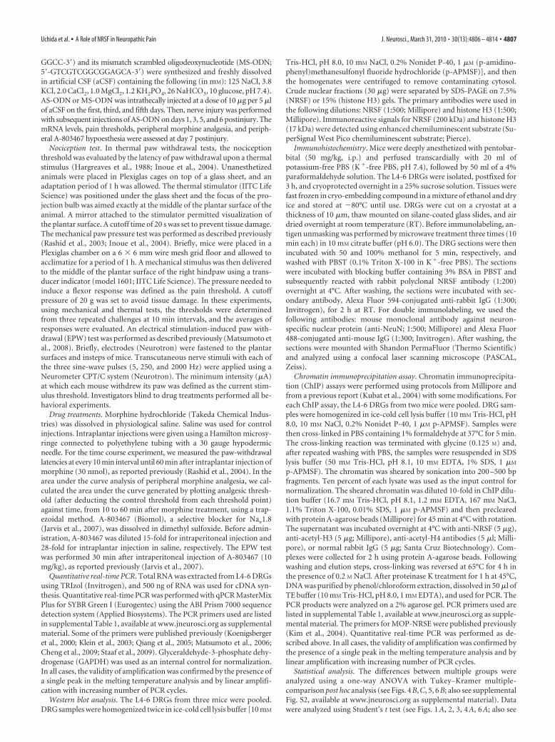
Histone hypoacetylation with an increase in NRSF binding atNRSE sequences within MOP and Nav1.8 genesNext, we used ChIP analysis to examine whether NRSF binds tothe NRSE sites of MOP and Nav1.8 genes after nerve injury. Nerve
injury caused a drastic increase in NRSFbinding to MOP-NRSE, Nav1.8-NRSE-1,and Nav1.8-NRSE-2 sequences (Fig. 3A),suggesting that these NRSE sequences arecapable of serving as NRSF-binding sites.Quantitative real-time PCR analysisshowed that there was a threefold increasein NRSF binding to MOP-NRSE andNav1.8-NRSE-2 (Fig. 3B), while the foldchange could not be calculated in theNRSF binding to Nav1.8-NRSE-1 becauseno significant signal was detected insham-operated preparations (supplementalFig. S1, available at www.jneurosci.org assupplemental material). In contrast, neg-ligible binding was observed followingprecipitation by normal IgG, confirmingthe specificity of the immunoprecipitation(Fig. 3A; supplemental Fig. S1, available atwww.jneurosci.org as supplemental ma-terial). In addition, we performed scan-ning ChIP analysis to assess the levels ofhistone H3 and H4 acetylation in thegenomic regions spanning NRSE sequencesof MOP and Nav1.8 genes. We found sig-nificant reductions of histone H3 and H4acetylation at MOP-NRSE and Nav1.8-NRSE-2 and H3 acetylation at Nav1.8-NRSE-1 at day 7 after injury (Fig. 3C,D).Together, these data suggest that nerve in-jury induces repressive chromatin statesaround the NRSE sequences of MOP andNav1.8 genes through NRSF-HDAC-me-diated mechanisms.
Blockade of nerve injury-inducedreductions in MOP and Nav1.8 geneexpressions by NRSF knockdownTo examine whether NRSF could con-tribute to the downregulation of MOPand Nav1.8 genes after nerve injury,mice were intrathecally pretreated withan AS-ODN against NRSF or a corre-sponding MS-ODN. Western blot anal-ysis revealed that NRSF protein levels inthe DRG were markedly reduced by AS-ODN, but not by MS-ODN (Fig. 4A).AS-ODN significantly blocked the nerveinjury-induced downregulation of MOPand Nav1.8 (Fig. 4 B). However, AS-ODN had no effects on basal MOP andNav1.8 mRNA levels in sham-operatedmice (Fig. 4 B). These findings stronglysuggest that NRSF-mediated mecha-nisms are responsible for the transcrip-tional suppression of MOP and Nav1.8genes in the DRG after nerve injury.
On the other hand, it has been reportedthat nerve injury downregulates transient
receptor potential melastatin 8 (TRPM8), TRP ankyrin 1 (TRPA1),and calcitonin gene-related peptide (CGRP) in the DRG (Hokfelt etal., 2006; Caspani et al., 2009; Staaf et al., 2009). Using TFSEARCHprogram, we found that these genes have putative NRSE sites, which
Figure 2. Epigenetic upregulation of NRSF gene expression. A, B, Time course of total (A) and exon-specific (B) NRSFmRNA expressions in the DRG after nerve injury. The mRNA expression levels were assessed using quantitative real-timePCR and normalized to that of GAPDH mRNA. Data are calculated as percentages of day 0. *p � 0.05 versus day 0. EI, EII, EIII,Exons I, II, III, respectively. C, Acetylation of histone H3 (AcH3) and H4 (AcH4) at NRSF promoter II (PII) at day 7 after injury,assessed using ChIP assay. Quantitative analysis was performed using real-time PCR, and the data were normalized to thecorresponding input. D, NRSF protein expression at day 7 postinjury, assessed using Western blot analysis. Results arenormalized to the histone H3 protein expression level. For C and D, data are calculated as percentages of sham-operatedgroup. *p � 0.05 versus sham-operated group. Data are expressed as the means � SEM from at least three mice.E, Immunohistochemical double labeling between NRSF (red) and NeuN (green), a neuronal marker, in the DRG of sham-operated and nerve-injured mice. Scale bars, 50 �m.
Uchida et al. • A Role of NRSF in Neuropathic Pain J. Neurosci., March 31, 2010 • 30(13):4806 – 4814 • 4809
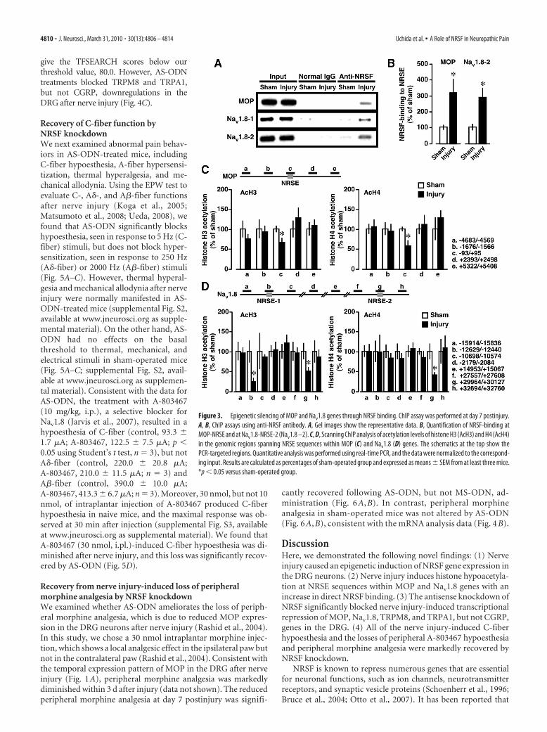
give the TFSEARCH scores below ourthreshold value, 80.0. However, AS-ODNtreatments blocked TRPM8 and TRPA1,but not CGRP, downregulations in theDRG after nerve injury (Fig. 4C).
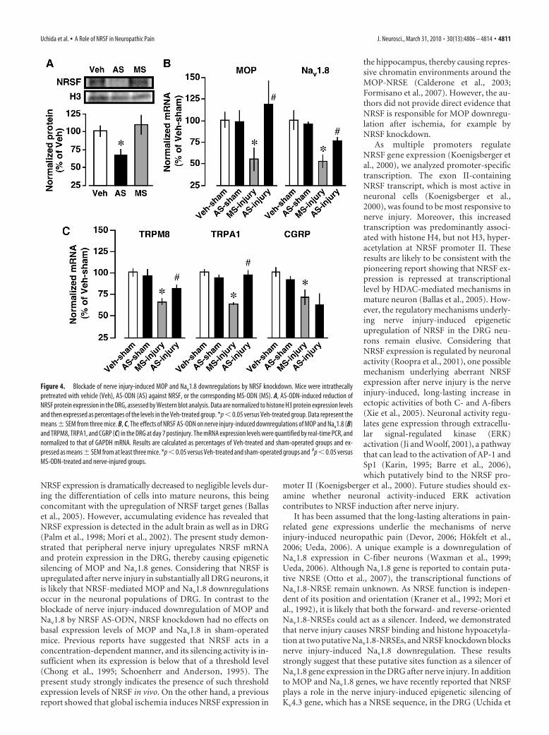
Recovery of C-fiber function byNRSF knockdownWe next examined abnormal pain behav-iors in AS-ODN-treated mice, includingC-fiber hypoesthesia, A-fiber hypersensi-tization, thermal hyperalgesia, and me-chanical allodynia. Using the EPW test toevaluate C-, A�-, and A�-fiber functionsafter nerve injury (Koga et al., 2005;Matsumoto et al., 2008; Ueda, 2008), wefound that AS-ODN significantly blockshypoesthesia, seen in response to 5 Hz (C-fiber) stimuli, but does not block hyper-sensitization, seen in response to 250 Hz(A�-fiber) or 2000 Hz (A�-fiber) stimuli(Fig. 5A–C). However, thermal hyperal-gesia and mechanical allodynia after nerveinjury were normally manifested in AS-ODN-treated mice (supplemental Fig. S2,available at www.jneurosci.org as supple-mental material). On the other hand, AS-ODN had no effects on the basalthreshold to thermal, mechanical, andelectrical stimuli in sham-operated mice(Fig. 5A–C; supplemental Fig. S2, avail-able at www.jneurosci.org as supplemen-tal material). Consistent with the data forAS-ODN, the treatment with A-803467(10 mg/kg, i.p.), a selective blocker forNav1.8 (Jarvis et al., 2007), resulted in ahypoesthesia of C-fiber (control, 93.3 �1.7 �A; A-803467, 122.5 � 7.5 �A; p �0.05 using Student’s t test, n � 3), but notA�-fiber (control, 220.0 � 20.8 �A;A-803467, 210.0 � 11.5 �A; n � 3) andA�-fiber (control, 390.0 � 10.0 �A;A-803467, 413.3 � 6.7 �A; n � 3). Moreover, 30 nmol, but not 10nmol, of intraplantar injection of A-803467 produced C-fiberhypoesthesia in naive mice, and the maximal response was ob-served at 30 min after injection (supplemental Fig. S3, availableat www.jneurosci.org as supplemental material). We found thatA-803467 (30 nmol, i.pl.)-induced C-fiber hypoesthesia was di-minished after nerve injury, and this loss was significantly recov-ered by AS-ODN (Fig. 5D).
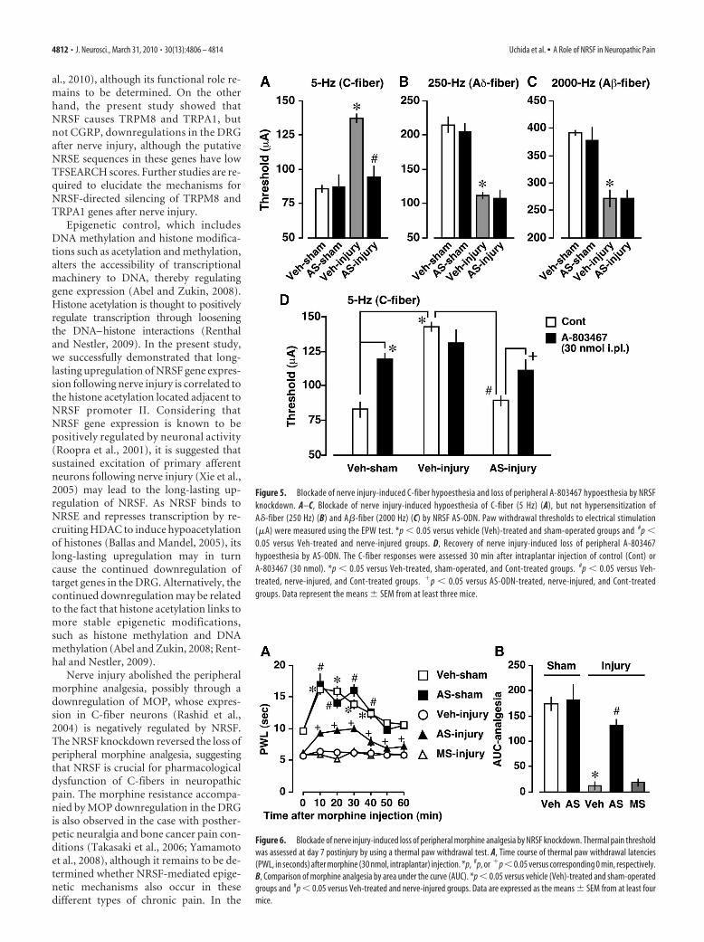
Recovery from nerve injury-induced loss of peripheralmorphine analgesia by NRSF knockdownWe examined whether AS-ODN ameliorates the loss of periph-eral morphine analgesia, which is due to reduced MOP expres-sion in the DRG neurons after nerve injury (Rashid et al., 2004).In this study, we chose a 30 nmol intraplantar morphine injec-tion, which shows a local analgesic effect in the ipsilateral paw butnot in the contralateral paw (Rashid et al., 2004). Consistent withthe temporal expression pattern of MOP in the DRG after nerveinjury (Fig. 1A), peripheral morphine analgesia was markedlydiminished within 3 d after injury (data not shown). The reducedperipheral morphine analgesia at day 7 postinjury was signifi-
cantly recovered following AS-ODN, but not MS-ODN, ad-ministration (Fig. 6 A, B). In contrast, peripheral morphineanalgesia in sham-operated mice was not altered by AS-ODN(Fig. 6 A, B), consistent with the mRNA analysis data (Fig. 4 B).
DiscussionHere, we demonstrated the following novel findings: (1) Nerveinjury caused an epigenetic induction of NRSF gene expression inthe DRG neurons. (2) Nerve injury induces histone hypoacetyla-tion at NRSE sequences within MOP and Nav1.8 genes with anincrease in direct NRSF binding. (3) The antisense knockdown ofNRSF significantly blocked nerve injury-induced transcriptionalrepression of MOP, Nav1.8, TRPM8, and TRPA1, but not CGRP,genes in the DRG. (4) All of the nerve injury-induced C-fiberhypoesthesia and the losses of peripheral A-803467 hypoesthesiaand peripheral morphine analgesia were markedly recovered byNRSF knockdown.
NRSF is known to repress numerous genes that are essentialfor neuronal functions, such as ion channels, neurotransmitterreceptors, and synaptic vesicle proteins (Schoenherr et al., 1996;Bruce et al., 2004; Otto et al., 2007). It has been reported that
Figure 3. Epigenetic silencing of MOP and Nav1.8 genes through NRSF binding. ChIP assay was performed at day 7 postinjury.A, B, ChIP assays using anti-NRSF antibody. A, Gel images show the representative data. B, Quantification of NRSF-binding atMOP-NRSE and at Nav1.8-NRSE-2 (Nav1.8 –2). C, D, Scanning ChIP analysis of acetylation levels of histone H3 (AcH3) and H4 (AcH4)in the genomic regions spanning NRSE sequences within MOP (C) and Nav1.8 (D) genes. The schematics at the top show thePCR-targeted regions. Quantitative analysis was performed using real-time PCR, and the data were normalized to the correspond-ing input. Results are calculated as percentages of sham-operated group and expressed as means � SEM from at least three mice.*p � 0.05 versus sham-operated group.
4810 • J. Neurosci., March 31, 2010 • 30(13):4806 – 4814 Uchida et al. • A Role of NRSF in Neuropathic Pain
NRSF expression is dramatically decreased to negligible levels dur-ing the differentiation of cells into mature neurons, this beingconcomitant with the upregulation of NRSF target genes (Ballaset al., 2005). However, accumulating evidence has revealed thatNRSF expression is detected in the adult brain as well as in DRG(Palm et al., 1998; Mori et al., 2002). The present study demon-strated that peripheral nerve injury upregulates NRSF mRNAand protein expression in the DRG, thereby causing epigeneticsilencing of MOP and Nav1.8 genes. Considering that NRSF isupregulated after nerve injury in substantially all DRG neurons, itis likely that NRSF-mediated MOP and Nav1.8 downregulationsoccur in the neuronal populations of DRG. In contrast to theblockade of nerve injury-induced downregulation of MOP andNav1.8 by NRSF AS-ODN, NRSF knockdown had no effects onbasal expression levels of MOP and Nav1.8 in sham-operatedmice. Previous reports have suggested that NRSF acts in aconcentration-dependent manner, and its silencing activity is in-sufficient when its expression is below that of a threshold level(Chong et al., 1995; Schoenherr and Anderson, 1995). Thepresent study strongly indicates the presence of such thresholdexpression levels of NRSF in vivo. On the other hand, a previousreport showed that global ischemia induces NRSF expression in
the hippocampus, thereby causing repres-sive chromatin environments around theMOP-NRSE (Calderone et al., 2003;Formisano et al., 2007). However, the au-thors did not provide direct evidence thatNRSF is responsible for MOP downregu-lation after ischemia, for example byNRSF knockdown.
As multiple promoters regulateNRSF gene expression (Koenigsberger etal., 2000), we analyzed promoter-specifictranscription. The exon II-containingNRSF transcript, which is most active inneuronal cells (Koenigsberger et al.,2000), was found to be most responsive tonerve injury. Moreover, this increasedtranscription was predominantly associ-ated with histone H4, but not H3, hyper-acetylation at NRSF promoter II. Theseresults are likely to be consistent with thepioneering report showing that NRSF ex-pression is repressed at transcriptionallevel by HDAC-mediated mechanisms inmature neuron (Ballas et al., 2005). How-ever, the regulatory mechanisms underly-ing nerve injury-induced epigeneticupregulation of NRSF in the DRG neu-rons remain elusive. Considering thatNRSF expression is regulated by neuronalactivity (Roopra et al., 2001), one possiblemechanism underlying aberrant NRSFexpression after nerve injury is the nerveinjury-induced, long-lasting increase inectopic activities of both C- and A-fibers(Xie et al., 2005). Neuronal activity regu-lates gene expression through extracellu-lar signal-regulated kinase (ERK)activation (Ji and Woolf, 2001), a pathwaythat can lead to the activation of AP-1 andSp1 (Karin, 1995; Barre et al., 2006),which putatively bind to the NRSF pro-
moter II (Koenigsberger et al., 2000). Future studies should ex-amine whether neuronal activity-induced ERK activationcontributes to NRSF induction after nerve injury.
It has been assumed that the long-lasting alterations in pain-related gene expressions underlie the mechanisms of nerveinjury-induced neuropathic pain (Devor, 2006; Hokfelt et al.,2006; Ueda, 2006). A unique example is a downregulation ofNav1.8 expression in C-fiber neurons (Waxman et al., 1999;Ueda, 2006). Although Nav1.8 gene is reported to contain puta-tive NRSE (Otto et al., 2007), the transcriptional functions ofNav1.8-NRSE remain unknown. As NRSE function is indepen-dent of its position and orientation (Kraner et al., 1992; Mori etal., 1992), it is likely that both the forward- and reverse-orientedNav1.8-NRSEs could act as a silencer. Indeed, we demonstratedthat nerve injury causes NRSF binding and histone hypoacetyla-tion at two putative Nav1.8-NRSEs, and NRSF knockdown blocksnerve injury-induced Nav1.8 downregulation. These resultsstrongly suggest that these putative sites function as a silencer ofNav1.8 gene expression in the DRG after nerve injury. In additionto MOP and Nav1.8 genes, we have recently reported that NRSFplays a role in the nerve injury-induced epigenetic silencing ofKv4.3 gene, which has a NRSE sequence, in the DRG (Uchida et
Figure 4. Blockade of nerve injury-induced MOP and Nav1.8 downregulations by NRSF knockdown. Mice were intrathecallypretreated with vehicle (Veh), AS-ODN (AS) against NRSF, or the corresponding MS-ODN (MS). A, AS-ODN-induced reduction ofNRSF protein expression in the DRG, assessed by Western blot analysis. Data are normalized to histone H3 protein expression levelsand then expressed as percentages of the levels in the Veh-treated group. *p � 0.05 versus Veh-treated group. Data represent themeans � SEM from three mice. B, C, The effects of NRSF AS-ODN on nerve injury-induced downregulations of MOP and Nav1.8 (B)and TRPM8, TRPA1, and CGRP (C) in the DRG at day 7 postinjury. The mRNA expression levels were quantified by real-time PCR, andnormalized to that of GAPDH mRNA. Results are calculated as percentages of Veh-treated and sham-operated groups and ex-pressed as means � SEM from at least three mice. *p � 0.05 versus Veh-treated and sham-operated groups and #p � 0.05 versusMS-ODN-treated and nerve-injured groups.
Uchida et al. • A Role of NRSF in Neuropathic Pain J. Neurosci., March 31, 2010 • 30(13):4806 – 4814 • 4811
al., 2010), although its functional role re-mains to be determined. On the otherhand, the present study showed thatNRSF causes TRPM8 and TRPA1, butnot CGRP, downregulations in the DRGafter nerve injury, although the putativeNRSE sequences in these genes have lowTFSEARCH scores. Further studies are re-quired to elucidate the mechanisms forNRSF-directed silencing of TRPM8 andTRPA1 genes after nerve injury.
Epigenetic control, which includesDNA methylation and histone modifica-tions such as acetylation and methylation,alters the accessibility of transcriptionalmachinery to DNA, thereby regulatinggene expression (Abel and Zukin, 2008).Histone acetylation is thought to positivelyregulate transcription through looseningthe DNA–histone interactions (Renthaland Nestler, 2009). In the present study,we successfully demonstrated that long-lasting upregulation of NRSF gene expres-sion following nerve injury is correlated tothe histone acetylation located adjacent toNRSF promoter II. Considering thatNRSF gene expression is known to bepositively regulated by neuronal activity(Roopra et al., 2001), it is suggested thatsustained excitation of primary afferentneurons following nerve injury (Xie et al.,2005) may lead to the long-lasting up-regulation of NRSF. As NRSF binds toNRSE and represses transcription by re-cruiting HDAC to induce hypoacetylationof histones (Ballas and Mandel, 2005), itslong-lasting upregulation may in turncause the continued downregulation oftarget genes in the DRG. Alternatively, thecontinued downregulation may be relatedto the fact that histone acetylation links tomore stable epigenetic modifications,such as histone methylation and DNAmethylation (Abel and Zukin, 2008; Rent-hal and Nestler, 2009).
Nerve injury abolished the peripheralmorphine analgesia, possibly through adownregulation of MOP, whose expres-sion in C-fiber neurons (Rashid et al.,2004) is negatively regulated by NRSF.The NRSF knockdown reversed the loss ofperipheral morphine analgesia, suggestingthat NRSF is crucial for pharmacologicaldysfunction of C-fibers in neuropathicpain. The morphine resistance accompa-nied by MOP downregulation in the DRGis also observed in the case with posther-petic neuralgia and bone cancer pain con-ditions (Takasaki et al., 2006; Yamamotoet al., 2008), although it remains to be de-termined whether NRSF-mediated epige-netic mechanisms also occur in thesedifferent types of chronic pain. In the
Figure 5. Blockade of nerve injury-induced C-fiber hypoesthesia and loss of peripheral A-803467 hypoesthesia by NRSFknockdown. A–C, Blockade of nerve injury-induced hypoesthesia of C-fiber (5 Hz) (A), but not hypersensitization ofA�-fiber (250 Hz) (B) and A�-fiber (2000 Hz) (C) by NRSF AS-ODN. Paw withdrawal thresholds to electrical stimulation(�A) were measured using the EPW test. *p � 0.05 versus vehicle (Veh)-treated and sham-operated groups and #p �0.05 versus Veh-treated and nerve-injured groups. D, Recovery of nerve injury-induced loss of peripheral A-803467hypoesthesia by AS-ODN. The C-fiber responses were assessed 30 min after intraplantar injection of control (Cont) orA-803467 (30 nmol). *p � 0.05 versus Veh-treated, sham-operated, and Cont-treated groups. #p � 0.05 versus Veh-treated, nerve-injured, and Cont-treated groups. �p � 0.05 versus AS-ODN-treated, nerve-injured, and Cont-treatedgroups. Data represent the means � SEM from at least three mice.
Figure 6. Blockade of nerve injury-induced loss of peripheral morphine analgesia by NRSF knockdown. Thermal pain thresholdwas assessed at day 7 postinjury by using a thermal paw withdrawal test. A, Time course of thermal paw withdrawal latencies(PWL, in seconds) after morphine (30 nmol, intraplantar) injection. *p, #p, or �p � 0.05 versus corresponding 0 min, respectively.B, Comparison of morphine analgesia by area under the curve (AUC). *p � 0.05 versus vehicle (Veh)-treated and sham-operatedgroups and #p � 0.05 versus Veh-treated and nerve-injured groups. Data are expressed as the means � SEM from at least fourmice.
4812 • J. Neurosci., March 31, 2010 • 30(13):4806 – 4814 Uchida et al. • A Role of NRSF in Neuropathic Pain
present study, we also demonstrated that the peripheral admin-istration of A-803467 produces C-fiber-selective hypoesthesia innaive mice, possibly through a blockade of Nav1.8, whose expres-sion in C-fiber neurons (Waxman et al., 1999) is negatively reg-ulated by NRSF. As seen in the case with morphine analgesia, theNRSF knockdown also reversed the nerve injury-induced lossof peripheral A-803467 hypoesthesia, being consistent with therecovery of Nav1.8 gene expression. Of interest are the findingsthat NRSF knockdown has no effects on A�- and A�-hypersensi-tization, thermal hyperalgesia, and mechanical allodynia afternerve injury. These results are consistent with the report that theablation of nociceptor neurons expressing Nav1.8 has no effectson neuropathic hyperalgesia, assessed only by thermal and me-chanical nociception tests (Abrahamsen et al., 2008). On theother hand, we also demonstrated that NRSF knockdown re-verses the nerve injury-induced downregulation of TRPM8 andTRPA1, which have been proposed to function as cold receptors(Levine and Alessandri-Haber, 2007), although their TFSEARCHscores for NRSE are not high enough. It would be an interestingsubject to examine whether NRSF-directed TRPM8 and TRPA1downregulations are crucial for cold hypoesthesia, which is anegative symptom of neuropathic pain as seen in the clinicalstudies (Devigili et al., 2008; Leffler and Hansson, 2008).
In conclusion, the present study demonstrated that NRSF-directed epigenetic gene silencing of Nav1.8 and MOP genes inthe DRG is responsible for pathological and pharmacologicaldysfunctions of C-fiber after nerve injury. Elucidation of regula-tory mechanisms for NRSE-NRSF systems after nerve injurymight provide novel therapeutic targets for the unmet negativesymptoms in neuropathic pain.
ReferencesAbel T, Zukin RS (2008) Epigenetic targets of HDAC inhibition in neuro-
degenerative and psychiatric disorders. Curr Opin Pharmacol 8:57– 64.Abrahamsen B, Zhao J, Asante CO, Cendan CM, Marsh S, Martinez-Barbera
JP, Nassar MA, Dickenson AH, Wood JN (2008) The cell and molecularbasis of mechanical, cold, and inflammatory pain. Science 321:702–705.
Akopian AN, Souslova V, England S, Okuse K, Ogata N, Ure J, Smith A, KerrBJ, McMahon SB, Boyce S, Hill R, Stanfa LC, Dickenson AH, Wood JN(1999) The tetrodotoxin-resistant sodium channel SNS has a specializedfunction in pain pathways. Nat Neurosci 2:541–548.
Ballas N, Mandel G (2005) The many faces of REST oversee epigenetic pro-gramming of neuronal genes. Curr Opin Neurobiol 15:500 –506.
Ballas N, Grunseich C, Lu DD, Speh JC, Mandel G (2005) REST and itscorepressors mediate plasticity of neuronal gene chromatin throughoutneurogenesis. Cell 121:645– 657.
Baron R (2006) Mechanisms of disease: neuropathic pain–a clinical per-spective. Nat Clin Pract Neurol 2:95–106.
Barre L, Venkatesan N, Magdalou J, Netter P, Fournel-Gigleux S, Ouzzine M(2006) Evidence of calcium-dependent pathway in the regulation of hu-man beta1,3-glucuronosyltransferase-1 (GlcAT-I) gene expression: a keyenzyme in proteoglycan synthesis. FASEB J 20:1692–1694.
Borrelli E, Nestler EJ, Allis CD, Sassone-Corsi P (2008) Decoding the epige-netic language of neuronal plasticity. Neuron 60:961–974.
Bruce AW, Donaldson IJ, Wood IC, Yerbury SA, Sadowski MI, Chapman M,Gottgens B, Buckley NJ (2004) Genome-wide analysis of repressor ele-ment 1 silencing transcription factor/neuron-restrictive silencing factor(REST/NRSF) target genes. Proc Natl Acad Sci U S A 101:10458 –10463.
Calderone A, Jover T, Noh KM, Tanaka H, Yokota H, Lin Y, Grooms SY,Regis R, Bennett MV, Zukin RS (2003) Ischemic insults derepress thegene silencer REST in neurons destined to die. J Neurosci 23:2112–2121.
Caspani O, Zurborg S, Labuz D, Heppenstall PA (2009) The contribution ofTRPM8 and TRPA1 channels to cold allodynia and neuropathic pain.PLoS One 4:e7383.
Cheng HT, Dauch JR, Hayes JM, Hong Y, Feldman EL (2009) Nerve growthfactor mediates mechanical allodynia in a mouse model of type 2 diabetes.J Neuropathol Exp Neurol 68:1229 –1243.
Chong JA, Tapia-Ramirez J, Kim S, Toledo-Aral JJ, Zheng Y, Boutros MC,
Altshuller YM, Frohman MA, Kraner SD, Mandel G (1995) REST: amammalian silencer protein that restricts sodium channel gene expres-sion to neurons. Cell 80:949 –957.
Costigan M, Scholz J, Woolf CJ (2009) Neuropathic pain: a maladaptiveresponse of the nervous system to damage. Annu Rev Neurosci 32:1–32.
Devigili G, Tugnoli V, Penza P, Camozzi F, Lombardi R, Melli G, Broglio L,Granieri E, Lauria G (2008) The diagnostic criteria for small fibre neu-ropathy: from symptoms to neuropathology. Brain 131:1912–1925.
Devor M (2006) Response of nerves to injury in relation to neuropathic pain.In: Wall and Melzack’s textbook of pain (McMahon SB, Koltzenburg M, eds),pp 905–927. Oxford: Churchill Livingstone.
Dickenson A, Kieffer B (2006) Opiates: basic mechanisms. In: Wall andMelzack’s textbook of pain (McMahon SB, Koltzenburg M, eds), pp 427–442. Oxford: Churchill Livingstone.
Fields HL, Rowbotham M, Baron R (1998) Postherpetic neuralgia: irritablenociceptors and deafferentation. Neurobiol Dis 5:209 –227.
Formisano L, Noh KM, Miyawaki T, Mashiko T, Bennett MV, Zukin RS(2007) Ischemic insults promote epigenetic reprogramming of mu opi-oid receptor expression in hippocampal neurons. Proc Natl Acad SciU S A 104:4170 – 4175.
Hargreaves K, Dubner R, Brown F, Flores C, Joris J (1988) A new and sen-sitive method for measuring thermal nociception in cutaneous hyperal-gesia. Pain 32:77– 88.
Hokfelt T, Zhang X, Xu XJ, Wiesenfeld-Hallin Z (2006) Central conse-quences of peripheral nerve damage. In: Wall and Melzack’s textbook ofpain (McMahon SB, Koltzenburg M, eds), pp 947–959. Oxford: ChurchillLivingstone.
Inoue M, Rashid MH, Fujita R, Contos JJ, Chun J, Ueda H (2004) Initiationof neuropathic pain requires lysophosphatidic acid receptor signaling.Nat Med 10:712–718.
Jarvis MF, Honore P, Shieh CC, Chapman M, Joshi S, Zhang XF, Kort M,Carroll W, Marron B, Atkinson R, Thomas J, Liu D, Krambis M, Liu Y,McGaraughty S, Chu K, Roelofts, Zhong C, Mikusa JP, Hernandez G, etal. (2007) A-803467, a potent and selective Nav1.8 sodium channelblocker, attenuates neuropathic and inflammatory pain in the rat. ProcNatl Acad Sci U S A 104:8520 – 8525.
Ji RR, Woolf CJ (2001) Neuronal plasticity and signal transduction in noci-ceptive neurons: implications for the initiation and maintenance ofpathological pain. Neurobiol Dis 8:1–10.
Karin M (1995) The regulation of AP-1 activity by mitogen-activated pro-tein kinases. J Biol Chem 270:16483–16486.
Kim CS, Hwang CK, Choi HS, Song KY, Law PY, Wei LN, Loh HH (2004)Neuron-restrictive silencer factor (NRSF) functions as a repressor in neu-ronal cells to regulate the mu opioid receptor gene. J Biol Chem279:46464 – 46473.
Klein JP, Tendi EA, Dib-Hajj SD, Fields RD, Waxman SG (2003) Patternedelectrical activity modulates sodium channel expression in sensory neu-rons. J Neurosci Res 74:192–198.
Koenigsberger C, Chicca JJ 2nd, Amoureux MC, Edelman GM, Jones FS(2000) Differential regulation by multiple promoters of the gene encod-ing the neuron-restrictive silencer factor. Proc Natl Acad Sci U S A97:2291–2296.
Koga K, Furue H, Rashid MH, Takaki A, Katafuchi T, Yoshimura M (2005)Selective activation of primary afferent fibers evaluated by sine-wave elec-trical stimulation. Mol Pain 1:13.
Kohno T, Ji RR, Ito N, Allchorne AJ, Befort K, Karchewski LA, Woolf CJ(2005) Peripheral axonal injury results in reduced mu opioid receptorpre- and post-synaptic action in the spinal cord. Pain 117:77– 87.
Kraner SD, Chong JA, Tsay HJ, Mandel G (1992) Silencing the type II so-dium channel gene: a model for neural-specific gene regulation. Neuron9:37– 44.
Kubat NJ, Amelio AL, Giordani NV, Bloom DC (2004) The herpes simplexvirus type 1 latency-associated transcript (LAT) enhancer/rcr is hyper-acetylated during latency independently of LAT transcription. J Virol78:12508 –12518.
Leffler AS, Hansson P (2008) Painful traumatic peripheral partial nerveinjury-sensory dysfunction profiles comparing outcomes of bedside ex-amination and quantitative sensory testing. Eur J Pain 12:397– 402.
Levine JD, Alessandri-Haber N (2007) TRP channels: targets for the relief ofpain. Biochim Biophys Acta 1772:989 –1003.
Malmberg AB, Basbaum AI (1998) Partial sciatic nerve injury in the mouse
Uchida et al. • A Role of NRSF in Neuropathic Pain J. Neurosci., March 31, 2010 • 30(13):4806 – 4814 • 4813
as a model of neuropathic pain: behavioral and neuroanatomical corre-lates. Pain 76:215–222.
Matsumoto M, Inoue M, Hald A, Xie W, Ueda H (2006) Inhibition ofpaclitaxel-induced A-fiber hypersensitization by gabapentin. J PharmacolExp Ther 318:735–740.
Matsumoto M, Xie W, Ma L, Ueda H (2008) Pharmacological switch inAbeta-fiber stimulation-induced spinal transmission in mice with partialsciatic nerve injury. Mol Pain 4:25.
Mori N, Schoenherr C, Vandenbergh DJ, Anderson DJ (1992) A common si-lencer element in the SCG10 and type II Na� channel genes binds a factorpresent in nonneuronal cells but not in neuronal cells. Neuron 9:45–54.
Mori N, Mizuno T, Murai K, Nakano I, Yamashita H (2002) Effect of age onthe gene expression of neural-restrictive silencing factor NRSF/REST.Neurobiol Aging 23:255–262.
Otto SJ, McCorkle SR, Hover J, Conaco C, Han JJ, Impey S, Yochum GS,Dunn JJ, Goodman RH, Mandel G (2007) A new binding motif for thetranscriptional repressor REST uncovers large gene networks devoted toneuronal functions. J Neurosci 27:6729 – 6739.
Palm K, Belluardo N, Metsis M, Timmusk T (1998) Neuronal expression ofzinc finger transcription factor REST/NRSF/XBR gene. J Neurosci18:1280 –1296.
Qiang M, Rani CS, Ticku MK (2005) Neuron-restrictive silencer factor reg-ulates the N-methyl-D-aspartate receptor 2B subunit gene in basal andethanol-induced gene expression in fetal cortical neurons. Mol Pharma-col 67:2115–2125.
Rashid MH, Inoue M, Kondo S, Kawashima T, Bakoshi S, Ueda H (2003)Novel expression of vanilloid receptor 1 on capsaicin-insensitive fibersaccounts for the analgesic effect of capsaicin cream in neuropathic pain.J Pharmacol Exp Ther 304:940 –948.
Rashid MH, Inoue M, Toda K, Ueda H (2004) Loss of peripheral morphineanalgesia contributes to the reduced effectiveness of systemic morphine inneuropathic pain. J Pharmacol Exp Ther 309:380 –387.
Renthal W, Nestler EJ (2009) Histone acetylation in drug addiction. SeminCell Dev Biol 20:387–394.
Roopra A, Huang Y, Dingledine R (2001) Neurological disease: listening togene silencers. Mol Interv 1:219 –228.
Schoenherr CJ, Anderson DJ (1995) The neuron-restrictive silencer factor(NRSF): a coordinate repressor of multiple neuron-specific genes. Science267:1360 –1363.
Schoenherr CJ, Paquette AJ, Anderson DJ (1996) Identification of potentialtarget genes for the neuron-restrictive silencer factor. Proc Natl Acad SciU S A 93:9881–9886.
Staaf S, Oerther S, Lucas G, Mattsson JP, Ernfors P (2009) Differential reg-ulation of TRP channels in a rat model of neuropathic pain. Pain144:187–199.
Takasaki I, Nojima H, Shiraki K, Kuraishi Y (2006) Specific down-regulation of spinal mu-opioid receptor and reduced analgesic effects ofmorphine in mice with postherpetic pain. Eur J Pharmacol 550:62– 67.
Taylor BK (2001) Pathophysiologic mechanisms of neuropathic pain. CurrPain Headache Rep 5:151–161.
Uchida H, Sasaki K, Ma L, Ueda H (2010) Neuron-restrictive silencer factorcauses epigenetic silencing of K(v)4.3 gene after peripheral nerve injury.Neuroscience 166:1– 4.
Ueda H (2006) Molecular mechanisms of neuropathic pain-phenotypicswitch and initiation mechanisms. Pharmacol Ther 109:57–77.
Ueda H (2008) Peripheral mechanisms of neuropathic pain-involvement oflysophosphatidic acid receptor-mediated demyelination. Mol Pain 4:11.
Waxman SG, Dib-Hajj S, Cummins TR, Black JA (1999) Sodium channelsand pain. Proc Natl Acad Sci U S A 96:7635–7639.
Xie W, Strong JA, Meij JT, Zhang JM, Yu L (2005) Neuropathic pain: earlyspontaneous afferent activity is the trigger. Pain 116:243–256.
Yamamoto J, Kawamata T, Niiyama Y, Omote K, Namiki A (2008) Down-regulation of mu opioid receptor expression within distinct subpopula-tions of dorsal root ganglion neurons in a murine model of bone cancerpain. Neuroscience 151:843– 853.
Zimmermann M (1983) Ethical guidelines for investigations of experimen-tal pain in conscious animals. Pain 16:109 –110.
4814 • J. Neurosci., March 31, 2010 • 30(13):4806 – 4814 Uchida et al. • A Role of NRSF in Neuropathic Pain