development/plasticity/repair anovelembryonicnestin ... · development/plasticity/repair...
TRANSCRIPT

Development/Plasticity/Repair
A Novel Embryonic Nestin-Expressing Radial Glia-LikeProgenitor Gives Rise to Zonally Restricted Olfactory andVomeronasal Neurons
Barbara Murdoch and A. Jane RoskamsDepartments of Zoology and Medicine, University of British Columbia, Vancouver, British Columbia, Canada V6T 1Z3
Persistent neurogenesis is maintained throughout development and adulthood in the mouse olfactory epithelium (OE). Despite this, theidentity and origin of different embryonic OE progenitors, their spatiotemporal induction and contribution to patterning during devel-opment, has yet to be delineated. Here, we show that the embryonic OE contains a novel nestin-expressing radial glia-like progenitor(RGLP) that is not found in adult OE, which is antigenically distinct from embryonic CNS radial glia. Nestin-cre-mediated lineage tracingwith three different reporters reveals that only a subpopulation of nestin-expressing RGLPs activate “CNS-specific” nestin regulatoryelements, and produce spatially restricted olfactory receptor neurons (ORNs) in zone 1 of the OE, and vomeronasal receptor neuronsrestricted to the VR1 zone. This dorsal-medial restriction of transgene-activating cells is also seen in the embryonic OE of Nestin-GFPtransgenic mice, in which green fluorescent protein (GFP) is found in a subpopulation of GFP�Mash1� neuronal progenitors, despitethe fact that endogenous Nestin expression is found in RGLPs throughout the OE. Embryonic OE progenitors produce three biologicallydistinct colony subtypes in vitro, a subpopulation of which include nestin-expressing RGLPs during in vitro colony formation. Whengenerated from Nestin-cre/ZEG mice, neurogenic colonies also produce GFP�Mash1� progenitors and ORNs. We thus identify a novelneurogenic precursor, the RGLP of the OE and vomeronasal organ (VNO), and provide the first evidence for intrinsic differences in theorigin and spatiotemporal potential of distinct progenitors during development of the OE and VNO.
Key words: radial glia; neural progenitors; Nestin-cre; olfactory progenitors; neurogenesis; ensheathing cell
IntroductionTo understand the mechanisms that drive nervous system devel-opment and regeneration, we need to establish when and wherespecific cell types originate, understand the lineage contributionof each progenitor, and how the interaction of sequentially gen-erated cells impacts the patterning of different nervous systemregions. In the adult nervous system, regeneration and adult neu-rogenesis is restricted to exclusive niches, one of which is themouse olfactory epithelium (OE) (Graziadei and Graziadei,1979; Schwob, 2002), in which newly differentiated olfactory re-ceptor neurons (ORNs) reintegrate into existing circuitry and
maintain the sense of smell in the adult organism (Farbman,1990; Roskams et al., 1996).
ORN precursor activation in adult OE is regulated by LIF(leukemia inhibitory factor), BMP4 (bone morphogenetic pro-tein 4), and basic FGF, in which globose basal cells (GBCs) serveas transit amplifying neuronal precursors for olfactory receptorneurons that activate Mash1 before neurogenin1 in both devel-opment and regeneration (Cau et al., 1997; Nan et al., 2001; Calofet al., 2002; Getchell et al., 2002; Bauer et al., 2003). In contrast,horizontal basal cells (HBCs) are more quiescent than GBCs,express EGFR (epidermal growth factor receptor), are epidermalgrowth factor (EGF)/TGF�-responsive in vitro and in vivo, sim-ilar to stem cells in many other tissues (Farbman and Buchholz,1996; Goldstein and Schwob, 1996; Getchell et al., 2000), and asubpopulation of selected EGF-responsive HBCs form multipo-tent clonal colonies in vitro (Carter et al., 2004), and demonstratelimited multipotency in vivo (Leung et al., 2007). The adult OE isdevelopmentally laminated epithelium, with progenitors at thebasal lamina, and mature neurons in the apical OE (Farbman,1992). In contrast, the embryonic OE is highly dynamic, withmultilayered highly neurogenic regions located adjacent to two-cell-layered, preneurogenic regions, containing potentially dif-ferent classes of progenitor. Our spatial and temporal under-standing of the interrelationship between different embryonicOE neuroglial progenitors has been stalled by a paucity of iden-tifiable genes we can use to distinguish, and assay the potential of,
Received May 1, 2007; revised Feb. 20, 2008; accepted March 12, 2008.This work was supported by The Jack Brown Family Foundation (A.J.R.). Studentship funding was provided by the
Heart and Stroke Foundation of Canada, The Canadian Stroke Network, and The Michael Smith Foundation for HealthResearch (B.M.). We thank the following colleagues for the reagents that made this project possible: Frank Margolis(OMP antibody and genomic OMP construct); Todd Anthony and Nat Heintz (BLBP antibody and BLBP-cre/Rosamice); Paul Orban, Ruth Slack, and Jackie Vanderluit (Nestin-cre lines); Grigori Enikolopov (Nestin-GFP mice); CorrineLobe (ZEG mice and NLS-Cre construct); and Jeff Rothstein (nGlast antibody). We also thank Sam Weiss and Derekvan der Kooy for sharing technical information. We thank Michael Underhill, Ian Tietjen, and members of theRoskams’ laboratory for critical review of this manuscript, Nicole Janzen for mouse colony maintenance, and ErinCurrie for excellent technical assistance.
Correspondence should be addressed to Dr. A. Jane Roskams, Department of Zoology, Life Sciences Centre,University of British Columbia, 2350 Health Sciences Mall, Vancouver, British Columbia, Canada V6T 1Z3. E-mail:[email protected].
DOI:10.1523/JNEUROSCI.5566-07.2008Copyright © 2008 Society for Neuroscience 0270-6474/08/284271-12$15.00/0
The Journal of Neuroscience, April 16, 2008 • 28(16):4271– 4282 • 4271
individual candidate progenitors, a common problem in stemcell-bearing tissues (Weissman et al., 2001).
The application of genetic fate mapping with progenitor-specific promoters driving site-specific recombinases (like Cre)now allows us to address fundamental questions of progenitorlineage contribution more definitively than traditional methodsof fate mapping (like retroviral tracing) (Joyner and Zervas,2006). Although this approach has been valuable in evaluatingprogenitor contributions in the adult OE (Leung et al., 2007),these approaches have yet to be used for testing potential in theembryonic olfactory system. In the developing CNS, radial gliaserve as both structural scaffolding for migrating neuroblasts andas embryonic neural progenitors (Anthony et al., 2004). Multi-potent radial glia are identified by shared expression of a selectgroup of antigens (Gotz et al., 2005), and lineage tracing usingCre driven by these radial glial gene promoters and enhancers[e.g., Nestin, brain lipid binding protein (BLBP)] has revealedtheir spatiotemporal lineage contribution during CNS develop-ment (Anthony et al., 2004; Imayoshi et al., 2006). Here, we testfor the existence of OE-based radial glia progenitors, and rely onin vivo genetic lineage tracing combined with in vitro progenitorassays to test the neurogenic potential of embryonic nestintransgene-activating OE progenitors. We reveal the existence of adistinct neural progenitor phenotype unique to the embryonicOE, and demonstrate a previously unappreciated spatial regula-tion of olfactory receptor neuron and vomeronasal receptor neu-ron genesis developmentally.
Materials and MethodsTissue preparation. Adult and postnatal mice were anesthetized with Xy-laket: 25 mg/ml ketamine HCl (MTC Pharmaceuticals, Cambridge, On-tario, Canada), 2.5 mg/ml xylazine (Bayer, Tarrytown, NY), 15% etha-nol, 0.55% NaCl (120 mg/kg ketamine and 12 mg/kg xylazine), perfusedwith cold PBS and 4% paraformaldehyde (PFA) in PBS and postfixed in4% PFA at 4°C (Carter et al. 2004). Embryos were immersion-fixed in 4%PFA overnight. The day of vaginal plug was defined as embryonic day 0.5(E0.5). Tissues were cryoprotected in sucrose, embedded in Tissue-Tekmedium (OCT; Sakura Finetek, Torrance, CA), and frozen in liquidnitrogen. Twelve-micrometer sections were stored at �20°C for subse-quent analysis (MacDonald et al., 2005; Carson et al., 2006).
Immunohistochemistry. Protocols for immunohistochemistry havebeen described previously (Au and Roskams, 2003; MacDonald et al.,2005; Carson et al., 2006). Sections were postfixed in 4% PFA, perme-abilized in 0.1% Triton X-100/PBS, and blocked with 4% normal serumbefore primary antibody incubation. Secondary antibodies (1:200) usedwere biotin, Alexa 594, or Alexa 488 conjugated (Invitrogen, Carlsbad,CA). Before blocking, Sus4 detection required a 60 s incubation of thesections with 0.12% trypsin/EDTA (Invitrogen), followed by washing inPBS (Carson et al., 2006). Colabeling with Sus4 and proliferating cellnuclear antigen (PCNA) was performed sequentially with fixation in 4%PFA after the Sus4 primary and secondary antibodies, followed by wash-ing and reblocking before antigen retrieval (MacDonald et al., 2005) andthe PCNA primary and secondary antibodies. Nestin monoclonal anti-body (rat 401 clone), but not nestin polyclonal rabbit antibody (Nestinclone 20), required antigen retrieval in 0.01 M citric acid microwaved for10 min before blocking. Cre detection required antigen retrieval beforeblocking/permeabilization in 10% serum/0.1% Triton X-100/PBS. Creantibodies were incubated in blocking solution and washed in 0.1% Tri-ton X-100/PBS. Cre signals were amplified using the Vectastain ABC kit(Vector Laboratories, Burlingame, CA) and Amplex Red ELISA kit 2,horseradish peroxidase conjugate (Invitrogen), following the manufac-turer’s instructions. For codetection of antigens with Cre, tissues werereblocked in 10% serum/0.1% Triton X-100/PBS before sequential pri-mary antibody incubation. Primary antibodies used were as follows: rab-bit anti-mouse nestin (Nestin clone 20; 1:500), mouse anti-rat �III-tubulin [neuron-specific tubulin (NST); TUJ1; 1:500] (both Covance,
Princeton, NJ); mouse anti-rat nestin (rat 401 clone; 1:100) (used in Fig.6 I, K; supplemental Fig. S1C, F, I, K, M, available at www.jneurosci.org assupplemental material), mouse anti-Mash1 (1:100; BD Pharmingen, SanDiego, CA); mouse anti-rat RC2 (1:100; Developmental Studies Hybrid-oma Bank, Iowa City, IA) (developed by M. Yamamoto, University ofTsukuba, Tsukuba, Japan), rabbit anti-Cre (1:5000; EMD Biosciences,San Diego, CA); mouse anti-PCNA (clone PC10; 1:5000), mouse anti-S100� (clone SH-B1; 1:400) (all Sigma-Aldrich, St. Louis, MO); goatanti-human Doublecortin (Dcx) (C-18; 1:200; Santa Cruz Biotechnol-ogy, Santa Cruz, CA; Millipore Bioscience Research Reagents, Temecula,CA), rabbit anti-BLBP (1:2000; Millipore Bioscience Research Reagents),anti-mouse olfactory cell adhesion molecule (OCAM) (1:400; R&D Sys-tems, Minneapolis, MN), rabbit anti-green fluorescent protein (GFP)(1:400; Abcam, Cambridge, MA), or mouse anti-GFP (1:100; MilliporeBioscience Research Reagents). Note: All GFP panels show anti-GFPimmunofluorescence, with the exception of the GFP-containing panelsin Figure 7, E–N, which indicate endogenous GFP fluorescence, whichwas confirmed using immunoperoxidase (VIP) immunohistochemistrywith anti-GFP antibodies. Gift antibodies were as follows: mouse anti-ratSus4 (1:100) from Dr. J. Schwob (Tufts University, Boston, MA), rabbitanti-BLBP (1:2000) from N. Heintz (Howard Hughes Medical Institute,The Rockefeller University, New York, NY), goat polyclonal olfactorymarker protein (OMP) (1:5000) from Frank L. Margolis (University ofMaryland, Baltimore, MD), and rabbit anti-nGLAST (1:100) from J.Rothstein (The Johns Hopkins University, Baltimore, MD). Nuclei werestained with 0.5 �g/ml diaminopyridine imidazole (DAPI) and sectionscoverslipped in Vectashield (Vector Laboratories) for fluorescent anti-gens or Aquapolymount for VIP. All images were visualized with anAxioskop 2 MOT microscope (Carl Zeiss, Jena, Germany) using a SPOTcamera (Diagnostic Instruments, Sterling Heights, MI) with NorthernEclipse software (Empix Imaging, Mississauga, Ontario, Canada) andcompiled using Adobe Photoshop 7.0.
Immunocytochemistry. Immunocytochemistry was performed as de-scribed previously (Au and Roskams, 2003; Carter et al., 2004), with cellsplated onto collagen:laminin (5:2 �g/cm 2)-coated glass coverslips andnuclei stained with DAPI. For nuclear antigen detection, cells were fixedfor 3 min in �20°C methanol and washed in PBS before blocking. Thepercentage of antigen-positive cells per colony was determined by count-ing as follows: (number of antigen-positive cells/total number of cells percolony measured by counting DAPI-stained nuclei) � 100.
Histochemistry. LacZ histochemistry was performed on cryosectionspostfixed in 4% PFA, permeabilized in 0.1% Triton X-100, and washed inPBS before adding staining buffer (2 mM MgCl2, 0.01% deoxycholate,0.02% Nonidet P-40, and 100 mM NaPO4, pH 7.3) containing 1 mg/mlX-gal, 5 mM potassium ferrocyanide, and 5 mM potassium ferricyanide.Staining proceeded at 37°C protected from light for 1–10 h. Negativecontrols did not demonstrate �-galactosidase staining and includedCD-1 nontransgenic mice, Nestin-cre or OMP-cre transgenic mice with-out the ZEG transgene. Results were confirmed using antibodies to �-ga-lactosidase; however, the detection level and reproducibility were not asgood as that with histochemistry.
OE and subventricular zone cell isolation. E13.5 OE and subventricularzone (SVZ) (for controls) were dissected from the same mice and trans-ferred to serum-free DMEM/F12 or PBS plus 0.6% glucose, respectively(Reynolds and Weiss, 1992). Pooled OE tissue or E13.5 ganglionic eminiwere minced into 1 mm 3 pieces, triturated 20 times with a polishedPasteur pipette, washed, filtered (40 �m), and rewashed before countingcells. Cells were counted using a hemocytometer and trypan blue exclu-sion, resuspended in media (0.7–3 � 10 5 cells/ml), and centrifuged at100 � g for 1 min before plating, to pellet tissue aggregates.
In vitro progenitor assays. Cells from both ganglionic emini and OEwere cultured in CNS neurosphere media: Neurocult, 1� proliferationsupplement (Stem Cell Technologies, Vancouver, British Columbia,Canada), 20 ng/ml growth factors (GFs) (EGF, basic FGF, or EGF plusFGF; Sigma-Aldrich), 100 U/ml penicillin, 100 �g/ml streptomycin, 2mM L-glutamine (Invitrogen). A single-cell suspension was determinedby inspecting the cells on a hemocytometer at the time of counting andafter plating, by observing the cells directly in each well. OE cells wereplated at clonal density; initially into uncoated 12-well plates (Falcon; BD
4272 • J. Neurosci., April 16, 2008 • 28(16):4271– 4282 Murdoch and Roskams • Novel Embryonic Chemosensory Neuron Progenitors
Biosciences Discovery Labware, Bedford, MA) to perform colony counts,and later onto collagen:laminin-coated coverslips for immunocyto-chemistry, in 1.5 ml of media with 0.7–2 � 10 5 cells/ml. SVZ cells wereplated at 10 –25 cells/�l, 5000 –10,000 cells per well. SVZ cells derivedfrom the same embryo as the OE cells, served as positive controls for ourculture conditions and growth factor efficacy. Clonal density was firstdetermined by mixing equal cell numbers from ubiquitous �-actindriven GFP-expressing mice (Richter et al., 2005) with GFP-nonexpressing mice to determine the density at which mostly only GFP-expressing or -nonexpressing colonies were obtained (data not shown).
Primary cultures were grown for 7–10 d in vitro (DIV) before countingnonadherent neurospheres or semiadherent colonies (OE). A neuro-sphere was counted if �50 �m in diameter, semiadherent colonies com-posed of more than eight cells were counted. Pooled OE colonies werepassaged and plated at their primary cell density (or as close as possiblewhere cell numbers were limiting) using primary culture conditions withfresh media and growth factors. Self-renewal was assessed by countingneurospheres or colonies after an additional 7–10 DIV.
Transgenic mice. Nestin-cre mice were obtained from The Jackson Lab-oratory (Bar Harbor, ME) (C57BL/6) (Tronche et al., 1999) and werecrossed with ZEG reporter mice obtained from C. Lobe (Sunnybrook andWomen’s College Health Science Centre, Toronto, Ontario, Canada)(Novak et al., 2000). Female ZEG reporters were either C57BL/6 or CD-1strains. Nestin-cre/ZEG transgenic mice, in which Cre recombinase ex-pression is under the control of the nestin promoter and second intronregulatory elements of the rat nestin gene (Zimmerman et al., 1994),express GFP throughout the CNS. Transgenic reporter linesGt(ROSA)26Sortm(EYFP) Cos (The Jackson Laboratory; expressing en-hanced yellow fluorescent protein from the ROSA26 locus) (Srinivas etal., 2001), produced a similar reporter expression pattern after Nestin-cre-mediated excision. An independently derived Nestin-cre line (no.2472; FVB/N strain) crossed with a reporter, Gtrosa26tm1Sor (Soriano etal., 1999) (from R. Slack, University of Ottawa, Ottawa Health ResearchInstitute, Ottawa, Ontario, Canada), showed strong �-galactosidase ex-pression in the forebrain and developing CNS (Berube et al., 2005). Micewere genotyped by PCR for Cre, GFP, and/or LacZ histochemistry. Creprimers were as follows: NLS CreA, 5�-CCCGGCAAAACAGGTAGTTA-3�; NLS CreS, 5�-CATTTGGGCCAGCTAAACAT-3� (94°C for 30 s, 55°Cfor 30 s, 72°C for 90 s; 454 bp product). GFP primers were as follows:XFPf, 5�-AAGTTCATCTGCACCACCG-3�; XFPr, 5�-TCCTTGA-AGAAGATGGTGCG-3� (35 cycles of 94°C for 30 s, 60°C for 60 s, 72°Cfor 60 s; 173 bp product; each for 35 cycles).
OMP-cre transgenic mice were generated after pronuclear injection ofan 11 kb EcoR1-digested fragment, containing both 5� and 3� flankingregions of the OMP coding sequence (Danciger et al., 1989). The pG-ROMP plasmid, containing an 11 kb EcoRI genomic rat OMP fragment(kindly provided by Frank Margolis), was digested with AatII/AarI toremove the OMP gene, and a 1057 bp AatII/Eco31l fragment from PCRamplified NLS-Cre (plasmid kindly provided by Dr. Corrine Lobe) wassubcloned into the vector. Expression and efficiency of Cre-lox recom-bination was verified by the specific colocalization of Cre with OMP andOMP with GFP proteins in the OE of OMP-cre/ZEG mice.
Nestin-GFP transgenic mice were obtained from Grigori Enikolopov(Cold Spring Harbor Laboratory, Cold Spring Harbor, NY) (Mignone etal., 2004) and drive expression of enhanced GFP via an identical nestinpromoter (5.8 kb) and enhancer (1.8 kb second intron) to that of theNestin-cre mice used here (Zimmerman et al., 1994; Yaworsky and Kap-pen, 1999). Mice were geno/phenotyped by PCR for GFP sequences andby fluorescence microscopy for the detection of GFP in the developingCNS. Schematics of Nestin-cre, Nestin-GFP, and OMP-cre constructsare shown in supplemental Figure S3 (available at www.jneurosci.org assupplemental material).
In vitro progenitor assays, Nestin-cre/ZEG mice. E13.5 embryos weregeno/phenotyped by PCR for Cre, and LacZ histochemistry. All Nestin-cre/ZEG embryos (but not littermate controls) expressed readily detect-able endogenous GFP in the CNS. The OE from Nestin-cre/ZEG or anequal number of littermate control embryos were pooled from each lit-ter, in which the average number of cells/embryo was 2.6 � 10 5. From atotal of four litters assayed, 15 of 44 embryos were Nestin-cre/ZEG.
Single-cell suspensions were plated at 0.7 � 10 5 cells/ml into CNS neu-rosphere media (as above) onto collagen:laminin-coated glass coverslipsin 12-well plates. Cells were processed for immunocytochemistry andcounting of semiadherent colony subtypes after 10 DIV. Immunocyto-chemistry was performed as described above, with cells fixed in 4% PFAfor 5 min and washed in PBS before blocking. Mouse monoclonal (In-vitrogen) and rabbit polyclonal antibodies (Millipore Bioscience Re-search Reagents; both 1:100) reliably detected GFP in vitro, using GFP-expressing olfactory ensheathing cells (Richter et al., 2005) as positivecontrols, and littermate OE cells and secondary antibody only as negativecontrols.
Statistics. Values are means � SEM. Variance between groups wasdetermined using ANOVA and statistical significance using Tukey’s hon-estly significant difference post hoc test. Correlations were determinedusing Pearson’s coefficient.
ResultsThe OE contains radial glia-like cells present duringearly developmentThe OE experiences three distinct phases of neurogenesis: embry-onic establishment [E10 to postnatal day 0 (P0)], postnatal ex-pansion (P1–P30), and adult maintenance (P30 to death). Dur-ing each phase, the OE undergoes a dynamic reorganization of itsactive progenitor subcompartments both in the apical-basal androstral-caudal planes. However, it is not known whether distinctOE zones are produced in different time windows. Understand-ing the nature of how different progenitors contribute to thispatterning will enable us to test the regulation of these distinctphases of developmental OE neurogenesis.
During development, the majority of dividing cells are foundin the most apical OE cell layer, and gradually transition to a morebasal site with postnatal to adult OE maturation (Smart, 1971).This organization is highly reminiscent of the organization ofembryonic CNS ventricular zone progenitors (Gotz et al., 2005),in which the apical OE would be most similar to the embryonicventricular zone. We thus tested whether embryonic OE mightcontain progenitors that morphologically or antigenically resem-ble multipotent CNS radial glia. Nestin, an intermediate filamentprotein characteristic of CNS neuroepithelial stem cells (Hock-field and McKay, 1985), was detected in embryonic OE as early asE10 (Murdoch and Roskams, 2007), but was more readily de-tected at E13.5, in cells with a radial glia-like morphology similarto those found in embryonic olfactory bulb (OB) that were de-tected in every developing turbinate of the embryonic OE (Fig.1A). Nestin detection in the embryonic CNS matches the re-ported radial glial expression pattern (Anthony et al., 2004), andsimilar patterns of nestin expression were detected in both theCNS and the OE using two independent polyclonal and mono-clonal anti-nestin antibodies (supplemental Fig. S1, available atwww.jneurosci.org as supplemental material). These antibodieswere also able to immunoprecipitate a single band correspondingto nestin from postnatal brain, embryonic OE, and olfactory en-sheathing cells (supplemental Fig. S1, available at www.jneuro-sci.org as supplemental material).
At E13.5, the majority of embryonic nestin� radial glia-likeprogenitors (RGLPs) coexpressed PCNA, a protein associatedwith the replication fork during S-phase (Fig. 1A–C) (Waseemand Lane, 1990), and were anchored at the OE basement mem-brane and apical surface, with nestin-rich processes spanning theOE (Fig. 1B,C). The most intense accumulation of nuclearPCNA, indicative of the peak of S-phase, was found in the verti-cally elongated nuclei of nestin� RGLPs situated at the base ofthe OE (Fig. 1C). In contrast, nestin� RGLPs undergoing cyto-kinesis segregated PCNA into their cytoplasm, and were found in
Murdoch and Roskams • Novel Embryonic Chemosensory Neuron Progenitors J. Neurosci., April 16, 2008 • 28(16):4271– 4282 • 4273
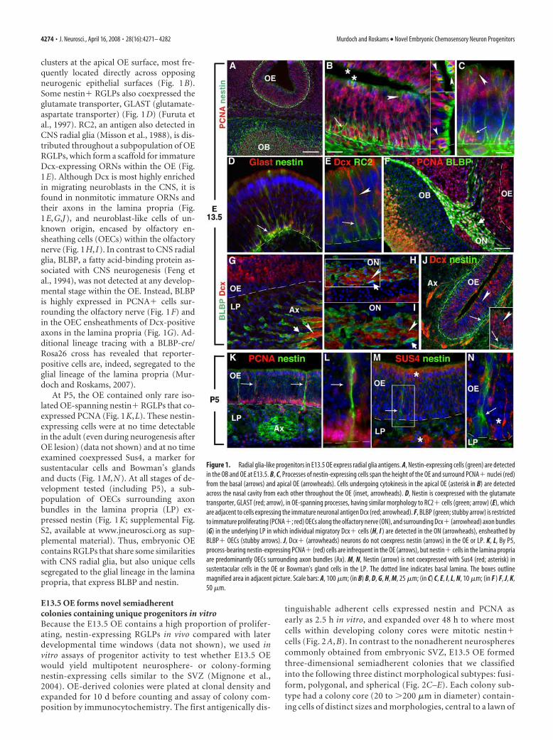
clusters at the apical OE surface, most fre-quently located directly across opposingneurogenic epithelial surfaces (Fig. 1B).Some nestin� RGLPs also coexpressed theglutamate transporter, GLAST (glutamate-aspartate transporter) (Fig. 1D) (Furuta etal., 1997). RC2, an antigen also detected inCNS radial glia (Misson et al., 1988), is dis-tributed throughout a subpopulation of OERGLPs, which form a scaffold for immatureDcx-expressing ORNs within the OE (Fig.1E). Although Dcx is most highly enrichedin migrating neuroblasts in the CNS, it isfound in nonmitotic immature ORNs andtheir axons in the lamina propria (Fig.1E,G,J), and neuroblast-like cells of un-known origin, encased by olfactory en-sheathing cells (OECs) within the olfactorynerve (Fig. 1H, I). In contrast to CNS radialglia, BLBP, a fatty acid-binding protein as-sociated with CNS neurogenesis (Feng etal., 1994), was not detected at any develop-mental stage within the OE. Instead, BLBPis highly expressed in PCNA� cells sur-rounding the olfactory nerve (Fig. 1F) andin the OEC ensheathments of Dcx-positiveaxons in the lamina propria (Fig. 1G). Ad-ditional lineage tracing with a BLBP-cre/Rosa26 cross has revealed that reporter-positive cells are, indeed, segregated to theglial lineage of the lamina propria (Mur-doch and Roskams, 2007).
At P5, the OE contained only rare iso-lated OE-spanning nestin� RGLPs that co-expressed PCNA (Fig. 1K,L). These nestin-expressing cells were at no time detectablein the adult (even during neurogenesis afterOE lesion) (data not shown) and at no timeexamined coexpressed Sus4, a marker forsustentacular cells and Bowman’s glandsand ducts (Fig. 1M,N). At all stages of de-velopment tested (including P5), a sub-population of OECs surrounding axonbundles in the lamina propria (LP) ex-pressed nestin (Fig. 1K; supplemental Fig.S2, available at www.jneurosci.org as sup-plemental material). Thus, embryonic OEcontains RGLPs that share some similaritieswith CNS radial glia, but also unique cellssegregated to the glial lineage in the laminapropria, that express BLBP and nestin.
E13.5 OE forms novel semiadherentcolonies containing unique progenitors in vitroBecause the E13.5 OE contains a high proportion of prolifer-ating, nestin-expressing RGLPs in vivo compared with laterdevelopmental time windows (data not shown), we used invitro assays of progenitor activity to test whether E13.5 OEwould yield multipotent neurosphere- or colony-formingnestin-expressing cells similar to the SVZ (Mignone et al.,2004). OE-derived colonies were plated at clonal density andexpanded for 10 d before counting and assay of colony com-position by immunocytochemistry. The first antigenically dis-
tinguishable adherent cells expressed nestin and PCNA asearly as 2.5 h in vitro, and expanded over 48 h to where mostcells within developing colony cores were mitotic nestin�cells (Fig. 2 A, B). In contrast to the nonadherent neurospherescommonly obtained from embryonic SVZ, E13.5 OE formedthree-dimensional semiadherent colonies that we classifiedinto the following three distinct morphological subtypes: fusi-form, polygonal, and spherical (Fig. 2C–E). Each colony sub-type had a colony core (20 to �200 �m in diameter) contain-ing cells of distinct sizes and morphologies, central to a lawn of
Figure 1. Radial glia-like progenitors in E13.5 OE express radial glia antigens. A, Nestin-expressing cells (green) are detectedin the OB and OE at E13.5. B, C, Processes of nestin-expressing cells span the height of the OE and surround PCNA� nuclei (red)from the basal (arrows) and apical OE (arrowheads). Cells undergoing cytokinesis in the apical OE (asterisk in B) are detectedacross the nasal cavity from each other throughout the OE (inset, arrowheads). D, Nestin is coexpressed with the glutamatetransporter, GLAST (red; arrow), in OE-spanning processes, having similar morphology to RC2� cells (green; arrow) (E), whichare adjacent to cells expressing the immature neuronal antigen Dcx (red; arrowhead). F, BLBP (green; stubby arrow) is restrictedto immature proliferating (PCNA�; red) OECs along the olfactory nerve (ON), and surrounding Dcx� (arrowhead) axon bundles(G) in the underlying LP in which individual migratory Dcx� cells (H, I ) are detected in the ON (arrowheads), ensheathed byBLBP� OECs (stubby arrows). J, Dcx� (arrowheads) neurons do not coexpress nestin (arrows) in the OE or LP. K, L, By P5,process-bearing nestin-expressing PCNA� (red) cells are infrequent in the OE (arrows), but nestin� cells in the lamina propriaare predominantly OECs surrounding axon bundles (Ax). M, N, Nestin (arrow) is not coexpressed with Sus4 (red; asterisk) insustentacular cells in the OE or Bowman’s gland cells in the LP. The dotted line indicates basal lamina. The boxes outlinemagnified area in adjacent picture. Scale bars: A, 100 �m; (in B) B, D, G, H, M, 25 �m; (in C) C, E, I, L, N, 10 �m; (in F ) F, J, K,50 �m.
4274 • J. Neurosci., April 16, 2008 • 28(16):4271– 4282 Murdoch and Roskams • Novel Embryonic Chemosensory Neuron Progenitors
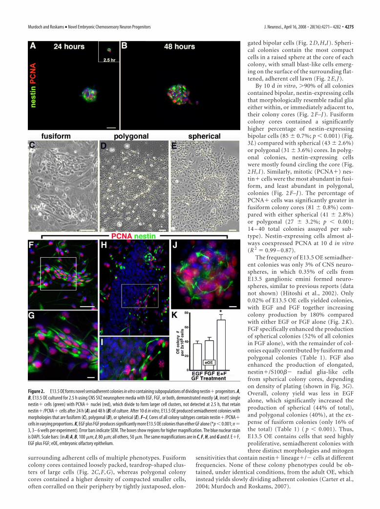
surrounding adherent cells of multiple phenotypes. Fusiformcolony cores contained loosely packed, teardrop-shaped clus-ters of large cells (Fig. 2C, F, G), whereas polygonal colonycores contained a higher density of compacted smaller cells,often corralled on their periphery by tightly juxtaposed, elon-
gated bipolar cells (Fig. 2 D, H,I ). Spheri-cal colonies contain the most compactcells in a raised sphere at the core of eachcolony, with small blast-like cells emerg-ing on the surface of the surrounding flat-tened, adherent cell lawn (Fig. 2 E, J ).
By 10 d in vitro, �90% of all coloniescontained bipolar, nestin-expressing cellsthat morphologically resemble radial gliaeither within, or immediately adjacent to,their colony cores (Fig. 2 F–J ). Fusiformcolony cores contained a significantlyhigher percentage of nestin-expressingbipolar cells (85 � 0.7%; p � 0.001) (Fig.3L) compared with spherical (43 � 2.6%)or polygonal (31 � 3.6%) cores. In polyg-onal colonies, nestin-expressing cellswere mostly found circling the core (Fig.2 H, I ). Similarly, mitotic (PCNA�) nes-tin� cells were the most abundant in fusi-form, and least abundant in polygonal,colonies (Fig. 2 F–J ). The percentage ofPCNA� cells was significantly greater infusiform colony cores (81 � 0.8%) com-pared with either spherical (41 � 2.8%)or polygonal (27 � 3.2%; p � 0.001;14 – 40 total colonies assayed per sub-type). Nestin-expressing cells almost al-ways coexpressed PCNA at 10 d in vitro(R 2 � 0.99 – 0.87).
The frequency of E13.5 OE semiadher-ent colonies was only 3% of CNS neuro-spheres, in which 0.35% of cells fromE13.5 ganglionic emini formed neuro-spheres, similar to previous reports (datanot shown) (Hitoshi et al., 2002). Only0.02% of E13.5 OE cells yielded colonies,with EGF and FGF together increasingcolony production by 180% comparedwith either EGF or FGF alone (Fig. 2 K).FGF specifically enhanced the productionof spherical colonies (52% of all coloniesin FGF alone), with the remainder of col-onies equally contributed by fusiform andpolygonal colonies (Table 1). FGF alsoenhanced the production of elongated,nestin�/S100�� radial glia-like cellsfrom spherical colony cores, dependingon density of plating (shown in Fig. 3G).Overall, colony yield was less in EGFalone, which significantly increased theproduction of spherical (44% of total),and polygonal colonies (40%), at the ex-pense of fusiform colonies (only 16% ofthe total) (Table 1) ( p � 0.001). Thus,E13.5 OE contains cells that seed highlyproliferative, semiadherent colonies withthree distinct morphologies and mitogen
sensitivities that contain nestin� lineage�/� cells at differentfrequencies. None of these colony phenotypes could be ob-tained, under identical conditions, from the adult OE, whichinstead yields slowly dividing adherent colonies (Carter et al.,2004; Murdoch and Roskams, 2007).
Figure 2. E13.5 OE forms novel semiadherent colonies in vitro containing subpopulations of dividing nestin� progenitors. A,B, E13.5 OE cultured for 2.5 h using CNS SVZ neurosphere media with EGF, FGF, or both, demonstrated mostly (A, inset) singlenestin� cells (green) with PCNA� nuclei (red), which divide to form larger cell clusters, not detected at 2.5 h, that retainnestin�/PCNA� cells after 24 h (A) and 48 h (B) of culture. After 10 d in vitro, E13.5 OE produced semiadherent colonies withmorphologies that are fusiform (C), polygonal (D), or spherical (E). F–J, Cores of all colony subtypes contain nestin�/PCNA�cells in varying proportions. K, EGF plus FGF produces significantly more E13.5 OE colonies than either GF alone (*p � 0.001; n �3, 3– 6 wells per experiment). Error bars indicate SEM. The boxes show regions for higher magnification. The blue nuclear stainis DAPI. Scale bars: (in A) A, B, 100 �m; J, 80 �m; all others, 50 �m. The same magnifications are in C, F, H, and G and I. E�F,EGF plus FGF; eOE, embryonic olfactory epithelium.
Murdoch and Roskams • Novel Embryonic Chemosensory Neuron Progenitors J. Neurosci., April 16, 2008 • 28(16):4271– 4282 • 4275

Cells derived from E13.5 OE coloniesexpress neuronal, glial, and radialglial antigensTo test the gliogenic and neurogenic poten-tial of embryonic OE-derived colonies, wenext tested for the expression of combina-tions of developmentally regulated neuro-nal and glial antigens found in vivo, in dif-ferent colony subtypes. BLBP was primarilyfound in cells in the center of fusiform col-ony cores, and at the edges of polygonal andspherical colony cores (Fig. 3A–C). The ma-jority of BLBP-expressing cells at 10 d invitro did not coexpress S100�, a calciumbinding protein found in glia (Au andRoskams, 2003), which was expressed bycells immediately adjacent to BLBP-expressing cells in polygonal and sphericalcolonies, and at the edges of fusiform colo-nies (Fig. 3A–C). A subpopulation ofglioblast-like cells adjacent to spherical col-onies coexpressed BLBP/S100� and asym-metrically distribute BLBP and S100� in di-viding cells (Fig. 3D).
Fusiform colonies primarily containcells expressing nestin, with rare peripheralcells expressing the glial and neuronal lin-eage markers S100� and �III NST (Fig.3E,H). Lower percentages of nestin� cellswere found in polygonal and spherical col-onies, compared with fusiform (Fig.3E–J,L). A large proportion of nestin�/S100�� cells appeared to be produced atthe periphery of all colony subtypes (Fig.3E–G). In comparison, cells radiating outfrom some spherical colony cores consist ofnestin� lineage-negative cells with radialglia-like morphology that are phenotypi-cally and antigenically distinct from the un-derlying S100�� cells that are larger andresemble OECs (Fig. 3G). �III NST is firstdetected in nestin� precursors, in which itcan be distributed equally or asymmetri-cally, to daughter cells (Fig. 3K). Typically,NST� neuronal cell bodies cluster at theedges of colony cores, with extensive pro-cesses that surround and penetrate the col-ony (Fig. 3H–J). Fusiform colonies containtwo to three times more nestin-expressingcells and the highest percentage of NST�cells, compared with spherical or polygonalcores (Fig. 3L). Concomitant with this,when we tested for the presence of Mash1-expressing cells, they were most frequentlyassociated with fusiform colonies (80.8 �5%; three experiments, 77 total coloniescounted) and spherical colonies (70.3 � 3.8%; n � 3, 159 totalcolonies assayed) at a higher frequency than polygonal colonies.These data indicate the highest percentage of proliferating, nes-tin� cells and neurons in neurogenic fusiform colony cores,compared with spherical (neuro/gliogenic, radial gliogenic) orpolygonal (mostly gliogenic) colonies.
A subpopulation of nestin-expressing RGLPs areORN precursorsTo test whether nestin� progenitors demonstrate neurogenicpotential in vivo, we crossed Nestin-cre transgenic mice, in whichCre recombinase is under the control of the “CNS-specific” reg-ulatory elements of the 5.8 kb rat nestin promoter and 1.8 kbsecond intron enhancer (Zimmerman et al., 1994), with a ZEG
Figure 3. Cells in E13.5 OE colony cores and their progeny express neuronal, glial, and radial glial antigens. A–C, Fusiform,polygonal, and spherical colony cores contain cells expressing BLBP (green) and/or S100� (red). D, BLBP and S100� areasymmetrically expressed in dividing (arrowheads) and nondividing (arrow) spherical colony progeny cells. E–J, Nestin (green)is expressed in the cores of each colony subtype, and S100� (red) expressing cells (E–G) are outside colony cores and clusteredat their edges (E, F, insets). G, Nestin� cells with radial glia-like morphology (arrowhead) radiate out from a spherical colony,and are distinct from either nestin�/S100�� (asterisk) or nestin�/S100�� (arrow) cells with glial morphology. Dividingcells segregate nestin and S100� to individual daughter cells (G, inset). H–J, NST� (red) nestin-negative neurons (arrowheads)are atop nestin� cells found in all colony subtype cores. K, A dividing colony progeny cell coexpresses nestin and perinuclear NST(arrowhead), which can be distributed to individual daughter cells after division (arrow). L, The percentage of nestin, BLBP,S100�, or NST-positive cells in cores of individual colony subtypes cultured in EGF plus FGF for 10 DIV. There are significantlymore nestin� cells in fusiform colony cores compared with spherical or polygonal cores, and significantly more in spherical coresthan polygonal cores (**p � 0.001, *p � 0.01; n � 3, average of 33 colonies tested per antigen). Error bars indicate SEM. Theblue nuclear stain is DAPI. The dotted line indicates the edge of colony core. Scale bars: (in A) A, C–E, G, H, J, K, insets, 25 �m;(in B) B, I, 50 �m; F, 100 �m.
4276 • J. Neurosci., April 16, 2008 • 28(16):4271– 4282 Murdoch and Roskams • Novel Embryonic Chemosensory Neuron Progenitors

(LacZ/enhanced GFP) reporter line (Novak et al., 2000). ZEGmice express �-galactosidase until Cre excision removes�-galactosidase and a STOP transcription signal, allowing forGFP expression in Cre-expressing cells and their progeny. Toensure that ORNs faithfully drive (and not silence) the ZEGtransgene in a temporal or zonal manner, we generated OMP-cretransgenic mice, in which Cre expression is driven by regulatorysequences controlling mature ORN-specific expression of theOMP gene (Danciger et al., 1989). When OMP-cre mice werecrossed with ZEG reporters, Cre expression was only detected inthe mature ORN layer of the OE at both P14 (data not shown)and adult OE (Fig. 4A–C), but the level of expression was highlyvariable (Fig. 4B), where Cre was only detected in a subpopula-tion of OMP� ORNs at any given time (Fig. 4C). However, ex-cision by Cre had clearly occurred, resulting in GFP expressioncoincident with OMP expression in mature ORNs and vomero-nasal receptor neurons (VNRNs), and throughout their axonbundles (Fig. 4D,E) (data not shown).
In contrast, in Nestin-cre/ZEG mouse crosses at P14 and adult,only a subpopulation of mature OMP� ORNs expressed GFP ina spatially restricted pattern. GFP was markedly absent frommany OE zones (Fig. 4F), despite widespread expression in theCNS (Fig. 4 J). Serial reconstruction of adult Nestin-cre/ZEG OErevealed GFP� ORNs are mostly restricted to the OE zone mostcommonly referred to as zone 1, the most dorsal-medial zone(Ressler et al., 1993) (Fig. 5B), which also contained some GFP�/OMP� cells in the adult basal progenitor and immature ORNlayers (Fig. 4G). GFP was occasionally detected in a subpopula-tion of immature ORNs, which coexpressed NST (Fig. 4H, I).GFP expression was not detected in sustentacular cells or hori-zontal basal cells of the OE, or OECs or Bowman’s glands of thelamina propria of Nestin-cre/ZEG mice (Fig. 4F–I) (data notshown). GFP expression was also more readily detected in a dis-tinct subset of OMP� and NST� neurons of the vomeronasalorgan (VNO), restricted to zones consistent with the nGi/V1RVNO subregion (Fig. 4K,L). This OE and VNO GFP expressionpattern was consistent when C57BL/6 Nestin-cre mice werecrossed with ZEG reporters from either C57BL/6 or CD-1 strains.
To confirm the regional restriction of GFP expressing cells inthe Nestin-cre/ZEG mice, we tested for coexpression with OCAM,a cell adhesion protein found throughout the OE, but excludedfrom zone 1 (Yoshihara et al., 1997). GFP� ORNs in the dorsal-medial OE were devoid of OCAM expression (Fig. 5A,B), a pat-tern that was consistent in crosses of this Nestin-cre line with analternative reporter line, Gt(ROSA)26Sortm (EYFP)cos (Srinivas etal., 2001), that expresses yellow fluorescent protein (YFP) fromthe Rosa26 locus (Fig. 5C,D). These data also clearly demonstratea zonal segregation of axons within axon bundles of the OE, inwhich some axon bundles appear to derive exclusively from zone1 (Fig. 5C), whereas others at the interface are mixed, with mes-axon groups of OCAM�/GFP� axons distinct from each other.We also crossed an independently derived Nestin-cre line in aFVB/N strain background that used identical nestin regulatory
elements (Berube et al., 2005) with a Rosa26 line, and obtainedreporter expression in a subpopulation of ORNs within zone 1(Fig. 5E,F). Together, these results demonstrate that cells thatcan activate nestin transgene expression show a consistent andrestricted pattern of ORN-specific expression in only the dorsal-medial OE that is not attributable to differences in transgeneintegration site, copy number, mouse line, or strain. To ensurethat the ZEG transgene was not silenced in some immature olfac-tory receptor neurons, or in specific OE zones, �-galactosidasehistochemical staining of Nestin-cre/ZEG mice detected LacZ incells of all ORN developmental stages, in basal cells, sustentacularcells, and Bowman’s glands cells, in all regions devoid of olfactoryreceptor neuron GFP expression (supplemental Fig. S3, availableat www.jneurosci.org as supplemental material). In addition,OMP-cre/ZEG mice showed little LacZ staining in mature ORNs,in which �-galactosidase excision had occurred, whereas susten-tacular, basal, and Bowman’s gland cells remained LacZ� (sup-plemental Fig. S3, available at www.jneurosci.org as supplemen-tal material).
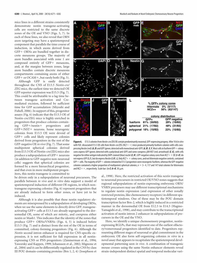
These results indicate that, at E13.5, the OE contains regions(like zone 1) that are induced, committed, and highly neuro-genic, but also contains other GFP� regions that are also highlyneurogenic, and with some regions still containing multipotentprogenitors that have yet to be committed. To test this, we nextplated OE cells from E13.5 Nestin-cre/ZEG mice at clonal density,to test whether embryonic progenitors in vitro demonstrate asimilar pattern of neurogenic potential to that observed in vivo(Fig. 6). Because of the low level of expression of endogenousGFP in some cells, we used anti-GFP immunodetection to en-hance GFP detection. After 10 d in vitro, 56% of all coloniesderived from Nestin-cre/ZEG mice contained GFP� cells (Fig.6A,B,E,F,L). All fusiform colonies were neurogenic, and allGFP� colonies contained NST�/GFP� cells (Fig. 6E–G).Within the GFP� colonies, 90% of the cells expressed detect-able levels of GFP (Fig. 6A,B,E,F), and occasional cells that ap-peared to be GFP� (or low) tended to be situated at the colonycore. GFP� colonies were predominantly neurogenic fusiform,containing a robust population of GFP�/nestin� or GFP�/NST� cells (Fig. 6A–H,L). Twenty-nine percent of GFP-negativecolonies were also fusiform and contained nestin�/GFP� bipo-lar RGLPs (Fig. 6 I–L), but the predominant GFP-negative colonyphenotype (48%), presumably from progenitors situated outsidezone 1, was the more multipotent spherical (Fig. 6L).
These in vitro/vivo results collectively suggest that, as early asE13.5, a subset of committed neurogenic progenitors is predes-tined to generate subsets of GFP� ORNs, with other progenitorsdestined to seed other GFP-negative OE regions. They also indi-cate that the subpopulation of nestin-expressing neurogenicRGLPs that drive the second intron enhancer elements of theNestin-cre transgene, give rise to zonally restricted ORNs andVNRNs, and are highly represented when assayed in vitro atE13.5. Despite the clear contribution of E13.5 OE progenitors tothe production of GFP� neurons in vitro, the OE of Nestin-cre/ZEG crosses at E13.5 was negative for GFP (Fig. 7A,B), withGFP� cells first detectable at E15.5 (Fig. 7C–E). To test whethertransgene-expressing precursors were present, but that GFP wasnot detectable because of a lag between the time taken to activateCre and drive excision to produce GFP to a detectable level, wenext used the identical nestin regulatory elements to directly driveGFP expression, and tested for coexpression and distribution ofnestin�/GFP� cells (Mignone et al., 2004). In the Nestin-GFPmice, GFP was detected throughout the E13.5 CNS, as previouslyshown (Fig. 7F) (data not shown), but was restricted to the
Table 1. Distribution of OE E13.5 colony subtypes with FGF or EGF
GF
Colony subtype
Spherical (%) Fusiform (%) Polygonal (%)
FGF 52 � 6 25 � 5 23 � 2EGF 44 � 4* 16 � 2 40 � 4*
Values are means � SEM. E13.5 colony subtypes were assessed after 10 d in vitro in serum-free cultures with basicFGF or EGF. In EGF, spherical and polygonal colonies are significantly greater than fusiform.
*p � 0.001; n � 5– 6 independent experiments.
Murdoch and Roskams • Novel Embryonic Chemosensory Neuron Progenitors J. Neurosci., April 16, 2008 • 28(16):4271– 4282 • 4277

dorsal-medial OE, in which it was found ina subpopulation of nestin-expressing cells,although endogenous nestin protein wasevident throughout the rest of the embry-onic OE (Fig. 7F–I). Because GFP is brokendown inefficiently in neurons (see GEN-SAT) (Maskos et al., 2002; Gong et al.,2003), its retention allows us to test whetherGFP� cells that are the progeny oftransgene-expressing precursors, also ex-press markers characteristic of differentORN developmental stages. In the Nestin-GFP E13.5 OE, GFP was also found in di-viding apical cells (Fig. 7J–L), a subpopula-tion of Mash1� neuronal progenitors withelongated nuclei of a migratory phenotype(Fig. 7M,N), and ORNs in the dorsal-medial region. These data indicate that onlya subpopulation of nestin-expressing cellsactivate the regulatory elements of this nes-tin transgene in the OE, and generate atleast some GFP� ORNs via Mash1-expressing intermediate precursors.
DiscussionThe identity and spatiotemporal regulationof embryonic OE progenitors at differentages is not well understood. Here, oursearch for a unique embryonic OE progen-itor has revealed a distinct nestin�/lin-eage� precursor that shares antigenic andmorphologic characteristics with, but ismolecularly distinct from, multipotentCNS radial glia. Nestin-cre-mediated lin-eage analysis demonstrates only a subset ofnestin-expressing precursors of embryonicOE and VNO drive nestin transgene expres-sion to produce ORNs and VNRNs in azonally restricted pattern, whose neuronalrestriction is recapitulated in colonies de-rived from E13.5 OE. These data suggestcommon conserved regulatory mecha-nisms of neurogenesis between differentchemosensory neuron progenitors.
In the embryonic OE, there are distinctapical and basal subsets of progenitors thatbecome progressively restricted to only thebasal OE in the adult (Smart, 1971; Mur-doch and Roskams, 2007) (Fig. 1). The en-richment of progenitors undergoing cyto-kinesis at the apical embryonic OE is highlyreminiscent of the early embryonic cortex,in which apical OE corresponds to the ven-tricular zone and the basal OE to the corti-
Figure 4. Nestin-Cre/ZEG lineage tracing reveals zonally restricted production of olfactory and vomeronasal receptor neu-rons. A–E, In adult OMP-cre/ZEG mice, Cre transgene expression (red) is detected in the neuronal layer throughout the OE. Cre isexpressed at variable levels (B) in a subpopulation of OMP� (green) ORNs (C). D, E, Throughout the OE, GFP reporter expressionoverlaps with OMP� ORNs. F–L, Nestin-cre/ZEG mice express GFP (green) (F ) in a subpopulation of ORNs in restricted zones.Most GFP� ORNs coexpress OMP (red) (F, G), and a smaller subpopulation coexpress NST (red) (H, I ). J, At P14, endogenous GFP
4
is detected in the OB, OE, and OMP (red) (K ) and NST (red) (L) inneuronal subpopulations in the VNO. The dotted line indicatesthe basal lamina. Asterisks indicate ORNs that do not expressGFP. Sus, Sustentacular cells; Sep, septum; Ax, axon bundles;BC, basal cells. The asterisks indicate dorsal recess. Scale bars:(in A, F ) A, D and F, H, respectively, 100 �m; (in B) B, C, K, L, 50�m; (in E) E, G, I, 50 �m; J, 50 �m.
4278 • J. Neurosci., April 16, 2008 • 28(16):4271– 4282 Murdoch and Roskams • Novel Embryonic Chemosensory Neuron Progenitors

cal plate (Gotz et al., 2005). Embryonic OE progenitors at differ-ent mitotic stages could thus be regulated by local cues, in whichmesenchymally derived neural inducers could stimulate mitosisor differentiation in basal, but not apical, progenitors (LaMantiaet al., 2000), which are more likely to respond to signals withinthe apical OE. This highly conserved morphological organizationof progenitors lead us to test whether any molecular similaritiesexist between the embryonic ventricular zone and OE. We foundnestin was expressed by a previously unidentified population ofmitotic, lineage-negative, epithelium-spanning embryonic OEcells that display many of the morphological and antigenic char-acteristics of CNS radial glia, including coexpression of RC2 andGLAST. We refer to these as RGLPs. A small population of RGLPsremain by P5, but are undetectable in the adult OE (Fig. 1).
Although nestin is clearly expressed by CNS progenitors, itsreported expression in the OE [in the endfeet of adult rat susten-tacular cells in vivo, purified lamina propria-derived OECs, basalcell lines, and OE-derived neurospheres in vitro (Doyle et al.,2001; Au and Roskams, 2003; Zhang et al., 2004)] has varieddepending on animal model (rat, mouse), age, or approach used,and when considered alone, is insufficient to indicate aprogenitor-like activity. In contrast to the CNS, the neurogenicradial glial protein, BLBP, is expressed only by cells in the OEClineage that ensheathe the axons of ORNs (Fig. 1) (Murdoch andRoskams, 2007). BLBP expression could be induced by Notch 1activation (in OEC gliogenic precursors) (Carson et al., 2006),and maintained in axon-ensheathing OECs, a hypothesis sup-ported by lineage tracing in BLBP-cre/Rosa26 crosses (Anthonyet al., 2004; Murdoch and Roskams, 2007).
Nestin is also the earliest detectable antigen in adherent cells
forming colonies from E13.5 OE, in whichthe majority of actively cycling cells after10 d are bipolar nestin-expressing cells sim-ilar to the RGLPs found in vivo (Fig. 2), andmay represent the earliest OE progenitoridentified to date. Embryonic OE yieldsthree distinct colonies of a three-dimensional (semiadherent) phenotype.Colony heterogeneity, coupled with dis-tinct morphological and antigen expressionprofiles of progeny after 10 DIV (Figs. 2, 3),suggests each colony subtype is likelyformed by progenitors at different stages ofinduction or commitment. All coloniescontain mitotic nestin� cells, in which nes-tin� cells at a distance from the colony coreassume a more differentiated glial (BLBP orS100�-expressing) phenotype. Fusiformcolonies contain the highest percentage ofmitotic bipolar PCNA�, nestin� cells,NST� neurons, and Mash1� mitotic cells(data not shown). That fusiform colony andNST� ORN production is specifically en-hanced by FGF (Table 1), a known regula-tor of neurogenesis in both the CNS(Vescovi et al., 1993) and OE (Calof et al.,1998), suggests that fusiform colonies con-tain the highest proportion of transit ampli-fying neurogenic precursors. Polygonal col-onies preferentially respond to EGF, whichspecifically enhances gliogenesis develop-mentally (Kuhn et al., 1997; Qian et al.,2000) (Table 1), and contain a high propor-
tion of BLBP�/S100�� expressing cells, in close proximity toexpansive populations of S100�� OEC-like cells (Au andRoskams, 2003; Carson et al., 2006). Polygonal colonies are thusmore likely founded by progenitors committed to (or defaulttoward) gliogenesis. Finally, bipotential spherical colonies ex-pand in response to EGF and FGF together (Table 1), in whichFGF specifically enhances the production of radially arrayed bi-polar nestin� RGLPs. Spherical colonies contain a high percent-age of nestin� mitotic cells, with NST� neurons and blast-likecells of both neuronal and glial lineages loosely attached to thesurface of the surrounding adherent cell layer (Figs. 2, 3). Giventhat spherical colonies are also mostly absent from P5 cultures,they may be formed by a population of more primitive embry-onic multipotent progenitors that do not persist into the adult.
Are the nestin� RGLPs detected in vivo, and enriched in mul-tipotent colonies in vitro, the multipotent progenitors of the em-bryonic OE? If so, then Nestin-cre/ZEG lineage tracing shouldreveal this. Instead, Nestin-cre-expressing cells produce onlyORNs regionally restricted to the OCAM-negative region classi-cally defined as zone 1 (Ressler et al., 1993), in addition to theVR1 region of the VNO (Figs. 4, 5). This OCAM-negative Nestintransgene-expressing zone also corresponds to the dorsomedialD-zone (Oka et al., 2003; Iwema et al., 2004; Miyamichi et al.,2005; Kobayakawa et al., 2007). Although ZEG transgene silenc-ing in other OE zones could account for this restriction, it isunlikely, because many ORNs outside zone 1 continue to expressLacZ, and OMP-cre/ZEG mice demonstrate Cre-mediated exci-sion in mature ORNs in all OE zones. Also, despite the expressionof endogenous nestin in RGLPs throughout the OE (from as earlyas E10), the use of additional reporters and alternative Nestin-cre
Figure 5. Nestin regulatory elements direct reporter expression to a subpopulation of cells in the OCAM-negative dorsal-medial zone. A, Nestin-cre/ZEG and Nestin-cre/Rosa YFP (C, D) mice express GFP/YFP (green) in the dorsal-medial OE, zone 1(zones 1– 4 indicated in B), devoid of OCAM expression (red) (A, C, D). C�, Inset, ORN axons segregate to form axon bundles (Ax)that are mostly either OCAM� (arrows) or YFP� (arrowheads), or OCAM�/YFP�. E, F, Identical patterns of reporter expres-sion (�-galactosidase; blue), in a subpopulation of zone 1 neurons, are seen in an independently derived Nestin-cre line whencrossed with a Rosa26R reporter mouse. Sep, Septum. The asterisks indicate dorsal recess. The dotted lines indicate zone 1 (A),zones 1– 4 (B), and basal lamina (D, F ). Scale bars: A, 200 �m; C, E, 50 �m; (in D) D, F, 50 �m.
Murdoch and Roskams • Novel Embryonic Chemosensory Neuron Progenitors J. Neurosci., April 16, 2008 • 28(16):4271– 4282 • 4279
mice lines in a different strains consistentlydemonstrate nestin transgene-activatingcells are restricted to the same discretezones of the OE and VNO (Figs. 5, 7). Ineach of these lines, we also reveal that ORNaxon targeting may have a spatiotemporalcomponent that parallels the time course ofinduction, in which axons derived fromGFP� ORNs are bundled together in dis-crete mesaxons groups. The majority ofaxon bundles associated with zone 1 arecomposed entirely of GFP� mesaxons,and, at the margins between zones, largeaxon bundles contain discrete mesaxonscompartments containing axons of eitherGFP� or OCAM�, but rarely both (Fig. 5).
Although GFP is easily detectedthroughout the CNS of E13.5 Nestin-cre/ZEG mice, the earliest time we detected OEGFP reporter expression was E15.5 (Fig. 7).This could be attributable to a lag time be-tween transgene activation and Cre-mediated excision, followed by sufficienttime for GFP accumulation (Miyoshi andFishell, 2006). In support of this, progenitorassays (Fig. 6) indicate that the E13.5 OE ofNestin-cre/ZEG mice is highly enriched inprogenitors that produce colonies contain-ing GFP�/nestin� progenitors andGFP�/NST� neurons. Some neurogeniccolonies from E13.5 OE were devoid ofGFP� cells and likely represent coloniesderived from progenitors in the rest of theGFP-negative OE in vivo (Fig. 7). That somemultipotent spherical colonies derivedfrom E13.5 OE of Nestin-cre/ZEG mice alsoproduce a subpopulation of GFP� neurons(in addition to GFP-negative non-neuronalcells) suggests that spherical colonies areformed by a more hierarchical progenitor,and that even in more multipotent progen-itors, this nestin transgene is committed tobe driven only in a subpopulation of neuronal precursors. Theparallels between in vivo and in vitro data support a model ofspatiotemporal induction of different OE regions, in which non-transgene expressing colonies (Fig. 6) represent progenitors thatare already induced to form other zones, or have yet to beinduced.
Although it is also possible that these nestin regulatory ele-ments are misexpressed by a subpopulation of developing ORNs,when we use the same elements to directly drive GFP, transgene-activating GFP� cells at E13.5 are already segregated to the dor-somedial OE, some of which are mitotic, and coexpress eithernestin or Mash1. This indicates that the identity of the zones thatproduce GFP� ORNs/VNRNs (Figs. 4, 5) is induced beforeE13.5 (Fig. 7) and is distinct from the rest of the OE in terms ofcommitted, colony-forming progenitors (Fig. 6). Although theNestin second intron enhancer is required for CNS-specific ex-pression, it is not sufficient for full expression in all nestin-expressing CNS or PNS progenitors (Zimmerman et al., 1994;Yaworsky and Kappen, 1999; Johansson et al., 2002; Mignone etal., 2004) and it can be differentially regulated in the CNS by classIII POU domain-containing proteins (Brn 1, 2, 4) (Josephson et
al., 1998). Here, the restricted activation of this nestin transgeneto neuronal precursors in restricted OE/VNO zones suggests thatregional subpopulations of nestin-expressing embryonic ORN/VNRN precursors may use different transcriptional mechanismsto regulate nestin expression (and expression of other zonallyrestricted proteins, like chemosensory receptors) in different spa-tiotemporal windows. One of these may be the POU domaintranscription factor Brn-2, which is highly induced in a restrictedmanner in the dorsomedial OE from E12.5 to E14.5 (Hagino-Yamagishi et al., 1998), and may contribute to the transcriptionalactivation of nestin intron 2 enhancer in subpopulations of pre-cursors in the OE and the VNO.
Here, we identify a unique chemosensory progenitor, nestin-expressing RGLPs, that may represent one of the earliest olfacto-ry/vomeronasal progenitors identified to date. Progenitors rep-resenting different stages of neuronal or glial commitment in theembryonic OE also form self-organizing colonies in vitro in anovel assay that appears to recapitulate the spatiotemporal devel-opmental patterning seen in vivo. A combination of transgenicmouse crosses using the same Nestin enhancer elements revealstrain-independent distinct spatial and temporal molecular vari-
Figure 6. E13.5 colonies from Nestin-cre/ZEG OE contain predominantly neuronal, GFP-expressing progeny. After 10 d in vitrowith FGF, dissociated E13.5 OE cells from Nestin-cre/ZEG (NCZ��) mice produced primarily fusiform colonies with cells coex-pressing Nestin (red) (A, D) and GFP (green; detected with monoclonal anti-GFP) (A, B). E, F, Most cells in fusiform GFP� colonycores express GFP (green; detected with a polyclonal anti-GFP) and some coexpress �III NST (red; arrowhead) (E, G), with cellsnegative for either antigen indicated by DAPI-stained (blue) nuclei (E, H ). GFP-negative colony cores from NCZ�� OE (I–K ) donot express GFP (I, J ), but do express Nestin (I, K ). L, Only NCZ�� colony cores, and not littermate negative controls, containedGFP� cells. The majority of GFP� colonies initiated be E13.5 progenitors were neurogenic fusiform, whereas the GFP-negativecolonies contained a higher proportion of multipotent spherical colonies; n � 3– 4, 117 and 147 total colonies for littermatesand NCZ��, respectively. Scale bar: (in C) A–K, 50 �m.
4280 • J. Neurosci., April 16, 2008 • 28(16):4271– 4282 Murdoch and Roskams • Novel Embryonic Chemosensory Neuron Progenitors

ations among OE and VNO progenitor subpopulations that mayshare a common developmental origin, or a common mode ofinduction and regulation. In so doing, we also reveal that ORNsderived from reporter-expressing or OCAM-expressing zonesare segregated within axon bundle subcompartments that reflecttheir spatiotemporal origin.
ReferencesAnthony TE, Klein C, Fishell G, Heintz N (2004) Radial glia serve as neuro-
nal progenitors in all regions of the central nervous system. Neuron41:881– 890.
Au E, Roskams AJ (2003) Olfactory ensheathingcells of the lamina propria in vivo and in vitro.Glia 41:224 –236.
Bauer S, Rasika S, Han J, Mauduit C, Raccurt M,Morel G, Jourdan F, Benahmed M, Moyse E,Patterson PH (2003) Leukemia inhibitory fac-tor is a key signal for injury-induced neurogen-esis in the adult mouse olfactory epithelium.J Neurosci 23:1792–1803.
Berube NG, Mangelsdorf M, Jagla M, Vanderluit J,Garrick D, Gibbons RJ, Higgs DR, Slack RS,Picketts DJ (2005) The chromatin-remodeling protein ATRX is critical for neuro-nal survival during corticogenesis. J Clin Invest115:258 –267.
Calof AL, Mumm JS, Rim PC, Shou J (1998) Theneuronal stem cell of the olfactory epithelium.J Neurobiol 36:190 –205.
Calof AL, Bonnin A, Crocker C, Kawauchi S, Mur-ray RC, Shou J, Wu HH (2002) Progenitorcells of the olfactory receptor neuron lineage.Microsc Res Tech 58:176 –188.
Carson C, Murdoch B, Roskams AJ (2006) Notch2 and Notch 1/3 segregate to neuronal and gliallineages of the developing olfactory epithelium.Dev Dyn 235:1678 –1688.
Carter LA, MacDonald JL, Roskams AJ (2004) Ol-factory horizontal basal cells demonstrate a con-served multipotent progenitor phenotype.J Neurosci 24:5670 –5683.
Cau E, Gradwohl G, Fode C, Guillemot F (1997)Mash1 activates a cascade of bHLH regulators inolfactory neuron progenitors. Development124:1611–1621.
Danciger E, Mettling C, Vidal M, Morris R, Margo-lis F (1989) Olfactory marker protein gene: itsstructure and olfactory neuron-specific expres-sion in transgenic mice. Proc Natl Acad Sci USA86:8565– 8569.
Doyle KL, Khan M, Cunningham AM (2001) Ex-pression of the intermediate filament proteinnestin by sustentacular cells in mature olfactoryneuroepithelium. J Comp Neurol 437:186 –195.
Farbman AI (1990) Olfactory neurogenesis: ge-netic or environmental controls? Trends Neu-rosci 13:362–365.
Farbman AI (1992) Cell biology of olfaction.Cambridge, UK: Cambridge UP.
Farbman AI, Buchholz JA (1996) Transforminggrowth factor-alpha and other growth factorsstimulate cell division in olfactory epithelium invitro. J Neurobiol 30:267–280.
Feng L, Hatten ME, Heintz N (1994) Brain lipid-binding protein (BLBP): a novel signaling sys-tem in the developing mammalian CNS. Neu-ron 12:895–908.
Furuta A, Rothstein JD, Martin LJ (1997) Gluta-mate transporter protein subtypes are expresseddifferentially during rat CNS development.J Neurosci 17:8363– 8375.
Getchell TV, Narla RK, Little S, Hyde JF, GetchellML (2000) Horizontal basal cell proliferation in the olfactory epithe-lium of transforming growth factor-alpha transgenic mice. Cell TissueRes 299:185–192.
Getchell TV, Shah DS, Partin JV, Subhedar NK, Getchell ML (2002) Leuke-mia inhibitory factor mRNA expression is upregulated in macrophagesand olfactory receptor neurons after target ablation. J Neurosci Res67:246 –254.
Goldstein BJ, Schwob JE (1996) Analysis of the globose basal cell compart-ment in rat olfactory epithelium using GBC-1, a new monoclonal anti-body against globose basal cells. J Neurosci 16:4005– 4016.
Gong S, Zheng C, Doughty ML, Losos K, Didkovsky N, Schambra UB, Nowak
Figure 7. Regional restriction of GFP� reporter cells in both embryonic Nestin-cre/ZEG mice and Nestin-GFP transgenic mice.A, In E13.5 Nestin-cre/ZEG mice, GFP (green) is readily detected in the CNS OB, but not the OE, in which NST (red) (A, B) is highlyexpressed in olfactory receptor neuron axons. C, First detected at E15.5 in the dorsal-medial OE and separate from emergingregions of OCAM expression (indicated by arrowheads), are the progeny of Nestin transgene-activation, GFP� cells. D, GFP�cells either coexpress NST (asterisk), or are NST negative, both above (arrow) and below (arrowhead) the NST� ORNs. E, RareGFP� cells, close to the basal lamina, coexpress nestin (red; arrowhead; inset, nestin alone). F, E13.5 Nestin-GFP embryos highlyexpress Nestin transgene-activated GFP that is restricted to a subpopulation of cells in the dorsal-medial OE, despite endogenousNestin expression throughout the OE. GFP can be detected in a subpopulation of Nestin� cells and cell processes spanning theOE (thick and thin arrowheads, respectively) (G–I ), surrounding cells undergoing cytokinesis at the apical OE (arrowheads)(J–L), and in neuronal precursors expressing Mash1 (arrowheads) (M, N ). A–D show anti-GFP immunofluorescence, and E–Nshow endogenous GFP fluorescence. Ax, Axon bundles; double asterisks indicate dorsal recess. The boxes show regions for highermagnification. The dotted lines indicate basal lamina. Scale bars: (in A, C, F, M ) A, C, F, M, N, 50 �m; (in B, D, G) B, D, E, G–L, 25�m.
Murdoch and Roskams • Novel Embryonic Chemosensory Neuron Progenitors J. Neurosci., April 16, 2008 • 28(16):4271– 4282 • 4281

NJ, Joyner A, Leblanc G, Hatten ME, Heintz N (2003) A gene expressionatlas of the central nervous system based on bacterial artificial chromo-somes. Nature 425:917–925.
Gotz M, Huttner WB, Haubensak W, Attardo A, Denk W (2005) The cellbiology of neurogenesis. Nat Rev Mol Cell Biol 6:777–788.
Graziadei PP, Graziadei GA (1979) Neurogenesis and neuron regenerationin the olfactory system of mammals. I. Morphological aspects of differen-tiation and structural organization of the olfactory sensory neurons.J Neurocytol 8:1–18.
Hagino-Yamagishi K, Saijoh Y, Yamazaki Y, Yazaki K, Hamada H (1998)Transcriptional regulatory region of Brn-2 required for its expression indeveloping olfactory epithelial cells. Brain Res Dev Brain Res 109:77– 86.
Hitoshi S, Alexson T, Tropepe V, Donoviel D, Elia AJ, Nye JS, Conlon RA,Mak TW, Bernstein A, van der Kooy D (2002) Notch pathway moleculesare essential for the maintenance, but not the generation, of mammalianneural stem cells. Genes Dev 16:846 – 858.
Hockfield S, McKay RD (1985) Identification of major cell classes in thedeveloping mammalian nervous system. J Neurosci 5:3310 –3328.
Imayoshi I, Ohtsuka T, Metzger D, Chambon P, Kageyama R (2006) Tem-poral regulation of Cre recombinase activity in neural stem cells. Genesis44:233–238.
Iwema CL, Fang H, Kurtz DB, Youngentob SL, Schwob JE (2004) Odorantreceptor expression patterns are restored in lesion-recovered rat olfactoryepithelium. J Neurosci 24:356 –369.
Johansson CB, Lothian C, Molin M, Okano H, Lendahl U (2002) Nestinenhancer requirements for expression in normal and injured adult CNS.J Neurosci Res 69:784 –794.
Josephson R, Muller T, Pickel J, Okabe S, Reynolds K, Turner PA, Zimmer A,McKay RD (1998) POU transcription factors control expression of CNSstem cell-specific genes. Development 125:3087–3100.
Joyner AL, Zervas M (2006) Genetic inducible fate mapping in mouse: es-tablishing genetic lineages and defining genetic neuroanatomy in the ner-vous system. Dev Dyn 235:2376 –2385.
Kobayakawa K, Kobayakawa R, Matsumoto H, Oka Y, Imai T, Ikawa M,Okabe M, Ikeda T, Itohara S, Kikusui T, Mori K, Sakano H (2007) In-nate versus learned odour processing in the mouse olfactory bulb. Nature450:503–508.
Kuhn HG, Winkler J, Kempermann G, Thal LJ, Gage FH (1997) Epidermalgrowth factor and fibroblast growth factor-2 have different effects onneural progenitors in the adult rat brain. J Neurosci 17:5820 –5829.
LaMantia AS, Bhasin N, Rhodes K, Heemskerk J (2000) Mesenchymal/epi-thelial induction mediates olfactory pathway formation. Neuron28:411– 425.
Leung CT, Coulombe PA, Reed RR (2007) Contribution of olfactory neuralstem cells to tissue maintenance and regeneration. Nat Neurosci10:720 –726.
MacDonald JL, Gin CS, Roskams AJ (2005) Stage-specific induction ofDNA methyltransferases in olfactory receptor neuron development. DevBiol 288:461– 473.
Maskos U, Kissa K, St Cloment C, Brulet P (2002) Retrograde trans-synaptictransfer of green fluorescent protein allows the genetic mapping of neu-ronal circuits in transgenic mice. Proc Natl Acad Sci USA99:10120 –10125.
Mignone JL, Kukekov V, Chiang AS, Steindler D, Enikolopov G (2004)Neural stem and progenitor cells in nestin-GFP transgenic mice. J CompNeurol 469:311–324.
Misson JP, Edwards MA, Yamamoto M, Caviness Jr VS (1988) Identifica-tion of radial glial cells within the developing murine central nervoussystem: studies based upon a new immunohistochemical marker. BrainRes Dev Brain Res 44:95–108.
Miyamichi K, Serizawa S, Kimura HM, Sakano H (2005) Continuous andoverlapping expression domains of odorant receptor genes in the olfac-tory epithelium determine the dorsal/ventral positioning of glomeruli inthe olfactory bulb. J Neurosci 25:3586 –3592.
Miyoshi G, Fishell G (2006) Directing neuron-specific transgene expressionin the mouse CNS. Curr Opin Neurobiol 16:577–584.
Murdoch B, Roskams AJ (2007) Olfactory epithelium progenitors: insightsfrom transgenic mice and in vitro biology. J Mol Histol 38:581–599.
Nan B, Getchell ML, Partin JV, Getchell TV (2001) Leukemia inhibitoryfactor, interleukin-6, and their receptors are expressed transiently in theolfactory mucosa after target ablation. J Comp Neurol 435:60 –77.
Novak A, Guo C, Yang W, Nagy A, Lobe CG (2000) Z/EG, a double reportermouse line that expresses enhanced green fluorescent protein upon Cre-mediated excision. Genesis 28:147–155.
Oka Y, Kobayakawa K, Nishizumi H, Miyamichi K, Hirose S, Tsuboi A,Sakano H (2003) O-MACS, a novel member of the medium-chain acyl-CoA synthetase family, specifically expressed in the olfactory epitheliumin a zone-specific manner. Eur J Biochem 270:1995–2004.
Qian X, Shen Q, Goderie SK, He W, Capela A, Davis AA, Temple S (2000)Timing of CNS cell generation: a programmed sequence of neuron andglial cell production from isolated murine cortical stem cells. Neuron28:69 – 80.
Ressler KJ, Sullivan SL, Buck LB (1993) A zonal organization of odorantreceptor gene expression in the olfactory epithelium. Cell 73:597– 609.
Reynolds BA, Weiss S (1992) Generation of neurons and astrocytes fromisolated cells of the adult mammalian central nervous system. Science255:1707–1710.
Richter MW, Fletcher PA, Liu J, Tetzlaff W, Roskams AJ (2005) Laminapropria and olfactory bulb ensheathing cells exhibit differential integra-tion and migration and promote differential axon sprouting in the le-sioned spinal cord. J Neurosci 25:10700 –10711.
Roskams AJ, Bethel MA, Hurt KJ, Ronnett GV (1996) Sequential expressionof Trks A, B, and C in the regenerating olfactory neuroepithelium. J Neu-rosci 16:1294 –1307.
Schwob JE (2002) Neural regeneration and the peripheral olfactory system.Anat Rec 269:33– 49.
Smart IH (1971) Location and orientation of mitotic figures in the develop-ing mouse olfactory epithelium. J Anat 109:243–251.
Soriano P, MacGregor GR, Zambrowicz BP (1999) Generalized lacZ expres-sion with the ROSA26 Cre reporter strain. Nat Genet 21:70 –71.
Srinivas S, Watanabe T, Lin CS, William CM, Tanabe Y, Jessell TM, Costan-tini F (2001) Cre reporter strains produced by targeted insertion ofEYFP and ECFP into the ROSA26 locus. BMC Dev Biol 1:4.
Tronche F, Kellendonk C, Kretz O, Gass P, Anlag K, Orban PC, Bock R, KleinR, Schutz G (1999) Disruption of the glucocorticoid receptor gene in thenervous system results in reduced anxiety. Nat Genet 23:99 –103.
Vescovi AL, Reynolds BA, Fraser DD, Weiss S (1993) bFGF regulates theproliferative fate of unipotent (neuronal) and bipotent (neuronal/astro-glial) EGF-generated CNS progenitor cells. Neuron 11:951–966.
Waseem NH, Lane DP (1990) Monoclonal antibody analysis of the prolif-erating cell nuclear antigen (PCNA). Structural conservation and the de-tection of a nucleolar form. J Cell Sci 96:121–129.
Weissman IL, Anderson DJ, Gage F (2001) Stem and progenitor cells: ori-gins, phenotypes, lineage commitments, and transdifferentiations. AnnuRev Cell Dev Biol 17:387– 403.
Yaworsky PJ, Kappen C (1999) Heterogeneity of neural progenitor cells re-vealed by enhancers in the nestin gene. Dev Biol 205:309 –321.
Yoshihara Y, Kawasaki M, Tamada A, Fujita H, Hayashi H, Kagamiyama H,Mori K (1997) OCAM: a new member of the neural cell adhesion mol-ecule family related to zone-to-zone projection of olfactory and vomero-nasal axons. J Neurosci 17:5830 –5842.
Zhang X, Klueber KM, Guo Z, Lu C, Roisen FJ (2004) Adult human olfac-tory neural progenitors cultured in defined medium. Exp Neurol186:112–123.
Zimmerman L, Parr B, Lendahl U, Cunningham M, McKay R, Gavin B, MannJ, Vassileva G, McMahon A (1994) Independent regulatory elements inthe nestin gene direct transgene expression to neural stem cells or muscleprecursors. Neuron 12:11–24.
4282 • J. Neurosci., April 16, 2008 • 28(16):4271– 4282 Murdoch and Roskams • Novel Embryonic Chemosensory Neuron Progenitors