dissecting the role of copi comple xes on influenza virus
TRANSCRIPT

Dissecting the Role of COPI Complexes in Influenza Virus Infection
Eileen Sun,a,c Jiang He,b,c Xiaowei Zhuangc,d,e
Program in Virology, Harvard Medical School,a Department of Molecular and Cellular Biology,b Department of Chemistry and Chemical Biology,c Department of Physics,d
and Howard Hughes Medical Institute,e Harvard University, Cambridge, Massachusetts, USA
As an obligate pathogen, influenza virus requires host cell factors and compartments to mediate productive infection and to pro-duce infectious progeny virus. Recently, several small interfering RNA (siRNA) knockdown screens revealed influenza virus hostdependency proteins, all of which identified at least two subunits of the coat protein I (COPI) complex. COPI proteins oligomer-ize to form coated vesicles that transport contents between the Golgi apparatus and the endoplasmic reticulum, and they havealso been reported to mediate endosomal trafficking. However, it remains unclear which steps in the influenza virus infectioncycle rely on the COPI complex. Upon systematic dissection of the influenza virus infection cycle, from entry to progeny virionproduction, we found that prolonged exposure to COPI complex disruption through siRNA depletion resulted in significantdefects in virus internalization and trafficking to late endosomes. Acute inhibition of COPI complex recruitment to the Golgiapparatus with pharmacological compounds failed to recapitulate the same entry defects as observed with the COPI-depletedcells but did result in specific decreases in viral membrane protein expression and assembly, leading to defects in progeny virionproduction. Taken together, our findings suggest that COPI complexes likely function indirectly in influenza virus entry but playdirect roles in viral membrane protein expression and assembly.
Influenza virus is a negative-sense strand RNA enveloped virusthat contains eight segmented genomes which encode 13 viral
proteins. The influenza virus virion envelope is derived from theinfected cell’s plasma membrane and contains three integral viralmembrane proteins: the M2 proton channel, hemagglutinin(HA), and neuraminidase (NA). Encapsulated within the viralenvelope is a coat of matrix protein 1 (M1), which forms contactswith each of the viral ribonucleoprotein (vRNP) complexes. Eachof the vRNPs consists of a strand of viral RNA bound to viralnucleoprotein (NP). In addition, each vRNP is bound to an RNA-dependent RNA polymerase complex containing three proteins:PA, PB1, and PB2 (1).
Influenza A virus is the causative agent of seasonal flu andhistorically has led to pandemic infections, such as the 1918 Span-ish influenza outbreak, which killed an estimated 50 million peo-ple worldwide (1). At a cellular level, influenza virus infectionstarts with virion attachment to cell surface sialylated glycopro-teins or glycolipids (2). The virus particle then triggers endocyto-sis through clathrin-dependent and clathrin-independent path-ways (3–6). Productive entry requires the virus to traffic tolow-pH endosomes (pH � 5.0), at which point HA mediates fu-sion between the viral envelope and the lipid bilayer of the endo-some (1, 4, 7–9). Upon pH-mediated fusion, the vRNPs are re-leased into the cytoplasm and subsequently transported into thenucleus to initiate viral replication and viral protein translation(10). Infected cells produce progeny virions by assembling viralproteins and vRNP complexes at the plasma membrane. Viral NAcleaves cell surface sialic acids to allow assembled virions to budand release from the infected cell’s membrane and thereby initiateanother infection cycle in neighboring uninfected cells (1).
Being an obligate pathogen encoding only 13 viral proteins,influenza virus hijacks host proteins, cellular compartments/or-ganelles in order to harbor infection and produce progeny virus.Four genome-wide knockdown screens, one in Drosophila cellsand three in human cells (11–14), recently identified host proteinsimportant for influenza virus infection. One group of host depen-dency proteins identified in all influenza screens included sub-
units of the coat protein I (COPI) complex (11–14). There arenine different COPI complex subunits: �, �1, �2, �, ε, �1, �2, �1,and �2. Each COPI complex contains a single copy of the �, �1,�2, �, and ε subunits in addition to one of the following isoformcombinations: �1/�1, �1/�2, or �2/�1 (15–17). Together, COPIcomplexes form a vesicle coat that traffics contents between Golgistacks and cargoes between the Golgi apparatus and the endoplas-mic reticulum (ER) (15, 17, 18). In addition, several studies re-ported a role for COPI complexes in the endocytic pathway, sinceperturbation of these complexes leads to defects in endosomalsorting, multivesicular body (MVB) formation, and/or mem-brane trafficking (19–24).
Previous studies indicated that COPI might play a role in in-fluenza virus entry. König and colleagues reported that �-COPI(ARCN1) knockdown inhibits vRNP nuclear import (14). Giventhe roles of COPI proteins in the endocytic pathway, König andcolleagues hypothesized that the block in vRNP nuclear importmight be due to defects in endosomal trafficking. Viral entry is amultistep process, however, and it remains unclear which step (ifany) is ARCN1 dependent.
Recently, Cureton and colleagues reported that disruption ofCOPI complexes differentially affects vesicular stomatitis virus(VSV) entry versus viral gene expression (25). The authors used atemperature-sensitive CHO cell line (IdlF), which degradesε-COPI at 40°C (23), to show that ε-COPI depletion inhibits virusbinding and internalization as well as transferrin (Tfn) uptake.However, these results were recapitulated only after prolongedtreatment with brefeldin A (BrefA), an inhibitor that preventsGDP-to-GTP exchange of ADP ribosylation factor 1 (ARF1), an
Received 4 September 2012 Accepted 12 December 2012
Published ahead of print 19 December 2012
Address correspondence to Xiaowei Zhuang, [email protected].
Copyright © 2013, American Society for Microbiology. All Rights Reserved.
doi:10.1128/JVI.02277-12
March 2013 Volume 87 Number 5 Journal of Virology p. 2673–2685 jvi.asm.org 2673
Dow
nloa
ded
from
http
s://j
ourn
als.
asm
.org
/jour
nal/j
vi o
n 24
Nov
embe
r 20
21 b
y 83
.48.
179.
46.

essential step for membrane recruitment of COPI to the Golgiapparatus (26). Acute (short-term) treatment with BrefA did notlead to the same effect on viral entry as observed with the IdlF cellsincubated at 40°C, but it did cause a specific decrease in VSV geneexpression.
The results from Cureton and colleagues suggest that indirecteffects caused by long-term inactivation of COPI lead to generaldefects in the clathrin-mediated endocytosis pathway (25). VSVexclusively uses clathrin-mediated endocytosis for infection (27–31). Influenza virus, on the other hand, can productively infectcells in the absence of clathrin-mediated endocytosis. Indeed, sev-eral reports have shown that depletion of key components of theclathrin complex fails to block influenza virus entry and infectionbecause alternative productive endocytic pathways, such as mac-ropinocytosis, can be hijacked by influenza virus (3, 5, 6, 32).Moreover, influenza virus requires lower-pH compartments forviral fusion than for VSV (4, 29, 33). Therefore, how COPI pro-teins are involved in each of the steps of influenza virus entry andinfection remains unclear.
Here, we dissected the different steps of the influenza virusinfection cycle (entry, replication, trafficking, and assembly) inorder to identify the steps that COPI proteins affect. We foundthat the first major entry block in COPI knockdown cells occurs atvirus internalization. In addition, COPI-depleted cells exhibiteddefects in early endosome-to-intermediate/late endosome traf-ficking for virus particles that did manage to enter the cells. Theentry block was not limited to influenza virus only but was alsoobserved for other cargoes (Tfn, epidermal growth factor [EGF],and dextran) that are internalized through either clathrin-medi-ated endocytosis or macropinocytosis. Given the large defect inviral entry upon small interfering RNA (siRNA) silencing ofCOPI, we used pharmacological inhibitors, namely, BrefA andgolgicide A (GCA), to acutely disrupt COPI complexes. With theinhibitor treatment, we found that COPI complexes were not di-rectly required for influenza virus entry. However, disruption offunctional COPI complexes directly inhibited viral membraneprotein expression, assembly of viral components at the plasmamembrane, and production of infectious progeny virus.
MATERIALS AND METHODSAntibodies for immunofluorescence assays or Western blotting. Weused the following primary antibodies for this study: rabbit anti-coatomersubunit � antibody (1:1,000 dilution) (catalog no. ab96725; Abcam),mouse anti-influenza A virus nucleoprotein antibody (AA5H) (1:1,000)(ab20343; Abcam), mouse antinucleoprotein (1:100) (sc-57882; SantaCruz Biotechnology), rabbit anti-�-COPI (1:1,000) (generously donatedby James Rothman), rabbit anti-COPZ1 (1:1,000) (SAB4500896; Sigma-Aldrich), rabbit anti-�1-COP1 (1:10,000) (generously donated by FelixWieland), rabbit anti-COPE (1:1,000) (ab88824; Abcam), mouse anti-�-actin antibody (loading control) (1:1,000) (ab8227; Abcam), rabbit-anti-phospho-signal transducer and activator of transcription 1 (STAT1)Tyr701 (1:1,000) (9167; Cell Signaling Technology), mouse anti-Cy5/Al-exa Fluor 647 (1:1,000) (C1117; Sigma-Aldrich), mouse anti-early endo-some antigen 1 (EEA1) (1:500) (610457; BD Transduction Laboratories),mouse anti-lysobisphosphatidic acid (LBPA) antibody (50 �g, 1:500) (Z-PLBPA; Echelon), mouse anti-influenza A virus M2 protein antibody(14C2) (1:1,000) (ab5416; Abcam), rabbit antineuraminidase (1:5,000)(kind gift from Gillian Air), and mouse anti-PB1 (1:100) (sc-17601; SantaCruz Biotechnology). We used the following secondary antibodies for thisstudy: Alexa Fluor 647 donkey anti-mouse IgG (1:1,000 dilution) (catalogno. A31571; Molecular Probes), Alexa Fluor 555 donkey anti-mouse IgG(1:1,000) (A31570; Molecular Probes), Alexa Fluor 488 donkey anti-
mouse IgG (1:1,000) (A-21202; Molecular Probes), Alexa Fluor 568 don-key anti-rabbit IgG (1:1,000) (A-100042; Molecular Probes), enhancedchemiluminescence (ECL) donkey anti-rabbit IgG, horseradish peroxi-dase (HRP) linked (1:5,000) (NA934; GE Healthcare), and ECL sheepanti-mouse IgG (1:5,000) (NXA931; GE Healthcare).
Cell culture. A549 lung carcinoma cells (ATCC) were cultured in Dul-becco’s modified Eagle medium (DMEM) (Invitrogen) supplementedwith 10% fetal bovine serum (Serum International), 1 mM nonessentialamino acids (ATCC), and antibiotics (25 U/ml penicillin and 25 �g/mlstreptomycin) (ATCC) and were maintained in a humidified environ-ment with 5% CO2 at 37°C. For siRNA knockdown experiments, A549cells were electroporated with 100 pmol of specified COPI siGENOMESMARTpool siRNA (Dharmacon) or AllStars negative-control siRNA(Qiagen) using a Lonza Amaxa Nucleofector with kit T (catalog no.VVCA-1002; Lonza) and program X-001. Experiments were performedwith the siRNA-treated A549 cells 48 h after electroporation. For plasmidexpression, A549 cells were electroporated with 2 �g of either COPE-green fluorescent protein (GFP) (OriGene) or Rab7-GFP (a generous giftfrom Qing Zhong) (34) plasmid using kit T and Nucleofector programX-001. Experiments using these plasmids were performed within 24 hafter electroporation with the plasmids.
A Live/Dead viability/cytotoxicity kit (catalog no. L-3224; Invitrogen)was used according to vendor instructions to measure cell viability, death,and growth. Stimulation of the interferon (IFN) response in A549 cellswas achieved with 10,000 U/ml IFN-� (catalog no. 11101-1; PBL Biomed-ical Laboratories) for 3 h prior to collecting cell lysate.
Spinning disk confocal imaging. Spinning disk confocal z-stacks wereobtained with a custom-built confocal microscope which has been de-scribed in detail (35). Briefly, multicolor fluorescent fixed-cell imagingwas achieved using an Yokogawa spinning disk confocal scan head at-tached to an Olympus IX-71 microscope (Olympus, Center Valley, PA)with an Olympus 60� oil immersion objective (numerical aperture,1.35). A 561-nm crystal laser, 647-nm krypton ion laser, and/or 457/488/514-nm argon laser was used to image the samples with an Andor 885electron-multiplying charge-coupled-device camera.
Cargo uptake assays and data analysis. siRNA-treated A549 cellswere rinsed extensively with phosphate-buffered saline (PBS) and coldbound with Alexa Fluor 568-transferrin (AF568-Tfn) (catalog no.T23365; Invitrogen) and Alexa Fluor 647-epidermal growth factor(AF647-EGF) (E35351; Invitrogen) for 30 min at 4°C. Unbound AF568-Tfn and AF647-EGF were removed with extensive cold PBS washes, andcells were incubated in warm DMEM with antibiotics at 37°C for differenttimes. For fluid-phase uptake, serum-starved A549 cells were incubatedwith 5 mg/ml tetramethylrhodamine (TMR)-dextran (D-1868; Invitro-gen) in warm DMEM with antibiotics at 37°C for different times.
At the indicated time points, surface-bound cargo was removed withacid buffer (PBS adjusted to pH 2.5 with acetic acid) for 2 min, and cellswere fixed with 3.2% paraformaldehyde (PFA) (Electron Microscopy Sci-ences) and imaged with a spinning disk confocal microscope. Confocalz-stacks were obtained for each field of view, which contained about 9 or10 cells per image. For the analysis of Tfn, EGF, or dextran internalization,a MatLab script based on k-clustering analysis was used to establishthresholds for the images to differentiate the background pixels from thesignal pixels. Briefly, an optimized threshold value was determined foreach maximal z-projection image with an iterative thresholding algorithmwhich found the maximal difference between the mean background pixelsand the mean signal pixels. The mean signal intensities of 15 to 20 maxi-mal z-projection images per time point were calculated and averaged.
To test the known effect of amiloride on macropinocytosis, A549 cellswere treated for 60 min with different concentrations of 5-(N-ethyl-N-isopropyl)amiloride (EIPA) (50 �M; catalog no. A3085; Sigma-Aldrich)in DMEM without penicillin-streptomycin (P/S) or fetal bovine serum(FBS). The cells were then incubated with 0.2 mg/ml AF647-dextran for30 min at 37°C prior to low-pH PBS washing, followed by PFA fixation.Additionally, inhibition of dynamin-dependent endocytosis was tested by
Sun et al.
2674 jvi.asm.org Journal of Virology
Dow
nloa
ded
from
http
s://j
ourn
als.
asm
.org
/jour
nal/j
vi o
n 24
Nov
embe
r 20
21 b
y 83
.48.
179.
46.

treatment for 30 min with different doses of Dynasore (D7693; Sigma-Aldrich) in DMEM without P/S or FBS prior to cold binding of 1 �g/mlAF647-Tfn or 1:1,000 AF647-EGF for 30 min. Unbound cargo was re-placed with a warmed inhibitor containing DMEM without FBS or P/S,and cells were incubated at 37°C for 10 min prior to low-pH PBS washing,followed by PFA fixation. For dextran, Tfn, and EGF uptake experiments,imaging of the samples and analyses were performed as described above.
Nonspecific amine-reactive labeling of virus. Fifty microliters of 1mg/ml purified influenza A virus X31, A/Aichi/68 (H3N2) (Charles RiverLaboratories), was mixed with 47 �l of 0.1 M freshly prepared carbonatebuffer (pH �8 to 9) and 6 �g Alexa Fluor 647 carboxylic acid, succinimi-dyl ester (catalog no. A-20106; Invitrogen), dissolved in dimethyl sulfox-ide (DMSO). The mixture was gently mixed in the dark for 60 min. ANap5 size exclusion column (GE Healthcare) was used to separate freedyes from labeled virus using HEPES 145 elution buffer (50 mM HEPES[pH 7.4], 145 mM NaCl). The excess AF647-labeled X31 was aliquoted,snap-frozen, and stored at 80°C (8). No significant difference in infec-tivity was observed between labeled and unlabeled X31 (data not shown).
X31 membrane labeling. One hundred microliters of 1 mg/ml puri-fied X31 was mixed with 3 �l of 25 mM 1,1=-dioctadecyl-3,3,3=,3=-tetram-ethylindodicarbocyanine, 4-chlorobenzenesulfonate salt (DiD solid)(catalog no. D-7757; Invitrogen), dissolved in DMSO. The mixture wasgently vortexed in the dark for 2 h. A Nap5 size exclusion column was usedto separate free dyes from labeled virus using HEPES 145 elution buffer.The excess DiD-labeled X31 was aliquoted, snap-frozen, and stored at80°C. Prior to the use of DiD-labeled X31 for the experiments, the viruswas filtered through a 0.2-�m filter (32). We confirmed with plaque as-says that the DiD-labeled X31 remained infectious.
Bulk viral fusion assay. For the pharmacological compound-treatedcells, all steps in the fusion assay before trypsinization, except the PBSwashes, included the compounds. siRNA- or pharmacological com-pound-treated A549 cells seeded in Lab-Tek 8-well glass dishes wererinsed extensively with PBS and cold bound with 2 � 104 PFU/ml DiD-X31 for 45 min on ice. Unbound virus was removed with extensive PBSwashes, and cells were incubated with DMEM for different times at 37°C.Afterwards, the cells were washed once with PBS and trypsinized. Thetrypsin was neutralized with PBS supplemented with 10% FBS and 30 mMsodium azide, and the cells were collected and placed on ice to prevent anyfurther fusion. Cells were fixed with 2% PFA for 20 min, washed once withPBS, and analyzed immediately with a flow cytometer (BD BiosciencesFortessa). The data were interpreted using FlowJo software.
Virus binding or elderberry lectin binding assays. After 48-h siRNAknockdown, A549 cells were washed extensively with PBS and cold boundwith different concentrations of AF647-X31 in DMEM for 45 min. Un-bound virus was removed through PBS washes, and the cells were imme-diately trypsinized. The trypsin was neutralized with PBS supplementedwith 10% FBS and 30 mM sodium azide. Cells were collected, fixed with2% PFA for 20 min, washed once with PBS, and analyzed immediatelywith a flow cytometer.
For the elderberry lectin binding assays, siRNA-treated cells weretrypsinized and fixed with 2% PFA. The cells were then treated with orwithout different doses of sialidase A (Prozyme) in PBS (pH �5) for 1 h at37°C. The pH was raised to �7 to stop the sialidase A reaction, and cellswere stained for 1 h with a 1:1,000 dilution of fluorescein isothiocyanate(FITC)-conjugated Sambucus nigra (elderberry) bark lectin (SNA-FITC)(Vector Laboratories). After three PBS washes, the samples were immedi-ately analyzed by flow cytometry.
Immunofluorescence for flow cytometry. siRNA- or pharmacologi-cal compound-treated A549 cells were infected with influenza virus at37°C for 9 or 12 h. Infected cells were trypsinized, fixed with 2% PFA inPBS for 20 min at room temperature, and washed once to remove thefixation buffer. The cells were permeablized in buffer P (PBS containing10% FBS and 0.075% saponin) for 5 min. The samples were incubatedwith mouse anti-influenza virus NP or anti-influenza virus M2 (1:1,000dilution in buffer P) for 60 min, washed three times with buffer P, incu-
bated with Alexa Fluor 647 donkey anti-mouse IgG (1:1,000 dilution inbuffer P) for 30 min, washed three times with buffer P, washed once withPBS, and analyzed by flow cytometry.
Immunofluorescence with endosomal markers. To delineate the cellmembrane, fixed A549 cells were first stained with SNA-FITC withoutpermeabilization. For colocalization studies with influenza virus andEEA1, fixed A549 cells were blocked and permeabilized with 3% bovineserum albumin (Jackson Laboratories) and 0.1% Triton X-100 (blockingbuffer) for 30 min at room temperature. The cells were incubated withmouse anti-EEA1 in blocking buffer for 1 h at room temperature, rinsedwith PBS, stained with Alexa Fluor 555 donkey anti-mouse IgG in block-ing buffer for 30 min, and washed with PBS.
For colocalization studies with influenza virus and LBPA, fixed A549cells were blocked and permeabilized with 3% bovine serum albumin(Jackson Laboratories) and 0.075% (wt/vol) saponin (blocking buffer) for30 min at room temperature. All subsequent washes and antibody incu-bations were performed with the blocking buffer, unless stated otherwise.The sample was incubated with mouse anti-LBPA for 1 h at room tem-perature, washed, stained with Alexa Fluor 555 donkey anti-mouse IgGfor 30 min, washed, and rinsed with PBS.
All samples were imaged with a customized spinning disk confocalmicroscope, which is described briefly above and described in more detailelsewhere (35). ImageJ software was used to visualize the fluorescenceconfocal z-stacks, and the fraction of internalized virus particles that co-localized with the respective endosomal marker was determined.
Pharmacological compounds. The following pharmacological com-pounds were used in this study: dimethyl sulfoxide (DMSO) (1:1,000)(catalog no. D2650; Sigma-Aldrich), brefeldin A (10 �g/ml) (B7651; Sig-ma-Aldrich), and golgicide A (10 �M) (345862; EMD). Both the brefeldinA and golgicide A were dissolved in DMSO, and incubations were per-formed in DMEM supplemented with 10% fetal bovine serum, 1 mMnonessential amino acids, and antibiotics. The final amount of DMSO didnot exceed 0.1% (vol/vol). Specific incubation times for each of the ex-periments are described in the figures and legends.
Plaque assays. We collected the supernatant from influenza-infectedcells at 18 or 24 h postinfection (hpi). Serial dilutions of the virus-con-taining supernatant were used to inoculate an �80% confluent mono-layer of Texas-MDCK cells (a generous gift from Robert Lamb) or Verocells (ATCC) grown in 6-well dishes for �1.5 to 2 h at 37°C. The cells werewashed with PBS, coated with 3 ml of 30% Noble agar (Affymetrix) inDMEM containing antibiotics and 2 �g/ml acetylated trypsin (Sigma-Aldrich), and then incubated at 37°C.
Two different detection methods were used to determine the viraltiters. For immunodetection of infected cell colonies (fluorescent foci),the Noble agar disks were removed at �36 hpi, and the cells were fixedwith 100% methanol for 8 to 10 min at 20°C. The cells were rinsed withPBS, blocked and permeabilized with 3% bovine serum albumin and0.1% Triton X-100 in PBS for 30 min, immunostained against NP inblocking buffer for 60 min, washed three times with PBS, incubated withAlexa Fluor 488 donkey anti-mouse IgG in blocking buffer for 30 min, andwashed three times with PBS. The fluorescent foci were visualized andquantified using a Typhoon fluorescence scanner (GE Healthcare).
The second detection method used to determine the viral titers wascounting the numbers of plaques observed 2.5 days postinfection. Todetect plaques, the agar disks were removed, and the cells were immedi-ately fixed and stained with a solution containing 1:1,000 (vol/wt) crystalviolet with 30% ethanol in water. The number of plaques for each condi-tion was counted. The viral titer was determined as (number of foci orplaques)/(dilution � inoculation volume [in ml]). Samples were tested inquadruplicate.
Western blotting. Cell lysates were diluted in Laemmli sample buffer(catalog no. 161-0737; Bio-Rad) containing dithiothreitol to achieve thesame amounts of protein for all samples. The samples were loaded and runon a 4% to 15% Tris-HCl polyacrylamide gel (Bio-Rad). The separatedproteins from the gel were transferred onto Hybond polyvinylidene diflu-
Role of COPI Complexes in Influenza Virus Infection
March 2013 Volume 87 Number 5 jvi.asm.org 2675
Dow
nloa
ded
from
http
s://j
ourn
als.
asm
.org
/jour
nal/j
vi o
n 24
Nov
embe
r 20
21 b
y 83
.48.
179.
46.
oride membranes (GE Healthcare), blocked with 5% nonfat milk in Tris-buffered saline (TBS)-Tween, incubated with the specified primary anti-bodies (reported in the figure legends) overnight at 4°C, washed withTBS-Tween, incubated with HRP-conjugated secondary antibodies for 60min at room temperature, washed with TBS-Tween, detected with Lumi-gen TMA-6 (TMA-100; Lumigen), and exposed/developed onto high-sensitivity Kodak film.
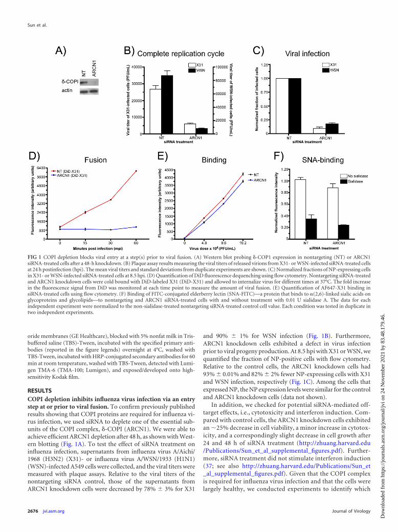
RESULTSCOPI depletion inhibits influenza virus infection via an entrystep at or prior to viral fusion. To confirm previously publishedresults showing that COPI proteins are required for influenza vi-rus infection, we used siRNA to deplete one of the essential sub-units of the COPI complex, �-COPI (ARCN1). We were able toachieve efficient ARCN1 depletion after 48 h, as shown with West-ern blotting (Fig. 1A). To test the effect of siRNA treatment oninfluenza infection, supernatants from influenza virus A/Aichi/1968 (H3N2) (X31)- or influenza virus A/WSN/1933 (H1N1)(WSN)-infected A549 cells were collected, and the viral titers weremeasured with plaque assays. Relative to the viral titers of thenontargeting siRNA control, those of the supernatants fromARCN1 knockdown cells were decreased by 78% 3% for X31
and 90% 1% for WSN infection (Fig. 1B). Furthermore,ARCN1 knockdown cells exhibited a defect in virus infectionprior to viral progeny production. At 8.5 hpi with X31 or WSN, wequantified the fraction of NP-positive cells with flow cytometry.Relative to the control cells, the ARCN1 knockdown cells had93% 0.01% and 82% 2% fewer NP-expressing cells with X31and WSN infection, respectively (Fig. 1C). Among the cells thatexpressed NP, the NP expression levels were similar for the controland ARCN1 knockdown cells (data not shown).
In addition, we checked for potential siRNA-mediated off-target effects, i.e., cytotoxicity and interferon induction. Com-pared with control cells, the ARCN1 knockdown cells exhibitedan �25% decrease in cell viability, a minor increase in cytotox-icity, and a correspondingly slight decrease in cell growth after24 and 48 h of siRNA treatment (http://zhuang.harvard.edu/Publications/Sun_et_al_supplemental_figures.pdf). Further-more, siRNA treatment did not stimulate interferon induction(37; see also http://zhuang.harvard.edu/Publications/Sun_et_al_supplemental_figures.pdf). Given that the COPI complexis required for influenza virus infection and that the cells werelargely healthy, we conducted experiments to identify which
FIG 1 COPI depletion blocks viral entry at a step(s) prior to viral fusion. (A) Western blot probing �-COP1 expression in nontargeting (NT) or ARCN1siRNA-treated cells after a 48-h knockdown. (B) Plaque assay results measuring the viral titers of released virions from X31- or WSN-infected siRNA-treated cellsat 24 h postinfection (hpi). The mean viral titers and standard deviations from duplicate experiments are shown. (C) Normalized fractions of NP-expressing cellsin X31- or WSN-infected siRNA-treated cells at 8.5 hpi. (D) Quantification of DiD fluorescence dequenching using flow cytometry. Nontargeting siRNA-treatedand ARCN1 knockdown cells were cold bound with DiD-labeled X31 (DiD-X31) and allowed to internalize virus for different times at 37°C. The fold increasein the fluorescence signal from DiD was monitored at each time point to measure the amount of viral fusion. (E) Quantification of AF647-X31 binding insiRNA-treated cells using flow cytometry. (F) Binding of FITC-conjugated elderberry lectin (SNA-FITC)—a protein that binds to �(2,6)-linked sialic acids onglycoproteins and glycolipids—to nontargeting and ARCN1 siRNA-treated cells with and without treatment with 0.01 U sialidase A. The data for eachindependent experiment were normalized to the non-sialidase-treated nontargeting siRNA-treated control cell value. Each condition was tested in duplicate intwo independent experiments.
Sun et al.
2676 jvi.asm.org Journal of Virology
Dow
nloa
ded
from
http
s://j
ourn
als.
asm
.org
/jour
nal/j
vi o
n 24
Nov
embe
r 20
21 b
y 83
.48.
179.
46.

steps of the influenza virus infection cycle COPI proteins me-diate.
We first probed the effect of COPI depletion on viral fusionusing a fluorescence dequenching assay. To detect viral fusion, weincorporated a saturating amount of a lipophilic dye, DiD (36),into enveloped viruses, such that the detected fluorescence signalfrom the virus particles became partially quenched due to inter-molecular dye interactions. For DiD-labeled viruses, pH-medi-ated fusion results in a dramatic increase in the fluorescence signal(dequenching) stemming from the diffusion of DiD moleculesfrom the viral envelope into the endosomal membrane (8, 38). Tomeasure the amount of viral fusion on a population level, we in-cubated siRNA-treated cells with DiD-labeled X31 on ice, allowedthe virus to infect the cells at 37°C for different times, and quan-tified the DiD fluorescence intensity using a flow cytometer. Asshown in Fig. 1D, we observed an enhancement in fluorescenceintensity with increasing incubation times at 37°C for the non-targeting siRNA-treated control cells. By the 60-min timepoint, the control cells exhibited a 5.70- 0.02-fold increase inthe DiD signal. In stark contrast, the signal enhancement inARCN1 knockdown cells was only 24% 0.03%. In addition,we tested COPE, COPG1, and COPZ1 knockdown cells withthe bulk DiD fusion assay to determine whether depletion ofother COPI subunits resulted in similar viral fusion defects.Indeed, knockdown of those COPI subunits also yielded signif-icant defects in viral fusion (http://zhuang.harvard.edu/Publications/Sun_et_al_supplemental_figures.pdf), althoughthe effect due to COPE depletion appeared to be weaker. Col-lectively, these results show that COPI subunit knockdownblocks a viral entry step(s) at or before viral fusion.
We next tested the amounts of Alexa Fluor 647-conjugatedX31 (AF647-X31) virus bound to control and ARCN1 knockdowncells by using flow cytometry. The largest difference in bindingwas observed with the lowest virus dose tested, at which ARCN1knockdown cells bound 20% 3% less virus than the nontarget-ing siRNA-treated control cells (Fig. 1E). We also quantified thelevel of �(2,6)-linked sialic acids presented on the surface of thesiRNA-treated cells by measuring the amount of FITC-conjugatedelderberry lectin (SNA-FITC) bound to nonpermeabilized fixedcells. This assay was validated with A549 cells treated with differ-ent sialidase A units, followed by SNA-FITC staining; exposure tohigher levels of sialidase A resulted in a dose-dependent decreasein SNA-FITC binding (http://zhuang.harvard.edu/Publications/Sun_et_al_supplemental_figures.pdf). Applying this assay to thenontargeting and ARCN1 siRNA-treated cells, we found that thelatter bound 17% 1% less elderberry lectin than the control cells(Fig. 1F). Because X31 and SNA can still bind to ARCN1-depletedcells, it is unlikely that siRNA-mediated COPI complex disruptionleads to a general defect in sialylation or presentation of glycopro-teins or glycolipids at the cell surface. Taken together, the data inFig. 1 show that COPI depletion with siRNA knockdown inhibitsviral infection and replication through an entry block after virusbinding but prior to or at viral fusion.
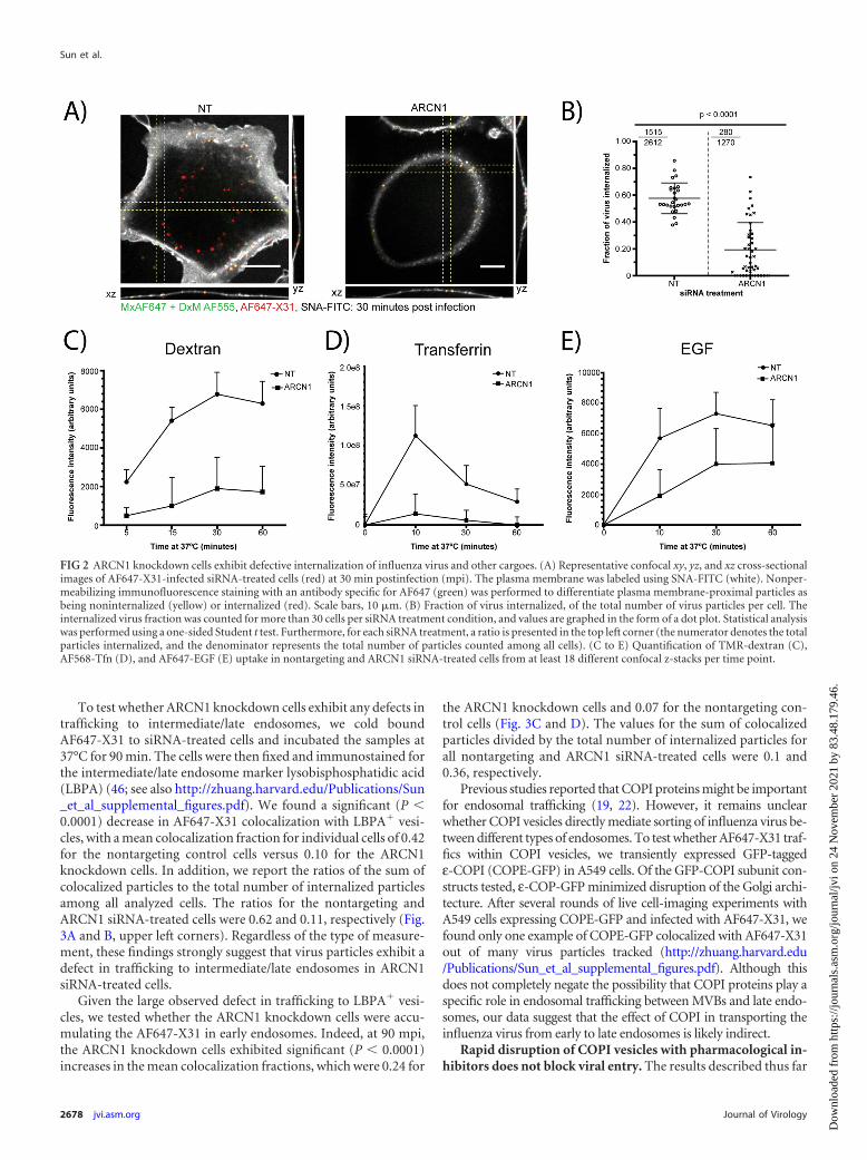
siRNA knockdown of COPI abrogates the internalization ofinfluenza virus, as well as other clathrin-mediated endocytosisor macropinocytosis cargos. The small defects in virus bindingcould not explain the dramatic decrease in viral fusion in theCOPI-depleted cells. Therefore, we subsequently tested the effectof COPI depletion on influenza virus internalization by quantify-ing the fraction of internalized AF647-X31 in the siRNA-treated
cells at 30 min postinfection (mpi). The cells were fixed and la-beled with SNA-FITC to delineate the plasma membrane. In ad-dition, to confidently identify the virus particles close to theplasma membrane as being internalized or noninternalized, weperformed nonpermeabilizing immunofluorescence staining tostain surface-bound noninternalized virus with an antibody spe-cific for AF647 (Fig. 2A). The ARCN1 knockdown cells exhibiteda significant (P � 0.0001) defect in virus uptake compared to thatof the control cells. Specifically, the mean internalization fractionsin the nontargeting siRNA-treated control cells and ARCN1knockdown cells were 0.58 and 0.19, respectively (Fig. 2B). Inaddition, we report a ratio that represents the sum of internalizedparticles divided by the total number of virus particles among allcells. The ratios for the control and ARCN1 knockdown cells were0.58 and 0.22, respectively (Fig. 2B, top left corners).
Given the substantial defect in influenza virus internalizationin the ARCN1 knockdown cells, we tested whether other cargoesof endocytic pathways previously described as productive influ-enza virus entry pathways, i.e., clathrin-mediated endocytosis andmacropinocytosis, were also inhibited. Tfn, EGF, and dextranwere used as representative markers of clathrin-mediated endocy-tosis and macropinocytosis. The cargoes were allowed to be inter-nalized for different amounts of time at 37°C, and surface-boundcargoes were stripped from the cells using a low-pH buffer (pH2.5) prior to fixation and imaging. We validated our analysismethod by quantifying the amount of cargo internalized upontreatment with well-established inhibitors of macropinocytosisand dynamin-dependent endocytosis. To verify our algorithm formacropinocytosis, we treated A549 cells with different concentra-tions of EIPA prior to allowing for fluid phase uptake; for dy-namin-dependent endocytosis, we treated A549 cells with differ-ent doses of Dynasore prior to Tfn or EGF uptake (39, 40). For allthree cargoes tested, we observed dose-dependent decreases inmeasured cargo uptake with increasing concentrations of inhibi-tor treatment (http://zhuang.harvard.edu/Publications/Sun_et_al_supplemental_figures.pdf). We applied the assay to thesiRNA-treated cells and found that the uptake of not only dextranbut also Tfn and EGF was significantly impaired (Fig. 2C to E).The largest observed defects in internalization were at the earliertime points tested, i.e., 15 min for dextran and 10 min for Tfn andEGF. EGF uptake was relatively less perturbed than Tfn uptake,because it has been shown that EGF might be internalized througha clathrin-independent pathway at high concentrations (36,41, 42).
Collectively, our results show that COPI depletion blocksinfluenza virus internalization, which in part contributes to thedefect in viral fusion shown in Fig. 1D. However, representa-tive cargoes for two different productive entry pathways forinfluenza virus— clathrin-mediated endocytosis and macropi-nocytosis (3–6)—also exhibited defects in internalization inARCN1-depleted cells.
ARCN1 knockdown cells exhibit impaired endosomal traf-ficking upon virus internalization. We next tested whetherARCN1 knockdown leads to any postinternalization defects.Upon internalization, influenza virus enters Rab5� endosomesnear the cell periphery and then is sorted into Rab7� endosomestoward the perinuclear region, in which late endosomes/lyso-somes are dense (43). Upon exposure to a pH of �5.0, the influ-enza virus envelope fuses with the endosomal membrane, releas-ing the vRNP into the cytoplasm for nuclear import (44, 45).
Role of COPI Complexes in Influenza Virus Infection
March 2013 Volume 87 Number 5 jvi.asm.org 2677
Dow
nloa
ded
from
http
s://j
ourn
als.
asm
.org
/jour
nal/j
vi o
n 24
Nov
embe
r 20
21 b
y 83
.48.
179.
46.
To test whether ARCN1 knockdown cells exhibit any defects intrafficking to intermediate/late endosomes, we cold boundAF647-X31 to siRNA-treated cells and incubated the samples at37°C for 90 min. The cells were then fixed and immunostained forthe intermediate/late endosome marker lysobisphosphatidic acid(LBPA) (46; see also http://zhuang.harvard.edu/Publications/Sun_et_al_supplemental_figures.pdf). We found a significant (P �0.0001) decrease in AF647-X31 colocalization with LBPA� vesi-cles, with a mean colocalization fraction for individual cells of 0.42for the nontargeting control cells versus 0.10 for the ARCN1knockdown cells. In addition, we report the ratios of the sum ofcolocalized particles to the total number of internalized particlesamong all analyzed cells. The ratios for the nontargeting andARCN1 siRNA-treated cells were 0.62 and 0.11, respectively (Fig.3A and B, upper left corners). Regardless of the type of measure-ment, these findings strongly suggest that virus particles exhibit adefect in trafficking to intermediate/late endosomes in ARCN1siRNA-treated cells.
Given the large observed defect in trafficking to LBPA� vesi-cles, we tested whether the ARCN1 knockdown cells were accu-mulating the AF647-X31 in early endosomes. Indeed, at 90 mpi,the ARCN1 knockdown cells exhibited significant (P � 0.0001)increases in the mean colocalization fractions, which were 0.24 for
the ARCN1 knockdown cells and 0.07 for the nontargeting con-trol cells (Fig. 3C and D). The values for the sum of colocalizedparticles divided by the total number of internalized particles forall nontargeting and ARCN1 siRNA-treated cells were 0.1 and0.36, respectively.
Previous studies reported that COPI proteins might be importantfor endosomal trafficking (19, 22). However, it remains unclearwhether COPI vesicles directly mediate sorting of influenza virus be-tween different types of endosomes. To test whether AF647-X31 traf-fics within COPI vesicles, we transiently expressed GFP-taggedε-COPI (COPE-GFP) in A549 cells. Of the GFP-COPI subunit con-structs tested, ε-COP-GFP minimized disruption of the Golgi archi-tecture. After several rounds of live cell-imaging experiments withA549 cells expressing COPE-GFP and infected with AF647-X31, wefound only one example of COPE-GFP colocalized with AF647-X31out of many virus particles tracked (http://zhuang.harvard.edu/Publications/Sun_et_al_supplemental_figures.pdf). Although thisdoes not completely negate the possibility that COPI proteins play aspecific role in endosomal trafficking between MVBs and late endo-somes, our data suggest that the effect of COPI in transporting theinfluenza virus from early to late endosomes is likely indirect.
Rapid disruption of COPI vesicles with pharmacological in-hibitors does not block viral entry. The results described thus far
FIG 2 ARCN1 knockdown cells exhibit defective internalization of influenza virus and other cargoes. (A) Representative confocal xy, yz, and xz cross-sectionalimages of AF647-X31-infected siRNA-treated cells (red) at 30 min postinfection (mpi). The plasma membrane was labeled using SNA-FITC (white). Nonper-meabilizing immunofluorescence staining with an antibody specific for AF647 (green) was performed to differentiate plasma membrane-proximal particles asbeing noninternalized (yellow) or internalized (red). Scale bars, 10 �m. (B) Fraction of virus internalized, of the total number of virus particles per cell. Theinternalized virus fraction was counted for more than 30 cells per siRNA treatment condition, and values are graphed in the form of a dot plot. Statistical analysiswas performed using a one-sided Student t test. Furthermore, for each siRNA treatment, a ratio is presented in the top left corner (the numerator denotes the totalparticles internalized, and the denominator represents the total number of particles counted among all cells). (C to E) Quantification of TMR-dextran (C),AF568-Tfn (D), and AF647-EGF (E) uptake in nontargeting and ARCN1 siRNA-treated cells from at least 18 different confocal z-stacks per time point.
Sun et al.
2678 jvi.asm.org Journal of Virology
Dow
nloa
ded
from
http
s://j
ourn
als.
asm
.org
/jour
nal/j
vi o
n 24
Nov
embe
r 20
21 b
y 83
.48.
179.
46.

help explain why several of the COPI subunits emerged as impor-tant host proteins in influenza virus infection in the siRNA ge-nome-wide knockdown screens (11, 13, 14). However, efficientsiRNA depletion can take a few days, potentially leading to theaccumulation of siRNA-mediated indirect effects due to long-term depletion of critical host proteins. In order to test whether wecould recapitulate the observations in the siRNA-treated cells andto determine whether there is a direct role for COPI during theinfluenza virus infection cycle, we conducted experiments usingpharmacological inhibitors that rapidly disrupt COPI vesicle for-mation.
The conversion of inactive cytosolic ADP ribosylation factor 1(ARF1)-GDP to the activated ARF1-GTP form triggers its attach-ment to the Golgi membrane and subsequent recruitment ofCOPI complexes (15–17). Two pharmacological inhibitors—BrefA and GCA—prevent the exchange of ARF1-GDP and ARF1-GTP via different mechanisms. BrefA binds to the protein-proteininterface of ARF1 and several different BrefA-sensitive guaninenucleotide exchange factors (GEFs) to prevent the exchange ofGDP and GTP (26, 47). Therefore, treatment with BrefA inhibitsnot only COPI membrane recruitment but also other ARF1-GTP-
dependent vesicular trafficking processes, such as activator pro-tein 1-dependent trans-Golgi network trafficking to endosomalcompartments (15, 16). A more specific inhibitor of COPI com-plex vesicle formation is GCA, which binds to Golgi apparatus-specific brefeldin A resistance guanine nucleotide exchange factor1 (GBF1), a COPI-specific ARF GEF (48).
We confirmed that the DMSO-treated control cells exhibitedcompact COPI perinuclear staining reminiscent of the Golgi appara-tus. In stark contrast, the BrefA- or GCA-treated cells exhibited dis-persed �-COPI staining, indicative of an inability to recruit the COPIcomplex to the Golgi apparatus (48; see also http://zhuang.harvard.edu/Publications/Sun_et_al_supplemental_figures.pdf). To testwhether the drug treatment also inhibits viral fusion, as observed forsiRNA COPI knockdown, we performed a viral fusion assay as de-scribed for Fig. 1D. Viral fusion was measured in A549 cells pretreatedand incubated with 10 �g/ml BrefA, 10 �M GCA, or 0.1% (vol/vol)DMSO (as a control for the total amount of DMSO added to theBrefA- or GCA-treated cells) during the entire experiment. In con-trast to the results shown in Fig. 1D, we found that the inhibition ofCOPI complex vesicle formation does not affect viral fusion (Fig. 4A).
Because internalization and transport to late endosomes con-
FIG 3 Defective endosomal trafficking of internalized influenza virus particles in ARCN1-depleted cells. (A) Representative spinning disk confocal images ofnontargeting and ARCN1 siRNA-treated cells infected with AF647-X31 for 90 min. The plasma membrane was stained with SNA-FITC, and the late endosomeswere immunostained with LBPA. Scale bars, 10 �m. (B) The fractions of internalized particles that colocalized with LBPA, a late endosome marker, were countedfor at least 20 cells per siRNA treatment. The dot plot average line represents the mean colocalization fraction of the analyzed cells; each dot within the plot is thecolocalized fraction from an individual cell. Statistical analysis was performed using a one-sided Student t test. In addition, for each siRNA treatment, a ratio ispresented in the top left corner (the numerator denotes the total number of internalized particles colocalized with LBPA, and the denominator represents the totalnumber of internalized virus particles among all cells). The dot plot mean and ratio in the top left corner for each respective treatment may not necessarily beequal, especially in the presence of substantial cell-to-cell variation. (C) Representative spinning disk confocal images of nontargeting and ARCN1 siRNA-treatedcells infected with AF647-X31 for 90 min, plasma membrane stained with SNA-FITC, and immunostained with EEA1, an early endosome marker. Scale bars, 10�m. (D) Quantification of the internalized particles that colocalize with EEA1 at 90 mpi was performed as described for panel B for more than 35 cells per siRNAtreatment. Statistical analysis was performed using a one-sided Student t test.
Role of COPI Complexes in Influenza Virus Infection
March 2013 Volume 87 Number 5 jvi.asm.org 2679
Dow
nloa
ded
from
http
s://j
ourn
als.
asm
.org
/jour
nal/j
vi o
n 24
Nov
embe
r 20
21 b
y 83
.48.
179.
46.

stituted the two major blocks in viral entry for ARCN1 knock-down cells, we tested whether BrefA and GCA treatment couldrecapitulate those same defects. Using the same immunofluores-cence-based internalization assay as described for Fig. 2A and B,we found that the mean internalization fractions were 0.58, 0.56,and 0.57 for DMSO-, BrefA-, and GCA-treated cells, respectively(Fig. 4B). Furthermore, we tested whether inhibitor treatmentaffects trafficking to intermediate/late endosomes by measuringthe fraction of internalized AF647-X31 within Rab7-GFP� endo-somes. The mean colocalization fractions for the virus and Rab7 inthe DMSO-, BrefA-, and GCA-treated cells were 0.74, 0.67, and
0.61, respectively (Fig. 4C). Our AF647-X31 internalization andRab7-GFP colocalization results were consistent with the viral fu-sion results; only minor differences were observed upon BrefAand GCA treatment, indicating that it is unlikely that rapid dis-ruption of COPI vesicles results in an entry defect.
Next, to test whether BrefA or GCA treatment affects influenzainfection, we pretreated A549 cells with 0.1% DMSO (as a con-trol), 10 �g/ml BrefA, or 10 �M GCA for 60 min, allowed X31 toinfect the cells at a multiplicity of infection (MOI) of �1 for 9 h,and measured NP expression with a flow cytometer. In stark con-trast to the results shown in Fig. 1C, we found that the disruption
FIG 4 Pharmacological disruption of COPI complexes does not block virus entry. (A) Representative flow cytometry-based quantification of DiD dequenchingfrom three independent experiments. The DiD signal was measured at different time points after DiD-X31 infection in A549 cells treated with 0.1% (vol/vol)DMSO, 10 �g/ml BrefA, or 10 �M GCA. Standard deviations represent duplicate samples for each time point. (B) Quantification of the fraction of internalizedAF647-X31 particles at 30 mpi upon treatment with inhibitors or DMSO, using the same immunofluorescence-based internalization assay in Fig. 2A. At least 17different cells were quantified for virus internalization. Statistical analysis was performed using one-way analysis of variance (ANOVA). (C) Quantification of thefraction of AF647-X31 particles colocalized with Rab7-GFP� endosomes at 90 mpi in A549 cells treated with 0.1% (vol/vol) DMSO, 10 �g/ml BrefA, or 10 �MGCA. At least 16 different cells were quantified. In the top left corner of each panel, we report the ratio that represents the sum of all AF647-X31 particlescolocalized with Rab7 versus the total number of internalized virus particles for all cells quantified. Statistical analysis was performed using one-way ANOVA. (D)Flow cytometry-based quantification of the percentage of NP� cells and the NP expression levels of the NP� cells for A549 cells treated with 0.1% (vol/vol)DMSO, 10 �g/ml BrefA, or 10 �M GCA during the entire infection with X31 (MOI, �1). (E) Flow cytometry-based quantification of the percentage of NP� cellsand the NP expression levels of the NP� cells for A549 cells infected with X31 (MOI, �1.5) and exposed to 0.1% (vol/vol) DMSO, 10 �g/ml BrefA, or 10 �M GCAonly during entry. (F) Flow cytometry-based quantification of the percentage of NP� cells and the NP expression levels of the NP� cells for A549 cells infectedwith X31 (MOI, �1.5) and exposed to 0.1% (vol/vol) DMSO, 10 �g/ml BrefA, or 10 �M GCA only after 90 mpi. In D to F, the bar chart data represent theaverages from two independent experiments (consisting of three replicate samples for each inhibitor or DMSO treatment), with the corresponding standarderrors of the mean (SEM).
Sun et al.
2680 jvi.asm.org Journal of Virology
Dow
nloa
ded
from
http
s://j
ourn
als.
asm
.org
/jour
nal/j
vi o
n 24
Nov
embe
r 20
21 b
y 83
.48.
179.
46.

of COPI complexes resulted in a moderate decrease in infection;relative to the DMSO control treatment, BrefA or GCA treatmentreduced the percentage of NP-expressing cells by 21.3% 7.9%or 40.6% 1.3%, respectively. In addition, we found a moderatedecrease in the NP expression level for NP� cells relative to thatfor the DMSO control, i.e., 31.1% 1.2% and 35.3% 3.6% inthe BrefA- and GCA-treated cells, respectively (Fig. 4D). How-ever, it remains unclear whether these defects resulted from anentry or postentry exposure to BrefA or GCA.
To assess whether exposure to BrefA or GCA during entry af-fects productive influenza virus infection, we pretreated cells withthe inhibitors for 60 min, followed by X31 infection in the pres-ence of the compounds for 90 min. Afterward, the compoundswere removed, and the infection was continued for an additional7.5 h before processing for flow cytometry. We found that treat-ment with BrefA or GCA during entry alone did not affect thepercentage of NP� cells or the NP expression levels (Fig. 4E).However, when BrefA and GCA were added and maintained onlyafter 90 mpi (Fig. 4F), a moderate decrease in NP expression wasobserved again (Fig. 4F), indicating that the effects of BrefA orGCA treatment on viral gene expression originate from postentrysteps. Collectively, these results indicate that functional COPI ves-icle recruitment to cellular membranes is not required for influ-enza virus internalization, endosomal trafficking, or viral fusionbut has a moderate effect on viral NP expression.
Rapid disruption of COPI complex function decreases viralmembrane protein expression and progeny virus production.To test whether COPI complex disruption inhibits viral mem-brane protein expression, we infected A549 cells with X31 (MOI,�1.5). At 90 mpi, we added 0.1% DMSO (as a control), 10 �g/mlBrefA, or 10 �M GCA to the virus-infected cells. After 9 hpi, wecollected the cells, fixed them, immunostained them for M2, andanalyzed the samples with a flow cytometer. Immunostaining ofthe X31-infected cells for M2 in the presence of detergents alloweddetection of total M2 expression. Compared with the results ofpostentry effects of BrefA and GCA on NP expression (Fig. 4F),M2 was more substantially perturbed upon treatment with BrefAor GCA than was the DMSO control (Fig. 5A).
To determine whether the disruption of COPI vesicle forma-tion was essential to M2 trafficking to the plasma membrane, weperformed the experiment as described above but immunostainedcells for M2 under nonpermeabilizing conditions. M2 cell surfaceexpression was decreased even more for the BrefA-treated cellsthan the total M2 level, indicating the presence of a traffickingdefect. Surprisingly, we observed only a minor defect in M2 traf-ficking in GCA-treated cells (Fig. 5A). Additionally, we confirmedour results through sequential immunofluorescence assays for M2without and with permeabilization. By 9 hpi, M2 readily traffickedto the plasma membrane in the DMSO-treated control cells. Instark contrast, no M2 was detected on the plasma membrane ofBrefA-treated cells, while some M2 was trafficked to the cell sur-face and some was trapped within some intracellular vesicles inGCA-treated cells (http://zhuang.harvard.edu/Publications/Sun_et_al_supplemental_figures.pdf). We confirmed with �-COPIstaining that COPI vesicle formation and concentration withinthe Golgi apparatus were still perturbed in both BrefA- and GCA-treated cells.
To test whether BrefA or GCA treatment also impaired theexpression or plasma membrane targeting of another influ-enza virus membrane protein, we stained for surface and total NA
(http://zhuang.harvard.edu/Publications/Sun_et_al_supplemental_figures.pdf). BrefA, and to a lesser extent GCA, impaired NA pro-tein expression. Furthermore, consistent with the M2 transportexperiments, BrefA treatment also inhibited NA trafficking to theplasma membrane. However, in contrast to the minor traffickingeffects on M2, GCA treatment strongly inhibited NA transport tothe cell surface (Fig. 5B). The discrepancy between M2 and NAplasma membrane trafficking in GCA-treated cells suggests thatthe two membrane proteins exhibit different transport require-ments.
It remains unclear whether BrefA and GCA treatment leads toadditional perturbations in the assembly of viral components atthe plasma membrane. Influenza virus assembly is initiated by HAand NA recruitment to cholesterol-enriched plasma membranedomains. M1 is believed to interact with the cytoplasmic tails ofHA and NA, and M1 interactions with vRNPs facilitate the incor-poration of the viral genome into the budding virions (49). ThevRNPs can be detected with PB1-specific antibodies. The PB1patches are sites enriched with assembling virus particles, or bud-ding zones. To determine whether functional COPI vesicle forma-tion is essential for virus assembly, we immunostained X31-in-fected cells treated with DMSO, BrefA, or GCA for M2 (to detectinfected cells) and PB1 (http://zhuang.harvard.edu/Publications/Sun_et_al_supplemental_figures.pdf). We found that 38% of theinfected DMSO-treated cells contained budding zones enrichedwith PB1 at 18 hpi. In stark contrast, only 1% and 2% of BrefA-and GCA-treated cells, respectively, infected with X-31 had bud-ding zones (Fig. 5C). The inability to traffic NA (and, to someextent, M2) to the plasma membrane in either the BrefA- or GCA-treated cells might help to explain the large defect in assembly andthe failure to concentrate progeny virions at budding zones withininfected cells.
To assess whether the defects in assembly were important forprogeny virus production, we performed the experiment depictedin Fig. 5D. The supernatant of the virus-infected cells was col-lected at 18 hpi, and the viral titers were measured with standardplaque assays. We found that the presence of either BrefA or GCAreduced viral titers by about 2 orders of magnitude relative tothose of the DMSO control (Fig. 5D). In summary, the resultsobtained from using the pharmacological inhibitors strongly sug-gest that the inactivation of COPI vesicle formation does not resultin an entry defect but does specifically inhibit viral membraneprotein expression, plasma membrane transport, assembly, andinfectious progeny virion production.
DISCUSSION
As an obligate pathogen encoding 13 viral proteins, influenza vi-rus requires host factors and compartments to mediate productiveinfection. Recently, genome-wide knockdown screens identifiedhost dependency proteins important for influenza virus infection.Despite relatively little overlap in identified host dependency pro-teins among the screens, all of the screens identified subunits ofthe COPI complex as critical host proteins mediating productivevirus infection. COPI proteins form complexes which oligomerizeto coat vesicles. These coated vesicles mediate retrograde traffick-ing between the Golgi apparatus and the ER, as well as between theGolgi stacks. Previous studies also identified a role for COPI pro-teins in endosomal trafficking. Given that COPI proteins mediatetrafficking between different cellular compartments in both thesynthesis and endosomal trafficking pathways, it remained un-
Role of COPI Complexes in Influenza Virus Infection
March 2013 Volume 87 Number 5 jvi.asm.org 2681
Dow
nloa
ded
from
http
s://j
ourn
als.
asm
.org
/jour
nal/j
vi o
n 24
Nov
embe
r 20
21 b
y 83
.48.
179.
46.

clear which steps in the influenza virus infection cycle COPI pro-teins regulate.
Identification of host dependency proteins through siRNA de-pletion can result from four general outcomes: (i) siRNA-induced
cytotoxicity, (ii) off-target effects (e.g., interferon induction orsiRNA sequence overlap with other mRNA sequences), (iii) directeffects of siRNA-mediated depletion of the gene of interest, or (iv)indirect effects mediated by long-term depletion of the target of
FIG 5 Functional COPI complex recruitment to the cis-Golgi is required for viral membrane protein expression and assembly of progeny virions. (A) Flowcytometry-based quantification of the percentage of M2-positive cells and the M2 expression levels in the M2-positive cells for A549 cells infected with X31 (MOI,�1.5) and exposed to 0.1% (vol/vol) DMSO, 10 �g/ml BrefA, or 10 �M GCA only after 90 mpi. Immunofluorescence staining was performed either in thepresence of saponin, to detect total M2 expression, or in the absence of detergent, to detect surface M2. The bar chart data represent the averages from twoindependent experiments (consisting of three replicate samples for each inhibitor or DMSO treatment) with the corresponding SEM. (B) Quantification of therelative surface or total NA expression based on the acquired confocal images. Error bars indicate standard deviations for at least 18 images per treatmentcondition. (C) Quantification of the fraction of infected cells treated with 0.1% (vol/vol) DMSO, 10 �g/ml BrefA, or 10 �M GCA that contain budding zones atthe cell periphery. The fraction in the top left corner of each panel represents the number of cells with a budding zone (numerator) relative to the total numberof virus-infected cells (denominator) for all cells counted. (D) Viral titers of supernatants collected from X31-infected A549 cells treated with 0.1% (vol/vol)DMSO, 10 �g/ml BrefA, or 10 �M GCA at 18 hpi, as measured with plaque assays (see Materials and Methods). The bar chart includes the standard deviationfrom samples tested in quadruplicate.
Sun et al.
2682 jvi.asm.org Journal of Virology
Dow
nloa
ded
from
http
s://j
ourn
als.
asm
.org
/jour
nal/j
vi o
n 24
Nov
embe
r 20
21 b
y 83
.48.
179.
46.

interest. We confirmed that, within our experimental time frame,siRNA-mediated cytotoxicity did not play a dominant role in ourstudies. No differences in cell viabilities were observed, with mi-nor changes in cytotoxicities and thus cell growth rates. Based onthe control experiments, we believe that the defects in influenzavirus infection upon ARCN1 depletion result in either direct orindirect effects specific to siRNA-mediated silencing. In order todetermine whether COPI depletion results in direct or indirecteffects, we used both siRNA silencing and rapid COPI vesicle dis-ruption via BrefA or GCA treatment to analyze how these twomethods of perturbation affect different steps in the influenzavirus infection cycle, i.e., binding, internalization, transport intolate endosomes, fusion, viral protein expression, trafficking, as-sembly, and progeny virion production.
König and colleagues identified ARCN1, an essential subunitof the COPI complex, as a critical host protein for influenza virusinfection (14). Upon further analysis, the authors also providedevidence that ARCN1 depletion affects influenza virus entry. Be-cause of the data suggesting a role for ARCN1 in mediating influ-enza virus entry and because ARCN1 is known to be an essentialcomponent of the COPI vesicle complex, we decided to focus ourstudies on understanding the role of ARCN1 in influenza virusinfection as a representative subunit of the COPI complex. Afterconducting a systematic dissection of the different entry steps, wefound that ARCN1-depleted cells exhibit a significant defect inviral fusion. The abrogation of viral fusion in ARCN1-depletedcells likely explains why König and colleagues observed a signifi-cant defect in vRNP accumulation in the nucleus compared to thatin the nontargeting control cells. Consistent with the viral fusiondata, we found that ARCN1 knockdown cells exhibited an �66%decrease in internalization, an �76% decrease in colocalization ofthe virus with the late endosome marker LBPA, and an �90%decrease in virus infection relative to the those of the nontargetingsiRNA-treated control cells. Taken together, our results show thatCOPI depletion leads to defects in virus internalization, transportto late endosomes, and viral fusion; viral fusion is likely a conse-quence of the first two effects.
Cureton and colleagues showed that COPI depletion also leadsto defects in VSV entry (25), although there are differences be-tween the two viruses in which particular steps are perturbed.Cureton and colleagues found that COPI depletion results in adecrease in viral fusion due to an �40% decrease in VSV bindingand a decreased rate of VSV internalization. However, the VSVparticles that manage to be internalized in COPI-depleted cellscan still traffic to early endosomes and undergo vRNP release,indicating that the endosome trafficking of VSV is not perturbed.In contrast, our findings show that COPI depletion did not per-turb influenza virus binding but did inhibit virus internalizationand transport to late endosomes.
Treatment of cells with two pharmacological compounds,BrefA and GCA, that rapidly prevent the recruitment of COPIcomplexes to the Golgi apparatus resulted in strikingly differentresults from the siRNA experiments. Specifically, we did not ob-serve any significant difference in influenza virus fusion or colo-calization with a late endosome marker when cells were treatedwith DMSO, BrefA, or GCA. Substantiating the results from BrefAand GCA treatment, no extensive colocalization was observed be-tween GFP-tagged ε-COPI, another essential component of theCOPI complex, and AF647-labeled influenza virus in live cellsduring entry. Combining the siRNA, pharmacological inhibitor,
and live cell imaging results, we conclude that functional COPIcomplex vesicle formation is not directly required for influenzavirus entry, although ARCN1 depletion leads to indirect effectsthat result in defects in influenza virus internalization, traffickingto late endosomes, and, consequently, viral fusion.
Functional COPI complex formation and COPI vesicles medi-ate trafficking between the Golgi apparatus and the ER, as well asbetween different Golgi stacks. The Golgi apparatus-derived ves-icles then traffic contents from the Golgi apparatus to other loca-tions within the cell, including late endosomes, lysosomes, and theplasma membrane (16, 17). Defects in Golgi vesicular traffickingover an extended period of time, such as during siRNA depletion,could result in improper trafficking of membrane proteins or lip-ids. Changes in protein or lipid compositions (50) at the plasmamembrane and endosomes may provide an explanation for theobserved defects in viral internalization, trafficking, and fusion.The potential changes in protein and lipid compositions are, how-ever, not reflected in the amount of �(2,6)-linked sialic acids (theattachment factor for influenza virus) presented on the surfaces ofcells, consistent with the observation that viral binding is not sub-stantially affected by ARCN1 knockdown.
The severely compromised uptake of general clathrin-medi-ated endocytosis and macropinocytosis cargoes in the ARCN1knockdown cells suggests that protein and/or lipid compositionsat the cell surface may be altered during the course of siRNA de-pletion. ARCN1 knockdown cells internalized �60% less virus by30 mpi than the nontargeting control. Other cargoes of clathrin-mediated endocytosis (Tfn and EGF) and macropinocytosis (dex-tran) also exhibited significant internalization defects in ARCN1knockdown cells relative to the nontargeting siRNA-treated cells.Consistent with our findings, Cureton and colleagues found thatthe uptake of VSV and Tfn was perturbed only after prolongedexposure to COPI disruption inhibitors (25).
Another phenomenon that might be related to the defect intrafficking of influenza virus to late endosomes is the anomalousLBPA� late endosome staining pattern observed in Fig. 3A. Incontrast to the dense perinuclear LBPA� late endosome stainingobserved with the nontargeting control cells, we observed a repro-ducible dispersion of late endosomes—with no apparent aberrantchanges to the early endosome organization—in ARCN1 knock-down cells. This observation is consistent with previous reportsshowing that COPI subunit depletion alters the late endosometrafficking pattern (51). It has also been found that the inhibitionof endosomal trafficking through blockade of the uptake pathwaysleads to the losses of lysosomal acidification and perinuclear local-ization (52). We also noted that the shape of the ARCN1 knock-down cells sometimes appeared different from that of the nontar-geting control cells, although the underlying mechanism for thechange in cell morphology remains unclear. It is possible that thechanges in the spatial distribution of late endosomes and the cellmorphology are also related to the inhibition of viral entry.
Together, these results show that prolonged exposure to siRNAdepletion of an essential COPI subunit results in an indirect effectof compromised cargo endocytosis. In contrast, the inhibition ofCOPI complex recruitment to the Golgi apparatus through acutepharmacological treatment directly affects viral protein expres-sion and infectious progeny virus production. Disruption ofCOPI complexes by BrefA and GCA had only moderate effects onNP expression. However, BrefA and GCA treatment inhibited M2and NA expression, suggesting that COPI complex formation is
Role of COPI Complexes in Influenza Virus Infection
March 2013 Volume 87 Number 5 jvi.asm.org 2683
Dow
nloa
ded
from
http
s://j
ourn
als.
asm
.org
/jour
nal/j
vi o
n 24
Nov
embe
r 20
21 b
y 83
.48.
179.
46.

specifically required for at least some membrane protein expres-sion. The inhibition might be due to retention of proteins withinthe ER from defective trafficking between the ER and the Golgiapparatus. Alternatively, treatment might cause rerouting of Golgiapparatus contents to alternative endosomal populations ratherthan intended target sites, such as the plasma membrane (53).
We also observed that BrefA- and GCA-treated cells exhibiteddefects in progeny virion assembly and production. Previousstudies found that NA, but not M2, associates with lipid rafts (49).For GCA-treated cells, transport of M2 to the plasma membranewas modestly affected, while both the plasma membrane targetingof NA and progeny virus assembly were strongly impaired. Theseresults suggest that the large assembly defect in GCA-treated cellsmight stem from inhibition of the trafficking of lipid raft-associ-ated membrane proteins (which are critical for initiating virusbudding and subsequent assembly steps) to the plasma mem-brane.
In summary, through dissection of the influenza virus infec-tion cycle with both siRNA silencing and pharmacological com-pound perturbation, we have identified which steps of influenzavirus infection are directly or indirectly dependent on functionalCOPI complex formation. Long-term siRNA COPI depletion incells leads to indirect effects on viral entry through defective cargouptake and vesicular trafficking to late endosomes, while acutetreatment with COPI complex inhibitors impairs viral membraneprotein expression and assembly, suggesting a direct role of COPIcomplexes in these later steps of infection. It was found recentlythat COPI proteins mediate many additional effects beyond thewell-studied trafficking roles in the synthesis and secretory path-ways, including lipid droplet formation and lipid metabolism, au-tophagosome formation, and endocytosis of EGF and Tfn (50, 51,54, 55), which might be related to the observed indirect effects ofCOPI depletion on viral entry. Future studies on how COPIsiRNA treatment affects protein or lipid expression, modification,and transport to different cellular compartments should be help-ful for understanding how COPI proteins regulate such diversecellular processes, as well as how these effects can lead to the ob-served defects of viral infection in COPI-depleted cells.
ACKNOWLEDGMENTS
We thank John Young for initial discussions that inspired this work, andwe thank James Hogle, Sean Whelan, Raymond Chung, David Cureton,and Joshua Vaughan for useful discussions. We also thank Patricia Rogersand Brian Tilton from the Bauer Core Facility for technical advice on flowcytometry, as well as Robert Lamb, James Rothman, Felix Wieland, Gil-lian Air, and Qing Zhong for reagents.
This work was funded by NIH grant R01GM068518 (to X.Z.). E.S. issupported by a National Science Foundation graduate research fellow-ship. X.Z. is a Howard Hughes Medical Institute investigator.
REFERENCES1. Palese P, Shaw ML. 2007. Orthomyxoviridae: the viruses and their repli-
cation, p 1647–1689. In Knipe DM, Howley PM, Griffin DE, Lamb RA,Martin MA, Roizman B, Straus SE (ed), Fields virology, 5th ed, vol 2.Lippincott Williams & Wilkins, Philadelphia, PA.
2. Wiley DC, Skehel JJ. 1987. The structure and function of the hemagglu-tinin membrane glycoprotein of influenza virus. Annu. Rev. Biochem.56:365–394.
3. de Vries E, Tscherne DM, Wienholts MJ, Cobos-Jimenez V, Scholte F,Garcia-Sastre A, Rottier PJ, de Haan CA. 2011. Dissection of the influ-enza A virus endocytic routes reveals macropinocytosis as an alternativeentry pathway. PLoS Pathog. 7:e1001329. doi:10.1371/journal.ppat.1001329.
4. Matlin KS, Reggio H, Helenius A, Simons K. 1981. Infectious entrypathway of influenza virus in a canine kidney cell line. J. Cell Biol. 91:601–613.
5. Rust MJ, Lakadamyali M, Zhang F, Zhuang X. 2004. Assembly ofendocytic machinery around individual influenza viruses during viral en-try. Nat. Struct. Mol. Biol. 11:567–573.
6. Sieczkarski SB, Whittaker GR. 2002. Influenza virus can enter and infectcells in the absence of clathrin-mediated endocytosis. J. Virol. 76:10455–10464.
7. Doms RW, Helenius A, White J. 1985. Membrane-fusion activity of theinfluenza-virus hemagglutinin: the low pH-induced conformationalchange. J. Biol. Chem. 260:2973–2981.
8. Lakadamyali M, Rust MJ, Babcock HP, Zhuang X. 2003. Visualizinginfection of individual influenza viruses. Proc. Natl. Acad. Sci. U. S. A.100:9280 –9285.
9. Skehel JJ, Bayley PM, Brown EB, Martin SR, Waterfield MD, White JM,Wilson IA, Wiley DC. 1982. Changes in the conformation of influenzavirus hemagglutinin at the pH optimum of virus-mediated membranefusion. Proc. Natl. Acad. Sci. U. S. A. 79:968 –972.
10. Whittaker GR, Kann M, Helenius A. 2000. Viral entry into the nucleus.Annu. Rev. Cell Dev. Biol. 16:627– 651.
11. Brass AL, Huang IC, Benita Y, John SP, Krishnan MN, Feeley EM, RyanBJ, Weyer JL, van der Weyden L, Fikrig E, Adams DJ, Xavier RJ, FarzanM, Elledge SJ. 2009. The IFITM proteins mediate cellular resistance toinfluenza A H1N1 virus, West Nile virus, and dengue virus. Cell 139:1243–1254.
12. Hao L, Sakurai A, Watanabe T, Sorensen E, Nidom CA, Newton MA,Ahlquist P, Kawaoka Y. 2008. Drosophila RNAi screen identifies hostgenes important for influenza virus replication. Nature 454:890 – 893.
13. Karlas A, Machuy N, Shin Y, Pleissner KP, Artarini A, Heuer D, BeckerD, Khalil H, Ogilvie LA, Hess S, Maurer AP, Muller E, Wolff T, RudelT, Meyer TF. 2010. Genome-wide RNAi screen identifies human hostfactors crucial for influenza virus replication. Nature 463:818 – 822.
14. König R, Stertz S, Zhou Y, Inoue A, Hoffmann HH, Bhattacharyya S,Alamares JG, Tscherne DM, Ortigoza MB, Liang Y, Gao Q, AndrewsSE, Bandyopadhyay S, De Jesus P, Tu BP, Pache L, Shih C, Orth A,Bonamy G, Miraglia L, Ideker T, Garcia-Sastre A, Young JA, Palese P,Shaw ML, Chanda SK. 2010. Human host factors required for influenzavirus replication. Nature 463:813– 817.
15. Beck R, Rawet M, Wieland FT, Cassel D. 2009. The COPI system:molecular mechanisms and function. FEBS Lett. 583:2701–2709.
16. Hsu VW, Yang JS. 2009. Mechanisms of COPI vesicle formation. FEBSLett. 583:3758 –3763.
17. Popoff V, Adolf F, Brugger B, Wieland F. 2011. COPI budding withinthe Golgi stack. Cold Spring Harb. Perspect. Biol. 3:a005231. doi:10.1101/cshperspect.a005231.
18. Gu F, Gruenberg J. 1999. Biogenesis of transport intermediates in theendocytic pathway. FEBS Lett. 452:61– 66.
19. Aniento F, Gu F, Parton RG, Gruenberg J. 1996. An endosomal betaCOP is involved in the pH-dependent formation of transport vesiclesdestined for late endosomes. J. Cell Biol. 133:29 – 41.
20. Daro E, Sheff D, Gomez M, Kreis T, Mellman I. 1997. Inhibition ofendosome function in CHO cells bearing a temperature-sensitive defect inthe coatomer (COPI) component -COP. J. Cell Biol. 139:1747–1759.
21. Gabriely G, Kama R, Gerst JE. 2007. Involvement of specific COPIsubunits in protein sorting from the late endosome to the vacuole in yeast.Mol. Cell. Biol. 27:526 –540.
22. Gu F, Aniento F, Parton RG, Gruenberg J. 1997. Functional dissectionof COP-I subunits in the biogenesis of multivesicular endosomes. J. CellBiol. 139:1183–1195.
23. Guo Q, Vasile E, Krieger M. 1994. Disruptions in Golgi structure andmembrane traffic in a conditional lethal mammalian cell mutant are cor-rected by -COP. J. Cell Biol. 125:1213–1224.
24. Whitney JA, Gomez M, Sheff D, Kreis TE, Mellman I. 1995. Cytoplas-mic coat proteins involved in endosome function. Cell 83:703–713.
25. Cureton DK, Burdeinick-Kerr R, Whelan SP. 2012. Genetic inactivationof COPI coatomer separately inhibits vesicular stomatitis virus entry andgene expression. J. Virol. 86:655– 666.
26. Lippincott-Schwartz J, Liu W. 2006. Insights into COPI coat assemblyand function in living cells. Trends Cell Biol. 16:e1– e4.
27. Cureton DK, Massol RH, Saffarian S, Kirchhausen TL, Whelan SPJ.2009. Vesicular stomatitis virus enters cells through vesicles incompletely
Sun et al.
2684 jvi.asm.org Journal of Virology
Dow
nloa
ded
from
http
s://j
ourn
als.
asm
.org
/jour
nal/j
vi o
n 24
Nov
embe
r 20
21 b
y 83
.48.
179.
46.

coated with clathrin that depend upon actin for internalization. PLoSPathog. 5:e1000394. doi:10.1371/journal.ppat.1000394.
28. Johannsdottir HK, Mancini R, Kartenbeck J, Amato L, Helenius A.2009. Host cell factors and functions involved in vesicular stomatitis virusentry. J. Virol. 83:440 – 453.
29. Matlin KS, Reggio H, Helenius A, Simons K. 1982. Pathway of vesicularstomatitis virus entry leading to infection. J. Mol. Biol. 156:609 – 631.
30. Sun XJ, Yau VK, Briggs BJ, Whittaker GR. 2005. Role of clathrin-mediated endocytosis during vesicular stomatitis virus entry into hostcells. Virology 338:53– 60.
31. Superti F, Seganti L, Ruggeri FM, Tinari A, Donelli G, Orsi N. 1987.Entry pathway of vesicular stomatitis virus into different host cells. J. Gen.Virol. 68:387–399.
32. Chen C, Zhuang XW. 2008. Epsin 1 is a cargo-specific adaptor for theclathrin-mediated endocytosis of the influenza virus. Proc. Natl. Acad. Sci.U. S. A. 105:11790 –11795.
33. White J, Matlin K, Helenius A. 1981. Cell fusion by Semliki Forest,influenza, and vesicular stomatitis viruses. J. Cell Biol. 89:674 – 679.
34. Sun QM, Westphal W, Wong KN, Tan I, Zhong Q. 2010. Rubiconcontrols endosome maturation as a Rab7 effector. Proc. Natl. Acad. Sci.U. S. A. 107:19338 –19343.
35. Vaughan JC, Brandenburg B, Hogle JM, Zhuang X. 2009. Rapid actin-dependent viral motility in live cells. Biophys. J. 97:1647–1656.
36. Chen H, De Camilli P. 2005. The association of epsin with ubiquitinatedcargo along the endocytic pathway is negatively regulated by its interac-tion with clathrin. Proc. Natl. Acad. Sci. U. S. A. 102:2766 –2771.
37. Randall RE, Goodbourn S. 2008. Interferons and viruses: an interplaybetween induction, signalling, antiviral responses and virus countermea-sures. J. Gen. Virol. 89:1– 47.
38. Stegmann T, Morselt HW, Scholma J, Wilschut J. 1987. Fusion ofinfluenza virus in an intracellular acidic compartment measured by fluo-rescence dequenching. Biochim. Biophys. Acta 904:165–170.
39. Doherty GJ, McMahon HT. 2009. Mechanisms of endocytosis. Annu.Rev. Biochem. 78:857–902.
40. Macia E, Ehrlich M, Massol R, Boucrot E, Brunner C, Kirchhausen T.2006. Dynasore, a cell-permeable inhibitor of dynamin. Dev. Cell 10:839 –850.
41. Jiang X, Sorkin A. 2003. Epidermal growth factor receptor internalizationthrough clathrin-coated pits requires Cbl RING finger and proline-richdomains but not receptor polyubiquitylation. Traffic 4:529 –543.
42. Sigismund S, Woelk T, Puri C, Maspero E, Tacchetti C, Transidico P,Di Fiore PP, Polo S. 2005. Clathrin-independent endocytosis of ubiq-uitinated cargos. Proc. Natl. Acad. Sci. U. S. A. 102:2760 –2765.
43. Lakadamyali M, Rust MJ, Zhuang X. 2006. Ligands for clathrin-mediated endocytosis are differentially sorted into distinct populations ofearly endosomes. Cell 124:997–1009.
44. Lakadamyali M, Rust MJ, Zhuang X. 2004. Endocytosis of influenzaviruses. Microbes Infect. 6:929 –936.
45. Mercer J, Schelhaas M, Helenius A. 2010. Virus entry by endocytosis.Annu. Rev. Biochem. 79:803– 833.
46. Kobayashi T, Stang E, Fang KS, de Moerloose P, Parton RG, GruenbergJ. 1998. A lipid associated with the antiphospholipid syndrome regulatesendosome structure and function. Nature 392:193–197.
47. Mossessova E, Corpina RA, Goldberg J. 2003. Crystal structure ofARF1*Sec7 complexed with brefeldin A and its implications for the gua-nine nucleotide exchange mechanism. Mol. Cell 12:1403–1411.
48. Sáenz JB, Sun WJ, Chang JW, Li J, Bursulaya B, Gray NS, Haslam DB.2009. Golgicide A reveals essential roles for GBF1 in Golgi assembly andfunction. Nat. Chem. Biol. 5:157–165.
49. Rossman JS, Lamb RA. 2011. Influenza virus assembly and budding.Virology 411:229 –236.
50. Misselwitz B, Dilling S, Vonaesch P, Sacher R, Snijder B, SchlumbergerM, Rout S, Stark M, von Mering C, Pelkmans L, Hardt WD. 2011. RNAiscreen of Salmonella invasion shows role of COPI in membrane targetingof cholesterol and Cdc42. Mol. Syst. Biol. 7:474. doi:10.1038/msb.2011.7.
51. Razi M, Chan EYW, Tooze SA. 2009. Early endosomes and endosomalcoatomer are required for autophagy. J. Cell Biol. 185:305–321.
52. Huotari J, Helenius A. 2011. Endosome maturation. EMBO J. 30:3481–3500.
53. Zhang CJ, Rosenwald AG, Willingham MC, Skuntz S, Clark J, Kahn RA.1994. Expression of a dominant allele of human ARF1 inhibits membranetraffic in vivo. J. Cell Biol. 124:289 –300.
54. Collinet C, Stoter M, Bradshaw CR, Samusik N, Rink JC, Kenski D,Habermann B, Buchholz F, Henschel R, Mueller MS, Nagel WE, FavaE, Kalaidzidis Y, Zerial M. 2010. Systems survey of endocytosis by mul-tiparametric image analysis. Nature 464:243–249.
55. Guo Y, Walther TC, Rao M, Stuurman N, Goshima G, Terayama K,Wong JS, Vale RD, Walter P, Farese RV. 2008. Functional genomicscreen reveals genes involved in lipid-droplet formation and utilization.Nature 453:657– 661.
Role of COPI Complexes in Influenza Virus Infection
March 2013 Volume 87 Number 5 jvi.asm.org 2685
Dow
nloa
ded
from
http
s://j
ourn
als.
asm
.org
/jour
nal/j
vi o
n 24
Nov
embe
r 20
21 b
y 83
.48.
179.
46.