distribution of human leukocyte antigens in a population...
TRANSCRIPT

Vol. 5, 873-877, November 1996 Cancer Epidemiology, Biomarkers & Prevention 873
3 The abbreviations used are: HTLV-I, human T-cell lymphotrophic virus, type I,
AU adult T-cell leukemia/lymphoma: HLA, human leukocyte antigen.
Distribution of Human Leukocyte Antigens in a Population of Black
Patients with Human T-Cell Lymphotrophic Virus Type
I-associated Adult T-Cell LeukemialLymphoma’
Jeffrey D. White,2 John A. Johnson, Jun-mo Nam,Beverly Cranston, Barrie Hanchard,Thomas A. Waldmann, and Angela Mantis
Metabolism Branch [J. D. W., J. A. J., T. A. W.l and Division of Cancer
Epidemiology and Genetics, Biostatistics and Viral Epidemiology Branch [J-
m. N., A. M.], National Cancer Institute, NIH, Bethesda, Maryland 20892, andDepartment of Pathology, University of the West Indies, Kingston, Jamaica
[B.C., B.H.]
Abstract
Human leukocyte antigens (HLAs) play an important rolein regulating the inunune response to infectious agents anddeterminants of malignant transformation. We comparedthe HLA frequencies of 25 black patients with adult T-cellleukemia/lymphoma (AU) referred to the National CancerInstitute for therapy with a racially similar, asymptomaticcontrol population of human T-cell lymphotrophic virus,type I (HTLV-I)-seropositive individuals (n 45).Serological typing was performed for MHC class I and Hantigens. Antigen frequencies were calcUlated, andcorresponding gene frequencies were estimated using themaximum likeithood method. Comparisons between theATL and control group were made with f or Fisher’sexact test. Three antigens (A36, B18, and DRS3) were foundto have a higher frequency in the ATh patients than in thecontrols (uncorrected two-tailed P < 0.05). The genefrequencies for these antigens also were statistically
significant in the uncorrected analysis. However, only A36approached statistical significance after correction of the Pvalue for multiple comparisons (P 0.08). The results ofthis pilot study indicate that black patients with Alt mayhave increased frequencies of certain class I HLA whencompared with a racially similar HTLV-I-positive reference
population. This suggests that either these antigens mayrepresent markers for a population at greater risk of
developing ATh once infected with HTLV-I or that theywere acquired at some point in the process of malignanttransformation or progression from the carrier state toonset of ATh. These antigens should be targeted in largerstudies to confirm or refute these findings.
Received 12/21/95; revised 7/31/96; accepted 8/1/96.
The costs of publication of this article were defrayed in part by the payment ofpage charges. This article must therefore be hereby marked advertisement in
accordance with 18 U.S.C. Section 1734 solely to indicate this fact.I This work was supported in part by National Cancer Institute Research Contract
N0l-CP-31006 from the NIH. Presented in part at the Annual Meeting of the
American Association for Cancer Research, Toronto, Ontario, Canada, March
18-22, 1995.
2 To whom requests for reprints should be addressed, at Metabolism Branch,
National Cancer Institute, Building 10, Room 4N1 15, Bethesda, MD 20892.
Phone: (301) 402-2912; Fax: (301) 402-1001.
Introduction
HTLV-13 is endemic to specific geographic regions of theworld, including the southwestern Japanese provinces of Ky-ushu and Shikoku, the Caribbean, and parts of sub-SaharanAfrica and South America (1, 2). This retrovirus is associatedwith a variety of illnesses but is most clearly linked etiologi-
cally with ATh and HTLV-I-associated myelopathy and trop-ical spastic paraparesis (3). However, only a small percentage( 1-5%) of people infected with HTLV-I will develop either of
these diseases. Early life infection with HTLV-I by maternal-to-infant transmission is thought to be critical for later devel-
opment of ATL (4). Other important determinants amongHTLV-I carriers for progression to ATh have not been defin-
itively identified.HLAs have been suspected to play a role in HTLV-I
infection and its oncogenic potential. Mann et a!. (5) demon-strated in vitro that additional HLA antigens were expressed onHTLV-I-positive cultured T cells from ATL patients. Several
Japanese investigators have identified certain HLA antigenswith an increased frequency among ATL patients comparedwith population controls and family members (6-8). It has
been suggested by Usuku et a!. (9) that specific HLA haplo-types may be markers of genetic susceptibility to either ATL orHTLV-I-associated myelopathy, also known as tropical spastic
paraparesis, by virtue of differences in host immune respon-siveness to HTLV-I-specific antigens. However, there is a
paucity of data to refute or support these observations fromother HTLV-I-endemic areas populated by other racial groups.
Different demographic and clinical features of All have
been shown for patient populations outside of Japan (10). Wehad the opportunity to evaluate black ATL patients from the
United States and the Caribbean. Our goals in the current studywere: (a) to determine HLA antigen and gene frequencies in a
population of black ATh patients; (b) to attempt to identifyantigens associated with ATL by comparing this group of black
All patients with a racially similar HTLV-I-positive controlgroup; (c) to compare results with published reports from
Japanese ATL patients; and (d) to assess the relationship be-
tween HLA and clinical characteristics of ATL patients.
Materials and Methods
ATh Patients. Thirty-one patients with AU were referred tothe Metabolism Branch of the National Cancer Institute be-tween April 1987 and May 1994 for protocols using interleukin2 receptor-directed therapies (1 1, 12). Diagnostic pathologicalmaterials were reviewed by the Hematopathology Section of
the Laboratory of Pathology of the National Cancer Institute.
on June 29, 2019. © 1996 American Association for Cancer Research. cebp.aacrjournals.org Downloaded from
874 HLA in ATL
Table 1 Demograp hic characteristics and HLA ph enotypes am ong black All patients
.Patient Age (yr) Sex
Country of.
birthATL subtype
HLA phenotypes” Survival
A B C DR DQ DRWb
(mo)
1
2
3
4
5
6
7
8
9
10
I I
12
13
14
15
16
17
18
19
20
2 I
22
23
24
25
39
31
31
53
32
38
65
22
44
48
53
40
31
42
62
53
24
40
65
34
52
40
39
37
63
Female
Female
Female
Female
Female
Female
Male
Female
Male
Female
Male
Male
Male
Male
Female
Male
Female
Male
Female
Male
Female
Female
Male
Female
Male
Jamaica
Grenada
United States
Trinidad
Jamaica
St. Vincent
United States
Haiti
Jamaica
Guyana
United States
Haiti
Jamaica
United States
Jamaica
Trinidad
Guyana
Jamaica
Jamaica
Jamaica
Jamaica
Jamaica
Jamaica
Jamaica
Jamaica
Acute
Chronic/crisis
Acute
Acute
Acute
Lymphoma
Acute
Acute
Chronic
Acute
Acute
Chronic
Acute
Lymphoma
Chronic
Lymphoma
Acute
Acute
Acute
Lymphoma
Chronic/crisis
Chronic
Acute
Chronic
Chronic
2, 28
2, 28
2, 30
2, 33
32, 36
2, 28
26, 34
2. 26
23, 33
nd
23, -
1, -
3, 36
28. 33
nd
2, 29
2, -
2, 36
23, 30
28, 34
30, 36
I. -
23, 33
I I, 26
2, 23
1 8, 53
35, 42
51, 49
45, 53
35, 53
39, 70
49, 53
35, 70
7, 52
nd
45, 63
53, -
13, 53
58, 70
nd
18, 44
7, 55
5, 57
7, 57
35, 44
13, 42
7, 18
7, 73
14, 57
49, 60
1 , 4 nd
4, 7 2, 3
7, - nd
4, - 9, 12
2, 4 1 1, 14
3, - 7. 15
4. - 7, 17
2, - 9. 17
4, 7 9, 13
nd 4, 6
-, - 5, 6
4, - 2, 6
4, 6 nd
3, 4 1 1, 18
nd 7, -
7, - nd
-, - nd
7, - I, 15
6, 7 8, 15
4, 7 1, 2
2, 7 1 . I 5
-, - 4, 7
7, - 9, 15
7, - 8, -
-, - 4, 17
nd
1, 4
nd
2, 5
5, -
2, 6
2, -
2, -
2, 6
7, -
1, -
1, -
nd
4, 7
2, -
nd
nd
5, 6
6, 7
1, -
5, 6
2, 3
2, 6
7, -
2, 8
nd
52, -
nd
52, 53
52, -
53, -
52, 53
52, 53
52, 53
52, 53
52, -
52, -
nd
52, -
53, -
nd
nd
-, -
-, -
-, -
- , -
53, -
53, -
52, -
52, 53
3
57+
12
23
26
2
59+
14
3+
3
35
21
7
64+
29
7
8
7+
15
19
5 +
129
7
44+
46+
“ - . blank HLA loci; nd, not done.
‘, +. patient still alive.
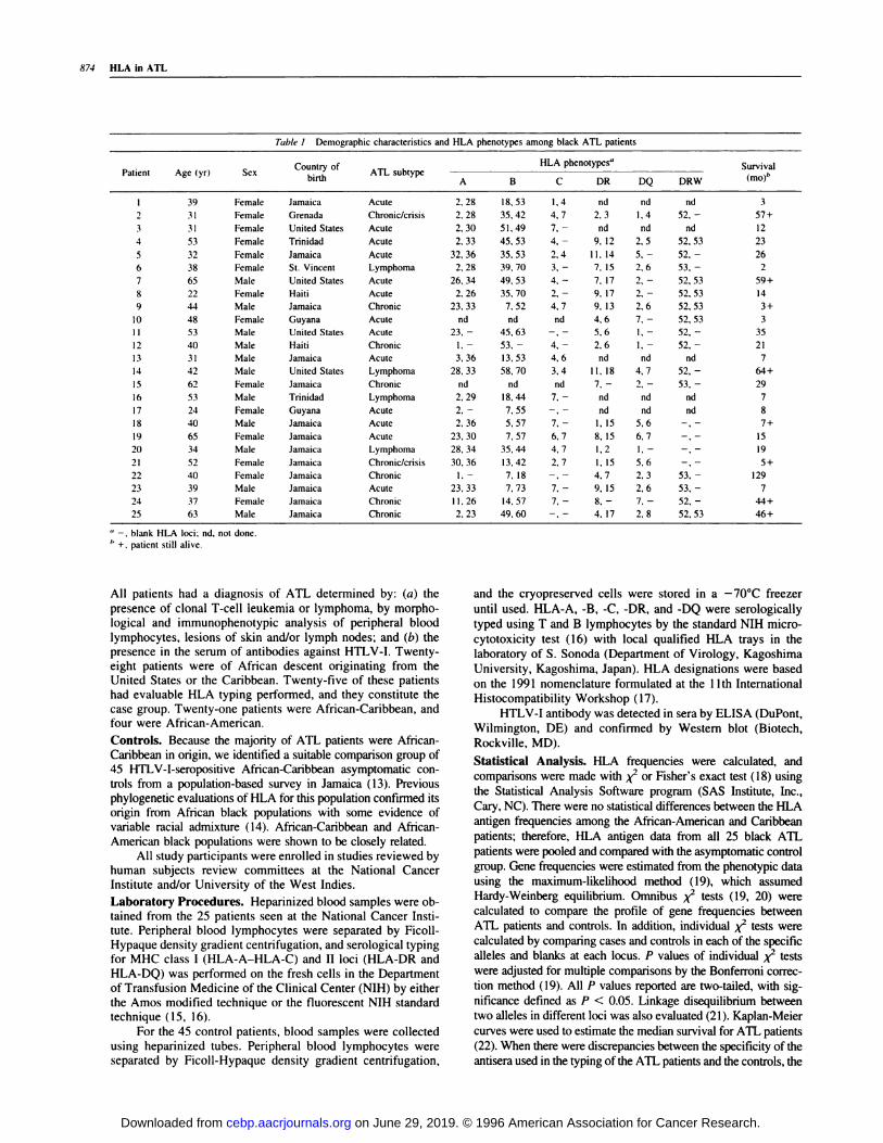
All patients had a diagnosis of ATL determined by: (a) the
presence of clonal T-cell leukemia or lymphoma, by morpho-
logical and immunophenotypic analysis of peripheral bloodlymphocytes, lesions of skin and/or lymph nodes; and (b) thepresence in the serum of antibodies against HTLV-I. Twenty-eight patients were of African descent originating from theUnited States or the Caribbean. Twenty-five of these patientshad evaluable HLA typing performed, and they constitute thecase group. Twenty-one patients were African-Caribbean, andfour were African-American.
Controls. Because the majority of All patients were African-Caribbean in origin, we identified a suitable comparison group of45 HTLV-I-seropositive African-Caribbean asymptomatic con-
trols from a population-based survey in Jamaica (13). Previousphylogenetic evaluations of HLA for this population confirmed itsorigin from African black populations with some evidence of
variable racial admixture (14). African-Caribbean and African-American black populations were shown to be closely related.
All study participants were enrolled in studies reviewed byhuman subjects review committees at the National CancerInstitute and/or University of the West Indies.
Laboratory Procedures. Heparinized blood samples were ob-
tamed from the 25 patients seen at the National Cancer Insti-tute. Peripheral blood lymphocytes were separated by Ficoll-Hypaque density gradient centrifugation, and serological typing
for MHC class I (HLA-A-HLA-C) and II loci (HLA-DR andHLA-DQ) was performed on the fresh cells in the Departmentof Transfusion Medicine of the Clinical Center (NIH) by either
the Amos modified technique or the fluorescent NIH standardtechnique (15, 16).
For the 45 control patients, blood samples were collected
using heparmnized tubes. Peripheral blood lymphocytes wereseparated by Ficoll-Hypaque density gradient centrifugation,
and the cryopreserved cells were stored in a -70#{176}Cfreezer
until used. HLA-A, -B, -C, -DR, and -DQ were serologically
typed using T and B lymphocytes by the standard NIH micro-
cytotoxicity test (16) with local qualified HLA trays in the
laboratory of S. Sonoda (Department of Virology, Kagoshima
University, Kagoshima, Japan). HLA designations were based
on the 1991 nomenclature formulated at the 1 lth International
Histocompatibility Workshop (17).
HTLV-I antibody was detected in sera by ELISA (DuPont,Wilmington, DE) and confirmed by Western blot (Biotech,
Rockville, MD).
Statistical Analysis. HLA frequencies were calculated, andcomparisons were made with � or Fisher’s exact test (18) using
the Statistical Analysis Software program (SAS Institute, Inc.,
Cary, NC). There were no statistical differences between the HLA
antigen frequencies among the African-American and Caribbean
patients; therefore, HLA antigen data from all 25 black ATh
patients were pooled and compared with the asymptomatic control
group. Gene frequencies were estimated from the phenotypic data
using the maximum-likelihood method (19), which assumed
Hardy-Weinberg equilibrium. Omnibus f tests (19, 20) were
calculated to compare the profile of gene frequencies between
ATh patients and controls. In addition, individual f tests were
calculated by comparing cases and controls in each of the specific
alleles and blanks at each locus. P values of individual f tests
were adjusted for multiple comparisons by the Bonferroni correc-
lion method (19). All P values reported are two-tailed, with sig-
nificance defined as P < 0.05. Linkage disequilibrium between
two alleles in different loci was also evaluated (21). Kaplan-Meier
curves were used to estimate the median survival for ATh patients
(22). When there were discrepancies between the specificity of the
antisera used in the typing ofthe ATh patients and the controls, the
on June 29, 2019. © 1996 American Association for Cancer Research. cebp.aacrjournals.org Downloaded from
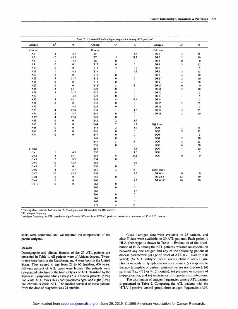
Table 2 HLA-A-HLA-D antigen frequencies among ATL patients”
Antigen G” % Antigen G” % Antigen G” %
A locus B locus DR locus
Al 2 8.7 B5 I 4.3 DR1 3 15
A2 10 43.5 B7 5 21.7 DR2 2 10
A3 1 4.3 B8 0 0 DR3 0 0
A9 0 0 B12 0 0 DR4 3 15
AlO 0 0 B13 2 8.7 DR5 I 5
All I 4.3 B14 I 4.3 DR6 3 15
A19 0 0 B15 0 0 DR7 4 20
A23 5 21.7 Bl6 0 0 DR8� 2 10
A24 0 0 B17 0 0 DR9 4 20
A25 0 0 Bl8’ 3 13 DR1O 0 0
A26 3 13 B21 0 0 DRII 2 10
A28 5 21.7 B22 0 0 DR12 I 5
A29 1 4.3 B27 0 0 DR13 I 5
A30 3 13 B35 4 17.4 DRI4 I 5
A31 0 0 B37 0 0 DR15 5 25
A32 I 4.3 B38 0 0 DR16 I 5
A33 4 17.4 B39 I 4.3 DRI7 3 15
A34 2 8.7 B40 0 0 DRI8 2 10
A36� 4 17.4 B41 0 0
A43 0 0 B42 2 8.7
A66 0 0 B44 2 8.7 DQ locus
A68 0 0 B45 2 8.7 DQI 3 15
A69 0 0 B46 0 0 DQ2 9 45
A74 0 0 B47
B48
B49
B50
0
0
3
0
0
0
13
0
DQ3
DQ4
DQS
DQ6
I
2
5
6
5
10
25
30
C locus B51 I 4.3 DQ7 4 20
Cwl 1 4.3 B52 I 4.3 DQ8 I 5
Cw2 3 13 B53 6 26.1 DQ9 0 0
Cw3 2 8.7 B54 0 0
Cw4 10 43.5 B55 I 4.3
Cw5 0 0 B56 0 0
Cw6 2 8.7 B57 3 13 DRW locus
Cw7 10 43.5 B58 I 4.3 DRW5I 0 0
Cw8 0 0 B59 0 0 DRW52 12 61)
Cw9 0 0 B60 I 4.3 DRW53’ 10 50
CwlO 0 0 B61
B62
B63
B64
B65
B67
B70
0
0
1
0
0
0
3
0
0
4.3
0
0
0
13
‘, Twenty-three patients had data for A-C antigens, and 20 had data for DR and DQ.
/� G, antigen frequency.
� Antigen frequency in ATL population significantly different from HTLV-I positive controls (i.e., uncorrected P � 0.05); see text.
Cancer Epidemiology, Biomarkers & Prevention 875
splits were combined, and we reported the comparisons of theparent antigens.
Results
Demographic and clinical features of the 25 ATh patients arepresented in Table 1 . All patients were of African descent. Twen-ty-one were born in the Caribbean, and 4 were born in the United
States. They ranged in age from 22 to 65 (median, 40) years.
Fifty-six percent of ATh cases were female. The patients werecategorized into three ofthe four subtypes ofATh classified by theJapanese Lymphoma Study Group (23). Thirteen patients (52%)
had acute ATh, four (16%) had lymphoma type, and eight (32%)had chronic or crisis ATh. The median survival of these patientsfrom the date of diagnosis was 21 months.
Class I antigen data were available on 23 patients, and
class II data were available on 20 ATL patients. Each patient’s
HLA phenotype is shown in Table 1 . Evaluation of the distri-
bution of HLA among the ATL patients revealed no association
between any one antigen and any of the following patient or
disease parameters: (a) age of onset of ATL (i.e. , <40 or �40
years); (b) ATL subtype (acute versus chronic versus lym-
phoma or acute or lymphoma versus chronic); (c) response to
therapy (complete or partial remission versus no response); (d)
survival (i.e., < 12 or �12 months); (e) presence or absence of
hypercalcemia; and (e) occurrence of opportunistic infections.
The distribution of antigen frequencies among ATh patients
is presented in Table 2. Comparing the ATh patients with the
HTLV-I-positive control group, three antigen frequencies (A36,
on June 29, 2019. © 1996 American Association for Cancer Research. cebp.aacrjournals.org Downloaded from

876 HLA in ATL
Table 3 Comparison of es timated individual ge ne frequencies with sign ificant difference or blank alleles bet ween All cases and HTLV-I positive controls
LocusATh patients” HUV-I controls” Individual test
(“�
Omnibus test.
[f� with df(P)lG n f ± SE G n f ± SE
A (15 alleles)
A36
B (23 alleles)
B18
Blank
DQ (6 alleles)
Blank
4
3
(P1 = 23)
0
0
0
0
8.7 ± 4.2
6.5 ± 3.6
0±2.3
13.4 ± 7.4
0
0
(n 45)
0
0
1 11.5±3.9
0 0 ± 3.3
0.005”
0.016
0.044
0.04
23.43 with 15 (0.075)
28.43 with 23 (0.20)
13.23 with 6 (0.04)
“ G, antigen frequency; n, number of individuals reacting to a single antigen only;f, estimated gene frequency in percent from phenotypic data using maximum-likelihood
method.h p 0.08 with Bonferroni adjustment for multiple comparisons.
Bl8, and DR53) were significantly more frequent in the AUgroup (uncorrected two-tailed P < 0.05). A36, B 18, and DR53were present in 17, 13, and 50%, respectively, in the AU patients
compared with 0, 0, and 22%, respectively, in the control popu-lation. To assure that these differences were not due to racial
heterogeneity among our two comparison groups, we did a subsetanalysis of only AU patients born in Jamaica. The significant
HLA antigen differences remained with increased frequencies forA36 (P = 0.01 1) and Bl8 (P = 0.035). However, when the Pvalues were corrected for the number of antigens tested, none wassignificant. Sixteen (64%) of the AU patients had one or more ofthese antigens with increased frequency. The antigen DR8 was
found in a higher frequency in the control group (33%) than in theAU group (10%; uncorrected P = 0.05).
Estimation of HLA gene frequencies for ATL patients and
comparison with those of HTLV-I-positive controls (Table 3)confirmed our results based on antigen frequencies. Gene fre-quencies for the antigens A36 and B I 8 were increased among
ATL patients. We did not estimate gene frequency for thesupertypic DR53 allele but determined frequencies for the morespecific alleles it represents, DR4, DR7, and DR9. Among ATL
patients, the increased gene frequency for A36 approachedsignificance (P = 0.08) after adjusting for multiple compari-sons. The Omnibus test approached significance for the A locus,reflecting the profile difference in frequencies observed be-
tween AU patients and the controls. This test was significantfor the DQ locus, for which we identified significant blankalleles, suggesting that there were some unspecified differenceswe could not determine with available typing methods. Wefound no survival or clinical differences between the ATL
patients with A36 and the remainder of the ATL patients. Therewas no evidence from the data that the two class I alleles, A36
and B18. were in linkage disequilibrium.
Discussion
AU is a malignancy of mature T lymphocytes occurring predom-inantly in patients infected with HTLV-I. The geographic cluster-ing of AU in southern Japan, the Caribbean islands, and parts ofthe United States coincides with areas endemic for I-ITLV-I (2,23). The great majority of AU patients have a clonal pattern of
HTLV-I provirus integrated in the DNA of their leukemic cells.The clinical manifestations of this disease are quite variable andinclude lymphadenopathy, leukemia, hepatosplenomegaly, skin
rash, lytic bone lesions, fever, opportunistic infections, and hyper-calcemia (23, 24). The leukemic cells are variably sized lympho-
cytes with basophilic cytoplasm and indented or deeply cleft nucleiwith clumped chromatin. Four clinicopathological subtypes havebeen defined that encompass the diverse clinical presentations:
smoldering, chronic, lymphoma. and the acute type. Both the
smoldering and chronic subtypes can enter a crisis phase in which
they behave like the aggressive, rapidly progressive acute subtype
(23).
Our data provide evidence that certain HLA alleles may be
associated with AU in a black population. In particular, anti-
gen frequencies of class I HLA-A36 and -B18 and class IIHLA-DR53 were elevated. These HLAs were increased infrequency when compared with those in HUV-I-positive con-
trols, suggesting either that some or all of them representmarkers for a population at greater risk of developing ATL once
infected with HTLV-I or that these antigens were acquired atsome point in the process of malignant transformation or pro-
gression from the carrier state to onset of ATL. The estimated
gene frequency for A36 was the only one that approached
significance (P = 0.08) after correction for multiple compari-
sons. To address the issue of comparability of the control and
patient populations, we analyzed our data looking at only theJamaican ATL patients (n = 21) and found that the same
significant antigens were identified in the uncorrected test.The T- and B-Cell Malignancy Study Group in Japan has also
identified an association between AU and certain class I and IIHLA antigens (7). When the comparison was made against a
control group of subjects from Kyushu and South Shikoku, Japan,AU-endemic areas from which 91% of their AU patients orig-
mated, higher frequencies were observed for A26 and B39, and
lower frequencies were observed for A24, B46, B52, B6l, and
DR7. These differences were statistically significant (uncorrected
P < 0.05) but were not significant after correction for the number
of antigens tested. There was no concurrence between their find-ings and those in the current study. Unfortunately, two of the
antigens with significantly different frequencies in the presentstudy (A36 and Bl8) apparently were not available in the antibodyor antisera panels used in the Japanese study. Therefore, although,
ethnicity could in part account for differences we observed, HLAtyping technology was also a factor. However, subsequent analy-
ses of the distribution of HLA in the general Japanese populationshow a low frequency ofA36, and further studies ofJapanese AU
patients likewise show no increased frequency of this antigen (25).
Independently, investigators have shown that ATL pa-tients from Japan and those of African descent from the Car-
ibbean have a low lymphocyte-proliferative response when
measured spontaneously or in reaction to exogenously admin-
istered HTLV-I virions (9, 26). These functionally deficientlymphocytes may in fact result from the altered expression ofclass I HLA among ATL patients, with resultant immune sup-pression and heightened susceptibility to opportunistic infec-(ions. Uno et al. (8) have shown that the enhanced expression
on June 29, 2019. © 1996 American Association for Cancer Research. cebp.aacrjournals.org Downloaded from

Cancer Epidemiology, Biomarkers & Prevention 877
of class I HLA on peripheral lymphocytes of AU patients hasan inverse relationship to natural killer cell activity. They offerthat such a mechanism may contribute to the ability of HTLV-I-infected malignant cells to escape from immune surveillance.Alternatively, others have suggested that the additional antigens
resulting from the malignant process and virus infection mayresult in molecular mimicry of a host protein, also causingevasion of immune surveillance. Certain HLAs have beenshown to have structural homology with HUV-I: the HL4-B
locus gene, with the envelope gene region of HTLV-I (27), andHL4-DRS3, with several amino acid sequences similar to
HTLV-I (28).An a priori hypothesis was that host factors such as HLA
might also affect the clinical features of a polymorphic disease
such as ATL. In this study, no significant relationships betweenHLA and clinical characteristics of AU patients were identi-fled. However, real differences could have been missed due to
the small sample size.The generalizability of our findings to other AU patients of
African descent may be limited by the fact that our AU popula-tion consisted of referral patients to a tertiary treatment center andhad a greater percentage of the chronic AU subtype (32 versus
3%) and longer survival (21 versus 5 months) than AU patientsfrom incident case series (29). Due to the small numbers ofpatients in the AU group, there is insufficient power in this study
to suggest that the findings are biologically significant. Nonethe-less, these data provide previously unavailable information on
associations of HLA with AU and identify antigens (A36) andgene loci in this population that will be used to narrow the focusfor larger studies. Further study with state-of-the-art HLA-typing
technology of HTLV-I-inlected cohorts and AU patients andfamily members in endemic populations will help determine thesignificance of these findings.
Acknowledgments
We thank the members of the HLA Typing Laboratory (Department of Transfu-
sion Medicine, Clinical Center, NIH) and the Laboratory of Prof. S. Sonoda for
providing HLA typing.
References
I . Madeleine. M. M., Wiktor, S. Z., Goedert, J. J., Manns, A., Levine, P. H.,
Biggar, R. J., and Blattner, W. A. HTLV-I and HTLV-II world-wide distribution:
reanalysis of 4,832 immunoblot results. mt. J. Cancer., 54: 255-260, 1993.
2. Tajima, K., and Hinuma, Y. Epidemiology of HTLV-1/II in Japan and the
world. In: K. Takatsuki, Y. Hinuma. and M. Yoshida (eds.), Gann Monograph on
Cancer Research: Advances in Adult T-Cell Leukemia and HTLV-l Research,No. 39, pp. 129-149. Boca Raton, FL: CRC Press, Inc., 1992.
3. Manns, A., and Blattner, W. A. The epidemiology of the human T-cell
lymphotropic virus type I and type II: etiologic role in human disease. Transfusion
(Arlingt.), 31: 67-75. 1991.
4. Manns, A., Cleghorn, F. R., Falk, R. T., Hanchard, B., Jaffe, E., Bartholomew,
C., Hartge, P., Benichou, J., Blattner, W. A., and HTLV Lymphoma Study Group.Role of HTLV-I in development of non-Hodgkin lymphoma in Jamaica and
Trinidad and Tobago. Lancet, 342: 1447-1450, 1993.
5. Mann, D. L., Popovic, M., Sarin, P., Murray, C., Reitz, M. S., Strong, D. M.,
Haynes, B. F., Gallo, R. C., and Blamer, W. A. Cell lines producing human T-celllymphoma virus show altered HLA expression. Nature (Land.), 305: 58-60, 1983.
6. Sonoda, S., Yashiki, S., Takahashi, K., Azima, N., Daitoku, Y., Matsumoto, M.,
Matsumoto, T., Tars, M., Shinmyozu, K., Sato, K., Inoko, H., Ando, A., and Tsuji.
K. Altered HLA antigens expressed on T and B lymphocytes ofadult T-cell leukemia’
lymphoma patients and their relatives. Int. J. Cancer, 40: 629-634, 1987.
7. The T- and B-Cell Malignancy Study Group. The third nation-wide study on
adult T-cell leukemia/lymphoma (ATh) in Japan: characteristic patterns of HLA
antigen and HTLV-I infection in AU patients and their relatives. mt. J. Cancer,
41: 505-512, 1988.
8. Uno, H., Kawano, K., Matsuoka, H., and Tsuda, K. HLA and T cell leukemia:
HLA-linked genes controlling susceptibility to human T cell leukemia virus typeI. Clin. Exp. Immunol.. 71: 21 1-216, 1988.
9. Usuku, K., Sonoda, S., Osame, M., Yashiki, S., Takahashi, K., Matsumoto, M.,
Sawada, T., Tsuji, K., Tarts, M., and Igata, A. HLA haplotype-linked high immuneresponsiveness against HTLV-I in H’I’LV-I-a.ssociated myelopathy: comparison withadult T-cell leukemiallymphoma. Ann. Neural.. 23 (Suppl.): S143-S150. 1988.
10. Levine. P. H., Manns, A., Jaffe, E. S., Colclough, G., Cavallaro, A., Reddy.
G., and Blattner, W. A. The effect of ethnic differences on the pattem of
HTLV-I-associated T-cell leukemia/lymphoma (HATL) in the United States. Int.
J. Cancer. 56: 177-181, 1994.
1 1. Waldmann, T. A., White. J. D., Goldman. C. K., Top. L.. Grant, A.. Bamford.
R., Roessler, E., Horak, I. D., Zaknocn, S., Kasten-Sportes, C., England, R..
Horak, E., Mishra, B., Dipre, M., Hale, P., Fleisher, T. A., Junghans. R. P.. Jaffe.
E. S.. and Nelson, D. L. The interleukin-2 receptor: a target for monoclonal
antibody treatment of human T-cell lymphotropic virus I-induced adult T-cell
leukemia. Blood, 82: 1701-1712, 1993.
12. Waldmann, T. A.. White. J. D., Carrasquillo. J. A.. Reynolds. J. C.. Gansow.
0., Jaffe, E. S., Fleisher, T. A., Goldman, C. K., Top. L.. Bamford, R., Junghans.
R. P., Zaknoen. S.. Roessler, E., Kasten-Sportes. C.. England. R., Luau, H.,
Johnson, J. A., Jackson-White, T., Manns, A., Hanchard, B., and Nelson, D. L.
Radioimmunotherapy of IL-2Rcs-expressing adult T-cell Leukemia with yttri-
um-90 labeled anti-Tac. Blood, 86: 4063-4075. 1995.
13. Murphy. E. L., Figueroa. J. P.. Gibbs, W. N.. Holding-Cobham. M.. Cranston.B., Malley. K.. Bodner. A. J., Alexander. S. S.. and Blattner. W. A. Human
T-lymphotropic virus type I (HTLV-I) seroprevalence in Jamaica. I. Demographic
determinants. Am. J. Epidemiol. 133: 1114-1124, 1991.
14. Blank. M., Blank, A., King, S.. Yashiki, S.. Kuwayama. M.. Fujiyama. C.,
Gongora. D.. Zaninovic, V., Cranston, B., Hanchard. B., Imanishi, T.. Manns. A.,
Blattner, W. A., Tajima. K., Hayami. M., Fujiyoshi. T.. and Sonoda, S. Distri-
bution of HLA and haplotypes of Colombian and Jamaican black populations.
Tissue Antigens. 45: 1 1 1-1 16. 1995.
15. Hopkins. K. A. The basic lymphocyte microcytotoxicity tests. I,,: F. L.
Phelan, E. M. Mickelson, H. S. Noreen, T. W. Shroyer. D. M. Cluff. and A.
Nikaein (eds.), American Society for Histocompatibility and Immuogenetics
Laboratory Manual, Ed. 3, pp. I.B.l.l.-I.B.15.7. Lenexa. KS: American Society
for Histocompatibility and Immunogenetics. 1994.
16. Terasaki, P. I., Bemoco, D., Park, M. S., Oztruck, G.. and Iwaki. Y. Micro-
droplet testing for HLA-A. -B, -C and -D antigens. Am. J. Clin. Pathol., 69:103-120, 1978.
17. Bodmer, J. G.. Marsh, S. G.. Albert, E. D., Bodmer. W. F.. Dupont. B.. Erlich,
H. A., Mach. B.. Mayr. W. R., Parham, P.. Sasazuki. T., Schreuder, G. M. T.,Strominger, J. L., Svejgaard. A.. and Terasaki, P. I. Nomenclature for factors of
the HLA system. 1991. Tissue Antigens. 39: 161-173. 992.
I 8. Armitage P. Statistical Methods in Medical Research. pp. I 3 I - I 38. New
York: John Wiley & Sons, 1971.
19. Gart, J. J., and Nam, J. Statistical methods for genetic studies of HLA and
cancer. In: R. G. Cornell (ed), Statistical Methods for Cancer Studies. pp.
229-266. New York: Marcel Dekker, 1984.
20. Nam, J., and Gart, J. J. The ML estimation and testing of generalized
ABO-like data with no observed double recessives. Biometrics, 41: 455-466.
21. Cavalli-Sforza, L. L., and Bodmer, W. F. The Genetics of Human Popula-
tions, pp. 283-288. San Francisco: W. H. Freeman, 1971.
22. Kaplan, E. L., and Meier, P. Nonparametric estimation from incomplete
observations. J. Am. Stat. Assoc., 53: 457-481, 1958.
23. Shimoyama, M., and members of The Lymphoma Study Group (1984-
1987). Diagnostic criteria and classification of clinical subtypes of adult T-cell
leukemia-lymphoma. Br. J. Haematol., 79: 428-437. 1991.
24. Kawano, F., Yamaguchi. K., Nishimura. H.. Tsuda, H., and Takatsuki. K.
Variation in the clinical courses of adult T-cell leukemia. Cancer (Phila.). 55:
851-856, 1985.
25. Sonoda, S., Yashiki, S., Kuwayama. M.. Fujiyoshi. T.. Jacobsen. S.. Manns,A., Alcalay, D., Pombo, M., Hammond, M. G., Nikbin, B., Blank, A., and Cartier,
L. HLA and HTLV Workshop report. Hum. Immunol.. 47: 120, 1996.
26. Kramer, A.. Jacobson, S.. Reuben, J. F., Murphy. E. L.. Wiktor. S. Z..Cranston, B., Figueroa. J. P.. Hanchard. B., McFarlin. D.. and Blattner. W. A.Spontaneous lymphocyte proliferation in symptom-free HTLV-l positive Jamai-
cans (Letter). Lancet, 2: 923-924. 1989.
27. Clarke. M. F., Gelmann, E. P., and Reitz. M. S., Jr. Homology of human
T-cell leukaemia virus envelope gene with class I HLA gene. Nature (Lond.). 305:
60-62, 1983.
28. Dorak. M. 1.. and Bumett, A. K. Molecular mimicry of an HLA-DR53
epitope by viruses (Letter). Immunol Today. 15: 138-139, 1994.
29. Hanchard, B., Williams, E., Williams, N. P., Green. M.. Wilks. R., Cranston.
B., Choy, L., Manns, A.. Blattner, W., and HTLV Lymphoma Study Group.
Geographic diversity in AlL: comparison of Jamaican and Japanese cases (Ab-
stract). AIDS Res. Hum. Retroviruses, 10: 476. 1994.
on June 29, 2019. © 1996 American Association for Cancer Research. cebp.aacrjournals.org Downloaded from

1996;5:873-877. Cancer Epidemiol Biomarkers Prev J D White, J A Johnson, J M Nam, et al. I-associated adult T-cell leukemia/lymphoma.black patients with human T-cell lymphotrophic virus type Distribution of human leukocyte antigens in a population of
Updated version
http://cebp.aacrjournals.org/content/5/11/873
Access the most recent version of this article at:
E-mail alerts related to this article or journal.Sign up to receive free email-alerts
Subscriptions
Reprints and
To order reprints of this article or to subscribe to the journal, contact the AACR Publications
Permissions
Rightslink site. Click on "Request Permissions" which will take you to the Copyright Clearance Center's (CCC)
.http://cebp.aacrjournals.org/content/5/11/873To request permission to re-use all or part of this article, use this link
on June 29, 2019. © 1996 American Association for Cancer Research. cebp.aacrjournals.org Downloaded from