dynamic ct scanning for visualization of the - ajnr · dynamic ct scanning for visualization of the...
TRANSCRIPT
Wendy A. Cohen 1
Richard S. Pinto Irvin I. Kricheff
This article appears in the March / April issue of AJNR and the May issue of AJR.
Received June 22. 1981; accepted after revision October 20, 1981.
Presented at the annua l meeting of the American Society of Neuroradiology, Chicago, April 198 1.
This work was supported in part by E. R. Squibb & Sons, Medical Developmental Department, Princeton , NJ 08540 and by General Electric, Milwaukee, WI 53201.
, All authors: Department of Radiology , New York University Medica l Center , 550 First Ave., New York , NY 10016. Address reprint requests to W. A. Cohen.
AJNR 3:185-189, March / April 1982 0195-6108/ 82/ 0302-0185 $00.00 © Ameri can Roentgen Ray Society
Dynamic CT Scanning for Visualization of the Parasellar Carotid Arteries
185
Evaluation of patients before transsphenoidal hypophysectomy for large intrasellar mass lesions has required bilateral internal carotid artery angiography. Using intravenous injection of contrast medium, a method has been developed to visualize the parasellar carotid arteries with rapid sequence sequential computed tomographic scanning . In 18 patients, the cavernous segments of the internal carotid arteries were well seen in 27 of 28 instances with technically complete examinations. The vascularity of the mass lesions and vascular encasement was also demonstrated.
The radiologic evaluation of abnormalities of the sella turcica before the development of computed tomography (CT) consisted of sku ll films, comple x motion tomography, angiography, and pneumoencephalography . Enlargement of the sella turcica on plain films is most often caused by pituitary adenoma and is less commonly due to other lesions, such as c ran iopharyngioma, meningioma, dysgerminoma, teratoma , aneurysm , or an aberrant course of the internal carotid arteries [1]. A parasellar aneurysm may present with symptoms secondary to endocrinologic dysfunction or to local compression of the optic nerves, optic chiasm, or cavernous sinus without history of prior subarachnoid hemorrh age [2-4]. Widespread experience with CT has confirmed its value in both diagnosing and demonstrating the extension of an intrasellar or parase llar process [5]. However, identification of vascular abnormalities within the pituitary fossa and suprasellar cistern has remained difficult , and the differential diagnosis of a parasellar aneurysm versus tumor remai ns.
Many large intrasellar tumors are resected by a transsphenoidal approach . Before operation , bilateral internal carotid angiography has been performed to demonstrate the vascular anatomy, confirm tumor extension, show possible vascular encasement by tumor, and el iminate the possibility of a vascular lesion [6]. High-resolution CT after intravenous in fu sion of contrast materi al will adequately demonstrate the supraclinoid carotid arteries and allow differentiation of cavernous sinus from bone [7], but is unable to resolve the course of the carotid artery through the cavernous sinus.
We report a technique using rapid sequence scanning (dynamic CT) with variable interscan location and timing to visualize the parase llar vascular structures after an intravenous bolus injection of contrast medium. This has el iminated conventional angiography as a prerequisite for transsphenoidal surgery.
Materials and Methods
Eighteen patients were suspected of having abnormalities in the reg ion of the se lla turc ica because of history, endoc rine changes, visual changes, abnormalit ies on sku ll films , or demonstrated parasellar lesions on routine noncontrast and contrast en hanced CT. Dynam ic CT was performed with a GE CT / T 8800 scanner using graphical data analysis capabilities available in experimental software and imag ing prog rams supplied by General
186 COHEN ET AL. AJNR:3 , MarchI April 1982
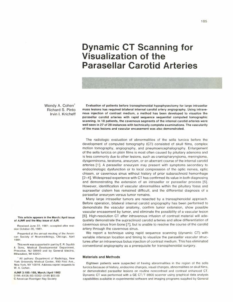
Fig. 1.-Lateral sku ll " scout view" showing scan position ing for coronal dynamic CT. Lines mark levels of scans. Line 2 is first scan as described in part 1 of Materials and Methods.
Electric (GEDIS 6 .1 6-6.21). The examination consisted of two parts with an optional third part.
For each part, baseline scans through the structures of interest were performed. Initial levels were chosen from a digitized image of th e lateral head (" scout view" ). The images were obtained with or without contrast enhancement. For the dynamic study an intravenous bolus injec tion of Renografin-76 was given through an 18 gauge angiocatheter in a large antecubital vein. All injections were with a mechan ical injector, delivering 40 ml of warmed contrast agent (11 g iodine) over 4-5 sec .
Arm to brain transit time in adults, determined empirically, is about 8 sec . Th e scanner required 2 sec for rotor preparation, thus the injec tion was started 4 sec before the commencement of th e initial scan. Max imum contrast density appeared in vascular structures in the second and third scans of a rapid sequence series.
Using the rapid sequence scanning method , a single scan was obtained over 4 .8 sec. Using the table increment mode, 6.5 sec. were required between scans, during which time the table changed locations to a preprog rammed position . If the scan loca tion was not changed, interscan tim e could be reduced to 1.5 sec.
Part 1 (Coronal Scanning)
The patient was placed in a position for direct coronal scanning , ly ing prone with his head extended and the scanner gantry tilted so that the scan plane was approximately perpendicular to the planum sphenoid ale. Gantry angulation and scan position were determined from the " lateral" scan view. Baseline 1 O-mm-thick adjacent noncontrast scans were obtained , starting 10 mm in front of the tips of the anterior c linoids to encompass a part of the planum sphenoidale and tuberculum , and ending behind th e dorsum sellae (fig . 1).
The routine dynamic CT series consisted of four to five scans taken at the same locations as the baseline scans using the tabl e incremen t mode and a bolus injection of contrast agent. Scanning started at the most anterior point of the baseline scans and moved
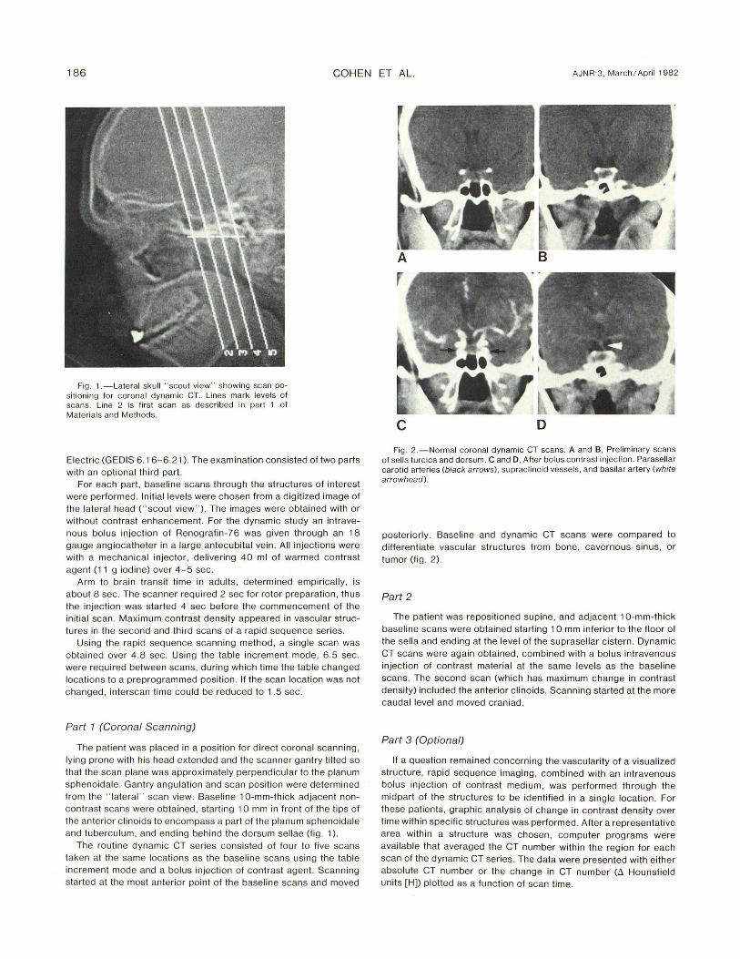
c o Fig. 2.-Norm al coronal dynamic CT scans. A and B , Preliminary scans
o f sella turcica and dorsum. C and D, After bolus con trast injec tion. Parasellar carotid arteries (black arrows), suprac linoid vessels, and basilar artery (white arrowhead).
posteriorly. Baseline and dynamic CT scans were compared to differentiate vascu lar structures from bone, cavernous sinus, or tumor (fig . 2).
Part 2
The patient was repositioned supine, and adjacent 10-mm-thick baseline scans were obtained starting 10 mm inferior to the floor of th e sella and ending at th e level of the suprasellar cistern. Dynamic CT scans were again obtained, combined with a bolus intravenous injec tion of contrast material at the same levels as the baseline scans. The second scan (which has maximum change in contrast density) included the anterior clinoids. Scanning started at the more caudal level and moved craniad.
Part 3 (Optional)
If a question remained concerning the vascu larity of a visualized structure, rapid sequence imaging , combined with an intravenous bolus injection of contrast medium, was performed through the midpart of the structures to be identified in a single location. For these patients, graph ic analysis of change in contrast density over tim e within speci fi c struc tures was performed. After a representative area within a structure was chosen, computer programs were available that averaged th e CT number within the region for each scan of the dynamic CT series. The data were presented with either absolute CT number or the change in CT number (t. Hounsfield units [H]) plotted as a function of scan time .
AJNR :3, Marchi April 1982 CT OF PARASELLAR CAROTID ARTERIES 187
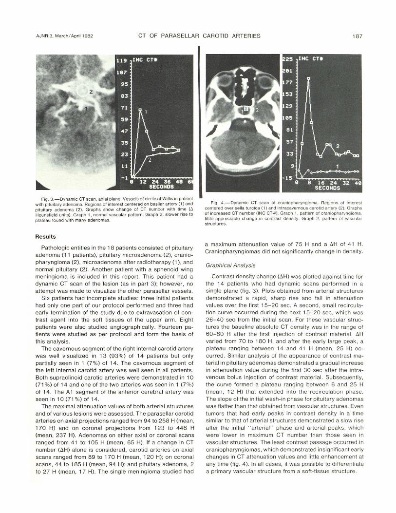
Fig . 3 .-0ynamic CT scan, axial plane. Vessels of circ le o f Willis in pati en t wi th pituitary adenoma. Regions of interest centered on basilar artery (1) and pitu itary adenoma (2) . Graphs show change of CT number with time (to
Hounsf ield units). Graph 1 , norm al vascular pattern . Graph 2, slower ri se to plateau found with many adenomas.
Results
Pathologic entities in the 18 patients consisted of pituitary adenoma (11 patients) , pituitary microadenoma (2) , craniopharyngioma (2), microadenoma after radiotherapy (1), and normal pituitary (2) . Another patient with a sphenoid wing meningioma is included in this report . This patient had a dynamic CT scan of the lesion (as in part 3); however, no attempt was made to visualize the other parasellar vessels .
Six patients had incomplete studies: three initial patients had only one part of our protocol performed and three had early termination of the study due to extravasation of contrast agent into the soft tissues of the upper arm. Eight patients were also studied angiographically. Fourteen patients were studied as per protocol and form the basis of this analysis.
The cavernous segment of the right internal carotid artery was well visualized in 13 (93%) of 14 patients but only partially seen in 1 (7%) of 14. The cavernous segment of the left internal carotid artery was well seen in all patients. Both supraclinoid carotid arteries were demonstrated in 10 (71 %) of 14 and one of the two arteries was seen in 1 (7 %) of 14. The A 1 segment of the anterior cerebral artery was seen in 10 (71 %) of 14.
The maximal attenuation values of both arterial structures and of various lesions were assessed . The parasellar carotid arteries on axial projections ranged from 94 to 258 H (mean , 170 H) and on coronal projections from 1 23 to 448 H (mean, 237 H). Adenomas on either axial or coronal scans ranged from 41 to 105 H (mean , 65 H). If a change in CT number (LlH) alone is considered, carotid arteries on axial scans ranged from 89 to 170 H (mean , 120 H); on coronal scans, 44 to 185 H (mean , 94 H); and pituitary adenoma, 2 to 27 H (mean , 17 H). The single meningioma studied had
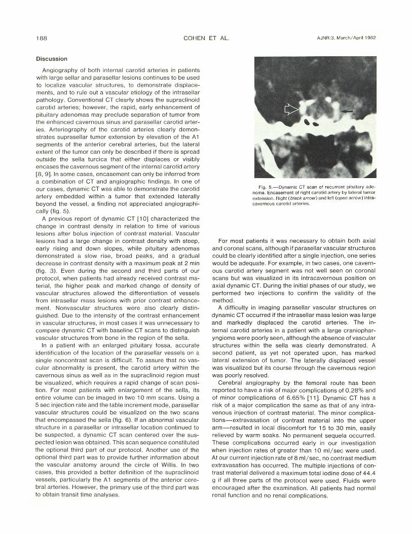
Fig. 4 .-0ynamic CT scan of c raniopharyngioma. Regions o f interest centered over sella turc ica (1) and intracavernous ca rotid arte ry (2). Graphs of inc reased CT number (INC CT# ). Graph 1, pattern of craniopharyngioma. li tt le appreciable change in contrast density . Graph 2, pattern o f vascular structures.
a maximum attenuation value of 75 H and a LlH of 41 H. Craniopharyngiomas did not significantly change in density .
Graphica l Analysis
Contrast density change (t.H) was plotted against time for the 14 patients who had dynamic scans performed in a single plane (fig . 3). Plots obtained from arteri al structures demonstrated a rapid , sharp ri se and fall in attenuati on values over the first 15-20 sec. A second , small recirculation curve occurred during the next 15-20 sec, which was 26-40 sec from the initial scan . For these vascular structures the baseline absolute CT density was in the range of 60-80 H after the first injecti on of contrast materi al. LlH varied from 70 to 180 H, and after the early large peak, a plateau ranging between 14 and 41 H (mean, 25 H) occurred . Similar analysis of the appearance of contrast material in pituitary adenomas demonstrated a gradual increase in attenuation value during the first 30 sec after the intravenous bolus injec tion of contrast material. Subsequently, the curve formed a plateau ranging between 6 and 25 H (mean , 12 H) that extended into the recircul ation phase . The slope of the initial wash-in phase for pituitary adenomas was flatter than that obtained from vascular struc tures . Even tumors that had early peaks in contrast density in a time similar to that of arteri al struc tures demonstrated a slow rise after the initial " arteri al" phase and arteri al peaks, which were lower in maxi mum CT number than those seen in vascular structures. The least contrast passage occurred in craniopharyngiomas, which demonstrated insignificant early changes in CT attenuation values and little enhancement at any time (fig. 4) . In all cases, it was possible to differentiate a primary vascular structure from a soft-ti ssue structure.
188 COHEN ET AL. AJNR:3, March! April 1982
Discussion
Angiography of both internal carotid arteries in pati ents with large sell ar and parasell ar lesions continues to be used to locali ze vascular struc tures, to demonstrate d isplacements, and to ru le out a vascular eti ology of the intrase llar pathology. Conventional CT clearly shows the suprac linoid carotid arteri es ; however, the rapid , earl y enhancement of pituitary adenomas may prec lude separation of tumor from the enhanced cavern ous sinus and parase llar carotid arteries. Arteri ography of the carotid arteri es c learly demonstrates suprasellar tumor extension by elevation of the A 1 seg ments of the anteri or cerebral arteri es, but the lateral extent of the tumor can only be described if there is spread outside the se lla turcica that either displaces or visibly encases the cavernous segment of the internal carotid artery [8, 9]. In some cases, encasement can on ly be inferred from a combinati on of CT and angiographic findings. In one of our cases, dynamic CT was able to demonstrate the carotid artery embedded within a tumor that extended laterally beyond the vessel , a finding not apprec iated angiographically (fig. 5).
A previous report of dynamic CT [10] characterized the change in contrast density in relation to time of various lesions after bolus injection of contrast materi al. Vascular lesions had a large change in contrast density with steep , early ri sing and down slopes, whil e pituitary adenomas demonstrated a slow rise, broad peaks, and a gradual decrease in contrast density with a maximum peak at 2 min (fig. 3). Even during the second and third parts of our protoco l, when patients had already received contrast mater ial, the higher peak and marked change of density of vascular structures allowed the differentiation of vessels from intrase llar mass lesions with prior contrast enhancement. Nonvascular structures were also c learly distinguished. Due to the intensity of the contrast enhancement in vascular struc tures, in most cases it was unnecessary to compare dynamic CT with baseline CT scans to distinguish vascular struc tures from bone in the region of the sella.
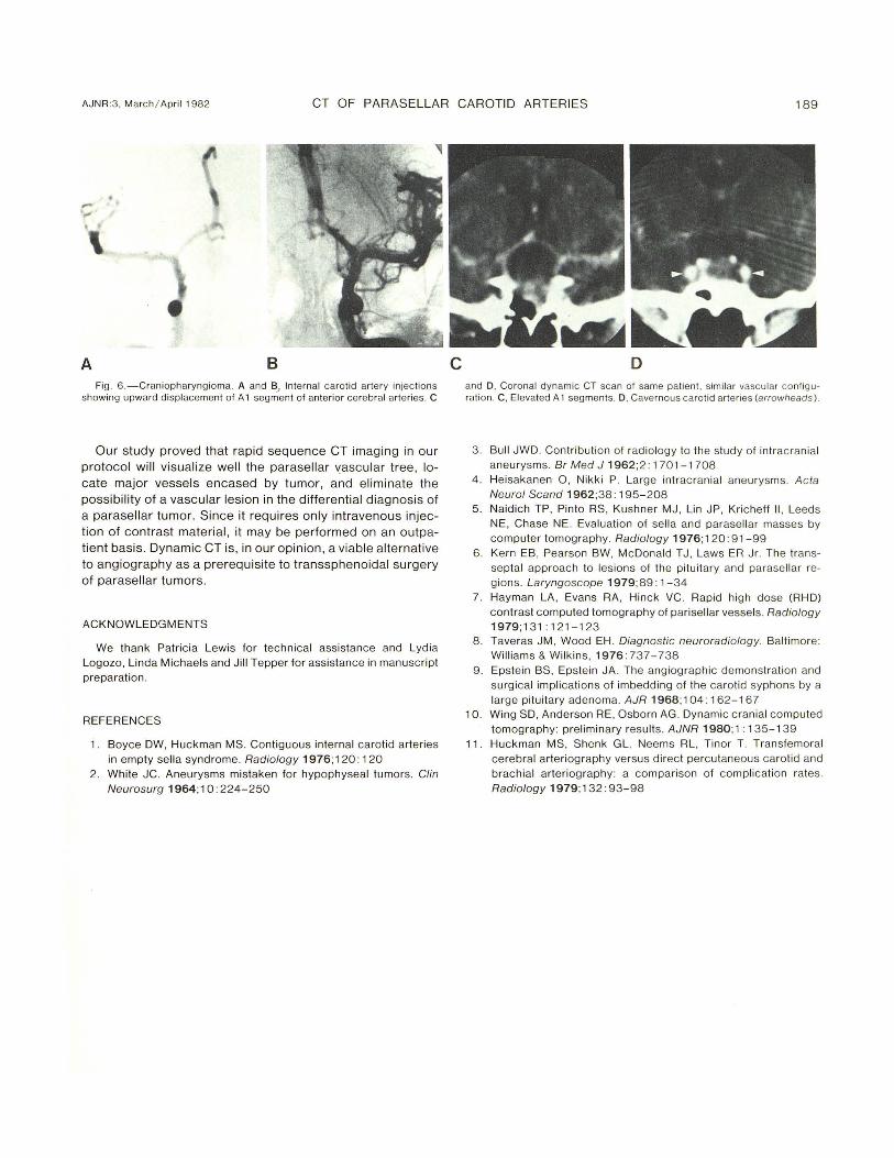
In a pati ent with an enlarged pituitary fossa, accurate identificati on of the locati on of the parase llar vessels on a single noncontrast scan is difficult. To assure that no vascular abnormality is present, the carotid artery within the cavern ous sinus as well as in the suprac linoid region must be vi sualized , which requires a rapid change of scan positi on. For most pati ents with enlargement of the sella, its entire volume can be imaged in two 10 mm scans. Using a 5 sec injecti on rate and the table increment mode, parasellar vascular structures could be visualized on the two scans that encompassed the se lla (fig . 6) . If an abnormal vascular structure in a parasell ar or intrasellar location continued to be suspected , a dynamic CT scan centered over the suspected les ion was obtained. This scan sequence constituted the optional third part of our protocol. Another use of the opti onal third part was to provide furth er information about the vascular anatomy around the circ le of Willi s. In two cases, this provided a better definition of the suprac linoid vesse ls, particularl y the A 1 segments of the ante ri or cerebral arteri es . However, the primary use of the third part was to obtai n transit time analyses.
Fig. 5. -Dynamic CT scan of recurrent pituitary adenoma. Encasement of right ca rotid artery by lateral tumor ex tension. Right (black arro w) and left (open arrow) intracavernous carotid arteries.
For most patients it was necessary to obtain both axial and coronal scans, although if parasellar vascular structures cou ld be clearly identified after a single injection , one series would be adequate. For example , in two cases, one cavernous carotid artery segment was not we ll seen on coronal scans but was visualized in its intracavernous position on axial dynamic CT. During the initial phases of our study, we performed two injections to confirm the validity of the method .
A difficulty in imaging parasellar vascular structures on dynamic CT occurred if the intrasellar mass lesion was large and markedly displaced the carotid arteries. The internal carotid arteries in a patient with a large craniopharyngioma were poorly seen, although the absence of vascular structures within the sella was clearly demonstrated . A second patient, as yet not operated upon , has marked lateral extension of tumor. The laterally displaced vessel was vi sualized but its course through the cavernous region was poorly resolved .
Cerebral angiography by the femoral route has been reported to have a risk of major complications of 0.28% and of minor compli cations of 6 .65% [11]. Dynamic CT has a risk of a major complication the same as that of any intravenous injection of contrast material. The minor compl ications-extravasation of contrast material into the upper arm-resulted in local discomfort for 15 to 30 min , easi ly relieved by warm soaks. No permanent sequela occurred. These complications occurred early in our investigation when injection rates of greater than 10 ml / sec were used . At our current injection rate of 8 ml / sec , no contrast medium extravasation has occurred . The multiple injections of contrast material delivered a maximum total iodine dose of 44.4 g if all three parts of the protocol were used. Fluids were encouraged after the examination. All patients had normal renal fun ction and no renal complications .
AJNR :3, March i April 1982 CT OF PARASELLAR CAROTID ARTERIES 189
A B Fig. 6.-Craniopharyngioma. A and B, Internal carotid artery injecti ons
showing upward displacement of A 1 segment of anterior ce rebral arteries. C
Our study proved that rapid sequence CT imaging in our protocol will visualize well the parasellar vascular tree, locate major vessels encased by tumor, and eliminate the possibility of a vascular lesion in the differential diagnosis of a parasellar tumor. Since it requires only intravenous injection of contrast material , it may be performed on an outpatient basis. Dynamic CT is, in our opinion, a viable alternative to angiography as a prerequisite to transsphenoidal surgery of parasellar tumors.
ACKNOWLEDGMENTS
We thank Patric ia Lewis for technical assistance and Lydia Logozo, Linda Michaels and Jill Tepper for assistance in manuscript preparation .
REFERENCES
1. Boyce OW, Huckman MS. Contiguous internal carotid arteries in empty sella syndrome. Radiology 1976;120 : 1 20
2. White JC . Aneurysms mistaken for hypophyseal tumors. Clin Neurosurg 1964;10 :224-250
c D and D, Coronal dynamic CT scan of same patient, similar vascular configuration. C, Elevated A 1 segments. D, Cavernous carotid art eries (arrowheads ).
3. Bull JWD . Contribution of rad iology to the study of in trac ranial aneurysms. Br Med J 1962;2: 1 701-1 708
4 . Heisakanen 0 , Nikki P. Large intrac ranial aneu rysms. Acta Neurol Scand 1962;38 : 1 95- 208
5. Naidich TP , Pinto RS , Kushner MJ, Lin JP, Kricheff II , Leeds NE, Ch ase NE. Evaluation of sella and parasellar masses by computer tomography. Radiology 1976; 120 : 9 1 - 99
6 . Kern EB, Pearson BW, McDonald TJ , Laws ER Jr. Th e transseptal approach to lesions of the pituitary and parasell ar reg ions. Laryngoscope 1979; 89: 1 - 34
7 . Hayman LA, Evans RA , Hinck VC . Rapid high dose (RHO) contrast computed tomography of pari sellar vessels. Radiology 1979;13 1 : 121-1 23
8. Taveras JM , Wood EH . Diagnostic neuroradio logy. Baltimore: Williams & Wilkin s, 1976 : 737 -738
9. Epstein BS, Epstein JA. The angiog raphic demonstration and surgica l implications of imbedd ing of the carotid syphons by a large pituitary adenoma. AJR 1968; 104: 1 62- 1 67
10. Wing SO, Anderson RE , Osborn AG . Dynamic c ranial computed tomography: preliminary results. AJNR 1980; 1 : 135-1 39
11 . Huc kman MS, Shenk GL, Neems RL , Tinor T. Transfemoral cerebral arteriography versus direct percutaneous carotid and brachial arteriography: a co mpari son of complicati on rates . Radiology 1979; 132 : 93- 98