ear new affection of ear and its treatment
TRANSCRIPT

Affection of ear and its treatment
Dr. Bikash PuriAssist. Professor
Nepal Polytechnic Institute, Chitwan

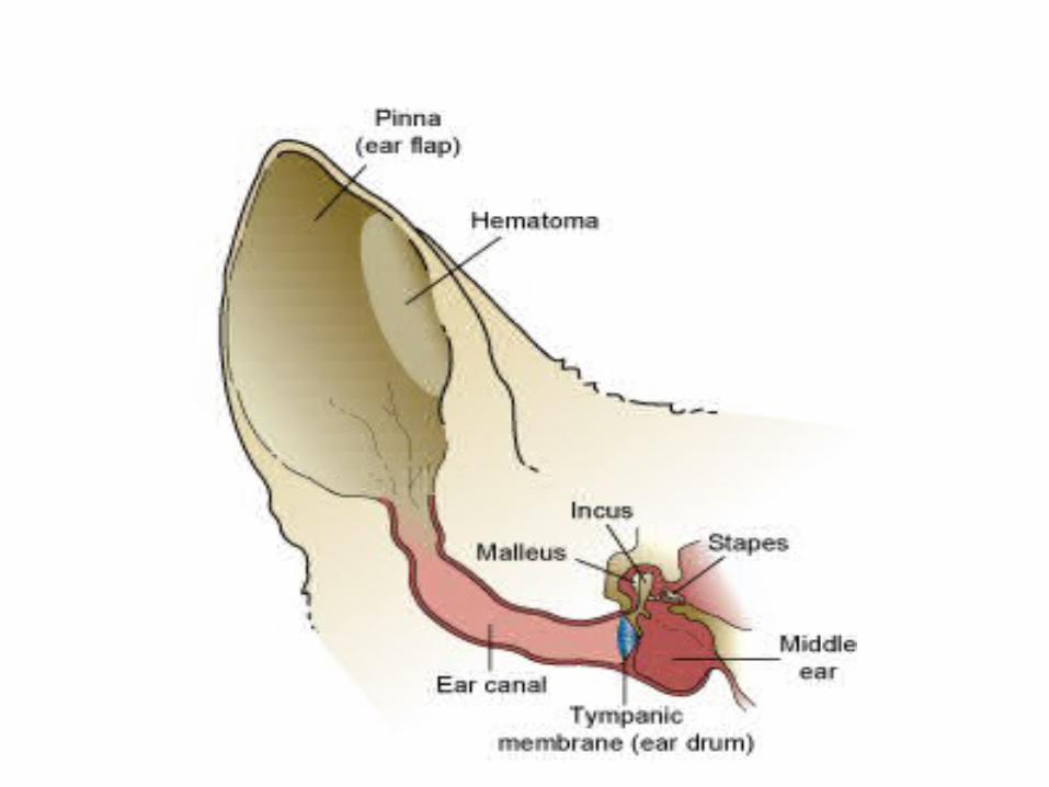
Aural Haematoma• A hematoma is a localized mass of blood that is confined within an organ or tissue.
• If it is affecting the pinna or ear flap, this is called an aural or ear hematoma.
• It is very common in dog however sporadic in case of buffalo, calves and goats.
However, dogs are commonly affected.
• Hematoma are normally confined to the concave surface of the ear, although it may be
on either or both sides of aurical cartilage, depending upon the severity and duration of
trauma.
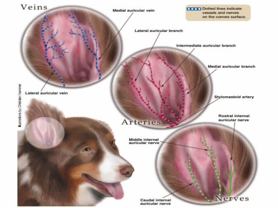
• The great auricular artery is the source of hemorrhage and bleeding continues only
when arterial pressure equals the intrahaematoma pressure.
• Additional pressure on the haematoma during head shaking or scratching causes further
separation f the tissue and allows hemorrhage to resume.

Etiology
• It is self inflicted and caused by scratching and head shaking
• Acute or chronic inflammation• Parasites e.g. ear mites or ticks• Foreign bodies in or near the ear canal.

TreatmentConservative management
A. Acute haematoma:
• Aspirate with needle.
• The source of irritant is removed and tissue are apposed
• A pressure bandage should be applied following aspiration.
B. Chronic haematoma:• Cannulation is considered. For cannulation disposable teat canula can be used.
• The canula is aseptically placed via a small stab incision in the dependent
portion of hematoma.
• Continious pressure bandage for 5-7 days decreases recurrence.

Surgical management
• If the haematoma is extensive, this conservative
method is time consuming and likely to fail.
Therefore surgical intervention is needed.
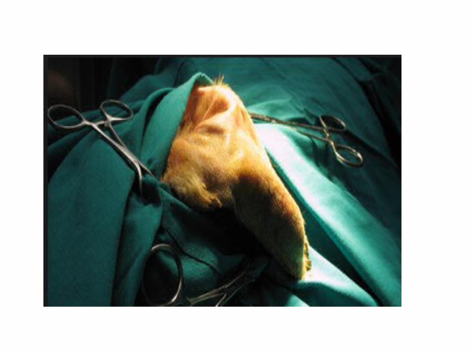
Control and Anesthesia• Control the animal in lateral recumbency keeping the affected
ear upwards after proper tranquilization or brief anesthesia.
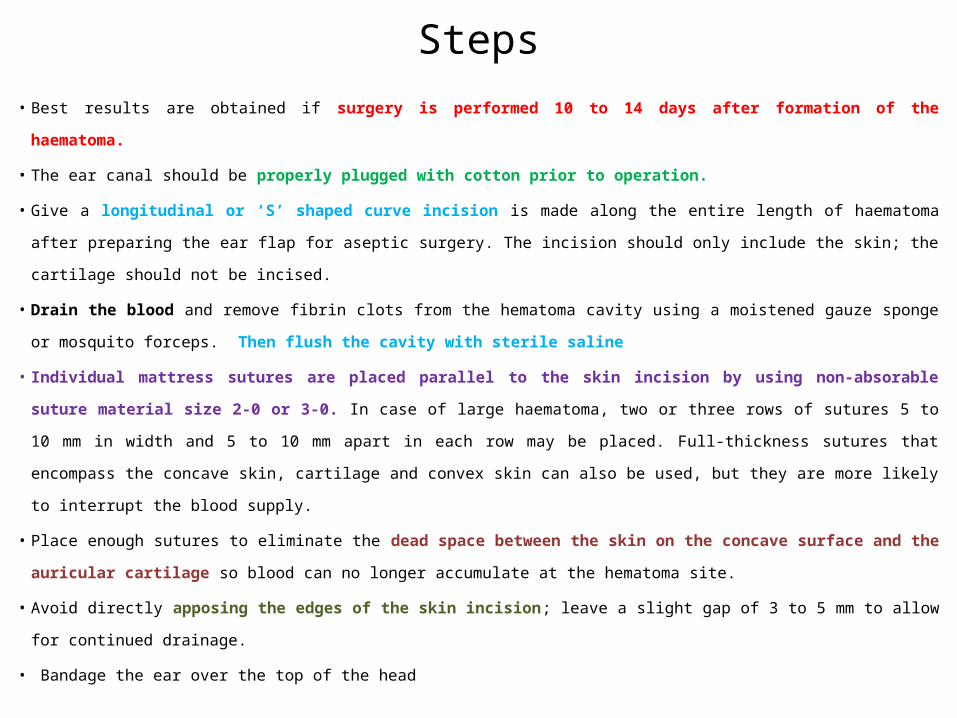
Steps• Best results are obtained if surgery is performed 10 to 14 days after formation of the haematoma.
• The ear canal should be properly plugged with cotton prior to operation.
• Give a longitudinal or ‘S’ shaped curve incision is made along the entire length of haematoma after preparing the
ear flap for aseptic surgery. The incision should only include the skin; the cartilage should not be incised.
• Drain the blood and remove fibrin clots from the hematoma cavity using a moistened gauze sponge or mosquito
forceps. Then flush the cavity with sterile saline
• Individual mattress sutures are placed parallel to the skin incision by using non-absorable suture material size 2-
0 or 3-0. In case of large haematoma, two or three rows of sutures 5 to 10 mm in width and 5 to 10 mm apart in
each row may be placed. Full-thickness sutures that encompass the concave skin, cartilage and convex skin can
also be used, but they are more likely to interrupt the blood supply.
• Place enough sutures to eliminate the dead space between the skin on the concave surface and the auricular
cartilage so blood can no longer accumulate at the hematoma site.
• Avoid directly apposing the edges of the skin incision; leave a slight gap of 3 to 5 mm to allow for continued
drainage.
• Bandage the ear over the top of the head

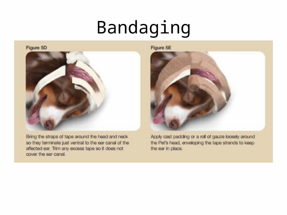
Bandaging

Post-Operative Care:
1) After treating haematoma, ear canal is cleaned and
proper medication should be applied.
2) Change the dressing as needed, usually every 3 days
and keep the animal sedated.
3) the ear should be usually bandaged.
4) Sutures are removed on 7th to 10th post-operative
day.

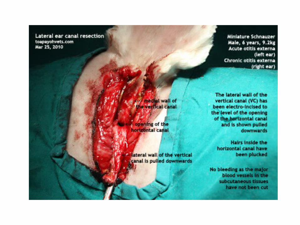
Zepp’s Operation/Lateral ear resection
INDICATIONS:1. Chronic otitis or otorrhoea for drainage.2. Neoplasms in ear canal for removal.3. Correction of anatomic deformity of ear canal.4. Congenital atresia or occlusion of the meatus5. Severe ulceration

SURGICAL ANATOMY:
• The external ear canal is not a straight tube.
• It has a horizental portion and a vertical portion formed by the
tubular portion of concha.
• The objective of this operation is to cut open the vertical portion to
facilitate drainage.
CONTROL AND ANAESTHESIA
• Lateral recumbency position with affected ear up under general
anaesthesia.
SITE OF OPERATION
• Ventral to cranio-caudal aspect of the ear canal
PRECAUTION:
Avoid damage to the parotid salivary gland.

SURGICAL TECHNIQUE• Measure the length of vertical portion of ear canal with the help of a probe.
• Make two parallel incisions starting at the indentations on either side of the tragus.
• The incision should be about a half times more then the length of vertical canal. Join
these incisions with horizontal incision on ventral side.
• Dissect out the skin flap and sub cutaneous tissue until it remains attached to the
external opening of the ear canal and reflected dorsally.
• Incise the lateral cartilage of vertical canal by two parallel incisions. Extend these
incision ventrally upto the level of the horizontal ear canal.
• Reflect the cartilage flap ventrally to expose the orifice of horizontal canal.
• Trim the skin and excess of cartilage and then suture the cartilage with skin by
applying simple interrupted suture pattern.


POST OPERATIVE CARE
1. Proper bandaging of the pinna.2. ASD of the surgical wound.3. Systemic antibiotics.4. Sutures removed after 8-10 days5. Use neck collar
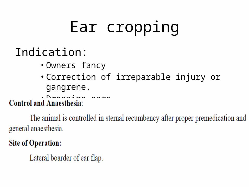
Ear cropping
Indication:• Owners fancy• Correction of irreparable injury or gangrene.• Drooping ears


Ulceration of the Concha• This condition is most common in long-eared dogs,
Etiology:
– usually the result of a wound which is prevented from healing by the constant shaking of the ears, or
by rubbing them on the ground or scratching them with a paw.
– The ulcer is usually situated towards the border of the flap, but the inner aspect of the latter may
become ulcerated when affected with canker or otorrhosa.
Treatment. —
– Arresting the shaking of the ears, or treating canker when it is present.
– If the ulcer be of long standing and too callous to granulate and cicatrize, it should be cauterized with
silver nitrate, or stimulated by the application of a little Tincture iodine, rubbed into the skin in the
vicinity and smeared over the ulcer.
– Otherwise thorough cleansing of the lesion and dressing it with iodoform powder prove sufficient with
immobilization.

Necrosis of conchal Cartilage
• Necrosis of the concha is most common in the horse, and is the result of an open wound
and infection of the cartilage.
• Symptoms:
– The symptoms are those of a sinus, there being an inflammatory swelling with one or more orifices
discharging a greyish, foetid, and sometimes blood-stained pus.
– The ear becomes deformed from contraction of the fibrous tissue replacing the destroyed
cartilage.
• Treatment.
– The treatment is on the usual principles for a sinus, comprising opening up of the passage,
providing for drainage, the injection of antiseptic or irritant liquids, or mechanical removal of the
necrotic cartilage with the knife or curette.
– When the ear is extensively diseased it is necessary to amputate the concha as follows:
Amputation of concha
• Restrain the animal properly with head well extended
• prepare the site of operation,
• inject a solution of Local anesthatic hypodermically,
• make an incision encircling the concha about one inch from its base,
• dissect back the skin and the upper border of the parotid gland for a short distance,
• divide the auricular muscles in this situation,
• ligature the anterior auricular artery, sever the ligament uniting the cartilage to the
auditory meatus, and remove the former.
• Apply a pad of gauze over the meatus to prevent the entrance of blood therein, and
suture the skin in front of and behind the orifice.

Otitis externa, otitis media, and otitis interna
Overview– An ear infection, or otitis, is an inflammation of the outer, middle, or inner ear canal.
– Most frequently, a dog will develop otitis in the outer ear that may worsen and spread into the middle ear.
– Once in the middle ear canal, the inflammation can move into the inner ear -- or, in cases in which the otitis
has originated in the middle ear, the infection can instead progress outward to the external ear.
– VERY COMMON IN Dogs and cats.
Etiology– Parasitic infestation: eg fleas,
– excess liquid in the ear from swimming,
– autoimmune diseases, skin parasites, and excess wax production.

Description• Typically, ear infections begin with otitis externa and then progress deeper into the canal to the
middle ear. When the inflammation in this region of the ear is chronic, the eardrum may rupture
and the infection may spread to the inner ear -- or, the infection may begin in the middle ear and
progress outward to the external ear. Of the three types of otitis, infections in the inner ear are
often the most severe and can lead to partial deafness and neurological problems.
• In serious cases of otitis, the skin begins to form into folds in which the infection can become
trapped, making cleaning and use of topical treatments very difficult. In addition, in cases of otitis
interna, the skin will secrete more wax and debris that allows yeast and bacteria to overgrow,
causing further disease. Severe inflammation leads to permanent skin thickening, mineralization,
and narrowing of the ear canals. Once this occurs, the only viable treatment will be surgical
removal of part or all of the ear canal.

Symptoms• Ears that are red, painful to the touch, and produce a foul-smelling discharge are
symptomatic of otitis.
• Typically, a dog with an ear infection will scratch and shake the ears or may tilt the affected
ear downwards. If both ears are affected, the animal may be deaf or off balance and
uncoordinated.
• Animals that are affected more severely may show some neurological signs such as rolling or
leaning to one side.
• Some animals may be nauseated and vomit. Also, some dogs may get a condition called
Horner's syndrome in which the pupils are sized differently and the nictitating membrane is
raised. This condition indicates that a nerve has been affected by the inflammation from the
middle ear. Any neurological clinical signs indicate significant middle ear or inner ear disease.

Diagnosis1. Based on the clinical signs, physical exam findings, and through the use of several other
diagnostic tools.
2. One such tool is cytology, which involves taking a swab of the ears and looking at the material
collected under a microscope for the presence of bacteria, yeast, mites, and other substances that
might cause an infection. Bacteria and yeast are normally present in low numbers in all dogs' ears,
but a large presence will lead to an ear infection.
3. Skin biopsies may be needed to determine any diseases such as an autoimmune disorder that
could cause a skin abnormality affecting the ear.
4. Skin scrapings may be needed to detect mites, tiny parasites that can infect the ears and cause
skin diseases.
5. X-rays of the skull can be used to examine the middle and inner ear for signs of disease.
6. In addition, cultures of an infected ear help determine the presence and type of bacteria, as well
as antibiotics that are appropriate for treatment.

Treatment• After the cause of the otitis has been diagnosed, the veterinarian usually will perform a thorough
cleaning of the ear canals while the dog is under sedation or general anesthesia.
• The use of topical medications, which are placed into the ear canal, is often very beneficial in
killing yeast, bacteria, and mites.
• Oral medications also may be used in conjunction with other treatments to help kill bacteria,
yeast, and mites.
• The surgical correction of the ear canals may be necessary in cases of severe infection. One type of
surgery, called a lateral ear canal resection, allows the ear to drain more easily, decreases
the amount of humidity in the ear, and makes topical treatments easier to apply or
• A total ear canal ablation -- a complete removal of the ear canal -- is performed on dogs with
severe, chronic ear disease that is nonresponsive to medical therapy.

Treatment
The goals of treatment are to clean the external and middle ear; remove infected, inflammatory, or foreign
• Perform under general anesthesia.• Soak the ear canal for 10 minutes with a
ceruminolytic ear cleaner, then flush using warm saline:
• Once the exudate and debris are removed from the ear canal, evaluate the tympanic membrane with an otoscope or video otoscope.
• If the tympanic membrane is not intact, perform cytology and bacterial C/S from the middle ear cavity
• Once the ear has been cleaned and flushed, begin systemic and topical antimicrobial/antifungal treatment based on cytologic results from the external and middle ear.

• The most common coccoid bacteria isolated from the middle ear of dogs with otitis media is S. intermedius; appropriate antibiotic choices include
• Cephalexin, 22mg/kg PO q12h• Amoxicillin and clavulanate, 13.75 to 22mg/kg
PO q12h

• The most common rod bacteria is Pseudomonas aeruginosa. A fluoroquinolone such as
• Enrofloxacin, 5 to 20 mg/kg PO q24h• Marbofloxacin, 2.75 to 5.5mg/kg q24h PO
• Certain systemic antibiotics (primarily aminoglycosides) are ototoxic and should be used cautiously.

Prognosis
• With proper treatment, otitis externa usually will resolve
within three to four weeks, although it may recur in
certain animals. The prognosis is good for otitis media and
otitis interna since medical treatment usually is effective.
• The key for long-term success is correcting or treating the
underlying problem that led to the development of the
otitis.