ecg quiz cardiology update 07 december 2014 · • pr segment (pericarditis) • qrs v1, v6...
TRANSCRIPT
ECG QUIZSAHA
October 2015
brian vezi
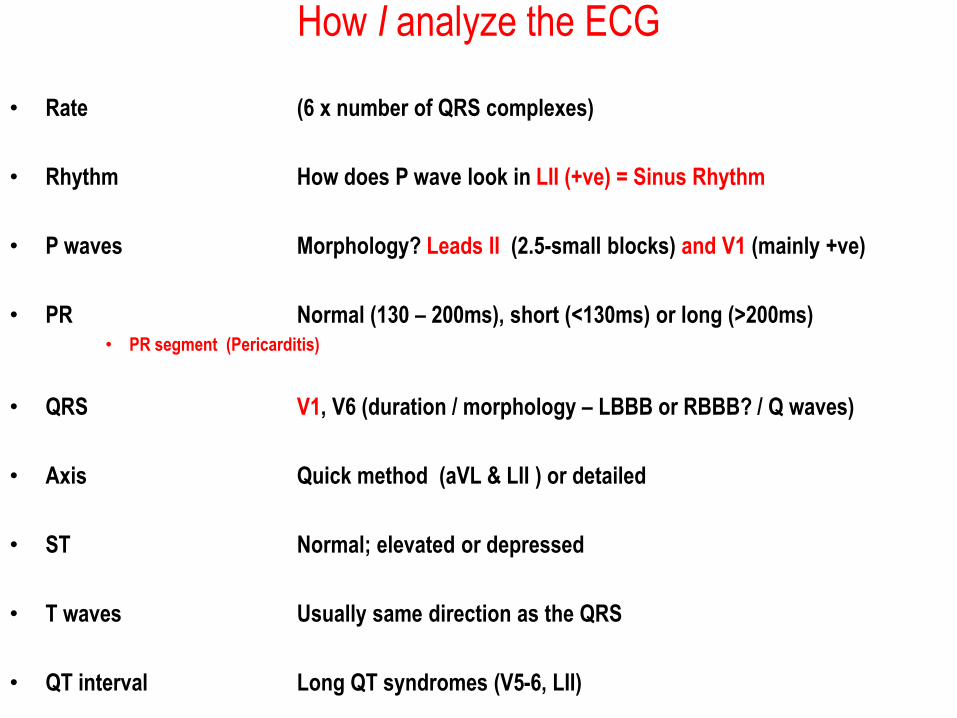
How I analyze the ECG
• Rate (6 x number of QRS complexes)
• Rhythm How does P wave look in LII (+ve) = Sinus Rhythm
• P waves Morphology? Leads II (2.5-small blocks) and V1 (mainly +ve)
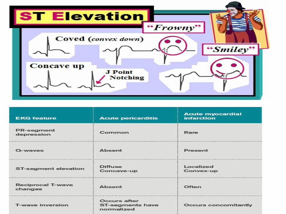
• PR Normal (130 – 200ms), short (<130ms) or long (>200ms)• PR segment (Pericarditis)
• QRS V1, V6 (duration / morphology – LBBB or RBBB? / Q waves)
• Axis Quick method (aVL & LII ) or detailed
• ST Normal; elevated or depressed
• T waves Usually same direction as the QRS
• QT interval Long QT syndromes (V5-6, LII)
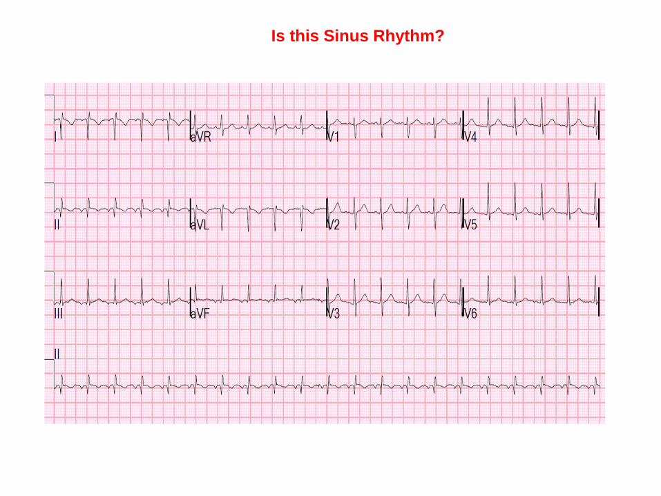
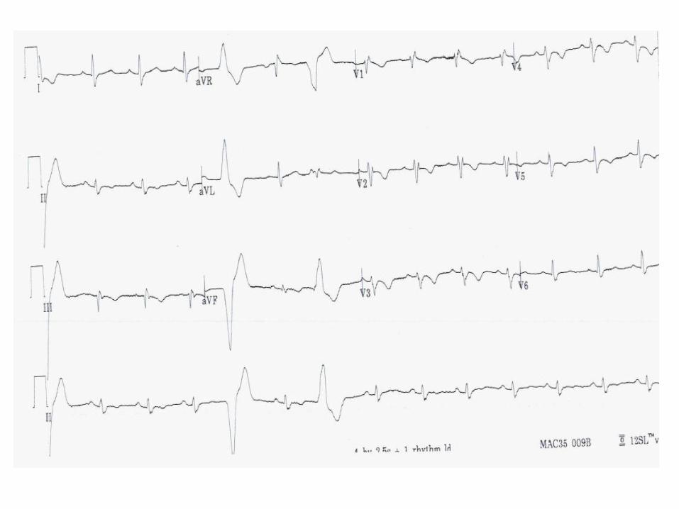
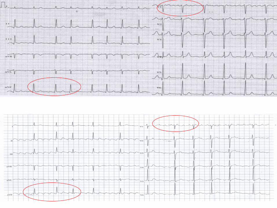
Is this Sinus Rhythm?
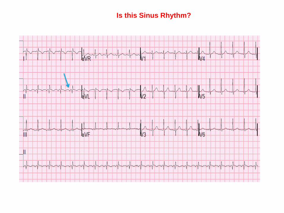
Is this Sinus Rhythm?
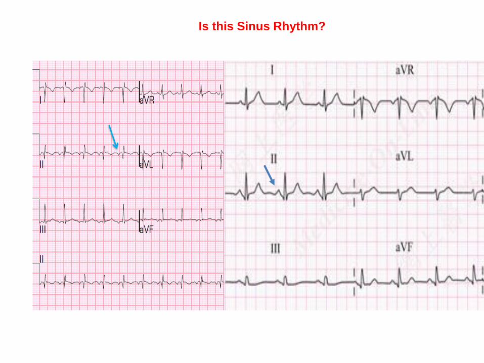
Is this Sinus Rhythm?
INSERT training slides on Block
• BRIAN to do
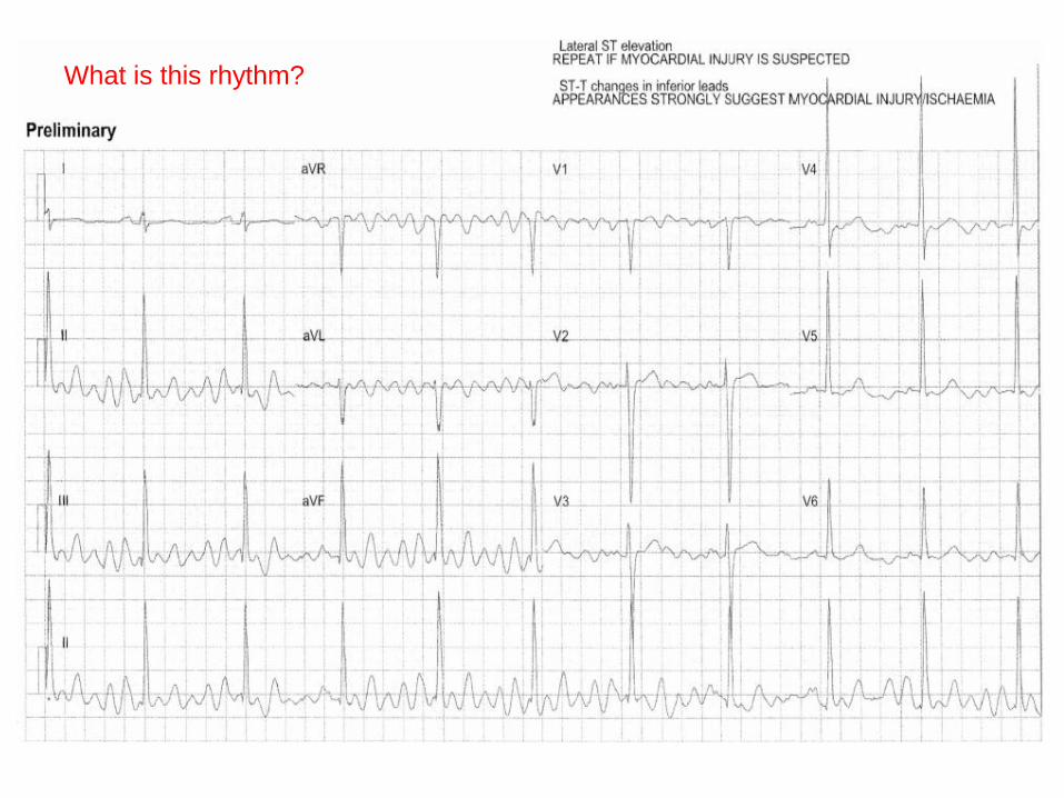
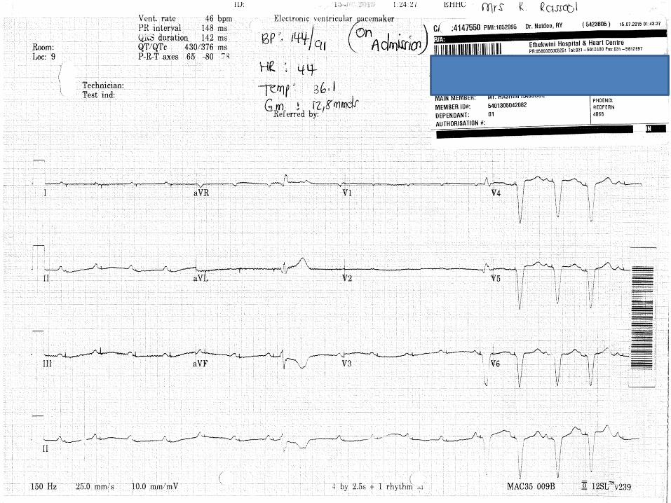
What is this rhythm?
Bradycardia
Pattern Recognition & Management
• If slow and regular Complete Heart Block
Mobitz II
Sinus Bradycardia
• If slow, irregular & ‘groups-of-QRS’Mobitz I
• If you see 3 P waves unconducted = High AV block
1
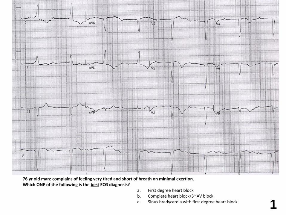
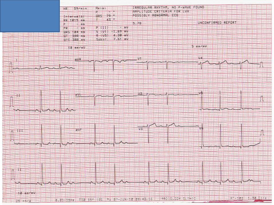
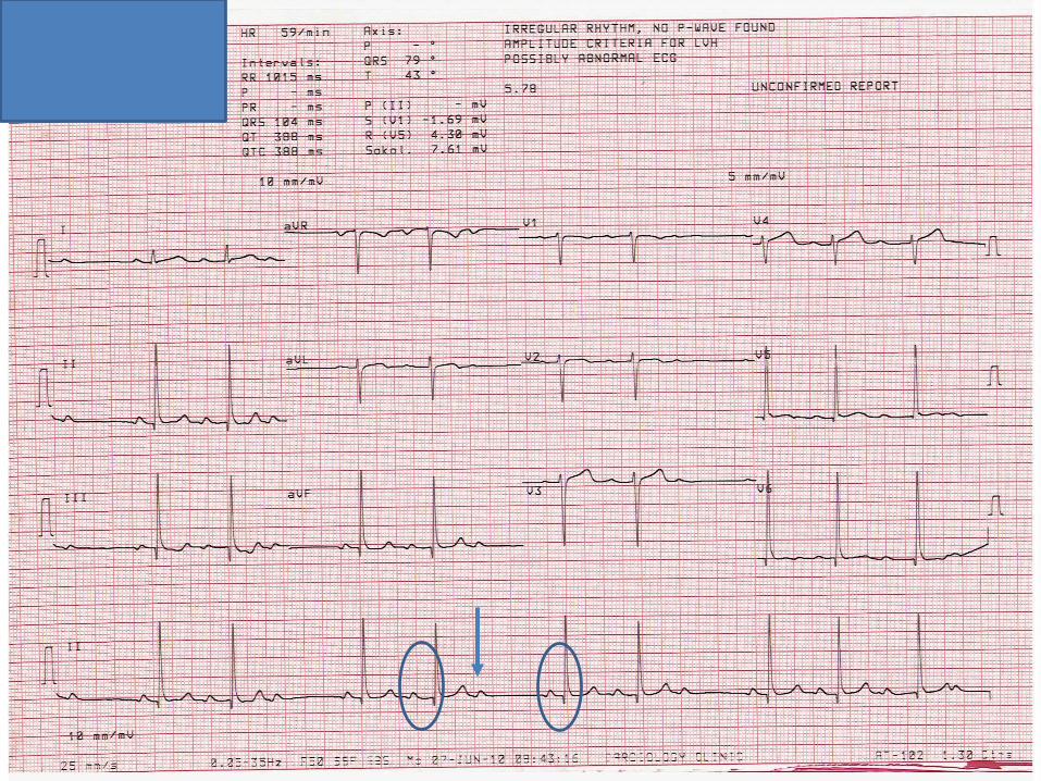
76 yr old man: complains of feeling very tired and short of breath on minimal exertion.Which ONE of the following is the best ECG diagnosis?
a. First degree heart block b. Complete heart block/3o AV blockc. Sinus bradycardia with first degree heart block
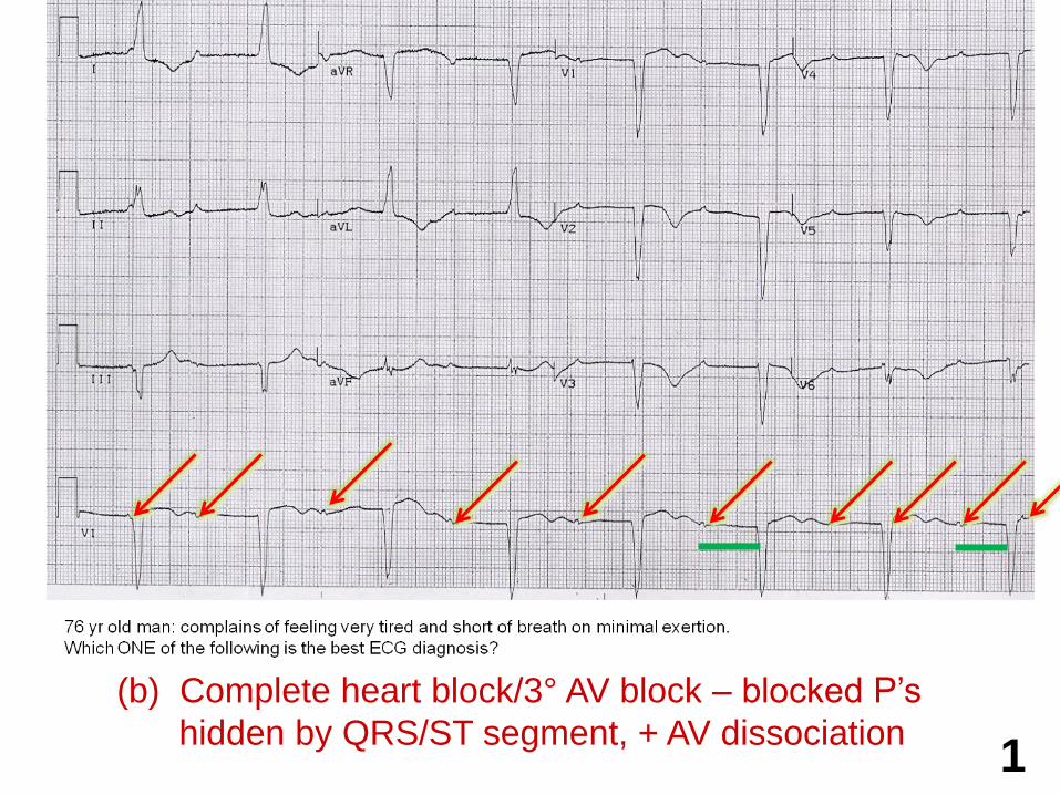
(b) Complete heart block/3° AV block – blocked P’s
hidden by QRS/ST segment, + AV dissociation1
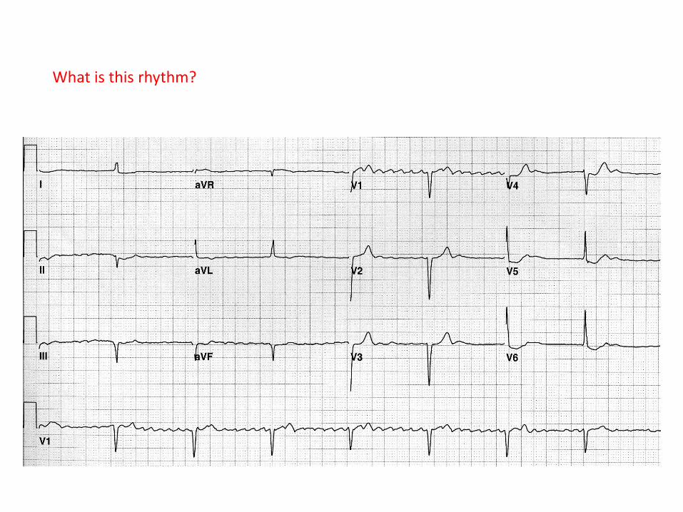
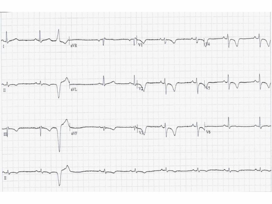
What is this rhythm?
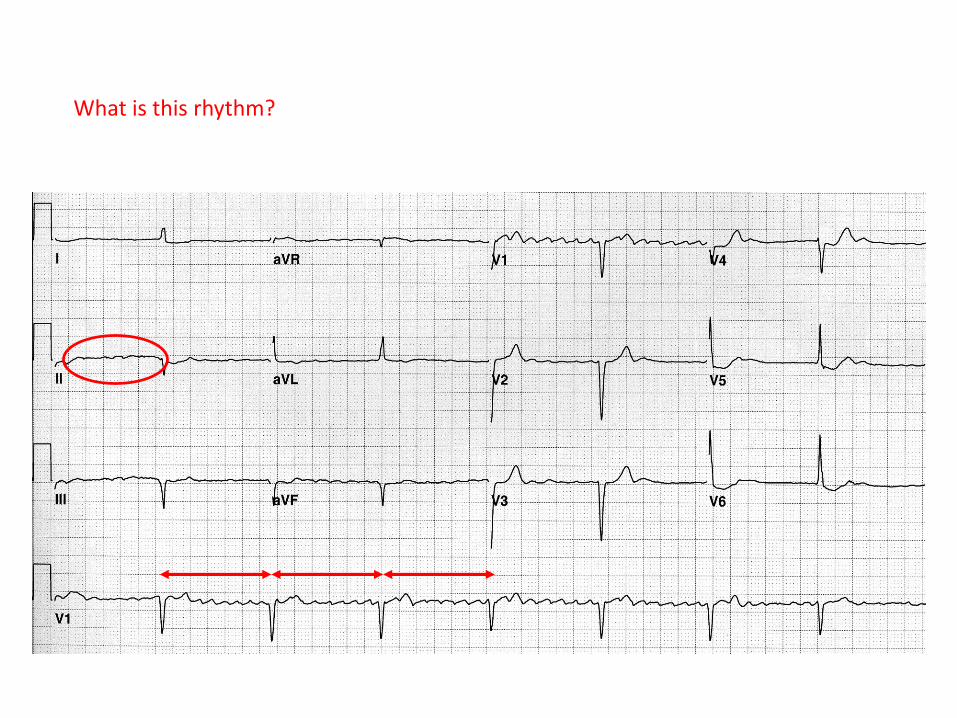
What is this rhythm?
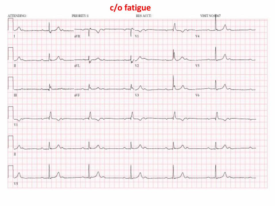
c/o fatigue
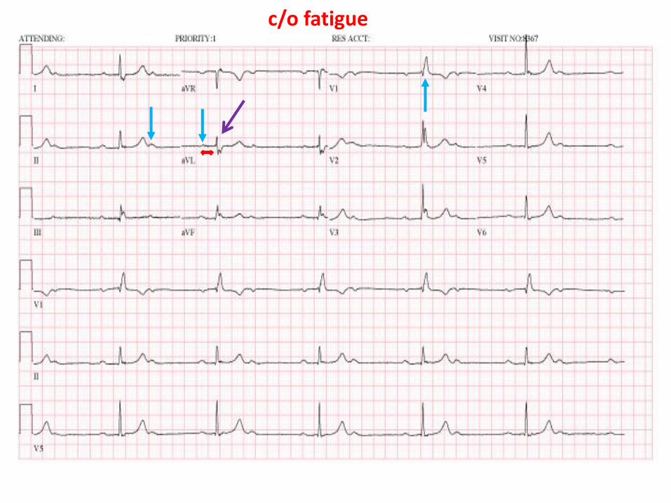
c/o fatigue

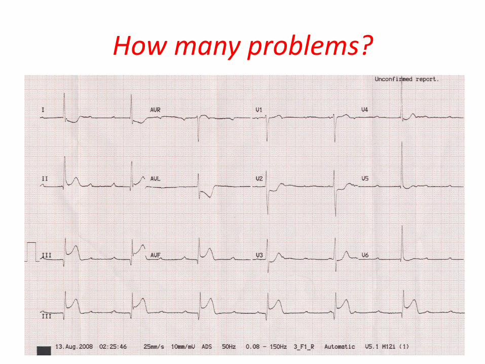
How many problems?
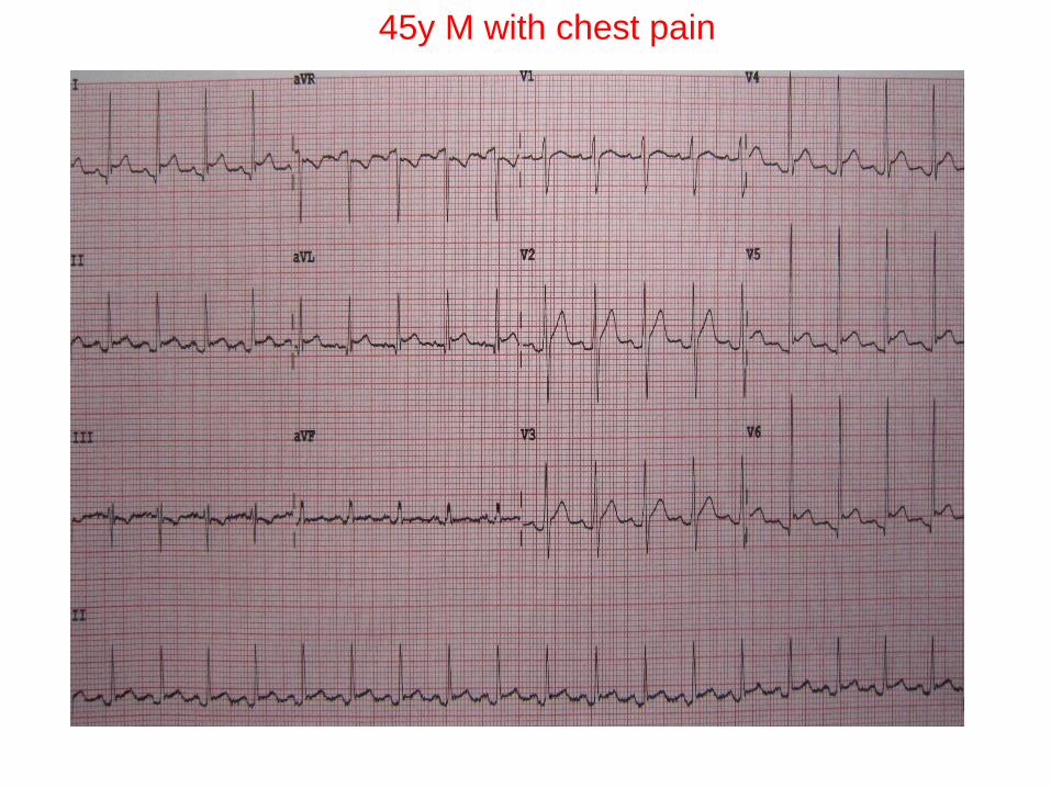
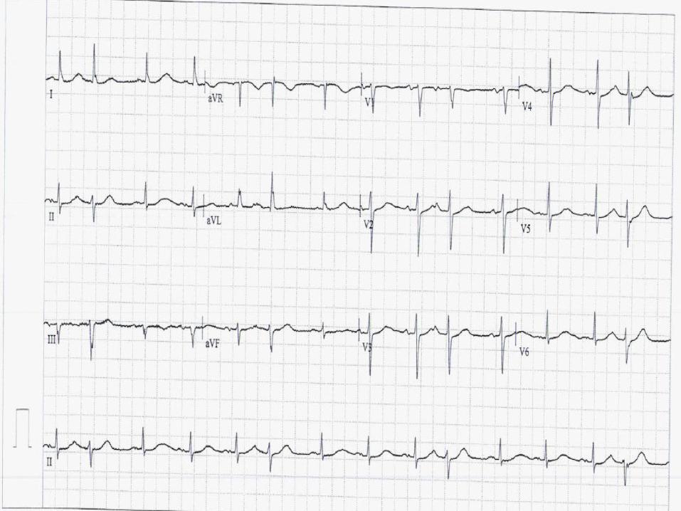
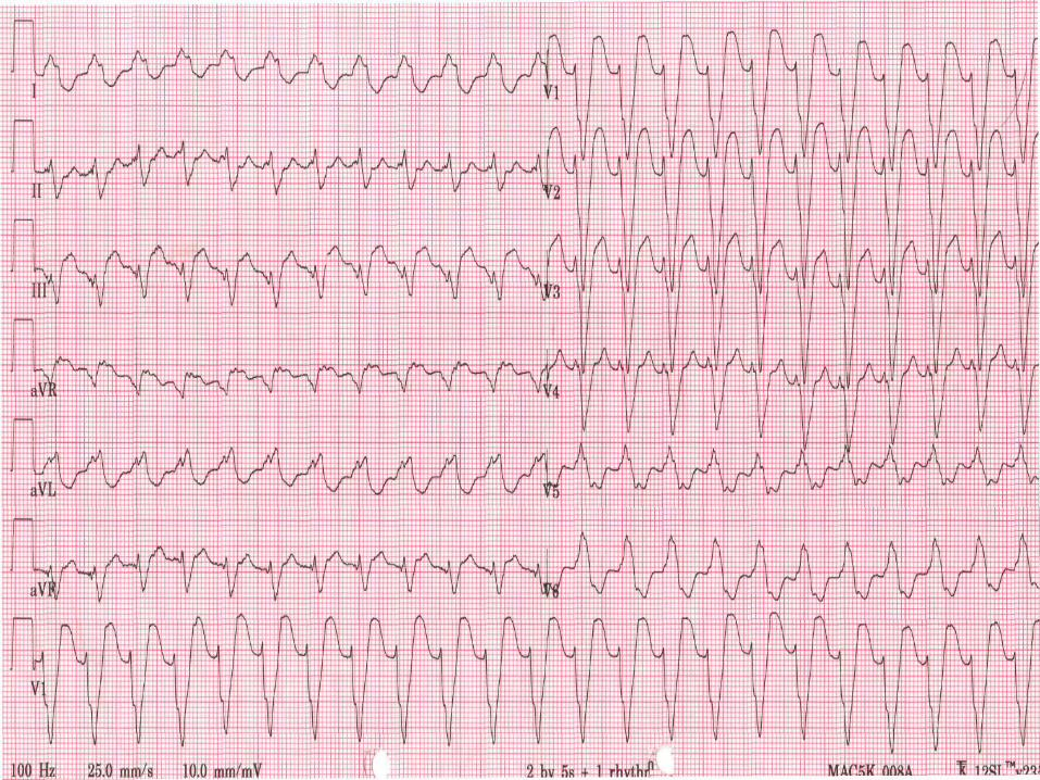
45y M with chest pain
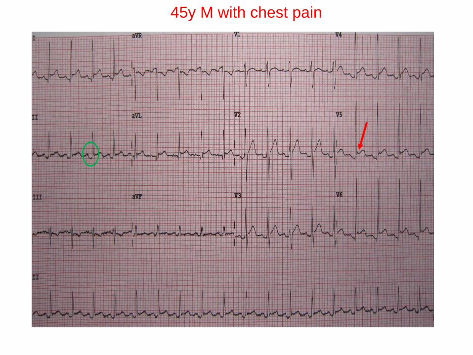
45y M with chest pain
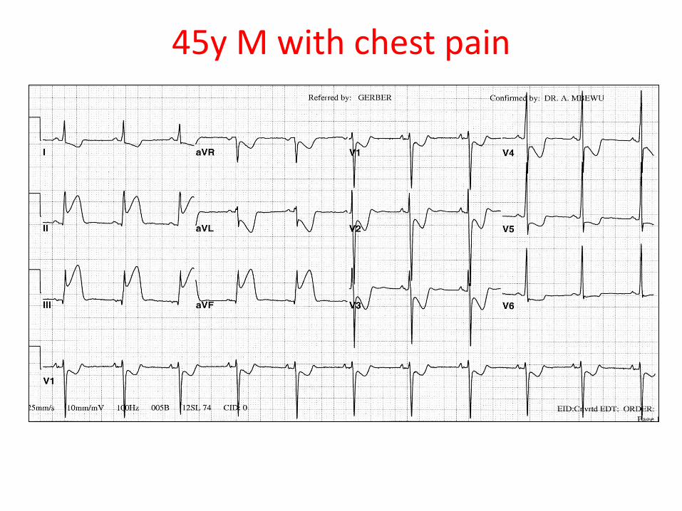
45y M with chest pain
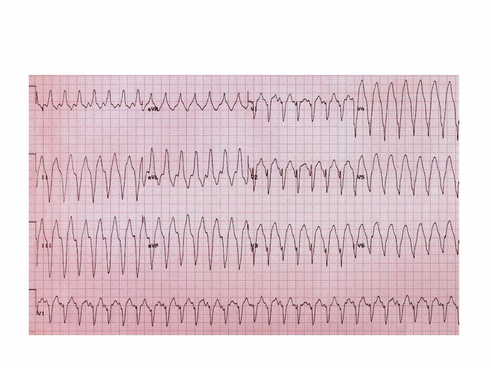
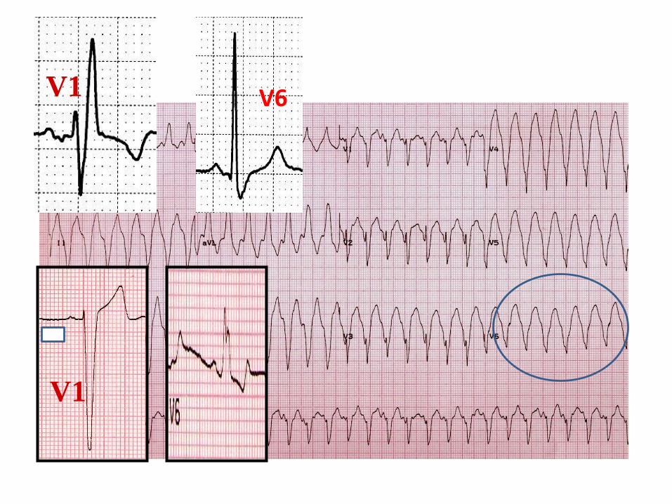
V1
V1
V6
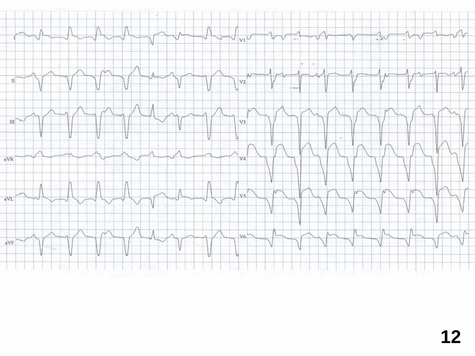
12
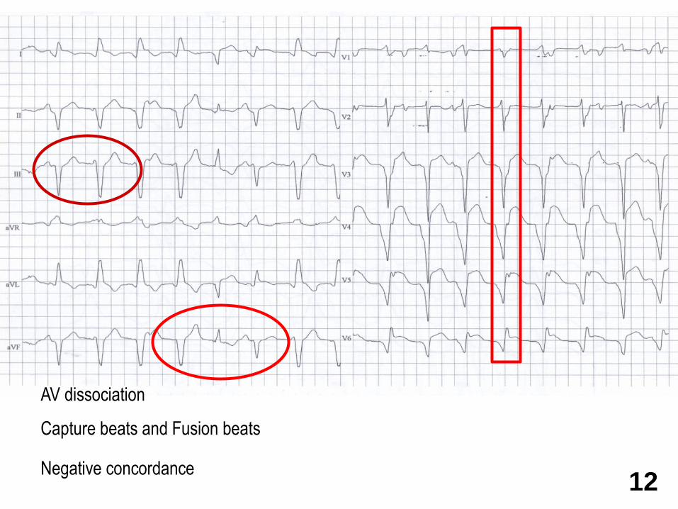
AV dissociation
Capture beats and Fusion beats
Negative concordance12
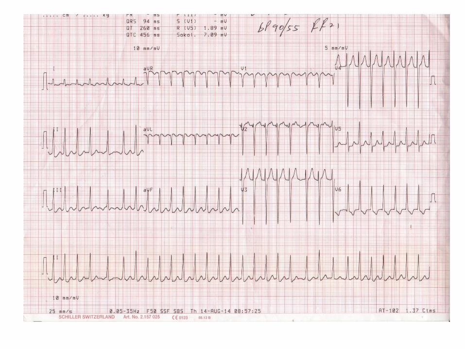
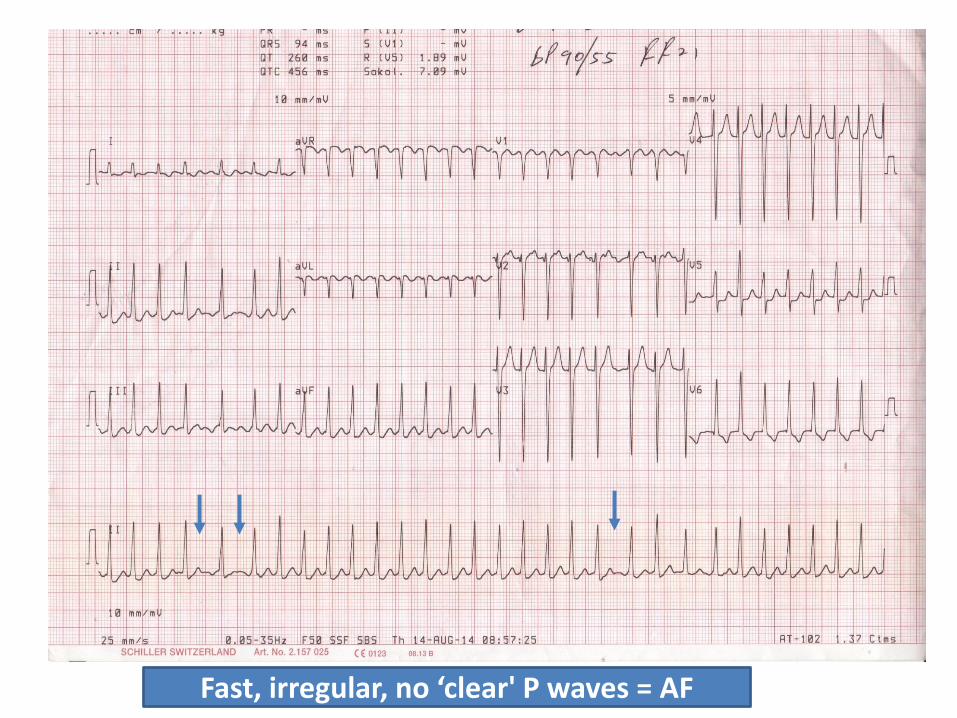
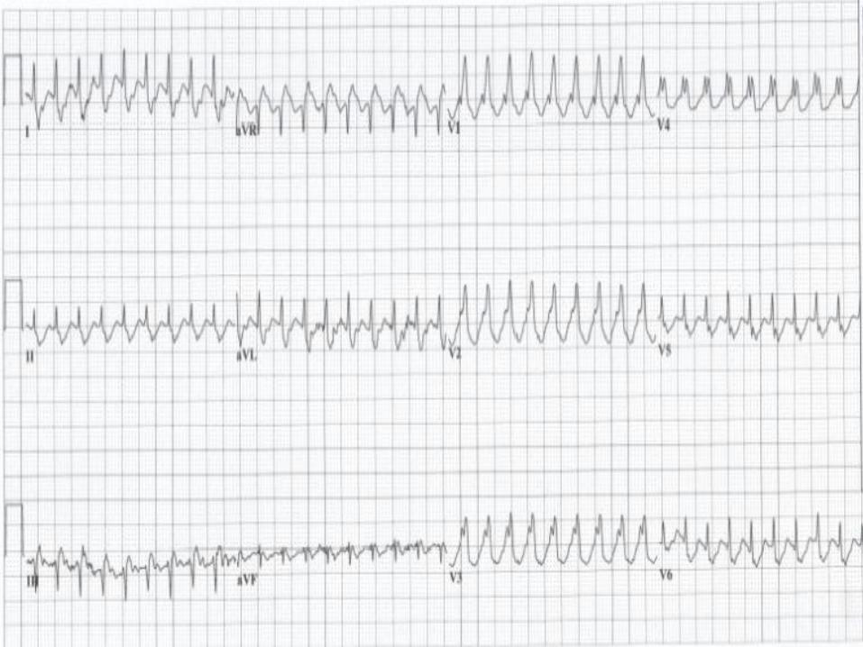
Fast, irregular, no ‘clear' P waves = AF
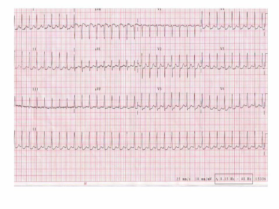
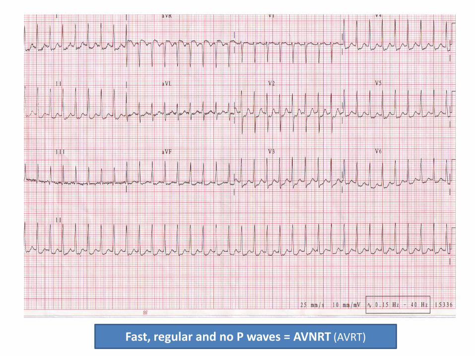
Fast, regular and no P waves = AVNRT (AVRT)
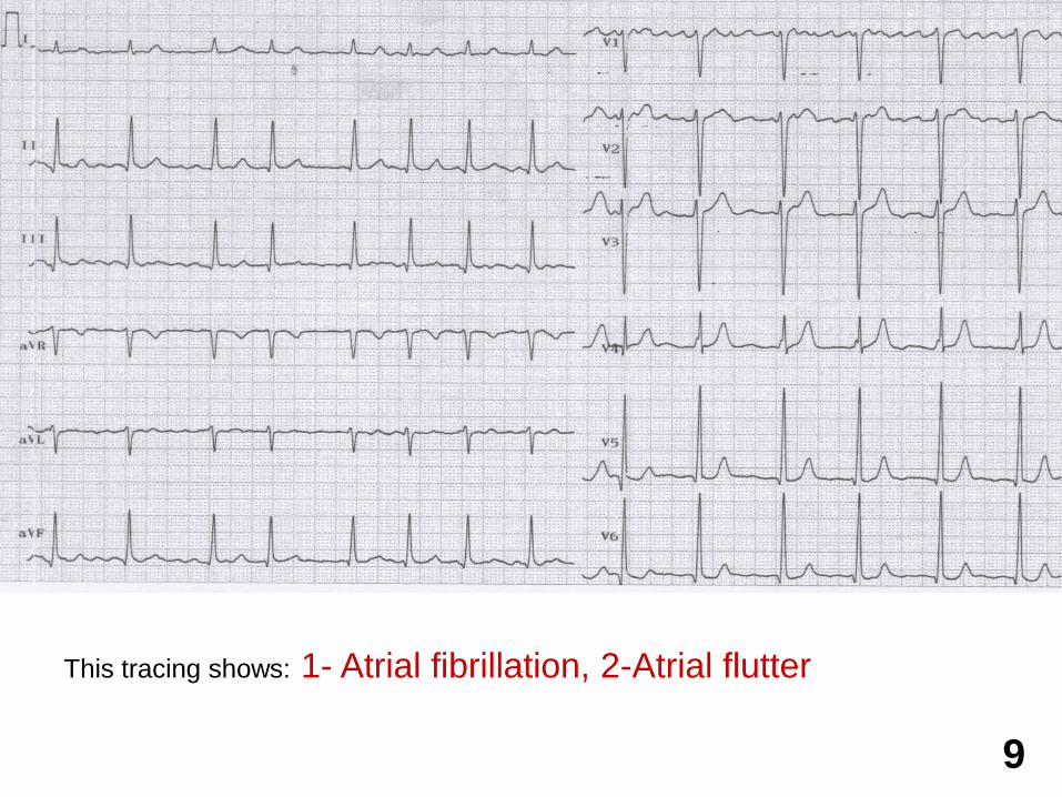
This tracing shows: 1- Atrial fibrillation, 2-Atrial flutter
9
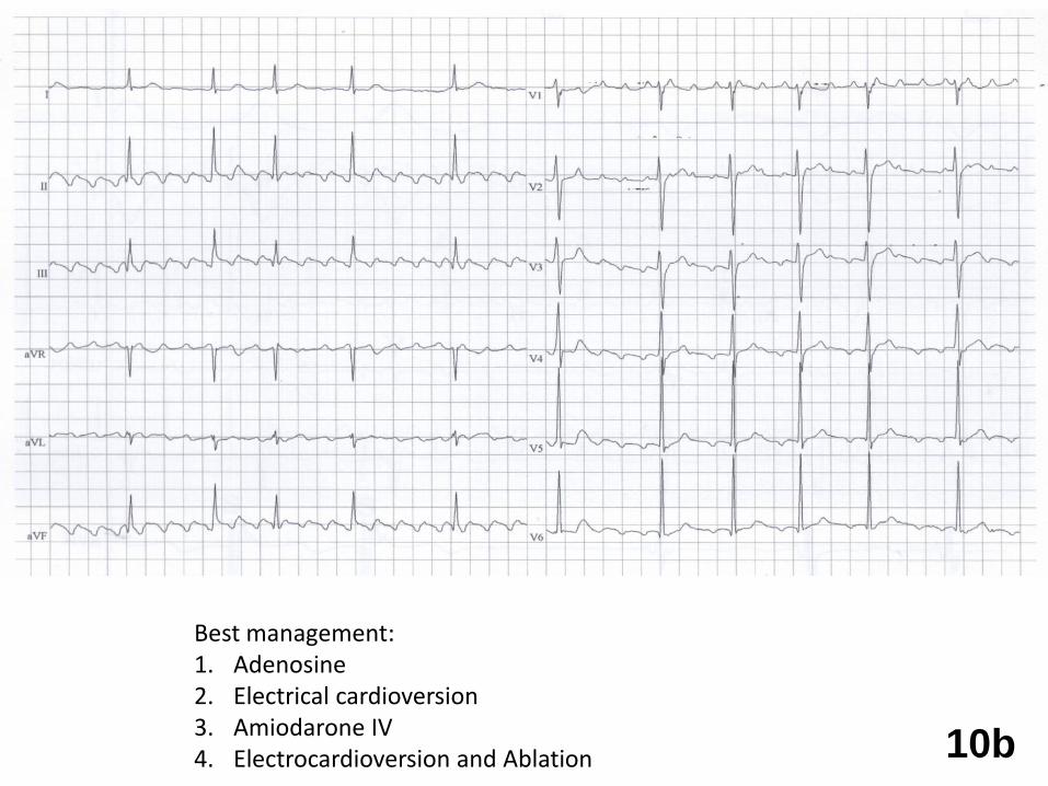
10b
Best management:1. Adenosine2. Electrical cardioversion3. Amiodarone IV4. Electrocardioversion and Ablation

Thank You
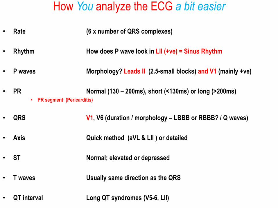
How You analyze the ECG a bit easier
• Rate (6 x number of QRS complexes)
• Rhythm How does P wave look in LII (+ve) = Sinus Rhythm
• P waves Morphology? Leads II (2.5-small blocks) and V1 (mainly +ve)
• PR Normal (130 – 200ms), short (<130ms) or long (>200ms)• PR segment (Pericarditis)
• QRS V1, V6 (duration / morphology – LBBB or RBBB? / Q waves)
• Axis Quick method (aVL & LII ) or detailed
• ST Normal; elevated or depressed
• T waves Usually same direction as the QRS
• QT interval Long QT syndromes (V5-6, LII)