echocardiography and surgery in a dog with left atrial
TRANSCRIPT

University of Pennsylvania University of Pennsylvania
ScholarlyCommons ScholarlyCommons
Departmental Papers (Vet) School of Veterinary Medicine
7-1990
Echocardiography and Surgery in a Dog With Left Atrial Rupture Echocardiography and Surgery in a Dog With Left Atrial Rupture
and Hemopericardium and Hemopericardium
Kenneth K. Sadanaga
Malcolm J. MacDonald
James W. Buchanan University of Pennsylvania, [email protected]
Follow this and additional works at: https://repository.upenn.edu/vet_papers
Part of the Cardiology Commons, Cardiovascular Diseases Commons, Comparative and Laboratory
Animal Medicine Commons, and the Veterinary Infectious Diseases Commons
Recommended Citation Recommended Citation Sadanaga, K. K., MacDonald, M. J., & Buchanan, J. W. (1990). Echocardiography and Surgery in a Dog With Left Atrial Rupture and Hemopericardium. Journal of Veterinary Internal Medicine, 4 (4), 216-221. http://dx.doi.org/10.1111/j.1939-1676.1990.tb00900.x
This paper is posted at ScholarlyCommons. https://repository.upenn.edu/vet_papers/128 For more information, please contact [email protected].

Echocardiography and Surgery in a Dog With Left Atrial Rupture and Echocardiography and Surgery in a Dog With Left Atrial Rupture and Hemopericardium Hemopericardium
Abstract Abstract Endocardial splitting and left atrial rupture were diagnosed in a dog with mitral regurgitation that experienced the sudden onset of collapsing episodes, weakness, depression, labored breathing, and weak pulses. Thoracic radiographs showed a rounded cardiac silhouette with prominent left atrium consistent with hemopericardium due to left atrial rupture. Two-dimensional echocardiography confirmed the presence of severe mitral valve disease, pericardial fluid, and a laminated blood clot caudal to the left ventricle. A sterile emergency thoracotomy was performed, the hemopericardium and blood clot were removed, and the rupture site in the left atrium was repaired with reinforced sutures. The dog recovered from surgery but died the next day, presumably from a ventricular arrhythmia. (Journal of Veterinary Internal Medicine 1990; 4:216–221)
Disciplines Disciplines Cardiology | Cardiovascular Diseases | Comparative and Laboratory Animal Medicine | Veterinary Infectious Diseases
This journal article is available at ScholarlyCommons: https://repository.upenn.edu/vet_papers/128

Echocardiography and Surgery in a Dog With Left Atrial Rupture and
Hemopericardium
Kenneth K. Sadanaga, VMD, Malcolm J. MacDonald, BVM&S, MRCVS,
and James W. Buchanan, DVM, M Med Sci
Endocardial splitting and left atrial rupture were diagnosed in a dog with mitral regurgitation that experienced the sudden onset of collapsing episodes, weakness, depression, labored breathing, and weak pulses. Thoracic radiographs showed a rounded cardiac silhouette with prominent left atrium consistent with hemopericardium due to left atrial rupture. Two-dimensional echocardiography confirmed the presence of severe mitral valve disease, pericardial fluid, and a laminated blood clot caudal to the left ventricle. A sterile emergency thoracotomy was performed, the hemopericardium and blood clot were removed, and the rupture site in the left atrium was repaired with reinforced sutures. The dog recovered from surgery but died the next day, presumably from a ventricular arrhythmia. (Journal of Veterinary Internal Medicine 1990; 4:216-221)
Case Report
History
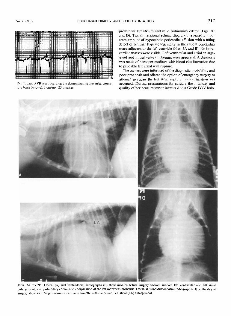
An I 1-year-old, 7.5-kg, female spayed Miniature Poodle was brought to the Veterinary Hospital of the University of Penn- sylvania (VHUP) because of a progressive four-month history of coughing, gagging, decreased exercise tolerance, and de- pression. Physical examination revealed a dog in a normal state of hydration and nutrition. Respiration rate was normal, and coughing was inducible by palpation of the cervical tra- chea. Auscultation revealed a Grade III/V holosystolic mur- mur in the mitral area and bilateral, pulmonary rales. Occa- sional atrial premature contractions were noted on the elec- trocardiogram (Fig. I) . Radiographs revealed marked left atrial enlargement (LAE), left ventricular enlargement (LVE), and compression of the left mainstem bronchus. Peribronchial and perihilar infiltrates were also evident (Figs. 2A and B). Two- dimensional echocardiography confirmed LAE, LVE, mitral valve thickening, and a shortening fraction of 40%. Diagnoses of chronic degenerative valvular disease, mitral valve regurgi- tation, bronchitis, and cardiogenic pulmonary edema were made.
The congestive heart failure signs were controlled with furo- semide ( 1.6 mg/kg every 8 h for two days and then every 12 h,
From the Sections of Surgery (Sadanaga) and Cardiology (MacDonald and Buchanan), Department of Clinical Studies, School of Veterinar). Medicine, University of Pennsylvania, Philadelphia, Pennsylvania.
Supported in part by the Bernard and Muriel Freeman Heart Re- search Fund.
Reprint requests: James W. Buchanan,'DVM, VHUP, 3850 Spruce Street, Philadelphia, PA 19 104.
orally) and digoxin therapy (0.0 16 mg/kg every I2 h for 2 days and then 0.008 mg/kg every 12 h, orally) for a duration of two months, after which the frequency and severity of the coughing increased in spite of medical therapy. One month later she collapsed for I5 minutes after a brief period of excitement and increased activity. She was recumbent yet conscious during this episode and recovered uneventfully. Nine days later she experienced two similar episodes and was brought to the VHUP Emergency Service on a Saturday afternoon when prior radiographs and medical records were unavailable.
Physical Exam inat ion On physical examination the dog was weak, depressed, and had a rectal temperature of 98.1 O F (36.7"C). Increased respira- tory efforts were made at a rate of 48 breaths per minute. Mucous membranes were pale and femoral pulses were weak but synchronous with a heart rate of 124 beats per minute. A Grade III/V holosystolic murmur was heard over the right and left apical precordium. The lung sounds were moist and harsh. A precordial thrill was not present. Furosemide ( 1 mg/kg) was administered intravenously, and the dog was placed in an ox- ygen cage and given oxygen by mask during diagnostic studies.
Diagnostic Studies
The packed cell volume was 45%. Total serum protein, blood glucose, sodium, potassium and BUN* were normal. An ECG showed sinus tachycardia at a rate of 165 beats per minute. Radiographs revealed a globoid cardiac silhouette with a
* Azostix, Miles Inc, Elkhart, IN 465 15
216
Vol. 4 . NO. 4 ECHOCARDIOGRAPHY AND SURGERY IN A DOG 217
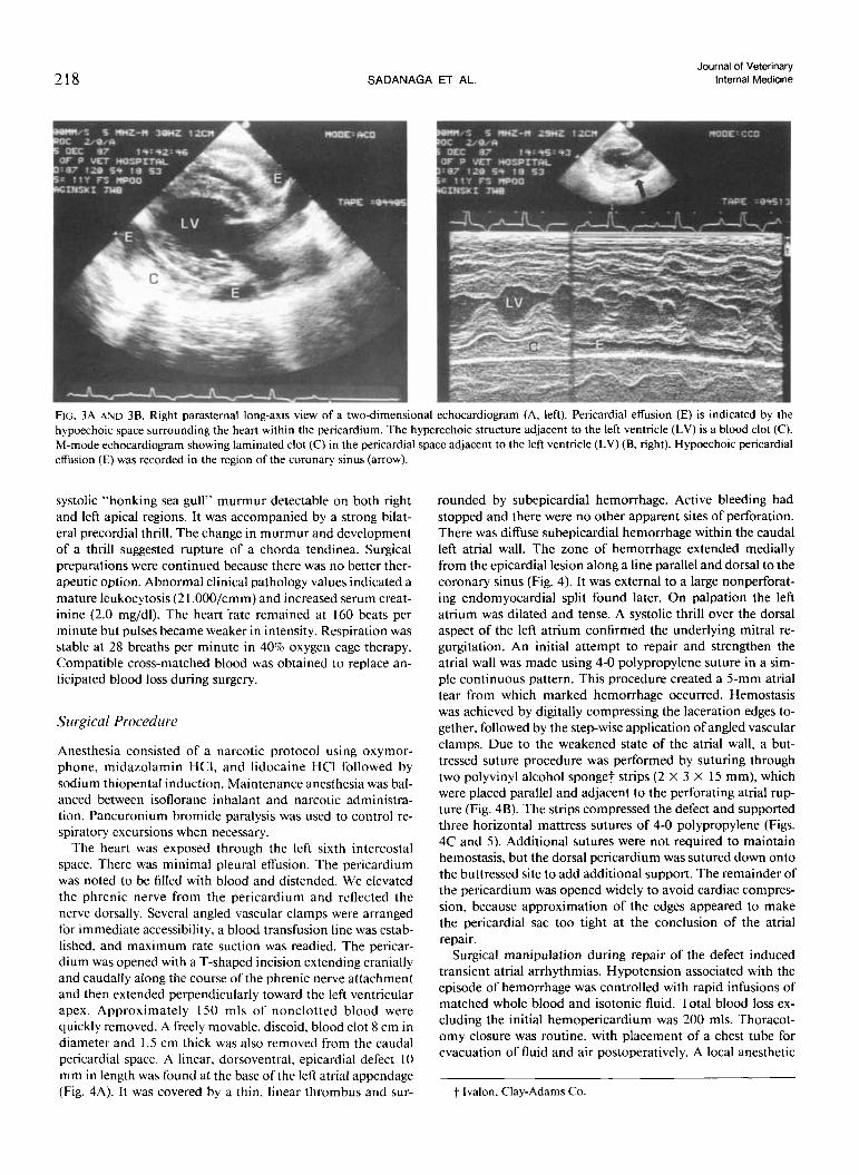
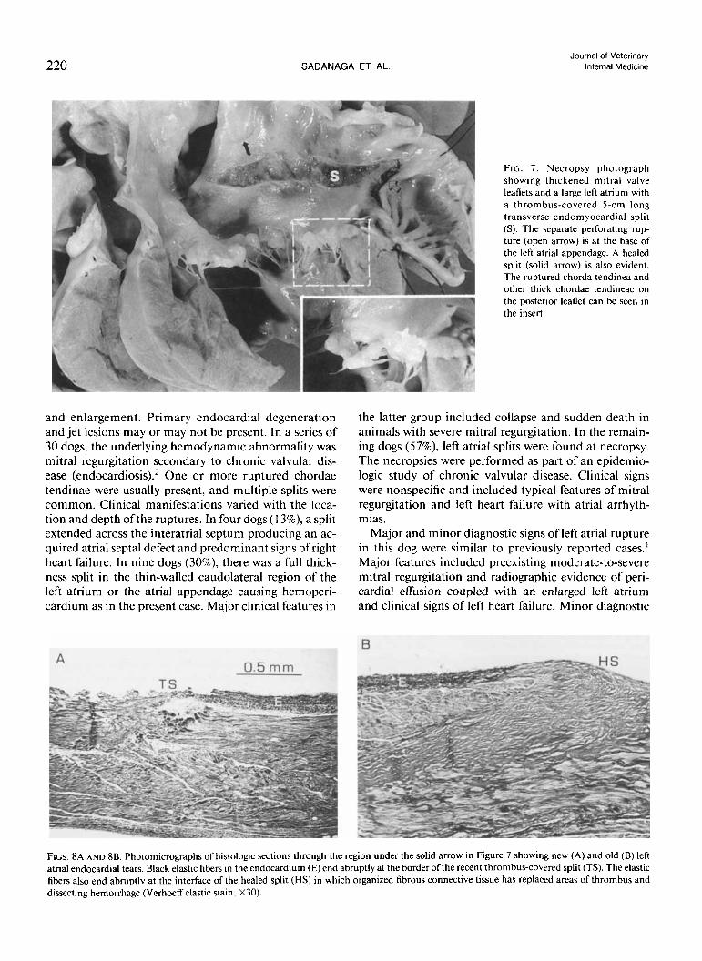
prominent left atrium and mild pulmonary edema (Figs. 2C and D). Two-dimensional echocardiography revealed a mod- erate amount of hypoechoic pericardial effusion with a filling defect of laminar hyperechogenicity in the caudal pericardial space adjacent to the left ventricle (Figs. 3A and B). No intra- cardiac masses were visible. Left ventricular and atrial enlarge- ment and mitral valve thickening were apparent. A diagnosis was made of hemopericardium with blood clot formation due to probable left atrial wall rupture.
The owners were informed of the diagnostic probability and poor prognosis and offered the option of emergency surgery to attempt to repair the left atrial rupture. This suggestion was accepted. During preparations for surgery the intensity and quality of her heart murmur increased to a Grade IV/V holo-
FIG. I . Lead AVR electrocardiogram demonstrating two atrial prema- ture beats (arrows). I cm/mv, 25 mm/sec.
FIGS. 2A TO 2D. Lateral (A) and ventrodorsal radiographs (B) three months before surgery showed marked left ventricular and left atrial enlargement, with pulmonary edema and compression of the left mainstem bronchus. Lateral (C) and dorsoventral radiographs (D) on the day of surgery show an enlarged, rounded cardiac silhouette with concurrent left atrial (LA) enlargement.
218 SADANAGA ET AL. Journal of Veterinary
Internal Medicine
FIG. 3.4 AND 38 . Right parasternal long-axis view of a two-dimensional echocardiogram (A, left). Pericardial effusion (E) is indicated by the hypoechoic space surrounding the heart within the pericardium. The hyperechoic structure adjacent to the left ventricle (LV) is a blood clot (C). M-mode echocardiogram showing laminated clot (C) in the pericardial space adjacent to the left ventricle (LV) (B, right). Hypoechoic pencardial effusion (E) was recorded in the region of the coronary sinus (arrow).
systolic “honking sea gull” murmur detectable on both right and left apical regions. It was accompanied by a strong bilat- eral precordial thrill. The change in murmur and development of a thrill suggested rupture of a chorda tendinea. Surgical preparations were continued because there was no better ther- apeutic option. Abnormal clinical pathology values indicated a mature leukocytosis (2 I ,000/cmm) and increased serum creat- inine (2.0 mg/dl). The heart ’rate remained at 160 beats per minute but pulses became weaker in intensity. Respiration was stable at 28 breaths per minute in 40% oxygen cage therapy. Compatible cross-matched blood was obtained to replace an- ticipated blood loss during surgery.
Surgical Procediire
Anesthesia consisted of a narcotic protocol using oxymor- phone, midazolamin HCI, and lidocaine HCI followed by sodium thiopental induction. Maintenance anesthesia was bal- anced between isoflorane inhalant and narcotic administra- tion. Pancuronium bromide paralysis was used to control re- spiratory excursions when necessary.
The heart was exposed through the left sixth intercostal space. There was minimal pleural effusion. The pericardium was noted to be filled with blood and distended. We elevated the phrenic nerve from the pericardium and reflected the nerve dorsally. Several angled vascular clamps were arranged for immediate accessibility, a blood transfusion line was estab- lished, and maximum rate suction was readied. The pericar- dium was opened with a T-shaped incision extending cranially and caudally along the course of the phrenic nerve attachment and then extended perpendicularly toward the left ventricular apex. Approximately 150 mls of nonclotted blood were quickly removed. A freely movable, discoid, blood clot 8 cm in diameter and 1.5 cm thick was also removed from the caudal pericardial space. A linear, dorsoventral, epicardial defect I0 mm in length was found at the base of the left atrial appendage (Fig. 4A). I t was covered by a thin, linear thrombus and sur-
rounded by subepicardial hemorrhage. Active bleeding had stopped and there were no other apparent sites of perforation. There was diffuse subepicardial hemorrhage within the caudal left atrial wall. The zone of hemorrhage extended medially from the epicardial lesion along a line parallel and dorsal to the coronary sinus (Fig. 4). It was external to a large nonperforat- ing endomyocardial split found later. On palpation the left atrium was dilated and tense. A systolic thrill over the dorsal aspect of the left atrium confirmed the underlying mitral re- gurgitation. An initial attempt to repair and strengthen the atrial wall was made using 4-0 polypropylene suture in a sim- ple continuous pattern. This procedure created a 5-mm atrial tear from which marked hemorrhage occurred. Hemostasis was achieved by digitally compressing the laceration edges to- gether, followed by the step-wise application of angled vascular clamps. Due to the weakened state of the atrial wall, a but- tressed suture procedure was performed by suturing through two polyvinyl alcohol sponge? strips (2 X 3 X 15 mm), which were placed parallel and adjacent to the perforating atrial rup- ture (Fig. 4B). The strips compressed the defect and supported three horizontal mattress sutures of 4-0 polypropylene (Figs. 4C and 5). Additional sutures were not required to maintain hemostasis, but the dorsal pericardium was sutured down onto the buttressed site to add additional support. The remainder of the pericardium was opened widely to avoid cardiac compres- sion, because approximation of the edges appeared to make the pericardial sac too tight at the conclusion of the atrial repair.
Surgical manipulation during repair of the defect induced transient atrial arrhythmias. Hypotension associated with the episode of hemorrhage was controlled with rapid infusions of matched whole blood and isotonic fluid. Total blood loss ex- cluding the initial hemopericardium was 200 mls. Thoracot- omy closure was routine, with placement of a chest tube for evacuation of fluid and air postoperatively. A local anesthetic
t Ivalon. Clay-Adam Co.

Vol. 4 . NO. 4
A
ECHOCARDIOGRAPHY AND SURGERY IN A DOG 219
B
administration were started. Cardiopulmonary signs remained stable. Twelve hours after surgery the frequency of VPCs in- creased, and intermittent isorhythmic atrio-ventricular disso- ciation developed with moderate ventricular tachycardia at a rate of 155 beats per minute (Fig. 6). Because peripheral pulses were good during the ventricular rhythm, antiarrhythmic drugs were not given. Approximately 10 minutes later the dog was found dead and could not be resuscitated.
Nrcropsy
Necropsy failed to establish the cause of death, which was presumably due to an arrhythmia. The surgical repair of the left atrial wall was intact, and there was no evidence of postop- erative hemorrhage or pneumothorax (Fig. 5) . The lungs ap- peared grossly normal and only residual pleural effusion was
and a ruptured primary chorda tendinea of the posterior mitral valve leaflet (Fig. 7). The left atrium had a Perforating (re-
FIG. 4A TO 4C. Diagram of left atrial rupture site (large arrow) and associated subepicardial hemorrhage (cross-hatched area) (A). Place- ment of polyvinyl alcohol polymer (Ivalon) strips adjacent to the atrial rupture (9). Completed repair (C). AA: aortic arch: PT: pulmonary trunk: LAA: left atrial appendage; LC:
seen. Dissection Of the heart thickened mitral
left coronary artery: CS: coronary sinus; RV: right ventricle; LV: left ventricle.
paired), vertical 1.5-cm rupture at the base of the atrial ap- pendage. The perforating rupture was perpendicular and sepa- rate from a 5-cm thrombus-covered, nonperforating endo-
(bupivacaine HCI) was injected in the adjacent intercostal spaces of the thoracotomy site to alleviate postoperative pain.
Postoperative management included monitoring a continu- ous lead I1 ECG and systemic arterial blood pressure, adminis- tration of intranasal oxygen at 1.5 liters per minute, and mea- surement of serial arterial blood gases. Initial respiratory aci- dosis and mild hypoxemia improved to normal in 1.5 hours. Systolic blood pressure remained stable at 85 to 102 mm Hg, and occasional (5-10 per, minute) ventricular premature con- tractions (VPC) were noted. Peripheral perfusion remained good, and there were no appreciable pulse deficits in associa- tion with the VPCs. Isotonic fluid was administered at 25 ml per hour. The hematocrit remained stable at 32% to 35%, but because total solids slowly declined to 4 g/dl a plasma infusion was initiated. Serial chest tube aspirations yielded minimal fluid and air volumes. Urine output was normal. The dog assumed sternal recumbency and became responsive within 3 hours after surgery. Ten hours after surgery the dog was stand- ing, the chest tube was removed, and oral captopril ( 1 mg/kg every 8 h, orally) and furosemide ( 1 mg/kg, every 12 h, orally)
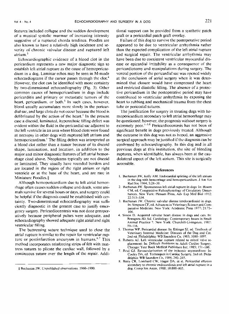
myocardial split in the caudal wall of the left atrium under the zone of subepicardial hemorrhage. The perforating rupture appeared to be adequately closed by the buttressed suture pro- cedure although there still was endocardial separation. Three smaller, healed splits, 1 to 3 cm in length, were present in the dorsal and cranial walls of the left atrium (Fig. 7). These were recognized by their typically depressed, crescent shape and were covered by glistening endothelium. ' Histologic sections stained for elastic fibers confirmed the endocardial separation in both recent and healed splits (Figs. 8A and B). The recent split was covered by a fresh thrombus. Dissecting hemorrhage was present in the underlying atrial myocardium. The healed split was filled with fibrous, connective tissue in the region of the split and in underlying areas of prior dissecting hemor- rhage. Moderate left ventricular hypertrophy was present but there were no gross ventricular myocardial lesions. Histology showed mild arteriosclerosis with medial thickening and hya- line degeneration in the lateral papillary muscle of the left ventricle.
Discussion
Left atrial endocardial and endomyocardial splitting occurs as a complication of increased left atrial pressure
FIG. 6. Lead I1 ECG showing isorhythmic A-V dissociation with mod- erate ventricular tachycardia at a rate of 155 beats/minute. I cm/mv. FIG. 5. Necropsy photograph of left atrial repair site (arrow) and sub-
epicardial hemorrhage (H). 25 mm/sec.
220 SADANAGA ET AL. Journal of Veterinary
Internal Medicine
and enlargement. Primary endocardial degeneration and jet lesions may or may not be present. In a series of 30 dogs, the underlying hemodynamic abnormality was mitral regurgitation secondary to chronic valvular dis- ease (endocardiosis).2 One or more ruptured chordae tendinae were usually present, and multiple splits were common. Clinical manifestations varied with the loca- tion and depth of the ruptures. In four dogs ( l 3%), a split extended across the interatrial septum producing an ac- quired atrial septa1 defect and predominant signs of right heart failure. In nine dogs (30%), there was a full thick- ness split in the thin-walled caudolateral region of the left atrium or the atrial appendage causing hemoperi- cardium as in the present case. Major clinical features in
FIG. 7. Necropsy photograph showing thickened mitral valve leaflets and a large left atrium with a thrombus-covered 5-cm long transverse endom yocardial split (S). The separate perforating rup- ture (open arrow) is at the base of the left atrial appendage. A healed split (solid arrow) is also evident. The ruptured chorda tendinea and other thick chordae tendineae on the posterior leaflet can be seen in the insert.
the latter group included collapse and sudden death in animals with severe mitral regurgitation. In the remain- ing dogs (57%), left atrial splits were found at necropsy. The necropsies were performed as part of an epidemio- logic study of chronic valvular disease. Clinical signs were nonspecific and included typical features of mitral regurgitation and left heart failure with atrial arrhyth- mias.
Major and minor diagnostic signs of left atrial rupture in this dog were similar to previously reported cases.' Major features included preexisting moderate-to-severe mitral regurgitation and radiographic evidence of peri- cardial effusion coupled with an enlarged left atrium and clinical signs of left heart failure. Minor diagnostic
FIGS. 8A AND 8B. Photomicrographs of histologic sections through the region under the solid arrow in Figure 7 showing new (A) and old (B) left atrial endocardial tears. Black elastic fibers in the endocardium (E) end abruptly at the border ofthe recent thrombus-covered split (TS). The elastic fibers also end abruptly at the interface of the healed split (HS) in which organized fibrous connective tissue has replaced areas of thrombus and dissecting hemorrhage (Verhoeff elastic stain, X30).
Vol. 4 . NO. 4 ECHOCARDIOGRAPHY AND SURGERY IN A DOG 22 1
features included collapse and the sudden development of a musical systolic murmur of increasing intensity suggestive of a ruptured chorda tendinea. Poodles are also known to have a relatively high incidence and se- verity of chronic valvular disease and ruptured left atrium.
Echocardiographic evidence of a blood clot in the pericardium represents a new major diagnostic sign to establish left atrial rupture as the cause of hemopericar- dium in a dog. Laminar echos may be seen in M-mode echocardiograms if the cursor passes through the However, the clot can be identified with more certainty by two-dimensional echocardiography (Fig. 3 ) . Other common causes of hemopericardium in dogs include pericarditis and primary or metastatic tumors of the heart, pericardium, or both.5 In such cases, however, blood usually accumulates more slowly in the pericar- dial sac, and large clots do not occur because the blood is defibrinated by the action of the heart.' In the present case a discoid, laminated, hyperechoic filling defect was evident within the fluid in the pericardial sac adjacent to the left ventricle in an area where blood clots were found at necropsy in other dogs with ruptured left atrium and hemopericardium.' The filling defect was interpreted as a blood clot rather than a tumor because of its discoid shape, lamination, and location, in addition to the major and minor diagnostic features of left atrial hemor- rhage cited above. Neoplasms typically are not discoid or laminated. They usually have rounded borders and are located in the region of the right atrium or right ventricle or at the base of the heart, and are rare in Miniature Poodles.$
Although hemopericardium due to left atrial hemor- rhage often causes sudden collapse and death, some ani- mals survive for several hours or days, and surgery could be helpful if the diagnosis could be established with cer- tainty. Two-dimensional echocardiography was suffi- ciently diagnostic in the present case to justify emer- gency surgery. Pericardiocentesis was not done preoper- atively because peripheral pulses were adequate, and echocardiography showed adequate right atrial and right ventricular filling.
The buttressing suture technique used to close the atrial rupture is similar to the repair for ventricular rup- ture or postinfarction aneurysm in This method incorporates reinforcing strips of felt with mat- tress sutures to plicate the cardiac wall, followed by a continuous suture over the length of the repair. Addi-
$ Buchanan JW. Unpublished observations, 1960- I990
tional support can be provided from a synthetic patch graft or a pericardial patch graft overlay
Failure of this dog to survive the postoperative period appeared to be due to ventricular arrhythmia rather than the expected complication of the left atrial rupture and surgical repair. The ventricular arrhythmia may have been due to coexistent ventricular myocardial dis- ease or epicardial irritability as a consequence of the pericardiotomy and manipulations during surgery. The ventral portion of the pericardial sac was opened widely at the conclusion of atrial surgery when it was deter- mined that closure would have compressed the heart and restricted diastolic filling. The absence of a protec- tive pericardium in the postoperative period may have contributed to ventricular arrhythmia by exposing the heart to rubbing and mechanical trauma from the chest tube or pericostal sutures.
The justification for surgery in treating dogs with he- mopericardium secondary to left atrial hemorrhage may be questioned; however, the prognosis without surgery is extremely p o ~ r . ' - ~ , ~ Pericardiocentesis has not been of significant benefit in dogs previously treated. Although the outcome in this dog was not as hoped, an aggressive surgical approach may be justified if the diagnosis can be confirmed by echocardiography. In this dog and in all previous dogs at this institution, the site of bleeding ruptures, when identifiable, has always been at the cau- dolateral aspect of the left atrium. This site is surgically accessible.
References I. Buchanan JW, Kelly AM. Endocardial splitting ofthe left atrium
in the dog with hemorrhage and hemopericardium. J Am Vet Rad Soc 1964; 5:28-39.
2. Buchanan JW. Spontaneous left atrial rupture in dogs: In: Bloom CM, ed. Comparative Pathophysiology of Circulatory Distur- bances. New York: Plenum Press, Adv Exp Med Biol 1972:
3. Buchanan JW. Chronic valvular disease (endocardiosis) in dogs. In: Simpson CF, ed. Advances in Veterinary Science and Com- parative Medicine. New York: Academic Press 1977; 21:75- 106.
4. Sisson D. Acquired valvular heart disease in dogs and cats. In: Bonagura JD, Ed. Cardiology: Contemporary Issues in Small Animal Practice 7. New York: Churchill-Livingston, 1987;
5. Thomas WP. Pericardial disease: In: Ettinger SJ, ed. Textbook of Veterinary Internal Medicine: Diseases of the Dog and Cat. 2nd ed. Philadelphia: WB Saunders Co, 1983: 1080-1097.
6. Roberts AJ. Left ventricular rupture related to mitral valve re- placement: In: Difficult Problems in Adult Cardiac Surgery. Chicago: Year Book Medical Publishers Inc, 1985: 151-160.
7. Reul GJ. Revascularization of the ischemic myocardium: In: Cooley DA, ed. Techniques in Cardiac Surgery. 2nd ed. Phila- delphia: WB Saunders Co, 1984; 240-245.
8. Berry CR, Lombard CW. Hager DA, et al. Pericardial effusion secondary to chronic endocardiosis and left atrial rupture i n a dog. Comp Sm Anim. 1988; 10:800-805.
2213 15-334.
59-1 16.