effect of ultra-low doses, asir and mbir on density and...
TRANSCRIPT
HEAD AND NECK
Effect of ultra-low doses, ASIR and MBIR on density and noiselevels of MDCT images of dental implant sites
Gerlig Widmann1& Reema Al-Shawaf2 & Peter Schullian1
& Ra’ed Al-Sadhan2&
Romed Hörmann3& Asma’a A. Al-Ekrish2
Received: 9 December 2015 /Revised: 28 July 2016 /Accepted: 29 August 2016# European Society of Radiology 2016
AbstractObjectives Differences in noise and density values in MDCTimages obtained using ultra-low doses with FBP, ASIR, andMBIR may possibly affect implant site density analysis. Theaim of this study was to compare density and noise measure-ments recorded from dental implant sites using ultra-lowdoses combined with FBP, ASIR, and MBIR.
Methods Cadavers were scanned using a standard protocoland four low-dose protocols. Scans were reconstructed usingFBP, ASIR-50, ASIR-100, and MBIR, and either a bone orstandard reconstruction kernel. Density (mean Hounsfieldunits [HUs]) of alveolar bone and noise levels (mean standarddeviation of HUs) was recorded from all datasets and mea-surements were compared by paired t tests and two-wayANOVAwith repeated measures.Results Significant differences in density and noise werefound between the reference dose/FBP protocol and almostall test combinations. Maximummean differences in HUwere178.35 (bone kernel) and 273.74 (standard kernel), and innoise, were 243.73 (bone kernel) and 153.88 (standardkernel).Conclusions Decreasing radiation dose increased density andnoise regardless of reconstruction technique and kernel. Theeffect of reconstruction technique on density and noise de-pends on the reconstruction kernel used.Key Points• Ultra-low-dose MDCT protocols allowed more than 90 %reductions in dose.
• Decreasing the dose generally increased density and noise.• Effect of IRTon density and noise varies with reconstructionkernel.
• Accuracy of low-dose protocols for interpretation of bonyanatomy not known.
• Effect of low doses on accuracy of computer-aided designmodels unknown.
Keywords Algorithms . Dental implants . Image processing,computer-assisted .Multidetector computed tomography .
Radiation dosage
Gerlig Widmann and Asma’a A. Al-Ekrish contributed equally to thiswork.
Electronic supplementary material The online version of this article(doi:10.1007/s00330-016-4588-8) contains supplementary material,which is available to authorized users.
* Asma’a A. [email protected]; [email protected]
Gerlig [email protected]
Reema [email protected]
Peter [email protected]
Ra’ed [email protected]
Romed Hö[email protected]
1 Department of Radiology, Medical University of Innsbruck,Innsbruck, Austria
2 Department of Oral Medicine and Diagnostic Sciences, College ofDentistry, King Saud University, P. O. Box 56810, Riyadh 11564,Kingdom of Saudi Arabia
3 Division of Clinical and Functional Anatomy, Medical University ofInnsbruck, Innsbruck, Austria
Eur RadiolDOI 10.1007/s00330-016-4588-8
AcronymsASIR Adaptive statistical iterative reconstructionMBIR Model-based iterative reconstructionFBP Filtered backprojection
Introduction
Assessment of bone density from multidetector computed to-mography (MDCT) images using Hounsfield units (HUs) isconsidered an objective, accurate, and stable measurement [1,2]. In imaging of dental implant sites, positive correlationshave been reported between various measures of primary im-plant stability and bone density measurements recorded fromMDCT [3–6] and quantitative CT [7–9]. Furthermore, due tothe accuracy in thresholding and segmentation ofMDCT data,it has been found to be one of the most accurate modalities inproduction of 3D models of the jaws [10–13], which is signif-icant when MDCT is used in the production of surgical stentsfor dental implant surgery.
However, the increasing use of MDCT has been cited as acause for the increasing collective dose of ionizing radiation topopulations [14], and cone beam CT (CBCT) has been advo-cated as imparting lower radiation doses to patients, and isincreasingly becoming more available to oral implantologists.MDCT, though, is still utilized by many practitioners, and theposition statement of the American Academy of Oral andMaxillofacial Radiology on selection criteria for the use ofradiology in dental implantology recognizes the continueduse and need for MDCT in oral implantology, but stresses thatdose-sparing protocols should be used whenever possiblewithout adversely affecting diagnostic accuracy [15]. The re-markable progress in MDCT technology has produced de-vices with more detectors, allowing faster scanning timesand low-dose exposures [16, 17], and the reported doses fromultra-low-dose MDCT protocols [18] are lower than thosereported for several dentomaxillofacial CBCT devices [19,20]. Further, the use of various iterative reconstruction tech-niques (IRTs), such as adaptive statistical iterative reconstruc-tion (ASIR) and model-based iterative reconstruction (MBIR)has shown improvements in MDCT image quality with re-duced radiation doses, when compared with the traditionallyused filtered backprojection technique (FBP) [18, 21].However, the resultant images reportedly demonstrate an"oversmoothing" effect [18, 22], of which the effect on densityand thresholding must be considered. Also, the HU scale mayvary when the MDCTexposure parameters are lowered, espe-cially x-ray beam energy (kVp) [23, 24].
At the time of writing, to our knowledge, there is no infor-mation in the published literature regarding the effect of con-siderable dose reductions in combination with various IRTs onthe combined HU and noise levels in dental implant site im-aging. The variations within these objective measurements
needs to be investigated to understand how the use of low-dose IRT protocols may affect analysis of bone density ofimplant sites and computer guided surgery. Therefore, thisstudy aimed to compare the objective density and noise mea-surements recorded from MDCT images of dental implantsites using ultra-low doses combined with FBP, ASIR andMBIR with those from a standard dose/FBP protocol, and toinvestigate the effect of dose and reconstruction technique onthe measurements.
Materials and methods
Cadaver selection
Two cadaveric heads (one edentulous and one partially den-tulous) with intact soft tissues were used in the study. Thebodies were donated by people who had given their informedconsent for their use for scientific and educational purposesprior to death. The study fulfilled all requirements necessaryfor studies on human cadavers according to the regulations ofthe Division of Clinical and Functional Anatomy, MedicalUniversity of Innsbruck [25, 26]. All cadavers were preservedusing an arterial injection of an alcohol-glycerin solution andimmersion in phenolic acid in water for 1 to 3 months [27].
Imaging of the cadavers
The cadaver heads including the mandible were scannedusing a 64-multi-slice CT scanner (Discovery CT750 HD,GE Healthcare, Vienna, Austria). The scan range includedthe entire skull and mandible. Each cadaver was exposedto a high-resolution reference protocol and four low-doseprotocols (Table 1). The pitch factor for all protocols was0.5. There was no movement of the cadavers during andbetween the five different exposures. In addition to theFBP standard reconstruction all images were reconstruct-ed using the following IRTs: ASIR-50, ASIR-100, andMBIR. Adaptive statistical iterative reconstruction (GEHealthcare) uses information obtained from the FBP algo-rithm but integrates a comparison of the pixel values withan ideal value to selectively identify and then subtractnoise from an image at adaptive blend levels which arefreely selectable, typically from 10 % to 100 %. [22] TheASIR reconstructions used in the present study wereASIR-50 (50 % FBP and 50 % ASIR) and ASIR-100(100 % ASIR). A bone convolution kernel was used inthe images of cadaver 1 (except with MBIR for whichonly a standard convolution kernel was available). A stan-dard reconstruction kernel was used in all the images ofcadaver 2. The volume CT dose index (CTDIvol) and doselength product (DLP) were obtained from the digital im-aging and communication in medicine (DICOM) tags. In
Eur Radiol
addition, effective doses were calculated using CT-ExpoV 2.1 (Medical University Hannover, Germany), an MSExcel application for assessing the radiation doses deliv-ered to patients undergoing CT examinations based on thescan parameters of the used scanner type. A protocol sum-mary can be found in Table 1.
Processing and viewing of 3D datasets
Each of the 40 MDCT data sets was converted to DICOMformat and uploaded onto a shared Google drive. TheDICOM data sets were then imported into a 3D imagereformatting software (OnDemand Software, version 1.0,Cybermed Inc., Seoul, South Korea). The images wereviewed on a 22" flat panel LCD colour monitor in land-scape mode (Dell P2210, Round Rock, TX, USA).Specifications of the monitor were as follows: aspect ra-tio: 16:10; screen resolution 1680 × 1050 (highest, recom-mended; calculated pixel size: 0.282 mm); colour resolu-tion: 32 bit; luminance 250 Cd/m2; contrast ratio (static):1000:1. The window width/level of the MDCT imageswas set to 3000/650, and the images were viewed andmeasurements recorded in a dimly lit room.
Sample selection
The maxillary and mandibular edentulous ridges of both theimaged cadavers provided the study sample sites. The samplesite inclusion criteria were all edentulous spaces within thejaws of the cadavers, at 5-mm intervals. The exclusion criteriafor the edentulous spaces were the following: presence offoreign objects or artificial defects at the residual ridge, andareas of residual ridges where the standardized position of theregion of interest (ROI) included non-osseous tissues. Basedupon the inclusion and exclusion criteria, 41 sample sites wereobtained (25 sites from cadaver 1 and 16 sites from cadaver 2).All the sample sites were in areas of the data sets which werenot degraded by artifacts from foreign bodies or dentalrestorations.
Reformatting of sample sites
One oral and maxillofacial radiologist (A.A.) with 10 yearsexperience in CT image processing and analysis performedthe reformatting of all the datasets to obtain transverse cross-sectional images of the sample sites. The 3D module of theOnDemand software was used to produce each cross-sectionindividually. The text overlay information was turned off in
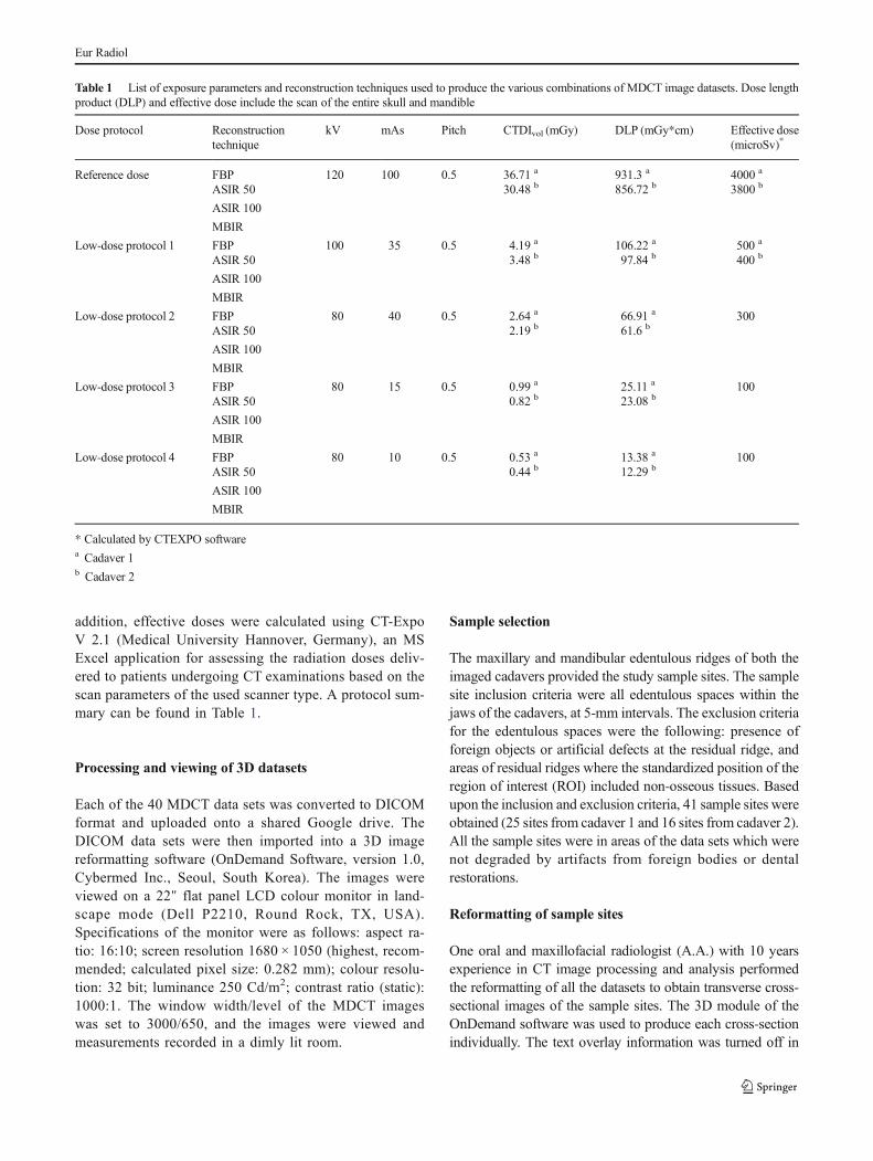
Table 1 List of exposure parameters and reconstruction techniques used to produce the various combinations of MDCT image datasets. Dose lengthproduct (DLP) and effective dose include the scan of the entire skull and mandible
Dose protocol Reconstructiontechnique
kV mAs Pitch CTDIvol (mGy) DLP (mGy*cm) Effective dose(microSv)*
Reference dose FBP 120 100 0.5 36.71 a
30.48 b931.3 a
856.72 b4000 a
3800 bASIR 50
ASIR 100
MBIR
Low-dose protocol 1 FBP 100 35 0.5 4.19 a
3.48 b106.22 a
97.84 b500 a
400 bASIR 50
ASIR 100
MBIR
Low-dose protocol 2 FBP 80 40 0.5 2.64 a
2.19 b66.91 a
61.6 b300
ASIR 50
ASIR 100
MBIR
Low-dose protocol 3 FBP 80 15 0.5 0.99 a
0.82 b25.11 a
23.08 b100
ASIR 50
ASIR 100
MBIR
Low-dose protocol 4 FBP 80 10 0.5 0.53 a
0.44 b13.38 a
12.29 b100
ASIR 50
ASIR 100
MBIR
* Calculated by CTEXPO softwarea Cadaver 1b Cadaver 2
Eur Radiol
order to mask the mA, and kVp. Since the cadaver heads werenot moved between the MDCT examinations, the default po-sition and orientation of the orthogonal sectional planes rela-tive to the jaws were consistent in all the MDCT datasets ofeach cadaver. Thus, standardization of the site and orientationof the reformatted sample sites was achievable through mea-sured shift and angulation of the orthogonal sectional planes.The contrast and density and zoom level were also standard-ized amongst all images. Each reformatted site was saved as abookmark on the master database of the reformatting software(on the hard drive of the computer) such that the examinerscan access the reformatted sites using the OnDemandsoftware.
Recording the test measurements
Two independent examiners who were blinded to the expo-sure parameters and reconstruction protocols recorded the testmeasurements. The examiners were OMF radiologists with 10and 5 years experience in MDCT image processing and anal-ysis. The ROI tool of the software was used to select a squarearea 4 × 4 mm in size which was positioned at the intersectionof the axial and para-sagittal reformatting lines at the samplesites. Tables 2 and 3 show sample sites, with ROIs, obtainedwith each of the 20 combinations of dose protocols and recon-struction techniques using a bone kernel and standard kernel,respectively. The mean values for the HUs within each ROIwere used to analyze density, and the standard deviation with-in each ROI was used to analyze the noise levels.
The first examiner recorded all the measurements once. Forreliability testing, 100 sample sites were selected to be equallydistributed amongst the imaging protocols; five samples wererandomly selected from each protocol (three sites randomlyselected from cadaver 1 and two sites randomly selected fromcadaver 2). An online random number generator was used forrandom selection of sites (http://stattrek.com/statistics/random-number-generator.aspx). The first and second examinersrecorded the measurements from the 100 sites independently,with at least a 1-week interval between the first and secondrecordings by the first examiner.
Statistical analysis
The recorded measurements were analyzed with the statisticalprogram: SPSS Versions 22 and 24 (IBM, Armonk, NY,USA). Intra- and inter-examiner reliability of the HU andnoise measurements were analyzed by calculating theintraclass correlation coefficient. Bland–Altman plots com-paring the examiners' readings were also obtained, and linearregression was used to test for proportional bias between theexaminers' measurements. The Kolmogorov–Smirnov andShapiro–Wilk tests of normality were performed for the sub-groups imaged with a standard kernel, and indicated normal
distribution of the data in these groups. Therefore, paired ttests were used to compare the HU and noise measurementsrecorded from each of the test combinations of protocols withthose from the reference dose/FBP technique protocol. Two-way ANOVAwith repeated measures was used to analyze theeffect of dose and reconstruction technique on the measure-ments. Level of significance was set at p = 0.05.
Results
Intra- and inter-examiner reliability of the mean HU were0.996 and 0.987, respectively (p < 0.001). For the noise mea-surements, they were 0.991 and 0.974, respectively(p < 0.001). Bland–Altman plots with linear regression (seeelectronic Supplementary Material) did not demonstrate sys-tematic variation between the examiners' measurements foreither HU or noise (p > 0.5).
Mean HU
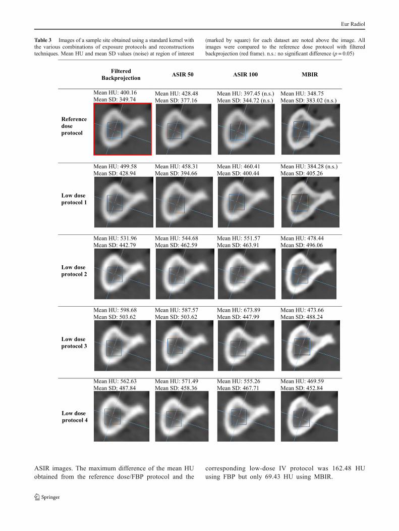
A statistically significant difference in the mean HUs wasfound between the reference dose/FBP protocol and almostall the test combinations (p < 0.021), regardless of the kernelused. Using bone kernel exceptions were those protocolsusing the reference dose with ASIR 50 (p = 0.533), ASIR100 (p = 0.096), and MBIR (P = 0.840; Table 2). MBIR wasonly possible with standard kernel, but was compared with thereference dose/FBP protocol bone kernel as well. Using thestandard kernel, exceptions were the reference dose withASIR 100 (p = 0.836) and low-dose protocol 1 with MBIR(p = 0.403; see Table 3). Overall, the maximum mean differ-ences in HU were 178.35 HU using the bone kernel and273.74 HU using the standard kernel.
When a bone kernel was used (except with MBIR), therewas a significant effect for the interaction between the doseand reconstruction techniques on the mean of the HU(p = 0.007). When the dose and reconstruction technique wereconsidered separately, there was a significant difference inmean HU between the different dose protocols (p < 0.001)with an overall pattern of increase in HU with decreasingdose, except at the lowest-dose protocols. No significant dif-ference in mean HU was found between the reconstructiontechniques (p = 0.353).
However, when a standard kernel was used with allreconstruction techniques, there was no significant effectfor the interaction between the dose and reconstructiontechniques on the mean of the HU (p = 0.318), althoughthere was a significant difference in mean HU betweenthe different dose protocols (p < 0.001) and between thereconstruction techniques (p < 0.001). Interestingly, usingthe standard kernel, use of MBIR decreased the meanHU when compared with the corresponding FBP and
Eur Radiol

Table 2 Images of a sample site obtained using a bone kernel (exceptwith MBIR) with the various combinations of dose protocols andreconstructions techniques. Mean HU and mean SD values (noise) at
region of interest (marked by square) for each dataset are noted above theimage. All images were compared to the reference dose protocol withfiltered backprojection (red frame). n.s.: no significant difference (p = 0.05)
Eur Radiol
ASIR images. The maximum difference of the mean HUobtained from the reference dose/FBP protocol and the
corresponding low-dose IV protocol was 162.48 HUusing FBP but only 69.43 HU using MBIR.
Table 3 Images of a sample site obtained using a standard kernel withthe various combinations of exposure protocols and reconstructionstechniques. Mean HU and mean SD values (noise) at region of interest
(marked by square) for each dataset are noted above the image. Allimages were compared to the reference dose protocol with filteredbackprojection (red frame). n.s.: no significant difference (p = 0.05)
Eur Radiol
Noise
A statistically significant difference in noise measurement(mean SD of HU measurements in bone) was found betweenthe reference dose/FBP protocol and almost all the test com-binations (p < 0.033). Using bone kernel exceptions werethose protocols using the reference dose with ASIR 50(p = 0.053), ASIR 100 (p = 0.400), and MBIR (P = 0.300; seeTable 2). Using a standard kernel, exceptions were the refer-ence dose with ASIR 100 (p = 0.705) and MBIR (p = 0.111;see Table 3). Overall, the maximum mean differences in noisewere 243.73 using a bone kernel and 153.88 using a standardkernel.
There was a significant effect for the interaction betweenthe dose and reconstruction techniques on the noise measure-ments when either a bone or standard kernel was used(p = 0.037 and 0.014, respectively). When the dose and recon-struction techniques were considered separately, there was asignificant difference in noise between the dose protocolswhen either a bone or standard kernel was used (p < 0.001),with progressively increasing noise as the dose decreased.However, there was no significant difference in noise levelsbetween the lowest dose protocols.
When a bone kernel was used there was also a significantdifference in noise between the reconstruction techniques(p = 0.006), with FBP and MBIR showing the highest andlowest noise levels, respectively, and no difference was foundbetween ASIR 50 and ASIR 100.When a standard kernel wasused there was no significant difference in noise between thereconstruction techniques (p = 0.197).
Discussion
The present study aimed to investigate the effect of usingultra-low doses in combination with various reconstructiontechniques on the objective density and noise measurementsof MDCT images of dental implant sites. The investigatedprotocols allowed for dose savings of about 89–98 % as com-pared with a current reference dose protocol for implant plan-ning and guided surgery. Reduction of radiation exposure toALARA Bas low as reasonably achievable^ levels is a majortask in radiology, driven by both legislative authorities andmedical societies. And although CBCT is becoming morewidely utilized for implant site analysis, ultra-low-doseMDCT may have an advantage over CBCT for analysis ofmultiple implant sites and if the CT examination is to be usedfor CT-guided production of surgical guides.
The lowest-dose protocols applied in the present study uti-lized the lowest mA and kVp possible with the MDCT deviceused. The calculated effective doses ranged from 100–500microSv. However, the scans included the entire head andmandible; so, limiting the field of view (FOV) to one jaw only
is expected to substantially lower the effective doses.Homolka et al. [28] showed that low-dose MDCT examina-tion of the maxilla may impart an effective dose of 22microSv, while that of a mandible may impart 123 microSv,which is comparable to a full-mouth survey with intra-oralfilms (150 microSv). Similar results were published byCordasco et al. [16] who calculated mean absorbed dose usingan ultra-low-dose MDCT protocol of CTDIvol of 2.5 mGy(comparable to the dose imparted by low-dose protocol 2 inthe present study), which is similar to that related to conven-tional radiographic exams (lateral cephalogram + posterior-anterior cephalogram + panoramic), with a difference of about0.06 mGy.
In order to standardize the area of interest and confine it toimplant sites only, the size of the ROI selected was the largestpossible size which would fit within the bone boundaries of allthe sample sites. The largest possible ROI size was used inorder to improve reproducibility of the measurements. Thevery high intra- and inter-examiner reliability obtained withthe present technique indicates that positioning of the ROIwasreproducible. Registration-based analysis is an alternativemethod which may also be used to ensure reproducible place-ment of the ROI.
The findings of the present study indicate that there was asignificant difference in mean HU and noise between most ofthe test protocols and the reference dose/FBP protocol. Theincrease in mean HU with decreasing doses is expected con-sidering the known effect of exposure parameters on the HUscale [24]. The lack of a significantly increasing mean HUobserved between the lowest dose protocols in the presentstudy may be due to the fact that the kVp, which is knownto have an effect on the HU scale [29], was constant at thelowest-dose protocols. In dentomaxillofacial radiology whereimages are usually reconstructed using bone kernels, whichare known to show higher accuracy compared to standardkernel for linear measurements of the jaws [30], additionalreconstruction using a standard kernel and MBIR may poten-tially reduce HU variability for bone density evaluation inlow-dose images.
The significant effect of the interaction between thedose and reconstruction techniques on mean HU foundin the present study is in agreement with the findings ofHerin et al. (2015) who reported higher HUs with a 70 %reduction in dose in combination with MBIR or ASIR 50[31]. Furthermore, the lack of a significant difference inHU between reconstruction techniques when a bone ker-nel was used is in agreement with the findings of previousstudies which found no difference in HU values of coro-nary artery plaque [32] or within the lungs [33] whenFBP, ASIR, or MBIR were used. However, the signifi-cantly lower mean HU obtained with MBIR and standardkernel, compared to ASIR, in the present study is in con-trast to the findings of Botsikas et al. (2014) who reported
Eur Radiol

significantly higher HU numbers for urinary tract stoneswith MBIR, compared to ASIR [34].
In general, standard kernels show less noise than bonekernels [35]. Interestingly, in the present study, the recon-struction technique had no significant effect on noiselevels when a standard kernel was used and with bonekernel a significant difference in noise between ASIR 50and ASIR 100 was not observed. These findings are incontrast with the findings of previous studies which re-ported that increasing the percentage of ASIR led to adecrease in image noise [33, 36], and that MBIR imagesdemonstrated less noise than ASIR [34, 37, 38]. The dif-ferences may possibly be explained by the fact that thenoise measurements in the present study were confined tobone, whereas the previous studies evaluated noise asstandard deviation of HU measures in soft tissue such asthe posterior fossa, lung, or fat, or within air, such as themaxillary sinus. Similar to the results of the present study,Schulz et al. showed that the effect of IRT on noise re-duction is more pronounced with bone (hard) kernelscompared to soft (standard) kernels [37].
The clinical significance of the variations in HU and noiseon implant treatment planning and surgery is still not clear.Standard-dose FBP protocols have been found to provide ac-curate 3Dmodels of the jaws [10–13]. Furthermore, ultra-low-dose protocols combined with IRT, utilizing dose reductionsof more than 90 % compared to standard protocols, did notsignificantly interfere with subjective image quality of 3Dmodels [38]. The accuracy of thresholding with these proto-cols is relevant to the CAD/CAM production of surgical stentsfor dental implant surgery; so, the effect of increased densityand noise on thresholding accuracy needs to be investigatedfor the various combinations of ultra-low-dose protocols.Other implant imaging tasks which may possibly be affectedby image noise are identification of anatomic margins andtrabecular bone changes. Relevant studies performed withthe various dose/reconstruction technique combinations mayinclude evaluation of contrast noise ratios and morphometricanalysis [39, 40].
The impact of the observed differences in noise and densitybetween the various protocols on objective bone quality eval-uation during implant site analysis is also still not clear. Forprevious investigators have related HU to bone density usingHU values acquired with standard dose protocols and FBP.Although a wide range of HUs may be seen within each ofthe bone quality types described by Lekholm and Zarb (1985)[41], Norton and Gamble (2001) [42] published quantitativeranges for these bone quality types. Type 1 bone had an HUgreater than +850. Type 2 and Type 3 bone were found to haveoverlapping HUs, mostly ranging from +500 to +800 HU.Type 4 bone had an HU range of less than 0 to +500.
An important aim of the present study was to demon-strate whether HU measurements remain stable with the
currently available ultra-low-dose technology and IRTs.Different dose/reconstruction protocols were found tohave different effects on HU. Although the observed max-imum mean differences in HU of 178.35 (bone kernel)and 273.74 (standard kernel) obtained with low-dose pro-tocols (compared with the standard protocol) may providea false classification of bone quality, MBIR showed alower discrepancy in HUs with ultra-low doses than theother reconstruction techniques. The difference of themean HU obtained from the reference dose/FBP protocoland the low-dose protocol IV was 162.48 HU using FBPbut only 69.43 HU using MBIR. Clinicians should beaware that MBIR may provide lower variability in HUsthan other reconstruction techniques, but that HU valuesare not absolute values.
Furthermore, although modification of implant treat-ment planning on the basis of bone density has been ad-vocated by some authors [43, 44], and primary implantstability has been correlated with bone density measure-ments recorded from MDCT [3–6], the effect of the com-binations of the ultra-low doses and various reconstruc-tion techniques on the determinants of implant stabilitystill needs to be investigated. There is still no referencestandard relating MDCT HU values to any of the deter-minants of implant site treatment planning and/or success[45]. In order to develop reference standards, reliable den-sity values must be correlated with the various measuresof implant stability and/or success for each combinationof MDCT dose/reconstruction technique [46].
Acknowledgments The authors wish to thank individuals who donatedtheir bodies and tissues for the advancement of education and research.The scientific guarantor of this publication is Dr. Gerlig Widmann. Theauthors of this manuscript declare no relationships with any companieswhose products or services may be related to the subject matter of thearticle. The authors state that this work has not received any funding.Amal Ahmed Gaber Abd-Alhafez, MSc (private consultant) and EidahAlenazi, MSc (Assistant Researcher, Statistics & Operations ResearchDepartment, Kind Saud University) kindly provided statistical advicefor this manuscript. Institutional review board approval was not requiredbecause the bodies used in the study were donated by people who hadgiven their informed consent for their use for scientific and educationalpurposes prior to death and the study fulfilled all requirements necessaryfor studies on human cadavers according to the regulations of theDivision of Clinical and Functional Anatomy, Medical University ofInnsbruck. Written informed consent was obtained from all subjects(patients) in this study. Some study subjects or cohorts have been previ-ously reported in the following experimental studies:Widmann G. et al.,Ultralow-dose computed tomography imaging for surgery of midfacialand orbital fractures using ASIR and MBIR. International Journal ofOral and Maxillofacial Surgery. 2015. 44(4): p. 441–446, andWidmann, G. et al., Ultralow-Dose CT of the Craniofacial Bone forNavigated Surgery Using Adaptive Statistical Iterative Reconstructionand Model-Based Iterative Reconstruction: 2D and 3D Image Quality.American Journal of Roentgenology, 2015. 204(3): p. 563–569.Methodology: retrospective, experimental, multicenter study.
Eur Radiol
References
1. Horner K (2013) Cone-beam computed tomography: time for anevidence-based approach. Prim Dent J 2:22–31
2. Nackaerts O, Maes F, Yan H, Couto Souza P, Pauwels R, Jacobs R(2011) Analysis of intensity variability in multislice and cone beamcomputed tomography. Clin Oral Implants Res 22:873–879
3. Turkyilmaz I, McGlumphy EA (2008) Influence of bone density onimplant stability parameters and implant success: a retrospectiveclinical study. BMC Oral Health 8:32
4. Turkyilmaz I, McGlumphy EA (2008) Is there a lower thresholdvalue of bone density for early loading protocols of dental implants?J Oral Rehabil 35:775–781
5. Ikumi N, Tsutsumi S (2005) Assessment of correlation betweencomputerized tomography values of the bone and cutting torquevalues at implant placement: a clinical study. Int J OralMaxillofac Implants 20:253–260
6. Merheb J, Van Assche N, Coucke W, Jacobs R, Naert I, QuirynenM (2010) Relationship between cortical bone thickness or comput-erized tomography-derived bone density values and implant stabil-ity. Clin Oral Implants Res 21:612–617
7. Aranyarachkul P, Caruso J, Gantes B et al (2005) Bone densityassessments of dental implant sites: 2. Quantitative cone-beamcomputerized tomography. Int J Oral Maxillofac Implants 20:416–424
8. Isoda K, Ayukawa Y, TsukiyamaY, SogoM,Matsushita Y, KoyanoK (2012) Relationship between the bone density estimated by cone-beam computed tomography and the primary stability of dentalimplants. Clin Oral Implants Res 23:832–836
9. Beer A, Gahleitner A, Holm A, Tschabitscher M, Homolka P(2003) Correlation of insertion torques with bone mineral densityfrom dental quantitative CT in themandible. ClinOral Implants Res14:616–620
10. Liang X, Lambrichts I, Sun Yet al (2010) A comparative evaluationof cone beam computed tomography (CBCT) and multi-slice CT(MSCT). Part II: On 3D model accuracy. Eur J Radiol 75:270–274
11. Primo B, Presotto A, de Oliveira H et al (2012) Accuracy assess-ment of prototypes produced using multi-slice and cone-beam com-puted tomography. Int J Oral Maxillofac Surg 41:1291–1295
12. Loubele M, Maes F, Schutyser F, Marchal G, Jacobs R, Suetens P(2006) Assessment of bone segmentation quality of cone-beam CTversus multislice spiral CT: a pilot study. Oral Surg Oral Med OralPathol Oral Radiol Endod 102:225–234
13. Mah P, Reeves TE,McDavidWD (2010)DerivingHounsfield unitsusing grey levels in cone beam computed tomography.Dentomaxillofac Radiol 39:323–335
14. United Nations Scientific Committee on the Effects of AtomicRadiation (2008) Sources and effects of ionizing radiation.Official records of the general assembly, sixty-third session, sup-plement No 46. United Nations, New York
15. Tyndall DA, Price JB, Tetradis S, Ganz SD, Hildebolt C, ScarfeWC(2012) Position statement of the American Academy of Oral andMaxillofacial Radiology on selection criteria for the use of radiol-ogy in dental implantology with emphasis on cone beam computedtomography. Oral Surg Oral Med Oral Pathol Oral Radiol 113:817–826
16. Cordasco G, Portelli M, Militi A et al (2013) Low-dose protocol ofthe spiral CT in orthodontics: comparative evaluation of entranceskin dose with traditional X-ray techniques. Prog Orthod 14:24
17. Jeong DK, Lee SC, Huh KH et al (2012) Comparison of effectivedose for imaging of mandible between multi-detector CTand cone-beam CT. Imaging Sci Dent 42:65–70
18. Widmann G, Dalla Torre D, Hoermann R et al (2015) Ultralow-dose computed tomography imaging for surgery of midfacial and
orbital fractures using ASIR and MBIR. Int J Oral Maxillofac Surg44:441–446
19. Ludlow J, Timothy R, Walker C et al (2015) Effective dose ofdental CBCT-a meta analysis of published data and additional datafor nine CBCT units. Dento Maxillo Facial Radiol 44:20140197
20. Kyriakou Y, Kolditz D, Langner O, Krause J, Kalender W (2011)Digital volume tomography (DVT) and multislice spiral CT(MSCT): an objective examination of dose and image quality.Röfo 183:144–153
21. Bulla S, Blanke P, Hassepass F et al (2012) Reducing the radiationdose for low-dose CTof the paranasal sinuses using iterative recon-struction: feasibility and image quality. Eur J Radiol 81:2246–2250
22. Silva AC, Lawder HJ, Hara A, Kujak J, Pavlicek W (2010)Innovations in CT dose reduction strategy: application of the adap-tive statistical iterative reconstruction algorithm. Am J Roentgenol194:191–199
23. Molteni R (2013) Prospects and challenges of rendering tissue den-sity in Hounsfield units for cone beam computed tomography. OralSurg Oral Med Oral Pathol Oral Radiol 116:105–119
24. NIST (National Institute of Science and technology) (Last updated:May 19, 2015) Tables of x-ray mass attenuation coefficients and massenergy-absorption coefficients from 1 keV to 20MeV for elements Z =1 to 92 and 48 additional substances of dosimetric interest*. Availablevia http://www.nist.gov/pml/data/xraycoef/index.cfmJuly6th, 2015
25. McHanwell S, Brenner E, Chirculescu A et al (2008) The legal andethical framework governing Body Donation in Europe - A reviewof current practice and recommendations for good practice. Eur JAnat 12:1–24
26. Riederer B, Bolt S, Brenner E et al (2012) The legal and ethicalframework governing Body Donation in Europe - 1st update oncurrent practice. Eur J Anat 16:1–21
27. Platzer W, Putz R, Poisel S (1978) Ein neues Konservierungs- undAufbewahrungssystem für anatomisches Material. Acta Anat(Basel) 102:60–67
28. Homolka P, Gahleitner A, Kudler H, Nowotny R (2001) A simplemethod for estimating effective dose in dental CT. Conversion fac-tors and calculation examples for a clinical low dose protocol.RoFo: Fortschritte auf dem Gebiete der Rontgenstrahlen und derNuklearmedizin 173:558–562
29. Todisco M, Trisi P (2005) Bone mineral density and bonehistomorphometry are statistically related. Int J Oral MaxillofacImplants 20:898–904
30. Loubele M, Van Assche N, Carpentier K et al (2008) Comparativelocalized linear accuracy of small-field cone-beam CT and multi-slice CT for alveolar bone measurements. Oral Surg Oral Med OralPathol Oral Radiol Endod 105:512–518
31. Hérin E, Gardavaud F, Chiaradia M et al (2015) Use of Model-Based Iterative Reconstruction (MBIR) in reduced-dose CT forroutine follow-up of patients with malignant lymphoma: dose sav-ings, image quality and phantom study. European radiology:1–9
32. Puchner SB, FerencikM,Maurovich-Horvat P et al (2015) Iterativeimage reconstruction algorithms in coronary CT angiography im-prove the detection of lipid-core plaque–a comparison with histol-ogy. Eur Radiol 25:15–23
33. Hague CJ, Krowchuk N, Alhassan D et al (2014) Qualitative andquantitative assessment of smoking-related lung disease: effect ofiterative reconstruction on low-dose computed tomographic exam-inations. J Thorac Imaging 29:350–356
34. Botsikas D, Stefanelli S, Boudabbous S, Toso S, Becker CD,Montet X (2014)Model-based iterative reconstruction versus adap-tive statistical iterative reconstruction in low-dose abdominal CTfor urolithiasis. AJR Am J Roentgenol 203:336–340
35. Hoxworth JM, Lal D, Fletcher GP et al (2014) Radiation dosereduction in paranasal sinus CT using model-based iterative recon-struction. AJNR Am J Neuroradiol 35:644–649
Eur Radiol
36. Hara AK, Paden RG, Silva AC, Kujak JL, Lawder HJ, Pavlicek W(2009) Iterative reconstruction technique for reducing body radia-tion dose at CT: feasibility study. Am J Roentgenol 193:764–771
37. Schulz B, BeeresM, Bodelle B et al (2013) Performance of iterativeimage reconstruction in CT of the paranasal sinuses: a phantomstudy. AJNR Am J Neuroradiol 34:1072–1076
38. Widmann G, Schullian P, Gassner E-M, Hoermann R, Bale R,Puelacher W (2015) Ultralow-dose CT of the craniofacial bonefor navigated surgery using adaptive statistical iterative reconstruc-tion and model-based iterative reconstruction: 2D and 3D imagequality. Am J Roentgenol 204:563–569
39. Pauwels R, Silkosessak O, Jacobs R, Bogaerts R, BosmansH, Panmekiate S (2014) A pragmatic approach to determinethe optimal kVp in cone beam CT: balancing contrast-to-noise ratio and radiation dose. Dentomaxillofac Radiol 43:20140059
40. Van Dessel J, Huang Y, Depypere M, Rubira-Bullen I, Maes F,Jacobs R (2013) A comparative evaluation of cone beam CT and
micro-CT on trabecular bone structures in the human mandible.Dentomaxillofac Radiol 42:20130145
41. Lekholm U (1985) Tissue-integrated prosthesis: osseointegration inclinical dentistry. Quintessence, Chicago, pp 199–209
42. Norton MR, Gamble C (2001) Bone classification: an objectivescale of bone density using the computerized tomography scan.Clin Oral Implants Res 12:79–84
43. Misch CE (2008) Contemporary implant dentistry. MosbyIncorporated, St. Louis
44. Ericsson I, Nilner K (2002) Early functional loading usingBrånemark dental implants. Int J Periodontics Restorative Dent22:9
45. Al-Ekrish AA (2014) Bone quality for implants. In: Tamimi D (ed)Specialty imaging dental implants. Elsevier, Altona, pp 1/30–31/37
46. Homolka P, Beer A, Birkfellner W et al (2002) Bone mineral den-sity measurement with dental quantitative CT prior to dental im-plant placement in cadaver mandibles: pilot study. Radiology 224:247–252
Eur Radiol