low-dose cta with asir - home - ge healthcare/me… · · 2012-06-04optima ct660 with asir scan...
TRANSCRIPT
44 A GE Healthcare CT publication • www.ctclarity.com
C A s E s T u d y L o w - d o s E C A r d i A C i m A G i n G
Contrast ProtocolBrand/type of contrast: Ioversol
Contrast injection rate: 5 cc/sec
Total contrast amount: 80 cc
Saline injection rate: 5 cc/sec
Total saline amount: 40 cc
Acquisition Protocol
Scanner: Optima CT660 with ASiR
Scan type/slice thickness:
Snapshot Pulse / 0.625 mm
Coverage: 40 mm
Rotation time: 0.35 sec
Total elapsed time: 5.1 sec
Total x-ray exposure time: 1.76 sec
mAs: 106.75
kV: 120
Recon kernel: Detail
SFOV: Cardiac large
DFOV: 25 cm
Heart rate: 47 BPM
BMI: 30
ASiR: 40%
Low-dose CTA With ASiRBy Roberto Cury, MD, cardiologist, IDS (Instituro de Diagnóstico de Sorocaba) and Melissa Megumi S. Kuriki, Advanced Application Specialist, GE Healthcare Latin America
When performing stent evaluation by CT, it is preferred to implement low radiation dose
particularly due to the patient’s previous exposure to radiation during the stent placement
procedure in the cath lab. The Optima CT660 may achieve low dose coronary CTA with
ASiR and provide high-quality images for visualizing the lumen and calcified plaque.**
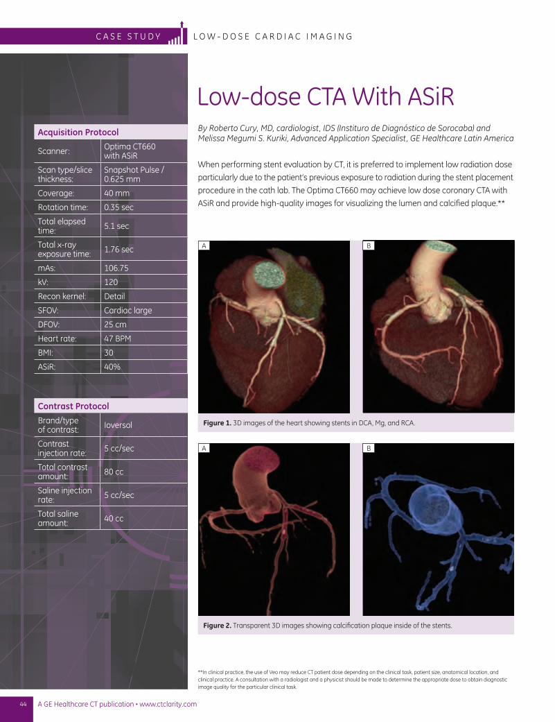
Figure 1. 3D images of the heart showing stents in DCA, Mg, and RCA.
A B
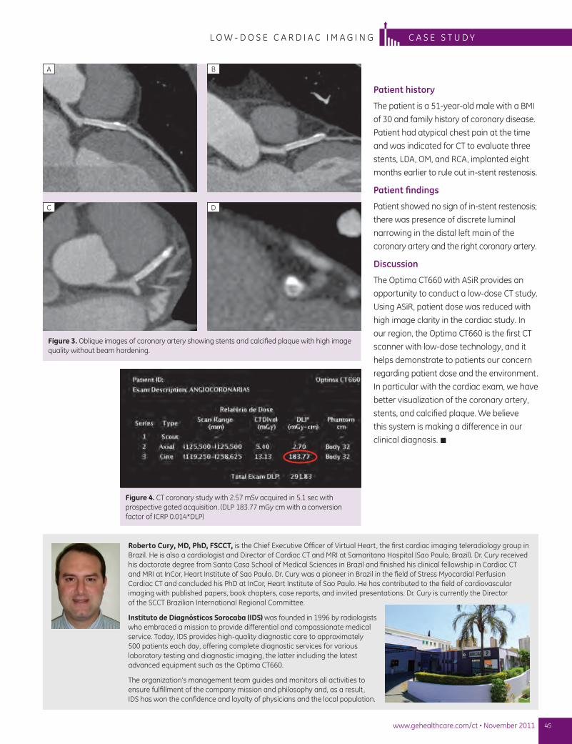
Figure 2. Transparent 3D images showing calcification plaque inside of the stents.
A B
**In clinical practice, the use of Veo may reduce CT patient dose depending on the clinical task, patient size, anatomical location, and clinical practice. A consultation with a radiologist and a physicist should be made to determine the appropriate dose to obtain diagnostic image quality for the particular clinical task.
45www.gehealthcare.com/ct • November 2011
c a s e s t u d yL o w - d o s e c a r d i a c i m a g i N g
Patient history
The patient is a 51-year-old male with a BMI
of 30 and family history of coronary disease.
Patient had atypical chest pain at the time
and was indicated for CT to evaluate three
stents, LDA, OM, and RCA, implanted eight
months earlier to rule out in-stent restenosis.
Patient findings
Patient showed no sign of in-stent restenosis;
there was presence of discrete luminal
narrowing in the distal left main of the
coronary artery and the right coronary artery.
Discussion
The Optima CT660 with ASiR provides an
opportunity to conduct a low-dose CT study.
Using ASiR, patient dose was reduced with
high image clarity in the cardiac study. In
our region, the Optima CT660 is the first CT
scanner with low-dose technology, and it
helps demonstrate to patients our concern
regarding patient dose and the environment.
In particular with the cardiac exam, we have
better visualization of the coronary artery,
stents, and calcified plaque. We believe
this system is making a difference in our
clinical diagnosis. n
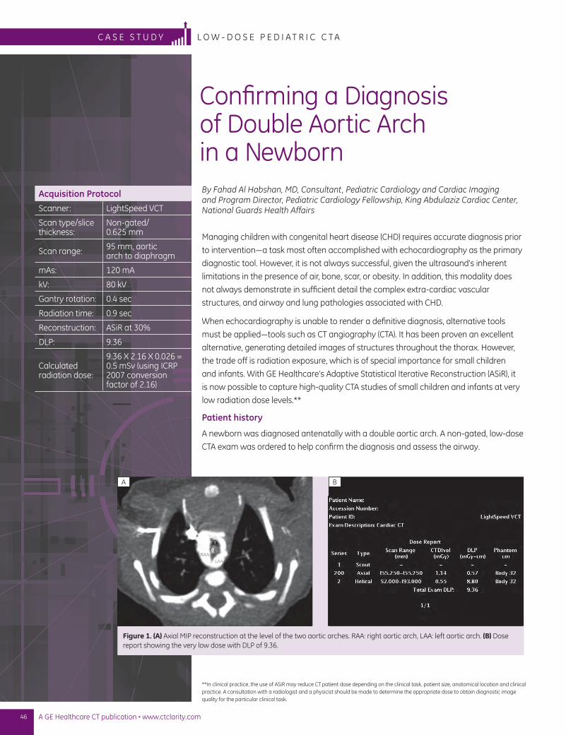
Figure 3. Oblique images of coronary artery showing stents and calcified plaque with high image quality without beam hardening.
A B
C D
Figure 4. CT coronary study with 2.57 mSv acquired in 5.1 sec with prospective gated acquisition. (DLP 183.77 mGy cm with a conversion factor of ICRP 0.014*DLP)
Roberto Cury, MD, PhD, FSCCT, is the Chief Executive Officer of Virtual Heart, the first cardiac imaging teleradiology group in Brazil. He is also a cardiologist and Director of Cardiac CT and MRI at Samaritano Hospital (Sao Paulo, Brazil). Dr. Cury received his doctorate degree from Santa Casa School of Medical Sciences in Brazil and finished his clinical fellowship in Cardiac CT and MRI at InCor, Heart Institute of Sao Paulo. Dr. Cury was a pioneer in Brazil in the field of Stress Myocardial Perfusion Cardiac CT and concluded his PhD at InCor, Heart Institute of Sao Paulo. He has contributed to the field of cardiovascular imaging with published papers, book chapters, case reports, and invited presentations. Dr. Cury is currently the Director of the SCCT Brazilian International Regional Committee.
Instituto de Diagnósticos Sorocaba (IDS) was founded in 1996 by radiologists who embraced a mission to provide differential and compassionate medical service. Today, IDS provides high-quality diagnostic care to approximately 500 patients each day, offering complete diagnostic services for various laboratory testing and diagnostic imaging, the latter including the latest advanced equipment such as the Optima CT660.
The organization’s management team guides and monitors all activities to ensure fulfillment of the company mission and philosophy and, as a result, IDS has won the confidence and loyalty of physicians and the local population.
46 A GE Healthcare CT publication • www.ctclarity.com
C A s E s T u d y l o w - d o s E p E d i A T r i C C T A
Acquisition ProtocolScanner: LightSpeed VCT
Scan type/slice thickness:
Non-gated/ 0.625 mm
Scan range: 95 mm, aortic arch to diaphragm
mAs: 120 mA
kV: 80 kV
Gantry rotation: 0.4 sec
Radiation time: 0.9 sec
Reconstruction: ASiR at 30%
DLP: 9.36
Calculated radiation dose:
9.36 X 2.16 X 0.026 = 0.5 mSv (using ICRP 2007 conversion factor of 2.16)
Confirming a Diagnosis of Double Aortic Arch in a NewbornBy Fahad Al Habshan, MD, Consultant, Pediatric Cardiology and Cardiac Imaging and Program Director, Pediatric Cardiology Fellowship, King Abdulaziz Cardiac Center, National Guards Health Affairs
Managing children with congenital heart disease (CHD) requires accurate diagnosis prior
to intervention—a task most often accomplished with echocardiography as the primary
diagnostic tool. However, it is not always successful, given the ultrasound’s inherent
limitations in the presence of air, bone, scar, or obesity. In addition, this modality does
not always demonstrate in sufficient detail the complex extra-cardiac vascular
structures, and airway and lung pathologies associated with CHD.
When echocardiography is unable to render a definitive diagnosis, alternative tools
must be applied—tools such as CT angiography (CTA). It has been proven an excellent
alternative, generating detailed images of structures throughout the thorax. However,
the trade off is radiation exposure, which is of special importance for small children
and infants. With GE Healthcare’s Adaptive Statistical Iterative Reconstruction (ASiR), it
is now possible to capture high-quality CTA studies of small children and infants at very
low radiation dose levels.**
Patient history
A newborn was diagnosed antenatally with a double aortic arch. A non-gated, low-dose
CTA exam was ordered to help confirm the diagnosis and assess the airway.
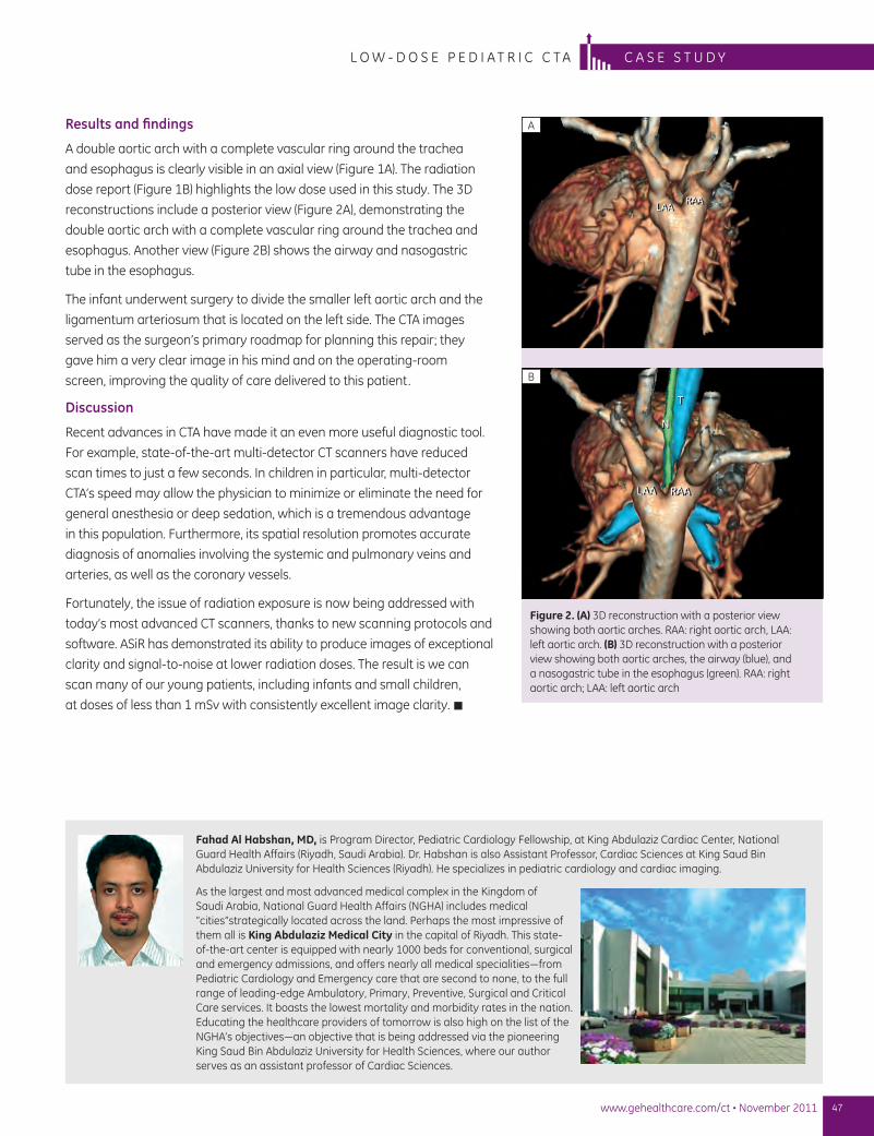
Figure 1. (A) Axial MIP reconstruction at the level of the two aortic arches. RAA: right aortic arch, LAA: left aortic arch. (B) Dose report showing the very low dose with DLP of 9.36.
**In clinical practice, the use of ASiR may reduce CT patient dose depending on the clinical task, patient size, anatomical location and clinical practice. A consultation with a radiologist and a physicist should be made to determine the appropriate dose to obtain diagnostic image quality for the particular clinical task.
A B
47www.gehealthcare.com/ct • November 2011
c a s e s t u d yl o w - d o s e p e d i a t r i c c t a
Results and findings
A double aortic arch with a complete vascular ring around the trachea
and esophagus is clearly visible in an axial view (Figure 1A). The radiation
dose report (Figure 1B) highlights the low dose used in this study. The 3D
reconstructions include a posterior view (Figure 2A), demonstrating the
double aortic arch with a complete vascular ring around the trachea and
esophagus. Another view (Figure 2B) shows the airway and nasogastric
tube in the esophagus.
The infant underwent surgery to divide the smaller left aortic arch and the
ligamentum arteriosum that is located on the left side. The CTA images
served as the surgeon’s primary roadmap for planning this repair; they
gave him a very clear image in his mind and on the operating-room
screen, improving the quality of care delivered to this patient.
Discussion
Recent advances in CTA have made it an even more useful diagnostic tool.
For example, state-of-the-art multi-detector CT scanners have reduced
scan times to just a few seconds. In children in particular, multi-detector
CTA’s speed may allow the physician to minimize or eliminate the need for
general anesthesia or deep sedation, which is a tremendous advantage
in this population. Furthermore, its spatial resolution promotes accurate
diagnosis of anomalies involving the systemic and pulmonary veins and
arteries, as well as the coronary vessels.
Fortunately, the issue of radiation exposure is now being addressed with
today’s most advanced CT scanners, thanks to new scanning protocols and
software. ASiR has demonstrated its ability to produce images of exceptional
clarity and signal-to-noise at lower radiation doses. The result is we can
scan many of our young patients, including infants and small children,
at doses of less than 1 mSv with consistently excellent image clarity. n
Figure 2. (A) 3D reconstruction with a posterior view showing both aortic arches. RAA: right aortic arch, LAA: left aortic arch. (B) 3D reconstruction with a posterior view showing both aortic arches, the airway (blue), and a nasogastric tube in the esophagus (green). RAA: right aortic arch; LAA: left aortic arch
A
B
Fahad Al Habshan, MD, is Program Director, Pediatric Cardiology Fellowship, at King Abdulaziz Cardiac Center, National Guard Health Affairs (Riyadh, Saudi Arabia). Dr. Habshan is also Assistant Professor, Cardiac Sciences at King Saud Bin Abdulaziz University for Health Sciences (Riyadh). He specializes in pediatric cardiology and cardiac imaging.
As the largest and most advanced medical complex in the Kingdom of Saudi Arabia, National Guard Health Affairs (NGHA) includes medical “cities”strategically located across the land. Perhaps the most impressive of them all is King Abdulaziz Medical City in the capital of Riyadh. This state-of-the-art center is equipped with nearly 1000 beds for conventional, surgical and emergency admissions, and offers nearly all medical specialities—from Pediatric Cardiology and Emergency care that are second to none, to the full range of leading-edge Ambulatory, Primary, Preventive, Surgical and Critical Care services. It boasts the lowest mortality and morbidity rates in the nation. Educating the healthcare providers of tomorrow is also high on the list of the NGHA’s objectives—an objective that is being addressed via the pioneering King Saud Bin Abdulaziz University for Health Sciences, where our author serves as an assistant professor of Cardiac Sciences.