ultralow dose chest ct · • uldct – filtered back projection asir uldct – asir50 • uldct...
TRANSCRIPT
Ultralow Dose Chest CTwith MBIR
Ella A. Kazerooni, M.D.Professor & Director
Cardiothoracic RadiologyAssociate Chair for Clinical Affairs
University of Michigan

Consultant: GE Healthcare
Disclosures

Low dose vs ultra low dose chest CT
0.2 mSvultraLDCT : MBIR
2‐3 mSvLDCT FBP
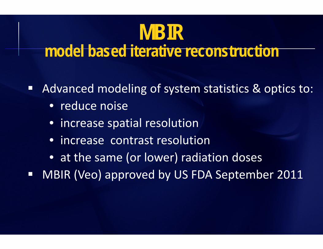
Advanced modeling of system statistics & optics to:• reduce noise• increase spatial resolution• increase contrast resolution• at the same (or lower) radiation doses
MBIR (Veo) approved by US FDA September 2011
MBIRmodel based iterative reconstruction

MBIRmodel based iterative reconstruction
NIH funded collaboration: PI Jeff Fessler PhD UM School of Engineering, GE Global Research and U of M Radiology Mitch Goodsitt PhD CT
physicist & Ella Kazerooni MD Thoracic Radiology


• Lung is a great target for low dose CT efforts• Inherent contrast between lung (air) and tissue (vessels & airways)
• Tissues extracted as arborizing structures• Lung segmentation straightforward• At or near CXR exposure CT exams• Trade off may be quantitative analysis
lowest exposure vs. most accurate measurement
Ultralow Dose Chest CT

National Lung Cancer Screening Trial
20% lung cancer mortality reduction
6.9% all cause mortality reduction
screen 320 individuals to save 1 from lung cancer death
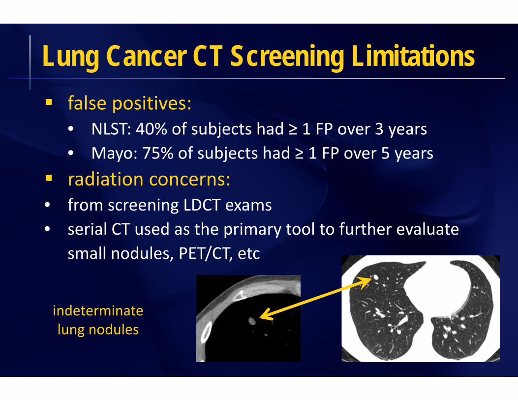
false positives:• NLST: 40% of subjects had ≥ 1 FP over 3 years• Mayo: 75% of subjects had ≥ 1 FP over 5 years
radiation concerns:• from screening LDCT exams• serial CT used as the primary tool to further evaluate
small nodules, PET/CT, etc
Lung Cancer CT Screening Limitations
indeterminate lung nodules

Radiation ExposureRadiation Risks Potentially Associated with Low Dose Screening
Brenner DJ et al. Radiology 2004;231:440‐445
• assumptions:• entire US population current/former smokers• age 50‐75 years• annual CT until age 75 (5.2 ± 0.9 mGy to lung; 60 mAs)• 50% compliance rate• atomic bomb survivor cohort for predicting risk
• expect 1.9 million lung cancers• 36,000 additional lung cancers attributed to CT• 1.8% increase in lung cancer (95% CI 0.5‐5.5%)

Parameter GE-VCT(64)64 slice | 0.5 sec
Siemens Sensation 64
64 x 0.0.6
Phillips MX8000 16 slice | 0.5 sec
16 x .75
Toshiba Aquillon16 slice | 0.5 sec
kV 120 120 120 120
Gantry Rotation Time 0.5 sec 0.5 sec 0.5 sec 0.5 sec
mA (Regular-Large Patient) 50-100 50-100 75-15- 80-160
mAs (Regular-Large) 25-50 25-50 37.5-75 40-80
Scanner Effective mAs (Reg-Large) 27-53 25-50 25-50 26.7-53.3
Detector Collimation (mm): T 0.625 mm 0.6 mm .75 mm 2 mm
Number of Active Channels: N 64 32 16 16
Detector configuration: N x T 64 x 0.625 mm 32 x 0.6 mm 16 x 0.75 16 x 2
MODE (Thick/Speed) or Console Collimation1 .625/.984/39.37 64 x 0.6 mm N/A N/A
Table Incrementation (mm/rotation): I 39.37 mm 19.2 mm 18 mm 48 mm
Pitch ([mm/rotation]/configuration): I/NT 0.984 1.0 1.5 1.5
Table speed (mm/second) 78.74 mm/sec 38.4 mm/sec 36 mm/sec 96 mm/sec
Scan time (40 cm thorax) 5.1 sec 11 sec 11 sec 4.2 sec
Max Nominal Reconstructed Slice Width 2.5 mm 2 mm 2 mm 2 mm
Reconstruction Interval 2.0 mm 1.8 mm 1.8 mm 1.8 mm
Reconstruction Algorithm STD B30f B or C FC 10
# Images/Data set (40 cm Thorax) 200 223 223 223
CTDIvol Dose in mGy (Regular-Large) 2.2-2.4 mGy 1.9-3.8 mGy 1.9-3.8 mGy 2.7-5.4 mGy
NLST CT Technique Chart - 2002-06

Detection Characterization
• Qualitative- Descriptive: ground glass, solid etc
• Semi‐quantitative- Caliper sizing
• Quantitative- Volumetric measurement
Follow up
Lung Nodule CT Tasks

MBIR Lung CT for Nodules
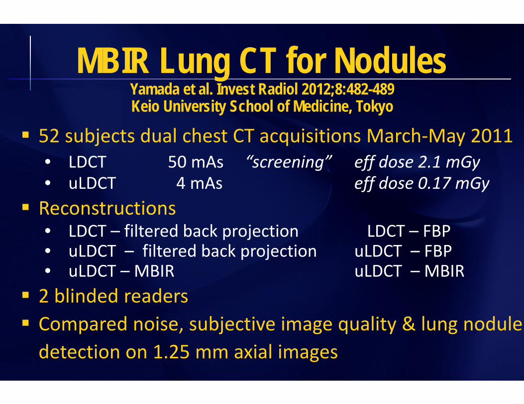
52 subjects dual chest CT acquisitions March‐May 2011• LDCT 50 mAs “screening” eff dose 2.1 mGy• uLDCT 4 mAs eff dose 0.17 mGy
Reconstructions• LDCT – filtered back projection LDCT – FBP• uLDCT – filtered back projection uLDCT – FBP• uLDCT – MBIR uLDCT – MBIR
2 blinded readers Compared noise, subjective image quality & lung nodule detection on 1.25 mm axial images
MBIR Lung CT for NodulesYamada et al. Invest Radiol 2012;8:482-489Keio University School of Medicine, Tokyo

“blotchy pixelated appearance” on all uLDCT‐MBIRs
MBIR Lung CT for NodulesYamada et al. Invest Radiol 2012;8:482-489Keio University School of Medicine, Tokyo
1 = none; 2 = present not impacting screening results3 = impacting screening results

“blotchy pixelated appearance”
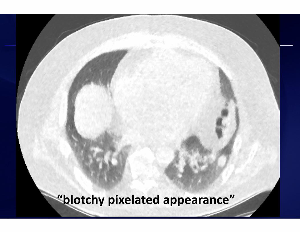
“blotchy pixelated appearance”

MBIR Lung CT for NodulesYamada et al. Invest Radiol 2012;8:482-489Keio University School of Medicine, Tokyo
Subjective image noise• uLDCT – MBIR = LDCT – FBP • uLDCT – MBIR < uLDCT – FBP (p < 0.001)

Artifacts• uLDCT – FBP > uLDCT – MBIR > LDCT – FBP p < 0.001
Image sharpness• uLDCT – MBIR & uLDCT – FBP equivalent• LDCT – FBP significantly more sharp (p < 0.001)
MBIR Lung CT for NodulesYamada et al. Invest Radiol 2012;8:482-489Keio University School of Medicine, Tokyo

Artifacts with MBIR

Nodule detection ‐ LDCT‐FBP used as “truth” Calcified nodules (n = 26, 2‐10 mm diameter)
uLDCT – MBIR & uLDCT – FBP detected all nodules Non calcified nodules (n = 184, 1‐30 mm, mean 4±3
uLDCT – MBIR > uLDCT – FBP (p < 0.002)TPF all 0.905 0.804TPF ≥ 4 mm 0.944 0.833TPF < 4 mm 0.884 0.789FPR all* 0.125 0.202
MBIR Lung CT for NodulesYamada et al. Invest Radiol 2012;8:482-489Keio University School of Medicine, Tokyo
* Upon review of misses reviewed – due to noise and artifacts

The patients… Age 38 ‐ 84 years (mean 66.3) Weight
• range: 37 ‐ 84 kg (82 ‐ 185 lbs)• mean: 55.9 ± 11.9 kg (123 ± 26 lbs)
BMI*• range: 15.4 ‐ 30.7 • mean: 21.7 ± 3.5
MBIR Lung CT for NodulesYamada et al. Invest Radiol 2012;8:482-489Keio University School of Medicine, Tokyo
*healthy weight 18.5‐25; overweight 25‐30; moderately obese 30‐35

100 subjects dual chest CT acquisitions July 2011• LDCT eff dose 4.04 mGy• uLDCT eff dose 0.55 mGy
Reconstructions• LDCT – filtered back projection with ASIR LDCT – ASIR50• uLDCT – filtered back projection ASIR uLDCT – ASIR50• uLDCT – MBIR uLDCT – MBIR
2 blinded readers Compared noise, subjective image quality & lung nodule detection on 0.625 mm axial images
MBIR Lung CT for NodulesMatsura et al. Eur Rad 2012;8:1613-1623
University of Tokyo

MBIR Lung CT for NodulesMatsura et al. Eur Rad 2012;8:1613-1623
University of Tokyo

MBIR Lung CT for NodulesMatsura et al. Eur Rad 2012;8:1613-1623
University of Tokyo
LDCT‐ASIR uLDCT‐ASIR uLDCT‐MBIR

Nodule detection not studied
MBIR Lung CT for NodulesMatsura et al. Eur Rad 2012;8:1613-1623
University of Tokyo

The patients… Age mean 65.6 ± 12.4 years Weight
• range: not reported• mean: 58 ± 13 kg (128 ± 29 lbs)
1 std dev 99‐157 lbs (approx 68% of subjects)2 std dev 70‐186 lbs (approx 95% of subjects)
BMI not reported
MBIR Lung CT for NodulesMatsura et al. Eur Rad 2012;8:1613-1623
University of Tokyo

42 subjects• SDCT or LDCT (11.2 or 2.7 mSV)• uLDCT (0.16 mSv) (in the range of 2view CXR)
Reconstructions• SDCT or LDCT – filtered back projection • uLDCT – filtered back projection • uLDCT – ASIR• uLDCT – MBIR
uLDCT– MBIR better image quality than uLDCT ASIR or FBP; quality inferior for ground glass and emphysema BMI mean 25.8 males, 27.8 females; “overweight”
MBIR Lung CT for NodulesNeroladaki et al. Eur Radiol 2012
Geneva University Hospital
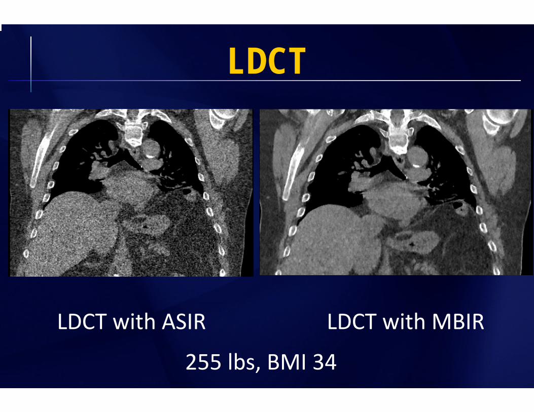
LDCT
LDCT with MBIRLDCT with ASIR
255 lbs, BMI 34

ultraLDCT
uLDCT with MBIRuLDCT with ASIR255 lbs, BMI 34
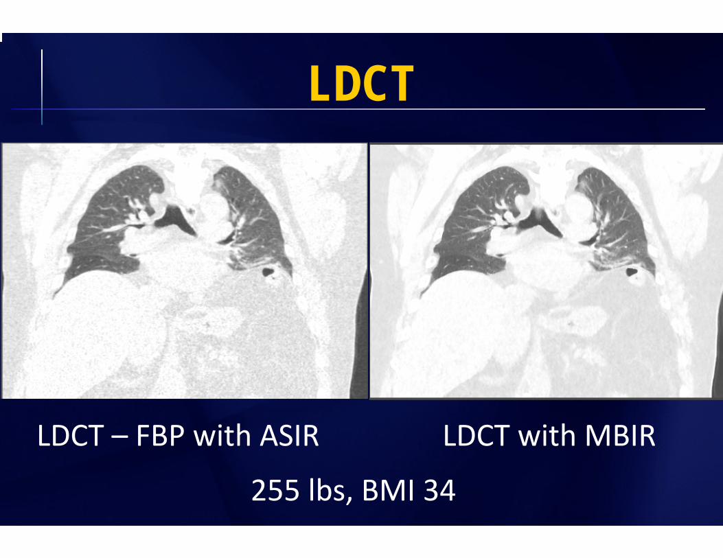
LDCT
LDCT with MBIRLDCT – FBP with ASIR
255 lbs, BMI 34

ultraLDCT
uLDCT with MBIRuLDCT – FBP with ASIR255 lbs, BMI 34

LDCT vs uLDCT
LDCTMBIR
LDCTASIR
uLDCTMBIR
uLDCTASIR

LDCT vs uLDCT
LDCTMBIR
LDCTASIR
255 lbsBMI 34
uLDCTMBIR
uLDCTASIR

LDCT vs uLDCT - GGO
LDCTMBIR
LDCTASIR
uLDCTMBIR
uLDCTASIR
200 lbsBMI 36.6

Low dose CT (conventional low dose) underestimates small nodule volume compared to standard dose chestCT (de Jong et al; 2012 Oct AJR) LDCT vs contrast enhanced standard chest CT CTDIvol 2.2 vs 5.5‐20 mGy 200 mm3 threshold or approx 8 mm below which nodules were undersized by approx 14‐16%
Impact of MBIR on Nodule Volume?TBD

• Substantial radiation exposure reduction• Reduces noise back to standard low dose CT levels• Blotchy pixelated appearance does not appear to subjectively impact diagnostic quality of chest CTs
• Not validated in US patient population size cohort• More work on diagnostic accuracy and impact on nodule quantification is needed
MBIR Lung CT for Nodules

Ultralow Dose Chest CTwith MBIR
Ella A. Kazerooni, M.D.Professor & Director
Cardiothoracic RadiologyAssociate Chair for Clinical Affairs
University of Michigan