effects of ionizing radiation on dna
DESCRIPTION
Effects of Ionizing Radiation on DNA. Effects. Single strand break Double-Strand Break Double-Strand Break in Same Rung of DNA Mutation. Single-Strand Break - PowerPoint PPT PresentationTRANSCRIPT

Effects of Ionizing Radiation on DNA

Effects
• Single strand break• Double-Strand Break• Double-Strand Break in Same Rung of DNA• Mutation

Single-Strand BreakIf ionizing radiation interacts with a DNA macromolecule, the energy transferred can rupture one of its chemical bonds, possibly severing one of the sugar-phosphate chain side rails or strands of the ladderlike molecular structure (single-strand break). This type of injury to DNA is called a point mutation. Gene mutations may result from a single alteration along the sequence of nitrogenous bases. Point mutations commonly occur with low-LET radiations. Repair enzymes, however, are capable of reversing this damage.

Double-Strand BreakFurther exposure of the affected DNA macromolecule to ionizing radiation may result in additional breaks in the sugar-phosphate molecular chain(s). These breaks might also be repaired, but double-strand breaks (one or more breaks in each of the two sugar-phosphate chains) are not repaired as easily as single-strand breaks. If repair does not take place, further separation may occur in the DNA chains, threatening the life of the cell..

Double-strand breaks occur more commonly with densely ionizing (high-LET) radiations and often are associated with the loss or gain of one or more nitrogenous bases. When high-LET radiation interacts with DNA molecules, the ionization interactions may be so closely spaced that, by chance, both strands of the DNA chain are broken. If both strands are broken at the same nitrogenous base “rung,” the result is the same as if both side rails of the ladder were cut at the same step or rung—the ladder would be cut into two pieces. If the DNA is cut into two pieces, the chromosome, which is composed of a long chain of twisted strands of DNA ladders, is itself broken. Thus some types of chromosomal damage that are particularly associated with high-LET radiation are related to double-strand breaks of DNA. Because the chance of repairing this damage is much slighter, the possibility of inducing a lethal alteration of nitrogenous bases within the genetic sequence is far greater.


Double-Strand Break in Same Rung of DNAWhen two interactions (hits), one on each of the two sugar-phosphate chains, occur within the same rung of the DNA ladderlike configuration, the result is a cleaved or broken chromosome, with each new portion containing an unequal amount of genetic material. If this damaged chromosome divides, each new daughter cell will receive an incorrect amount of genetic material. This will culminate in the death or impaired functioning of the new daughter cell.

MutationIn general, the interaction of high-energy radiation with a DNA molecule causes either a loss of or change in a nitrogenous base on the DNA chain. The direct consequence of this damage is an alteration of the base sequence. Because the genetic information to be passed on to future generations is contained in the strict sequence of these bases, the loss or change of a base in the DNA chain is a mutation. It may not be reversible and may cause acute consequences for the cell but, more important, if the cell remains viable, incorrect genetic information will be transferred to one of the two daughter cells when the cell divides.

Covalent Cross-LinksCovalent cross-links are chemical unions created between atoms by the single sharing of one or more pairs of electrons. Covalent cross-links involving DNA are another effect initiated by high-energy radiation. At low energies, however, covalent cross-links are probably caused by the process of indirect action. Following irradiation, some molecules can produce small, spurlike molecules that become very interactive (“sticky”) when exposed to radiation. This can cause these molecules to attach to other macromolecules or to other segments of the same macromolecule chain. Cross-linking can occur in many different patterns. For example, a cross-link can form between two places on the same DNA strand. This joining is termed an intrastrand cross-link. Cross-linking may also occur between complementary DNA strands or between entirely different DNA molecules. These joinings are termed interstrand cross-links. Finally, DNA molecules also may become covalently linked to a protein molecule. All these linkages are potentially fatal to the cell if they are not properly repaired.


Effects of Ionizing Radiation on Chromosomes
Large-scale structural changes in a chromosome brought about by ionizing radiation may be as grave for the cell as are radiation-induced changes in DNA. When changes occur in the DNA molecule, the chromosome exhibits the alteration. Because DNA modifications are discrete, they do not inevitably result in observable structural chromosome alterations.

Radiation-Induced Chromosome Breaks
After irradiation and during cell division, some radiation-induced chromosome breaks may be viewed microscopically. These alterations manifest themselves during the metaphase and anaphase of the cell division cycle, when the length of the chromosomes is visible. Because the events that have happened before these phases of cell division are not visible, they can only be assumed to have occurred. What can be seen, however, is the effect of these events—the gross or visible alterations in the structure of the chromosome. Both somatic cells and reproductive cells are subject to chromosome breaks induced by radiation.

Chromosomal FragmentsAfter chromosome breakage, two or more chromosomal fragments are produced. Each of these fragments has a fractured extremity. These broken ends appear sticky and have the ability to adhere to another such sticky end. The broken fragments may rejoin in their original configuration, fail to rejoin and create an aberration (lesion or anomaly), or rejoin other broken ends and create new chromosomes that may not look structurally altered compared with the chromosome before irradiation

Chromosome AnomaliesTwo types of chromosome anomalies have been observed at metaphase. They are called (1) chromosome aberrations and (2) chromatid aberrations. Chromosome aberrations result when irradiation occurs early in interphase, before DNA synthesis takes place. In this situation, the break caused by ionizing radiation is in a single strand of chromatin; during the DNA synthesis that follows, the resultant break is replicated when this strand of chromatin lays down an identical strand adjacent to itself if repair is not complete before the start of DNA synthesis. This leads to a chromosome aberration in which both chromatids exhibit the break. This break is visible at the next mitosis. Each daughter cell generated will have inherited a damaged chromatid as a consequence of a failure in the repair mechanism. Chromatid aberrations, on the other hand, result when irradiation of individual chromatids occurs later in interphase, after DNA synthesis has taken place. In this situation, only one chromatid of a pair might suffer a radiation-induced break. Therefore only one daughter cell is affected.

Structural Changes in Biologic Tissue Caused by Ionizing RadiationIonizing radiation interacts randomly with matter. Because of this phenomenon, exposure to radiation produces a variety of structural changes in biologic tissue. Some of these changes are as follows:
•A single-strand break in one chromosome•A single-strand break in one chromatid•A single-strand break in separate chromosomes•A strand break in separate chromatids•More than one break in the same chromosome•More than one break in the same chromatid•Chromosome stickiness, or clumping together

Consequences to the Cell from Structural Changes in Biologic Tissue: 1.Restitution, whereby the breaks rejoin in their original configuration with no visible damage . In this case no damage to the cell occurs because the chromosome has been restored to the condition it was in before irradiation. The process of healing by restitution is believed to be the way in which 95% of single-chromosome breaks mend.
2.Deletion, whereby a part of the chromosome or chromatid is lost at the next cell division, creating an aberration known as an acentric fragment.
3.Broken-end rearrangement, whereby a grossly misshapen chromosome may be produced. Ring chromosomes, dicentric chromosomes, and anaphase bridges are examples of such distorted chromosomes.
4.Broken-end rearrangement without visible damage to the chromosomes, whereby the chromosome's genetic material has been rearranged even though the chromosome appears normal. Translocations are an example of such rearrangements. Changes such as these inevitably result in mutation because the positions of the genes on the chromosomes have been rearranged, thus altering the heritable characteristics of the cell.

Restitution
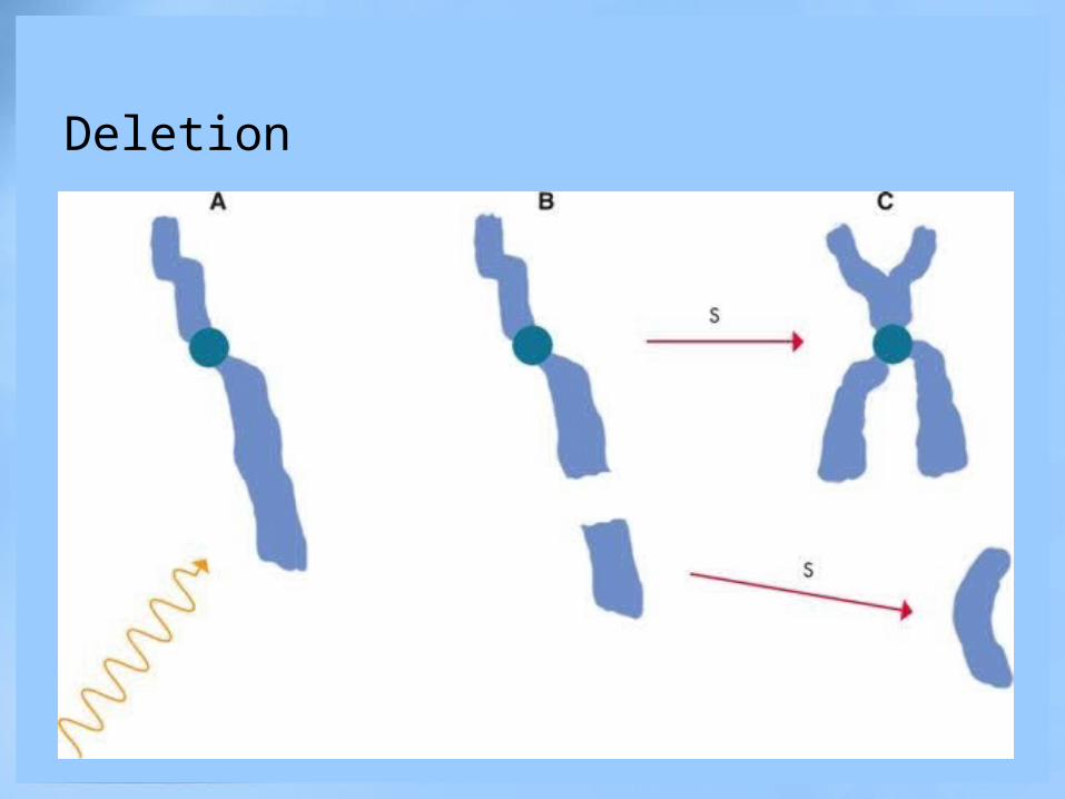
Deletion

The process of broken-end rearrangement may result in grossly misshapen chromosomes. A, Two chromosome breaks occur in a single chromosome as a result of the interactions of two photons. B, The fragments from opposite ends unite before the DNA synthesis phase. C, The ends of the chromosome that are still attached to the centromere also unite, forming a “ring” chromosome. D, Chromosome breaks occur in two different chromosomes. E, The fragments are fully separated from the rest of their respective chromosomes. F, The ends of the chromosomes and the ends of the fragments have joined before DNA synthesis, forming a dicentric (two centromeres) and an acentric (no centromere) fragment. G, After DNA synthesis (labeled S), the chromosome is elongated but cannot split in two. The two centromeres are “bridged.” This type of chromosomal damage leads to reproductive death of the cell (i.e., it cannot replicate or divide into two cells)

The process of broken-end rearrangement may result in no visible damage to the chromosome, although the chromosome's genetic material has been rearranged—a result that will drastically alter its function within the cell, probably leading to cell death or failure to replicate.

Target TheoryAmid the many different types of molecules that lie within the cell, a master, or key, molecule that maintains normal cell function also is believed to be present. This master molecule is necessary for the survival of the cell. Because this molecule is unique in any given cell, no similar molecules in the cell are available to replace it; if the master molecule is inactivated by exposure to radiation, the cell will die. Experimental data strongly support this concept and indicate that DNA is the irreplaceable master, or key, molecule that serves as the vital target. Destruction of some of the molecules that are plentiful in the cell does not result in cell death. The reason for this is simply that cells have an abundance of similar molecules to take over and perform necessary functions for them in the event of their demise. If only a few non-DNA cell molecules are destroyed by radiation exposure, the cell will probably not show any evidence of injury after irradiation.


CELLULAR EFFECTS OF IRRADIATIONIonizing radiation can adversely affect the cell. Damage to the cell's nucleus reveals itself in one of the following ways:
1.Instant death2.Reproductive death3.Apoptosis, or programmed cell death (interphase death)4.Mitotic, or genetic, death5.Mitotic delay6.Interference of function7.Chromosome breakage

SURVIVAL CURVES FOR MAMMALIAN CELLSCells vary in their radiosensitivity. This fact is particularly important in determining the type of cancer cells that will respond to radiation therapy. A classic method of displaying the sensitivity of a particular type of cell to radiation is the cell survival curve. A cell survival curve is constructed from data obtained by a series of experiments. First, the cells are made to grow “in culture,” meaning in a laboratory environment such as a Petri dish. Then the cells are exposed to a specified dose of radiation. After radiation exposure, the ability of the cells to divide, or form new “colonies” of cells, is measured. The fraction of cells that are able to form new colonies through cell division is then reported as the fraction of cells that have survived irradiation. The process is repeated for a range of radiation doses, and the results are graphed with the logarithm of the surviving fraction on the vertical axis and the dose on the horizontal axis.


