effects of pm2.5 and gases exposure during prenatal and
TRANSCRIPT
RESEARCH Open Access
Effects of PM2.5 and gases exposure duringprenatal and early-life on autism–likephenotypes in male rat offspringBaharan Emam1, Abbas Shahsavani1,2*, Fariba Khodagholi3, Saeed Motesaddi Zarandi1, Philip K. Hopke4,5,Mostafa Hadei6,7, Hamidreza Behbahani3 and Maryam Yarahmadi8
Abstract
Background: Epidemiological studies have reported associations between elevated air pollution and autism spectrumdisorders (ASD). However, we hypothesized that exposure to air pollution that mimics real world scenarios, is apotential contributor to ASD. The exact etiology and molecular mechanisms underlying ASD are not well understood.Thus, we assessed whether changes in OXTR levels may be part of the mechanism linking PM2.5/gaseous pollutantexposure and ASD. The current in-vivo study investigated the effect of exposure to fine particulate matter (PM2.5) andgaseous pollutants on ASD using behavioral and molecular experiments. Four exposure groups of Wistar rats wereincluded in this study: 1) particulate matter and gaseous pollutants exposed (PGE), 2) gaseous pollutants only exposed(GE), 3) autism-like model (ALM) with VPA induction, and 4) clean air exposed (CAE) as the control. Pregnant dams andmale pups were exposed to air pollutants from embryonic day (E0) to postnatal day (PND21).
Results: The average ± SD concentrations of air pollutants were: PM2.5: 43.8 ± 21.1 μg/m3, CO: 13.5 ± 2.5 ppm, NO2:0.341 ± 0.100 ppm, SO2: 0.275 ± 0.07 ppm, and O3: 0.135 ± 0.01 ppm. The OXTR protein level, catalase activity (CAT), andGSH concentrations in the ALM, PGE, and GE rats were lower than those in control group (CAE). However, thedecrements in the GE rats were smaller than other groups. Also in behavioral assessments, the ALM, PGE, and GE ratsdemonstrated a repetitive /restricted behavior and poor social interaction, but the GE rats had weaker responsescompared to other groups of rats. The PGE and GE rats showed similar trends in these tests compared to the VPA rats.
Conclusions: This study suggested that exposure to ambient air pollution contributed to ASD and that OXTR proteinmay serve as part of the mechanism linking them.
Keywords: Air pollution, Fine particulate matter, Behavioral assessment, OXTR protein
BackgroundAutism spectrum disorder (ASD) is a pervasive neurodeve-lopmental disorder recognized by social communicationdeficits and restricted/repetitive patterns of behavior [1]. Itis estimated that the global prevalence of ASD is 1 in 132persons [2] and the prevalence rate is still increasing [3].The prevalence of ASD is four to five times higher in malesthan females [4]. ASD has attracted public attentionbecause of its high social costs and substantial impacts on
society [5]. Although genetics likely plays an important rolein ASD, environmental exposures to pollutants particularlyduring the early life periods could be another potential riskfactor [6, 7]. Environmental factors such as exposure to airpollution may contribute to ASD etiology [8–10].Previous studies point to a biological pathway linked to
autism through a systemic inflammatory response that canaffect the development of the central nervous system [9].Developmental exposure to traffic-related air pollution(TRAP) has been associated with increased ASD risk [11].Environment exposures during perinatal and postnatal pe-riods may be crucial in ADS since brain development takesplace in these periods, and exposure to environmental che-micals may cause neurodevelopmental disorders [12, 13].
© The Author(s). 2020 Open Access This article is distributed under the terms of the Creative Commons Attribution 4.0International License (http://creativecommons.org/licenses/by/4.0/), which permits unrestricted use, distribution, andreproduction in any medium, provided you give appropriate credit to the original author(s) and the source, provide a link tothe Creative Commons license, and indicate if changes were made. The Creative Commons Public Domain Dedication waiver(http://creativecommons.org/publicdomain/zero/1.0/) applies to the data made available in this article, unless otherwise stated.
* Correspondence: [email protected] of Environmental Health Engineering, School of Public Healthand Safety, Shahid Beheshti University of Medical Sciences, Tehran, Iran2Environmental and Occupational Hazards Control Research Center, ShahidBeheshti University of Medical Sciences, Tehran, IranFull list of author information is available at the end of the article
Emam et al. Particle and Fibre Toxicology (2020) 17:8 https://doi.org/10.1186/s12989-020-0336-y

Limited prior animal studies also suggested a connec-tion between exposure to air pollution and ASD [14].Most of these studies exposed rats or mice to high con-centrations of air pollution. For instance, in a study wasconducted by Li et al. (2018), rats were exposed to PM2.5
with doses of 2 or 20 mg/kg body weight per day [9],and reported that both groups of exposed rats showedtypical behavioral features of autism. In another study,mice developmentally exposed to high concentrations ofdiesel exhaust particles exhibited altered behavioral phe-notypes including effects on locomotor activity and re-petitive behaviors [15].It has been suggested that airborne particulate matter
may act like a Trojan horse [16] and represents an effect-ive delivery system for diverse environmental toxicants toreach the brain. Additionally, associated water solublecompounds may provide a toxic stimulus independent ofthe particle composition itself and may be transported tothe brain by the circulation system [17]. The toxicity ofparticulate matter in the lung have been linked to boththe particulate constituents including metallic elements,oxidants, and oxidant forming species [18, 19] and thephysical characteristic of particles itself [20]. Many com-pounds present in the particulate matter are neurotoxic[19]. For example, environmental exposure to neurotoxi-cants such as iron (Fe), copper (Cu), manganese (Mn),aluminum (Al), zinc (Zn), and lead (Pb) can induce oxida-tive stress [21, 22], and the brain is vulnerable to oxidativestress due to its great metabolic activity and low levels ofantioxidants such as catalase (CAT) [23]. Previous studieshave suggested that autism could result from the inter-action between genetic and environmental factors withoxidative stress as the link between them [24]. Disturbingredox signaling, imbalance in the cellular redox state to-wards the pro-oxidant status, oxidative stress, and theresulting systemic inflammation are a possible mechanismof air pollution induced autism [25]. In addition, oxidativemodification can modulate activity of several proteins thathave relevant roles in normal brain function. Reactive oxy-gen species (ROS) play a crucial role in cell signaling. Oxi-dative stress also plays a role in controlling the activitiesof receptor proteins [26].Extensive research has established the possible ability of
the hypothalamic neuropeptide oxytocin (OXT) to modu-late social behaviors across species including humans [27].Results from the animal studies led to examination of theeffects of OXT administration to humans and investiga-tions of the etiology and treatment of psychiatricdisorders, especially ASD [28]. Because social behaviorsymptoms are a clear manifestation of this disorder, theOXT system has been implicated in the biology of ASD,and has become a promising treatment option for the so-cial symptoms of ASD. Increased research efforts into thepotential involvement of OXT and its receptor (OXTR) in
ASD include genetic studies, analysis of OXT levels in bio-logical fluids and the assessments of OXT treatment inhumans [29]. However, recent evidence investigating andimplicating the role of OXTR protein in ASD has been in-creasing [29].Epidemiological studies suggest an association between
elevated air pollution and ASD. Since the effects of lowerconcentration of air pollution (that reflect the realworld) on autism spectrum disorder is poorly under-stood. Hence we hypothesized exposure to ambient airpollution is a potential contributor to ASD. Previousstudies have reported that simultaneous exposure to par-ticulate matter and gaseous pollutants during pregnancyhave been associated with ASD [30]. However, there islimited evidence showing a relationship between onlygaseous pollutants exposure and ASD. Therefore, we in-vestigated the effect of gaseous pollutant only exposureson ASD. Studies on rodents have found that prenatal ex-posure to VPA induces an animal model of ASD show-ing similar structural, functional, and behavioral featuresto human autistic patients [31–33]. Hence we comparedthe effects of the air pollution to that of VPA in rats. Inthe present study, we investigated the effects of prenataland early life PM2.5 and gaseous pollutants (NO2, CO,O3, SO2) exposure on a battery of behavioral dimensions,OXTR protein expression, and antioxidants (CAT) en-zymatic activity and GSH concentration in the brain.Since the precise etiology of ASD remains poorly under-stood [34]., we assessed whether changes in OXTR levelsmay serve as part of the mechanism linking PM2.5/gas-eous pollutants and ASD.
ResultsThe concentration of PM2.5 and gaseous pollutantsThe average concentration of PM2.5 in the exposureperiod (E0 until PND22) was 43.82 ± 21.12 μg/m3. Theaverage concentrations of CO, NO2, SO2 and O3 were13.5 ± 2.5 ppm, 0.341 ± 0.100 ppm, 0.275 ± 0.07 ppm, and0.135 ± 0.010 ppm, respectively. The concentrations ofgases in both chambers one and two were equal. Controlgroup of rats were exposed to clean air with these char-acteristics: PM2.5 < 5 μg/m
3, SO2 < 0.02 ppm, NO2 < 0.04ppm, CO < 2.4 ppm, and O3 < 0.02 ppm.
The concentration of metals and PAHsConcentrations of PM2.5-bounded heavy metals were deter-mined, and the mean values can ordered as follow: Ca >Al >Na >Cu > Fe >Cd >Cr >Ni > Pb >Zn >Mn>As>V (Add-itional file 1: Table S1). The mean total concentration of 16PAHs was 45.88 ± 21.02 ng/m3 (Additional file 1: Table S2).The order of average concentrations of the observed PAHswas phenanthrene> naphtalene> benzo(k)fluoranthene> flor-ene> pyrene> anthracene> acenaphtylen> benzo(b)fluor-anthene> chrysene> fluorantene> benzo(a) anthracene >
Emam et al. Particle and Fibre Toxicology (2020) 17:8 Page 2 of 16

acenaphten> dibenzo(a,h)anthracene> benzo (g,h,i)perylene>benzo(a)p- yrene> indeno (1,2,3-cd)pyrene.
Open fieldThe open field test assesses locomotor activity and ex-ploratory drive (23). To directly assess whether prenataland early life exposures in the PGE and GE rats altermotor activity, the rats were assessed for locomotor ac-tivity (Fig. 1). There were no significant differencesamong the exposed rats and controls in total distancetravelled and velocity.
Social preference testThe main aim of this study was to assess whether prenataland early life air pollution exposure could induce autistictraits in PGE and GE rats. To eliminate possible con-founding, we set a control group (CAE rats) to distinguishbetween the effects of PM2.5 and gasses exposure (PGErats) and gaseous exposure (GE rats) on the behavioraloutcomes of pups. We also created an autism-like modelby injecting doses of VPA to produce ALM rats, and com-pare these animals to the other exposure groups.The first session of the social preference test allowed es-
timation of the social affiliation and motivation of subjectrat. Typically, a wild type animal will spend a significantamount of time in the compartment with the strangercompared to the compartment with empty cup. Such be-havior indicates normal sociability, social motivation, andaffiliation [35, 36]. In the sociability session of the test,every group demonstrated a significant preference forspending time in the chamber containing stranger1 com-pared to the empty chamber. The time that the ALM ratsspent in the chamber containing the stranger1 was signifi-cantly lower than that for the GE rats (Fig. 2a).The second session of the test estimated social novelty
and social memory. The ability to differentiate socialnovelty was determined by measuring time spent in thechamber containing stranger2 compared to the time
spent in the chamber containing stranger1 that is nowthe familiar rat. The ALM and PGE rats showed no pref-erence between the stranger2 and stranger1 rats. Thetime spent by ALM rats in the chamber containingstranger2 was significantly lower than that for the PGErats (Fig. 2b).
Y-mazeWe assessed spontaneous alternation in the Y-maze toanalyze respective behavior and /or working memory.Spontaneous alternation in a Y- maze represents a com-mon exploratory strategy and repetitive behavior [33,37]. The Y-maze labyrinth is a hippocampus dependenttask of the spatial working memory. A decrease in thepercentage of alternation in this test was detected inALM, PGE, and GE rats, comparing to CAE rats (Fig. 3).The reduction in alternation in the Y-maze test also in-dicated restricted behavioral patterns (another core ofASD), in ALM, PGE, and GE rats [38].
Marble buryingRepetitive behavior was assessed by the marble buryingtest. The ALM and PGE rats buried more marbles thanthe CAE rats (Fig. 4a). For all the affected rats (ALM,PGE, and GE), the duration of exploration decreased vsCAE rats (Fig. 4b). For all the affected rats (ALM, PGE,and GE), there were increasing trends in the interactiontime with the marbles compared to CAE rats althoughthe difference was not significant (Fig. 4c). In this test,the frequency of self- grooming events and repetitivedigging behavior were quantified. For the PGE and GErats, the duration of self–grooming increased comparedto the CAE rats (Fig. 4d) and the duration of digging inthe ALM rats increased vs the CAE rats (Fig. 4e).
Oxidative stressOxidative stress was assessed by measuring the antioxi-dant enzymes i.e. catalase (CAT) and GSH in prefrontal
Fig. 1 Open field test; PGE and GE rats showed no differences in locomotor behaviors. Prenatal exposure of PGE and GE rats did not affect: adistance travelled and b velocity in the open field task when compared to CAE control rats. (one-way ANOVA with Tukey s̉ multiplecomparisons). Number of rats per group: CAE = 8, GE = 13, PGE = 8, ALM = 8
Emam et al. Particle and Fibre Toxicology (2020) 17:8 Page 3 of 16

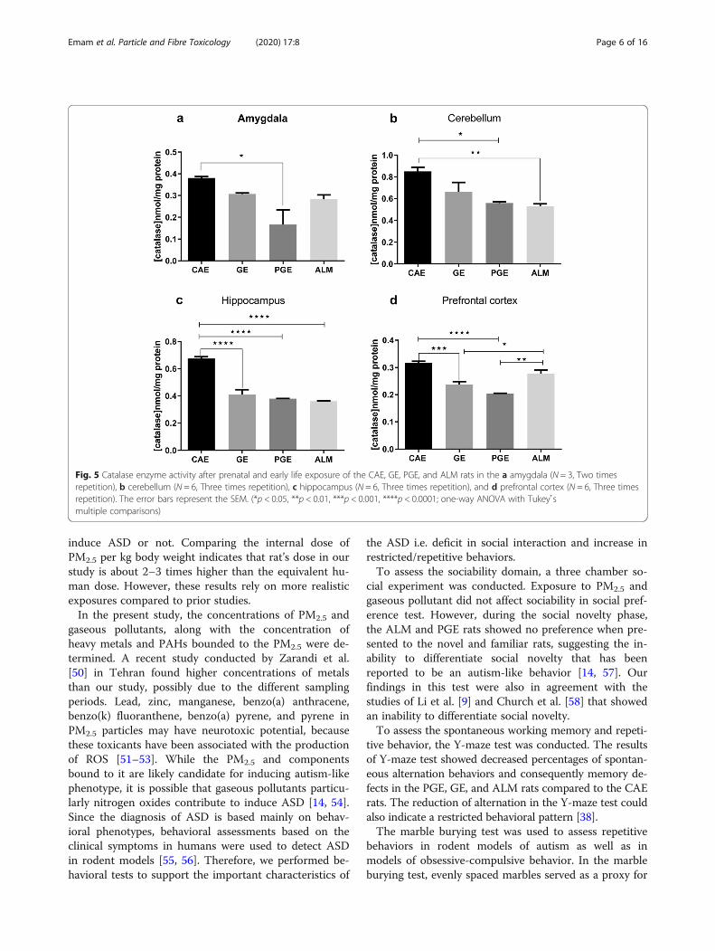
cortex, amygdala, hippocampus, and cerebellum of theexcised brain tissue. Overall, the CAT activity decreasedsignificantly in all brain regions of the PGE rats thatwere tested compared to the CAE rats as a control(Fig. 5a). Although, the CAT activity in ALM rats didnot significantly decrease vs CAE rats in amygdala andprefrontal cortex, this result may be due to an increasedlevel of ROS-mediated cell damage, specifically ·OH thatcould result in an increase of CAT activity to maintainredox homoeostasis (Fig. 5. a, d) [25]. A significant de-crease of CAT activity in the cerebellum was observed inthe ALM and PGE rats compared to the CAE rats(Fig. 5b). The PGE, GE, and ALM rats exhibited signifi-cantly decreased CAT activity in hippocampus compared
to the CAE rats (Fig. 5c). Thus, in the cerebellum, andamygdala, the activity of CAT activity was relativelysimilar to that in the PGE and GE rats vs CAE rats. ThePGE and GE rats showed significantly decreased CATactivity in the prefrontal cortex compared to the CAErats (Fig. 5d).As illustrated in Fig. 6, the PGE and ALM rats exhibited
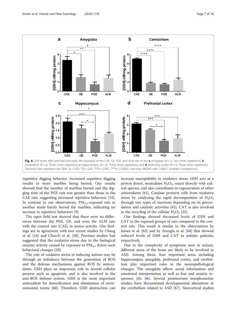
decreased levels of GSH in the prefrontal cortex, amyg-dala, hippocampus and cerebellum compared to the CAEgroup. The PGE, GE, and ALM rats showed significantlydecreased levels of GSH in the amygdala and cerebellum(Fig. 6. a, b). Unlike the GE group, the ALM and PGE ratsshowed significantly decreased levels of GSH in the hippo-campus and prefrontal cortex (Fig. 6. c, d).
Expression of oxytocin receptorWestern blot tests were conducted to determine theOXTR protein expression level. The ALM, PGE, and GErats exhibited significantly decreased OXTR levels in theamygdala, cerebellum, hippocampus, and prefrontal cor-tex compared to the CAE rats (Fig. 7 a, b, c, d). Theseresults indicate that the exposed rats showed the valuessimilar to the autism-like model rats.
DiscussionIn this study, several groups of rats were exposed to am-bient air pollution without any modifications during theperiod of E0 to PND21 covering the main neurodevelop-mental events. As a result, the PGE, GE and ALM groupof rats demonstrated decreased OXTR protein levels, re-duced catalase activity and GSH concentration com-pared to the CAE group of rats (as a control group).However, the decrements in the GE rats were smallerthan other symptomatic rats group. The PGE and GErats showed similar results for these endpoints compared
Fig. 2 Three Chambered Social Preference Test. a In the sociability phase of the three chambered social preference test, sociability was assessedby measuring the cumulative time spent by the test rat in chamber containing the stranger1 vs. empty cup. There were no differences amongthe CAE, GE, PGE, and ALM rats in exhibiting preference toward stranger1over the empty setup (*p < 0.05, **p < 0.01, ***p < 0.001 Two-wayANOVA with Tukey s̉ test for the multiple comparisons). b In the social novelty test, the cumulative times spent by a test rat in the chambercontaining stranger2 vs. stranger1 were compared. The error bars represent the standard error of the mean. (*p < 0.05, **p < 0.01 Two-wayANOVA with Tukey s̉ test for the multiple comparisons). Number of rats per group: CAE = 10, GE = 11, PGE = 9, ALM = 8
Fig. 3 Y maze; a strong decrease in the percentage of alternationwas detected in ALM, PGE, and GE rats. Error bars represent the SEM.(*p < 0.05, **p < 0.01 one-way ANOVA with Tukey s̉ multiplecomparisons. Number of rats per group: CAE = 9, GE = 10,PGE = 8, ALM = 13
Emam et al. Particle and Fibre Toxicology (2020) 17:8 Page 4 of 16

to the VPA rats. However, the GE rats showed smallerdecrements in these measures.The immature brains of fetuses or toddlers are more sus-
ceptible for environmental toxicants because the baby’sbrain weight at birth is about 24% of its adult weight andthe nerve cells are not fully developed until around the ageof 2 years old. The developing nervous system is sensitiveto environmental toxicants because temporal and regionaldevelopmental processes i.e., proliferation, migration, differ-entiation, synaptogenesis, myelination and apoptosis, growduring this period. During this vulnerable period, a widerange of chemicals may interfere with one or more ofthese processes through inhalation. The susceptibleportion of the neurodevelopmental period in a rat areE0 to PND21 [12].The immature rat’s vulnerability to environmental
toxins may be related not only to the neurodevelopmentalstages but also to the failure of other protective barrierse.g., the placental barrier and the blood brain barrier. Theplacental barrier should protect the fetus against the pas-sage of the harmful substances like environmental toxinsfrom the mother’s body. However, the placenta is not aneffective protective barrier against environmental toxins
during this time of extreme fetal vulnerability [39]. Al-though previous studies have described the associationsbetween prenatal ambient air pollution exposure and im-paired birth outcomes [40], it remains uncertain exactlywhat adverse effects are induced in the fetus. Various po-tential mechanism have been proposed including both dir-ect particle translocation and/or through indirectmechanism such as intrauterine inflammation [41–43]. Inrecent years, studies were conducted showing that onlynano-sized particles can pass the placental barrier [44, 45].However, a recent study [46] found that black carbon par-ticles were able to translocate from the mother’s lungs tothe placenta. An indirect mechanism may also be involvedsince the exposure to particulate air pollution and its con-stituents e.g. PAHs and metals, can induce oxidative stressand inflammation that leads to developmental toxicity andadverse health outcomes [47, 48] by negatively affectingplacental transport [49].The aim of this study was to use ambient air pollution
concentrations without any modification to mimic realworld exposure scenarios. Since prior studies typicallyused high exposures, it is not clear that exposure atcommonly observed air pollution concentrations can
Fig. 4 Marble burying test; a ALM and PGE rats buried more marbles vs CAE rats. However, GE rats buried less marbles than ALM rats. (*p < 0.05;*< 0.01one-way ANOVA with Tukey s̉ multiple comparisons) b GE,PGE and ALM rats spent less time in exploring in plexiglas test cage.(*p < 0.05;* <0.01one-way ANOVA with Tukey s̉ multiple comparisons) c ALM,PGE and GE rats spent more time in interacting with marbles vs CAE group, butthis increasing trend is not statistically significant.(one-way ANOVA with Tukey s̉ multiple comparisons) d GE and PGE rats spent more time inself-grooming as a repetitive behavior vs CAE group.(*p < 0.05;* < 0.01one-way ANOVA with Tukey s̉ multiple comparisons) e ALM rats spentmore time in digging behavior vs CAE group,and also there is a trend of increasing digging behavior in PGE rats vs CAE group,but this is notstatistically significant. Error bars represent the SEM. (*p < 0.05; one-way ANOVA with Tukey s̉ multiple comparisons). Number of rats per group:CAE = 8, GE = 8, PGE = 8, ALM = 10
Emam et al. Particle and Fibre Toxicology (2020) 17:8 Page 5 of 16
induce ASD or not. Comparing the internal dose ofPM2.5 per kg body weight indicates that rat’s dose in ourstudy is about 2–3 times higher than the equivalent hu-man dose. However, these results rely on more realisticexposures compared to prior studies.In the present study, the concentrations of PM2.5 and
gaseous pollutants, along with the concentration ofheavy metals and PAHs bounded to the PM2.5 were de-termined. A recent study conducted by Zarandi et al.[50] in Tehran found higher concentrations of metalsthan our study, possibly due to the different samplingperiods. Lead, zinc, manganese, benzo(a) anthracene,benzo(k) fluoranthene, benzo(a) pyrene, and pyrene inPM2.5 particles may have neurotoxic potential, becausethese toxicants have been associated with the productionof ROS [51–53]. While the PM2.5 and componentsbound to it are likely candidate for inducing autism-likephenotype, it is possible that gaseous pollutants particu-larly nitrogen oxides contribute to induce ASD [14, 54].Since the diagnosis of ASD is based mainly on behav-ioral phenotypes, behavioral assessments based on theclinical symptoms in humans were used to detect ASDin rodent models [55, 56]. Therefore, we performed be-havioral tests to support the important characteristics of
the ASD i.e. deficit in social interaction and increase inrestricted/repetitive behaviors.To assess the sociability domain, a three chamber so-
cial experiment was conducted. Exposure to PM2.5 andgaseous pollutant did not affect sociability in social pref-erence test. However, during the social novelty phase,the ALM and PGE rats showed no preference when pre-sented to the novel and familiar rats, suggesting the in-ability to differentiate social novelty that has beenreported to be an autism-like behavior [14, 57]. Ourfindings in this test were also in agreement with thestudies of Li et al. [9] and Church et al. [58] that showedan inability to differentiate social novelty.To assess the spontaneous working memory and repeti-
tive behavior, the Y-maze test was conducted. The resultsof Y-maze test showed decreased percentages of spontan-eous alternation behaviors and consequently memory de-fects in the PGE, GE, and ALM rats compared to the CAErats. The reduction of alternation in the Y-maze test couldalso indicate a restricted behavioral pattern [38].The marble burying test was used to assess repetitive
behaviors in rodent models of autism as well as inmodels of obsessive-compulsive behavior. In the marbleburying test, evenly spaced marbles served as a proxy for
Fig. 5 Catalase enzyme activity after prenatal and early life exposure of the CAE, GE, PGE, and ALM rats in the a amygdala (N = 3, Two timesrepetition), b cerebellum (N = 6, Three times repetition), c hippocampus (N = 6, Three times repetition), and d prefrontal cortex (N = 6, Three timesrepetition). The error bars represent the SEM. (*p < 0.05, **p < 0.01, ***p < 0.001, ****p < 0.0001; one-way ANOVA with Tukey s̉multiple comparisons)
Emam et al. Particle and Fibre Toxicology (2020) 17:8 Page 6 of 16
repetitive digging behavior. Increased repetitive diggingresults in more marbles being buried. Our resultsshowed that the number of marbles buried and the dig-ging time of the PGE rats was greater than those in theCAE rats, suggesting increased repetitive behaviors [33].In contrast to our observations, PM2.5-exposed rats inanother study barely buried the marbles, indicating noincrease in repetitive behaviors [9].The open field test showed that there were no differ-
ences between the PGE, GE, and even the ALM ratswith the control rats (CAE) in motor activity. Our find-ings are in agreement with two recent studies by Changet al. [14] and Church et al. [58]. Previous studies hadsuggested that the oxidative stress due to the biologicalenzyme activity caused by exposure to PM2.5 drives suchbehavioral changes [59].The role of oxidative stress in inducing autism may be
through an imbalance between the generation of ROSand the defense mechanisms against ROS by antioxi-dants. GSH plays an important role in several cellularprocess such as apoptosis, and is also involved in theanti-ROS defense system. GSH is the most importantantioxidant for detoxification and elimination of envir-onmental toxins [60]. Therefore, GSH destruction can
increase susceptibility to oxidative stress. GSH acts as aproton donor, neutralizes H2O2, reacts directly with rad-ical species, and also contributes to regeneration of otherantioxidants [61]. Catalase protects cells from oxidativestress by catalyzing the rapid decomposition of H2O2
through two types of reactions depending on its peroxi-dation and catalytic activities [62]. CAT is also involvedin the recycling of the cellular H2O2 [25].Our findings showed decreased levels of GSH and
CAT in the exposed groups of rats compared to the con-trol rats. This result is similar to the observations byJames et al. [63] and by Zoroglu et al. [64] that showedreduced levels of GSH and CAT in autistic patients,respectively.Due to the complexity of symptoms seen in autism,
different areas of the brain are likely to be involved inASD. Among them, four important areas includinghippocampus, amygdala, prefrontal cortex, and cerebel-lum play important roles in the neuropathologicalchanges. The amygdala affects social information andemotional interpretation as well as fear and anxiety re-sponses [65, 66]. Several postmortem morphometricstudies have documented developmental alterations inthe cerebellum related to ASD [67]. Neocortical studies
Fig. 6 GSH levels after prenatal and early–life exposure of the CAE, GE, PGE, and ALM rats in the a amygdala (N = 3, Two times repetition), bcerebellum (N = 6, Three times repetition), c hippocampus (N = 6, Three times repetition), and d prefrontal cortex (N = 6, Three times repetition).The error bars represent the SEM. *p < 0.05, **p < 0.01, ***p < 0.001, ****p < 0.0001; one-way ANOVA with Tukey s̉ multiple comparisons)
Emam et al. Particle and Fibre Toxicology (2020) 17:8 Page 7 of 16

on autism have found a 67% increase in the number ofneurons in prefrontal cortex [68]. Similarly, a connectionhas been reported between ASD and neuronal size ab-normalities in medial temporal lobe structures includingthe hippocampus [69, 70].One of the main objectives of this study was to assess
the effect of exposure to the mixture of PM2.5 and gases(PGE) and gases alone (GE) on the levels of the oxytocinreceptor protein (OXTR). Some associations have beenpreviously observed between OXTR haplotypes, ASD,IQ, and total VABS scores in humans [71]. In thepresent study, decreased levels of OXTR in the pre-frontal cortex, amygdala, hippocampus, and cerebellumwere observed in the exposed and ALM rats comparedto the control rats. These results are similar to those ofBertelsen et al. [72] in which the level of OXTR de-creased in the autistic rat model induced by VPA. Thepossible mechanism for down-regulation of OXTR pro-tein at nerve terminals was assumed to be the increasedoxysterols levels during neuronal injury [73]. There areno prior reports of the role of OXT and OXTR in ROSproduction in the nervous system. Thus, further studiesare required to establish this relationship. Denda et al.[74] found that OXT is expressed in human skin kerati-nocytes and released in response to a calcium influx viaP2X receptors. Also, there are evidence that OXT is notonly expressed in keratinocytes, but also in human skin-derived dermal fibroblasts [75], which are in common
with neurons in ectodermal derivation. In addition, therole of OXTR expression in fibroblast production hasbeen reported [76]. Expression of OXT occurs in all epi-dermal layers, while expression of the OXTR only occursin the basal layers [75].There is evidence that with inhibition of OXTR signal-
ing in integumentary system, OXT exerts its effectsthrough the alteration of oxidative stress, intracellularGSH levels, and cytokine release by dermal fibroblasts andkeratinocytes. Increments of susceptibility to oxidativestress occur following the reduction of OXTR in dermalcells [75]. Simultaneous to increment of ROS levels inOXTR reduction in dermal fibroblasts and keratinocytes,a decrement of intracellular GSH concentrations has beenreported [75]. Some other studies have also shown therole of OXT and OXTR in oxidative stress and GSHlevels. For instance, sepsis-induced pelvic inflammationcaused by the increase of ROS and reduction of GSHlevels, could be treated with OXT administration [77] orwith atosiban, an OXTR antagonist that increased oxida-tive stress in the cardiomyocytes of the newborn ratswhose mothers received atosiban during gestation [78].Previous studies revealed that OXTR is associated with so-cial cognition [79–81] in autism [82]. The loss of oxytocinor OXTR may result in reduced social recognition [83]and social interaction [84].In recent years, several studies have shown the associ-
ation between exposure to particulate matter and increased
Fig. 7 OXTR levels after prenatal and early–life exposure of the CAE, GE, PGE, and ALM rats in the a amygdala (N = 4, Three times repetition), bcerebellum (N = 4, Three times repetition), c hippocampus (N = 4, Three times repetition), and d prefrontal cortex (N = 4, Three times repetition).Error bars represent the SEM. *p < 0.05, **p < 0.01, ***p < 0.001, ****p < 0.0001; one-way ANOVA with Tukey s̉ multiple comparisons)
Emam et al. Particle and Fibre Toxicology (2020) 17:8 Page 8 of 16

risk of ASD [85, 86]. In addition, the relationship betweenambient particulate matter and autism-like phenotype hasbeen recently assessed in rodent models. In a study by Liet al. [9], Sprague-Dawley rats were exposed to PM2.5 by in-tranasal instillation. However, Li et al. [9] suggested that thewhole-body inhalation exposure (such as the current study)is more physiologically relevant to human context ratherthan intranasal instillation. In addition, the dose to the ratpups in the Li et al. (2018) study was determined using theadult rat’s respiratory rate. Thus, this dose was higher thantypical exposures in humans. In the current study, rats wereexposed to the concentrations of PM2.5 and gaseous pollut-ants at ambient levels. Therefore, this model of exposuremay better reflect the actual human exposure to PM2.5 andgaseous pollutants.
ConclusionsIn the present study, our results showed that prenataland early life exposure to a mixture of PM2.5 and gas-eous pollutants (PGE rats) and gaseous pollutants alone(GE rats) caused behavioral deficits, including increasedrepetitive behavior, poor social interaction and an inabil-ity to differentiate social novelty. Air pollution also af-fected oxidative stress biomarkers like CAT and GSH,and may induce down-regulation of OXTR protein inmale rats. To our knowledge, this is the first report thatcompares the effects of exposure to PM2.5 and gaseouspollutants (PGE rats) and gaseous pollutants alone (GE
rats) with VPA-induced rat models of ASD (ALM rats).These experimental findings support the hypothesis thatan etiological association exists between a complex mix-ture of air pollution and physiopathology of ASD. Fur-ther mechanistic research should be performed todetermine the mode of neurodevelopmental toxicity ofair pollution on ASD.
MethodsRatsWistar rat litters of both sexes were purchased from Pas-teur Institute (Tehran, Iran). The rats were housed atthe Shahid Beheshti University of Medical Sciencesunder specific pathogen free (SPF) and standard condi-tions, including access to supplies of water and food adlibitum under a 12 h light/dark cycle. The rats weremaintained under constant environmental conditions in-cluding temperature of 20–25 °C and relative humidityof 40–60%. The ethical use of animal models was ap-proved by Shahid Beheshti University of Medical Sci-ences’ Ethics Committee.
Study location and method of exposureThe pilot animal (Fig. 8) room was located on theroof of the School of Public Health and Safety of theShahid Beheshti University of Medical Sciences(35.7991 N, 51.3947 E) at a height of 20 m (fourthfloor) above the ground.
Fig. 8 Schematic of exposure method: 1- Oil-free compressor, 2- Thermometer, 3- Air purifier, 4- Moisture Sensor, 5- Dust Track, 6- Time lamp, 7-HEPA Filter, and 8- Echo PM
Emam et al. Particle and Fibre Toxicology (2020) 17:8 Page 9 of 16

Four groups of rats including PGE (particulate matterand gaseous pollutants), GE (gaseous pollutants), CAE(clean air) and ALM (autism like model exposed to cleanair) rats were exposed in the animal pilot study room.The GE rats in chamber 1 were exposed to filtered am-bient air at a flow rate of 20 L/min provided by an oil-free compressor and a HEPA filter (model H13) to re-move particulate matter. PGE rats in chamber 2 wereexposed to ambient air at a flow rate of 20 L/min usingan Echo PM2.5 Low Volume Sampler (LVS) (TCRTecora Italy) without a filter to provide exposure to bothPM2.5 and gaseous pollutants. The CAE and ALM ratswere housed in chamber 3, and exposed to cleaned am-bient air at 20 L/min flow rate provided by an oil-freecompressor and two purifier systems (Model: Air TouchA5, Honeywell, and model: KAIST-AIR Home) to re-move both particulate and gaseous pollutants. PregnantWistar rats in the ALM group received Valproic Acid(VPA: 350 mg/kg) at gestational day 12.5 to induce ASD.
Drug administrationBefore the start of the experiment, the rats at the age of10 weeks were acclimated to the pilot animal room for 1week. The rats were then mated in-house at the age of11 to 13 weeks, and pregnancy was determined by a va-ginal plug on embryonic day 1 (E1).To produce the ALM rats [87], the sodium salt of val-
proic acid (P4543 - Sigma) was prepared in 0.9% salinesolution (100mg/ml, PH 7.3). On E12.5, when the preg-nant rat’s weight was 200–225 g, VPA-dams received asingle intraperitoneal (i.p.) injection of VPA at a dose of350 mg/kg body weight [88]. Control dams received asingle injection of saline solution (i.p., 0.9%) (Fig. 9).
Exposure periodsPregnant dams and pups were exposed to ambient airthat contained PM2.5, CO, NO2, SO2, and O3 for 12 hper day and 5 days per week (Saturday to Wednesday)from embryonic day 0 (E0) to postnatal day 21 (PND21).The concentrations of PM2.5 and gaseous pollutants(CO, NO2, SO2 and O3) in each chamber were measuredusing a DustTrak model 8520 and an AeroQual 500,respectively.The rats in control groups were exposed to clean air
during the same period. The exposure period waschosen based on the human epidemiological studies sug-gesting that air pollution exposure during all the threetrimesters of pregnancy and the first 9 months of an in-fant’s life is related to ASD risk [30, 89, 90]. The expos-ure time was designed to cover the mainneurodevelopmental events (including: the timing ofneurogenesis, synaptogenesis, gliogenesis, oligodendro-cyte maturation) that were happening in this window ofsusceptibility, the period of E0 to PND21 [91]. For theexposure of pregnant dams and pups, female rats (11–13 week old) were time mated with males (3 femaleswith 1 male per cage) on Friday evenings. The copula-tory plug was checked the next morning before the Sat-urday onset of the weekly exposure. Rats with confirmedvaginal plugs were removed from breeding cage on em-bryonic day zero and housed individually in cages in ei-ther chambers one, two or three. Chambers 1 and 2housed nine pregnant dams; while chamber 3 housedeighteen dams for duration of their pregnancy, partur-ition, and weaning of the litters. The use of Friday timedmattings ensured that E0 occurred on a Saturday andensured that the exposure timing was the same for all
Fig. 9 Experimental design of study: rats were time mated and exposed to PM2.5/gases and gases alone from E0 to PND21. Biochemical testswere started at PND22 by extraction of brain tissue. Behavioral assessments were started at PND 29. * VPA-dams received a single intraperitoneal(i.p.) injection of VPA at a dose of 350 mg/kg body weight
Emam et al. Particle and Fibre Toxicology (2020) 17:8 Page 10 of 16

rats. All dams were exposed on the same gestationaldays E0–4, E7–11, E14–18 and all pups were exposedon PND1-PND7, PND10–14, PND17-PND21. Exposurestopped in chambers one and two while rats were givingbirth on E19 and E20. On PND14, male and female pupswere separated and female pups were removed fromtheir mothers. The male pups remained with theirmothers until weaning occurred on PND 21. On PND22, the pups were divided into two groups. One groupwere euthanized by decapitation for the biochemicaltests and the second group were transferred to housingracks (two rats per cage) 1 week before the starting thebehavioral tests (on PND29) and for the duration of thebehavioral testing period. Behavioral tests only were per-formed on male rat offspring and all behavioral testswere performed between 9:00 and 15:00. The behavioralassessments were made sequentially on a single cohortof male rats. This lack of replication is a limitation ofthis study (Fig. 9).
Determination of PM2.5 characteristicsPM2.5 was sampled continuously during the exposurehours using an Echo PM Low Volume Sampler near theair intake for the exposure chambers [92]. PM2.5 was col-lected on 47mm quartz filters at a flow rate of 20 L/min.Before sampling, the filters were washed with double-distilled water, and placed in an oven at 100–105 °C for 2h [93]. After sampling, the filters were stored in aluminumfoil at − 10 °C to prevent evaporation and photo-degradation of PM components. For each 2 days, a newfilter was used for sampling. Also, field blanks were usedto control for the possible contamination during the sam-pling procedures. For this, a blank filter was treated withthe same manner as a filter used for sampling (placed intothe sampler, placed into the aluminum foil, etc.), exceptthat the sampler was not operated.To determine the elemental composition of PM, one-
half of filters were shredded, and put into a Teflon con-tainer with 2.5 ml of concentrated of HClO4 (70%) and2.5 ml HNO3 (69%). Samples were heated at 170 °C for4 h, and dried on a hot plate at 100 C. After adding 2.5ml of double-distilled water and 2.5 ml of HNO3,thesamples were shaken at 180 rpm for 30 min [93]. Finally,the samples were filtered through Whatman No. 42 fil-ters and diluted with double-distilled water to 10 ml.The samples were stored in plastic vials at − 4 °C untilthe analysis by ICP/MS (Agilent, Model: 7900) [94]. La-boratory blanks were used to control for the effects oflaboratory contaminations. The metal concentrations areprovided in the Additional file 1: Table S1. The concen-trations of metals were analyzed in triplicate, and the av-eraged values were used. Field and laboratory blankswere used to control for the effects of field and labora-tory contaminations. The limit of detection (LOD) and
the limit of quantitation (LOQ), of Pb was 5 and 1 μg/L,respectively. The LOD and LOQ for other elements were25 and 5 μg/L, respectively.To determine the concentrations of polycyclic aro-
matic hydrocarbons (PAHs), the other section of the fil-ter was placed in a Teflon container, and extracted with2.5 ml of dichloromethane (CH2CL2) and 2.5 ml ofmethanol (CH3OH) using an ultrasonic bath at 20 kHzfor 30 min (Elmasonic S 80 H). The extracts were fil-tered with 0.22 μm Millipore filters (Hesperia CA, USA).PAHs concentrations were determined using GC/MS(Agilent, model: 5890 A). The PAH results are providedin Additional file 1: Table S2. The concentrations ofPAHs were analyzed in triplicate, and the mean concen-trations were reported. In the analysis of all of the blanksamples, PAHs were not observed. The values of LODand LOQ for 16 PAHs were < 2 and < 10 ng/L,respectively.
Behavioral testsOpen fieldThe open field test was conducted as previously describedby Chang et al. [14] with some modifications. At PND29for the male rat offspring, each rat was placed in a cleanacrylic cage (60*60*60) with no bedding for 30min. To as-sess general locomotor activity levels and willingness toexplore in a novel environment, the rat’s location duringthe test was tracked using Ehtovision XT 7 system fromthe video recorded using a Microsoft LifeCam HD-6000.
Social preference testThis test was conducted as previously described by Liet al. [9] with modifications. The three sessions in thistest was conducted in a three chambered box (60*40*22cm) equipped with retractable doorways that permittedaccess to each chamber.This test was conducted at PND32 for each male rat off-
spring. In session 1 (habituation), a pup was placed in themiddle chamber with the doorways open, and allowed toexplore the other two side chambers. For session 2 (soci-ability), at the end of the period of habituation, pups weremade to interact with a never-before-met and age-matchedrat enclosed in a wire cup placed in a side chamber. Anempty wire cup was placed in the other side chamber. Dur-ing session 3 (social novelty), a new and unfamiliar rat wasplaced in the wire cup that had been empty during the pre-vious session. Each session lasted 10min, and the timespent in each chamber was manually recorded.
Y-mazeThe test was conducted as previously described by Grab-rucker et al. [38]. Spontaneous alternation behavior wasassessed at PND43 for each male rat offspring in a sym-metrical Y Maze (3 arms, 40*9 cm with 16 cm wall
Emam et al. Particle and Fibre Toxicology (2020) 17:8 Page 11 of 16
height). Arm choices by the rats (all four paws enteringone arm) were recorded while the rats were allowed toexplore the Y-shaped labyrinth for 5 min. Alternationwas determined by recording the order of the visitedarms (A, B, or C). Overlapping triplets of three armvisits were counted as one complete spontaneous alter-nation. The percentage of alternation was calculatedusing the equation:
Percentage of alteration ¼ number of spontaneous alternationtotal number of arm visits−2
� 100
ð1Þ
where an alternation was recorded as consecutively visit-ing the three arms.
Marble buryingThis test was conducted as previously described by Kuet al. [95] with some modifications. On PND46 for eachmale rat offspring, the subject was placed in a standardPlexiglas test cage (42*24*17 cm) with a 5 cm deep layerof corncob bedding, and allowed to explore freely for 10min. Each subject was placed in a transfer cage and 18marbles (1.3 cm diameter, red) were placed on the bed-ding surface in a 3*6 pattern. The subject was thenplaced in the test cage, and allowed to explore for 10min. After ten minutes, the subject was removed fromthe test cage, and the number of buried marbles wascounted. A marble was considered as buried only if atleast two-third of the marble was covered by the beddingmaterial.
Tissue collectionStudies have increasingly recognized that developingbrains are more sensitive to both neuronal apoptosis thatare likely to be affected by age-related injury vulnerability[96–101]. Since approximately 90% of the rat cortex isformed by PND 20 and the weight of rat cortex reachesapproximately to 90% of its weight by PND 20, the typicalage of weaning. Also, myelination peaks at approximatelyPND 20, when maturation markers especially myelin basicprotein are detectable [102]. Thus, the biochemical assess-ments should be performed after the rats attain PND20 toensure valid test results. Since exposures continued untilPND 21, the following day (PND22) was selected forconducting the biochemical tests.On PND22, sixteen male offspring in each group were
anesthetized via CO2 and rapidly decapitated. The pre-frontal cortex, amygdala, cerebellum, and hippocampuswere rapidly dissected out, and frozen in liquid nitrogenat − 80 °C until use.
GSHThe test was conducted as described by Ellman et al.[103]. Sixty μg of protein supernatant containing 19.8mgof DTNB (D8130-1G SIGMA) in 100ml, 0.1% sodium ni-trate and phosphate buffer (PH 7.4) were used for theGSH assay. The absorbance was measured at 412 nm byan ELISA reader. Ellman’s colorimetric method measuresthe formation of GS-TNB complex from DTNB (5,5̉dithiobis (2- nitro benzoic acid)) in which its reductioncaused the development of a yellow color.
CatalaseTo determine the level of catalase activity, the concentra-tions of the yellow stable complex of ammonium molyb-date and hydrogen peroxide were measured at 405 nmwith an ELISA reader [104]. First, 20 μL of the sample wasloaded in each well of the 96-well microplate. Then,100 μL hydrogen peroxide (65mM) was added to eachwell. This mixture was incubated for 4min at 25 °C. Then,100 μl of ammonium molybdate (32.4 mM) was added toeach well, and the absorbance was read at 405 nm.
Western blotWestern blotting was used to assess the level of OXTR inthe prefrontal cortex, amygdala, hippocampus, and cerebel-lum of the male rats. On PND 22, the prefrontal cortex,amygdala, hippocampus, and cerebellum were excised.Next, they were lysed on ice via lysis buffer [50mM Tris-HCl (PH 8), 0.25% sodium deoxycholate, 0.1% sodiumdodecyl sulfate (SDS), 150mM NaCl, 1mM EDTA,0.1%Triton X-100, complete protease inhibitor cocktail,phosphatase inhibitors cocktail] for 2min. Then, the lysateswere cleared with centrifugation at 16,100 Xg for 10min at4 °C. In order to determine the protein concentration, Brad-ford’s method [105] using bovine serum albumin (BSA) asstandard was performed to denature proteins, equal vol-umes of 5X sample buffer were added to lysate proteins.Then 60 μg of total proteins were loaded into SDS-polyacrylamide gel electrophoresis and then transferredonto polyvinylidene difluoride membranes. Then blots wereblocked by blocking solution [2% non-fat dry milk in tris-buffered saline Tween 20 (TBST) (containing 0.2% Tween20, 50mM Tris–HCl pH 7.4, 150mM NaCl] for one hourand half. At this point, the membranes were incubated withthe primary antibody (Anti-Oxytocin Receptor antibody[EPR12789]-20 C) (Abcam ab181077-100 μl) at 4 °C over-night. Next day, the blots were washed with TBST threetimes for 10min each. After incubating with the anti-rabbithorseradish peroxidase secondary antibody (Anti-rabbitIgG, HRP-linked Antibody) (cellsignaling) for 90min at theroom temperature, immunoreactivity was detected withECL kit and captured by Kodak x-ray films. To compensatefor loading errors, membranes were stripped with strippingbuffer [100mM 2-mercaptoethanol, 2% (w/v) SDS, 62.5
Emam et al. Particle and Fibre Toxicology (2020) 17:8 Page 12 of 16
mM Tris-HCl (PH 7)] followed by incubating with anti β-actin antibody. Densitometric data of protein bands wereobtained with ImageJ 1.41o.
Statistical analysisAll data were analyzed by using Graph Pad software(Graph Pad Prism v6.0). For the three-chambered socialpreference test, time spent in the ‘stranger1’ versus‘empty’ chambers (for the sociability phase) or ‘stranger1’versus ‘stranger2’ chambers (for the social novelty phase)were compared with a two-way analysis of variance(ANOVA) followed by Tukey ̉s test for the multiplecomparisons. A one-way ANOVA followed by Tukey ̉stest was used for the analysis of the other test results.Multiple comparison corrections were not done. Thesignificance levels of 0.05, 0.01, 0.001and 0.0001 wereapplied in all analyses.
Supplementary informationSupplementary information accompanies this paper at https://doi.org/10.1186/s12989-020-0336-y.
Additional file 1: Table S1. Concentration of Metals bound PM2.5 inexposure period. Table S2. Concentration of 16-PAHs bound PM2.5 in ex-posure period.
AcknowledgementsThis work is a part of the Mastership student’s thesis of Baharan Emam atSchool of Public Health and Safety, Shahid Beheshti University of MedicalSciences, Tehran, Iran. The authors wish to thank Shahid Beheshti Universityof Medical Sciences (grant number # 15515)
Authors’ contributionsBE participated in study design, in vivo section, experiments, data analysis,and manuscript writing. AS, FK, and SMZ participated in study design,experiments, data analysis, and manuscript writing. PKH, MH, HMB and MYparticipated in study design, parts of experiments, data analysis, andmanuscript writing. All authors read and approved the final manuscript.
FundingThis study was funded by Shahid Beheshti University of Medical Sciences,Tehran, Iran (Grant number: 15515).
Availability of data and materialsThe datasets used and/or analyzed during the current study are availablefrom the corresponding author on reasonable request.
Ethics approval and consent to participateAll ethical aspects of this study were approved by Shahid Beheshti Universityof Medical Sciences’ Ethics Committee.
Consent for publicationNot applicable.
Competing interestsThe authors declare that they have no competing interests.
Author details1Department of Environmental Health Engineering, School of Public Healthand Safety, Shahid Beheshti University of Medical Sciences, Tehran, Iran.2Environmental and Occupational Hazards Control Research Center, ShahidBeheshti University of Medical Sciences, Tehran, Iran. 3Neuroscience ResearchCenter, Shahid Beheshti University of Medical Sciences, Tehran, Iran.4Department of Public Health Sciences, University of Rochester School of
Medicine and Dentistry, Rochester, NY 14642, USA. 5Center for Air ResourcesEngineering and Science, Clarkson University, Potsdam, NY 13699, USA.6Department of Environmental Health Engineering, School of Public Health,Tehran University of Medical Sciences, Tehran, Iran. 7Students’ ScientificResearch Center (SSRC), Tehran University of Medical Sciences, Tehran, Iran.8Center of Environmental and Occupational health, Ministry of Health andMedical Education, Tehran, Iran.
Received: 29 September 2019 Accepted: 6 January 2020
References1. American Psychiatric Association. Diagnostic and Statistical Manual of
Mental Disorder. 5th ed; 2013. p. 24–9.2. Baxter AJ, Brugha TS, Erskine HE, Scheurer RW, Vos T, Scott JG. The
epidemiology and global burden of autism spectrum disorders. PsycholMed. 2015;45(3):601–13. https://doi.org/10.1017/s003329171400172x.
3. French LRBA, Hyde KL, Fombonne E. Epidemiology of autism Spectrumdisorders. Neurosci Autism Spectr Disord. 2013;43:3–24.
4. Blumberg SJ, Bramlett MD, Kogan MD, Schieve LA, Jones JR, Lu MC.Changes in prevalence of parent-reported autism spectrum disorder inschool-aged U.S. children: 2007 to 2011–2012. Natl Health Stat Report. 2013;65:1–11 1 p following.
5. Wallace S, Fein D, Rosanoff M, Dawson G, Hossain S, Brennan L, et al. Aglobal public health strategy for autism spectrum disorders. Autism Res.2012;5(3):211–7. https://doi.org/10.1002/aur.1236.
6. Grønborg TK, Schendel DE, Parner ET. Recurrence of autism Spectrumdisorders in full- and half-siblings and trends over time: a population-basedcohort study. JAMA Pediatr. 2013;167(10):947–53. https://doi.org/10.1001/jamapediatrics.2013.2259 http://www.ncbi.nlm.nih.gov/pmc/articles/PMC4610344/.
7. Kim YS, Leventhal BL. Genetic epidemiology and insights into interactivegenetic and environmental effects in autism spectrum disorders. BiolPsychiatry. 2015;77(1):66–74. https://doi.org/10.1016/j.biopsych.2014.11.001.
8. Rossignol DA, Genuis SJ, Frye RE. Environmental toxicants and autismspectrum disorders: a systematic review. Transl Psychiatry. 2014;4:e360.https://doi.org/10.1038/tp.2014.4.
9. Li K, Li L, Cui B, Gai Z, Li Q, Wang S, et al. Early postnatal exposure toairborne fine particulate matter induces autism-like phenotypes in male rats.Toxicol Sci. 2018;162(1):189–99. https://doi.org/10.1093/toxsci/kfx240.
10. Costa LG, Chang YC, Cole TB. Developmental neurotoxicity of traffic-relatedair pollution: focus on autism. Curr Environ Health Rep. 2017;4(2):156–65.https://doi.org/10.1007/s40572-017-0135-2.
11. Becerra TA, Wilhelm M, Olsen J, Cockburn M, Ritz B. Ambient air pollutionand autism in Los Angeles county, California. Environ Health Perspect. 2013;121(3):380–6. https://doi.org/10.1289/ehp.1205827.
12. Rice D, Barone S Jr. Critical periods of vulnerability for the developingnervous system: evidence from humans and animal models. Environ HealthPerspect. 2000;108(Suppl 3):511–33. https://doi.org/10.1289/ehp.00108s3511.
13. Rodier PM, Ingram JL, Tisdale B, Nelson S, Romano J. Embryological originfor autism: developmental anomalies of the cranial nerve motor nuclei. JComp Neurol. 1996;370(2):247–61. https://doi.org/10.1002/(sici)1096-9861(19960624)370:2<247::aid-cne8>3.0.co;2-2.
14. Chang Y-C, Cole TB, Costa LG. Prenatal and early-life diesel exhaustexposure causes autism-like behavioral changes in mice. Part Fibre Toxicol.2018;15(1):18. https://doi.org/10.1186/s12989-018-0254-4 https://doi.org/10.1186/s12989-018-0254-4.
15. Thirtamara Rajamani K, Doherty-Lyons S, Bolden C, Willis D, Hoffman C,Zelikoff J, et al. Prenatal and early-life exposure to high-level diesel exhaustparticles leads to increased locomotor activity and repetitive behaviors inmice. Autism Res. 2013;6(4):248–57. https://doi.org/10.1002/aur.1287.
16. Ortega R, Bresson C, Darolles C, Gautier C, Roudeau S, Perrin L, et al. Low-solubility particles and a Trojan-horse type mechanism of toxicity: the caseof cobalt oxide on human lung cells. Part Fibre Toxicol. 2014;11:14. https://doi.org/10.1186/1743-8977-11-14 https://www.ncbi.nlm.nih.gov/pubmed/24669904.
17. Peters A. Ea. translocation and potential neurological effects of fine andultrafine particles a criticalupdate. Part Fibre Toxicol. 2006;3:13.
18. Muhlfeld C, et al. Interactions of nanoparticles with pulmonarystructures and cellular responses. AmJ Physiol Lung Cell Mol Physiol.2008;294:L817–29.
Emam et al. Particle and Fibre Toxicology (2020) 17:8 Page 13 of 16

19. Simkhovich BZ. Ea. air pollution and cardiovascular injury epidemiology,toxicology, and mechanisms. J Am Coll Cardiol. 2008;52:719–26.
20. Ma JYMJ. The dual effect of the particulate and organic components ofdiesel exhaust particles on the alteration of pulmonary immune/inflammatory responses and metabolic enzymes. Environ Sci Health Part CEnviron Carcinog Ecotoxicol Rev. 2002;20:117–47.
21. Colin-Barenque LFT. Oxidative stress and metals. In: Fortoul T, editor. Metals andtoxicological implications in Health. Kerala: Research Signpost; 2007. p. 15–25.
22. KA J. The relevance of metals in the pathophysiology ofneurodegeneration,pathological considerations. Int Rev Neurobiol. 2013;110:1–47.
23. MohanKumar SMCA, Block M, Veronesi B. Particulate matter, oxidative stressand neurotoxicity. Neurotoxicology. 2008;29(3):479–88.
24. Grice DE, Buxbaum JD. The genetics of autism spectrum disorders.NeuroMolecular Med. 2006;8(4):451–60. https://doi.org/10.1385/NMM:8:4:451https://doi.org/10.1385/NMM:8:4:451.
25. María Elena González-Fraguela M-LDH, Vera H, Maragoto C, Noris E, BlancoL, Galvizu R. Maria Robinson oxidative stress markers in children with autismSpectrum disorders. Br J Med Med Res. 2013;3(2):307–17.
26. Arons MH, Thynne CJ, Grabrucker AM, Li D, Schoen M, Cheyne JE, et al.Autism-associated mutations in ProSAP2/Shank3 impair synaptictransmission and neurexin-neuroligin-mediated transsynaptic signaling. JNeurosci. 2012;32(43):14966–78. https://doi.org/10.1523/jneurosci.2215-12.2012.
27. Johnson ZV, Young LJ. Oxytocin and vasopressin neural networks:Implications for social behavioral diversity and translational neuroscience.Neurosci Biobehav Rev. 2017;76:87–98. https://doi.org/10.1016/j.neubiorev.2017.01.034.
28. Modi ME, Young LJ. The oxytocin system in drug discovery for autism:animal models and novel therapeutic strategies. Horm Behav. 2012;61(3):340–50. https://doi.org/10.1016/j.yhbeh.2011.12.010.
29. Freeman SM, Palumbo MC, Lawrence RH, Smith AL, Goodman MM, Bales KL.Effect of age and autism spectrum disorder on oxytocin receptor density inthe human basal forebrain and midbrain. Translational Psychiat. 2018;81:257.https://doi.org/10.1038/s41398-018-0315-3.
30. Volk HE, Lurmann F, Penfold B, Hertz-Picciotto I, McConnell R. Traffic relatedair pollution, particulate matter, and autism. JAMA psychiatry. 2013;70(1):71–7. https://doi.org/10.1001/jamapsychiatry.2013.266 http://www.ncbi.nlm.nih.gov/pmc/articles/PMC4019010/.
31. Yamaguchi H, Hara Y, Ago Y, Takano E, Hasebe S, Nakazawa T, et al.Environmental enrichment attenuates behavioral abnormalities in valproicacid-exposed autism model mice. Behav Brain Res. 2017;333:67–73. https://doi.org/10.1016/j.bbr.2017.06.035.
32. Nicolini C, Fahnestock M. The valproic acid-induced rodent model ofautism. Exp Neurol. 2018;299:217–27. https://doi.org/10.1016/j.expneurol.2017.04.017.
33. Campolongo M, Kazlauskas N, Falasco G, Urrutia L, Salgueiro N, Hocht C,et al. Sociability deficits after prenatal exposure to valproic acid are rescuedby early social enrichment. Mol Autism. 2018;9:36. https://doi.org/10.1186/s13229-018-0221-9.
34. Win-Shwe TT, Nway NC, Imai M, Lwin TT, Mar O, Watanabe H. Socialbehavior, neuroimmune markers and glutamic acid decarboxylase levels ina rat model of valproic acid-induced autism. J Toxicol Sci. 2018;43(11):631–43. https://doi.org/10.2131/jts.43.631.
35. Peralta F, Fuentealba C, Fiedler J, Aliaga E. Prenatal valproate treatmentproduces autistic-like behavior and increases metabotropic glutamatereceptor 1A-immunoreactivity in the hippocampus of juvenile rats. Mol MedRep. 2016;14:2807–14. https://doi.org/10.3892/mmr.2016.5529.
36. Kaidanovich-Beilin O, Lipina T, Vukobradovic I, Roder J, Woodgett JR.Assessment of social interaction behaviors. J Vis Exp. 2011;48. https://doi.org/10.3791/2473.
37. Roullet FI, Crawley JN. Mouse models of autism: testing hypotheses aboutmolecular mechanisms. Curr Top Behav Neurosci. 2011;7:187–212. https://doi.org/10.1007/7854_2010_113 http://www.ncbi.nlm.nih.gov/pmc/articles/PMC3396120/.
38. Grabrucker S, Boeckers TM, Grabrucker AM. Gender dependent evaluation ofautism like behavior in mice exposed to prenatal zinc deficiency. FrontBehav Neurosci. 2016;10:37. https://doi.org/10.3389/fnbeh.2016.00037 http://www.ncbi.nlm.nih.gov/pmc/articles/PMC4776245.
39. Perlroth NH, Castelo Branco CW. Current knowledge of environmentalexposure in children during the sensitive developmental periods. J Pediatr
(Rio J). 2017;93(1):17–27. https://doi.org/10.1016/j.jped.2016.07.002 http://www.sciencedirect.com/science/article/pii/S0021755716302352.
40. Lamichhane DK, Leem JH, Lee JY, Kim HC. A meta-analysis of exposure toparticulate matter and adverse birth outcomes. Environ Health Toxicol.2015;30:e2015011. https://doi.org/10.5620/eht.e2015011.
41. Stapleton PA. Gestational nanomaterial exposures: microvascularimplications during pregnancy, fetal development and adulthood. J Physiol.2016;594(8):2161–73. https://doi.org/10.1113/JP270581 https://www.ncbi.nlm.nih.gov/pubmed/26332609.
42. Nachman RM, Mao G, Zhang X, Hong X, Chen Z, Soria CS, et al. IntrauterineInflammation and Maternal Exposure to Ambient PM2.5 duringPreconception and Specific Periods of Pregnancy: The Boston Birth Cohort.Environ Health Perspect. 2016;124(10):1608–15. https://doi.org/10.1289/ehp243.
43. Valentino SA, Tarrade A, Aioun J, Mourier E, Richard C, Dahirel M, et al.Maternal exposure to diluted diesel engine exhaust alters placental functionand induces intergenerational effects in rabbits. Part Fibre Toxicol. 2016;13(1):39. https://doi.org/10.1186/s12989-016-0151-7.
44. Hougaard KS, Campagnolo L, Chavatte-Palmer P, Tarrade A, Rousseau-Ralliard D, Valentino S, et al. A perspective on the developmental toxicity ofinhaled nanoparticles. Reprod Toxicol. 2015;56:118–40. https://doi.org/10.1016/j.reprotox.2015.05.015.
45. Muoth C, Aengenheister L, Kucki M, Wick P, Buerki-Thurnherr T. Nanoparticletransport across the placental barrier: pushing the field forward!Nanomedicine (London, England). 2016;11(8):941–57. https://doi.org/10.2217/nnm-2015-0012.
46. Bové H, Bongaerts E, Slenders E, Bijnens EM, Saenen ND, Gyselaers W, et al.Ambient black carbon particles reach the fetal side of human placenta. NatCommun. 2019;10(1):3866. https://doi.org/10.1038/s41467-019-11654-3https://doi.org/10.1038/s41467-019-11654-3.
47. Vadillo-Ortega F, Osornio-Vargas A, Buxton MA, Sanchez BN, Rojas-Bracho L,Viveros-Alcaraz M, et al. Air pollution, inflammation and preterm birth: apotential mechanistic link. Med Hypotheses. 2014;82(2):219–24. https://doi.org/10.1016/j.mehy.2013.11.042.
48. Al-Gubory KH. Environmental pollutants and lifestyle factors induceoxidative stress and poor prenatal development. Reprod BioMed Online.2014;29(1):17–31. https://doi.org/10.1016/j.rbmo.2014.03.002.
49. Lager S, Powell TL. Regulation of nutrient transport across the placenta. JPregnancy. 2012;2012:179827. https://doi.org/10.1155/2012/179827.
50. Motesaddi Zarandi S, Shahsavani A, Khodagholi F, Fakhri Y. Co-exposure toambient PM2.5 plus gaseous pollutants increases amyloid β1–42accumulation in the hippocampus of male and female rats. Toxin Rev. 2019:1–10. https://doi.org/10.1080/15569543.2019.1611604 https://doi.org/10.1080/15569543.2019.1611604.
51. Jedrychowski WA, Perera FP, Camann D, Spengler J, Butscher M, Mroz E,et al. Prenatal exposure to polycyclic aromatic hydrocarbons and cognitivedysfunction in children. Environ Sci Pollut Res Int. 2015;22(5):3631–9. https://doi.org/10.1007/s11356-014-3627-8 https://www.ncbi.nlm.nih.gov/pubmed/25253062.
52. Zhang Y, Ji X, Ku T, Li G, Sang N. Heavy metals bound to fine particulatematter from northern China induce season-dependent health risks: A studybased on myocardial toxicity. Environ Pollut. 2016;216:380–90. https://doi.org/10.1016/j.envpol.2016.05.072 http://www.sciencedirect.com/science/article/pii/S0269749116304602.
53. Fortoul T, Rodriguez-Lara V, Gonzalez-Villalva A, Rojas-Lemus M, Colin-Barenque L, Bizarro-Nevares P, García-Peláez I, Ustarroz-Cano M, López-Zepeda S, Cervantes-Yépez S, López-Valdez N, Meléndez-García N, Espinosa-Zurutuza M, Cano-Gutierrez G, Cano-Rodríguez MC. Health Effects of Metalsin Particulate Matter. In: Current Air Quality Issues, vol. 571; 2015. p. 608.https://doi.org/10.5772/59749.
54. Flores-Pajot M-C, Ofner M, Do MT, Lavigne E, Villeneuve PJ. Childhoodautism spectrum disorders and exposure to nitrogen dioxide, andparticulate matter air pollution: A review and meta-analysis. Environ Res.2016;151:763–76. https://doi.org/10.1016/j.envres.2016.07.030 http://www.sciencedirect.com/science/article/pii/S0013935116303176.
55. Pasciuto E, Borrie SC, Kanellopoulos AK, Santos AR, Cappuyns E, D'Andrea L,et al. Autism Spectrum disorders: translating human deficits into mousebehavior. Neurobiol Learn Mem. 2015;124:71–87. https://doi.org/10.1016/j.nlm.2015.07.013.
56. Servadio M, Vanderschuren LJ, Trezza V. Modeling autism-relevantbehavioral phenotypes in rats and mice: Do ‘autistic’ rodents exist? Behav
Emam et al. Particle and Fibre Toxicology (2020) 17:8 Page 14 of 16
Pharmacol. 2015;26(6):522–40. https://doi.org/10.1097/fbp.0000000000000163.
57. Moy SS, Nadler JJ, Young NB, Perez A, Holloway LP, Barbaro RP, et al. Mousebehavioral tasks relevant to autism: phenotypes of 10 inbred strains. BehavBrain Res. 2007;176(1):4–20. https://doi.org/10.1016/j.bbr.2006.07.030.
58. Church JS, Tijerina PB, Emerson FJ, Coburn MA, Blum JL, Zelikoff JT, et al.Perinatal exposure to concentrated ambient particulates results in autism-like behavioral deficits in adult mice. Neurotoxicology. 2018;65:231–40.https://doi.org/10.1016/j.neuro.2017.10.007 http://www.sciencedirect.com/science/article/pii/S0161813X17302115.
59. Ma M, Li S, Jin H, Zhang Y, Xu J, Chen D, et al. Characteristics and oxidativestress on rats and traffic policemen of ambient fine particulate matter fromShenyang. Sci Total Environ. 2015;526:110–5. https://doi.org/10.1016/j.scitotenv.2015.04.075.
60. Chauhan A, Chauhan V. Oxidative stress in autism. Pathophysiology. 2006;13(3):171–81. https://doi.org/10.1016/j.pathophys.2006.05.007.
61. Schafer FQ, Buettner GR. Redox environment of the cell as viewed throughthe redox state of the glutathione disulfide/glutathione couple. Free RadicBiol Med. 2001;30(11):1191–212.
62. Al-Abrash AS, Al-Quobaili FA, Al-Akhras GN. Catalase evaluation in differenthuman diseases associated with oxidative stress. Saudi Med J. 2000;21(9):826–30.
63. James SJ, Cutler P, Melnyk S, Jernigan S, Janak L, Gaylor DW, et al. Metabolicbiomarkers of increased oxidative stress and impaired methylation capacityin children with autism. Am J Clin Nutr. 2004;80(6):1611–7. https://doi.org/10.1093/ajcn/80.6.1611.
64. Zoroglu SS, Armutcu F, Ozen S, Gurel A, Sivasli E, Yetkin O, et al. Increasedoxidative stress and altered activities of erythrocyte free radical scavengingenzymes in autism. Eur Arch Psychiatry Clin Neurosci. 2004;254(3):143–7.https://doi.org/10.1007/s00406-004-0456-7.
65. Amaral DG, Bauman MD, Schumann CM. The amygdala and autism:implications from non-human primate studies. Genes Brain Behav. 2003;2(5):295–302.
66. Winston JS, Strange BA, O'Doherty J, Dolan RJ. Automatic and intentionalbrain responses during evaluation of trustworthiness of faces. Nat Neurosci.2002;5(3):277–83. https://doi.org/10.1038/nn816.
67. Hutsler JJ, Zhang H. Increased dendritic spine densities on corticalprojection neurons in autism spectrum disorders. Brain Res. 2010;1309:83–94. https://doi.org/10.1016/j.brainres.2009.09.120.
68. Courchesne E, Mouton PR, Calhoun ME, Semendeferi K, Ahrens-Barbeau C,Hallet MJ, et al. Neuron number and size in prefrontal cortex of childrenwith autism. JAMA. 2011;306(18):2001–10. https://doi.org/10.1001/jama.2011.1.638.
69. Bauman M, Kemper TL. Histoanatomic observations of the brain in earlyinfantile autism. Neurology. 1985;35(6):866–74. https://doi.org/10.1212/wnl.35.6.866.
70. Chaddad A, Desrosiers C, Hassan L, Tanougast C. Hippocampus andamygdala radiomic biomarkers for the study of autism spectrum disorder.BMC Neurosci. 2017;18(1):52. https://doi.org/10.1186/s12868-017-0373-0.
71. de Wied D, Diamant M, Fodor M. Central nervous system effects of theneurohypophyseal hormones and related peptides. Front Neuroendocrinol.1993;14(4):251–302. https://doi.org/10.1006/frne.1993.1009.
72. Bertelsen F, Folloni D, Moller A, Landau AM, Scheel-Kruger J, Winterdahl M.Suppressed play behaviour and decreased oxytocin receptor binding in theamygdala after prenatal exposure to low-dose valproic acid. BehavPharmacol. 2017;28(6):450–7. https://doi.org/10.1097/fbp.0000000000000316.
73. Loke SY, Tanaka K, Ong WY. Comprehensive gene expression analyses ofthe rat prefrontal cortex after oxysterol treatment. J Neurochem. 2013;124(6):770–81. https://doi.org/10.1111/jnc.12142.
74. Denda S, Takei K, Kumamoto J, Goto M, Tsutsumi M, Denda M. Oxytocin isexpressed in epidermal keratinocytes and released upon stimulation withadenosine 5′-[gamma-thio] triphosphate in vitro. Exp Dermatol. 2012;21(7):535–7. https://doi.org/10.1111/j.1600-0625.2012.01507.x.
75. Deing V, Roggenkamp D, Kühnl J, Gruschka A, Stäb F, Wenck H, et al. Oxytocinmodulates proliferation and stress responses of human skin cells: implicationsfor atopic dermatitis. Exp Dermatol. 2013;22(6):399–405. https://doi.org/10.1111/exd.12155 https://onlinelibrary.wiley.com/doi/abs/10.1111/exd.12155.
76. Stock S, Uvnas-Moberg K. Increased plasma levels of oxytocin in responseto afferent electrical stimulation of the sciatic and vagal nerves and inresponse to touch and pinch in anaesthetized rats. Acta Physiol Scand.1988;132(1):29–34. https://doi.org/10.1111/j.1748-1716.1988.tb08294.x.
77. Iseri SO, Sener G, Saglam B, Gedik N, Ercan F, Yegen BC. Oxytocin protectsagainst sepsis-induced multiple organ damage: role of neutrophils. J SurgRes. 2005;126(1):73–81. https://doi.org/10.1016/j.jss.2005.01.021.
78. Simsek Y, Celik O, Karaer A, Yilmaz E, Gul M, Ozerol E, et al. Elevated cardiacoxidative stress in newborn rats from mothers treated with atosiban. ArchGynecol Obstet. 2012;285(3):655–61. https://doi.org/10.1007/s00404-011-2069-5.
79. Landgraf R, Neumann ID. Vasopressin and oxytocin release within the brain:a dynamic concept of multiple and variable modes of neuropeptidecommunication. Front Neuroendocrinol. 2004;25(3–4):150–76. https://doi.org/10.1016/j.yfrne.2004.05.001.
80. Kirsch P, Esslinger C, Chen Q, Mier D, Lis S, Siddhanti S, et al. Oxytocinmodulates neural circuitry for social cognition and fear in humans. JNeurosci. 2005;25(49):11489–93. https://doi.org/10.1523/jneurosci.3984-05.2005.
81. Kosfeld M, Heinrichs M, Zak PJ, Fischbacher U, Fehr E. Oxytocin increasestrust in humans. Nature. 2005;435(7042):673–6. https://doi.org/10.1038/nature03701 https://doi.org/10.1038/nature03701.
82. Kosaka H, Munesue T, Ishitobi M, Asano M, Omori M, Sato M, et al. Long-term oxytocin administration improves social behaviors in a girl withautistic disorder. BMC Psychiatry. 2012;12(1):110. https://doi.org/10.1186/1471-244X-12-110 https://doi.org/10.1186/1471-244X-12-110.
83. Lee HJ, Caldwell HK, Macbeth AH, Tolu SG, Young WS 3rd. A conditionalknockout mouse line of the oxytocin receptor. Endocrinology. 2008;149(7):3256–63. https://doi.org/10.1210/en.2007-1710.
84. Pobbe RL, Pearson BL, Blanchard DC, Blanchard RJ. Oxytocin receptor andMecp2 308/Y knockout mice exhibit altered expression of autism-relatedsocial behaviors. Physiol Behav. 2012;107(5):641–8. https://doi.org/10.1016/j.physbeh.2012.02.024.
85. Raz R, Roberts AL, Lyall K, Hart JE, Just AC, Laden F, et al. Autism spectrumdisorder and particulate matter air pollution before, during, and afterpregnancy: a nested case-control analysis within the Nurses' health study IIcohort. Environ Health Perspect. 2015;123(3):264–70. https://doi.org/10.1289/ehp.1408133 https://www.ncbi.nlm.nih.gov/pubmed/25522338.
86. Talbott EO, Arena VC, Rager JR, Clougherty JE, Michanowicz DR, Sharma RK,et al. Fine particulate matter and the risk of autism spectrum disorder.Environ Res. 2015;140:414–20. https://doi.org/10.1016/j.envres.2015.04.021.
87. Rodier PM, Ingram JL, Tisdale B, Croog VJ. Linking etiologies in humans andanimal models: Studies of autism. Reprod Toxicol. 1997;11(2):417–22. https://doi.org/10.1016/S0890- 6238(97)80001-U http://www.sciencedirect.com/science/article/pii/S089062389780001U.
88. Snow WM, Hartle K, Ivanco TL. Altered morphology of motor cortexneurons in the VPA rat model of autism. Dev Psychobiol. 2008;50(7):633–9.
89. Guxens M, Ghassabian A, Gong T, Garcia-Esteban R, Porta D, Giorgis-Allemand L, et al. Air pollution exposure during pregnancy and childhoodautistic traits in four European population-based cohort studies: the ESCAPEproject. Environ Health Perspect. 2016;124(1):133–40. https://doi.org/10.1289/ehp.1408483.
90. Volk HE, Hertz-Picciotto I, Delwiche L, Lurmann F, McConnell R. Residentialproximity to freeways and autism in the CHARGE study. Environ HealthPerspect. 2011;119(6):873–7. https://doi.org/10.1289/ehp.1002835.
91. Semple BD, Blomgren K, Gimlin K, Ferriero DM, Noble-Haeusslein LJ. Braindevelopment in rodents and humans: Identifying benchmarks of maturationand vulnerability to injury across species. Prog Neurobiol. 2013;106(107):1–16. https://doi.org/10.1016/j.pneurobio.2013.04,001.
92. US EPA: Method 5 - particulate matter (PM). Edited by air emissionmeasurement center (EMC) UE, Available Online: https://www.epa.gov/emc/method-5-particulate-matterpm2017
93. Ashrafi K, Fallah R, Hadei M, Yarahmadi M, Shahsavani A. Sourceapportionment of Total suspended particles (TSP) by positive matrixfactorization (PMF) and chemical mass balance (CMB) modeling in Ahvaz,Iran. Arch Environ Contam Toxicol. 2018;75. https://doi.org/10.1007/s00244-017-0500-z.
94. Perez N, Pey J, Querol X, Alastuey A, Lopez J, Viana M. Partitioning of majorand trace components in PM10-PM2.5-PM1 at an urban site in SouthernEurope. Atmos Environ. 2008;42:1677–91. https://doi.org/10.1016/j.atmosenv.2007.11.034.
95. Katherine M, Ku RKW, Silverman JL, Berman RF, Bauman MD. BehavioralPhenotyping of Juvenile Long-Evans and Sprague-Dawley Rats: Implicationsfor Preclinical Models of Autism Spectrum Disorders. PLoS One. 2016;11(16):150–8. https://doi.org/10.1371/journal.pone.0158150.
Emam et al. Particle and Fibre Toxicology (2020) 17:8 Page 15 of 16

96. Bayir H, Kochanek PM, Kagan VE. Oxidative stress in immature brain aftertraumatic brain injury. Dev Neurosci. 2006;28(4–5):420–31. https://doi.org/10.1159/000094168.
97. Blomgren K, Hagberg H. Free radicals, mitochondria, and hypoxia-ischemiain the developing brain. Free Radic Biol Med. 2006;40(3):388–97. https://doi.org/10.1016/j.freeradbiomed.2005.08.040.
98. Blomgren K, Leist M, Groc L. Pathological apoptosis in the developing brain.Apoptosis. 2007;12(5):993–1010. https://doi.org/10.1007/s10495-007-0754-4.
99. Blomgren K, Zhu C, Hallin U, Hagberg H. Mitochondria and ischemicreperfusion damage in the adult and in the developing brain. BiochemBiophys Res Commun. 2003;304(3):551–9. https://doi.org/10.1016/s0006-291x(03)00628-4.
100. Ikonomidou C, Kaindl AM. Neuronal death and oxidative stress in thedeveloping brain. Antioxid Redox Signal. 2011;14(8):1535–50. https://doi.org/10.1089/ars.2010.3581.
101. Chen CY, Noble-Haeusslein LJ, Ferriero D, Semple BD. Traumatic injury tothe immature frontal lobe: a new murine model of long-term motorimpairment in the absence of psychosocial or cognitive deficits. DevNeurosci. 2013;35(6):474–90. https://doi.org/10.1159/000355874 https://www.karger.com/DOI/10.1159/000355874.
102. Wiggins RC. Myelination: a critical stage in development. Neurotoxicology.1986;7(2):103–20.
103. Ellman GL. Tissue sulfhydryl groups. Arch Biochem Biophys. 1959;82(1):70–7.https://doi.org/10.1016/0003-9861(59)90090-6 http://www.sciencedirect.com/science/article/pii/0003986159900906.
104. Goth L. A simple method for determination of serum catalase activity andrevision of reference range. Clin Chim Acta. 1991;196(2–3):143–51. https://doi.org/10.1016/0009-8981(91)90067-m.
105. Bradford MM. A rapid and sensitive method for the quantitation of microgramquantities of protein utilizing the principle of protein-dye binding. AnalBiochem. 1976;72:248–54. https://doi.org/10.1006/abio.1976.9999.
Publisher’s NoteSpringer Nature remains neutral with regard to jurisdictional claims inpublished maps and institutional affiliations.
Emam et al. Particle and Fibre Toxicology (2020) 17:8 Page 16 of 16