elucidating the molecular basis of copper stress in
TRANSCRIPT

1
ELUCIDATING THE MOLECULAR BASIS OF COPPER STRESS IN
Erwinia amylovora
Begoña Águila Clares
ADVISORS:
María Milagros López
Ester Marco Noales
March 2017

2

3
ELUCIDATING THE MOLECULAR
BASIS OF COPPER STRESS IN Erwinia amylovora
This thesis has been performed in:
With the collaboration of:

4

5
El talento, en buena medida, es una cuestión de insistencia
(For the most part, talent is a matter of persistence)
-Francisco Umbral (1932-2007); Spanish writer

6
7
ACKNOWLEDGEMENTS
This thesis was supported by the Spanish Ministry of Science
and Innovation with reference No. AGL2008-05723-C02-0, by two
fellowships of the Spanish Personnel Training Programme (2011 and
2012), and by Michigan State University AgBioresearch. It was
performed at Instituto Valenciano de Investigaciones Agrarias (IVIA,
Spain), Estación Experimental del Zaidín (EEZ-CSIC, Spain) and at
Michigan State University (MSU, USA).
This thesis was a long and hard road, there were a lot of
headaches and disappointments along the way, however I want to
remember the good things, like the opportunity this thesis has given
me to experience other cultures, to meet new and amazing people
and to grow professionally and personally.
First of all, I want to thank my advisors Ester and María for
giving me the opportunity to get trained as a researcher, for believing
that I was able to confront this challenge. Thanks for all your valuable
time spend in this thesis, all the support and the courage to make this
happened. I hope that I have met your expectations. Thanks
especially to Ester for her huge effort with my thesis when she was
living one of the worst times of her life, thanks a lot!
Thanks to my American advisor, Professor George W. Sundin
from Michigan State University (MSU, USA) for accepting me in his
lab and put all the facilities and tools up at my disposal. Thanks to Jeff

8
Landgraf (Research Technology Support Facility, MSU) and Ryan
McNally (MSU) for technical assistance in microarray hybridization.
Thanks to José Miguel Quesada and Manuel Espinosa, both from
Estación Experimental del Zaidín (EEZ-CSIC, Spain). The first one, for
all the support in setting up the real-time PCRs assays and the last
one, for hosting me in his lab and giving us those rewarding
comments. Thanks to Ian Toth from Scottish Crop Research Institute
(SCRI) for providing us with the customized microarray for E.
amylovora. Thanks to Juan Carbonell (UV, Spain) for the statistical
work. Thanks to José Fco. Català Senent (IVIA, Spain) for his
contribution to the copA paper and our good times spent at Elena’s
Laboratory.
Thanks to Elena González Biosca from Univerity of Valencia
(UV, Spain), she was first my Professor at the University and my first
contact with “scientific world” almost at the same time. Then she
was my Advisor of the Master’s thesis and now she will be forever a
good friend. I will always remember the first time we met, in practice
class, she wanted to open a new research line at the Microbiology
and Ecology Department of the University and my research instinct
started to awaken when she said that the new line would be called
Plant Bacteriology. I will never forget our two-year collaboration
performing my first experiments but also equipping a new laboratory.
It was an amazing experience that I will never forget. It was very hard
to say bye when I won a grant to start my thesis at the IVIA. I hope
you did not mind. Also, thanks a lot to her for giving us the
9
opportunity to work with the mutant of E. amylovora in rpoS gene
that was obtained by her team.
Thanks to Ramón (IVIA, Spain) for his support in molecular
work and his funny meeting times. You are crazy, man! Thanks also
for re-starting the copA paper, although the experience was not too
much gratifying for you I will always be grateful to you for this.
Thanks to my tutor at the University, Rosa Peiró (UPV, Spain)
for helping us with all the paperwork and for being always there. I
could not have a better tutor, you are the best!

10
I also want to thank the great friends who I am proud to
have, they made this experience far easier and were an important
source of release throughout this journey.
Thanks thin guy for your sense of humor that always got to
put a smile back on my face despite despair moments. Thanks to
remind me that I was not the only one with difficulties and that I can
count on you every time I need it. I miss working with you a lot, when
the long and hard days of work seemed to me just seconds.
Thanks to my lovely mermaid for helping me in the last stage
of my experimental work, I don’t know what I would have done
without you, thanks for hanging out with me in those complicated
moments and for consider me your confident and friend. I will never
forget you.
Thanks to my “Lunch Mates” for make me the lunch time the
best thing of all day. For allowing my brain switch from science to
real world, to give the exact everydayness to my life keeping me
down to earth, thanks for those funny moments… I am already
missing you guys and I will miss those times forever.
Thanks American sweetheart for being with me in the tunnel,
for all the kind, gratifying and sincere conversations and for making
me feel that I could see in you a great, good and lasting friend. Also
thanks for taking me to visit Grand Rapids and its lovely ArtPrize
festival, it was an amazing experience.

11
Thanks to my “American mother” (sorry mom) for taking care
of me during the second period abroad, for treating me like one of
your daughters, for your interest about my dairy work, for wishing
me good morning and good night every day, for taking me to visit
Detroit, for introducing me to your friends and family, for making me
feel like home… Definitely, you will always be in my heart.
Thanks Bangor guy for your wise advises, although you are
younger than me! Always is good to see things from another
perspective but, as you told me, never to lose sight of why we are
here and why we came for. Thanks for those Skype conversations
that made me to realize the definition of a good friend. I miss you a
lot!
Thanks to my USA lab mates for helping me in the second
period abroad. Especially Luisa, thanks for accepting to be part of this
thesis and spending part of your time in it, thanks a lot! Thank you
and Alejo for taking me to visit Chicago in the first period abroad.
Thanks also to my Chinese colleague Jei (from the lab in front of
mine) for those kind walks at the mall, for all the deep conversations
(in spite of my bad English), thanks for being my friend in United
States.
Thank YOU, for believing in me since day one, for your
support all the way, even in the hardest moments ever. I couldn’t
have made it without you, without your understanding and respect.
I’m very proud to have you in my life and I’m very sorry for the

12
collateral effects that this whole process made in our relationship.
Now, finally, it is all gone and it was well worth it because it made our
relationship stronger.
Thanks Mom for all the courage and the support, thanks for
bringing me Spain to Michigan in those long Skype conversations.
That was very important for me, to feel close to you and close to
home. Thanks for trying to understand this crazy world of science. I
will never be enough grateful to you for all you made and still you
keep making for me everyday… thank you ALWAYS.
To everybody who has supported me along this journey in so
many ways, and probably I’m missing them now, please accept these
words as a thank you. It has been appreciated!
13
INDEX SUMMARY 17 RESUMEN 20 RESUM 23
1. INTRODUCTION 27 1.1. Fire blight of Rosaceae 29 1.1.1. Symptomatology of fire blight and host range of Erwinia amylovora
29
1.1.2. Economic impact 40 1.1.3. Global distribution 42 1.1.4. Situation in Spain 48 1.1.5. Fire blight epidemiology and control 50 1.1.5.1. Fire blight cycle 50 1.1.5.2. Preventive and cultural measures 53 1.1.5.3. Chemical control 54 1.1.5.4. Biological control 56 1.1.5.5. Cultivar susceptibility and genetic control 58 1.1.5.6. Integrated control in Spain and the European Union (EU)
58
1.1.5.6. Legislation 59 1.2. Copper and field application 62 1.2.1. The history of the use of copper 62 1.2.2. Copper formulations and mode of action 64 1.2.3. Field applications in the EU 65 1.3. Copper, friend and foe for bacterial cells: targets for copper action and strategies
67
1.3.1. Copper and generation of reactive oxygen species 68 1.3.2. Copper uptake and homeostasis 69 1.3.3. Multicomponent copper efflux systems 72 1.3.4. Biosynthesis of extracellular polymeric substances (EPS)
73
1.3.5. The viable but nonculturable (VBNC) state can be 74
14
induced by copper 1.4. Erwinia amylovora, the causal agent 76 1.4.1. Taxonomy and general features 76 1.4.2. Virulence factors 78 1.5. E. amylovora genetics 82 1.5.1. E. amylovora genome 82 1.5.2. Plasmids 87 1.5.3. Genetics of host-pathogen interaction 89 1.5.4. E. amylovora under stress 91 1.5.5. Transcriptomic approach for gene expression evaluation
94
2. OBJECTIVES 97 3. MATERIALS AND METHODS 101 3.1. Bacterial strains and growth conditions 103 3.2. Minimal inhibitory concentration (MIC) of copper 104 3.3. Short-term assay for E. amylovora survival in AB medium supplemented with different concentrations of copper
107
3.4. RNA isolation 107 3.5. Stress-related selected genes. Primers and probes for relative quantitation of gene expression by real-time RT-PCR
108
3.6. Real-time RT-PCR standard curve for estimating PCR amplification efficiency of rpoS gene
110
3.7. Long-term assay for E. amylovora survival in AB medium supplemented with copper sulfate and expression of rpoS gene under these conditions
112
3.8. Microarray experiment 113 3.8.1. Erwinia amylovora microarray 113 3.8.2. Experimental set-up for preparation of samples for microarry hybridization
115
3.8.3. Microarray data analysis 118

15
3.9. Validation of the differentially expressed genes by quantitative real-time PCR
118
3.10. Mutant construction and complementation 122 3.11. Experiments with mutants of E. amylovora to evaluate the role of different genes
125
3.11.1. The possible role of rpoS gene in the survival of strain CFBP 1430
125
3.11.2. Assays to evaluate the possible role of copA, soxS, arcB, yjcE, ygcF, yhhQ, galF and EAM_3469 genes of strain Ea1189 after copper shock
125
3.11.2.1. Copper tolerance in vitro 125 3.11.2.2. Effect of introduction of target genes in the mutants
126
3.12. The copA case 127 3.12.1. Expression curve of copA in E. amylovora after copper-shock induction
127
3.12.2. Copper tolerance in planta 129 4. RESULTS 131 4.1. Minimal inhibitory concentration for copper 133 4.2. Short-term assay for E. amylovora survival in AB medium supplemented with different copper concentrations
134
4.3. Efficiency of RNA extraction 134 4.4. Sensitivity of rpoS, katG and dsbC primers 136 4.5. Real-time PCR standard curve for rpoS gene 137 4.6. Long-term assay for E. amylovora survival in AB medium supplementd with copper sulfate and expression of rpoS gene through time
139
4.7. General characteristics of the E. amylovora Ea1189 strain transcriptome in response to a copper shock
141
4.8. Evaluation of differentially expressed genes under copper conditions by quantitative real-time PCR
147
4.9. Mutants of selected genes of E. amylovora 151 4.9.1. Assays to evaluate the possible role of rpoS gene 151 4.9.2. Assays to evaluate the possible role of copA, soxS, 153
16
arcB, yjcE, ygcF, yhhQ, galF and EAM_3469 genes 4.9.2.1. Copper tolerance in vitro 153 4.9.2.2. Introduction of target genes in the mutants 154 4.10. The copA case 157 4.10.1. Induced expression of copA gene 157 4.10.2. Involvement of the copA gene in copper tolerance in planta
158
5. GENERAL DISCUSSION 161 5.1. Survival of E. amylovora under several copper conditions and the role of a general stress regulator
167
5.2. Global response against a copper shock 170 5.2.1. Copper transport genes (copper homeostasis) 172 5.2.2. Oxidative stress (copper toxicity) 178 5.3. Response model to copper in E. amylovora 181 6. CONCLUSIONS 185 7. REFERENCES 190
17
SUMMARY
Erwinia amylovora, a quarantine organism of the European
Union (EU), is the causal agent of fire blight. This disease causes
substantial economic losses in all countries where it is present and its
control turns out difficult, due to the absence of effective chemical
and biological treatments and the ability of persistence and
dissemination of E. amylovora. Cupric treatments constitute the base
of the integrated management of fire blight in the European Union
countries, because the antibiotics, although have been proved useful
against this disease, are forbidden in the EU for plant treatments.
This thesis, mostly performed in a P2 security lab, is aimed to
dilucidate molecular mechanisms implicated in the response of E.
amylovora to copper sulfate as a stress factor, considering that
copper is a well known toxic element for bacterial cells over a certain
threshold concentration. The global objective was first addressed
with the study of a selection of genes that have been related in other
bacterial models with copper stress or with stress in general. The
quantification of the rpoS gene expression in presence of copper
showed that, at least in long-term survival, this gene may be involved
in the E. amylovora response to copper stress.
Second, a transcriptomic study was performed by microarray
after subdue the bacteria to a copper shock treatment. The analysis
of the microarray results showed that 44 genes were differentially
expressed in presence of this metal. Each one of these genes was
studied by gene ontology and, after comparing them with databases

18
published in NCBI, they were classified in functional categories. The
gene expression of twenty-five out of fourty-four differentially
expressed genes was validated by real-time PCR. In the validation,
copA gene was expressed more than 19-fold in presence than in
absence of copper and, because of that, it was selected together with
other seven genes (soxS, yjcE, ygcF, yhhQ, galF, arcB, EAM_3469),
which also showed an increased expression, to generate mutants of
E. amylovora. The responses of mutants to copper, and the fact that
the wild phenotype was restored in the complemented mutants, has
shown the role of copA, soxS, yjcE, ygcF, arcB and yhhQ genes in the
E. amylovora in vitro survival against copper stress. Besides, the
implication of copA gene has also been proved in planta, in copper
treated shoots from pear trees. Finally, all the results obtained along
this thesis have allowed to elaborate a putative model of the
different genetic mechanisms that seem are involved in the
interaction between E. amylovora and copper. The most important
mechanism seems to be to face up reactive oxygen species (ROS) by
the activation of the soxS and yjcE genes. The activity of these genes
is supported by CopA protein, which pumps copper from inside the
cell out to the periplasmic space. The activation of arcB gene, which
allows the change from aerobic metabolism to anaerobic
metabolism, would also help E. amylovora to reduce ROS.
Taking together, the results of this thesis have allowed an
approximation to the genetic basis of E. amylovora response to
copper stress and they constitute a start point to move forward in

19
the knowledge of the molecular mechanisms underlying that
response.
20
RESUMEN
Erwinia amylovora, organismo de cuarentena en la Unión
Europea (UE), es el agente causal del fuego bacteriano. Esta
enfermedad produce grandes pérdidas económicas en todos los
países en los que está presente y su control resulta muy difícil,
debido a la carencia de tratamientos químicos y biológicos eficaces y
a la persistencia y facilidad de diseminación de E. amylovora. Los
tratamientos con compuestos cúpricos constituyen la base de la
gestión integrada del fuego bacteriano en los países de la UE, puesto
que el uso de antibióticos, aunque se ha demostrado útil contra esta
enfermedad, está prohibido en la UE para el tratamiento de
bacteriosis en plantas.
Esta tesis, realizada en su mayoría en un laboratorio de
seguridad biológica P2, pretende dilucidar mecanismos moleculares
implicados en la respuesta de E. amylovora al sulfato de cobre como
factor de estrés, ya que este metal es un elemento tóxico para las
células bacterianas por encima de una determinada concentración
umbral. El objetivo global se abordó, en primer lugar, con el estudio
de una selección de genes que se han relacionado en otros modelos
bacterianos con el estrés que produce el cobre o con el estrés en
general. La cuantificación de la expresión del gen rpoS en presencia
de cobre mostró que este gen puede estar implicado en la
supervivencia a largo plazo de E. amylovora para combatir el estrés
que produce este metal.

21
En una segunda aproximación, se realizó un estudio
transcriptómico mediante microarray tras someter a la bacteria a un
breve tratamiento de cobre. El análisis de los resultados del
microarray reveló que 44 genes se expresaban de forma diferencial
en presencia del metal. Cada uno de ellos se estudió mediante gene
ontology y por comparación con las bases de datos publicadas en el
NCBI, y así se clasificaron en categorías funcionales. Las categorías de
estrés y transporte fueron las más abundantes, tanto respecto a los
genes que aumentaron su expresión tras la aplicación de cobre como
a los que la disminuyeron. De los 44 genes que se expresaron de
forma diferencial, se validó la expresión de 25 de ellos por PCR en
tiempo real. En dicha validación, el gen copA se expresó 19 veces más
en presencia que en ausencia de cobre, por lo que fue seleccionado,
junto con siete genes más (soxS, yjcE, ygcF, yhhQ, galF, arcB,
EAM_3469), en los que el incremento en la expresión fue menos
pronunciado, para generar mutantes de E. amylovora. La respuesta
de los mutantes a la presencia de cobre, y la restauración de
fenotipos al complementar las mutaciones generadas, han revelado
el papel de los genes copA, soxS, yjcE, ygcF, arcB y yhhQ en la
supervivencia in vitro de E. amylovora frente al estrés por cobre.
Además, la implicación del gen copA se ha demostrado también in
planta en brotes de peral tratados con cobre. Finalmente, todos los
resultados obtenidos han permitido elaborar un posible modelo de
los diferentes mecanismos genéticos que parecen estar implicados en
la interacción de E. amylovora con el cobre. El mecanismo más
importante parece ser combatir las especies reactivas del oxígeno
22
(ERO), mediante la activación de la expresión de los genes soxS e yjcE.
La actividad de estos genes está apoyada, además, por la proteína
CopA, que bombea cobre desde el interior celular al espacio
periplásmico. La activación del gen arcB, que permite el cambio de un
metabolismo aerobio a uno anaerobio, también ayudaría a la
reducción de las ERO. En definitiva, los resultados han permitido una
aproximación al sustrato genético de la respuesta de E. amylovora al
estrés por cobre, y constituyen un punto de partida para avanzar en
el conocimiento de los mecanismos moleculares implicados en dicha
respuesta.
23
RESUM
E. amylovora, organisme de quarantena a la Unió Europea
(UE), és l’agent causal del foc bacterià. Aquesta malaltia produeix
grans pèrdues econòmiques a tots els països on està present, i el seu
control resulta molt difícil, a causa de l’ absència de productes
químics i biològics eficaços i també per la capacitat de persistència i
disseminació d’E. amylovora. Els tractaments amb composts cúprics
constitueixen la base de la gestió integrada del foc bacterià als països
europeus, ja que l’ús d’antibiòtics, tot i que s’ha demostrat eficaç per
a combatre aquesta malaltía, està prohibit a la UE per al tractament
de bacteriosi en plantes.
Aquesta tesi, realitzada majoritàriament a un laboratori de
seguretat biològica P2, pretén dilucidar mecanismes moleculars
implicats en la resposta d’E. amylovora davant del coure com a factor
d’estrés, ja que el coure és un element tòxic per la cèl.lula per
damunt d’una determinada concentració umbral. L’objectiu global es
va abordar, en primer lloc, amb l’estudi d’una selecció de gens
relacionats en altres models bacterians amb l’estrés que produeix el
coure, o amb l’estrés en general. La quantificació de l’expressió del
gen rpoS en presència de coure va mostrar que aquest gen pot estar
implicat en la supervivència a llarg termini d’E. amylovora per a
combatre l’estrés que produeix aquest metall.
En una segona aproximació, es va realitzar un estudi
transcriptòmic mitjançant microarrays després de sotmetre els
bacteris a un breu tractament de coure. L’anàlisi dels resultats dels

24
microarrays va revelar que 44 gens s’expressen de forma diferencial
en presència del metall. Cadascun d’ells es va estudiar mitjançant
gene ontology i, per comparació amb les bases de dades publicades
al NCBI, es van classificar en categories funcionals. Les categories
d’estrés i transport van ser les més enriquides, tant en els gens que
augmentaren la seua expressió després de l‘aplicació de coure com
en aquells que la van reduir. Dels 44 gens que s’expressaren de forma
diferencial, es va validar l’expressió de 25 d’ells per PCR a temps real.
En la validació, el gen copA es va expressar 19 vegades més en
presència que en absència de coure, per aquesta raó va ser
seleccionat junt amb set gens més (soxS, yjcE, ygcF, yhhQ, galF, arcB,
EAM_3469), en els que l’increment de l’expressió va ser menys
pronunciada, per a generar mutants d’E. amylovora. La resposta dels
mutants a la presència de coure, i la restauració dels fenotips
originals al complementar les mutacions generades, han revelat el
paper dels gens copA, soxS, yjcE, ygcF, arcB i yhhQ en la
supervivència in vitro d’E. amylovora davant a l’estrés per coure. A
més a més, la implicació del gen copA s’ha demostrat també in
planta, en brots de perera tractats amb coure. Finalment, tots els
resultats obtinguts han permès elaborar un possible model dels
diferents mecanismes genètics que semblen estar implicats en la
interacció d’E. amylovora amb el coure. El mecanisme més important
sembla ser combatre les especies reactives de l’oxigen (ERO),
mitjançant l’activació de l’expressió dels gens soxS i yjcE. L’activitat
d’aquestos gens és recolzada també per l’acció de la proteïna copA,
que bombeja coure des de l’interior cel.lular a l’espai periplàsmic.

25
L’activació del gen arcB, que permet el canvi d’un metabolisme
aerobi a un metabolisme anaerobi, també ajudaria a reduir la
producción de les ERO. En conclusió, els resultats han suposat una
aproximació al substrat genètic de la resposta d’E. amylovora a
l’estrés per coure, i constitueixen un punt de partida per avançar en
el coneixement dels mecanismes moleculars implicats en aquesta
resposta.

26

INTRODUCTION
27
1
INTRODUCTION

1.1. Fire blight of Rosaceae
28

INTRODUCTION
29
1.1. Fire blight of Rosaceae
Fire blight is a destructive and highly infectious disease of apple,
pear and other plants of the Spiraeoideae subfamily of the Rosaceae.
It is apparently indigenous to North America, as the disease was first
noticed in the late 18th century in New York State and was not
reported in any other country until over a century later. Thus, it was
detected in New Zealand in 1919, Europe in 1957 and Africa in 1964
(van der Zwet et al., 2012). Today, the most important question is
how to stop fire blight spreading all over the world. At the present
time, chemicals against fire blight and the cultivation of fire blight
resistant hosts seem to be the most helpful plant protection
measurements against this disease (Fischer, 2012). Nevertheless it is
very important not to forget the role that ornamental plants may
play as disease reservoirs (Giayetto and Rossini, 2011).
1.1.1. Symptomatology of fire blight and host range of Erwinia
amylovora
The symptoms of fire blight are easily recognized and, with few
exceptions, are readily distinguished from those of other apple and
pear diseases. According to van der Zwet et al. (2012), the name fire
blight describes the most characteristic symptom of the disease, a
blackening of twigs, flowers, and foliage looking like they had been
burnt by fire (Fig. 1). The disease is also known by other names,
depending on the plant part affected, such as blossom blight, twig
blight, fruit blight, and trunk and collar blight (Eastgate, 2000). Often,

1.1. Fire blight of Rosaceae
30
when succulent shoots are affected, they bend forming the
characteristic shepherd’s crook (van der Zwet et al., 2012) (Fig. 2).
Blossom blight is usually the first symptom of fire blight (van
der Zwet et al., 2012). Blossoms first appear water-soaked; they then
wilt, shrivel, and turn brownish to black. According to van der Zwet et
al. (2012), the blight progresses into the peduncle, which also may
appear water-soaked and turns dark green and then black. In the
most favorable weather conditions for fire blight development, that
is, when it is warm and humid, ooze droplets sometimes exude from
the peduncle. Tissues affected by fire blight turn black, appear dried
and shriveled, but usually remain attached to the tree. This disease
has a rapid spread using the midrib and main veins to invade
adjacent tissues.

INTRODUCTION
31
Figure 1. General view of a pear tree with a branch affected by fire blight (Photo by E. Marco-Noales, Spain)

1.1. Fire blight of Rosaceae
32
Figure 2. The characteristic fire blight symptom of shepherd’s crook in a pear shoot (Photo by E. Marco-Noales, Spain)
INTRODUCTION
33
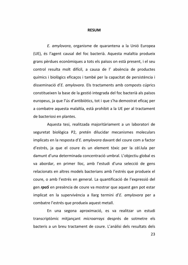
Fruit also becomes blighted at any time but most commonly
during the growing season following a severe hailstorm (van der Zwet
and Keil, 1972; van der Zwet et al., 1974). The affected part of the
fruit is first appearing oily or water-soaked and later, as infection
progress, becomes brown to black. Infection spreads directly through
lenticels in the skin, through wounds, or from an infected spur into
the fruit (Fig. 3) (van der Zwet et al., 2012). Infected fruit, such as
infected tissues, remains attached to the spur, with a mummified
appearance (Fig. 4), and may even produce ooze favoured by hail
damage and warm weather (van der Zwet et al., 2012).
Leaves may become infected after blight bacteria enter directly
through stomata, trichomes, and hydathodes, but more often
through wounds caused by insects, hail, or wind whipping (van der
Zwet et al., 2012). When infection occurs in the leaf blade, a necrotic
section usually appears within 48h. This part of the leaf may dry, but
infection often spreads through the secondary veins into the midrib,
then into the petiole and the supporting stem (Fig. 5) (van der Zwet
et al., 2012). There is often a characteristic blackening of the petiole
and leaf midrib and ooze drops are usually present. Leaves can also
show symptoms when the branch is infected and they can not
receive enough nutritive substances, showing the typical necrotic
aspect (van der Zwet et al., 2012).
1.1. Fire blight of Rosaceae
34
Figure 3. Fire blight infected pear (Photo by E. Marco-Noales, Spain)

INTRODUCTION
35
Figure 4. Blossom blight and ooze production (red arrow) in pear (Photo by E. Marco-Noales, Spain)

1.1. Fire blight of Rosaceae
36
Figure 5. Necrotic pear-shoot after fire blight infection (Photo by E. Marco-Noales, Spain)

INTRODUCTION
37
In fire blight-susceptible hosts, the disease may advance
downward from blossoms, shoots, or fruit through the larger scaffold
limbs to older branches and eventually into the trunk (van der Zwet
et al., 2012), and the advance can also be observed in the subcortex
tissue. The disease may cause small or large cankers in limb and trunk
tissues (Fig. 6), which consist mainly of dead and collapsed bark
cortex and phloem tissues. For the fire blight pathogen, the main
overwintering sites are indeterminate cankers formed at the base of
blighted shoots and fruit spurs, water sprouts, limbs, or branches or
trunk, staying alive usually in the healthy tissue immediately adjacent
to the edge of the visible canker (van der Zwet et al., 2012). Often,
when conditions are conducive, abundant ooze flows along the bark,
accompanying the progress of infection. Flies, using the ooze to feed
on, may be also an instrumental vehicle to spread the disease (van
der Zwet et al., 2012; Ordax et al., 2015).
Fire blight was first described as a disease of apple and pear
(van der Zwet et al., 2012). Since that time knowledge of the disease
has grown exponentially, and many more host plants have become
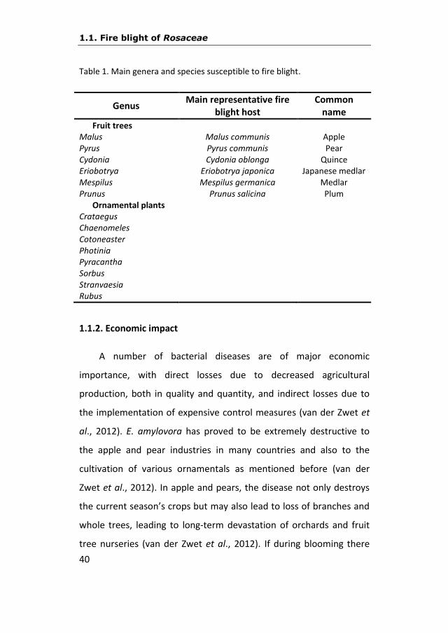
known (Table 1). To determine the types and number of host plants
susceptible to the disease, most natural blight observations and
artificial inoculations of plants were made during the period 1925-
1935 (Rosen and Groves, 1928; Rosen, 1929; Pierstorff, 1931;
Thomas and Thomas, 1931; Thomas and Parker, 1933; Thomas and
Ark, 1934; Parker et al., 1956). Since that time, many more records
have been collected. Besides genera Malus and Pyrus, 129 species in

1.1. Fire blight of Rosaceae
38
37 genera of the Rosaceae family have been reported being
susceptible to fire blight. Of these genera, six are fruit crops: Cydonia,
Eriobotrya, Fragaria, Mespilus, Prunus and Rubus. The remaining 21
genera, with 37 species, are nearly all ornamental host plants and
trees (Thomas and Thomas, 1931; Shaw, 1934; Thomas and Ark,
1934; Jock et al., 2000; Vogelsanger et al., 2006; Bastas and Sahin,
2014). Among them, those that are most susceptible and are the
cause of more economic loses, and usually exhibit the most severe
blight, are species of Cotoneaster, Crataegus, Pyracantha, and Sorbus
(van der Zwet et al., 2012) (Table 1).

INTRODUCTION
39
Figure 6. Canker caused by E. amylovora in a pear tree. (Photo by E. Marco-Noales, Spain)
1.1. Fire blight of Rosaceae
40
Table 1. Main genera and species susceptible to fire blight.
Genus Main representative fire
blight host Common
name
Fruit trees Malus Malus communis Apple Pyrus Pyrus communis Pear Cydonia Cydonia oblonga Quince Eriobotrya Eriobotrya japonica Japanese medlar Mespilus Mespilus germanica Medlar Prunus Prunus salicina Plum
Ornamental plants Crataegus Chaenomeles Cotoneaster Photinia Pyracantha Sorbus Stranvaesia Rubus
1.1.2. Economic impact
A number of bacterial diseases are of major economic
importance, with direct losses due to decreased agricultural
production, both in quality and quantity, and indirect losses due to
the implementation of expensive control measures (van der Zwet et
al., 2012). E. amylovora has proved to be extremely destructive to
the apple and pear industries in many countries and also to the
cultivation of various ornamentals as mentioned before (van der
Zwet et al., 2012). In apple and pears, the disease not only destroys
the current season’s crops but may also lead to loss of branches and
whole trees, leading to long-term devastation of orchards and fruit
tree nurseries (van der Zwet et al., 2012). If during blooming there
INTRODUCTION
41
have been favorable weather conditions for the pathogen, blossoms
are affected, because of that yield is considerably reduced and in
some cases nullified and also the next year’s productivity is
significantly affected because of the destruction of fruiting spurs (van
der Zwet et al., 2012). In susceptible hosts, the infection spreads so
rapidly through the tree under favorable conditions. Once trees are
infected, they cannot be saved even in spite of drastic and immediate
surgery and usually die in a short time after the first visual sign of
infection (van der Zwet et al., 2012).
It is difficult to get information about the economic losses
caused annually by fire blight but they are quite high (van der Zwet
and Keil 1979; van der Zwet et al., 2012). It is necessary to add to the
cost of direct production losses, associated costs of the control
measures (treatments, intensive vigilance, analysis, infected trees
destruction, etc…) and the obligatory varietal structure modification
of the fruit growing sector. The fast dissemination of fire blight and
the progressive death of susceptible cultivars (especially pear trees)
have made to quit its cultivation in some areas of United States of
America (USA) and the European Union (EU).
To get an idea of the significance of losses, we can look at some
numbers through years. In USA, detailed accounts of the early history
of fire blight in California (USA) have been published, suggesting that
two-thirds of the pear trees cultivar “Bartlett” were eliminated, at a
cost of $5 million, during the period 1903-1908 (Woods, 1909;
Gardner and Ark, 1924; Baker, 1971). More recently, fire blight was
1.1. Fire blight of Rosaceae
42
particularly severe in 1991 in south-western Michigan, where the
estimate of losses was $3.8 million (van der Zwet and Beer, 1995). In
Netherland, in 1982, a particularly severe year, the combined
economic impact of the disease on nurseries and fruit orchards and
the total cost of eradication and control were estimated at $6 million
(Vanneste, 2000). At the end of 1996, in Hungary more than 65,000
trees and shrubs were eradicated by the Ministry of Agriculture at a
total cost of $1.1 million (Vanneste, 2000). The more detailed recent
data are those about the epidemy of Michigan (USA) in 2000, having
an evaluation of crop costs and trees losses near to 80 million dollars
(van der Zwet et al., 2012). In Spain, only in Aragón and in a period
between the years 2000 and 2004, costs of the disease (inspections
and eradications) were estimated in more than one million euros
(Palacio-Bielsa et al., 2012). And these are only some examples of the
economical losses caused by fire blight.
This disease also have negative consecuences for the producers
and the nursery sector (Lanthier, 2011), because the prohibition to
export plants and in some cases fruits from countries with E.
amylovora to countries that are free of the disease.
1.1.3. Global distribution
After its origin in the Hudson valley of New York in 1780, fire
blight has moved into most states of the USA. This process took a
period of more than hundred years at the same time as the
movement of humans and the advance of industrialization (van der

INTRODUCTION
43
Zwet and Keil, 1979; van der Zwet et al., 2012). By 1925 fire blight
had spread across USA border into Canada and Mexico and had
moved overseas to New Zealand (van der Zwet et al., 2012). From
1925, the disease spread to 42 additional countries, and up to date it
has been reported in a total of 53 countries (Table 2, Fig. 7).
According to van der Zwet et al. (2012), sometime during the 1950s
the fire blight organism was most probably disseminated, via infested
bud wood or trees, to two different areas from North America to
north-western Europe and to the north-east corner of Africa. Without
a doubt, human activity has been very influential in the spread of fire
blight and another important factor for the fast spreading of the
disease has been the increasing use of susceptible cultivars and
rootstocks, as well as high tree densities in nurseries and young
planting orchards (Vanneste, 2000).
In Europe, in 1958 the disease was first detected in England
(Crosse et al., 1958), suggesting, although never proved, that the
introduction was due to contaminated fruit crates, which were
recycled in those orchards in Kent where the initial blight symptoms
were observed in 1956/57 (Lelliott, 1959). In 1966 it appeared on the
mainland of the European continent, in The Netherlands
(Netherlands Plant Protection, 1966) and the Baltic coast of Poland
(Borecki et al., 1967). It was also detected in Denmark in 1968, in
Germany in 1971, and in Belgium and France in 1972 (Vanneste,
2000). Because the first reports of the disease in Denmark (1968) and
the northern coast of former West Germany (1971), Belgium (1972)

1.1. Fire blight of Rosaceae
44
and France (1972) all appeared within six years, it has been suggested
although not proved, that migratory birds may have been
instrumental in the dissemination of the bacterium across the English
Channel to the western and northern European coastlines (Meijneke,
1972; van der Zwet, 1994; Billing and Berrie, 2002).
In the early 1960s, fire blight also appeared in Egypt in north-east
corner of the African continent (El-Helaly et al., 1964), whereas the
first report of fire blight in Israel dates from 1985 (Zutra and Shabi,
1985), and on the island of Cyprus from 1986 (Psallidas and Dimova,
1986). In 1985, fire blight was reported from Turkey and the
following year from Crete (Greece) (EPPO, 1987). Once fire blight
became established in the Egypt-Cyprus-Israel triangle, it was only a
matter of time before the disease appeared in neighbouring
countries (Vanneste, 2000). By 1988, the disease was reported in
Lebanon (EPPO, 1988), in 1990 in Jordan (Tehabsim et al., 1992), and
in 1995 in Iran (Afunian and Rahimian, 1996). After Crete and Turkey,
fire blight was found into the mainland areas of Greece and then into
Bulgaria (Bobev, 1990), Romania (Baicu et al., 1994), Macedonia
(Mitrev, 1996), and Hungary (Hevesi, 1996). Once fire blight was
established throughout the southern Balkans, it came as no surprise
the observation of symptoms in the southern part of Italy (Cariddi
and Piglionica, 1992). In 1995, the first outbreak of fire blight was
identified in northern Spain (de la Cruz Blanco, 1996). Since then, the
disease has been reported in new countries and now E. amylovora
has been identified in all the EU members including Finland. In 2000,

INTRODUCTION
45
host plants of E. amylovora in the Royal Botanic Gardens of
Melboune (Australia) were positive for the presence of the pathogen,
although it was not isolated from wood samples (Jock et al., 2000). In
2008, fire blight was first reported in Morocco on pear, apple and
quince (Fatmi et al., 2008) and in 2013 a report showed that the
characterization of a selection of strains from Middle Atlas
Mountains of Morocco had notable similarities with a Spanish strain
obtained from plants imported from Belgium (Hannou et al., 2013).
Recently, in 2012, E. amylovora was reported from different host
plants and locations in Serbia and Montenegro (Ivanovic et al., 2012)
and it is also present in other countries like Tunisia, first reported in
2014 in pear (Rhouma et al., 2014) or Algeria, that had its first
characterization of isolates in 2012 (Laala et al., 2012).
The last first report of E. amylovora has been on pear trees in
Finland (Soukainen et al., 2015), that have a Protected Zone status
against fire blight in the EU, and in Kyrgyzstan, Kazakhstan and South
Korea (Myung et al., 2016; Zhao and Sundin, 2017).
1.1. Fire blight of Rosaceae
46
Table 2. Countries where fire blight is present (EPPO, 2017).
Area Countries
Africa Tunisia Algeria Morocco Egypt America Bermuda Mexico Canada United States Guatemala Asia Iran Kazakhstan Israel Lebanon Jordan Syria Kyrgyzstan South Korea Europe Albania Lithuania Armenia Luxemburg Austria FYROM* Belarus Moldova Belgium Montenegro Bosnia-Herzegovina Netherlands Bulgaria Norway Croatia Poland Cyprus Romania Czech Republic Rusia Denmark Serbia Estonia Slovakia Finland Slovenia France Spain Germany Sweden Greece Switzerland Hungary Turkey Ireland Ukraine Italy United Kingdom Latvia Oceania New Zealand
*Former Yugoslav Republic of Macedonia, mentioned as Macedonia in the EPPO database.

INTRODUCTION
47
Figure 7. Global distribution of fire blight (EPPO 2017). Red circle indicates countries. Red cross indicates regions or states inside some countries.
1.1. Fire blight of Rosaceae
48
1.1.4. Situation in Spain
In 1984, while fire blight was spreading across north-western
Europe, Sampayo and Palazón (1984) published a detailed list of
preventive measures to try to keep the disease out of Spain. In spite
of this, the first report of fire blight in Spain dates from 1995 (de la
Cruz Blanco, 1996), as mentioned before. The disease was observed
in August 1995, mainly on cider apple trees, a few kilometers south
of the French border, in Guipúzcoa, near the Atlantic coast. In the
following two years, fire blight was observed in pear, apple, quince,
loquat, and several ornamental hosts (López et al., 1999). The spatial
and temporal distribution of the disease foci strongly suggest that
infested plant material was responsible for the introduction of the
disease (López et al., 2002). Moreover, molecular analysis of a large
number of Spanish strains of E. amylovora support the hypothesis of
several introductions in Spain of infected plant material from
different European countries (Donat et al., 2007).
Spain was recognized as “protected zone” (PZ) for fire blight in
2000 (DOCE 2000). This is the most important protection considered
in the European Union Phytosanitary legislation. Countries or places
where a pathogen is not established and perform surveys to try to
find it may obtain the status of PZ, and introduction of vegetal
material and its movement are subjected to high severe quarantine
requirements (Real Decreto 58/2005).

INTRODUCTION
49
In 2011 and the following years, there were new fire blight
outbreaks in many of the pome fruit production zones of the national
territory. Even though measures to eradicate the disease or delay its
establishment have been adopted in some cases, the situation today
in 2017 is not encouraging. It is considered that the disease is
established in the following areas that have lost, therefore, the PZ
status: Castilla y León, Extremadura, La Rioja, Castilla La Mancha,
Murcia, Navarra, Guipúzcoa (País Vasco), Aragón, Murcia and
Valencia (green spots in Fig. 8). Recently, several regions of a city of
Catalonia named Lleida lost its PZ status as well (official
communication).
Figure 8. Current distribution of fire blight in Spain (www.magrama.gob.es 2017).

1.1. Fire blight of Rosaceae
50
1.1.5. Fire blight epidemiology and control
1.1.5.1. Fire blight cycle
Fire blight bacteria hibernate in the bark at the edge of cankers
formed during previous growing seasons. As weather becomes warm
in the spring and temperature reach 18-30ºC the bacteria multiply
and ooze to the surface in sticky droplets (Palacio-Bielsa and Cambra,
2009). Under low relative humidity the bacteria can survive in the dry
exudates for over a year (Rosen, 1938). Relatively few cankers survive
winter, becoming active and producing bacteria in the spring.
However, a single active canker will produce millions of bacteria,
enough to infect an entire orchard. Cankers produce bacteria in
droplets of ooze that are transferred to flowers by splashing rain or
by insects, mostly bees, flies and ants (Fig. 9).
According to van der Zwet et al. (2012), once on the flower
stigmas, the bacteria can grow epiphytically under favorable
circumstances and reaching 106-107 colony-forming units (CFU) per
healthy flower. The presence of high densities of epiphytic bacteria
on healthy flowers facilitates an efficient movement of the bacteria
from flower to flower by rain or by any insect that visits the flowers
(Fig. 9). Blossom infection occurs when the bacteria are washed by
rain to natural openings at the flower base. These openings located in
the hypanthium are specialized stomata, termed nectar glands.
Blossoms wilt and die in about 1-2 weeks after infection occurs, and
the bacteria that ooze from them provide inoculum for secondary
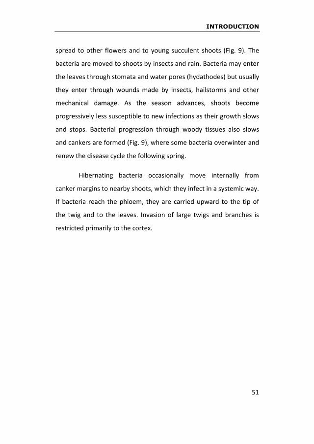
INTRODUCTION
51
spread to other flowers and to young succulent shoots (Fig. 9). The
bacteria are moved to shoots by insects and rain. Bacteria may enter
the leaves through stomata and water pores (hydathodes) but usually
they enter through wounds made by insects, hailstorms and other
mechanical damage. As the season advances, shoots become
progressively less susceptible to new infections as their growth slows
and stops. Bacterial progression through woody tissues also slows
and cankers are formed (Fig. 9), where some bacteria overwinter and
renew the disease cycle the following spring.
Hibernating bacteria occasionally move internally from
canker margins to nearby shoots, which they infect in a systemic way.
If bacteria reach the phloem, they are carried upward to the tip of
the twig and to the leaves. Invasion of large twigs and branches is
restricted primarily to the cortex.
1.1. Fire blight of Rosaceae
52
Figure 9. Fire blight cycle.
INTRODUCTION
53
1.1.5.2. Preventive and cultural measures
To prevent fire blight, it is basic not to illegally introduce vegetal
material of Rosaceae from infected areas or countries. The plant
purchase must be made exclusively from authorized nurseries,
requiring the phytosanitary passport which guarantees the
requirement compliance established in the European legislation (BOE
2005; DOCE 2000). The elimination of symptomatic or infected plants
laid down in the Real Decreto 1201/99 (BOE 1999) as eradication
measures has been very effective (Palacio-Bielsa et al., 2012) and it is
necessary to carry out the eradication measures as faster as possible
to reduce inoculum and avoid E. amylovora dissemination.
Control of fire blight will be only possible if disease symptoms are
detected prematurely in the orchard. It has been observed that fire
blight progresses faster when more symptomatic plant material is
present in an orchard or closer to it. Therefore, it is very important to
examine the orchard during and after bloom, rain, storms and above
all after hail. Moreover, June, July and September are in general the
most critical months for symptom detection since trees have an
active vegetative growth and more susceptible material is available.
All factors that favor plant susceptibility and/or pathogen
spreading should be controlled. Pruning is recommended only when
trees are hibernating to remove all suspicious cankers, favoring the
maximum ventilation of the orchard, combined with disinfecting
frequently all the tools used, and burning the plant debris. Other
recommended practices include the removing of secondary bloom, a

1.1. Fire blight of Rosaceae
54
limited use of nitrogen fertilizer to prevent vegetative overgrowth,
and avoidance of spraying watering.
1.1.5.3. Chemical control
Chemicals are applied to reduce the number of E. amylovora
cells or, at least, inhibit their multiplication. To achieve these effects,
it is necessary to eliminate sources of inoculum, such as
overwintering cankers on fruit trees or alternate hosts, or to protect
potential invasion sites, such as blossoms, shoots, leaves, or fruit,
especially after wounding (Palacio-Bielsa and Cambra, 2009). In this
sense, to be effective against this pathogen, any bactericide should
be applied during the three distinct periods of the host: when the
tree or plant is dormant, in bloom, and during the postbloom period.
To prevent the development of new blossom infections, chemical
agents are used during the dormant period and before budbreak,
since E. amylovora may overwinter in cankers. During bloom the
objective is to decrease pathogen populations and blossom necrosis
and in the postbloom to avoid shoot infections (van der Zwet et al.,
2012).
Two groups of chemical agents, antibiotics and copper
compounds, have played the most important role in controlling fire
blight of apples and pears since the 1930s. In the early 1950s,
discovery and application of antibiotics was the most important
development in fire blight control, due to their successful use in
treating human diseases (van der Zwet et al., 2012). Moreover, for

INTRODUCTION
55
fire blight control, in general, streptomycin gave better result than
copper, and normally it caused no fruit russet. This lack of injury
probably appealed to many growers even though several treatments
are necessary and the antibiotic cost is considerably higher than that
of copper (van der Zwet et al., 2012). In early antibiotic research, a
widely used formulation contained 15% streptomycin sulfate and
1.5% oxytetracycline (Terramycin) (Dunegan et al., 1954; Higdon,
1954; Ark, 1958; Keil, 1963). Laboratory studies showed that E.
amylovora developed resistance far more slowly with this
combination than with streptomycin alone (English and van Halsema,
1954). Then, it was used commercially for many years, until
oxytetracycline was removed during the 1960s, apparently because
the combination showed no advantage when used in the field (van
der Zwet et al., 2012). According to Stockwell and Duffy (2013),
streptomycin is applied in several countries of the United States to
control fire blight before infection have place, in springtime, because
after infection antibiotics are ineffective. Antibiotics have been
indispensable for crop protection in USA for more than 50 years
without reports of adverse effects on human health or persistent
impacts on the environment, since antibiotics are active on plants for
less than a week (Stockwell and Duffy, 2013). A study with
streptomycin to control fire blight in experimental orchards of
Europe was recently carried out by Walsh et al. (2014). They
demonstrated that there was not abundance of streptomycin or
tetracycline resistant genes in the bacteria neither a negative impact
on the bacterial community after three streptomycin treatments.

1.1. Fire blight of Rosaceae
56
Numerous copper compounds have given variable results in the
control of fire blight, ranging from poor to excellent (Rosen, 1932,
1934; Pinckard et al., 1936; Sherbakof and Andes, 1939; Veerkamp,
1945; Gardiner, 1951-1957; Agrios, 1968; Hickey et al., 1998; Bastas
et al., 2010). Copper sulfate mixed with lime (Bordeaux mixture) has
been used more often than any other form of copper. It is applied
only during the dormant period and the bloom period, to avoid fruit
russet that is directly proportionate to the copper content of the
formulation. Probably because of this fruit injury, copper is not used
more often.
A wide and renew overview about the use of copper in the
management of bacterial diseases of fruit trees will be explained in
section 1.2.
1.1.5.4. Biological control
The possibility of biological control of fire blight, using different
microorganisms, has been investigated, discussed, and reviewed for
more than four decades (Schroth et al., 1974; Aldwinckle and Beer,
1979; Pusey, 2002; Stockwell et al., 2002; Ozaktah and Bora, 2004;
Cabrefiga et al., 2014). At first, mainly antagonistic bacteria were
tested as potential biological control agents, but since then several
natural compounds (plant extracts and etheric oils) have also been
assayed against the fire blight pathogen (Briffaerts et al., 1996; Zeller
and Laux, 2006; Farkas et al., 2012). Increased research on the
biocontrol of this disease has been motivated by the development of

INTRODUCTION
57
resistance in E. amylovora to the antibiotic streptomycin, and the
restrictions to the use of several chemicals and antibiotics in
countries of the EU and other states (Zeller and Laux, 2001, 2002a,
2002b; Montesinos et al., 2009; BOE 2012).
The surface of the stigma, located on top of the floral pistil, is
the site where bacterial bio-control agents must interact with and
successfully antagonize E. amylovora (Hattingh et al., 1986; Thomson,
1986; Wilson et al., 1989; Vanneste, 1995; Farkas et al., 2012). In fact,
biological control of fire blight is successful when a bacterial
antagonist establishes and develops a large population on the
stigmatic surface prior to the establishment of E. amylovora (Wilson
et al., 1992; Johnson et al., 1993; Wilson and Lindow, 1993). These
populations, through a combination of mechanisms, can suppress the
establishment and epiphytic growth of the pathogen (Farkas et al.,
2012). Decrease of E. amylovora population on stigmatic surfaces
reduces the probability of floral infection and spread of the pathogen
to other blossoms. Effective biological control requires colonization
of the most stigmas of the flowers in the orchard by the bacterial
antagonists (Johnson et al., 1993; Lindow et al., 1996), and requires a
larger population of them on these surfaces (Farkas et al., 2012). Fire
blight is a good candidate for biological control because the bacterial
antagonists need to persist on the nutrient-rich, stigmatic surfaces
for only about one week to suppress blossom infection effectively
(Cabrefiga et al., 2007; Cabrefiga et al., 2011; Farkas et al., 2012;
Cabrefiga and Montesinos, 2017).

1.1. Fire blight of Rosaceae
58
1.1.5.5. Cultivar susceptibility and genetic control
A very limited number of apple and pear cultivars are
responsible for a large proportion of annual world production
(Vanneste, 2000). To retain a cultivar with fruiting desirable
characteristics and to introduce disease-resistance genes by
conventional breeding methods is virtually impossible, because of
apple and pear heterozygosity, long generation time and self-
incompatibility. All this make back-cross programs of several
generation prohibitively long term and expensive (Vanneste, 2000).
The use of biotechnology can now overcome these handicaps by
introducing resistance genes directly into current valuable
commercial cultivars and thereby transforming them into resistant
forms of the same cultivars (van der Zwet et al., 2012).
Targeting gene expression to fire blight-susceptible tissues or
during specific developmental stages could be advantageous in
providing resistance where and when needed (Vanneste, 2000).
Exogenous application of plant resistance inducers (PRIs) able to
activate plant defenses is the most novel approach for new
integrated pest management practices (de Bernonville et al., 2014).
1.1.5.6. Integrated control in Spain and the European Union (EU)
Optimal control of fire blight seems to be only achieved by
eliminating all diseased plant material and by reducing host
susceptibility with available cultural measures (Deckers and Porreye,
1987; Deckers et al., 1987). The disease control must be considered
INTRODUCTION
59
as an integrated strategy, selecting the lower sensitivity varieties and
applying the prophylactic measures, the cultural techniques and
necessary treatments to reduce the inoculum amount (Johnson and
Stockwell, 1998; Palacio-Bielsa and Cambra, 2009; Johnson and
Temple, 2013; Smith, 2014).
Genetic improvement of apple and pear trees to obtain resistant
varieties with commercial interest did not provide the expected
results despite that it has been obtained significative advances (van
der Zwet et al., 2012). Also, transgenic and cisgenic varieties of apple
and pear trees with significant levels of fire blight resistance have
been obtained (Litz and Padilla, 2012), but not authorized and
commercialized in EU yet.
A combination of all available control measures (sanitation,
selection of resistant varieties, and cultural practices) is the preferred
way to keep the disease to a minimum.
1.1.5.7. Legislation
In Spain, the regulation against fire blight started with a Ministry
Order in 1975 about the prohibition of host plants import from
contaminated countries. In 1999, it was published the Real Decreto
1201/1999 (BOE 1999) on the National Program for eradication and
control of fire blight, that was based on the European legislation but
adapted to the Spanish situation. This law was modified in 2005 (Real
Decreto 1512/2005), 2010 (Real Decreto 246/2010) and 2011 (Real
Decreto 1786/2011). The following rules were determined as
1.1. Fire blight of Rosaceae
60
obligatory in disease free zones (PZ): a) official declaration of the
disease, b) destruction of the affected vegetal material by particular
people and public entities, and c) systematic research by the
Autonomous Communities. Obligatory phytosanitary measures were
established in this Real Decreto in zones where the disease was
present, to slow down its propagation, as well as the prohibition to
plant host ornamental species in public road and gardens in the
hazard zones decided by each Autonomous Community (Montesinos
et al., 1999).
The European Economic Community (EEC) published in 1977 the
Directive 77/93/CEE about the circulation of damaging organisms of
plants where E. amylovora appeared as a quarantine organism
(present in some countries of the EEC but not in all of them).
Restrictions were imposed to the distribution of vegetal material
coming from epidemic zones, which were considered as Protected
Zones (PZ) for this disease, being Spain among them. The EEC
member countries not yet affected by fire blight established a
supervision network for maintaining these areas free of the disease.
According to that, all the vegetal material susceptible to the disease
must be commercialized under the phytosanitary passport with the
initials PZ (Montesinos et al., 1999).
In 2000, the Directive 2000/29/CE was published in the Official
Diary of the European Economic Communities. The Directive is
relative to the protection measures against the introduction in the
Community of damaging organisms for the vegetables or vegetal

INTRODUCTION
61
products and against their dissemination inside the Community. In
Spain, the Plant Health law (Law 43/2002, of November 20th of 2002)
regulates thoroughly general aspects relative to the prevention and
fight against the different pests and diseases. The Autonomous
Communities, basing on the Real Decreto 1201/1999 and 1512/2005,
have adopted in Spain other complementary measures to reinforce
the effects that are being pursued (Palacio-Bielsa and Cambra, 2009).
1.2. Copper and field application
62
1.2. Copper and field application
Bacterial infections of plants are some of the most difficult
diseases to control because there is still little effective chemistry
available (Civerolo, 1982; López et al. 2003; Janse, 2004, 2005).
Antimicrobials for prophylactic treatment of bacterial diseases of
plants are limited in availability, use, and efficacy, and therapeutic
use is largely ineffective (Vidaver, 2002). One type of the products
more frequently used are copper formulations that have been
extensively used in agriculture since more than 200 years ago, with a
significant track history of relative success, and nowadays the
utilisation of different copper-based bactericides is a piece in the
whole of controlling bacterial diseases.
1.2.1. The history of the use of copper
Copper is considered a unique metal known for its
antimicrobial properties throughout millennia. It was the first metal
used by humans, probably because of its metallic native form
(Elguindi et al., 2011). Thus, use of copper has been reported as far as
in ancient Chinese civilization around 2500 B.C (Yu et al., 1995); in
ancient Egypt to sterilize drinking water and chest wounds (Dollwet
and Sorenson 1985); by Greeks in 400 B.C., for treating pulmonary
diseases and purifying drinking water (Dollwet and Sorenson, 1985);
and by ancient Aztecs, in Mexico, for treating skin conditions. And
during the circa 1850 cholera epidemic in Paris, copper workers were
found to be immune to it (Michels et al., 2005). Throughout history,
men have exploited the antimicrobial attributes of copper.

INTRODUCTION
63
As early as the beginning of 18th century, Prevost (1807) had
demonstrated that bunt of wheat, caused by Tilletia caries, could be
controlled to some degree by copper sulfate. He provided the first
scientific evidence that this compound would kill fungal structures.
Previously, Schulthess (1761) had observed that copper sulfate could
provide some control of bunt but did not know the mode of action
(van Zweiten et al., 2007). It was in the latter half of the 19th century
that chemical disease control really started to develop. Because of its
high phytotoxicity, copper was not used as a foliar pesticide until
1885 (Millardet, 1885), when French scientist Millardet accidentally
observed that a mixture of copper sulfate, lime and water, that had
been applied to grapevines near roadways in order to discourage
thieves from stealing the grapes, was not phytotoxic but exhibited a
fungicidal action against Plasmopara viticola (Russell, 2005). His
vinter’s spray formulation was then the fungicide of choice in USA
and was named “Bordeaux mixture” (Borkow and Gabbay, 2005). It
had represented the first large scale use of a fungicide in Europe and
America (Dunegan and Doolittle, 1953; Floyd, 1991). It later proved
also to be a good bactericide; in fact, it has been used widely against
bacterial diseases on different crops (Jiang et al., 2008). Already in
one of the first publications on bacterial diseases of plants (Smith,
1920), it is reported that the number of infections in walnut blight in
California was reduced by 50% using Bordeaux mixture, and that in
Italy this mixture was recommended for olive trees following hail-
storms to protect them against tuberculose.
1.2. Copper and field application
64
1.2.2. Copper formulations and mode of action
Copper bactericides work by coating the leaf surface with
minute particles of copper which then react with acid and moisture
on the surface to release copper ions that kill bacteria and prevent
fungal spores from germinating. Over time the protective coating
provided by copper bactericides is diluted by the action of rain, wind
and the growth of the crop. Interestingly, despite the long-term use
of copper as antimicrobial, its precise mode of action has not been
fully elucidated (Dupont et al., 2011; Fones and Preston, 2012).
The antimicrobial activity of copper is due to the soluble
fraction of Cu++ or Cu+ metal. Copper compounds used in agriculture
are generally composed of the active ingredient Cu++ combined with
an anionic component to form inorganic or organic salts, or quelates.
The other ingredients improve water miscibility, adherence and
spreadibility to plant surfaces, and other additional properties.
Copper, although no target specific, kills all living cells.
Hovewer, plants are less susceptible to copper toxicity than
microorganisms, due to tissue barriers and structures, such as leaf
cuticle, fruit wax or trunk suber, which avoid the entry of copper. The
role of copper for control of bacterial plant diseases is primarily
based on its direct action on the pathogenic cells. Copper can interact
with many vital cell structures due to reactive nature of Cu++ with
anionic cell components. Moreover, increased intracellular copper
levels may induce the synthesis of reactive oxygen species (ROS),
causing an additional oxidative damage to lipids, proteins and DNA in

INTRODUCTION
65
the internal structures of the bacterial cell. The result of these actions
is a loss of the functionality of the bacterial cell envelope by
membrane disruption, a protein-enzyme denaturation, inhibition of
DNA replication, transcription, and protein synthesis. Therefore,
copper exhibits a bactericidal activity. However, there is a second
mechanism of action of copper, which is mediated by the host. It
consists on a stress response induced by copper in the plant, with
overproduction of ROS and increased levels of antioxidant enzymes
(SOD, GPX, APX) (Ros-Barceló, 2006; Yadav et al., 2010).
1.2.3. Field applications in the EU
As described before, copper compounds through agriculture
history have been used for vegetable and fruit crops to restrict the
spread of plant pathogens, both bacteria and fungi. They have
constitued for decades the main protection instrument against plant
diseases. However, today there is a strong tendency to reduce the
use of copper and copper-based products to minimize their
environmental impact. Development of copper resistant bacterial
strains and accumulation of copper in soil are detrimental effects
derived from use of this chemical.
In the EU, plant protection products containing copper must fulfil
the safety requirements laid down in BOE (2012). According to it,
several active substances were evaluated, and the following products
were included in the Annex I of the Directive: copper hydroxide,
copper oxychloride, copper oxide, Bordeaux mixture, and tribasic
copper sulfate. The phytosanitary products that contain these

1.2. Copper and field application
66
substances can be therefore authorized in the Member States.
Nevertheless, due to the fact that the risk assessment of copper
compounds revealed eco-toxicological concerns, a restriction on the
inclusion period is deemed necessary to allow Member States to
review after a shorter period copper containing plant protection
products already on the market.
Chemical treatments are very useful and contribute to reduce
losses in fruit production due to bacterial infections. However, no
complete protection can be expected by chemical treatments alone,
but they should be considered as a part of an integrated control
system aiming to decrease disease incidence. The objective for a
sustainable disease management is to improve the efficiency of
disease control while reducing the amount of copper application to
an environmentally acceptable level. An attempt to adequately
control diseases and limit unnecessary chemical applications is to
select the copper compounds to be used, applying them in a strategic
way, timed according to pathogen activity, and test alternative
products and farming practices.

INTRODUCTION
67
1.3. Copper, friend and foe for bacterial cells: targets for copper
action and strategies
Copper is a transition metal utilized by bacteria in many cellular
processes. However, while copper plays critical roles it can be toxic
when levels are beyond cellular needs. Bacterial cells may prevent
copper toxicity in part by keeping copper compartmentalized in the
cell periphery. Trace copper is sufficient for survival as there are few
Cu-dependent enzymes and they are most often localized within the
cell periphery. The most widespread Cu-dependent enzyme in
bacteria is cytochrome c oxidase (Cox), located in the cell membrane
(Festa and Thiele, 2012).
Our current understanding of the molecular mode of action of
copper ions against bacteria is still limited and the microorganism
more studied is Escherichia coli. Rensing and Grass (2003)
demostrated that the copper translocating P-type ATPase, CopA, was
the central component in E. coli for copper homeostasis, responsible
for removing excess copper from the cytoplasm. Macomber and
Imlay (2009) showed that copper toxicity involved the action of ROS
and that the primary target of copper in E. coli was the iron-sulfur
cluster of proteins. A connection between copper and cell integrity
was discovered when Espirito Santo et al. (2011) challenged E. coli to
dry copper surfaces. Cells suffered extensive membrane damage
within minutes of exposure to dry copper.
1.3. Copper friend and foe for bacterial cells
68
1.3.1. Copper and generation of reactive oxygen species
Metals can act directly as antimicrobial toxins, and, consequently,
they can be used directly to limit pathogen growth (Fones and
Preston, 2012). Besides that, the ability of copper to undergo redox
changes between Cu+ and Cu++, although makes it essential for life by
its indispensable role in various enzymatic processes, also makes it
dangerous since it is able to elicit the production of ROS. Aerobically,
copper readily catalyzes reactions that result in the production of
hydroxyl radicals through the Fenton-like and Haber-Weiss reactions
(Cu+ + H2O2 → HO• + HO- + Cu++; O2•- + Cu++ → Cu+ + O2) (Halliwell and
Gutteridge, 1984, 1990; Rensing and Franke-McDevitt, 2013). The
highly reactive oxygen intermediates are responsible for lipid
peroxidation, oxidation of proteins and damage to nucleic acids
(Halliwell and Gutteridge, 1984; Imlay and Linn, 1988; Stadtman,
1992). The effect of Cu+ on generation of ROS can also be indirect,
since free copper ions are able to oxidize sulfhydryl-groups, such as
cysteine in proteins or the cellular redox-buffer glutathione (Stohs
and Bagchi, 1995; Helbig et al., 2008). Moreover, Cu+ can attack and
destroy iron-sulfur clusters realeasing iron which can in turn cause
oxidative damage through iron-based Fenton chemistry (Keyer and
Imlay, 1996; Rensing and Franke-McDevitt, 2013).
It was believed that copper ion toxicity in bacteria was only
mediated by oxidative DNA damage. However, under anaerobic
conditions copper ions reduce the growth rate of E. coli even more
strongly than under aerobic conditions (Outten et al., 2001).

INTRODUCTION
69
Figure 10 shows a summary of the multitarget action of copper in
a Gram-negative bacterial cell.
Figure 10. The multitarget action of copper in a Gram-negative bacterial cell. IM: inner membrane, CW: cell wall, EM: external membrane, EPS: exopolysacharide (Courtesy of Prof. E. Montesinos, University of Girona, Spain).
1.3.2. Copper uptake and homeostasis
Copper compounds have been used to control bacterial diseases
since long time ago and consequently pathogens have been under
copper pressure all that time. They have developed detoxification
strategies and copper-resistance mechanisms to face up to the
bactericide properties of high level copper concentrations (Rensing
and Grass, 2003; Teitzel and Parsek, 2003; Waldron and Robinson,
2009; Fones and Preston, 2012).
1.3. Copper friend and foe for bacterial cells
70
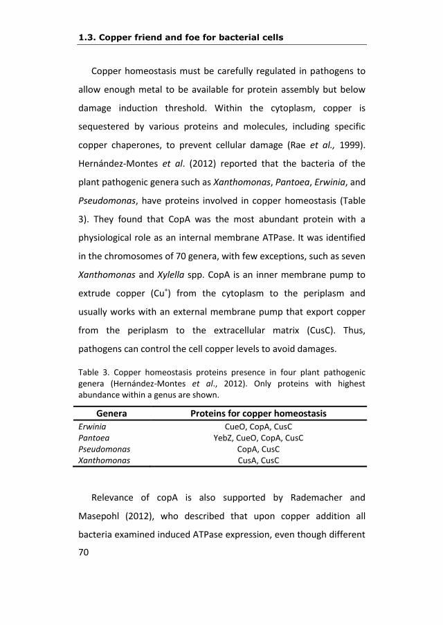
Copper homeostasis must be carefully regulated in pathogens to
allow enough metal to be available for protein assembly but below
damage induction threshold. Within the cytoplasm, copper is
sequestered by various proteins and molecules, including specific
copper chaperones, to prevent cellular damage (Rae et al., 1999).
Hernández-Montes et al. (2012) reported that the bacteria of the
plant pathogenic genera such as Xanthomonas, Pantoea, Erwinia, and
Pseudomonas, have proteins involved in copper homeostasis (Table
3). They found that CopA was the most abundant protein with a
physiological role as an internal membrane ATPase. It was identified
in the chromosomes of 70 genera, with few exceptions, such as seven
Xanthomonas and Xylella spp. CopA is an inner membrane pump to
extrude copper (Cu+) from the cytoplasm to the periplasm and
usually works with an external membrane pump that export copper
from the periplasm to the extracellular matrix (CusC). Thus,
pathogens can control the cell copper levels to avoid damages.
Table 3. Copper homeostasis proteins presence in four plant pathogenic genera (Hernández-Montes et al., 2012). Only proteins with highest abundance within a genus are shown.
Genera Proteins for copper homeostasis
Erwinia CueO, CopA, CusC Pantoea YebZ, CueO, CopA, CusC Pseudomonas CopA, CusC Xanthomonas CusA, CusC
Relevance of copA is also supported by Rademacher and
Masepohl (2012), who described that upon copper addition all
bacteria examined induced ATPase expression, even though different
INTRODUCTION
71
species utilize structural and functionally different regulators to
control ATPase gene transcription. As for the ATPase gene regulation
and as a general rule, Gram-negative bacteria activate ATPase gene
transcription with increasing copper concentrations, mostly by CueR-
like one-component regulator or by CusRS-like two-component
systems (Rademacher and Masepohl, 2012). Yamamoto and Ishihama
(2005) described that CueR from E. coli is the main system regulating
copper-response under aerobic conditions. On the other hand, the
CusSR system may play an important role in copper tolerance under
anaerobic conditions because self-phosphorylation of CusS is
activated in that situation.
Other partners playing an important role in copper homeostasis
are the multicopper oxidase proteins (MCOs). The most outstanding
toxicity of copper is given when Cu+ is in the cytoplasm, whereas Cu++
is less toxic for the cell. In fact, many bacteria synthesize MCOs as
additional copper defence determinants. Comparative analysis
showed that MCOs can be found in approximately 13% of the
bacterial genomes (Ridge et al., 2008). Although the sequence
homology among MCOs is low, amino acid alignments show that the
overall structures and copper-binding motifs are highly conserved
(Reiss et al., 2013). Based on the type of substrates, several types of
MCOs can be differentiated (Sakurai and Kataoka, 2007). MCO
expression in Gram-negative bacteria is activated by either CueR or
CusRS homologues as we mentioned above for copper-ATPase
proteins. Although MCOs function occurs in the periplasm, there is
no apparent preference for CusRS systems, which sense periplasmic

1.3. Copper friend and foe for bacterial cells
72
copper concentrations, over CueR sensors, which respond to the
cytoplasmic copper status (Rademacher and Masepohl, 2012).
1.3.3. Multicomponent copper efflux systems
Resistance-nodulation-cell division (RND) superfamily efflux
systems are responsible for the active transport of toxic compounds
from the Gram-negative bacterial cell. Besides copper-ATPases and
MCOs, many Gram-negative bacteria synthesize RND-type
multicomponent copper efflux systems (Rademacher and Masepohl,
2012). These pumps typically assemble as tripartite complexes,
spanning the inner and outer membranes of the cell envelope. These
complexes consist of an inner-membrane substrate-binding
transporter (or pump), a periplasmic membrane fusion protein (or
adaptor), and an outer membrane-anchored channel (Delmar et al.,
2013). The assembled tripartite efflux complex is responsible for
removing toxic compound out of the cell, and mediating bacterial
resistance to these noxious chemicals (Delmar et al., 2013). Taking E.
coli as a model (Outten et al., 2001; Rensing and Grass, 2003), we
know that it harbours the divergently transcribed cusCFBA and cusRS
operons, which encode an RND-type copper efflux system and a
copper-responsive two-component system, respectively. Membrane-
bound CusS is thought to sense the periplasmic copper status. Upon
binding of Cu+, CusS is expected to autophosphorylate and donates
the phosphoryl group to CusR, which in turn, activates transcription
of the cusCFBA and cusRS operons (Outten et al., 2001; Franke et al.,
2003; Rensing & Grass, 2003; Gudipaty et al., 2012). Primarily, these

INTRODUCTION
73
RND systems excrete copper from the periplasm and, maybe less
important for copper export, from the cytoplasm. As it would be
expected for bacteria lacking an outer membrane, copper induced
RND systems are absent in Gram-positive species (Rademacher and
Masepohl, 2012).
1.3.4. Biosynthesis of extracellular polymeric substances (EPS)
A common feature of many bacteria is the biosynthesis of EPS that
can offer a protective barrier under environmental stress conditions.
These extracellular polymeric substances may protect cells by binding
toxic metal ions (Geddie and Suhterland, 1993) and, in fact, bacteria
can increase their EPS production in the presence of toxic metals as a
defense mechanism (Decho, 1994). Commonly, exopolysaccharides
are composed of glucose, fructose, manose, pyruvate and fucose, as
well as mannuronic and glucuronic-acid complexes (Brisou, 1995). In
addition, the EPS matrix comprises a mixture of
heteropolysaccharides, proteins and nucleic acids (Filipe et al., 1999).
Exopolysaccharides and other biopolymers exhibit excellent metal-
binding properties with varying degrees of specificity and affinity. The
binding of cations to bacterial biopolymers generally occurs through
electrostatic interaction with negatively charged functional groups
such as uronic acids, phosphoryl groups of amino acids, acidic amino
acids and phosphate-containing nucleotides (Beech and Sunner,
2004). It has been described that copper increases production of the
main exopolysaccharide of E. amylovora, amylovoran (Bereswill et
al., 1998). Amylovoran and levan contribute to long term survival of
1.3. Copper friend and foe for bacterial cells
74
this pathogen in a culturable state in presence of copper and can be
used as carbon sources under deprivation conditions providing to E.
amylovora survival advantage (Ordax et al., 2010).
1.3.5. The viable but nonculturable (VBNC) state can be induced by
copper
Copper induces a particular physiological state, the VBNC state, in
several plant-associated bacteria, such as Agrobacterium
tumefaciens, Ralstonia solanacearum, E. amylovora, and
Xanthomonas citri subsp. citri as part of the survival strategies of
these microorganisms (Alexander et al., 1999; Grey and Steck, 2001;
Ordax et al., 2006; del Campo et al., 2009). Effectiveness of copper to
control bacterial plant disease is often measured by the absence of
bacterial growth on a solid medium. However, the failure to produce
a visible colony may not necessarily mean that the bacterial cell is
dead. VBNC state can be induced in bacteria under some stress
conditions, losing their culturability on nonselective solid medium but
remaining viable and virulent, potentially creating a hidden reservoir
of the pathogen. Induction of the VBNC state is a general mechanism
to retain viability, at least in a proportion of cells, and it can be
considered a long-term dormant-like survival mechanism for non-
spore-forming bacteria. Resuscitation of nonculturable cells does not
always occur by a simple reversal of the stress that induced the
nonculturability, and there appears to be no a universal resuscitation
condition (Kell et al., 1998). In the case of E. amylovora and X. citri
subsp. citri cells, resuscitation has been reported even after several

INTRODUCTION
75
months under copper stress (Ordax et al., 2006; Golmohammadi et
al., 2013).

1.4. Erwinia amylovora, the causal agent
76
1.4. Erwinia amylovora, the causal agent
1.4.1. Taxonomy and general features
E. amylovora was the first bacterium for which its pathogenicity
to plants was demonstrated, and as a plant pathogenic bacterium
was shown to be the causative agent of fire blight (Burrill, 1883). It is
a Gram-negative bacterium belonging to the family of
Enterobacteriaceae, which includes some human and animal
pathogens such as Escherichia coli, Yersinia spp., Shigella spp. and
Salmonella spp. Cells are rod-shaped, have a size of about 0,3μm x 1-
3μm, and are motile in culture media by means of two to seven
peritrichous flagella per cell (van der Zwet et al., 2012). On the plant
surface, the motility of E. amylovora is easily expressed (Paulin,
2000), but no motile bacterial cells have been observed in the
intercellular spaces of infected plant tissues (Cesbron et al., 2006). E.
amylovora is a facultative anaerobe and is quoted as being weakly
fermentative (Holt et al., 1994). The optimal temperature for its
growth is 25-27ºC, although it can grow between 3-5ºC and up to
37ºC (Billing et al., 1961).
Its appearance on plate depends on the culture medium (Fig.
11). For example in KB medium (EPPO, 2013) E. amylvora colonies are
visible at 24h and are creamy white, circular and non-fluorescent
under UV light color at 366nm after 48h (Fig. 11 A). This allows
distinction from fluorescent pseudomonads. Colonies of E. amylovora
on Levan medium (EPPO, 2013) are apparent at 24h and are whitish,

INTRODUCTION
77
circular, domed, smooth and mucoid after 48h (Fig. 11 B). E.
amylovora colonies on CCT medium (EPPO, 2013) are visible at about
48h and are pale violet, circular, highly convex to domed, smooth and
mucoid after 72h, showing slower growth than on KB and Levan
media (Fig. 11 C). CCT medium inhibits most pseudomonads but not
Pantoea agglomerans (EPPO, 2013) that is a frequent member of the
native microbiota in E. amylovora host plants. KB, Levan and CCT are
the three media recommended by EPPO to maximum recovery of E.
amylovora from samples in poor condition.
Figure 11. Erwinia amylovora appearance in different solid media. Courtesy of Laboratory of Bacteriology of IVIA, Spain.
A) KB medium
B) Levan medium

1.4. Erwinia amylovora, the causal agent
78
C) CCT medium
According to phenotypic and genomic studies, the specie E.
amylovora is very homogeneous at the biochemical and serological
levels (Paulin, 2000; Smits et al., 2010), but exhibits differences
among strains in several characteristics, including host range,
virulence (Norelli et al., 1984; Cabrefiga and Montesinos, 2005; Wang
and Zhao, 2009) and presence of different plasmids (Llop et al.,
2012).
1.4.2. Virulence factors
Pathogenicity and virulence in E. amylovora mainly depend on
several factors (van der Zwet et al., 2012): (1) the synthesis of
amylovoran and levan, (2) a type III secretion system, (3) the effector
protein DspE, (4) the iron-scavenging siderophore desferrioxamine
and (5) the presence of plasmids pEA29 and pEI70 (Llop et al., 2011).

INTRODUCTION
79
Amylovoran is a polymer of a pentasaccharide repeating units
that consists mainly of four galactose residues and one glucuronic
acid residue (Nimtz et al., 1996). It is produced in the early stationary
growth phase and its size varies according to environmental
conditions (Bellemann et al., 1994; Geider et al., 1999; Li et al., 2014).
Levan is a neutral polyfructan (β-2, 6-D-fructofuranan) synthesized by
E. amylovora via the secreted enzyme levansucrase from
environmental sucrose (van der Zwet et al., 2012). Lack of levan
synthesis can result in slow symptom development on shoots of host
plants (Bogs and Geider, 2000). E. amylovora cells moving through
plant vessels result in bacterial aggregate and produce accumulation
of EPS, and disruption of water flow (Sjulin and Beer, 1978). The
bacterial aggregation causes leakage of the vessels and extrusion of
bacteria into the parenchyma, forces bacterial ooze out of the plant
surface, and thus causes shoot wilting (Brisset and Paulin, 1991;
Vanneste, 1995; Zhao et al., 1996). It has been shown that virulence
of E. amylovora is dependent on the production of EPS, since strains
that do not have the capacity to synthesize EPS are nonpathogenic
and are unable to move into plant vessels (van der Zwet et al., 2012).
E. amylovora requires a functional Hrp secretion-translocation
pathway, in order to be pathogenic (Klement, 1982; Goodman and
Novacky, 1994). The Hrp type III secretion system (TTSS) delivers
effector proteins into host plants (He et al., 1994; Kim et al., 1997a,
1997b, 1997c; Bogdanove et al., 1998a, 1998b; Kim and Beer, 2000;
Oh et al., 2005). The hrp/dsp gene cluster of E. amylovora contains

1.4. Erwinia amylovora, the causal agent
80
the hrp/hrc and the HEE regions (Oh and Beer, 2005). The hrp/hrc
region contains 25 genes, including four regulatory genes which
control the expression of other genes, and nine hrc (hypersensitive
response-conserved) genes (Bogdanove et al., 1998b). The HEE
region has seven genes including two encoding harpin proteins (hrpN
and hrpW) and two dsp genes (Oh and Beer, 2005). HrpN is
considered to be involved in disease development (Kim et al., 1997a).
As for dsp genes DspA/E, Boureau et al. (2006) confirmed that they
act as major inducers of cell death during disease and hypersensitive
response (HR). DspA/E-mediated necrosis may be associated with an
alteration of defense responses (Boureau et al., 2006), maybe
inhibiting photosysthesis in young leaves.
The production of siderophores provides an efficient strategy to
allow pathogens to overcome conditions of iron limitation in host
tissues, and also to protect cells against iron toxicity. In iron-limited
environments, E. amylovora produces trihydroxamate siderophores
belonging to the desferrioxamine (DFO) family (Feistner et al., 1993;
Kachadourian et al., 1996). Mutants of E. amylovora CFBP 1430
lacking a functional high affinity in the iron transport system
mediated by DFO are impaired in their ability to initiate fire blight
symptoms (Dellagi et al., 1998). Feistner and coworkers (Feistner et
al., 1993; Feistner, 1995) identified and characterized the
siderophores of E. amylovora in which proferrioxamines were found,
with desferrioxamines D2 and E being the major siderophores of all
strains studied.
INTRODUCTION
81
The importance of the role of plasmids pEA29 and pEI70 in the
virulence of E. amylovora has recently been demonstrated at least for
some strains. Llop et al. (2011) proved that when pEI70 is introduced
into strains with low levels of aggressiveness, the intensity of
symptoms increased. On the other hand, after curing this plasmid
from strains that showed a standard level of aggressiveness, the
intensity of symptoms decreased to levels similar to strains without
plasmids or cured of pEA29. Moreover, in this study, wild-type strains
lacking pEA29, but containing plasmid pEI70, presented similar levels
of aggressiveness compared to the reference strain CFBP 1430 that
harbors only pEA29. In contrast, no significant effect of the
introduction of pEA29 was detected on the incidence of infection in
strains only harboring pEI70. This plasmid seems to provide some
features that compensate for the lack of pEA29 and could explain the
standard aggressiveness levels generally observed in the strains
harboring it (Llop et al., 2011).
1.5. Erwinia amylovora genetics
82
1.5. E. amylovora genetics
1.5.1. E. amylovora genome
The first two published genomes of E. amylovora were those of a
North American strain isolated from apple, Ea273 (ATCC 49946)
(Sebaihia et al., 2010) and the strain CFBP 1430 (Smits et al., 2010),
isolated in France in 1972 from Crataegus. Afterwards, the draft
genome of a Rubus strain, ATCC BAA-2158 (Powney et al., 2011) was
published. Draft sequences of seven isolates from Maloideae and two
additional Rubus strains were also generated (Mann et al., 2012), and
raw assemblies for eight further Maloideae isolates are available
(Smits et al., 2011). The total collection of the genomes of these
sequenced strains provides a good overview of the diversity within
the species.
The genome of E. amylovora ATCC 49946 strain consists of a
circular chromosome of 3,805,874 bp and two plasmids, AMYP1
(28,243 bp) and AMYP2 (71,487 bp) (Sebaihia et al., 2010) (Fig. 12).
Coding regions in the chromosome account for 85.1% of the total
sequence, with 3,483 identified coding sequences (CDS) in 2010
(Sebaihia et al., 2010). The smaller plasmid, AMYP1, has been
reported as pEA29 (McGhee and Jones, 2000), with nearly identical
sequences. The larger plasmid, AMYP2, renamed pEA72 for
consistency in nomenclature, contains 87 predicted CDSs, with two
predicted mobile-element-related CDSs and one pseudogene. Among
the CDSs with annotated functions are a cluster of genes (AMYP2_49

INTRODUCTION
83
to AMYP2_62) that encode a putative type IV fimbrial system (pil
genes).
Figure 12. Genome of Erwinia amylovora Ea273 and plasmids that harbors. Circular representation. http://www.plantpath.cornell.edu/labs/beer/Research.html
The complete genome sequence of E. amylovora CFBP 1430
strain consists of the 3,8Mb circular chromosome and the nearly
ubiquitous plasmid pEA29. The genome of this strain was assembled
against the sequence of strain ATCC 49946 (Smits et al., 2010).
Overall, the genome sequences of the European and USA strains
were found to be nearly identical (>99.99%), with only a low number
of nucleotide differences, reinforcing indications of low diversity
within this pathogen. This high genetic-homogeneity suggests that
minimal evolution has occurred since E. amylovora global dispersal.
The E. amylovora CFBP 1430 chromosome is 301 bp smaller than that

1.5. Erwinia amylovora genetics
84
of E. amylovora ATCC 49946 and this is largely due to intergenomic
intergenic transcribed spacer (ITS) variation. This level of
conservation between a European and USA strain reflects the
relatively recent dispersal of the species and indicates a low rate of
significant evolution (Smits et al., 2010).
The genome of all these E. amylovora strains is less than 4Mb
long, whereas most free-living enterobacteria, including plant
pathogens, have genomes of 4.5 Mb to 5.5 Mb. The E. amylovora
strains have many more predicted pseudogenes than other
enterobacteria with similar lifestyles. It has also revealed clear signs
of pathoadaptation to the rosaceous plant environment (Sebaihia et
al., 2010), since genome contains proteins more similar to other
plant pathogens than proteins of closely related enterobacteria.
Examples of this probable pathoadaptation are: the type III effectors
are homologous to those of plant-pathogenic pseudomonads; a
sorbitol-metabolizing cluster that may confer a competitive
advantage for survival in rosaceous plants; or the remarkable
reduced genome size and erosion or loss of genes involved in
anaerobic respiration and nitrate assimilation, relative to other plant-
and animal-pathogenic members of the Enterobacteriaceae (Sebaihia
et al., 2010).
Although many of the virulence factors found in E. amylovora
are present in the genomes of other Erwinia species such as E.
pyrifoliae and E. tasmaniensis, a comparative analysis demonstrated
that many others are specific for E. amylovora and, therefore, may
INTRODUCTION
85
contribute to the broad-spectrum and severe-disease phenotype of
this species (Oh and Beer 2005). Currently, the major factors
considered critical for pathogenicity or virulence on rosaceous hosts
are those related to Hrp T3SS and the amylovoran biosynthesis
(Bernhard et al. 1996; Oh and Beer 2005; Oh et al. 2005; Zhao et al.,
2009).
E. amylovora can be divided into two host-specific groups:
strains infecting a broad range of hosts within the Rosaceae
subfamily Spiraeoideae (eg., Malus, Pyrus, Crataegus, Sorbus) and
strains infecting Rubus (raspberries and blackberries). Mann et al.
(2013) performed a comparative genomic analysis of 12 strains
representing distinct populations belonging to each host-specific
grouping, with the objective of describing the pan-genome of E.
amylovora. This pan-genome is highly conserved relative to other
phytopathogenic bacteria, comprising on average 89% conserved
core genes. Analysis of the annotated sequences revealed that 86%
of the average E. amylovora genome consists of CDS and has an
average CDS density of approximately 1 per kb. Comparison of
average amino acid identities (AAI) between the strains indicated
that the core genome of E. amylovora is highly conserved (>99% AAI
among all strains). The pan-genome of E. amylovora was calculated
to contain 5,751 CDS of which 3,414 CDS were considered as core.
The chromosomes of Spiraeoideae-infecting strains were highly
homogeneous, while greater genetic diversity was observed among

1.5. Erwinia amylovora genetics
86
Rubus-infecting strains, the majority of which was attributed to
variable genomic islands.
According to Mann et al. (2013), in comparison with other
studies, the pan-genome of E. amylovora has a high percentage of
CDS per individual genome classified as core. This highlights the
relatively small amount of intra-species genetic diversity observed in
E. amylovora even with the inclusion of the more genetically diverse
Rubus-infecting strains. It has been speculated that E. amylovora has
a relatively low genetic diversity (compared to other plant pathogens
like P. syringae) because it undergoes limited genetic recombination
and it has a high degree of specialization to a narrow ecological
niche. In Spiraeoideae-infecting strains, the genome is exposed to
limited selection pressure due to pome fruit breeding strategies
favoring high-value varieties that often are highly susceptible to fire
blight (McManus and Jones, 1995; Smits et al., 2011).
The number of genomes required to estimate the size of a
species’ pan-genome has been mathematically modeled (Hogg et al.,
2007; Tettelin et al., 2008), leading to the concept of “open” and
“closed” pan-genomes. In an open pan-genome new genes are added
to the gene repertoire of the species with every new strain
sequenced (Tettelin et al., 2008). Based on EDGAR analysis (Blom et
al., 2009), using two complete genome sequences and ten draft
genome sequences of E. amylovora, its pan-genome is predicted to
be open. Singleton development analysis estimated that 52 novel
CDS (including plasmids) or 40 novel CDS (excluding plasmids) would

INTRODUCTION
87
be added to the pan-genome with each additional genome of E.
amylovora sequenced (Mann et al., 2013).
1.5.2. Plasmids
One of the most obvious differences among E. amylovora strains
is the presence of different plasmids (Llop et al., 2012). Nine
plasmids, and someone else of minor size, have been reported in E.
amylovora up to date. The more studied is plasmid pEA29, that was
first described by Merckaert et al. (1982) and since then the
information about it is very abundant. Its size can actually vary from
27.6 to 34.9 kb (Schnabel and Jones, 1998; Kim and Geider, 1999;
McGhee and Jones, 2000), due to a variable number of short
sequence repeats harboured. The plasmid encodes a thiamine
biosynthesis operon and several candidate genes that could affect
virulence and survival in plants (McGhee and Jones, 2000). It could
play a role in the physiology or metabolism of extracellular
polysaccharide production, and these traits are associated with full
virulence of the pathogen (McGhee and Sundin, 2008).
The other less studied plasmids are the following, in decreasing
order of size. Plasmid pEA72 was described in strain ATCC 49946, and
is considered cryptic (Sebaihia et al., 2010).
Plasmid pEI70 (Llop et al., 2006) has characteristics that make it
unusual: it is conjugative (Llop et al., 2011), is widespread in 11
European countries, and induces faster development of symptoms
when introduced in low virulence E. amylovora strains (Llop et al.,
1.5. Erwinia amylovora genetics
88
2008 and 2011). This plasmid is thought to provide a fitness
advantage to the host bacterium, but its specific role has not been
determined yet (Llop et al. 2011).
A plasmid called pCPP60 (Steinberg et al., 1990) was renamed as
pEA72 in strain ATCC 49946 (AMY P2 in Sebaihia et al., 2010), but it
could be pEI70 in another ones (Llop et al., 2012).
Plasmid pEA68 was recently discovered in E. amylovora strain
692 (LMG 28361), isolated in Poland from Sorbus (mountain ash)
(Ismail et al., 2014). Although the spread of pEA68 is currently limited
to Europe, pEA68 together with pEA72 and pEA78 (both found in
North America) belong to a new plasmid family that is present in two
continents (Ismail et al., 2014).
Plasmid pEL60 (Foster et al., 2004), reported in strains from
Lebanon, may be of environmental significance related to UV radition
tolerance.
Plasmid pEA34 was reported from some strains with resistance
to streptomycin (Chiou and Jones, 1991, 1993) and seems to be
present only in strains from Michigan (USA) and plasmid pEU30 was
reported from USA strains (Foster et al., 2004). Genetic content of
pEU30 is similar to that of other plasmids inhabiting plant pathogenic
bacteria, with no specific genes apparently related to virulence or
fitness.

INTRODUCTION
89
Plasmid pEA8.7 was reported from a survey in California (USA)
looking for strains showing streptomycine resistance (Palmer et al.,
1997), and it is closely related, if not identical, to a broad host-range
plasmid reported in a wide variety of clinical bacteria.
1.5.3. Genetics of host-pathogen interaction
Extensive genetic studies have been performed to identify and
characterize the genes involved in the ability of E. amylovora to cause
fire blight (Oh and Beer, 2005). The main virulence factors have been
described previously in section 1.4.2. which are the protein
secretion/translocation pathway, called the hrp type III secretion
system (hrp-T3SS), the type 3 effector DspA/E, and the
exopolysaccharide amylovoran (ams) and levan (rls) required for E.
amylovora pathogenicity (Oh and Beer, 2005).
Related to the hrp-T3SS, was reported that a hrp-T3SS mutant
and an ams mutant can complement each other, and co-inoculation
of both mutants restored pathogenicity (Zhao et al., 2009). The hrp-
T3SS mutant was altered in a basic attack mechanism required at the
beginning of the infection process, whereas the ams mutant was
blocked later on, as amylovoran is required for the bacterial
progression in planta through biofilm formation (Koczan et al., 2009).
The first type III effectors identified in E. amylovora were harpins
and DspA/E. Recent data suggest that harpins HrpN and HrpW could
be injected inside the plant cell (Bocsanczy et al., 2008; Boureau et
al., 2011), and that HrpN localizes in the plant plasma membrane as
1.5. Erwinia amylovora genetics
90
part of the translocon apparatus (Malnoy et al., 2012). DspA/E is
central to the disease process, since it is required to block callose
deposition (DebRoy et al., 2004) and to repress PR1 expression
(Boureau et al., 2006), suggesting that it contributes to disease
development inhibiting salicylic acid-dependent innate immunity
(Malnoy et al., 2012).
Many studies, including genome sequencing, have reached the
conclusion that only five effector genes (eop1, eop3, avrRpt2Ea,
dspA/E, and hopC1) exist in the genome of E. amylovora, which are
subjected to direct hrpL regulation, a master regulator of T3SS (Zhao
et al., 2005; Nissinen et al., 2007; Zhao et al., 2006). A fast
transcriptional induction of hrp expression was observed within 48
hours in flower challenge as monitored by qPCR (Pester et al., 2012);
thereafter, expression of hrpL and hrpA genes, enconding for major
players in T3SS, declines, which correlates well with the onset of
transcriptional plant response (Pester et al., 2012), indicating that E.
amylovora outcompetes plant defense in time during natural flower
infections (Pester et al., 2012). Interestingly, hrp expression varied
between single flowers, and this variability may mimic the natural
conditions and thus reflect the epidemiological behaviour of E.
amylovora (Pester et al., 2012).
It has been recently described by Zeng and Sundin (2014) that
small RNAs also regulate multiple virulence determinants. According
to these authors, there is an RNA chaperone (Hfq) that enhances the
stability of small RNAs and facilitate their regulatory function. Small

INTRODUCTION
91
RNAs are important post-trancriptional regulators in bacteria and in
this sense they found that the two of them, ArcZ and RprA, were
required for the full virulence of E. amylovora.
1.5.4. E. amylovora under stress
Bacteria are constantly exposed to environmental stresses, such
as nutritional downshifts, variations in pH and osmolarity, DNA
damage by reactive oxygen species (ROS), etc. (Landini et al., 2014).
Modulation of expression, as regulated by specific as well as global
strategies, has a central role in bacterial adaptation. The global
regulation of transcription is a general response to the environmental
changes dependent on alternate sigma factors, histone-like proteins,
and other transcriptional regulators (Hildebrand et al., 2006).
Copper stress is caused because copper ions, although essential
for life, are very toxic when allowed to accumulate to levels beyond
cellular needs. The fact that copper resistance mechanisms are
frequently found among pathogens, and required for virulence,
suggests that this is an important aspect of survival in the host (Festa
and Thiele, 2012). Pathogenic microorganisms implement tightly
controlled copper homeostatic mechanisms to utilize copper yet
resist its toxicity (Festa and Thiele, 2012). Organisms avoid free
copper ions within the cell by developing copper translocation routes
based in precise sequences of specific protein-protein interactions
(Djoko et al., 2007; Bagai et al., 2008). In most cases, expression of
the genes for the copper resistance factors acting in the periplasm is

1.5. Erwinia amylovora genetics
92
under the control of a two-component regulatory system (Munson et
al., 2000; Franke et al., 2001; Yamamoto and Ishihama, 2005;
Gudipaty et al., 2012). In enterobacteria, three mechanisms are used
to prevent the toxic action of copper ions in the periplasm: oxidation,
sequestration and efflux (Nies and Herzberg, 2013).
Regarding the response to copper conditions of E. amylovora, in
the context of its survival under this stress, and according to other
bacterial models, we can hypothesize the involvement of some genes
related with a general stress response (rpoS) (Battesti, et al., 2011;
Landini et al., 2014), an oxidative stress response (katG) (Madar et
al., 2013) and a specific copper stress response (dsbC) (Hiniker et al.,
2005):
-rpoS: The general stress response is governed by the alternative
sigma factor σs. The rpoS encoding σs is associated with RNA
polymerase and, through transcription of genes belonging to the rpoS
regulon, allows the activation of a “general stress response”, which
protects bacterial cells from harmful environmental conditions
(Landini et al., 2014).
-katG: This gene codes a periplasmic catalase which is under s
regulon control. This enzyme removes reactive oxygen species that
are produced in some stress conditions and that are detrimental for
the bacterial survival (Loewen et al., 1985). The VBNC state has been
related with the oxidative stress (Bloomfield et al., 1998).
INTRODUCTION
93
-dsbC: Copper, a redox-active metal, catalyzes periplasmic
disulfide bond formation under aerobic conditions. The primary role
of DsbC may be to rearrange incorrect disulfide bonds that are
formed during certain oxidative stress (Hiniker et al., 2005).
E. amylovora faces to copper exhibiting survival strategies like
the VBNC state (Ordax et al., 2006, 2008, 2010), but the molecular
level of this interaction is still unrevealed. In other enterobacteria,
such as E. coli, the transcriptional response under copper stress has
been reported, using DNA microarray and in vivo and in vitro
transcription assays (Yamamoto and Ishihama, 2005). In this case,
microarray analysis indicated that at least 29 genes (28 up-regulated
and one down-regulated) were marked and specifically affected
(Yamamoto and Ishihama, 2005), suggesting that copper homeostasis
in E. coli is maintained mainly by controlling the export of excess
copper out of the cells. In this sense, copA gene, that codes for Cu+-
translocating P-type ATPase pump, and cueO gene, encoding the
enzyme that is considered to convert periplasmic Cu+ into the less-
toxic oxidative form Cu++, were more than threefold up-regulated by
copper (Yamamoto and Ishihama, 2005). Copper-responsive genes
seem to be organized into a hierarchy of the regulation network, with
several regulons which sense and respond to different concentrations
of external copper (Yamamoto and Ishihama, 2005).

1.5. Erwinia amylovora genetics
94
1.5.5. Transcriptomic approach for gene expression evaluation
Studying which genes are induced or not in E. amylovora under
copper stress can help us to understand how this pathogen is
affected at molecular level by the presence of this toxic metal. With
the development of microarray technology, it is possible to examine
how active thousands of genes are at any given time.
According to Pallás et al. (2008), in order to build a microarray,
first it is necessary to immobilize thousands of molecules of DNA
above an inert substrat (generally a glass of a microscope slide size),
placed in an organized way (hence the term microarray or
micromatrix) and with a high density. Each microarray point belongs
to one molecule, generally one gene (or an oligonucleotide whose
sequence is contained in a gene). These immobilized acid nucleic
points will be subjected to hybridization with labelled molecules of
acid nucleics from biological samples. If the immobilized molecule of
one microarray point, meet complementary labelled molecules,
those will be selectively retained, while those unable to hybridize will
be removed during the washed process. The label process is made by
union of fluorescent molecules. Afterwards, the hybridized slide must
be read by a scann that bombs the slide with a determined
wavelength laser and captures the emitted light by the fluorescence
of each point. The final result is a table that can provide us an
estimation of the expression level of each gene in a particular
biological sample. The key of the process lies in using at the same
time two kinds of probes labelled with two different fluorofores and
INTRODUCTION
95
coming from two different physiological situations. Since the scann
can read the slides in more than one wavelength, the final result
allow us to compare the relative amount of each gene in the two
mentioned situations, considering that this amount is variable
depending on the situation, whatwill be reflected in the degree of
hybridization with its complementary sequence. Therefore, we will
have the response to the question of what genes are differentially
expressed in the two compared situations.
The availability of complete genome sequences of E. amylovora
has enabled genome-level transcriptomic studies utilizing different
types of oligonucleotide microarrays. One of the most used includes
3,483 chromosomal sequences from E. amylovora ATCC 49946 and
483 sequences from known E. amylovora plasmids (McNally et al.,
2012). A hrpL mutant of E. amylovora Ea1189 was used to validate
the array and revealed that 19 genes exhibited positive direct or
indirect regulation by HrpL and five genes were negatively regulated
(McNally et al., 2012). This work also identified novel genes of the
HrpL regulon of E. amylovora, including EAM_2938, which encodes a
putative membrane protein and has a strong virulence phenotype
when mutationally interrupted (McNally et al., 2012).
A second study utilized this same microarray to define the RcsB
and RcsC regulons of E. amylovora during immature pear infection,
identifying 648 differentially regulated genes, which include those
related to amylovoran biosynthesis, cell wall proteins, and cell
membrane proteins (Wang et al., 2012). Of particular interest was

1.5. Erwinia amylovora genetics
96
the identification that the RcsBCD phosphorelay system regulates the
expression of EAM_2938, also regulated by HrpL as described above
(Wang et al., 2012).
Microarray experiments greatly facilitate studies focused on a
deeper, more comprehensive understanding of E. amylovora
pathogenesis, a broader identification of virulence gene regulatory
circuit, and the discovery of new virulence genes (Malnoy et al.,
2012). And, in general, not only limited to pathogenesis issue,
microarray techonology allows to go further in many other aspects of
the E. amylovora biology.

OBJECTIVES
97
2
OBJECTIVES

98

OBJECTIVES
99
To accomplish the general objective of determining underlying
mechanisms of the E. amylovora response facing copper stress
conditions, we planned the following partial objectives:
1. To evaluate the expression of rpoS gene in the survival of E.
amylovora strain CFBP1430 in the presence of 0.005mM
copper sulfate.
2. To analyze the differential gene expression of E. amylovora
strain Ea1189 after a copper sulfate shock of 0.5mM through
a transcriptomic study.
3. To assess the real role of copA gene in E. amylovora strain
Ea1189 survival during copper exposure in planta conditions.

100

MATERIALS AND METHODS
101
3
MATERIALS AND
METHODS

MATERIALS AND METHODS
102
MATERIALS AND METHODS
103
3.1. Bacterial strains and growth conditions
The bacterial strains and plasmids used are listed in Table 4. In the
experiments performed at IVIA, that is, survival studies and the
specific evaluation of expression of rpoS gene, the French reference
strain CFBP 1430 was used. The Spanish strain IVIA-1892 was also
included as a control for minimal inhibitory concentration (MIC)
determination. For the transcriptomic study performed in Michigan
State University, the obtention of mutants and their evaluation, the
German strain Ea1189, which has been deeply studied there by Dr.
Sundin team since long time ago, was selected. To assess the role of
some genes differentially expressed in this study, mutants of strain
Ea1189 were built with the helper plasmid pKD46.
Besides E. amylovora, a bacterial strain of E. coli was used for the
cloned process. Strain S-17 of E. coli and a total of six genes
separately cloned into plasmid pBBR1-MCS-1 were employed for
complementation assays of the mutants.
In order to identify the importance of selected genes for E.
amylovora survival after copper sulfate exposure, one mutant of E.
amylovora strain CFBP 1430 and eight mutants of strain Ea1189 were
challenged to copper sulfate .
E. amylovora strains were routinely cultured at 26-28ºC in King’s B
(KB) (King et al., 1954) or Luria–Bertani (LB) (Bertani, 1951) media,
unless otherwise stated. Mutants were cultured in the same media
supplemented with 100μg/mL ampicillin, 20μg/mL chloramphenicol

MATERIALS AND METHODS
104
or 30μg/mL kanamycin. E. coli strain S-17 was cultured in LB at 37ºC.
Copper sulfate used for all the experiments was pentahydrated
(Sigma-Aldrich C7631).
Because of the quarantine status of E. amylovora in the EU, the
experiments of this thesis performed in Spain, were made inside a P2
security lab.
3.2. Minimal inhibitory concentration (MIC) of copper
To know the copper concentration that constitutes the growth
threshold for E. amylovora cells in our conditions, we performed an
assay to determine the MIC of copper in a non bounding copper solid
medium. To select the medium to be employed, first the free copper
ion bounding ability of casitone yeast extract (CYE) (Zevenhuizen et
al., 1979, Al-Daoude et al., 2009, Sholberg et al., 2001), LB and KB
media was compared by using Copper Test MQuantTM strips (Merck;
Darmstadt, Germany) which provide a semi-quantitative measure of
Cu+ and Cu++ ion concentrations. Then, for MIC assays, drops of 10μl
of bacterial suspensions of approximately 1x10⁸ CFU/ml of E.
amylovora strains Ea1189, CFBP 1430 and IVIA-1892 were spotted on
CYE and KB media containing copper sulfate at different
concentrations from 0 to 4mM. Plates were incubated at 26ºC for
72h.

MATERIALS AND METHODS
105
Table 4. Bacterial strains and plasmids used in this thesis.
Strains and plasmids Relevant characteristics* Source or reference
Strains Erwinia amylovora
Wild type IVIA-1892 Wild type strain isolated from
Pyrus communis Donat et al., 2007
CFBP 1430 Wild type strain isolated from Crataegus sp.
Collection Française de Bactéries
Phytopathogènes Ea1189 Wild type strain isolated from
apple Burse et al., 2004
Mutants CFBP 1430∆rpoS rpoS deletion mutant, Km
R Santander et al., 2014
Ea1189∆arcB arcB deletion mutant, AmpR,
CmR
This thesis
Ea1189∆copA copA deletion mutant, AmpR,
CmR
This thesis
Ea1189∆galF galF deletion mutant, AmpR,
CmR
This thesis
Ea1189∆EAM_3469 EAM_3469 deletion mutant, Amp
R, Cm
R
This thesis
Ea1189∆soxS soxS deletion mutant, AmpR,
CmR
This thesis
Ea1189∆ygcF ygcF deletion mutant, AmpR,
CmR
This thesis
Ea1189∆yhhQ yhhQ deletion mutant, AmpR,
CmR
This thesis
Ea1189∆yjcE yjcE deletion mutant, AmpR,
CmR
This thesis
Escherichia coli S17-1 Pro
- Res
- Mod
+ recA;
integrated copy of RP4-Tet::Mu-Kan::Tn7; Mob
+ Tp
R
Simon et al., 1983
Plasmids
pKD46 λ red recombinase helper plasmid; Amp
R
Datsenko and Wanner, 2000
pBBR1-MCS-2 RK2-based broad-host-range cloning vector; Km
R
Kovach et al., 1995
pArcB 2,829-pb PCR fragment This thesis
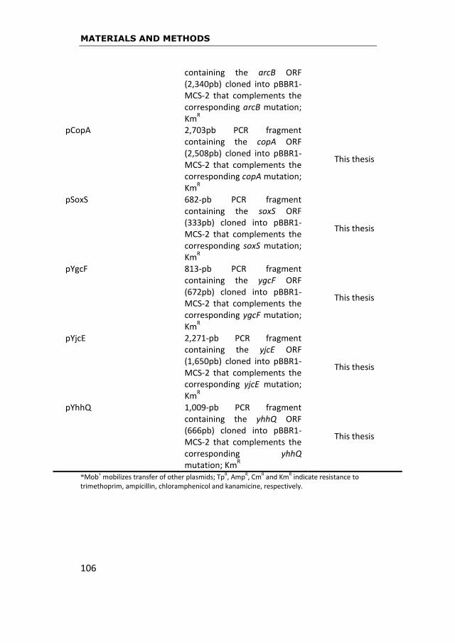
MATERIALS AND METHODS
106
*Mob+ mobilizes transfer of other plasmids; TpR, AmpR, CmR and KmR indicate resistance to trimethoprim, ampicillin, chloramphenicol and kanamicine, respectively.
containing the arcB ORF (2,340pb) cloned into pBBR1-MCS-2 that complements the corresponding arcB mutation; Km
R
pCopA 2,703pb PCR fragment containing the copA ORF (2,508pb) cloned into pBBR1-MCS-2 that complements the corresponding copA mutation; Km
R
This thesis
pSoxS 682-pb PCR fragment containing the soxS ORF (333pb) cloned into pBBR1-MCS-2 that complements the corresponding soxS mutation; Km
R
This thesis
pYgcF 813-pb PCR fragment containing the ygcF ORF (672pb) cloned into pBBR1-MCS-2 that complements the corresponding ygcF mutation; Km
R
This thesis
pYjcE 2,271-pb PCR fragment containing the yjcE ORF (1,650pb) cloned into pBBR1-MCS-2 that complements the corresponding yjcE mutation; Km
R
This thesis
pYhhQ 1,009-pb PCR fragment containing the yhhQ ORF (666pb) cloned into pBBR1-MCS-2 that complements the corresponding yhhQ mutation; Km
R
This thesis
MATERIALS AND METHODS
107
3.3. Short-term assay for E. amylovora survival in AB medium
supplemented with different concentrations of copper
Containers with 250mL of AB medium (Alexander et al., 1999) were
supplemented or not with copper sulfate at four concentrations
chosen below the MIC: 0.005, 0.01, 0.05 and 0.5mM and then
inoculated with a suspension of CFBP 1430 E. amylovora strain at a
final concentration of 107CFU/mL. Containers were incubated for 7
days at 26ºC without shaking. Culturability of E. amylovora cells after
copper exposure was measured by plating serial ten dilutions on KB
at different time points and plates were incubated at 26ºC for 72h.
3.4. RNA isolation
For the gene expression assays detailed in 3.5., five different
commercial kits to extract total RNA were tested with the purpose of
selecting that yielding enough and highest quality RNA: RNeasy Mini
Kit (Qiagen), PureLink RNA mini kit (Invitrogen), RiboPure bacteria
isolation kit (Life technologies), mirVana miRNA isolation kit
(Invitrogen) and RealTime ready cell Lysis kit (Roche). Total RNA was
extracted with each different kit, from a suspension of approximately
109 CFU/mL of strain CFPB 1430 growed in KB medium for 72h and
then measured with NanoDrop 2000 UV-Vis spectrophotometer
(Thermo Scientific). Since only the last kit included a final step to
remove DNA, for the rest of kits we added this step with the TURBO
DNA-free kit (Life Technologies). With this exception, in all cases, kits
were used according to manufacturer directions.
MATERIALS AND METHODS
108
3.5. Stress-related selected genes. Primers and probes for relative
quantitation of gene expression by real-time RT-PCR
For an overview of stress-related genes potentially playing a role
in E. amylovora survival under copper conditions, we selected rpoS,
katG and dsbC genes for expression evaluation assays in presence of
copper. These three genes, previously described in section 1.5.4, are
involved in the response to general (rpoS), oxidative (katG) and
copper (dsbC) stresses, in other bacterial models (Battesti, et al.,
2011; Landini et al., 2014; Madar et al., 2013; Hiniker et al., 2005).
Primers and probes (TaqMan) for target sequences of these genes
were designed based on the genome sequence of the European
reference E. amylovora strain CFBP 1430 (Smits et al., 2010) and
using Primer Express® Software v.3.0.1. (Applied Biosystems):
- rpoS gene: forward primer, 5’–GTTTTGGCCTGTTAGGCTATGAA-3’;
reverse primer, 5’–CTCACGGGTCAAACCAATTTC-3’; probe: FAM-
CGGCCACGCTGGAAGATGTAGGC-TAM.
- katG gene: forward primer, 5’–CCCGCTCAATGTGAATTTCA-3’;
reverse primer, 5’–CAGTGATTTGAGGTCGGCTTTA-3’; probe: FAM-
ATGAGTTCCGCAAACTGGATTATTCCCAG-TAM.
- dsbC gene: forward primer, 5’–TATCCGGCAGTCGTTAAGCA-3’;
reverse primer, 5’–TGGCGAAGGTTGGATCTCA-3’; probe: FAM-
ACTGGGCTTACAACAGA-TAM.
MATERIALS AND METHODS
109
The RNA used for real-time RT-PCR assays had different origins. To
test the sensitivity of the amplification protocol, RNA was extracted
from a suspension of approximately 109 CFU/mL of strain CFBP 1430
growed in KB medium for 72h, in the case of the elaboration of
standard curve for real-time RT-PCR (section 3.6.), RNA was obtained
from a transcription reaction with MEGAscript T7 transcription kit
(Life Technologies) and in the case of section 3.4 and 3.7., RNA was
extracted from strain CFBP 1430 cells incubated 7 or 50 days in
containers with AB medium and different concentrations of copper.
The real-time PCR assay was performed in a 12µL reaction mixture
containing 2μL RNA, 6μL 2X TaqMan AgPath-ID MasterMix (with ROX)
(Life Technologies), 0.5μL M-MLV reverse transcriptase (Life
Technologies), 0.12μL each of forward and reverse primers (100μM),
and 0.3μL of probe (6μM). Amplification and detection were
performed with a LightCycler Real-Time PCR detection system
(Roche) under the following conditions: RNA was reverse transcribed
at 45ºC for 10min, followed by PCR activation at 95ºC for 10min and
45 cycles of amplification (95ºC for 15sec and 60ºC for 45sec).
Analysis of assays was conducted with LightCycler 480 software
(version 1.5; Roche).

MATERIALS AND METHODS
110
3.6. Real-time RT-PCR standard curve for estimating PCR
amplification efficiency of rpoS gene
The rpoS gene was selected to follow amplification efficiency because
of its possible potential role in survival of E. amylovora under copper
stress. To addres the performance of the standard curve for rpoS
gene we used two strategies.
In a first strategy, based on clonning the rpoS gene into a vector, the
fragment contained the ORF and the entire promoter region of the
gene was amplified using new designed external forward primer (5’-
TGAACTCAACGCCAAGCAGC-3’) and reverse external primer (5’-
ATTTCACGCAGACGACGCAG-3’) from CFBP 1430 strain using Primer
Express® Software v.3.0.1. (Applied Biosystems). The PCR product
was cloned into pGEM-T easy vector system (Promega) and then
introduced in E. coli according to the manufacturer instructions. The
transformant colonies of E. coli were confirmed by PCR. Cloned
pGEM-T was extracted from E. coli with Real Miniprep turbo kit
(REAL) and digested with Not1. The multiple copies of rpoS DNA were
transcribed to RNA with MEGAscript T7 transcription kit (Life
Technologies). Serial 10-fold dilutions of rpoS RNA were prepared and
stored at -80ºC prior to use in standard curve generation.
In a second strategy, rpoS gene was amplified using the external
forward primer described above but cointaining the promoter of T7
RNApol (5’-TAATACGACTCACTATAGGGTGAACTCAACGCCAAGCAGC-
3’) and the reverse primer as described before. The PCR product was

MATERIALS AND METHODS
111
cleaned with UltraClean PCR Clean-up kit (MoBio). Then, rpoS DNA
was transcribed to RNA with MEGAscript T7 transcription kit and
purified with MEGAclear transcription Clean-up kit (Life
Technologies). Serial 10-fold dilutions of rpoS RNA were prepared and
stored at -80ºC prior to use in standard curve generation.
In all cases, the quantity of RNA transcripted was measured with
NanoDrop 2000 UV-Vis spectrophotometer (Thermo Scientific),
Quant-iT RiboGreen RNA assay kit (Life Technologies) and Qubit®
Fluorometric Quantitation (Life Technologies) device.
The real-time RT-PCR assay was performed as described before in
3.5.
PCR amplification efficiency is the rate at which a PCR amplicon is
generated (ABI Guide, 2004), commonly presented as a percentage
value. If a particular PCR amplicon doubles in quantity during the
geometric phase of its PCR amplification then the PCR assay has
100% efficiency. The slope of a standard curve is commonly used to
estimate the PCR amplification efficiency of a real-time PCR reaction.
A real-time PCR standard curve is graphically represented as a semi-
log regression line plot of CT value vs. log of input nucleic acid. A
standard curve slope of -3.32 indicates a PCR reaction with 100%
efficiency (ABI Guide, 2004). A calculation for estimating the
efficiency (E) of a real-time PCR assay according to ABI Guide is:
E= (10-1/slope) X 100

MATERIALS AND METHODS
112
3.7. Long-term assay for E. amylovora survival in AB medium
supplemented with copper sulfate and expression of rpoS gene
under these conditions
Containers with 250mL of AB medium were supplemented or not
with copper sulfate at 0.005mM and then inoculated with a
suspension of CFBP 1430 E. amylovora strain at a final concentration
of 107CFU/mL. This copper concentration was selected when the
results of section 3.3. were obtained. Containers were incubated for
50 days at 26ºC without shaking. Culturability of E. amylovora strain
CFBP 1430 cells after copper exposure was measured by plating serial
ten dilutions on KB medium at different time points. Plates were
incubated at 26ºC for 72h. To determine if nonculturable cells were
dead or still viable and have entered in the VBNC state, aliquots from
containers were stained with the bacterial viability kit LIVE/DEAD
(BacLight, Life Technologies) according to manufacturer instructions,
and then observed with a Nikon ECLIPSE E800 epifluorescence
microscope.
At each time point, an aliquot from containers was saved with
glycerol at -20ºC. When appropriate, RNA was extracted from those
samples with RNeasy Mini Kit (Qiagen), cleaned with TURBO DNA-
free kit (Life Technologies) and analyzed by real-time RT-PCR with
primers for rpoS gene. Primers, probes and protocol to run these RT-
PCRs were described before in section 3.5. This experiment was
repeated twice.

MATERIALS AND METHODS
113
Data were stadistically analyzed using general linear model
technique. It is possible to perform this using regression analysis by
dichotomizing all factors. This technique is similar to regression
models but the idea behind it is a generalization of analysis of
variance models. P-value used for rejecting the null hypothesis was
0.05 as is usual in other studies of the discipline.
3.8. Microarray experiment
As a more powerful approach to the objective of studying the gene
expression under copper stress conditions, we selected a
transcriptomic tool that could provide a general view of the genes
affected when E. amylovora faces a copper shock.
3.8.1. Erwinia amylovora microarray
The microarray used was previously designed at the James Hutton
Institute (JHI; formely Scottish Crop Research Institute (SCRI)) and
synthesized by Agilent Technologies, Inc. (Palo Alto, CA, USA), it
contains 3,483 target sequences (annotated genes and pseudogenes)
of the main E. amylovora ATCC 49946 genome (accession
NC_013971; Sebaihia et al., 2010), in a design on a single 8 x 15k
format slide (McNally et al., 2012).
Each slide contained eight arrays and each array had nearly 12,000
spots, containing the probes in triplicate. Besides the 3,483 target
sequences of the E. amylovora ATCC 49946 genome, a further 483
target genes or simple gene predictions from five sequenced

MATERIALS AND METHODS
114
plasmids were present: plasmids 1 and 2 for the same strain
(accessions NC_013972, NC_013973; McGhee and Jones 2000;
Sebaihia et al., 2010), pEL60 and pEU30 (accessions NC_005246,
NC_005247; Foster et al., 2004) and pEI70 (accession NC_018999,
from the Spanish strain E. amylovora IVIA-1614-2a; Llop et al., 2011).
Up to five sense orientation candidate probes per target were
designed with the Agilent eArray webtool, using temperature
matching methodology, a preferred probe melting temperature of
80ºC, no 3’ bias and a target length of 60bp. Any short probes were
later extended to 60bp using the Agilent linker. BLASTN (Altschul et
al., 1997) and Biopython (Cock et al., 2009) were used to identify
potential cross-hybridization in order to rank the candidate probes.
The selection of one probe per genome target, and up to five probes
per plasmid target, allowed all of the probes to be present in
triplicate.
We performed experiments using two microarray slides to evaluate
the transcriptomic response of E. amylovora strain Ea1189 after a
copper shock. Each slide contained the RNA extraction of the strain
after being treated or not with 0.5mM copper sulfate. Moreover,
RNA came from two different bacteria cultures (biological replicates)
submitted to the same experimental conditions. Therefore, three
independent experiments were carried out with a total of six
replicates (two replicates from slide 1 and four replicates from slide
2). The experimental array design of each single 8 x 15k format slide
is detailed in Table 5.

MATERIALS AND METHODS
115
Table 5. Experimental design of the array to evaluate E. amylovora gene expression after copper shock. NT means non-treated, T means treated, BR means biological replicate. Time of copper exposure, 0 or 5min.
SLIDE 1
NT0 NT5
T0 T5
SLIDE 2
NT0 NT0 NT5 NT5
T0 T0 T5 T5
3.8.2. Experimental set-up for preparation of samples for
microarray hybridization
As described, all these molecular assays were run with strain Ea1189
of E. amylovora. A suspension from five colonies coming from a plate
of LB medium growed 72h was obtained. Then, a flask with 100mL of
LB broth was inoculated with 100μL of the suspension and incubated
16h at 28ºC and 200rpm to a density of approximately 109CFU/mL.
After that period of time, the culture was splited in two fractions of
50mL. One fraction was copper-shock challenged to 0.5mM CuSO4 for
5 min, whereas the other one was used as control (not copper
BR 1
BR 2 BR 2
BR 1

MATERIALS AND METHODS
116
challenged). This copper concentration was selected according to
Yamamoto and Ishihama (2005). Cells were then treated with
RNAprotect (Qiagen). Total RNA from 1mL culture was extracted by
using SV Total RNA Isolation System (Promega) (Fig. 13). It was
quantified with NanoDrop 2000 UV-Vis spectrophotometer (Thermo
Scientific) and quality checked using an RNA 6000 Nano Kit on a 2100
Bionalyzer (Agilent Technologies). Fluorescent labelling of total RNA
was performed as described by Venkatesh et al. (2006) (Fig. 13). The
design incorporated a dye-swap and balanced labelling of all samples.
Levels and efficiencies of labelling were estimated using the
NanoDrop spectrophotometer mentioned before. Microarray
hybridization, washing and scanning were performed at the JHI
Sequencing and Microarray Facility of Michigan State University
(USA) as described previously (Stushnoff et al., 2010) (Fig. 13). They
were conducted according to the manufacturer’s protocols (Agilent
Two-Color Microarray-Based Gene Expression Analysis, version 5.5).
Briefly, 20µl labeled samples were added to 5µl 10X blocking agent
(Agilent 5188-5242), heat denatured at 98ºC for 3 min, and then
cooled to room temperature. GE hybridization buffer HI-RPM 2X
(25µl) was added and mixed prior to hybridization at 65ºC for 17h at
10 rpm. Array slides were dismantled in Wash 1 buffer (Agilent, 5188-
5327) and washed in Wash 1 buffer for 1 min, then in Wash 2 buffer
(Agilent, 5188-5327) for 1 min, and centrifuged dry. Hybridized slides
were scanned using an Agilent G2505B scanner at resolution of 5µm
at 532nm (Cy3) and 633nm (Cy5, for two-channel analysis)
wavelengths with extended dynamic range (laser settings at 100%

MATERIALS AND METHODS
117
and 10%) (Fig. 13). Microarray images were imported into Agilent
Feature Extraction (FE) (v.9.5.3) software and aligned with the
appropriate array grid template file (021826_D_F_20081029).
Intensity data and quality control (QC) metrics were extracted using
the recommended FE protocol (GE2-v5_95_Feb07). Entire FE
datasets for each array were loaded into GeneSpring (v.7.3) software
for further analysis.
Figure 13. General view of an oligonucleotide microarray performance. http://www.u.arizona.edu/~gwatts/azcc/help_old.html RNA is extracted from control and test cells. Then, the isolated RNA is labeled differently and hybridized with a microarray. The detection of the different fluorescence from different hybridized probes is detected by a scan.
MATERIALS AND METHODS
118
3.8.3. Microarray data analysis
Data were normalized using default settings: for two-channel arrays,
data were transformed to account for dye-swaps and data from each
array were normalized, according to McNally et al. (2012), using the
Lowess algorithm to minimize differences in dye incorporation
efficiency. Unreliable data flagged as absent in all replicate samples
by the FE software were discarded. Significantly changing gene lists
were generated from combined replicate datasets for each time
point using volcano plot filtering (fold-change ratio >1.5x; Student’s t-
test with P value <0.05). In each independent experiment, technical
replicates were analyzed individually. A gene was classified as
differentially expressed when it was present at least in one biological
replicate of both independent experiments and not in opposite
expression in any other replicates. Since fold-change ratios were
lower than 1.5, we selected genes with values ≥1 for up-regulated
genes and values ≤1 for down-regulated genes, in a way similar to the
study of Mitchell et al. (2010). Those genes differentially expressed in
both independent experiments were selected for further analysis.
Functional classification of the differentially expressed genes was
based on the gene ontology using the GPRO v1.1.0 software.
3.9. Validation of differentially expressed genes by quantitative
real-time PCR
Twenty-five out of fourty-four genes differentially expressed after
copper shock were selected for quantitative real-time PCR validation.

MATERIALS AND METHODS
119
These validation assays were performed in collaboration with
Estación Experimental del Zaidín (EEZ), Granada, Spain. Those genes
were selected attending to its up-regulation and/or product
description obtained from data bases, regarding their possible
relation with the response of E. amylovora to copper stress. The
selected genes are described in Table 6.
Table 6. Selection of E. amylovora genes differentially expressed after microarray analysis for validation through quantitative real-time PCR.
Gene Product description according to NCBI
copA Copper exporting ATPase ygcF Conserved hypothetical protein yhhQ Hypothetical protein tatC Twin-arginine protein translocation system soxS Transcriptional regulator galF UTP-glucose-1-phosphate uridylyltransferase apt Adenine phosphoribosyltransferase ydhC Inner membrane transport cheA Chemotaxis protein nodT RND efflux system, multidrug resistance protein CusA-like EAM_1634 Hypothetical protein gltI Glutamate and aspartate ABC transporter rimI Ribosomal-protein-alanine acetyltransferase arcB Aerobic respiration sensor-response protein tag3 3-methyl-adenine DNA glycosylase I EAM_2853 Putative helicase/relaxase yjcE Na
+/H
+ exchanger
uspA Universal stress protein smdB Multidrug efflux pump ABC transporter yfcA Inner membrane protein EAM_3469 Phage holin tufA Protein chain elongation factor ET-Tu dfoA Desferrioxamine siderophore biosynthesis oppA Oligopeptide ABC ppc Phosphoenolpyruvate carboxylase
MATERIALS AND METHODS
120
Gene expression was measured by quantitative real-time PCR
performed in triplicate using new RNA samples taken from cultures
of strain Ea1189 treated with copper as described above for the
microarray hybridization assays. RNA was extracted as described in
section 3.8.2. and after its quality was confirmed, it was transcribed
to cDNA using SuperScript II Reverse transcriptase (Invitrogen)
according to the manufacturer’s protocol. The optimization of
quantitative real-time PCR was set up first using the 16S gene as an
internal control at the melting temperature of each primer, using the
iQSybrGreen Supermix (BioRad) (Pffafl, 2001). Primers were designed
using Primer Express® Software v.3.0.1. (Applied Biosystems) based
on the genome sequence of the North American strain isolated from
apple, Ea273 (ATCC 49946) (Sebaihia et al., 2010), they were used at
20μM and the sequences are listed in Table 7. All reactions were
conducted with a real-time PCR System iQ5 (Bio-Rad) and
amplification was carried out with one cycle at 95°C for 5 min,
followed by 40 cycles of denaturation at 95°C for 30s and annealing
at 63°C for 30s. Nonspecific primer activity was monitored using a
dissociation curve and the resulting threshold cycles (CT) were
determined using Bio-Rad iQ5 Software 2.1 (Bio-Rad). The 16S rDNA
gene was used as an endogenous control in quantitative real-time
PCR to calculate the relative expression. Data of CT were analyzed
quantitatively via the comparative cycle threshold (∆∆CT) method
(Pffafl, 2001) to generate relative fold-change values comparing WT
and mutant strains. Each quantitative real-time PCR analysis was
performed in triplicate.

MATERIALS AND METHODS
121
Table 7. Primers used for validation of microarray expressed genes by quantitative real-time PCR.
Gene Sequence (5’-3’)
Forward Reverse
copA CCG GCG CAA CAG AGT GA CAC ATT GTG CAA CGC ATT CC
ygcF CAT TGC ACG GCG GAA AC CGC ACT AAC GCC TGG TTG A
yhhQ AGC GGC ATG GTG GAT AGC AAC GGA TCG CTG CTT TGA T
tatC CAC CCC GAT CAA GCT AAC CA GCG CTC GTG GCG ATA GAG
soxS CGA GCA AAA CGA TTA TGG CTA TC GAA CAC CCG CTG GAA ACA CT
galF GCT GCA TGA TGA AGC GTT TG TCG CCG CCA GGT TGT AAC
apt AAC CGG CGG CAC CAT T TCG CCA CTG AGG TCA AAC AA
ydhC GGG TCT TTC ACC TGC CGA TA AGC GCA CGA CAG CTA AAG C
cheA GAC GTT GCG CGT CCT GAT A CAG CAC GCT AAA CCC AAA GG
nodT CGA GCG GGC AAA GAT CAC CAG ATG CGA TTC GGC AGT AA
EAM_1634 CAG AAA CGC CAA CAC AGG AA GCC GTC ATG CGT TTG GTT
gltI CGT CCG TCC CCT TCT CCT A CCT GGA GAT CGG GTT TGT TC
rimI GCG GTA GAT CCG GCA TTT C CCT CCA GCC ACA GGG TCA T
arcB AGA TGT CGC GGC GTG AA TCA AGA AAA GAG CGC AGA AAT G
tag3 CGG CTC CAC CAC CTG TTA CT TGT CGG GAT GGC GAA AAC
EAM_2853 TTG CAG GAG GCA CGG TTA C GCG GAG GAA TAT GGC CTG TT
yjcE GCG GAA CAT ATT GGC GTT TC CGG AGC ATG GCG AAT AAT G
uspA CGG TGA ATC GCC TGC TAA AA AAT GCG CAC ATC GAC CAG TA
smdB TGG CAG GTG AGC GAA TTT TC TCA GGT CGC GAA TGT CGA T
yfcA TGC TGT TTC TCG TGG CGT TA CAG GGC GGG AAC CGT TA
EAM_3469 TGG CGT TCT GGC TCA ATG T CCC CGA GAT TTG CCA GAG TA
tufA CCA CGC CGA CTA TGT GAA AA GGC ATT GGG CCG TCA GT
dfoA AAC GGC GTT TTT CTC GAT CA GGC GCT TTC CAG CAT CAT
oppA GAA GGC GTA ACG GAA TCG AA GCC GCC CTC TTT GCT TTC
ppc GGG CCC CGC TGT ATA CCT GCA CGC CAA AGC ACT TCA C
16S TCC CTA GCT GGT CTG AGA GGA T GCT GCC TCC CGT AGG AGT CT

MATERIALS AND METHODS
122
3.10. Mutant construction and complementation
Standard recombinant DNA techniques were used as described by
Sambrook et al. (1989). To make specific mutants of E. amylovora
Ea1189 strain, non-polar chromosomal mutations were generated
using the phage λ red recombinase system (pKD46) previously
described (Datsenko and Wanner 2000; Zhao et al., 2009; McNally et
al., 2012). We decided to make mutants in copA, soxS, arcB, yjcE,
ygcF, yhhQ, galF and EAM_3469 genes, because their higher
expression level as measured by quantitative real-time PCR and/or
their interest as potentially related to the response of E. amylovora
facing copper stress. Single-gene recombinatorial deletions were
confirmed by PCR using the primers described in Table 8, based on
the genome sequence of the North American strain, Ea273
(ATCC49946) (Sebaihia et al., 2010). Afterwards, we selected copA,
soxS, arcB, yjcE, ygcF and yhhQ mutants to confirm that their
phenotypes were due to the absence of the target gene. Thus,
fragments containning copA, soxS, arcB, yjcE, ygcF or yhhQ genes
were obtained by PCR amplification of genomic DNA from strain
Ea1189 using a specifically designed primer set (see Table 8), and
cloned between the XbaI and SacI sites of the broad-host-range
vector pBBR1-MCS-2 (Kovach et al., 1995). These fragments
contained the ORF and the entire promoter region of each gene (see
corresponding plasmids in Table 4). These recombinant plasmids
were introduced into the pertinent Ea1189 mutant by bi-parental
cross-streak mating using E.coli S17-1 harboring the plasmid of

MATERIALS AND METHODS
123
interest as the conjugative donor (Simon et al., 1983; Abarca-Grau et
al., 2012). The phenotype of these complemented mutants was
checked as described in section 3.11.

MATERIALS AND METHODS
124
Table 8. Primers used for mutant construction and complementation of genes of E. amylovora strain Ea1189.
Name Sequence (5’-3’)
Mutant construction
arcB_F arcB_R
AAATATTCGCTCCGATGCAC
GCAGGCGAATGATTCAAAAT
copA_F copA_R
TAAAAGCGGATTGCTTTGCT CACCTCTTGCGCTTATTTCC
soxS_F soxS_R
GCC TTTTATCGCGGAGTAGA TGAAGAGTGCTGTTGCAAGG
ygcF_F ygcF_R
TATCCGTGCAATCTGCCATA AACCCGTTCAGCCCTTTTAT
yhhQ_F yhhQ_R
TGAAGGACAGGACTGGGTTC ATC GGT TTT GAT GCT TCA GG
yjcE_F yjcE_R
CGCTTAACGCACTTCCTGTT GGGATGGGAGTAGCCATTCT
EAM_3469_F EAM_3469_R
TGTTCCGTGGAAAGGTAAGG TTGCTGCCTGTAGTGCAATC
galF_F galF_R
CGAAGCCCGCTGTATTTTT CACGGCTGTGCTCAGATTT
Gene cloning Product size (pb)
copA(XbaI)_F copA(SacI)_R
CCATCTAGAATGTTCATGGTTTCCCCTTG 2,703 CCAGAGCTCAATAACCGCAAAGCCATCTG
soxS(XbaI)_F soxS(SacI)_R
CCATCTAGACTAAGGCGTAGCTGGCAGAG 687 CCTGAGCTCGCTGTTGCAAGGAGTGTCAA
arcB(XbaI)_F arcB(SacI)_R
CCATCTAGAAAAAAGGCGACAAGCCATTA 2,829 CCTGAGCTCGGTTACCAATTTCCCTGGTG
yjcE(XbaI)_F yjcE(SacI)_R
CCATCTAGACCAGTTTGGGGCCGTAATTT 2,271 CCTTCTAGACTGTTTTCCATGCCAAGCCT
ygcF(XbaI)_F ygcF(SacI)_R
CCATCTAGAACATCCTGTATGGGGAGACG 813 CCTGAGCTCATGAGTGCGGACTTGACTTG
yhhQ(XbaI)_F yhhQ(SacI)_R
CCATCTAGACGGTAATATGCCGCAAAAGT 1,009 CCTGAGCTCCTGAAGCCATCGGTTTTGAT
MATERIALS AND METHODS
125
3.11. Experiments with mutants of E. amylovora to evaluate the
role of different genes
3.11.1. The posible role of rpoS gene in the survival of strain CFBP
1430
E. amylovora CFBP 1430∆rpoS mutant was kindly provided by R.D.
Santander (Santander et al., 2014). Containers with 4mL of AB
medium were supplemented or not with copper sulfate at 0.005,
0.01 and 0.5mM trying to simulate the conditions of section 3.3. and
then inoculated with a suspension of mutant strain at a final
concentration of 108CFU/mL. Containers were incubated for 64h at
26ºC without shaking. Culturability of wild type and mutant strains
throughout copper exposure was monitored by plating serial ten
dilutions from the containers on KB medium at different time points.
Plates were incubated at 26ºC for 72h.
3.11.2. Assays to evaluate the posible role of copA, soxS, arcB, yjcE,
ygcF, yhhQ, galF and EAM_3469 genes of strain Ea1189 after copper
shock
3.11.2.1. Copper tolerance in vitro. Wild type E. amylovora strain
Ea1189 was grown 16h in LB broth at 28°C in a shaking incubator to a
density of approximately 109CFU/mL and the culture was then splited
in two fractions as indicated in section 3.8.2. One fraction was
copper-shock challenged for 5 minutes to different copper sulfate
concentrations ranged from 0.5 to 35mM, to determine the maximal
copper concentration that had no impact on its culturability, whereas

MATERIALS AND METHODS
126
the other fraction was used as control (not copper challenged). Then,
an aliquot from each fraction was washed once with PBS and the
culturability of wild type was measured by plating serial ten dilutions
on LB medium. Plates were incubated at 26ºC for 72h.
Then, the concentration of copper sulfate which did not affect the
growth of E. amylovora Ea1189 (10mM) was selected to test the
culturability of selected mutants. Each mutant from a 72h culture
was grown 16h in LB broth at 28°C in a shaking incubator to a density
of approximately 109CFU/mL and the culture was then splited in two
fractions, as described before for wild type strain. One fraction was
challenged to 10mM copper sulfate for 5 minutes and the other one
used as control. An aliquot from each fraction was washed once with
PBS and the culturability was measured by plating serial ten dilutions
on LB medium supplemented with 100μg/mL ampicillin and 20μg/mL
chloramphenicol. Plates were incubated at 26ºC for 72h.
3.11.2.2. Effect of introduction of target genes in the mutants. Six
out of the eight genes selected to make mutants in E. amylovora,
were chosen to be introduced in the mutant strains (complemented
mutants). We selected those genes because of their product
description from data bases and their possible relation with the
response of E. amylovora to copper stress. After recombinant
plasmids containing the ORF and the entire promoter region of each
target gene, copA, soxS, arcB, yjcE, ygcF and yhhQ were introduced
into each corresponding E. amylovora Ea1189 mutant
(Ea1189∆geneX), the phenotypes were tested challenging each
MATERIALS AND METHODS
127
complemented mutant (Ea1189∆geneX(pGenX)) to 10mM copper
sulfate. Thus, E. amylovora Ea1189 wild type, the six mutants in the
target genes, and the new six complemented mutants were grown in
separate flasks with LB broth for 16h at 28°C in a shaking incubator.
Then, the culture of each strain was splited in two fractions. One
fraction was copper-shock challenged to 10mM copper sulfate for 5
min, whereas the other one was used as control. An aliquot from
each fraction was then washed once with PBS and the culturability
was measured by plating serial ten dilutions on LB medium
supplemented or not with 100μg/mL ampicillin and 20μg/mL
chloramphenicol. Plates were incubated at 26ºC for 72h.
3.12. The copA case.
As in the quantitative real-time PCR validation assays, copA gene
exhibited the highest expression of all genes tested, and it is reported
that it codes for a copper exporting ATPase, we selected it for further
studies.
3.12.1. Expression curve of copA in E. amylovora after copper-shock
induction. Wild type strain of E. amylovora Ea1189 was grown 16h in
LB broth at 28°C with shaking to a density of approximately
109CFU/mL. Then, aliquots of 10 mL were subjected to copper shock
with different copper sulfate concentrations ranging from 0.005 to
0.5mM for 5 minutes in a rocking shaker, with one fraction used as
untreated control (not copper-challenged). A volume of 0.5 mL of
each culture was treated with 1 mL of RNAprotect (Qiagen) and total

MATERIALS AND METHODS
128
RNA was extracted using the miRNeasy Mini kit (Qiagen) with an on-
colum RNAse-free DNAse treatment (Qiagen). cDNA was synthesized
from 1µg of purified RNA using TaqMan reverse transcription (RT)
reagents (Applied Biosystems), according to the manufacturer’s
protocol. Quantitative real-time PCR was used to determine the
relative expression levels of copA after induction with the different
treatments; 16S rDNA gene was used as an endogenous control.
qPCR was performed on a CFX96 Real-Time PCR Detection System
(BioRad Laboratories) with the SYBR® Green PCR Master Mix (Applied
Biosystems). Efficiencies for each primer set were obtained from a
standard curve using ten-fold dilution series up 106 of one cDNA
sample of the untreated control at the melting temperature of the
copA primer. Primers of copA and 16S rDNA genes were the same
used for validation expression after microarray assay (section 3.9)
and the sequences are listed in Table 7. Optimization of primer
concentration was carried out by qPCR using combinations of
different primer concentrations (50, 300 and 900 nM) and the
undiluted cDNA sample of the untreated WT strain. Optimal
concentration was selected as the combination with the lowest Ct
value. The thermal cycling conditions were one cycle at 95°C for 5
min, followed by 40 cycles of denaturation at 95°C for 30s and
annealing at 63°C for 30s. Each biological replicate was run in
triplicate. Melting curves were generated to evaluate primer
specificity and rule out primer dimer formation. CT values were
analyzed quantitatively via the comparative cycle threshold (∆∆CT)
method (Pffafl, 2001) to generate relative fold-change values

MATERIALS AND METHODS
129
comparing WT and copper-shock induced WT strains. Each
quantitative real-time PCR analysis was performed in triplicate.
3.12.2. Copper tolerance in planta. Wild type Ea1189, mutant
Ea1189∆copA and complemented mutant Ea1189∆copA(pCopA)
were assayed at the same time in this experiment.
Fresh in planta pear shoots taken from 8 years old pear trees cv.
‘Conference’ growing inside a mesh were inoculated in a P2 security
lab by immersion in a suspension of aprox. 109 CFU/mL of each E.
amylovora strain, potted in closed flask of approx. 13cm diameter
and with 1% agar. These flasks were incubated at 25ºC in a climate
chamber at 8h light-16h darkness for 24h. Then, after 24h
inoculation, 4mL of a solution of copper sulfate at 10mM was sprayed
on leaves, and the first sampling was made (time 0). Pots were again
incubated in the climate chamber and periodically sampled at
different time points throughout 29 hours. The procedure of sample
processing at each time point was as follows: one leaf was pull out
and two square pieces of aproximatelly 1cm2 were cut with a sterile
scalpel. Leaf pieces were crushed in 1mL PBS 0.5X. Culturable cell
counts were then determined by plating ten serial dilutions on LB
medium and incubating the plates at 26ºC. CFU counts were
performed after 72h.
Data were statistically analyzed using a generalized mixed model
technique (McCulloch and Searle, 2001), where sprout nested in

MATERIALS AND METHODS
130
experiment was a random effect while the rest of factors were
considered fixed. P-value was 0.05.

RESULTS
131
4
RESULTS

RESULTS
132

RESULTS
133
4.1. Minimal inhibitory concentration (MIC) for copper
To evaluate which medium was the most recommendable to perform
the MIC assays, we tested three media, CYE, LB and KB for copper
bounding ability. Out of the three tested growth media, LB and KB
bounded each of them 60% of the copper added, whereas CYE
medium did not exhibit ability to bound it, as measured by Copper
test Microquant. CYE medium was then selected to assay the MIC for
copper sulfate compared to KB medium.
A suspension of each strain of E. amylovora (CFBP 1430, Ea1189 and
IVIA-1892) was prepared and plotted on CYE and KB media
supplemented or not with copper sulfate. All the strains grew at
copper sulfate concentrations below 1mM on KB and CYE media, but
only the strains plotted on KB medium grew at this concentration
(Fig. 14). Therefore, the concentration of 1mM was established as the
MIC for copper sulfate in E. amylovora under assayed conditions in
CYE medium.
Figure 14. MIC for copper in two media. Left side, CYE medium with no growth at 1mM copper sulfate of the strains assayed. Right side, KB medium with growth of all strains at 1mM copper sulfate.
RESULTS
134
4.2. Short-term assay for E. amylovora survival in AB medium
supplemented with different copper concentrations
Once determined the MIC for copper sulfate on solid medium,
culturability of E. amylovora strain CFBP 1430 in liquid AB medium at
concentrations lower than 1mM was assayed. As shown in Fig. 15,
culturable cells decreased in approximately one and a half
logarithmic order through the seven days of the experiment, from
107 CFU/mL to 4.5 x 10⁵ CFU/mL, in absence of copper. Treatment
with 0.005mM copper sulfate resulted in a loss of culturability of
almost three orders of magnitude in the same time period, dropping
to 2 x 10⁴ CFU/mL at day seven (Fig. 15). At the other copper sulfate
concentrations of 0.01, 0.05 and 0.5mM, culturability of E. amylovora
CFBP 1430 was under the detection limit just after 24 hours of
exposure (Fig. 15).
4.3. Efficiency of RNA extraction
In all the experiments, performed with strain CFBP 1430, no
differences either in quantity or in quality were found between the
five commercial kits used for RNA extraction after repeated trials
with each of them and under the same conditions. Then, considering
its simplicity and easy to use, we selected RNeasy Mini Kit (Qiagen)
for all the RNA extractions, followed by a treatment with TURBO
DNA-free kit (Life Technologies) to remove DNA from RNA.

RESULTS
135
Figure 15. Short-term assay of the E. amylovora survival in AB medium supplemented with copper.

RESULTS
136
4.4. Sensitivity of rpoS, katG and dsbC primers
The sensitivity of the protocol for detection of strain CFBP 1430 by
the primers from rpoS and katG genes by real-time PCR was
103CFU/mL, as observed in amplification curves of the different cell
concentrations (Fig. 16 A and B). The amplification with katG primers,
however, showed not good reproducibility.
Figure 16. Primers sensitivity of stress-related genes in E. amylovora CFBP 1430 strain. A) rpoS and B) katG by real-time PCR. Fluorescence means wave length emited by TaqMan probe at each decimal dilution. C) Sensitivity of dsbC primers by conventional PCR. In A, B and C, decimal numbers indicate CFU/mL.
A)
B)
108 107 106 105 104 103
104
105
106
107
108 103
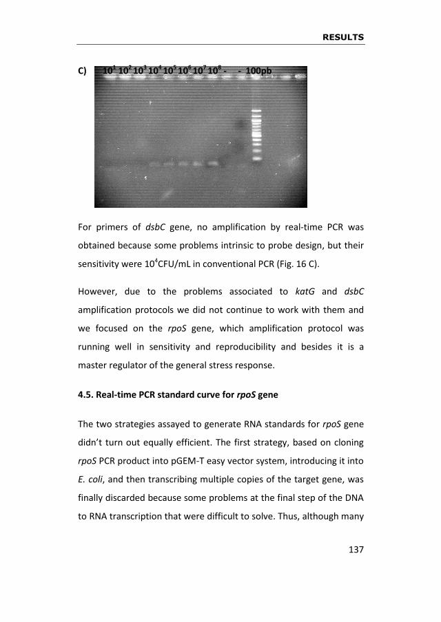
RESULTS
137
C) 101 102 103 104 105 106 107 108 - - 100pb
For primers of dsbC gene, no amplification by real-time PCR was
obtained because some problems intrinsic to probe design, but their
sensitivity were 104CFU/mL in conventional PCR (Fig. 16 C).
However, due to the problems associated to katG and dsbC
amplification protocols we did not continue to work with them and
we focused on the rpoS gene, which amplification protocol was
running well in sensitivity and reproducibility and besides it is a
master regulator of the general stress response.
4.5. Real-time PCR standard curve for rpoS gene
The two strategies assayed to generate RNA standards for rpoS gene
didn’t turn out equally efficient. The first strategy, based on cloning
rpoS PCR product into pGEM-T easy vector system, introducing it into
E. coli, and then transcribing multiple copies of the target gene, was
finally discarded because some problems at the final step of the DNA
to RNA transcription that were difficult to solve. Thus, although many

RESULTS
138
modifications were introduced in the protocol, an insufficient
amount of digested plasmid was achieved.
Therefore, we finally chose the second strategy, based on
amplification of rpoS gene using the external forward primer
containing the promoter of T7 RNApolymerase, which avoids an
excessively long clonation process since the amplification product of
the gene is directly transcribed to RNA. This strategy worked very
well and it did not present any handicap.
The quantity of RNA was comparatively measured with NanoDrop
2000 UV-Vis spectrophotometer (Thermo Scientific), Quant-iT
RiboGreen RNA assay kit (Life Technologies) and Qubit® Fluorometric
Quantitation (Life Technologies) device. Finally, the last one
demonstrated to have the most reliable measure because of its
accuracy based on the detection of RNA-specific fluorescence, and it
was used to quantifying RNA in the standard curve for rpoS gene.
The semi-log regression line of CT values versus log of input nucleic
acid is represented in Fig. 17. According to this standard curve, the
efficiency of PCR amplification for rpoS gene was around 82%, since a
100% efficient reaction will yield a 10-fold increase in PCR amplicon
every 3.32 cycles during the exponential phase of amplification
(log210 = 3.3219), and the slope of the standard curve for rpoS gene
in our assay was -2.71, more positive than -3.32 characteristic of
100% efficiency.

RESULTS
139
Figure 17. Calibration curve, using RNA standards for rpoS gene, graphically represented as a semi-log regression line plot of Ct (cycle threshold) value vs log of input nucleic acid.
4.6. Long-term assay for E. amylovora survival in AB medium
supplemented with copper sulfate and expression of rpoS gene
through time
The lowest copper concentration assayed in short-term survival
assays (0.005mM) which allowed the maintining of culturability in the
short-term (Fig. 15), was selected for long-term assays. At long term,
culturability of E. amylovora CFBP 1430 strain decreased in more
than three logarithmic orders after 50 days in AB medium with no
copper, whereas viability only went down one log order in the same
time period (Fig. 18 A).

RESULTS
140
Figure 18. E. amylovora strain CFBP 1430 survival in AB medium supplemented with 0.005mM copper sulfate and expression of the rpoS gene through time. CT means cycle threshold.
A)
B)
CT
CT
RESULTS
141
However, in the presence of copper the reduction in population
numbers was more drastic than in absence of copper, both in
culturability and viability, since culturable cells decreased in five log
orders and viable cells in approximately two from the time 0 (Fig. 18
B). The trend of the respective curves for both culturable (p=0.0472)
and viable (p=0.0099) cells was, in fact, significantly different
between copper and no copper treatments.
Throughout this long-term period, expression of rpoS gene was
evaluated by real-time PCR at each time point. The number of CT in
AB medium with no copper increased the first 15 days from 22.9 to
29.8, and then remained approximately constant until day 50 (Fig. 18
A). In contrast, in the presence of copper the value of CT, after a slight
initial increase, remained approximately constant around 25 (Fig. 18
B). This increase in CT, regardless the presence of copper, indicated a
reduction in rpoS expression throughout the time. However, the fact
that the value of CT was lower in presence than in absence of copper
all the time assayed (p<0.0001) suggests a higher level of expression
of the gene under copper stress.
4.7. General characteristics of the E. amylovora Ea1189 strain
transcriptome in response to a copper shock
After the study of the role of rpoS gene in copper survival of E.
amylovora strain CFBP 1430 and in order to have a more complete
picture of the expressed genes by copper stress, a two-color whole
genome microarray approach was used to identify differentially
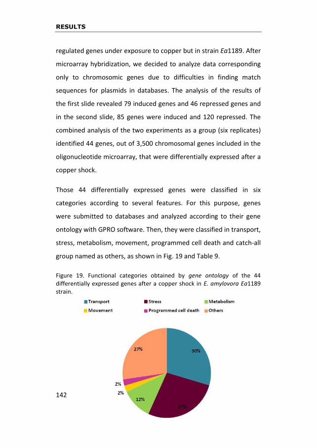
RESULTS
142
regulated genes under exposure to copper but in strain Ea1189. After
microarray hybridization, we decided to analyze data corresponding
only to chromosomic genes due to difficulties in finding match
sequences for plasmids in databases. The analysis of the results of
the first slide revealed 79 induced genes and 46 repressed genes and
in the second slide, 85 genes were induced and 120 repressed. The
combined analysis of the two experiments as a group (six replicates)
identified 44 genes, out of 3,500 chromosomal genes included in the
oligonucleotide microarray, that were differentially expressed after a
copper shock.
Those 44 differentially expressed genes were classified in six
categories according to several features. For this purpose, genes
were submitted to databases and analyzed according to their gene
ontology with GPRO software. Then, they were classified in transport,
stress, metabolism, movement, programmed cell death and catch-all
group named as others, as shown in Fig. 19 and Table 9.
Figure 19. Functional categories obtained by gene ontology of the 44 differentially expressed genes after a copper shock in E. amylovora Ea1189 strain.

RESULTS
143
Table 9. Genes differentially expressed in E. amylovora Ea1189 in response to a copper shock. Bold words indicate up-regulated genes.
Functional categorya
Gene IDb Gene namec
Product description Fold
changed
Transport EAM_0320 yjcE Na+/H+ exchanger 1.2
EAM_1003 smdB Multidrug efflux pump ABC transporter
1.5
EAM_1136 gltI Glutamate and aspartate ABC transporter
1.3
EAM_1661 ydhC Inner membrane transport protein
1.6
EAM_1897 oppA Oligopeptide ABC transporter
1.3
EAM_2340 yfcA Inner membrane protein 1.7
EAM_2748 nodT RND efflux system, multidrug resistance protein (partial) detox protein CusA-like
1.3
EAM_0222 secE Preprotein translocase subunit
-1.3
EAM_0954 proY Proline-specific permease -1.2
EAM_1044 copA Copper exporting ATPase -1.1
EAM_1612 spy Periplasmic protein spheroplast protein Y
-1.2
EAM_1884 ompW Outer membrane protein -1.2
EAM_3433 tag3 3-methyl-adenine DNA glycosylase I
-1.3
Stress EAM_0207 tatC Twin-arginine protein translocation system
1.2
EAM_0221 tufA Protein chain elongation factor EF-Tu
1.6
EAM_0360 dfoA Desferrioxamine siderophore biosynthesis
1.1
EAM_0568 rimI Ribosomal-protein-alanine acetyltransferase
1.2
EAM_1023 apt Adenine phosphoribosyltransferase
1.2
EAM_2126 galF UTP-glucose-1-phosphate uridylyltransferase
1.2
EAM_3348 soxS Transcriptional regulator 1.2
EAM_0073 rpmB 50S ribosomal protein L28 -1.4
EAM_0127 rpmE 50S ribosomal protein L28 -1.4
EAM_1116 cspE Cold shock-like protein -1.5
EAM_2370 uspA Universal stress protein -1.2

RESULTS
144
a) Functional category obtained by gene ontology study using GPRO software v1.1.0. b) Number of the gene according to E. amylovora ATCC 49946 sequencing genome. c) Gene name according to highest similarity query with protein BLAST database. Genes in bold black are
those that were up-regulated. d) Obtained with a p-value ≤ 0.05.
Transport category contained 13 genes whose functions were related
with exchangers, ABC transporters, membrane proteins and efflux
systems. We found several interesting genes as copA gene, coding for
SERP1273
EAM_2853 Putative helicase/relaxase -1.5
Metabolism EAM_0136 ppc Phosphoenolpyruvate carboxylase
1.2
EAM_0704 lpxC UDP-3-O-acyl N-acetylglucosamine deacetylase
-1.4
EAM_0861 Hypothetical polysaccharide deacetylase
-1.1
EAM_2964 1-acyl-sn-glycerol-3-phosphate acyltransferase
-1.1
EAM_3110 arcB Aerobic respiration sensor-response protein
-1.5
Movement EAM_2538 cheA Chemotaxis protein 1.9
Programmed cell death
EAM_3469
Phage holin 1.1
Others EAM_2701 ygcF Conserved hypothetical protein
1.3
EAM_0537 Hypothetical protein 1.8
EAM_2645 Hypothetical protein 1.2
EAM_2818 Hypothetical protein 1.4
EAM_3301 yhhQ Hypothetical protein 1.2
EAM_3421 Hypothetical protein 1.3
EAM_0684 No significant similarities found
-1.7
EAM_0927 psiF Phosphate starvation-inducible protein
-1.3
EAM_1312 ybjP Putative lipoprotein -1.2
EAM_1634 Hypothetical protein -1.1
EAM_2851 Hypothetical protein -1.1
EAM_3016 Hypothetical protein -1
RESULTS
145
a copper exporting ATPase; yjcE gene, coding for a Na+/H+ exchanger;
two inner membrane proteins YdhC and YfcA and one outer
membrane protein OmpW; smdB gene, coding for a multidrug efflux
pump ABC; nodT gene, coding for a RND efflux system; oppA gene,
coding for a oligopeptide ABC transporter; gltI gene, coding for a
glutamate and aspartate ABC transporter or tag3 gene, coding for a
3-methyl-adenine DNA glycosylase I. Fold-changes expression of
these genes ranged between -1.1 and 1.7 (Table 9).
Stress category contained 12 genes whose functions were related
with translocation systems, elongation factors (EF-Tu), siderophore
biosynthesis, transcriptional regulators or stress proteins. We
considered the transcriptional regulator soxS the most relevant gene
of this category. However, we also found other interesting genes
such as dfoA gene involved in desferrioxamine biosynthesis; cspE
gene, coding for a cold shock-like protein or uspA coding for a
universal stress protein; tatC gene, coding for a twin-arginine protein
translocation system; tufA gene, coding for a protein chain
elongation factor; rimI gene, coding for a ribosomal-protein-alanine
acetyltransferase; apt gene, coding for a adenine
phosphoribosyltransferase; galF gene, coding for a UTP-glucose-1-
phosphate uridylyltransferase and EAM_2853 gene, coding for a
putative helicase/relaxase. Fold-changes expression range was
between -1.5 and 1.6 (Table 9).
Metabolism category was smaller than the previous ones because it
only contained 5 genes whose functions were related with Krebs’s

RESULTS
146
cycle and a respiration sensor-response protein. This last function,
coded by arcB gene, was apparently the most important founded in
this category because it is related to an aerobic-anaerobic switch that
could be connected with an answer to redox events produced by
copper. Most of the genes in this category were down-regulated with
fold-changes ranging from -1.1 to -1.5. Only ppc gene, coding for a
phosphoenolpyruvate carboxylase that catalizes the β-carboxilation
of phosphoenolpyruvate (PEP), was 1.2fold-change up-regulated. This
is a high energetic reaction that supplies dicarboxilic acids to the
tricarbolixic acid’s cycle (Krebs’s cycle) (Table 9).
Only one gene, cheA, coding for a chemotaxis protein, was allocated
to movement category. And only other gene, EAM_3469, was
classified in the group of programmed cell death since it codes for a
phage holin. Both genes were up-regulated with fold-changes of 1.9
and 1.1, respectively (Table 9).
No function was found for twelve out of the 44 genes differentially
expressed after copper shock. We classified them inside a category
named “others”. Only the sequences for ygcF, yhhQ, psiF and ybjP
genes were annotated but others like the one for EAM_1634 gene
were not and were classified only as hypothetical proteins. We
selected ygcF and yhhQ for further analysis because they were up-
regulated and, in the case of ygcF, also due to be conserved. We also
selected EAM_1634 gene because it was down-regulated (Table 9).

RESULTS
147
4.8. Evaluation of differentially expressed genes under copper
conditions by quantitative real-time PCR
To validate gene expression patterns observed in our microarray
analysis by an specific and independent technology, the expression of
25 genes, selected among those identified as differentially expressed,
was examined by quantitative real-time PCR after optimizing RNAs
according to 16S rRNA levels. According to the formula E=10-1/slope, in
an ideal situation, the quantity of PCR product should duplicates in
each amplification cycle. As it is shown in Table 10, the optimization
of each pair of primers for quantitative real-time PCR is around 2,
what means they have 100% efficiency. Selection of genes was made
according to their probable relation with the function resulted from
the annotation with databases and GPRO software, and the putative
function in the case of the hypothetical proteins. These genes were:
yjcE, smdB, gltI, ydhC, oppA, yfcA, nodT, copA and tag3 from
transport category; tatC, tufA, dfoA, rimI, apt, galF, soxS, uspA and
EAM_2853 genes from stress; ppc and arcB genes from metabolism;
cheA gene from movement; EAM_3469 gene from programmed cell
death; and ygcF, yhhQ, and EAM_1634 genes from “others”
category.
Expression of selected target genes by real-time PCR upon exposure
to copper was not completely in accordance with the microarray
results. The most remarkable divergence was the copA gene, which
exhibited down-regulation in the microarray analysis but had the
highest up-regulation when its expression was measured by
RESULTS
148
quantitative real-time PCR, with a value of more than 19-fold
increase (Fig. 20). Other genes that showed down-regulation in the
microarray but were induced as monitored by PCR were EAM_1634,
arcB, tag3, EAM_2853 and uspA, with values of induction of 1.79-,
1.60-, 1.51-, 1.49- and 1.37-fold increase, respectively (Fig. 20).
The relative expression of the rest of the analyzed genes ranged from
0.96-fold induction for gene dfoA to 3.82-fold induction for gene
ygcF, and all of them were confirmed as up-regulated (Fig. 20).
Finally, only oppA and ppc genes, both up-regulated in the
microarray analysis, were revealed as down-regulated by quantitative
real-time PCR in response to copper.
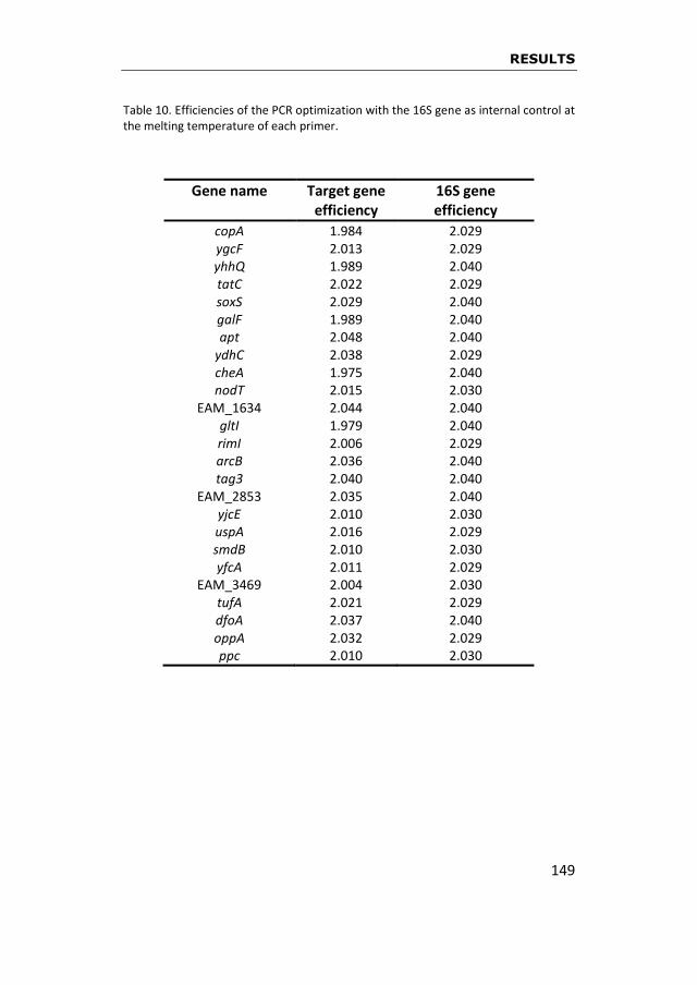
RESULTS
149
Table 10. Efficiencies of the PCR optimization with the 16S gene as internal control at the melting temperature of each primer.
Gene name Target gene efficiency
16S gene efficiency
copA 1.984 2.029 ygcF 2.013 2.029 yhhQ 1.989 2.040 tatC 2.022 2.029 soxS 2.029 2.040 galF 1.989 2.040 apt 2.048 2.040
ydhC 2.038 2.029 cheA 1.975 2.040 nodT 2.015 2.030
EAM_1634 2.044 2.040 gltI 1.979 2.040 rimI 2.006 2.029 arcB 2.036 2.040 tag3 2.040 2.040
EAM_2853 2.035 2.040 yjcE 2.010 2.030 uspA 2.016 2.029 smdB 2.010 2.030 yfcA 2.011 2.029
EAM_3469 2.004 2.030 tufA 2.021 2.029 dfoA 2.037 2.040 oppA 2.032 2.029 ppc 2.010 2.030

RESULTS
150
Figure 20. Relative expression of 25 differentially expressed genes in E. amylovora Ea1189 after a copper shock. Color indicates the functional category assigned before in section 4.7. For more info also see Fig. 19 and table 8.
The 16S rDNA gene was used as an endogenous control in quantitative real-time PCR to calculate the relative expression. Data of CT were analyzed quantitatively via the comparative cycle threshold (∆∆CT) method (Pffafl, 2001) to generate relative fold-change values comparing WT and mutant strains. * Inactivated genes for further studies.

RESULTS
151
4.9. Mutants of selected genes of E. amylovora
4.9.1. Assays to evaluate the posible role of rpoS gene
A mutant of CFBP 1430 strain in rpoS gene (Santander et al., 2014),
was used to evaluate its role in culturability in the presence of
copper. As shown in Fig. 21, mutant culturability was lower than that
of wild type, even in AB medium with no copper (Fig. 21). Whereas
the wild type strain remained near 108 CFU/mL for more than 60 days
(Fig. 21 A), the mutant dropped two logarithmic orders in the same
period (Fig. 21 B).
The difference between the wild type and the mutant was more
pronounced in the presence of copper. At the lowest copper
concentration (0.005mM) culturability of the mutant was reduced in
more than six log orders, and at 0.01mM it already was under the
detection limit at 24h (Fig. 21 B). This was contrasting to the
culturability of the wild type, which was decreasing gradually through
time (Fig. 21 A). At the highest copper concentration (0.5mM) the
culturability of both the wild type and the mutant was under the
detection limit already at 24h (Fig. 21).

RESULTS
152
Figure 21. (A) Survival of the E. amylovora CFBP 1430 wild type incubated in AB medium supplemented with copper sulfate along 64 hours. (B) Survival of the E. amylovora CFBP 1430 rpoS
- mutant incubated in AB medium
supplemented with copper sulfate along 64 hours.
A)
B)
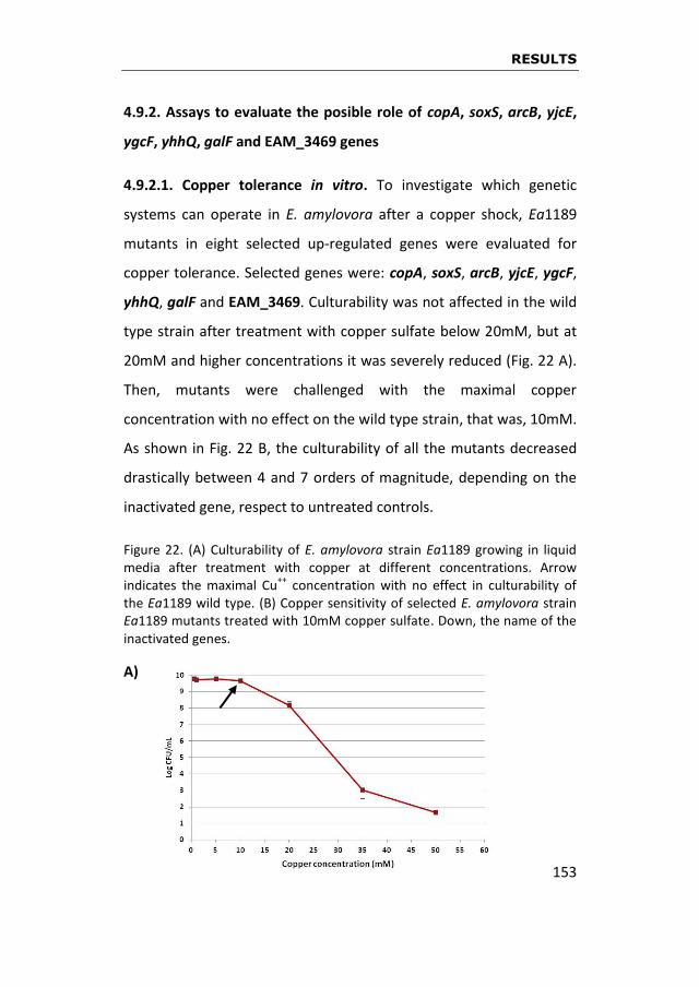
RESULTS
153
4.9.2. Assays to evaluate the posible role of copA, soxS, arcB, yjcE,
ygcF, yhhQ, galF and EAM_3469 genes
4.9.2.1. Copper tolerance in vitro. To investigate which genetic
systems can operate in E. amylovora after a copper shock, Ea1189
mutants in eight selected up-regulated genes were evaluated for
copper tolerance. Selected genes were: copA, soxS, arcB, yjcE, ygcF,
yhhQ, galF and EAM_3469. Culturability was not affected in the wild
type strain after treatment with copper sulfate below 20mM, but at
20mM and higher concentrations it was severely reduced (Fig. 22 A).
Then, mutants were challenged with the maximal copper
concentration with no effect on the wild type strain, that was, 10mM.
As shown in Fig. 22 B, the culturability of all the mutants decreased
drastically between 4 and 7 orders of magnitude, depending on the
inactivated gene, respect to untreated controls.
Figure 22. (A) Culturability of E. amylovora strain Ea1189 growing in liquid media after treatment with copper at different concentrations. Arrow indicates the maximal Cu
++ concentration with no effect in culturability of
the Ea1189 wild type. (B) Copper sensitivity of selected E. amylovora strain Ea1189 mutants treated with 10mM copper sulfate. Down, the name of the inactivated genes.
A)

RESULTS
154
B)
4.9.2.2. Introduction of target genes in the mutants. The ORF of
each selected gene as well as the upstream mutagenic region were
amplified by PCR, cloned into pBBR1 and introduced into each
respective Ea1189 mutant. The cloned copA gene restored almost
completely the culturability of the mutant, which dropped more than
six orders of magnitude after a copper shock respect to no copper
treatment and respect to that of wild type strain under the same
copper shock conditions (Fig. 23 A). Culturability of arcB, soxS and
yhhQ mutants decreased approximately by two orders of magnitude
respect to wild type strain (Fig. 23 B, C, E), whereas that of ygcF and
yjcE mutants was reduced about four orders of magnitude (Fig. 23 D,
F). When the respective genes were introduced in each mutant, the
wild phenotypes were restored in all cases (Fig. 23), indicating an
important role of copA, arcB, soxS, yhhQ, ygcF and yjcE genes for E.
amylovora survival facing copper shock.

RESULTS
155
Figure 23. Genes confirmed to have a role in the defense of E. amylovora against a copper shock. A) copA gene B) arcB gene, C) soxS gene, D) ygcF gene, E) yhhQ and F) yjcE gene. Down, WT means wild type, Ea1189∆geneX means mutant strain and Ea1189∆geneX(pGeneX) means complemented mutant.
A)
B)
C)

RESULTS
156
D)
E)
F)

RESULTS
157
The genes EAM_3469 and galF were not complemented due to lack
of time in the period of this thesis but we will keep them in mind for
future research.
4.10. The copA case
4.10.1. Induced expression of copA gene
In order to assess the differential expression of copA gene in E.
amylovora strain Ea1189, a relative quantification by real-time PCR
was performed after submitting the strain to a copper-shock
induction. As shown in figure 24, copA gene was induced in the
presence of copper. A very low copper concentration of 0.005mM
increased the expression of copA 1.5-fold compared with the
expression at no copper exposure (1-fold). Interestingly, the relative
expression of copA gene was observed to raise as soon as the copper
concentration was increased, reaching 16.8-fold expression when the
strain was challenged with copper sulfate 0.5mM (Fig. 24).
Figure 24. Relative expression curve of copA gene in E. amylovora after copper-shock induction.

RESULTS
158
4.10.2. Involvement of the copA gene in copper tolerance in planta
Due to its high expression level in the presence of copper (Fig. 20),
coupled with its demonstrated role in copper tolerance in vitro (Fig.
23 A), copA gene was selected to further investigate whether it also
plays a role in copper tolerance in vivo in E. amylovora Ea1189. The
culturability of copA mutant in planta was firstly assayed without
copper treatment, and it was slightly lower than that of wild type
(Fig. 25, blue and pink dashed lines), but the trend of both curves,
those of wild type and mutant, through time was not significantly
different (p=0.12). Interestingly, when the cloned copA gene was
inserted in the mutant, the numbers of culturable cells recovered
those of wild type (Fig. 25, green dashed line). Then, the effect of
copper was comparatively evaluated in the three bacterial
populations: the wild type, the copA mutant and the mutant
complemented with copA gene. As seen in Fig. 25, copper affected to
both mutant (blue continuous line) and wild type (pink continuous
line) strains, but this effect was more drastic for the mutant. Thus,
culturability of wild type was decreased in planta after two hours of
copper exposure (Fig. 25, pink continuous versus pink dashed lines),
reducing its population almost two orders of magnitude and
mantaining at this level throughout the experiment. As for the
mutant strain, significant differences in culturability were observed
respect to wild type strain at 29h after the copper exposure
(p=0.009), with a loss of about four orders of magnitude from the
inoculation time (Fig. 25, blue continuous line). However at this time,

RESULTS
159
the culturability of the mutant was restored when the cloned copA
gene had been introduced (Fig. 25, green continuous line). Although
this restoration was only partial, as occurred in in vitro assays, the
survival of wild type and the complemented mutant was not
significantly different (p=0.25) through time, confirming the effect of
copper in copA mutant.

RESULTS
160
Figure 25. Role of copA gene in E. amylovora strain Ea1189 to 10mM copper sulfate in planta. Dashed lines means untreated and continuous lines means after being treated with 10mM of copper sulfate. Wild type strain is represented by a pink circle, mutant Ea1189∆copA is represented by a blue rhombus and complemented mutant Ea1189∆copA(pCopA) is represented by a green triangle.

GENERAL DISCUSSION
161
5
GENERAL
DISCUSSION

GENERAL DISCUSSION
162
GENERAL DISCUSSION
163
Copper-based compounds are among the antibacterial agents
most widely used in agriculture, and they have been extensively
employed for decades to deal with the devastating plant disease fire
blight (van der Zwet et al., 2012). But it has also been described that
they are not completely efficient to control the disease because,
when plants are treated with copper, E. amylovora can develop a
survival strategy that allows it to persist in time: the viable but
nonculturable (VBNC) state (Ordax et al., 2006). In fact, the role of
copper as a stress factor for E. amylovora was described by the
research group in which the present thesis was performed (Ordax,
2008), so the goals of this work were planned as a continuation of the
previous findings. Thus, once determined that copper triggers a
survival response in E. amylovora, we were interested in unraveling
the molecular mechanisms involved in this interaction.
First, we addressed this issue by selecting some genes, such as
the Sigma factor rpoS, that were previously described as important in
the bacterial response to several stresses (Battesti et al., 2011;
Landini et al., 2014), although not specifically reported in relation to
copper. Secondly, we decided to go forward with a transcriptomic
study to evaluate the global response of E. amylovora to copper,
taking advantage of the customized microarray for E. amylovora by
courtesy of Ian Toth (The James Hutton Institute (JHI), formely
Scottish Crop Research Institute (SCRI)). The experimental design,
based on the work of Yamamoto and Ishihama (2005) for another
enterobacterium, E. coli, consisted on challenging E. amylovora to a

GENERAL DISCUSSION
164
copper sulfate shock and to assess the genes induced under this
stressful condition.
All the data obtained from the microarray allowed designing
experiments to evaluate the role of some selected genes in vitro and
lately in vivo. We made a selection of genes differentially expressed
by copper to validate their expression using quantitative real-time
PCR. Then, we constructed E. amylovora mutants in those genes
exhibiting a higher expression level as measured by quantitative real-
time PCR and/or their interest as potentially related to the response
of E. amylovora facing copper stress. Then, we tested the phenotype
of these mutants after challenging them to copper and we
complemented the mutants with the introduction of the
corresponding gene and verified that the phenotype of the wild type
strain was restored. Finally, we selected one of the genes to test its
role also in vivo. In fact, the validation in planta of the role of copA
gene in the survival of E. amylovora to copper exposure is one of the
most relevant results of this work. Nevertheless, other remarkable
result is the general overview of the genes involved in the survival of
E. amylovora to copper stress. Up to date, the global transcriptional
response of E. amylovora against copper had not been elucidated
yet. The type of copper stress selected in this study was a rapid shock
of 5 min, not only reproducing the conditions of a previous work with
E. coli (Yamamoto and Ishihama, 2005) but also to avoid the influence
of long-term exhaustion of nutrients, the production of toxic
metabolites coming from the active metabolism or other factors that
could interact with the direct copper effect.

GENERAL DISCUSSION
165
The information provided by this work constitutes a first step in
the way of a future research about possible targets for more efficient
chemical treatments for fire blight disease. According to the data
obtained, it is possible that a treatment against fire blight based on a
combination of copper sulfate and an inhibitor of CopA protein would
be more effective than copper alone. It is an issue for future
research.
After summarizing the global sense and the main achievements
of this thesis, the different tasks addressed in the whole work are
discussed in detail. As a previous step in the study of the molecular
response of E. amylovora to copper, the minimal inhibitory
concentration (MIC) of this metal was firstly determined to decide
the range of copper concentrations to be assayed. On one side, since
King’s B (KB) and Luria Bertani (LB) media bounded copper at the
same extent, we selected KB for comparative purposes because it
was used in previous MIC studies with E. amylovora (Ordax, 2008).
On the other side, we used CYE medium (Zevenhuizen et al., 1979)
and our results confirmed those reporting its usefulness for MIC
determination since it does not bind any copper (Sholberg et al.,
2001; Al-Daoude et al., 2009). In fact, the MIC obtained in CYE, 1mM,
was lower than in KB because KB has a greater ability to bind copper,
then reducing the amount of free copper that can affect bacterial
cells. Ordax (2008) also obtained a MIC higher than 1mM in KB
(2.5mM), even in liquid cultures, where all components, including
copper, are more accessible to cells, what usually leads to a decrease
GENERAL DISCUSSION
166
of the MIC, but not in KB due to its copper complexing ability.
Therefore, the MIC for the three studied strains was established in
CYE at 1mM, pretty similar to that obtained in other studies
performed using E. amylovora isolates from other geographical
origins, such as Syria (Al-Daoude et al., 2009) and British Columbia
(Sholberg et al., 2001), which were considered as copper tolerant
strains (growth at 1.2mM copper sulfate).

GENERAL DISCUSSION
167
5.1. Survival of E. amylovora under several copper conditions and the role of a general stress regulator
The survival of E. amylovora in the presence of two copper
concentrations below the established MIC (0.005 and 0.5mM) was
studied. The lowest copper concentration assayed (0.005mM) was
found to cause a significant decrease of two logarithmic orders in
CFBP1430 strain population, while the shock with higher copper
concentrations (up to 0.5mM) caused a more severe decrease. In
long-term survival assays in distilled water without addition of
copper, numbers of culturable cells were reduced in approximately
three logarithmic orders after 50 days due to the effect of nutrient
starvation (Biosca et al., 2006; Ordax et al., 2006). When adding
copper (0.005mM), the culturability dropped five log orders in the
same time period. In both cases (in absence or presence of copper) a
percentage of the population adopted the VBNC state, as previously
reported (Biosca et al., 2006; Ordax et al., 2006). This was indicated
by the fact that the number of viable cells, according to a culture-
independent method (Live & Dead), was not reduced at the same
extent as the number of culturable ones. Nevertheless, the fraction
of bacterial population in VBNC state was higher in presence of
copper due to the joined effect of two stress factors, copper and
starvation. The induction of this state by copper has not only been
demonstrated in E. amylovora (Ordax et al., 2006) but also in other
phytopathogenic bacteria (Alexander et al., 1999; Ghezzi and Steck,
1999; del Campo et al., 2009; Um et al., 2013). The present results
GENERAL DISCUSSION
168
are, therefore, consistent with those previously obtained by Ordax et
al. (2006). Interestingly, copper at all the assayed concentrations did
neither cause cellular lysis nor kill E. amylovora cells in other way
(Ordax et al., 2006). Bacteria are able to face up to stress through
adaptation responses, and many of these responses involve specific
pathways that include a given regulator and a set of regulated genes
(the regulon) which helps the cell to survive to the stress (Battesti et
al., 2011). In consequence, it is very interesting to evaluate the role
of these genes to elucidate molecular mechanisms underlying the
response of E. amylovora to copper stress. As a first approach, we
analyzed the expression of the general stress response regulator
gene, rpoS, because RpoS regulon has been reported as playing a
critical role in E. coli survival against acid (Small et al., 1994), heat
(Hengge-Aronis et al., 1991), oxidative stress (Sammartano et al.,
1986), starvation (Lange and Hengge-Aronis, 1991), and near-UV
exposure (Sammartano et al., 1986). In E. amylovora, an altered
expression of this gene under starvation stress has been recently
described (Santander et al., 2014), but nothing is known about its
relation with copper stress. In other Enterobacteriaceae as E. coli, the
RpoS regulon comprises 10% of its genes (Lacour and Landini, 2004;
Patten et al., 2004; Weber et al., 2005; Dong et al., 2008; Dong and
Schellhorn, 2009), what gives an idea of its relevance.
We observed that when E. amylovora was exposed to copper, the
level of rpoS expression was significantly higher than in absence of
copper. In both cases, with and without copper, there was a
GENERAL DISCUSSION
169
progressively reduction of rpoS expression through time, probably
because cells needed to save energy since they were maintained in a
non chemostat container with mineral medium for a very long period
(50 days). However, the reduction in the rpoS expression was
significantly less pronounced in the presence of copper, indicating
that this gene was more active than in absence of the metal. The link
between rpoS and copper is probably related to oxidative stress
provoked by copper excess. In E. coli, it has been described that
copper overload induces the expression of RpoS, OxyR and SoxRS
regulons (Macomber et al., 2007), and in P. aeruginosa that there is
an increase of a periplasmic protein as a reaction to copper
imbalance, leading to oxidative stress (Vijgenboom et al., 1997;
Raimunda et al., 2013). Moreover, our results are in accordance with
previous works on the role of the RpoS in the defense response of E.
amylovora (Santander et al., 2014) and also in other phytopathogenic
bacteria such as Xanthomonas campestris and Pseudomonas
fluorescens (Rao and Sureshkumar, 2000; Stockwell et al., 2009) in
defense against reactive oxygen species (ROS).

GENERAL DISCUSSION
170
5.2. Global response against a copper shock
With the aim to dig into the entire system of E. amylovora
genome-wide response to external copper, we carried out a search
for copper-responsive genes by a transcriptomic approach. The
experimental design was based on a study performed with copper
and the model bacterium E. coli (Yamamoto and Ishihama, 2005),
another member of Enterobacteriaceae, because no previous work
with E. amylovora on this issue has been reported.
We first carried out a systematic search for copper-
responsive genes by using DNA microarray technique. A copper shock
markedly activated 23 genes of the pathogen, being ca. 50% of those
induced genes classified into the categories of transport (yjcE, smdB,
gltI, ydhC, oppA, ycfA and EAM_2748) and stress (tatC, tufA, dfoA,
rimL, apt, galF and soxS), and some also into metabolism (ppc),
movement (cheA), and programmed cell death (EAM_3469),
remaining 6 as unclassified genes (EAM_0537, EAM_2645, ygcF,
EAM_2818, yhhQ and EAM_3421).
In the transcriptional assay previously conducted with E. coli
(Yamamoto and Ishihama, 2005), 30 copper-shock activated genes
were identified and classified into their putative regulons: 5 genes
were classified into the CusR regulon, 6 into the CueR regulon, 11
into the CpxR regulon and another 8 genes remained unclassified.
That previous study concluded that the copper stimulon is formed by
the activated genes under the control of at least four regulons: CueR,

GENERAL DISCUSSION
171
CusR, CpxR and YedW, in a hierarchical regulation network to
maintain copper homeostasis. Most of the genes identified in our
study differ from those identified in the E. coli copper shock
microarray study (Yamamoto and Ishihama, 2005). If the conditions
of each study are compared, although we isolated mRNA 5 min after
the copper shock, as in the E. coli study, the growth phase of the
challenged cells was different. Transcriptome of E. coli was obtained
in exponential growth while that of E. amylovora was obtained in late
exponential growth or beginning of the stationary phase. This
difference may have contributed to observed differences in gene
expression. However, both our current study with E. amylovora and
the previous study with E. coli included the hitherto identified copA
gene encoding a Cu+-translocating P-type ATPase pump, which
exports copper from the cytoplasm to the periplasm and is directly
involved in maintaining copper homeostasis in E. coli (Rensing and
Grass, 2003; Yamamoto and Ishihama, 2005). Besides copA, the
transcription factor for superoxide response regulon SoxS is copper-
activated both in E. coli (Yamamoto and Ishihama, 2005; Pletzer et
al., 2014) and in E. amylovora, as showed in the present work.
In plant pathogenic bacteria, there are no previous studies
evaluating transcriptional gene expression after copper stress. The
present study raises for the first time the association between copper
stress and its effects in the plant pathogenic bacterium E. amylovora,
copper homeostasis and production of oxidative radicals.

GENERAL DISCUSSION
172
5.2.1. Copper transport genes (copper homeostasis)
As it was described before, copper ion can be a toxic element
if it is present over a certain threshold, so metal homeostasis has an
essential role in bacterial survival. As a consequence of an aerobic
metabolism, copper catalyzes Fenton-like and Haber-Weiss reactions
that result in the production of hydroxyl radicals (Cu+ + H2O2 → HO• +
HO- + Cu++; O2•- + Cu++ → Cu+ + O2) (Halliwell and Gutteridge, 1990,
1984; Rensing and Franke-McDevitt, 2013). The main pool of
intracellular copper ions, as reported in E. coli, is located in the
periplasm (Outten et al., 2001), and the reactive oxygen species
(ROS) produced by copper do not reach the cytosolic targets
(Macomber et al., 2007; Lemire et al., 2013). However, if the
accumulation of ROS in the periplasm is over a certain threshold, a
leakage of these toxic species could happen into the cytosol, where
they trigger the activation of the ROS defense systems. To avoid
these events in cascade, metal efflux and uptake systems are
transport mechanisms displayed by bacterial cells to achieve copper
homeostasis.
The copper homeostasis protein repertoire described in E.
coli comprises two systems: one CueR-regulated, which plays a key
role in aerobic copper tolerance (Grass and Rensing, 2001; Outten et
al., 2001), and other regulated by cusSR for anaerobic conditions
(Outen et al., 2001). According to Hernández-Montes et al. (2012),
this dual copper homeostasis protein repertoire is only present in 3%
of all organisms tested (among others 268 gamma proteobacteria,
GENERAL DISCUSSION
173
two strains of E. amylovora (TAX552 and ATCC 49946), one strain of
E. pyrifoliae and one strain of E. tasmaniensis).
A recent phylogenomic study revealed that the genomes of
two E. amylovora strains (ATCC 49946 and TAX552) encode at least
three periplasmic proteins involved in copper homeostasis: CopA,
CueO and an orphan CusC (Hernandez-Montes et al., 2012). While
CopA pumps out excess copper from the cytoplasm to the periplasm,
CueO oxidizes Cu+ to Cu++ in the periplasm thereby reducing the
concentration of the more toxic Cu+ ion (Grass and Rensisng, 2001).
In E. coli, under anaerobic conditions the two-component regulatory
system CusSR activates the transcription of the cusCBAF operon that
encodes for a complex that pumps Cu+ from the cytoplasm directly to
the extracellular space (Grass and Rensisng, 2001). In most bacteria,
this complex consists of the inner membrane pump CusA, the
periplasmic protein CusB and the outer membrane protein CusC,
forming a channel through the periplasm (Hernandez-Montes et al.,
2012). In the genome of E. amylovora, only an orphan cusC gene is
present but, surprisingly, it is located in an operon together with two
other putative multidrug efflux transporter genes, emrB and emrA.
Thus, it is possible that they may form another complex pump also
dedicated to extrude Cu+ directly from the cytoplasm to the external
space, as the cusCBAF system does. However, since we did not
observe an upregulation of either emrB or emrA, these multidrug
efflux transporter genes may not respond to copper in aerobic
metabolism in E. amylovora, confirming that described for E. coli
(Yamamoto and Ishihama, 2005). It is important to note here that, in

GENERAL DISCUSSION
174
contrast to other enterobacteria, E. amylovora is not very efficient in
anaerobic metabolism (Sebaihia et al., 2010).
In this study, seven genes classified into transport category
were over-expressed in the presence of copper. Expression analyses
clearly show that copA gene is that most remarkably induced by
copper. This gene codes for a Cu+-translocating P-type ATPase pump
that exports copper from the cytoplasm to the periplasm. Although
Cu++ was added to the medium, it is reasonable to assume that it was
reduced to Cu+ intracellularly, and that Cu+ was the actual inducer of
a response (Rensing et al., 2000). The relevance of copA among the
mechanisms developed by E. amylovora against copper agrees with
results observed in previous studies with different gamma
proteobacteria: Hernández-Montes et al. (2012) found that CopA is
the most abundant protein for copper homeostasis in bacteria, with
an important physiological role as an internal membrane ATPase. It
was identified in the chromosomes of 70 bacterial genera, also the
genus Erwinia and it was found in all organisms cited before (among
others 268 gamma proteobacteria, two strains of E. amylovora
(TAX552 and ATCC 49946), one strain of E. pyrifoliae and one strain of
E. tasmaniensis) as the core protein for copper homeostasis. It is
interesting to note that CopA protein was also found in other genera
belonging to Enterobacteriaceae family as Dickeya, Pantoea,
Pectobacterium or Pseudomonas, which contain plant pathogenic
bacteria; on the contrary, it is not present in the genome of other
phytopathogenic bacteria, such as four strains of Xylella fastidiosa
and seven out of nine strains of Xanthomonas spp. The remarkable
GENERAL DISCUSSION
175
induction of copA gene by copper is in agreement with other gene
expression studies after copper exposure in non phytopathogenic
bacteria, such as Acidithiobacillus ferrooxidans, Streptococcus
pneumoniae or Listeria monocytogenes (Salazar et al., 2013; Shafeeq
et al., 2011; Corbett et al., 2011). Moreover, recently, Zhao et al.
(2014) have shown that copper homeostasis also plays an important
role in oxidative resistance of Deinococcus radiodurans and that this
role is played, among others, by copA.
We did observe some differences in gene expression
between the microarray results and qPCR for a few of the genes
examined, most notably copA. These differences could be due to the
limitations of the microarray technique. The analysis of the global
expression of one genome at a specific point in time is the result of
all genes from the genetic pool in that particular time. Then, the
results of a massive transcriptomic analysis technique should not be
taken to do an analysis of the differential expression of each
individual gene of the genetic pool, since some discrepancies can rise,
as has been described in other bacterial models (Thieme et al., 2008;
Karunakaran et al., 2009).
Expression analysis clearly showed that the copA gene in E.
amylvora is not only remarkably induced by copper but also fine-
tuned regulated. Three lines of evidence suggest that the expression
of copA is regulated by copper through the transcriptional regulator
CueR. First, sequence analysis outward from copA gene revealed that
in E. amylovora strain Ea1189 both genes are contiguous in the same

GENERAL DISCUSSION
176
locus, and they are transcribed in opposite directions (Figure S1).
Second, CueR activates transcription of copA in the presence of
copper ion in E. coli (Outten et al., 2000). And third, the CueR binding
site, including the CueR-box sequences, where the Cu+-CueR
transcriptional factor binds to the copA promoter, is known in E. coli
(Yamamoto and Ishihama, 2005). Sequence analysis revealed that the
copA promoter of E. amylovora strain Ea1189 contains the CueR box
consensus sequence, including the inverted repeat with just one
mismatch (Figure S2).
Our gene-disruption studies confirmed for the first time a
biological role of copA gene in E. amylovora survival after copper
shock because it was partially complemented in vitro and in planta by
addition of the copA gene with its native promoter in trans. In the E.
amylovora chromosome, copA is located adjacent to cueR, the main
gene involved in copA regulation, and it its possible that the genetic
context of copA may be important in this case. The 10-100%
reduction in survival of the wild type Ea1189 in planta upon
treatment with copper is a typical result observed following
treatment of a coper-sensitive bacterial pathogen on plant surfaces
with a copper bactericide (ex. Garrett and Schwartz 1998; Quesada et
al., 2010). Further reductions in survival of the ∆copA mutant could
be a reflection of increasing availability of copper on the plant
surfaces over time. These results suggest that the copA gene plays an
important role for survival of E. amylovora against copper both in
vitro and in planta.

GENERAL DISCUSSION
177
The present study also showed that other differentially
expressed genes classified into the transport family may direct or
indirectly be also involved in maintaining copper homeostasis in E.
amylovora. Concretely, the ydhC and smdB genes, belonging to drug
resistance efflux transporter family, and the transmembrane protein
YfcA might act reducing toxicity in the cytoplasm due to ion copper
effect. Further gene disruption studies will be required to
demonstrate their current role in copper tolerance in E. amylovora.
This is also the case of yjcE gene, coding for a Na+/H+ exchanger (NHE)
with similarity to NHEs in eukaryotes (Verkhovskaya et al., 2001) and
having a potential role in eliminating excess acid from cells.
Expression analysis and gene disruption studies with yjcE show that it
should play a role for copper tolerance in E. amylovora; this could be
accomplished through maintaining pH homeostasis in cells which
could reduce the cycling of Cu+ and Cu++ ions. It has also been
demonstrated in human cells (Akram et al., 2006) that an increase in
intracellular superoxide anion induces synthesis of an NHE that
strongly correlates with the resistance of cells to death.

GENERAL DISCUSSION
178
5.2.2. Oxidative stress (copper toxicity)
The strong relationship between the presence of a stress
factor and the production of superoxide radicals has been reported
by microarray studies in other bacterial models as Pyrococcus
furiosus, Nitrosomonas europaea, Herminiimonas arsenicoxydans or
Bacillus cereus (Williams et al., 2007; Park and Ely, 2008; Cleiss-
Arnold et al., 2010; Mols et al., 2010). In the case of E. amylovora, the
effect of copper in previous studies on culturability led to conclude
that this metal acts as a stress factor also for this pathogen (Ordax et
al., 2006). Besides maintaining copper homeostasis, bacteria also
have to overcome its toxic effect. As commented before, the main
toxicity of copper could derive from the production of reactive
oxygen species (ROS), as previously described by Halliwell and
Gutteridge (1984) in E. coli (see section 1.3.1. of the Introduction),
since copper dissociates the oxygen peroxide produced by an aerobic
metabolism into hydroxyl radicals. This is genetically supported by
the fact that, in the present study, seven differentially-expressed
genes were stress-related and they may direct or indirectly be
involved in overcoming the toxic effect of this metal in E. amylovora.
We identified three ROS related genes that were
differentially expressed after a copper shock. A gene-disruption study
was performed for the AraC family DNA-binding transcriptional
regulator, SoxS, and the ∆soxS mutant was highly affected in copper
tolerance, losing more than six orders of magnitude in population
numbers. It is well known that copper sulphate is a powerful inducer

GENERAL DISCUSSION
179
of the soxRS regulon of E. coli through oxidative stress (Greenberg et
al., 1990; Kimura and Nishioka, 1997). An intracellular redox signal
activates SoxR, a superoxide response sensor protein encoded by
soxR. It binds to DNA at a specific site activating the expression of
soxS gene, and SoxS protein is a direct inducer of target genes of
bacterial response to oxidative stress (Lushchak, 2011). The soxS
gene has been already characterized in E. amylovora (Pletzer et al.,
2014) and its regulon was identified in E. coli (Martin and Rosner,
2002). This study shows that copper is one of the signals for
activation of soxRS system in E. amylovora and that some SoxRS-
regulated genes of its regulon should play and important role in its
copper tolerance.
Another ROS-related gene found in this work was arcB.
Expression analysis and gene disruption studies with arcB show that
the two-component regulatory system ArcAB is activated by copper
and it is required for copper tolerance in E. amylovora. ArcB activates
its cognate transcriptional regulator ArcA to repress genes
contributing to aerobic metabolism and activates genes necessary for
anaerobic metabolism (Loui et al., 2009). Thus, in an anaerobic
habitat, bacteria avoid more ROS production and, consequently,
oxidative damage. This study shows that copper is one of the signals
for activation of ArcAB system and that some ArcAB-regulated genes
of its regulon should play an important role in copper tolerance in E.
amylovora. It seems that, for E. amylovora, this strategy to avoid ROS
production is not preferred because anaerobic metabolism is not the
optimal one for an efficient growth in E. amylovora. The fact that this

GENERAL DISCUSSION
180
gene appears in previous studies related to ROS in the bacterial
model E. coli (Luchi and Weiner, 1996; Loui et al., 2009) may support
the hypothesis that ArcAB system is a metabolic strategy against
copper toxicity in E. amylovora not described before.
The third ROS-related gene has been described above, yjcE. It
is also connected with copper homeostasis. Our observation that E.
amylovora ∆yjcE mutant was also strongly affected after copper
shock supports the hypothesis that pH homeostasis can affect copper
sensitivity.
Another interesting gene classified into the stress category is
galF. The enzyme GalF is involved in the formation of UDP-glucose
from α-D-glucose-1P, which is essential for polysaccharide
production. Up to date, no direct relation between this gene and
copper or others stress factors had been described. However, we
observed an increase of almost 3-fold expression in this gene and the
reduced culturability of the ∆galF mutant strain for more than six
orders of magnitude after copper exposure, suggesting that this gene
is in fact involved, at least indirectly through protection by
exopolysaccharide production, in the response of E. amylovora
against copper stress.

GENERAL DISCUSSION
181
5.3. Response model to copper in E. amylovora
The data obtained show that E. amylovora responds to
copper shock by over-expressing several genes related to two genetic
systems that represent two complementary strategies: (i) the control
of copper homeostasis through the expression of, at least, the copA
gene, and (ii) the overcoming of copper toxicity due to oxidative
stress through the expression of some ROS-related genes.
All results together allowed us elaborating a putative model
of the different genetic mechanisms that probably are involved in the
interaction between E. amylovora and copper in the assayed
conditions (Figure 26). Copper probably crosses the external
membrane and enters periplasm via porins, as in E. coli (Teitzel et al.,
2006) and many others Gram-negative bacteria (de Brujin, 2016), and
then reaches the cytoplasm crossing the inner membrane via certain
transport proteins. Once in the cytoplasm, copper generates ROS
through Fenton-like reactions. In order to prevent the oxidative
damage, E. amylovora displays several mechanisms. The most
important one seems to be a compendium of actions aimed to
maintain copper homeostasis and to face up ROS by the activation of
soxS (Fig. 26A) and yjcE (Fig. 26B) gene expression. The first one
activates the expression of other genes related with the elimination
of ROS, and the second one contributes to neutralize the acid pH
generated by ROS inside the cytoplasm. The activity of these genes is
supported by the action of CopA protein (Fig. 26C), which exports
copper from inside the cell to the periplasm space. Besides, the
GENERAL DISCUSSION
182
activation of arcB gene (Fig. 26D) allows changing from aerobic to
anaerobic metabolism, which would drastically reduce the
production of ROS. Finally, the role of the two hypothetical proteins
YgcF and YhhQ (Fig. 26E) remains to be elucidated.
The results of this thesis have constituted an approach to the
genetic substrate of E. amylovora response against copper stress, and
they are a starting point to make progress in the knowledge of the
complex molecular mechanisms involved in the copper response of
the fire blight pathogen.

GENERAL DISCUSSION
183
Figure 26. Putative model of the effect of copper stress in Erwinia amylovora

GENERAL DISCUSSION
184

CONCLUSIONS
185
6
CONCLUSIONS

CONCLUSIONS
186

CONCLUSIONS
187
This work was designed to improve the knowledge of the
molecular mechanisms involved in the response of E. amylovora to
copper stress, a topic not previously afforded in plant pathogenic
bacteria. The new results obtained are summarized in the following
conclusions:
1. Copper, even at very low concentrations such as 0.005mM,
constitutes a stressful factor for E. amylovora and triggers a
response, observed at least as a reduced culturability, with a genetic
substrate constituted by the coordinated action of several genes
belonging to different functional categories.
2. The general stress response regulator gene rpoS contributes to the
survival of E. amylovora under copper exposure, since its expression
was consistently higher in presence than in absence of copper when
E. amylovora was challenged to copper and starvation stressful
conditions.
3. A set of at least fourty four genes exhibit a differential expression
after copper shock. Twenty-five of them belong to functional
categories of transport and stress. Genes related to metabolism
(five), movement (one) and programmed cell death (one) also show
an altered expression. The validation of the differential expression of
twenty-five selected genes showed that twenty-three of them
increased its expression after copper shock.
4. The genes arcB, copA, EAM_3469, galF, soxS, ygcF, yhhQ and yjcE
are involved in the survival of E. amylovora when it is exposed to
CONCLUSIONS
188
copper sulfate, from concentrations as low as 10mM. The role of six
of these genes, arcB, copA, soxS, ygcF, yhhQ and yjcE, was confirmed
under in vitro conditions, since the complementation of the mutants
in those genes restored the wild type phenotype.
5. The differential expression under copper shock of seven genes
related to transport suggests its direct or indirect involvement in
maintaining copper homeostasis in E. amylovora. Genes ydhC and
smdB, that have concern in drug efflux, and YfcA, that is a
transmembrane protein, probably act reducing the toxicity of copper
present in cytoplasm, whereas yjcE gene coding for a Na+/H+
exchanger probably acts neutralizing the acid pH generated by
reactive oxygen species provoked by copper.
6. From the seven transport genes differentially expressed under
copper exposure, the copA gene, which codes for a Cu+-translocating
P-type ATPase pump that exports copper from the cytoplasm to the
periplasm, is the most remarkably induced by copper and even fine-
tune regulated, playing an important role as defense mechanism of E.
amylovora against copper stress both in vitro and in planta. The
expression of copA gene seems to be transcriptionally regulated by
copper through the regulator CueR but not through CusSR.
7. The increased expression of soxS gene, involved in superoxide
resistance, under copper shock confirms its connection with oxidative
stress and it also shows, for the first time, that copper can be one of
the signals for activation of SoxRS system.

CONCLUSIONS
189
8. The induction of arcB gene after activation of ArcAB system
triggered by copper suggests another strategy, complementary to the
role of soxS, to stop reactive oxygen species production through an
aerobic/anaerobic switch, since ArcB activates ArcA to repress genes
contributing to aerobic metabolism and activates those necessary for
anaerobic one.
9. The gene galF is also involved in the response of E. amylovora
against copper probably through an increased production of
exopolysaccharide that confers protection to the bacterial cell against
copper and other kinds of stress.
10. A putative model of response of E. amylovora against copper
stress has been elaborated following the experimental evidences
from this work. It involves at least two complementary strategies:
one aimed to control copper homeostasis, and the other to reduce
copper toxicity by counteracting the production of ROS.

CONCLUSIONS
190

REFERENCES
191
7
REFERENCES

REFERENCES
192
REFERENCES
193
Abarca-Grau, A.M., Burbank, L.P., de Paz, H.D., Crespo-Rivas, J.C., Marco-Noales, E., López, M.M., Vinardell, J.M., Von Bodman, S.B., Penyalver, R. 2012. Role for Agrobacterium rhizogenes K84 cell envelope polysaccharides in surface interactions. Appl. Environ. Microb. 78:1644-1651.
ABI Guide, 2004. ABI Guide to performing relative quantitation of gene expression using real-time quantitative PCR. Applied Biosystems Part number 4371095 Rev A. https://www.thermofisher.com/es/es/home/brands/applied-biosystems.html Afunian, M.R. and Rahimian, H. 1996. Investigation on the characteristics of Iranian isolates of Erwinia amylovora. Acta Hort. 411:187.
Agrios, G.N. 1968. Cause, development and control of fire blight. Mass. Fruit Grow. Assoc. Annu. Rep. 74:30-35.
Akram, S., Teong, H.F.C., Fliegel, L., Pervaiz, S., Clément, M.V. 2006. Reactive oxygen species-mediated regulation of the Na+-H+ exchanger 1 gene expression connects intracellular redox status with cells’ sensitivity to death triggers. Cell Death Differ. 13:628-641.
Al-Daoude, A., Arabi, M.I.E., Ammouneh, H. 2009. Studying Erwinia amylovora isolates from Syria for copper resistante and streptomycin sensitivity. J. Plant Pathol. 91:203-205. Aldwinckle, H.S. and Beer, S.V. 1979. Fire blight and its control. Hortic. Rev. 1:423-474. Alexander, E., Pham, D., Steck, TR. 1999. The viable-but-nonculturable condition is induced by copper in Agrobacterium tumefaciens and Rhizobium leguminosarum. Appl. Environ. Microb. 65:3754-3756.

REFERENCES
194
Altschul, S.F., Madden, T.L., Schaffer, A.A., Zhang, J., Zhand, Z., Miller, W., Lipman, D.J. 1997. Gapped BLAST and PSI-BLAST: a new generation of protein database search programs. Nucleic Acids Res. 25:3389-3402. Ark, P.A. 1958. Control of fireblight of pear with Agrimycin formulations. Plant Dis. Rep. 42:1397-1398.
Bagai, I., Rensing, C., Blackburn, N.J., McEvoy, M.M. 2008. Direct metal transfer between periplasmic proteins identifies a bacterial copper chaperone. Biochemistry 47:11408-11414.
Baicu, T., Severin, V., Braniste, N., Rachiteanu, A., Amzat, V., van der Zwet, T. 1994. First occurrence of fire blight in Romania. EPPO Bulletin 24:129-134.
Baker, K.F. 1971. Fire blight of pome fruits: the genesis of the concept that bacteria can be pathogenic to plants. Hilgardia 40:603-633.
Bastas, K.K., Maden, S., Katircioglu, Y.Z., Boyraz, N. 2010. Determination of application time for chemical control of fire blight disease in pear varieties. J. Agr. Sci. 16:150-161.
Bastas, K.K. and Sahin, F. 2014. Screening of blackberrys and raspberrys cultivars for susceptibility to fire blight disease in Turkey. Hortscience 49:1492-1497.
Battesti, A., Majdalani, N., Gottesman, S. 2011. The RpoS-mediated general stress response in Escherichia coli. Annu. Rev. Microbiol. 65:189-213.
Beech, I.B. and Sunner, J. 2004. Biocorrosion: towards understanding interactions between biofilms and metals. Curr. Opin. Biotechnol. 15: 181-186. Belleman, P., Bereswill, S., Berger, S., Geider, K. 1994. Visualization of capsule formation by Erwinia amylovora and assays to determine amylovoran synthesis. Int. J. Biol. Macromol. 16:290-296.

REFERENCES
195
Bereswill, S., Jock, S., Bellemann, P., Geider, K. 1998. Identification of Erwinia amylovora by growth in the presence of copper sulfate and bycapsule staining with lectin. Plant Dis. 82:158-164. Bernhard, F., Schullerus, D., Belleman, P., Geider, K., Nimtz, M., Majerczak, D.R., Coplin, D.L. 1996. Genetics and complementation of DNA regions involved in amylovoran synthesis of Erwinia amylovora and stewartan synthesis of Erwinia stewartii. Acta Hort. 411:269-274. Bertani, G. 1951. Studies on lysogenesis. I. The mode of phage liberation by lysogenic Escherichia coli. J. Bacteriol. 62:293-300. Billing, E., Baker, L.A.E., Crosse, J.E., Garret, C.M.E. 1961. Characteristics of English isolates of Erwinia amylovora (Burrill) Winslow et al. J. Appl. Bacteriol. 24:195-211.
Billing, E. and Berrie, A.M. 2002. A re-examination of fire blight epidemiology in England. Acta Hort. 590:61-67.
Biosca, E.G., Marco-Noales, E., Ordax, M., López, M.M. 2006. Long-term starvation-survival of Erwinia amylovora in sterile irrigation water. Acta Hort. 704:107-112.
Blom, J., Albaum, S.P., Doppmeier, D., Pühler, A., Vorhölter, F.J. Zakrzewski, M., Goesmann, A. 2009. EDGAR: a software framework for the comparative analysis of prokaryotic genomes. BMC Bioinformatics 10:154.
Bloomfield, S.F., Stewart, G.S.A.B., Dodd, C.E.R., Booth, I.R., Power, E.G.M. 1998. The viable but non-culturable phenomenon explained? Microbiology 144:1-3.
Bobev, S. 1990. Fire blight of tree fruits in Bulgaria –a characterization of its pathogen. Higher Institute of Agricultural and Scientific Works 35:227-231.
Bocsanczy, A.M., Nissinen, R.M., Oh, C.S., Beer, S.V. 2008. HrpN of Erwinia amylovora functions in the translocation of DspA/E into plant cells. Mol. Plant Pathol. 9:425-434.
REFERENCES
196
Bogdanove, A.J., Bauer, D.W., Beer, S.V. 1998a. Erwinia amylovora secretes DspE, a pathogenicity factor and functional AvrE homolog, through the Hrp (type III secretion) pathway. J. Bacteriol. 180:2244-2247.
Bogdanove, A.J., Kim, J.F., Wei, Z., Kolchinsky, P., Charkowski, A.O., Conlin, A.K., Collmer, A., Beer, S.V. 1998b. Homology and functional similarity of an hrp-linked pathogenicity locus, dspEF, of Erwinia amylovora and the avirulence locus avrE of Pseudomonas syringae pathovar tomato. Proc. Natl. Acad. Sci. 95:1325-1330.
Bogs, J. and Geider, K. 2000. Molecular analysis of sucrose metabolism of Erwinia amylovora and influence on bacterial virulence. J. Bacteriol. 182:5351-5358.
Boletin Oficial del Estado (BOE). 1999. Real Decreto 1201/1999, de 9 de Julio, por el que se establece el programa nacional de erradicación y control del fuego bacteriano de las rosáceas. BOE, 184 1999:28784-28788
Boletin Oficial del Estado (BOE). 2005. Real Decreto 58/2005, de 21 de enero, por el que se adoptan medidas de protección contra la introducción y difusión en el territorio nacional y de la Comunidad Europea de organismos nocivos para los vegetales o productos vegetales, así como para la exportación y tránsito hacia países terceros. BOE 19, 2005:2583-2665.
Boletin Oficial del Estado (BOE). 2012. Real Decreto 1311/2012, de 14 de septiembre, por el que se establece el marco de actuación para conseguir un uso sostenible de los productos fitosanitarios. BOE 223, 2012:65127-65171.
Borecki, Z., Basak, W., Zawadzka, B., Millikan, D. 1967. Fire blight in continental Europe. Plant Dis. Rep. 51:3.
Borkow, G. and Gabbay, J. 2009. Copper, an ancient remedy returning to fight microbial, fungal and viral infections. Curr. Chem. Biol. 3:272-278.

REFERENCES
197
Boureau, T., el Maarouf-Bouteau, H., Garnier, A., Brisset, M.N., Perino, C., Pucheu, I., Barny, M.A. 2006. DspA/E, a type III effector essential for Erwinia amylovora pathogenicity and growth in planta, induces cell death in host apple and nonhost tobacco plants. Mol. Plant-Microbe Interact. 19:16-24.
Boureau, T., Siamer, S., Perino, C., Gaubert, S., Patrit, O., Degrave, A., Fagard, M., Chevreau, E., Barny, M.A. 2011. The HrpN effector of Erwinia amylovora, which is involved in type III translocation, contributes directly or indirectly to callose elicitation on apple leaves. Mol. Plant-Microbe Interact. 24:577-584.
Burrill, T.J. 1883. New species of Micrococcus. Am. Naturalist. 17:319.
Burse, A. Weingart, H., Ullrich, M.S. 2004. NorM, an Erwinia amylovora multidrug efflux pump involved in in vitro competition with other epiphytic bacteria. Appl. Environ. Microb. 70:693-703.
Briffaerts, K., Daemen, E., Deckers, T. 1996. Biologische bestrijding van bacterievuur in de fruitteelt. Parasitica 52:163-175.
Brisou, J.F. 1995. Biofilms: methods for enzymatic release of microorganism. CRC press. Brisset, M.N. and Paulin, J.P. 1991. Relationships between electrolyte leakage from Pyrus communis and virulence of Erwinia amylovora. Physiol. Mol. Plant Pathol. 39:443-453.
Cabrefiga, J. and Montesinos, E. 2005. Analysis of aggressiveness of Erwinia amylovora using disease-dose and time relationships. Phytopathology 95:1430-1437.
Cabrefiga, J., Bonaterra, A., Montesinos, E. 2007. Mechanisms of antagonism of Pseudomonas fluorescens EPS62e against Erwinia amylovora, the causal agent of fire blight. Int. Microbiol. 10:123-132.
Cabrefiga, J., Francés, J., Montesinos, E., Bonaterra, A. 2011. Improvement of fitness and efficacy of a fire blight biocontrol agent via nutritional enhancement combined with osmoadaptation. Appl. Environ. Microbiol. 77:3174-3181.
REFERENCES
198
Cabrefiga, J., Frances, J., Montesinos, E., Bonaterra, A. 2014. Improvement of a dry formulation of Pseudomonas fluorescens EPS62e for fire blight disease biocontrol by combination of culture osmoadaptation with a freeze-drying lyoprotectant. J. Appl. Microbiol. 117:1122-1131.
Cabrefiga, J. and Montesinos, E. 2017. Lysozyme enhances the bactericidal effect of BP100 peptide against Erwinia amylovora, the causal agent of fire blight of rosaceous plants. BMC Microbiol. 17:39.
Cariddi, C. and Piglionica, V. 1992. First record of fire blight in Italy. Phytoparasitica 19:265-266.
Cesbron, S., Paulin, J.P., Tharaud, M., Barny, M.A., Brisset, M.N. 2006. The alternative sigma factor HrpL negatively modulates the flagellar system in the phytopathogenic bacterium Erwinia amylovora under hrp-inducing conditions. FEMS Microbiol. Lett. 257:221-227.
Chiou, C. and Jones, A.L. 1991. The analysis of plasmid-mediated resistance in Erwinia amylovora. Phytopathology 81:710-714.
Chiou, C.S. and Jones, A.L. 1993. Nucleotide sequence analysis of a transposon (Tn5393) carrying streptomycin resistance genes in Erwinia amylovora and other gram-negative bacteria. J. Bacteriol. 175:732-740.
Civerolo, E. L. 1982. Disease management by cultural practices and environmental control. In: Phytopathogenic prokaryotes. New York Academic. Cleiss-Arnold, J., Koechler, S., Proux, C., Fardeau, M.L., Dillies, M.A., Coppee, J.Y., Arsène-Ploetze, F., Bertin, P.N. 2010. Temporal transcriptomic response during arsenic stress in Herminiimonas arsenicoxydans. BMC Genomics 11:709. Cock, P.J.A., Antao, T., chang, J.T., Chapman, B.A., Cox, C.J., Dalke, A., Friedberg, I., Hamelryck, T., Kauff, F., Wilczynski, B., Hoon, M.J. 2009. Biopython: freely available Python tools for computational molecular biology and bioinformatics. Bioinformatics 25:1422-1423.

REFERENCES
199
Corbett, D., Schuler, S., Glenn, S., Andrew, P.W., Cavet, J.S., Roberts, I.S. 2011. The combined actions of the copper-responsive repressor CsoR and copper-metallochaperone CopZ modulate CopA-mediated copper efflux in the intracellular pathogen Listeria monocytogenes. Mol. Microbiol. 81:457-472. Crosse, J.E., Bennett, M., Garrett, C.M.E. 1958. Fire blight of pear in England. Ann. Appl. Biol. 48:541-558. Datsenko, K.A. and Wanner, B.L. 2000. One-step inactivation of
chromosomal genes in Escherichia coli K-12 using PCR products. Proc.
Natl. Acad. Sci. 97:6640-6645.
DebRoy, S., Thilmony, R., Kwack, Y.B., Nomura, K., He, S.Y. 2004. A family of conserved bacterial effectors inhibits salicylic acid-mediated basal immunity and promotes disease necrosis in plants. Proc. Natl. Acad. Sci. 101:9927-9932. Decho, A.W. 1994. Exopolymers in microbial mats: assessing their adaptative roles. Microbial Mats. 35:215-219. Deckers, T. and Porreye, W. 1987. Faites saillants observés en 1985, 1986, et 1987 sur le feu bactérien et leur implication sur la lutte chimique. Fruit Belge 55:300-306.
Deckers, T., Geenen, J., P., Porreye, W. 1987. Strategy for fire blight control (Erwinia amylovora (Burr.). Winslow et al. under natural infection conditions. Acta Hortic. 217:203-210.
de Bernonville, T.D., Marolleau, B., Staub, J., Gaucher, M., Brisset, M.N. 2014. Using molecular tools to decipher the complex world of plant resistance inducers: an apple case study. J. Agr. Food Chem. 62:11403-11411.
de Brujin, F.J. 2016. Stress and environmental regulation of gene expression and adaptation in bacteria. Wiley-Blackwell.

REFERENCES
200
del Campo, R., Russi, P., Mara, P., Mara, H., Peyrou, M., de León, I. P. and Gaggero, C. 2009. Xanthomonas axonopodis pv. citri enters the VBNC state after copper treatment and retains its virulence. FEMS Microbiol. Lett. 298:143-148. de la Cruz Blanco, J. 1996. Lucidencias climáticas y fitosanitarias en los cultivos españoles durante 1995. Phytoma-España 78:22-27.
Delmar, J.A., Su, C.C., Yu, E.W. 2013. Structural mechanisms of heavy-metal extrusion by the Cus efflux system. Biometals 26:593-607. Dellagi, A., Brisset, M.N., Paulin, J.P., Expert, D. 1998. Dual role of desferrioxamine in Erwinia amylovora pathogenicity. Mol. Plant-Microbe Interact. 11:734-742.
Diario Official de las Comunidades Europeas (DOCE). 2000. Directiva 2000/29/CE del Consejo de 8 de mayo de 2000 relativa a las medidas de protección contra la introducción en la Comunidad de organismos nocivos para los vegetales o productos vegetales y contra su propagación en el interior de la Comunidad. DOCE 169:1-112.
Djoko, K.Y., Xiao, Z., Huffman, D.L., Wedd, A.G. 2007. Conserved mechanism of copper binding and transfer. A comparision of the copper-resistance proteins PcoC from Escherichia coli and CopC from Pseudomonas syringae. Inorg. Chem. 46:4560-4568.
Dollwet, H.H.A. and Sorenson, J.R.J. 1985. Historic uses of copper compounds in medicine. Trace Elem. Med. 2:80-87. Donat, V., Biosca, E.G., Peñalver, J., López, M.M. 2007. Exploring diversity among Spanish strains of Erwinia amylovora and possible infection sources. J. Appl. Microbiol. 103:1639-1649.
Dong, T., Kirchhof, M.G., Schellhorn, H.E. 2008. RpoS regulation of gene expression during exponential growth of Escherichia coli K12. Mol. Genet. Genomics 279:267-277.

REFERENCES
201
Dong, T. and Schellhorn, H.E. 2009. Control of RpoS in global gene expression of Escherichia coli in minimal media. Mol. Genet. Genomics 281:19-33.
Dunegan, J.C. and Doolittle, S.P. 1953. How fungicides have been developed. Year book Agr. Dunegan, J.C., Kienholtz, J.R., Wilson, R.A., Morris, W.T. 1954. Control of pear blight by a streptomycin-Terramycin mixture. Plant Dis. Rep. 38:666-669.
Dupont, C.L., Grass, G., Rensing, C. 2011. Copper toxicity and the origin of bacterial resistance: new insights and applications. Metallomics 11:1109-1118. Eastgate, J.A. 2000. Erwinia amylovora: The molecular basis of fire-blight disease. Mol. Plant Pathol. 1:325-329.
Edmunds, A.C., Castiblanco, L.F., Sundin, G.W., Waters, C.M. 2013. Cyclic di-GMP modulates the disease progression of Erwinia amylovora. J. Bacteriol. 195:2155-2165.
Elguindi, J., Hao, X., Lin, Y., Alwathnani, H.A., Wei, G., Rensing, C. 2011. Advantages and challenges of increased antimicrobial copper use and copper mining. Appl. Microbiol. Biotechnol. 91:237-249. El-Helaly, A.F., Abo-El-Dahab, M.K., El-Goorani, M.A. 1964. The occurrence of the fire blight disease of pear in Egypt. Phytopathol. Mediterr. 3:156-163. English, A.R. and van Halsema, G. 1954. A note on the delay in the emergence of resistant Xanthomonas and Erwinia strains by the use of streptomycin plus Terramycin combinations. Plant Dis. Rep. 38:429-431.
European and Mediterranean Plant Protection Organization (EPPO) Bulletin. 2013. Diagnostic. PM 7/20 (2)* Erwinia amylovora 43:21-45. https://www.eppo.int/DATABASES/pqr/pqr.htm

REFERENCES
202
European and Mediterranean Plant Protection Organization (EPPO). 2017. EPPO Plant Quarantine Retrieval System. Online distribution and distribution map of Erwinia amylovora. https://www.eppo.int/DATABASES/pqr/pqr.htm Espirito Santo, C., Lam, E.W., Elowsky, C.G., Quaranta, D., Domaille, D.W., Chang, C.J., Grass, G. 2011. Bacterial killing by dry metallic copper surfaces. Appl. Environ. Microbiol. 77:794-802. Farkas, A., Mihalik, E., Dorgai, L., Buban, T. 2012. Floral traits affecting fire blight infection and management. Trees-Struct. Funct. 26:47-66. Fatmi, M., Bougsiba, M., Saoud, H. 2008. First report of fire blight caused by Erwinia amylovora on pear, apple and quince in Morocco. Plant Dis. 92:314. Feistner, G.J., Stahl, D.C., Gabrik, A.H. 1993. Proferrioxamine siderophores of Erwinia amylovora: A capillary liquid chromatographic/electrospray tandem mass spectrometry study. Org. Mass Spectrom. 28:163-175.
Feistner, G.J. 1995. Proferrioxamine synthesis in Erwinia amylovora in response to precursor of hydroxylysine feeding: Metabolic profiling with liquid chromatography-electrospray mass spectrometry. Biometals 8:318-327.
Festa, R.A. and Thiele, D.J. 2012. Copper at the front line of the Host-Pathogen battle. PLoS pathogens:e1002887. Filipe, C.D.M., Grady, C.P.L., Daigger, G.T., Lim, H.C. 1999. Biological wastewater treatment. Inc. New York. Fischer, M. 2012. Fire blight: a whip for fruit production? Erwerbs-Obstbau 54:63-67.

REFERENCES
203
Floyd, R.M. 1991. Bordeaux mixture and similar copper fungicides. Department of Agriculture – Western Australia. Farmnote 78. http://agric.firstsoftwaresolutions.com/resultbyField.jsp?term=Floyd%2C+R.+M.+(Western+Australian+Dept.+of+Agriculture%2C+Perth.+Plant+Pathology+Branch)&field=author&displayFormat=BIBLIOGRAPHY Fones, H. and Preston, G.M. 2012. The impact of transition metals on bacterial plant disease. FEMS Microbiol. Rev. 37:495-519. Foster, G.C., McGhee, G.C., Jones, A.L., Sundin, G.W. 2004. Nucleotide sequences, genetic organization, and distribution of pEU30 and pEL60 from Erwinia amylovora. Appl. Environ. Microbiol. 70:7539-7544.
Franke, S., Grass, G., Nies, D.H. 2001. The product of the ybdE gene of the Escherichia coli chromosome is involved in detoxification of silver ions. Microbiology 147:965-972. Franke, S., Grass, G., Rensing, C., Nies, D. H. 2003. Molecular analysis of the copper-transporting efflux system CusCFBA of Escherichia coli. J. Bacteriol. 185:3804-3812. Garrett, K.A. and Schwartz, H.F. 1998. Epiphytic Pseudomonas syringae on dry beans treated with copper-based bactericides. Plant Dis. 82:30-35. Gardner, M.W. and Ark, P.A. 1924. Early references to pear blight in California. Plant Dis. Rep. 48:871-872.
Gardiner, J.C. 1951-1957. Control of fire blight on apple and pear. Can. Dep. Agric. Annu. Rep. (1951-2952): 31; (1953-1954): 34; (1954-1955):32-33; (1955-1956):36; (1956-1957):40-41.
Geddie, J.L. and Sutherland, I. W. 1993. Uptake of metals by bacterial polysaccharides. J. Appl. Bacteriol. 74:467-472.

REFERENCES
204
Geider, K., Zhang, Y., Ullrich, H., Langlotz, C. 1999. Expression of virulence factors secreted by Erwinia amylovora. Acta Hortic. 489:347-351.
Ghezzi, J.I. and Steck, T.R. 1999. Induction of the viable but non-culturable condition in Xanthomonas campestris pv. campestris in liquid microcosms and sterile soil. FEMS Microbiol. Ecol. 30:203-208.
Giayetto, A. and Rossini, M. 2011. Prediction of symptoms occurrence or fire blight (Erwinia amylovora) by the MARYBLYT™ program in Rio Negro, Argentina. Acta Hort. 909:517-520.
Golmohammadi, M., Cubero, J., López, M.M., LLop, P. 2013. The viable but non-culturable state in Xanthomonas citri subsp. citri is a reversible state induced by low nutrient availability and copper stress conditions. J. Life Sci. 7:1019-1029. Goodman, R.N. and Novacky, A.J. 1994. The hypersensitive reaction in plants to pathogens. APS.
Grass, G. and Rensing, C. 2001. CueO is a multi-copper oxidase that confers copper tolerance in Escherichia coli. Biochem. Biophys. Res. Commun. 286:902-908. Greenberg, J.T., Monach, P., Chou, J.H., Josephy, P.D., Demple, B. 1990. Positive control of a global antioxidant defense regulon activated by superoxide-generating agents in Escherichia coli. Proc. Natl. Acad. Sci. 87:6181-6185. Grey, B.E. and Steck, T.R. 2001. The viable but nonculturable state of Ralstonia solanacearum may be involved in long-term survival and plant infection. Appl. Environ. Microbiol. 67:3866-3872.
Gudipaty, S.A., Larsen, A.S., Rensing, C., McEvoy, M.M. 2012. Regulation of Cu+/Ag+ efflux genes in Escherichia coli by the sensor kinase CusS. FEMS Microbiol. Lett. 330:30-37. Halliwell, B. and Gutteridge, J.M. 1990. Role of free radicals and catalytic metal ions human disease: an overview. Methods. Enzymol. 186:1-85.

REFERENCES
205
Halliwell, B. and Gutteridge, J.M. 1984. Oxygen toxicity, oxygen radicals, transition metals and disease. Biochem. J. 219:1-14. Hannou, L., Llop, P., Faure, D., Lopez, M.M., Moumni, M. 2013. Characterization of Erwinia amylovora strains from Middle Atlas Mountains in Morocco by PCR based on tandem repeat sequences. Eur. J. Plant Pathol. 136:665-674. Hattingh, M.J., Beer, S.V., Lawson, E.W. 1986. Scanning electron microscopy of apple blossoms colonized by Erwinia amylovora and E. herbicola. Phytopathology 76:900-904.
He, S.Y., Bauer, D.W., Collmer, A., Beer, S.V. 1994. Hypersensitive response elicited by Erwinia amylovora harpin requires active plant metabolism. Mol. Plant-Microbe Interact. 7:289-292.
Helbig, K., Bleuel, C., Krauss, G.J., Nies, D. H. 2008. Glutathione and transition-metal homeostasis in Escherichia coli. J. Bacteriol. 190:5431-5438. Hengge-Aronis, R., Klein, W., Lange, R., Rimmele, M., Boos, W. 1991. Trehalose synthesis genes are controlled by the putative sigma factor encoded by rpoS and are involved in stationary-phase thermotolerance in Escherichia coli. J. Bacteriol. 173:7918-7924. Hernández-Montes, G., Argüello, J.M., Valderrama, B. 2012. Evolution and diversity of periplasmic proteins involved in copper homeostasis in gamma proteobacteria. BMC Microbiol. 12:249-262. Hevesi, M. 1996. Az Erwinia amylovora (Burrill) Winslow et al. hazai meg jelenese alman. Novehyvedelem 32:225-228.
Hickey, K.D., May, J., McGlaughlin, E. 1998. Fire blight incidence and phytotoxicity to leaves and fruit on “Rome Beauty” trees sprayed with antibiotics and copper compounds in 1997. Fungic. Nematicide Tests 53:17.
Higdon, R.J. 1954. Antibiotic control of fire blight. Oreg. State Hortic. Soc. Proc. 69:47-48.

REFERENCES
206
Hildebrand, M., Aldridge, P., Geider, K. 2006. Characterization of hns genes from Erwinia amylovora. Mol. Gen. Genomics 275:310-319.
Hiniker, A., Collet, J.F., Bardwell, J.C.A. 2005. Copper stress causes an in vivo requirement for the Escherichia coli disulfide isomerase DsbC. J. Biol. Chem. 280:33785-33791.
Hogg, J.S., Hu, F.Z., Janto, B., Boissy, R. Hayes, J., Keefe, R., Post, J.C., Ehrlich, G.D. 2007. Characterization and modeling of the Haemophilus influenzae core and supragenomes based on the complete genomic sequences of Rd and 12 clinical nontypeable strains. Genome Biol. 8:103.
Holt, J.G., Krieg, N.R., Sneath, P.H.A., Staley, J.T., Williams, S.T. 1994. Bergey’s manual of determinative bacteriology. Williams and Wilkins.
Imlay, J.A. and Linn, S. 1988. DNA damage and oxygen radical toxicity. Science 240:1302-1309. Ismail, E., Blom, J., Bultreys, A., Ivanovic, M., Obradovic, A., van Doorn, J., Bergsma-Vlami, M., Maes, M., Willems, A., Duffy, B., Stockwell, V.O., Smits, T.H.M., Pulawska, J. 2014. A novel plasmid pEA68 of Erwinia amylovora and the description of a new family of plasmids. Arch. Microbiol. 196:891-899. Ivanovic, M., Obradovic, A., Gasic, K., Minsavage, G.V., Dickstein, E.R., Jones, J.B. 2012. Exploring diversity of Erwinia amylovora population in Serbia by conventional and automated techniques and detection of new PFGE patterns. Eur. J. Plant Pathol. 133:545-557.
Janse, JD. 2004. Management of bacterial diseases of plants: regulatory aspects. In: Encyclopedia of plant and crop science. Inc. New York. Janse, JD. 2005. Phytobacteriology: principles and practice. CABI Publishing.

REFERENCES
207
Jiang, B.L., Liu, J., Chen, L.F., Ge, Y.Y., Hang, X.H., He, Y.Q., Tang, D.J., Lu, G.T., Tang, J.L. 2008. DsbB is required for the pathogenesis process of Xanthomonas campestris pv. campestris. Mol. Plant-Microbe Interact. 21:1036-1045. Jock, S., Rodoni, B., Gillings, M., Kim, W.S., Copes, C., Merriman, P., Geider, K. 2000. Screening of ornamental plants from the Botanic Gardens of Melbourne and Adelaide for the occurrence of Erwinia amylovora. Australas. Plant Path. 29:120-128.
Johnson, D.B., Stockwell, V.O., McLaughlin, M.J., Sugar, D., Loper, J.E., Roberts, R.G. 1993. Effect of bacterial antagonists on establishment of honey bee-dispersed Erwinia amylovora in pear blossoms and on fire blight control. Phytopathology 83:995-1002.
Johnson, D.B. and Stockwell, V.O. 1998. Management of fire blight: a case study in microbial ecology. Ann. Rev. Phytopathol. 36:227-248.
Johnson, D.B. and Temple, T.N. 2013. Evaluation of strategies for fire blight control in organic pome fruit without antibiotics. Plant Dis. 97:402-409.
Kachadourian, R., Dellagi, A., Laurent, J., Bricard, L., Kunesch, G., Expert, D. 1996. Desferrioxamine-dependent iron transport in Erwinia amylovora CFBP 1430: Cloning of the gene encoding the ferrioxamine receptor FoxR. Biometals 9:143-150.
Karunakaran, R., Ramachandran, V.K., Seaman, J.C., East, A.K., Mouhsine, B., Mauchline, T.H., Prell, J., Skeffington, A., Poole, P.S. 2009. Transcriptomic analysis of Rhizobium leguminosarum biovar viciae in symbiosis with host plants Pisum sativum and Vicia cracca. J. Bacteriol. 191:4002-4014.
Keil, H.L. 1963. Fire blight of apple and its control. Mountaineer Grow 34:17-23.
Kell, B.D., Kaprelyants, A.S., Weichart, D.H., Harwood, C.R., Barer, M.R. 1998. Viability and activity in readily culturable bacteria: a review and discussion of the practical issues. Antonie Leeuwenhoek 73:169-187.
REFERENCES
208
Keyer, K. and Imlay, J. A. 1996. Superoxide accelerates DNA damage by elevating free-iron levels. Proc. Natl. Acad. Sci. 93:13635-13640. Kim, W.S. and Geider, K. 1999. Analysis of variable short sequence DNA repeats on the 29kb plasmid of Erwinia amylovora strains. Eur. J. Plant Pathol. 105:703-713. Kim, J.F., Bogdanove, A.J., Bauer, D.W., Wei, Z.M., Beer, S.V. 1997a. Hrp-secreted proteins and avirulence proteins homologs of Erwinia amylovora. Phytopathology 87:52.
Kim, J.F., Wei, Z.M., Beer, S.V. 1997b. The hrpA and hrpC operons of Erwinia amylovora encode components of a type III pathway that secretes harpin. J. Bacteriol. 179:1690-1697.
Kim, J.F., Zumoff, C.H., Beer, S.V. 1997c. HrpW, a new harpin of Erwinia amylovora is a member of a family of pectate lyases. Phytopathology 87:52.
Kim, J.F. and Beer, S.V. 2000. Hrp genes and harpins of Erwinia amylovora: A decade of discovery. In: Fire Blight: The disease and its causative agent, Erwinia amylovora. CABI Publishing
Kimura, T. and Nishioka, H. 1997. Intracellular generation of superoxide by copper sulphate in Escherichia coli. Mutat. Res. 389:237-242.
King, E.O., Ward, M.K., Raney, D.E. 1954. Two simple media for demonstration of pyocyanin and fluorescin. J. Lab. Clin. Med. 44:301-307.
Klement, Z. 1982. Hypersensitivity. In: Phytopathogenic. Inc. New York.
Koczan, J.M., McGrath, M.J., Zhao, Y., Sundin, G.W. 2009. Contribution of Erwinia amylovora exopolysaccharides amylovoran and levan to biofilm formation: implications in pathogenicity. Phytopathology 99:1237-1244.

REFERENCES
209
Kovach, M.E., Elzer, P.H., Hill, D.S., Robertson, G.T. Farris, M.A., Roop, R.M., Peterson, K.M. 1995. Four new derivates of the broad-host-range cloning vector pBBR1MCS, carrying different antibiotic-resistance cassettes. Gene 166:175-176.
Laala, S., Manceau, C., Valentini, E., Kerkoud, M., Kheddam, M. 2012. Fire blight survey and first characterization of Erwinia amylovora isolates from Algeria. J. Plant Pathol. 94:693-696.
Lacour, S. and Landini, P. 2004. SigmaS-dependent gene expression at the onset of stationary phase in Escherichia coli: function of sigmaS-dependent genes and identification of their promoter sequences. J. Bacteriol. 186:7186-7195.
Landini, P., Egli, T., Wolf, J., Lacour, S. 2014. sigma S, a major player in the response to environmental stresses in Escherichia coli: role, regulation and mechanisms of promoter recognition. Env. Microbiol. Rep. 6:1-13.
Lange, R. and Hengge-Aronis, R. 1991. Identification of a central regulator of stationary-phase gene expression in Escherichia coli. Mol. Microbiol. 5:49-59.
Lanthier, M. 2011. Summer outbreaks of fire blight in tree nurseries in South Alberta, Canada. Acta Hort. 896:293-300.
Lee, J.H., Sundin, G.W., Zhao, Y. 2016. Identification of the HrpS binding site in the hrpL promoter and effect of the RpoN binding site of HrpS on the regulation of the type III secretion system in Erwinia amylovora. Mol. Plant Pathol. 17:691-702.
Lelliott, R.A. 1959. Fire blight of pears in England. Agriculture 65:564-568.
Lemire, J.A., Harrison, J.J., Turner, R.J. 2013. Antimicrobial activity of metals: mechanisms, molecular targets and applications. Nat. Rev. Microbiol. 11:371-384.

REFERENCES
210
Li, W., Anocona, V., Zhao, Y.F. 2014. Co-regulation of polysaccharide production, motility, and expression of type III secretion genes by EnvZ/OmpR and GrrS/GrrA systems in Erwinia amylovora. Mol. Gen. Genomics 289:63-75.
Lindow, S.E., McGourty, G., Elkins, R. 1996. Interactions of antibiotics with Pseudomonas fluorescens strain A506 in the control of fire blight and frost injury to pear. Phytopathology 86:841-848.
Litz, R.E., Padilla, G. 2012. Genetic transformation of fruit trees. In: Genomics of tree crops. Springer Science and Bussiness Media.
Loewen, P.C., Triggs, B.L., George, C.S., Hrabarchuk, B.E. 1985. Genetic mapping of katG, a locus that affects synthesis of the bifunctional catalase-peroxidase hydroperoxidase I in Escherichia coli. J. Bacteriol. 162:661-667.
López, M.M., Gorris, M.T., Llop, P., Cambra, M., Rosello, M., Berra, D., Borruel, M., Plaza, B., Garcia, P., Palomo, J.L. 1999. Fire blight in Spain: Situation and monitoring. Acta Hortic. 489:187-191.
López, M.M., Llop, P., Rico, A., Ortiz, A., Murillo, J., Donat, V., Peñalver, J., Llorente, I., Badosa, E., Montesinos, E. 2002. Chronicle of a disease foretold (that advances slowly): The 2001 Spanish situation. Acta Hortic. 590:35-38.
López, M.M., Bertolini, E., Olmos, A., Caruso, P., Gorris, M.T., Llop, P., Penyalver, R., Cambra, M. 2003. Innovative tools for detection of plant pathogenic viruses and bacteria. Int. Microbiol. 6:233-243. Llop, P., Barbé, S., López, M.M. 2012. Functions and origin of plasmids in Erwinia species that are pathogenic to or epiphytically associated with pome fruit trees. Trees Struct. Funct. 26:31-46.
Llop, P., Cabrefiga, J., Smits, T.H.M., Dreo, T., Barbé, S., Pulawska, J., Bultreys, A., Blom, J., Duffy, B., Montesinos, E., López, M.M. 2011. Complete sequence, biogeography, and role in aggressiveness of a novel Erwinia amylovora plasmid pEI70. PLoS ONE 6:e28651.

REFERENCES
211
Llop, P., Donat, V., Rodríguez, M., Cabrefiga, J., Ruz, L., Palomo, J.L., Montesinos, E., López, M.M. 2006. An indigenous virulent strain of Erwinia amylovora lacking the ubiquitous plasmid pEA29. Phytopathology 96:900-907.
Loui, C., Alexander, C.C., Sangwei, L. 2009. Role of the ArcAB two-component system in the resistance of Escherichia coli to reactive oxygen stress. BMC Microbiol. 9:183. Luchi, S., and Weiner, L. 1996. Cellular and molecular physiology of Escherichia coli in the adaptation to aerobic environments. J. Biochem. 6:1055-1063. Lushchak, V.I. 2011. Adaptative response to oxidative stress: Bacteria, fungi, plants and animals. Comp. Biochem. Physiol. 153:175-190. Macomber, L., Rensing, C., Imlay, J.A. 2007. Intracellular copper does not catalyze the formation of oxidative DNA damage in Escherichia coli. J. Bacteriol. 189:1616-1626. Macomber, L. and Imlay, J.A. 2009. The iron-sulfur clusters of dehydratases are primary intracellular targets of copper toxicity. Proc. Natl. Acad. Sci. 106:8344-8349. Madar, D., Dekel, E., Bren, A., Zimmer, A., Porat, Z., Alon, U. 2013. Promoter activity dynamics in the lag phase of Escherichia coli. BMC Syst. Biol. 7:136. Malnoy, M., Martens, S., Norelli, J.L., Barny, M.A., Sundin, G.W., Smits, T.H.M., Duffy, B. 2012. Fire blight: Applied genomic insights of the pathogen and host. Annu. Rev. Phytopathol. 50:475-494.
Mann, R.A., Smits, T.H.M., Bühlmann, A., Blom, J., Goesmann, A., Frey, J.E., Plummer, K.M., Beer, S.V., Luck, J., Duffy, B., Rodoni, B. 2013. Comparative genomics of 12 strains of Erwinia amylovora identifies a Pan-Genome with a large conserved core. PLoS ONE 8:e55644.
REFERENCES
212
Martin, R.G., Rosner, J.L. 2002. Genomics of the marA/SoxS/rob regulon of Escherichia coli: identification of directly activated promoters by application of molecular genetics and informatics to microarray data. Mol. Microbiol. 44:1611-1624. Maxson-Stein, K., McGhee, G.C., Smith, J.J., Jones, A.L., Sundin, G.W. 2003. Genetic analysis of a pathogenic Erwinia sp. isolated from pear in Japan. Phytopathology 93:1393-1399.
McCulloch, C.E. and Searle, S.R. 2001. Generalized, linear and mixed models. Wiley.
McGhee, G.C. and Jones, A.L. 2000. Complete nucleotide sequence of ubiquitous plasmid pEA29 from Erwinia amylovora strain Ea88: gene organization and intraspecies variation. Appl. Environ. Microbiol. 66:4897-4907.
McGhee, G.C. and Sundin, G.W. 2008. Thiamin biosynthesis and its influence on exopolysaccharide production: a new component of virulence identified on Erwinia amylovora plasmid pEA29. Acta Hort. 793:271-277.
McManus, P.S. and Jones, A.L. 1995. Genetic fingerprinting of Erwinia amylovora strains isolated from tree-fruit crops and Rubus spp. Phytopathology 85:1547-1553.
McNally, R.R., Toth, I.K., Cock, P.J.A., Pritchard, L., Hedley, P.E., Morris, J.A., Zhao, Y., Sundin, G.W. 2012. Genetic characterization of the HrpL regulon of the fire blight pathogen Erwinia amylovora reveals novel virulence factors. Mol. Plant Pathol. 13:160-173.
Meijneke, C.A.R. 1972. Perevuur en zijn verspreiding. Gewasbescherming 3:128-136.
Merckaert, C., Thiry, M.E., Thiry, G.J., Ledous, L. 1982. Characterization of the plasmids present in Erwinia amylovora. Arch. Int. Physiol. Biochem. 90:54-55.
REFERENCES
213
Michels, H.T., Wilks, S.A., Noyce, J.O., Keevil, C.W. 2005. Copper alloys for human infectious disease control. Materials and Technology Conference, September 25-28, Pittsburch, PA Copper for the 21st century symposium. Millardet, A. 1885. The discovery of Bordeaux mixture. Translated from French by Schneiderhan FJ. APS. http://www.apsnet.org/apsstore/shopapspress/Pages/40047.aspx Mitchell, H.L., Dashper, S.G., Catmull, D.V., Paolini, R.A., Cleal, S.M., Slakeski, N., Tan, K.H., Reynolds, E.C. 2010. Treponema denticola biofilm-induced expression of a bacteriophage, toxin-antitoxin systems and transposases. Microbiology 156:774-788. Mitrev, S. 1996. Fire blight of pomaceous fruit trees in Macedonia-characteristics of the pathogen Erwinia amylovora. Acta Hort. 411:189.
Mols, M., van Kranenburg, R., Tempelaars, M.H., van Schaik, W., Moezelaar, R., Abee, T. 2010. Comparative analysis of transcriptional and physiological responses of Bacillus cereus to organic and inorganic acid shocks. Int. J. Food Microbiol. 137:13-21.
Montesinos, E., Lopez, M.M., Murillo, J. 1999. Importancia y situación actual del fuego bacteriano (Erwinia amylovora) en España: Epidemiología, daños y prevención. Phytoma 114:128-136.
Montesinos, E., Llorente, I., Badosa, E., Cabrefiga, J., Bonaterra, A., Ruiz, L., Moragrega, C., Frances, J. 2009. Sistemas de predicción y métodos de control del fuego bacteriano. In: El fuego bacteriano de las rosáceas (Erwinia amylovora). Ministerio de Medio Ambiente y Medio Rural y Marino.
Munson, G.P., Lam, D.L., Outten, F.W., O’Halloran, T.V. 2000. Identification of a copper-responsive two-component system on the chromosome of Escherichia coli K-12. J. Bacteriol. 182:5864-5871.
Myung, I.S., Lee, J. Y., Yun, M.J., Lee, Y.H., Lee, Y.K., Park, D.H., Oh, C.S. 2016. Fire blight of apple, caused by Erwinia amylovora, a new disease in Korea. Plant Dis. 100:1774.

REFERENCES
214
Netherlands Plant Protection Service. 1966. A case of fire blight in The Netherlands. EMPPO.
Nies, D.H. and Herzberg, M. 2013. A fresh view of the cell biology of
copper in Enterobacteria. Mol. Microbiol. 87:447-454.
Nimtz, M.L., Mort, A., Domke, T., Wray, V., Zhang, Y., Qiu, F., Coplin, D., Geider, K. 1996. Structure of amylovoran, the capsular exopolysaccharide from the fire blight pathogen Erwinia amylovora. Carbohydr. Res. 287:59-76.
Nissinen, R.M., Ytterberg, A.J., Bogdanove, A.J., van Wijk, K.J. Beer, S.V. 2007. Analyses of the secretomes of Erwinia amylovora and selected hrp mutants reveal novel type III secreted proteins and an effect of HrpJ on extracellular harpin levels. Mol. Plant Pathol. 8:55-67.
Norelli, J.L., Aldwinckle, H.S., Beer, S.V. 1984. Differential host x pathogen interactions among cultivars of apple and strains of Erwinia amylovora. Phytopathology 74:136-139.
Oh, C.S., Kim, J.F., Beer, S.V. 2005. The Hrp pathogenicity island of Erwinia amylovora and identification of three novel genes required for systemic infection. Mol. Plant Pathol. 6:125-138.
Oh, C.S. and Beer, S.V. 2005. Molecular genetics of Erwinia amylovora involved in the development of fire blight. FEMS Microbiol. Lett. 253:185-192.
Ordax, M., Marco-Noales, E., López, M.M., Biosca, E.G. 2006. Survival strategy of Erwinia amylovora against copper: induction of the viable-but-nonculturable state. App. Environ. Microbiol. 72:3482-3488.
Ordax, M. 2008. Supervivencia de Erwinia amylovora en condiciones de estrés: influencia de la presencia de cobre y la limitación de nutrientes. Universidad Politécnica de Valencia. Doctoral Thesis.

REFERENCES
215
Ordax, M., Marco-Noales, E., López, M.M., Biosca, E.G. 2010. Exopolysaccharides favor the survival of Erwinia amylovora under copper stress through different strategies. Res. Microbiol. 161:549-555. Ordax, M., Piquer-Salcedo, J.E., Santander, R.D., Sabater-Muñoz, B., Biosca, E.G., López, M.M., Marco-Noales, E. 2015. Medfly Ceratitis capitata as potential vector for fire blight pathogen Erwinia amylovora: survival and transmission. PLoS ONE 10:e0127560 Outten, F.W., Outten, C.E., Hale, J. And O’Halloran, T.V. 2000. Transcriptional activation of an Escherichia coli copper efflux regulon by the chromosomal MerR homolog, CueR. J. Biol. Chem. 275:31024-31029. Outten, F.W., Huffman, D.L., Hale, J.A., O’Halloran, T.V. 2001. The independent cue and cus systems confer copper tolerance during aerobic and anaerobic growth in Escherichia coli. J. Biol. Chem. 276:30670-30677. Ozaktah, H. and Bora, T. 2004. Biological control of fire blight in pear orchards with a formulation of Pantoea agglomerans strain Eh24. Braz. J. Microbiol. 35:224-229. Palacio-Bielsa, A. and Cambra, M.A. 2009. El fuego bacteriano: la enfermedad. In: El fuego bacteriano de las rosáceas (Erwinia amylovora). Ministerio de Medio Ambiente y Medio Rural y Marino.
Palacio-Bielsa, A., Lopez-Quilez, A., Llorente, I., Ruz, L., Lopez, M.M., Cambra, M.A. 2012. Criteria for efficient prevention of dissemination and successful eradication of Erwinia amylovora (the cause of fire blight) in Aragon, Spain. Phytopathol. Mediterr. 51:505-518.
Palmer, E.L., Teviotdale, B.L., Jones, A.L. 1997. A relative of the broad-host-range plasmid RSF1010 detected in Erwinia amylovora. Appl. Environ. Microbiol. 63:4604-4607. Pallás, V., Escobar, C., Rodríguez-Palenzuela, P., Marcos, J.F. 2008. Herramientas biotecnológicas en fitopatología. Mundi-Prensa.
REFERENCES
216
Park, S. and Ely, R.L. 2008. Genome-wide transcriptional responses of Nitrosomonas europaea to zinc. Arch. Microbiol. 189:541-548. Parker, K.G., Fisher, E.G., Mills, W.D. 1956. Fire blight on pome fruits and its control. Coll. Agric. Ext. Bull. 966.
Patten, C.L., Kirchhof, M.G., Schertzberg, M.R., Morton, R.A., Schellhorn, H.E. 2004. Microarray analysis of RpoS-mediated gene expression in Escherichia coli K-12. Mol. Genet. Genomics 272:580-591. Paulin, J.P. 2000. Erwinia amylovora: general characteristics, biochemistry and serology. In: Fire blight. The disease and its causative agent, Erwinia amylovora. CABI Publishing.
Pester, D., Milcevicova, R., Schaffer, J., Wilhelm, E., Blümel, S. 2012. Erwinia amylovora expresses fast and simultaneously hrp/dsp virulence genes during flower infection on apple trees. PLoS ONE 7:e32583.
Pffafl, M.W. 2001. A new mathematical model for relative quantification in real-time RT-PCR. Nucleic Acids Res. 29:2002-2007.
Pierstorff, A.L. 1931. Studies on the fire-blight organism, Bacillus amylovorus. Agric. Exp. Stn. Mem. 136.
Pinckard, J.A., Keitt, G.W., Riker, A.J. 1936. Progress of spraying experiments for control of apple fire blight. Phytopathology 26:104.
Pletzer, D., Schweizer, G., Weingart, H. 2014. AraC/XylS family stress response regulators Rob, SoxS, PliA, and OpiA in the fire blight pathogen Erwinia amylovora. J. Bacteriol. 196:3098-3110.
Powney, R., Smits, T.H.M., Sawbridge, T., Frey, B., Blom, J., Frey, J.E., Plummer, K.M., Beer, S.V., Luck, J., Duffy, B., Rodoni, B. 2011. Genome sequence of an Erwinia amylovora strain with restricted pathogenicity to Rubus plants. J. Bacteriol. 193:785-786.
REFERENCES
217
Prevost, B. 1807. Memoire sur la cause immediate de la carie ou charbon des bles, et de plusieurs autres maladies des plantes, et sur les preservatifs de la carie. Paris. English translation by G W Keitt as: Memoir on the immediate cause of bunt or smut of wheat, and of several other diseases of plants, and on preventives of bunt. APS.
Psallidas, P.G. and Dimova, M. 1986. Occurrence of the disease fire blight of pomaceous trees in Cyprus-characteristics of the pathogen Erwinia amylovora. Ann. Inst. Phytopathol. 15:61-70.
Pusey, P.L. 2002. Biological control agents for fire blight of apple compared under conditions limiting natural dispersal. Plant Dis. 86:639-644.
Quesada, J.M., Penyalver, R., Pérez-Penadés, J., Salcedo, C.I., Carbonell, E.A., Lopez, M.M. 2010. Comparison of chemical treatments for reducing epiphytic Pseudomonas savastanoi pv. savastanoi populations and for improving subsequent control of olive know disease. Crop Protect. 29:1413-1420.
Rademacher, C. and Masepohl, B. 2012. Copper-responsive gene regulation in bacteria. Microbiology 158:2451-2464. Rae, T.D., Schmidt, P.J., Pufahl, R.A., Culotta, V.C., O’Halloran, T.V. 1999. Undetectable intracellular free copper: the requirement of a copper chaperone for superoxide dismutase. Science 284:805-808. Raimunda D., Padilla-Benavides T., Vogt, S., Boutigny, S., Tomkinson K.N., Finney, L.A., Argüello, J.M. 2013. Periplasmic response upon disruption of transmembrane Cu+ transport in Pseudomonas aeruginosa. Metallomics 5:144-151. Rao, Y.M. and Sureshkumar, G.K. 2000. Oxidative-stress-induced production of pyocyanin by Xanthomonas campestris and its effect on the indicator target organism, Escherichia coli. J. Ind. Microbiol. Biot. 25:266-272.

REFERENCES
218
Reiss, R., Ihssen, J., Richter, M., Eichhorn, E., Schilling, B., Thöny-Meyer, L. 2013. Laccase versus laccase-like multi-copper oxidase: a comparative study of similar enzymes with diverse substrate spectra. PLoS ONE 8:e65633. Rensing, C., Fan, B., Sharma, R., Mitra, B., Rosen, B. 2000. CopA: An Escherichia coli Cu(I)-translocating P-type ATPase. Proc. Natl. Acad. Sci. 97:652-656. Rensing, C. and Grass, G. 2003. Escherichia coli mechanisms of copper homeostasis in changing environment. FEMS Microbiol. Rev. 27:197-213. Rensing, C. and Franke-McDevitt, S. 2013. The copper metallome in prokaryotic cells. In: Metal ions in life sciences. Springer Verlag. Rhouma, A., Helali, F., Chettaoui, M., Hajjouji, M., Hajlaoui, M.R. 2014. First report of fire blight caused by Erwinia amylovora on pear in Tunisia. Plant Dis. 98:158. Ridge, P.G., Zhang, Y., Gadyshev, V.N. 2008. Metallomics and the cell. PLoS ONE 3:e1378. Ros-Barceló, A., Gómez-Ros, L.V., Ferrer, M.A., Hernández, J.A. 2006. The apoplastic antioxidant enzymatic system in the woodforming tissues of trees. Trees 20:145-156. Rosen, H. R. 1929. The life history of the fire blight pathogen, Bacillus amylovorus, as related to the means of overwintering and dissemination. Ark. Agric. Exp. Stn. Bull. 244.
Rosen, H.R. 1932. Control of the blossom blight stage of fire blight. Science 76:447-448.
Rosen, H.R. 1934. Control of fire blight. Phytopathology 24:842.
Rosen, H.R. 1938. Life span and morphology of fire blight bacteria as influenced by relative humidity, temperature, and nutrition. J. Agr. Res. 56:239-258.

REFERENCES
219
Rosen, H.R. and Groves, A.B. 1928. Studies on fire blight: Host range. J. Agric. Res. 37:493-505.
Russell, P. E. 2005. A century of fungicide evolution. J. Agr. Sci. 143:11-25. Sakurai, T. and Kataoka, K. 2007. Basic and applied features of multicopper oxidases, CueO, bilirubin oxidase, and laccase. Chem. Rec. 7:220-229. Salazar, C., Acosta, M., Galleguillos, P., Shmaryahu, A., Quatrini, R., Holmes, D.S., Demergasso, C. 2013. Analysis of gene expression in response to copper stress in Acidithiobacillus ferrooxidans strain D2, isolated from a copper bioleaching operation. Int. Sci. Ind. Knowledge Biohydromet. 825:157-161. Sambrook, J., McCallum, P., Russell, D. 2000. Molecular cloning: A laboratory manual. Cold Spring Harbor Laboratory Press. Sammartano, L.J., Tuveson, R.W., Davenport, R. 1986. Control of sensitivity to inactivation by H2O2 and broad-spectrum near-UV radiation by the Escherichia coli katF (rpoS) locus. J. Bacteriol. 168:13-21. Sampayo, M. and Palazón, I. 1984. Prevention against fireblight in an uncontaminated country, Spain. Acta Hortic. 151:335-338.
Santander, R.D., Monte-Serrano, M., Rodríguez-Herva, J.J., López-Solanilla, E., Rodríguez-Palenzuela, P., Biosca, E.G. 2014. Exploring new roles for the rpoS gene in the survival and virulence of the fire blight pathogen Erwinia amylovora. FEMS Microbiol. Ecol. 90:895-907.
Schnabel, E.L. and Jones, A.L. 1998. Instability of a pEA29 marker in Erwinia amylovora previously used for strain classification. Plant Dis. 82:1334-1336.
REFERENCES
220
Schroth, M.N., Thomson, S.V., Hildebrand, D.C., Moller, W.J. 1974. Epidemiology and control of fire blight. Annu. Rev. Phytopathol. 12:389-412.
Schulthess, H. 1761. Vorschlag einiger durch die Erfahrung bewa¨ hrter Hilfsmittel gegen den Brand im Korn. Abhandlung Zurich Naturfor Schenden Gesellschaft 1:498-506.
Sebaihia, M., Bocsanczy, A.M., Biehl, B.S., Quail, M.A., Perna, N.T., Glasner, J.D., DeClerck, G.A., Cartinhour, S., Schneider, D.J., Bentley, S.D., Parkhill, J., Beer, S.V. 2010. Complete genome sequence of the plant pathogen Erwinia amylovora strain ATCC 49946. J. Bacteriol. 192:2020-2021.
Shafeeq, S., Yesilkaya, H., Kloosterman, T.G., Narayanan, G., Wandel, M., Andrew, P.W., Kuipers, O.P., Morrissey, J.A. 2011. The cop operon is required for copper homeostasis and contributes to virulence in Streptococcus pneumoniae. Mol. Microbiol. 81:1255-1270.
Shaw, L. 1934. Studies on resistance of apple and other rosaceous plants to fire blight. J. Agric. Res. 49:283-313.
Sherbakoff, C.D. and Andes, J.O. 1939. Apple and pear fire blight. Ten. Agric. Exp. Stn. Circ. 64.
Sholberg, P.L., Bedford, K.E., Haag, P., Randall, P. 2001. Survey of Erwinia amylovora isolates from British Columbia for resistance to bactericides and virulence on apple. Can. J. Plant Pathol. 23:60-67.
Simon, R., Priefer, U., Pühler, A. 1983. Vector plasmid for in vivo and
in vitro manipulations of gram-negative bacteria. In: Molecular
genetics of the Bacteria-Plant interaction. Springer-Verlag.
Sjulin, T.M. and Beer, S.V. 1978. Mechanism of wilt induction by amylovorin in cotoneaster shoots and its relation to wilting of shoots infected by Erwinia amylovora. Phytopathology 68:89-94.

REFERENCES
221
Small, P., Blankenhorn, D., Welty, D., Zinser, E., Slonczewski, J.L. 1994. Acid and base resistance in Escherichia coli and Shigella flexneri: role of rpoS and growth pH. J. Bacteriol. 176:1729-1737.
Smith, K.M. 1920. Investigation of the nature and cause of the damage to plant tissue resulting from the feeding of Capsid bugs. Ann. Appl. Biol. 7:40-55.
Smith, T.J. 2014. Fire blight: barriers to control in the past and present/future control strategies. Acta Hort. 1056:29-38.
Smits, T.H.M., Rezzonico, F., Duffy, B. 2011. Evolutionary insights from Erwinia amylovora genomics. J. Biotechnol. 155:34-39.
Smits, T.H.M., Rezzonico, F., Kamber, T., Blom, J., Goesmann, A., Frey, J.E., Duffy, B. 2010. Complete genome sequence of the fire blight pathogen Erwinia amylovora CFBP 1430 and comparison to other Erwinia spp. Mol. Plant-Microbe Interact. 23:384-393.
Soukainen, M., Santala, J., Tegel, J. 2015. First report of Erwinia amylovora, the causal agent of fire blight, on pear in Finland. Plant Dis. 99:1033.
Stadtman, E.R. 1992. Protein oxidation and aging. Science 257:1220-1224. Steinberger, E.M., Cheng, G.Y., Beer, S.V. 1990. Characterization of a 56kb plasmid of Erwinia amylovora Ea322: its non involvement in pathogenicity. Plasmid 24:12-24. Stockwell, V.O., Johnson, K.B., Sugar, D., Loper, J.E. 2002. Antibiosis contributes to biological control of fire blight by Pantoea agglomerans strain Eh252 in orchards. Phytopathology 92:1202-1209. Stockwell, V.O., Hockett, K., Loper, J.E. 2009. Role of RpoS in stress tolerance and environmental fitness of the phyllosphere bacterium Pseudomonas fluorescens strain 122. Phytopathology 99:689-695.
REFERENCES
222
Stockwell, V.O. and Duffy, B. 2013. Use of antibiotics in plant agriculture. Revue Scientifique Technique-Office International Epizooties 31:199-210. Stohs, S.J. and Bagchi, D. 1995. Oxidative mechanisms in the toxicity of metal ions. Free Radical Biol. Med. 18:321-336. Stushnoff, C., Ducreux, L.J., Hancock, R.D., Hedley, P.E., Holm, D.G., McDougall, G.J., McNicol, J.W., Morris, J., Morris, W.L., Sunqurtas, J.A., Verrall, S.R., Zuber, T., Taylor, M.A. 2010. Flavonoid profiling and transcriptome analysis reveals new gene-metabolite correlations in tubers of Solanum tuberosum L. J. Exp. Bot. 4:1225-1238.
Tehabsim, A., Masannat, K., Janse, J.D. 1992. Fire blight (Erwinia amylovora) on pome fruits in Jordan. Phytopathol. Mediterr. 31:117-118.
Teitzel, G.M. and Parsek, M.R. 2003. Heavy metal resistance of biofilm and planktonic Pseudomonas aeruginosa. Appl. Environ. Microbiol. 69:2313-2320. Tettelin, H., Riley, D., Cattuto, C., Medini, D. 2008. Comparative genomics: the bacterial pan-genome. Curr. Opin. Microbiol. 12:472-477. Thieme, D., Neubauer, P., Nies, D.H., Grass, G. 2008. Sandwich hybridization assay for sensitive detection of dynamic changes in mRNA transcript levels in crude Escherichia coli cell extracts in response to copper ions. Appl. Environ. Microbiol. 74:7463-7470. Thomas, H.E. and Ark, P. 1934. Fire blight of pears and related plants. Calif. Agric. Exp. Stn. Bull. 586.
Thomas, H.E. and Parker, K.G. 1933. Fire blight of pear and apple. Agric. Exp. Stn. Bull. 557.
Thomas, H.E. and Thomas, H.E. 1931. Plants affected by fire blight. Phytopathology 21:425-435.
REFERENCES
223
Thomson, S.V. 1986. The role of the stigma in fire blight infections. Phytopathology 76:476-482.
Triplett, L.R., Melotto, M., Sundin, G.W. 2009. Functional analysis of the N terminus of the Erwinia amylovora secreted effector DspA/E reveals features required for secretion, translocation, and binding to the chaperone DspB/F. Mol. Plant-Microbe Interact. 22:1282-1292.
Um, H.Y., Kong, H.G., Lee, H.J., Choi, H.K., Park, E.J., Kim, S.T., Murugiyan, S., Chung, E., Kang, K.Y., Lee, S.W. 2013. Altered gene expression and intracellular changes of the viable but nonculturable state in Ralstonia solanacearum by copper treatment. Plant. Pathol. J. 29:374-385.
van Zweiten, M., Stovold, G.L., Van Zweiten, L. 2007. Alternatives to copper for disease control in the Australian organic industry. Australia Government Rural Industries and Development Corporation. van der Zwet, T., Orolaza-Halbrendt, N., Zeller, W. 2012. Fire Blight. History, biology and management. APS.
van der Zwet, T. and Keil, H.L. 1972. Importance of pear tissue injury to infection by Erwinia amylovora and control with streptomycin. Can. J. Microbiol. 18:893-900.
van der Zwet, T., Oitto, W.A., Westwood, M.N. 1974. Variability in degree of fire blight resistance within and between Pyrus species, interspecific hybrids, and seedling progenies. Euphytica 23:295-304.
van der Zwet T. and Keil H.L. 1979. Fireblight: a bacterial disease of Rosaceous plants. USDA Agriculture Handbook 510.
van der Zwet, T. 1994. The various means of dissemination of the fire blight bacterium E. amylovora. EPPO Bulletin 24:209-214.
van der Zwet, T. and Beer, S.V. 1995. Fire blight-Its nature, prevention and control. United States Department of Agriculture Bulletin 631.
REFERENCES
224
Vanneste, J.L. 1995. Erwinia amylovora. In: Pathogenesis and Host Specificity in Plant diseases: Histopathological, Biochemical, Genetic, and Molecular Bases. Prokaryotes.
Vanneste, J.L. 2000. Fire blight. The disease and its causative agent. CABI Publishing.
Veerkamp, L.W. 1945. Bordeaux sprays for blight control. Oreg. State Hortic. Soc. Anu. Rep. 37:25-29.
Verkhovskaya, M.L., Barquera, B., Wikstrom, W. 2001. Deletion on one of two Escherichia coli genes encoding putative Na+/H+ exchangers (ycgO) perturbs cytoplasmic alkali cation balance at low osmorality. Microbiology 147:3005-3013.
Venkatesh, B., Babujee, L., Liu, H., Hedley, P., Fujikawa, T., Birch, P.,
Toth, I., Tsuyumu, S. 2006. The Erwinia chrysanthemi 3937 PhoQ
sensor kinase regulates several virulence determinants. J. Bacteriol.
188:3088-3098.
Vidaver, A.K. 2002. Uses of antimicrobials in plant agriculture. Clin. Infect. Dis. 34:107-110. Vijgenboom, E., Busch, J.E., Canters, G.W. 1997. In vivo studies disprove an obligatory role of azurin in denitrification in Pseudomonas aeruginosa and show that azu expression is under control of RpoS and ANR. Microbiology 143:2853-2863. Vogelsanger, J., Schoch, B., Vignutelli, A., Barofflo, C., Bunter, M., Holliger, E., Duffy, B. 2006. Monitoring fire blight on wild hosts and ornamentals. Acta Hort. 704:197-201. Waldron, K.J. and Robinson, N.J. 2009. How do bacterial cells ensure that metalloproteins get the correct metal. Nat. Rev. Microbiol. 7:25-35. Walsh, F., Pedullat, C., Duffy, B., Smith, D.P., Owens, S.M., Frey, J.E., Holliger, E. 2014. Impact of streptomycin applications on antibiotic resistance in apple orchards. Agrarforschung Schweiz 5:300-305.

REFERENCES
225
Wang, D. and Zhao, Y. 2009. Molecular signature of Erwinia amylovora virulence. APS 99:137. Wang, D., Qi, M., Calla, B., Korban, S.S., Clough, S.J., Cock, P.J., Sundin, G.W., Toth, I., Zhao, Y. 2012. Genome-wide identification of genes regulated by the Rcs phosphorelay system in Erwinia amylovora. Mol. Plant-Microbe Interact. 25:6-17.
Weber, H., Polen, T., Heuveling, J., Wendisch, V.F., Hengge, R. 2005. Genome-wide analysis of the general stress response network in Escherichia coli: sigmaS-dependent genes, promoters, and sigma factor selectivity. J. Bacteriol. 187:1591-1603.
Williams, E., Lowe, T.M., Savas, J., DiRuggiero, J. 2007. Microarray analysis of the hyperthermophilic archaeon Pyrococcus furiosus exposed to gamma irradiation. Extremophiles 11:19-29.
Wilson, M., Sigee, D.C., Epton, H.A.S. 1989. Erwinia amylovora infection of hawthorn blossoms: II. The stigma. J. Phytopathol. 127:15-28.
Wilson, J., Epton, H.A.S., Sigee, D.C. 1992. Interactions between Erwinia herbicola and E. amylovora on the stigma of hawthorn blossoms. Phytopathology 82:914-918.
Wilson, M. and Lindow, S.E. 1993. Interactions between the biological control agent Pseudomonas fluorescens strain A506 and Erwinia amylovora in pear blossoms. Phytopathology 83:117-123.
Woods, A.F. 1909. The wastes of the farm: pome fruits. In: United States Department of Agriculture Yearbook (1908). Government printing office.
Yadav, A.K., Srivastava, A.K., Yandigeri, M.S., Kashyap, S.K., Modi, D.R., Arora, D.K. 2010. Characterization of indigenous copper-resistant Streptomycetes from chickpea (Cicer arietinum L.) fields. Ann. Microbiol. 60:605-614.

REFERENCES
226
Yamamoto, K. and Ishihama, A. 2005. Transcriptional response of Escherichia coli to external copper. Mol. Microbiol. 56:215-227.
Yu, W., Foster, H.D., Zhang, T. 1995. Discovering Chinese mineral drugs. J. Orthomol. Med. 10:31-58. Zeller, W. and Laux, P. 2001. Neueste Ergebnisse zur Feuerbrand-bekaempfung mit alternative biologischen Verfahren. Gesunde Pflanz. 53:158-162.
Zeller, W. and Laux, P. 2002a. Zum Stand der biologische Bekempfung des Feuerbrandes and Kernobst. Mitt. Biol. Bundesanst. Land-Forstwirtsch. 389:61-66.
Zeller, W. and Laux, P. 2002b. Perspektiven zur Bekaempfung des Feuerbrandes mit biologischen und anderen alternative Praeparaten. Gesunde Pflanz. 54:86-91.
Zeller, W. and Laux, P. 2006. Biocontrol of fire blight with the bacterial antagonist Rhanella aquatilis Ra39 in combination with aromatics compounds. Acta Hortic. 704:341-344.
Zeng, Q. and Sundin, G.W. 2014. Identification and characterization of Hfq and Hfq-regulated small RNAs in Erwinia amylovora. Acta Hort. 1056:143-148.
Zevenhuizen, L.P.T.M., Dolfing, J., Eshuis, E.J., Scholten-Koerselman, I.J. 1979. Inhibitore effects of copper on bacteria related to the free ion concentration. Microb. Ecol. 5:139-146.
Zhao, L., Wei, Z.M., Beer, S.V. 1996. The hrp gene cluster of Erwinia amylovora causes electrolyte leakage and necrosis in apple leaves and facilitates bacterial multiplication. Acta Hortic. 411:219-222.
Zhao, Y.F., Blumer, S.E., Sundin, G.W. 2005. Identification of Erwinia amylovora genes induced during infection of immature pear tissue. J. Bacteriol. 187:8088-8103.

REFERENCES
227
Zhao, Y.F., He, S.Y., Sundin, G.W. 2006. The Erwinia amylovora avrRpt2EA gene contributes to virulence on pear and AvrRpt2EA is recognized by Arabidopsis RPS2 when expressed in Pseudomonas syringae. Mol. Plant-Microbe Interact. 19:644-654.
Zhao, Y.F. and Sundin, G.W. 2017. Exploring linear and cyclic (di)-nucleotides as messengers for regulation of T3SS and biofilm formation in Erwinia amylovora. J. Plant Pathol. In press.
Zhao, Y., Wang, D., Nakka, S., Sundin, G.W., Korban, S.S. 2009. Systems level analysis of two-component signal transduction systems in Erwinia amylovora: Role in virulence, regulation of amylovoran biosynthesis and swarming motility. BMC Genomics 10:245.
Zhao, Z.C., Zhou, Z.F., Li, L., Xian, X.Y., Ke, X.B., Chen, M., Zhang, Y.X. 2014. A copper-responsive gene cluster is required for copper homeostasis and contributes to oxidative resistance in Deinococcus radiodurans R1. Mol. Biosyst. 10:2607-2616.
Zutra, D. and Shabi, E. 1985. First evidence of fire blight in Israel. Phytoparasitica 13:151.

REFERENCES
228