emergency in neurologyneurology.dote.hu/2017-2018/emergency2018shortfinal.pdf · headaches •...
TRANSCRIPT

Emergency in neurology

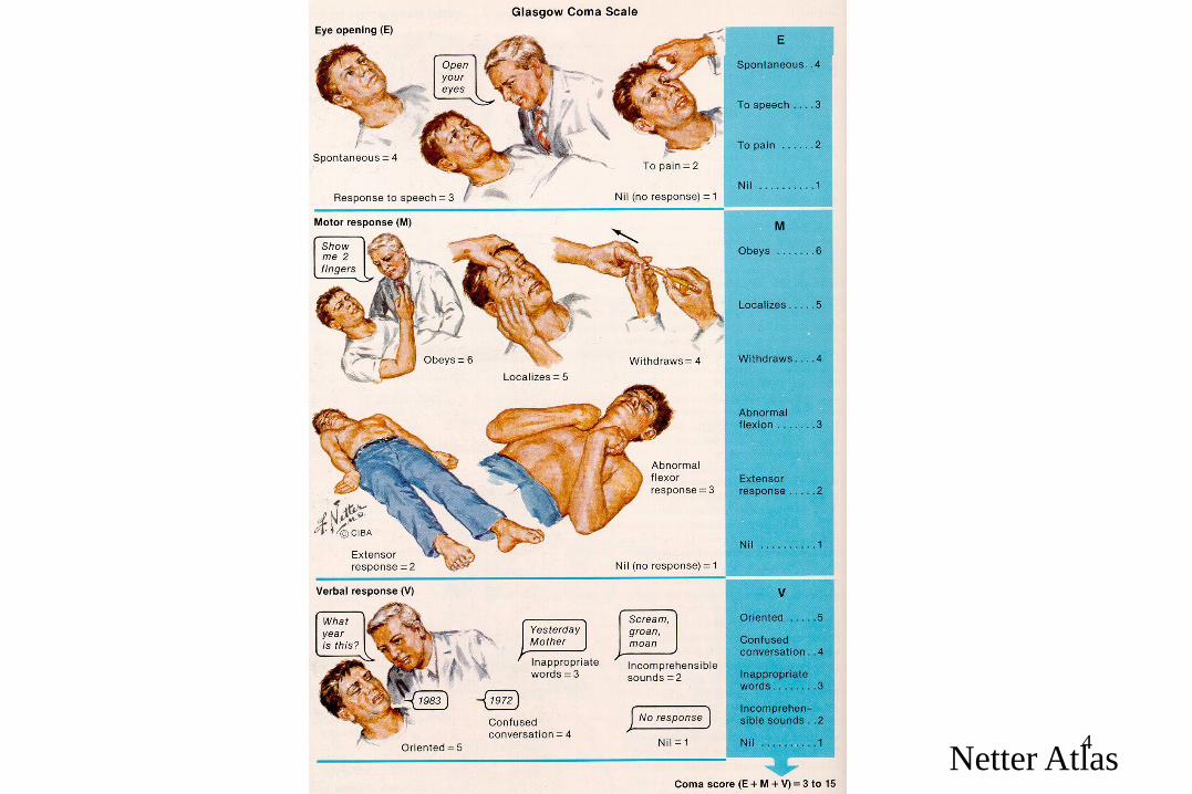
COMA
– Smell?
– respiratory rate & patterns (
– Look for abnormal posturing.
• Decorticate (Flexion of UE with Extension of LE)
• Decerebrate (Extension of all Ext.)
– Look for needle marks, cyanosis, signs of trauma
Netter Atlas
4Netter Atlas
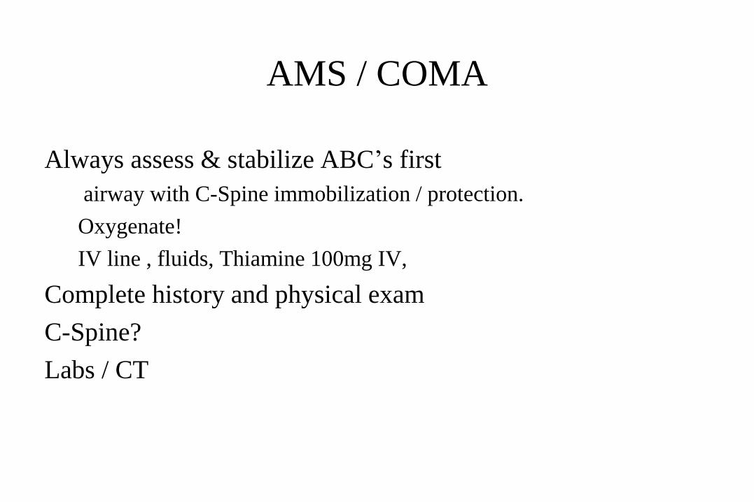
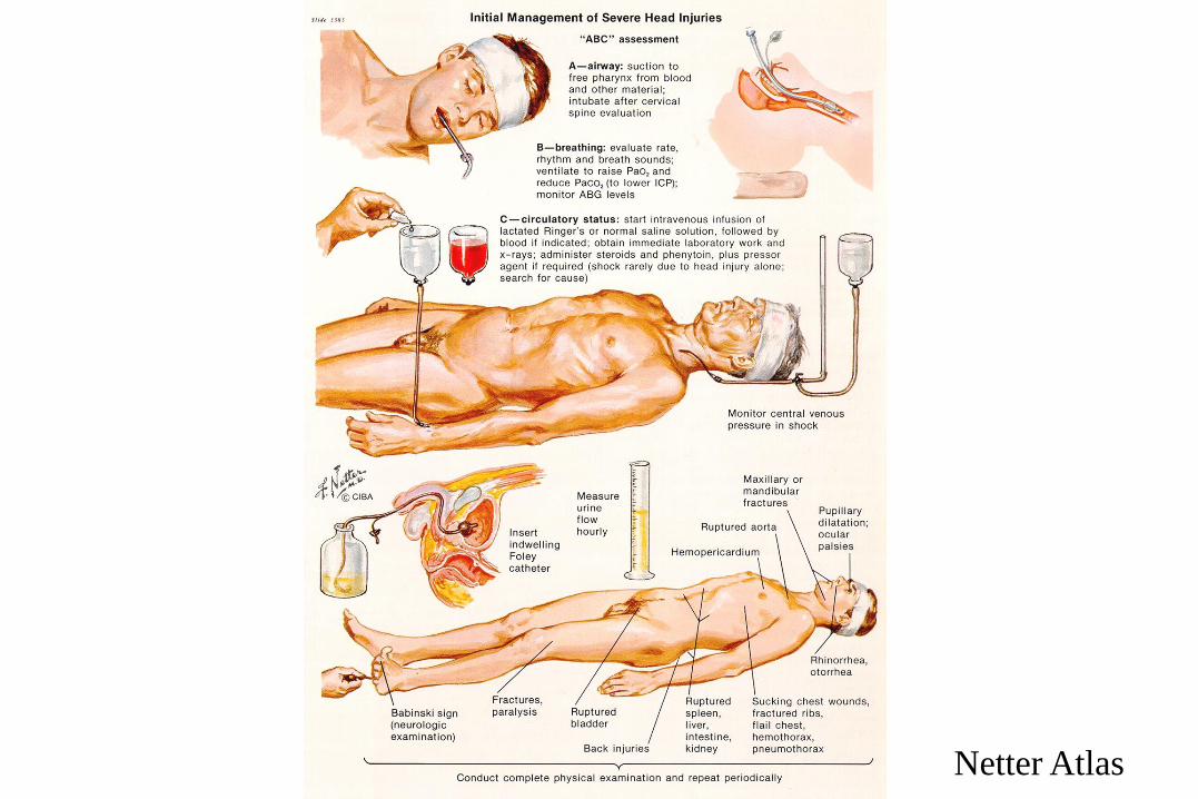
AMS / COMA
Always assess & stabilize ABC’s first
airway with C-Spine immobilization / protection.
Oxygenate!
IV line , fluids, Thiamine 100mg IV,
Complete history and physical exam
C-Spine?
Labs / CT

Headaches
• History!!
• New or changing the characteristics
• Autonomous or symptom of some other dis?
• Neurol. symptoms?
– Start with the simplest ther.
– Go up till the max dosage
– At the beginning of the HA

Headache
Migraine
Severe headache either preceeded by a visual “aura”(scintillating scotoma or VF cut) or motor disturbance.
Nausea, vomiting, light sensitivity, sound sensitivity
Factors that may provoke an attack include:
Menstruation, Sleep/food deprivation
Physical activity or certain foods (chocolate)
Contraceptives

Netter Atlas

Migraines
History & PE
CRUCIAL to obtain HA history from patient
Is this HA similar to others or is it “worst HA of life”
Medications
Foods
Menses
FULL PE including Neuro exam
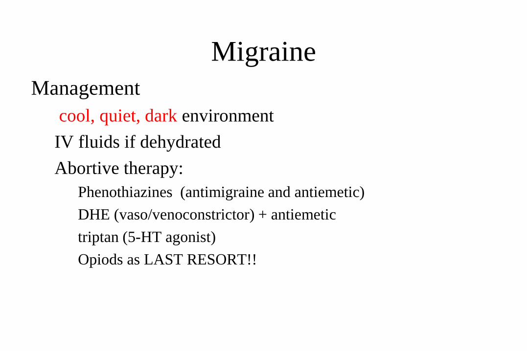
Migraine
Management
cool, quiet, dark environment
IV fluids if dehydrated
Abortive therapy:
Phenothiazines (antimigraine and antiemetic)
DHE (vaso/venoconstrictor) + antiemetic
triptan (5-HT agonist)
Opiods as LAST RESORT!!

Headaches
Cluster Headaches
boring headache on one side behind the eye.
facial flushing, tearing, nasal stuffiness
TX: 100% O2 by N/C at 6-8 l/min
- If no relief, Sumatriptan

Subarachnoid hemorrhage SAH
Abrupt onset of severe thunderclap “worst HA of life”.
Usually associated nausea and vomiting
Nonfocal neurologic exam (usually)
Etiology: aneurysm
DX: CT +LP A MUST
If CT (-), MUST LP

Temporal Arteritis
– granulomatous inflammation of the external carotid artery
– Clinically presents as:
• Severe unilateral HA over Temporal area
•Usually in middle aged females.
• PE reveals: a tender, warm, frequently pulseless temporal artery, with decreased visual acuity on the affected side.
Temporal Arteritis
DX:clinically + ESR elevation, usually >50 mm/h
Confirm with biopsy of artery
TX: HIGH dose steroids are VISION SAVING!
Start on prednisone IMMEDIATELY
Prednisone 60 – 80 mg Q day

Vertigo
• Central
• mild
• foc. neurol. sympt.
• Deviation and nystagmus
direction:the same
• Peripheral
• intensive
• NO foc. neurol. Sympt.
• Deviation and nystagmus
direction:different

Stroke
differential diagnosis---CT/MRI!!!
16
Agyi infarktus 80% Vérzés 20%ischemia 80% sp. Hemorrhage 10-15%
subarachnoidal bleeding

Ischemic Stroke Syndromes:
thrombotic vs. embolic
Thrombotic Syndromes
usually slow, progressive onset
Sx develop shortly after awakening and are progressive
Embolic Syndromes
Usually abrupt onset with maximal deficit that tends to
improve

18
Netter Atlas
Ischemic Stroke Syndromes
Middle Cerebral Artery Occlusion (MCA)
# 1 type
Contralateral hemiplegia, hemianesthesia and homonymous
hemianopsia
Upper extremity deficit >> Lower extremity
Aphasia (if dominant hemisphere involved)
Conjugate gaze impaired

20Netter Atlas

62 yrs stroke at admission
One day later
2 days later
CT

Ischemic Stroke
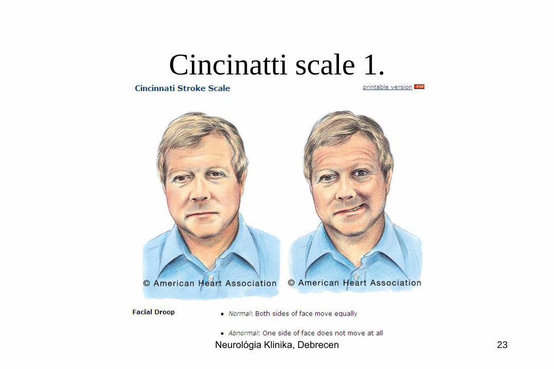
Cincinatti scale 1.
Neurológia Klinika, Debrecen 23

Cincinatti scale 2.
Neurológia Klinika, Debrecen 24
Cincinatti scale 3.
• Slurred speech, aphasia
Neurológia Klinika, Debrecen 25

TIA
(TRANSIENT ISCHEMIC ATT.)• Transient symptoms
• Minutes
• No residual tissue deficit (diff. MRI?)
TIA is emergency!!! High risk
of devastating stroke

Open the artery as soon as
possible
1 1,5 3 4,5 6 hours after stroke
45 patients
21 patients
9 patients
4 patients
2 patients

12 h if basilar artery occluded either iv or ia. lysis
6-8 hours IF ICA or MCA occlusion:intraarterial or
mechanical thrombectomy (MET) BUT start with iv.
If specific constellation of MRI and sympt. up till 24 h!!!
Within 4,5 hours (some subgroups 3 hours) iv. lysis if small vessel occlusion
Time window?
depends on the occluded vessel
and elapsed time
eafter stroke?
time

If out of time window?
• 100-300 mg aspirin
• Monitoring of BP and ECG
• Do NOT decrease BP till 220/110 Hgmm!
• Pulsoximetry, 2-4 lit oxigen, if less 94%
• Normoglycemia
• LMWH or heparin to prevent DVT deep venous thromb.
• Nasogastric tube if dysphagia
• Antipyretic ther.
• If seizure antiepilept.
• antibiotics

Diagnosis acute stroke
Blood•platelets
•Hgb
•glucose
•INR!
•O2 sat
•hypercoagulable
Heart
•BP monitoring
•ECG monitoring
TTE
TEE
Carotid, vertebral•US
•CTA
•MRA
•DSA
•CT+CT angio!!
•MRI•Diff. WI+GRE

Netter Atlas

Subarachnoid Hemorrhage SAH
Highest incidence in 35-65 year old.
Usually from the rupture of a berry aneurysm
Clinically:abrupt onset of “worst headache of life”
Nuchal rigidity, photophobia, vomiting, retinal hemorrhages.
Diagnosis: CT + LP!!!!CT only 92% sens. within 24 hours of event, loses sensitivity >24 hours out from headache.
72 hours out CANNOT r/o without LP!
Management: Consider adding Nimodipine 60 mg Q6 to reduce vasospasm

33Netter Atlas

34Netter Atlas

Seizure

Seizures & Status Epilepticus
Background:
1 – 2% of the general population has seizures
Primary
Idiopathic epilepsy:onset ages 10-20
Secondary
Intracranial pathology
Trauma, Abscess, Infarct, tumor
Extracranial Pathology
Toxic, metabolic, hypertensive, eclampsia

Netter Atlas

Seizure Types
Generalized Convulsive Seizures (Grand Mal):Tonic , clonic movements, (+) LOC, apnea, incontinence and a post-ictal state
Non Convulsive Seizures (Petit Mal)Absence seizures – “blank staring spells”
Myoclonic – brief contractions of selected muscle groups
Partial SeizuresCharacterized by presence of auditory or visual hallucinations
Simple = somatic complaints + no LOC
Complex = somatic complaints + Altered Mental Status or LOC

39Netter Atlas

Approach for 1st Seizure, New Seizure, or
Substance/ Trauma Induced Seizure
As always ABC’s First
IV, O2, Monitor.
Send blood for CBC, Chem 20, Tox screen as appropriate
Anticonvulsant levels
Prolactin levels / Lactate level
CXR / UA/ Head CT
Is patient actively seizing? Post ictal? Pseudoseizure?
Consider treatment options
Complete History and Physical Exam
Including detailed Neuro Exam
Repeat Neuro evaluations a must!

Approach to Breakthrough Seizure
As Before, But History, History, History!!
Main causes of Breakthrough Seizure:
Noncompliance with anticonvulsant regimen
Start of new medication (level alteration)
Antibiotics, OCP’s
Infection
Fever
Changes in body habitus, eating patterns
Status Epilepticus
Definition:operationally defined as seizure lasting greater than 5 minutes OR two seizures between which there is incomplete recovery of consciousness.
Treatment algorhythm:
As before ABC’s
IV, O2, Monitor
Consider ALL potential causesINH (pyridoxime/B-6 deficiency)
Eclampsia
Alcoholic (thiamine/B-1 deficiency)
Other Tox ingestion (TCA’s, sulfonylurea)
Trauma

Status Epilepticus Treatment
FIRST LINE TREATMENT
Lorazepam 2mg/min IV up to 10 mg max.
OR Diazepam 5mg/min IV or PR up to 20mg
SECOND LINE TREATMENT
Phenytoin or Fosphenytoin
20mg/kg IV at rate of 50mg/min
THIRD LINE TREATMENT
Get Ready to intubate at this point!!
Phenobarbial 10-20mg/kg @ 60 mg/min

Status Epilepticus Treatment
FINAL TREATMENT
Barbiturate Coma
Pentobarbitol 5mg/kg @ 25 mg/min
Stat Neurology consult for evaluation and EEG
Pentobarbitol titrated to EEG response.
Always get a through HISTORY
Possible trauma
Medications in house
Other diseases?
Overall appearance of patient
Status Epilepticus Adjunctive Treatment by
History
Thiamine 100mg IV, 1-2 amps D 50
If suspect alcoholic, malnourished, hypoglycemia
Magnesium Sulfate 20cc of 10% solution
As above of if eclampsia (BP does NOT have to be 200/120!!)
Pyridoxine 5 gms IV
INH or B-6 deficiency

Why emergency?
Guillain-Barré sy respir. Insuffic.

47Netter Atlas

Emergent Peripheral Neuropathies
Guillain-Barre SyndromeMost common acute polyneuropathy.
2/3’s of patients will have preceeding URI or gastroenteritis 1-3 weeks prior to onset.
Presents as:paresthesias followed by ascending paralysis starting in legs and moving upwards.
Remember Miller-Fischer variant: has minimal weakness and presents with ataxia, areflexia, and ophthalmoplegia.
DX:LP will show cytochemical dissociation (only days after the onset!).
Normal cells with HIGH protein.
TX: Self limiting, early and aggressive airway stabilization.

Emergent Peripheral Neuropathies
Myasthenia Gravis
Most common disorder of neuromuscular transmission.
An autoimmune disease that destroys acetylcholine receptors (AchR) which leads to poor neurotransmission and weakness.
Proximal >> Distal muscle weakness
Commonly will present as:
Muscle weakness exacerbated by activity and is relieved by rest
Clinically:ptosis, diplopia and blurred vision are the most common complaints. Pupil is spared!

Emergent Peripheral Neuropathies
Myasthenia Gravis
Myasthenic crisis = a true emergency!!
Occurs in undiagnosed or untreated patients
Due to relative Ach (acetylcholine) deficiency
Patients present with profound weakness and impending respiratory failure
TX:Stabilize and manage airway
Consider edrophonium 1 -2 mg IV
(AchE inhibitor)
Plasmapheresis and/or immunglobulin therapy

Netter Atlas

Netter Atlas

Infectious Neurologic Emergencies
Meningitis: inflammation of the meninges
History:
Acute Bacterial Meningitis:
Rapid onset of symptoms <24 hours
Fever, Headache, Photophobia
Stiff neck, Confusion

Meningitis
Lymphocytic Meningitis (Aseptic/Viral)Gradual onset of symptoms as previously listed over 1-7 days.
Etiology:Viral
Atypical MeningitisHistory (medical/social/environmental) crucial
Insidious onset of symptoms over 1-2 weeks
Etiology:TB(#1)
Coccidiomycosis, cryptococcus

Meningo-encephalitis
Physical Exam PearlsInfants and the elderly lack the usual signs and symptoms, only clue may be headache
Look for papilledema, focal neurologic signs, ophthalmoplegia and rashes
As always full examChecking for above
Brudzinski’s sign
Kernig’s sign
KEY POINT:If you suspect meningococcemia do NOT delay antibiotic therapy, MUST start within 20 minutes of arrival!!!!!

Meningitis
Emergent CT Prior to LP
Those with profoundly depressed MS
Seizure
Head Injury
Focal Neurologic signs
Immunocompromised with CD4 count <500
DO NOT DELAY ANTIBIOTIC THERAPY!!

Meningitis
Lumbar Puncture Results
TEST NORMAL BACTERIAL VIRAL
Pressure <170 >300 200
Protein <50 >200 <200
Glucose >40 <40 >40
WBC’s <5 >1000 <1000
Cell type Monos >50% PMN’s Monos
Gram Stain Neg Pos Neg

Meningitis Management
Antibiotics By Age Group
Neonates(<1month) = Ampicillin + Gent. Or Cefotaxime + Gent
Infants (1-3mos) = Cefotaxime or Ceftriaxone+ Ampicillin
Children (3mos-18yrs) = Ceftriaxone
Adults (18yr-up) = Ceftriaxone + Vancomycin
Elderly/Immunocomp = Ceftriaxone +Ampicillin +Vancomycin

Meningitis Management
Steroids
In children, dexamethasone has been shown to be of benefit in
reducing sensiorneural hearing loss, when given before the first dose
of antibiotic.
Indications:
Children> 6 weeks with meningitis due to H. flu or S. pneumo.
Adults with positive CSF gram stain
Dose: 0.15mg/kg IV
Encephalitis
Always think of in the young/elderly or immunocompromised with FEVER + AMS
Common Etiologies:
Viral
West Nile
Herpes Simplex Virus (HSV)
Varicella Zoster Virus (VZV)
Arboviruses
Eastern Equine viruses
St. Louis Encephalitis

Encephalitis
Defined as: inflammation of the brain itself
Most cases are self limited, and unless virulent strain, or immunocompromised, will resolve.
The ONLY treatable forms of encephalitis are:
HSV
Zoster
Encephalitis
Management:
Emergent MR
ABC’s with supportive care.
Lumbar puncture:
Send for ELISA and PCR
Acyclovir 10 mg/kg Q 8 hours IV for HSV and Zoster
Steroids not shown to be of benefit.

Headache

Complications Head traumaFluid-elektrolyte >50% aldosterone and ADH: sodium
and fluid retention
Respiratory system aspiration, infection and atelectasia,
adult-respiratory-distress syndrome
(ARDS)
Gastrointestinal stomach erosion
Cardiovascular cardiac arhythmias, arrest
Hemostasis DIC, coagulopathy, 5% - 10%

Closed head Injury Facts
The single most important factor in the neurologic assessment of the head injured patient is level of consciousness (LOC)
Always assume multiple injuries with serious mechanism.ESPECIALLY C - SPINE!!!!
Unless hypotensive WITH bradycardia and WARM extremities (spinal cord injury); hypotension is ALWAYS secondary to hypovolemia from blood loss in the trauma patient!
The most common intracranial bleed in CHI is subarachnoid hemorrhage.
Closed Head Injuries with Hemorrhage
Cerebral Contusion
Focal hemorrhage and edema under the site of impact.
Susceptible areas are those in which the gyri are in close contact with the skull
Frontal lobe
Temporal lobes
Diagnostic Test of Choice:NC Head CT
Treatment: Supportive with measures to keep ICP normal.
Repeat Neuro checks.
Repeat Head Ct in 24 hours.

Contusion
Subdural Hematoma
Occurs secondary to acceleration/decelleration injury with resultant tearing of the bridging veins that extend from the subarachnoid space to the dural sinuses.
Blood dissects over the cerebral cortex and collects under the dura overlying the brain.
Patients at risk:alcoholics
elderly
anticoagulant users
Appears as “sickle shape” and does not extend across the midline

Subdural hematoma

Epidural Hematoma
Occurs from blunt trauma to head especially over the parietal/temporal area.
Presents as LOC which then patient has lucid interval then progressive deterioration, coma , death. ( Patient talks to you & dies!)
Commonly associated with linear skull fracture
Mechanism of bleed is due to tear of artery, usually middle meningeal.
Sometimes ipsilateral pupillary dilatation with contralateral hemiparesis.
CT Scan :a BICONVEX (lens) density which can extend across the midline

Epidural hematom

Management of Closed Head Injuries
As always ABC’s with C-Spine precautions
IV, O2, Monitor.
Stabilize and resuscitateSao2>95%
SBP>90
Treat Fever
Head of Bed 30% (once C-Spine cleared)
Stat head CT with Stat Neurosurgical evaluation for surgical lesions.
Repeat Exams, looking for signs of herniation.

Signs of Herniation / Increased ICP
Headache, nausea, vomiting
Decreasing LOC
Decreased respiratory rate
Cushing reflex (hypertension/bradycardia/bradynpea)
Papilledema
Development of signs of herniation
Fixed and dilated pupil
Contralateral hemiparesis
PosturingThis teaching material is a slightly modified version of the teaching material
(https://phdres.caregate.net)