endocrine 12 may 2015 final
TRANSCRIPT

Reproduction endocrinology

Introduction In most species of mammals, the multiple
differences between the male and the female depend primarily on a single chromosome (the Y chromosome) and a single pair of endocrine structures,
the testes in the male and
the ovaries in the female.

Introduction The differentiation of the primitive gonads
into testes or ovaries in utero is genetically determined in humans
But the formation of male genitalia depends on the presence of a functional, secreting testis
In the absence of testicular tissue, development is female

The male sexual behavior and, in some species, the male pattern of gonadotropin secretion are due to the action of male hormones on the brain in early development.
After birth, the gonads remain quiescent until adolescence, when they are activated by gonadotropins from the anterior pituitary.
Hormones secreted by the gonads at this time cause the appearance of features typical of the adult male or female and the onset of the sexual cycle in the female.

In human females, ovarian function regresses after a
number of years and sexual cycles cease (the menopause).
In males, gonadal function slowly declines with advancing age.
But the ability to produce viable gametes persists

In both sexes, the gonads have a dual function: the production of germ cells (gametogenesis)
and the secretion of sex hormones. The androgens are the steroid sex hormones
that are masculinizing in their action. The estrogens are those that are feminizing. Both types of hormones are normally secreted
in both sexes.

The testes secrete large amounts of androgens, principally testosterone.
But they also secrete small amounts of estrogens.
The ovaries secrete large amounts of estrogens and small amounts of androgens.
Androgens are secreted from the adrenal cortex in both sexes, and some of the androgens are converted to estrogens in fat and other extra-gonadal and extra-adrenal tissues.


The ovaries also secrete progesterone, a steroid that has special functions in preparing the uterus for pregnancy.
Particularly during pregnancy, the ovaries secrete the polypeptide hormone relaxin, which loosens the ligaments of the pubic
symphysis softens the cervix, facilitating delivery of the fetus.

In both sexes, the gonads secrete other polypeptides, including
inhibin B, a polypeptide that inhibits follicle-stimulating hormone (FSH) secretion.
The secretory and gametogenic functions of the gonads are both dependent on the secretion of the
anterior pituitary gonadotropins, FSH, and luteinizing hormone (LH).

The sex hormones and inhibin B feed back to inhibit gonadotropin secretion.
In males, gonadotropin secretion is noncyclic; but in post-pubertal females an orderly, sequential secretion of gonadotropins is necessary for the occurrence of
menstruation, pregnancy, and lactation.

12
Similarities and differences between males and females, but same goal: new life
Primary sex organs: gonads Testes in males Ovaries in females
› These produce the gametes (sex cells) Sperm in males Ovum (egg) in females
› Endocrine function also: secretion of hormones Accessory sex organs
› Internal glands and ducts› External genitalia

Menopause The human ovaries become unresponsive to
gonadotropins with advancing age, and their function declines, so that sexual cycles disappear (menopause).
This unresponsiveness is associated with and probably caused by a decline in the number of primordial follicles, which becomes precipitous at the time of menopause .

The ovaries no longer secrete progesterone and 17-estradiol in appreciable quantities,
Estrogen is formed only in small amounts by aromatization of androstenedione in peripheral tissues.
The uterus and the vagina gradually become atrophic.

As the negative feedback effect of estrogens and progesterone is reduced.
The secretion of FSH is increased, and plasma FSH increases to high levels.
While LH levels are moderately high.

In women, a period called perimenopause precedes menopause, and can last up to 10 y.
During perimenopause the menses become irregular and the level of inhibins decrease, usually between the ages of 45 and 55.
The average age at onset of the menopause has been increasing since the end of the 19th century and is currently 52 y.

Blue squares, premenopausal women (regular menses); red squares, perimenopausal women (irregular menses for at least 1 y); red triangles, postmenopausal women

The loss of ovarian function causes many symptoms such as;
sensations of warmth spreading from the trunk to the face (hot flushes; also called hot flashes) and
night sweats.
In addition, the onset of menopause increases the risk of many diseases such as;
osteoporosis, ischemic heart disease, and renal disease.

Hot flushes are said to occur in 75% of menopausal women and may continue intermittently for as long as 40 y.
They also occur when early menopause is produced by bilateral ovariectomy, and they are prevented by estrogen treatment.
In addition, they occur after castration in men.
Their cause is unknown.

However, they coincide with surges of LH secretion. LH is secreted in episodic bursts at intervals of 30 to 60 min or more (circhoral secretion), and in the absence of gonadal hormones these bursts are large.
Each hot flush begins with the start of a burst. However, LH itself is not responsible for the symptoms, because they can continue after removal of the pituitary.
Instead, it appears that some estrogen-sensitive event in the hypothalamus initiates both the release of LH and the episode of flushing.

The Male Reproductive System
Structure The testes are made up of loops of convoluted
seminiferous tubules, in the walls of which the spermatozoa are formed from the primitive germ cells (spermatogenesis).
Both ends of each loop drain into a network of ducts in the head of the epididymis. From there, spermatozoa pass through the tail of the epididymis into the vas deferens.

They enter through the ejaculatory ducts into the urethra in the body of the prostate at the time of ejaculation.
Between the tubules in the testes are nests of cells containing lipid granules, the interstitial cells of Leydig, which secrete testosterone into the bloodstream.
The spermatic arteries to the testes are tortuous,
This anatomic arrangement may permit countercurrent exchange of heat and testosterone.


Endocrine Function of the Testes
Chemistry & Biosynthesis of Testosterone
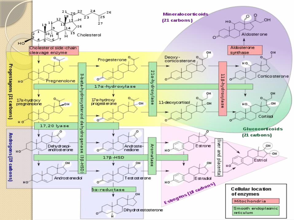
Testosterone, the principal hormone of the testes, is a C19 steroid with an –OH group in the 17 position .
It is synthesized from cholesterol in the Leydig cells and is also formed from andro-stenedione secreted by the adrenal cortex.

Pregnenolone is therefore hydroxylated in the 17 position and then subjected to side chain cleavage to form dehydro-epiandrosterone.
Andr-ostenedione is also formed via progesterone and 17-hydroxyprogesterone, but this pathway is less prominent in humans.
Dehydro-epiandrosterone and androstenedione are then converted to testosterone.


Secretion
The testosterone secretion rate is 4 to 9 mg/d (13.9–31.33 mol/d) in normal adult males.
Small amounts of testosterone are also secreted in females, with the major source being the ovary, but possibly from the adrenal as well.

Transport & Metabolism Ninety-eight percent of the testosterone in plasma is
bound to protein: 65% is bound to a -globulin called gonadal steroid-binding globulin (GBG) or sex steroid-binding globulin, and 33% to albumin
The plasma testosterone level (free and bound) is 300 to 1000 ng/dL (10.4–34.7 nmol/L) in adult men compared with 30 to 70 ng/dL (1.04–2.43 nmol/L) in adult women.
It declines somewhat with age in males.

A small amount of circulating testosterone is converted to estradiol, but most of the testosterone is converted to 17-ketosteroids, principally androsterone and its isomer etiocholanolone, and excreted in the urine.
About two thirds of the urinary 17-ketosteroids are of adrenal origin, and one third are of testicular origin.

Although most of the 17-ketosteroids are weak androgens (they have 20% or less the potency of testosterone), it is worth emphasizing that not all 17-ketosteroids are androgens and not all androgens are 17-ketosteroids.
Etiocholanolone, for example, has no androgenic activity, and testosterone itself is not a 17-ketosteroid.


Actions In addition to their actions during development,
testosterone and other androgens exert an inhibitory feedback effect on pituitary LH
secretion; develop and maintain the male secondary sex
characteristics; exert an important protein-anabolic, growth-promoting effect; along with FSH, maintain spermatogenesis.

Secondary Sex Characteristics The widespread changes in hair distribution, body
configuration, and genital size that develop in boys at puberty—the male secondary sex characteristics.
The prostate and seminal vesicles enlarge, and the seminal vesicles begin to secrete fructose.
This sugar appears to function as the main nutritional supply for the spermatozoa.

The psychic effects of testosterone are difficult to define in humans, but in experimental animals, androgens provoke boisterous and aggressive play.
The effects of androgens and estrogens on sexual behavior are considered.
Although body hair is increased by androgens, scalp hair is decreased.
Hereditary baldness often fails to develop unless dihydrotestosterone is present.


Anabolic Effects Androgens increase the synthesis decrease the breakdown of protein Thus leading to an increase in the rate of growth.
It used to be argued that they cause the epiphyses to fuse to the long bones,
Thus eventually stopping growth, but it now appears that epiphysial closure is due to estrogens.

Anabolic Effects Secondary to their anabolic effects, androgens cause
moderate Na+
K+
H2O Ca2+
SO4–
PO4–
retention; and they also increase the size of the kidneys.

Doses of exogenous testosterone that exert significant anabolic effects are also;
masculinizing increase libido

Testicular Production of Estrogens
Over 80% of the estradiol and 95% of the estrone in the plasma of adult men is formed by extragonadal and extraadrenal aromatization of circulating testosterone and androstenedione.
The remainder comes from the testes. Some of the estradiol in testicular venous blood
comes from the Leydig cells, but some is also produced by aromatization of androgens in Sertoli cells.

Testicular Production of Estrogens
In men, the plasma estradiol level is 20 to 50 pg/mL (73–184 pmol/L) and the total production rate is approximately 50 g/d (184 nmol/d).
In contrast to the situation in women, estrogen production moderately increases with advancing age in men.

The Female Reproductive System
The Menstrual Cycle The reproductive system of women , unlike
that of men, shows regular cyclic changes
That teleologically (purpose especially in nature) may be regarded as periodic preparations for fertilization and pregnancy.

The Female Reproductive System
In humans and other primates, the cycle is a menstrual cycle.
Its most conspicuous feature is the periodic vaginal bleeding that occurs with the shedding of the uterine mucosa (menstruation).

The length of the cycle is notoriously variable in women, but an average figure is 28 days from the start of one menstrual period to the start of the next.
By common usage, the days of the cycle are identified by number, starting with the first day of menstruation.


Ovarian Cycle From the time of birth, there are many
primordial follicles under the ovarian capsule. Each contains an immature ovum.
At the start of each cycle, several of these follicles enlarge, and a cavity forms around the ovum (antrum formation).
This cavity is filled with follicular fluid.

Ovarian Cycle
In humans, usually one of the follicles in one ovary starts to grow rapidly on about the sixth day and becomes the dominant follicle.
While the others regress, forming atretic follicles.

The atretic process involves apoptosis.
It is uncertain how one follicle is selected to be the dominant follicle in this follicular phase of the menstrual cycle.
But it seems to be related to the ability of the follicle to secrete the estrogen inside it that is needed for final maturation.
When women are given highly purified human pituitary gonadotropin preparations by injection, many follicles develop simultaneously.


The primary source of circulating estrogen is the granulosa cells of the ovaries.
However, the cells of the theca interna of the follicle are necessary for the production of estrogen as they secrete androgens that are aromatized to estrogen by the granulosa cells.
At about the 14th day of the cycle, the distended follicle ruptures, and the ovum is extruded into the abdominal cavity. This is the process of ovulation.
The ovum is picked up by the fimbriated ends of the uterine tubes (oviducts).

It is transported to the uterus and, unless fertilization occurs, out through the vagina.
The follicle that ruptures at the time of ovulation promptly fills with blood, forming what is sometimes called a corpus hemorrhagicum.
Minor bleeding from the follicle into the abdominal cavity may cause peritoneal irritation and fleeting lower abdominal pain ("mittelschmerz" German: "middle pain).

The granulosa and theca cells of the follicle lining promptly begin to proliferate, and the clotted blood is rapidly replaced with yellowish, lipid-rich luteal cells, forming the corpus luteum.
This initiates the luteal phase of the menstrual cycle, during which the luteal cells secrete estrogen and progesterone.
Growth of the corpus luteum depends on its developing an adequate blood supply, and there is evidence that vascular endothelial growth factor (VEGF) is essential for this process.
If pregnancy occurs, the corpus luteum persists and usually there are no more periods until after delivery.
If pregnancy does not occur, the corpus luteum begins to degenerate about 4 d before the next menses (24th day of the cycle) and is eventually replaced by scar tissue, forming a corpus albicans.
The ovarian cycle in other mammals is similar, except that in many species more than one follicle ovulates and multiple births are the rule.
Corpora lutea form in some submammalian species but not in others.

In humans, no new ova are formed after birth.
During fetal development, the ovaries contain over 7 million primordial follicles.
However, many undergo atresia (involution) before birth and others are lost after birth.
At the time of birth, there are 2 million ova, but 50% of these are atretic.

The million that are normal undergo the first part of the first meiotic division at about this time and enter a stage of arrest in prophase in which those that survive persist until adulthood.
Atresia continues during development, and the number of ova in both of the ovaries at the time of puberty is less than 300,000.

Only one of these ova per cycle (or about 500 in the course of a normal reproductive life) normally reaches maturity; the remainder degenerate.
Just before ovulation, the first meiotic division is completed.
One of the daughter cells, the secondary oocyte, receives most of the cytoplasm, while the other, the first polar body, fragments and disappears.
The secondary oocyte immediately begins the second meiotic division, but this division stops at metaphase and is completed only when a sperm penetrates the oocyte.


At that time, the second polar body is cast off and the fertilized ovum proceeds to form a new individual.

Uterine Cycle At the end of menstruation, all but the deep
layers of the endometrium have sloughed.
A new endometrium then regrows under the influence of estrogens from the developing follicle.
The endometrium increases rapidly in thickness from the 5th to the 14th days of the menstrual cycle.

As the thickness increases, the uterine glands are drawn out so that they lengthen.
But they do not become convoluted or secrete to any degree.
These endometrial changes are called proliferative, and this part of the menstrual cycle is sometimes called the proliferative phase.
It is also called the preovulatory or follicular phase of the cycle.

After ovulation, the endometrium becomes more highly vascularized and slightly edematous under the influence of estrogen and progesterone from the corpus luteum.
The glands become coiled and tortuous and they begin to secrete a clear fluid.
Consequently, this phase of the cycle is called the secretory or luteal phase.
Late in the luteal phase, the endometrium, like the anterior pituitary, produces prolactin, but the function of this endometrial prolactin is unknown.


Follicular life cycle through menstrual cycle

The endometrium is supplied by two types of arteries.
The superficial two thirds of the endometrium that is shed during menstruation, the stratum functionale, is supplied by long, coiled spiral arteries.
Whereas the deep layer that is not shed, the stratum basale, is supplied by short, straight basilar arteries.

When the corpus luteum regresses, hormonal support for the endometrium is withdrawn.
The endometrium becomes thinner, which adds to the coiling of the spiral arteries.
Foci of necrosis appear in the endometrium, and these coalesce.


In addition, spasm and degeneration of the walls of the spiral arteries take place, leading to spotty hemorrhages that become confluent and produce the menstrual flow.
The vasospasm is probably produced by locally released prostaglandins.
Large quantities of prostaglandins are present in the secretory endometrium and in menstrual blood, and infusions of prostagladin F2 (PGF2) produce endometrial necrosis and bleeding.

From the point of view of endometrial function, the proliferative phase of the menstrual cycle represents restoration of the epithelium from the preceding menstruation, and the secretory phase represents preparation of the uterus for implantation of the fertilized ovum.
The length of the secretory phase is remarkably constant at about 14 d, and the variations seen in the length of the menstrual cycle are due for the most part to variations in the length of the proliferative phase.
When fertilization fails to occur during the secretory phase, the endometrium is shed and a new cycle starts.

Sequential hormonal changes during menstrual cycle1. degeneration of corpus luteum estrogen,
progesterone, inhibin FSH & LH
2. follicles develop estrogen levels
3. plasma estrogen levels increase
4. ~day 7, dominant follicle secretes high levels of estrogen
5. plasma estrogen level increases sharply
Sequential hormonal changes during menstrual cycle
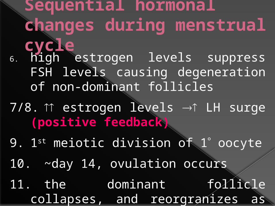
6. high estrogen levels suppress FSH levels causing degeneration of non-dominant follicles
7/8. estrogen levels LH surge (positive feedback)
9. 1st meiotic division of 1 oocyte
10. ~day 14, ovulation occurs
11. the dominant follicle collapses, and reorgranizes as the corpus luteum

Sequential hormonal changes during menstrual cycle
12. corpus luteum secretes estrogen & progesterone
13. plasma levels of estrogen & progesterone increase, suppressing release of GnRH, LH, & FSH
14. ~day 25, corpus luteum spontaneously degenerates
15. secretion & plasma levels of estrogen & progesterone
16. estrogen & progesterone FSH & LH levels which begin follicular development of the next menstrual cycle
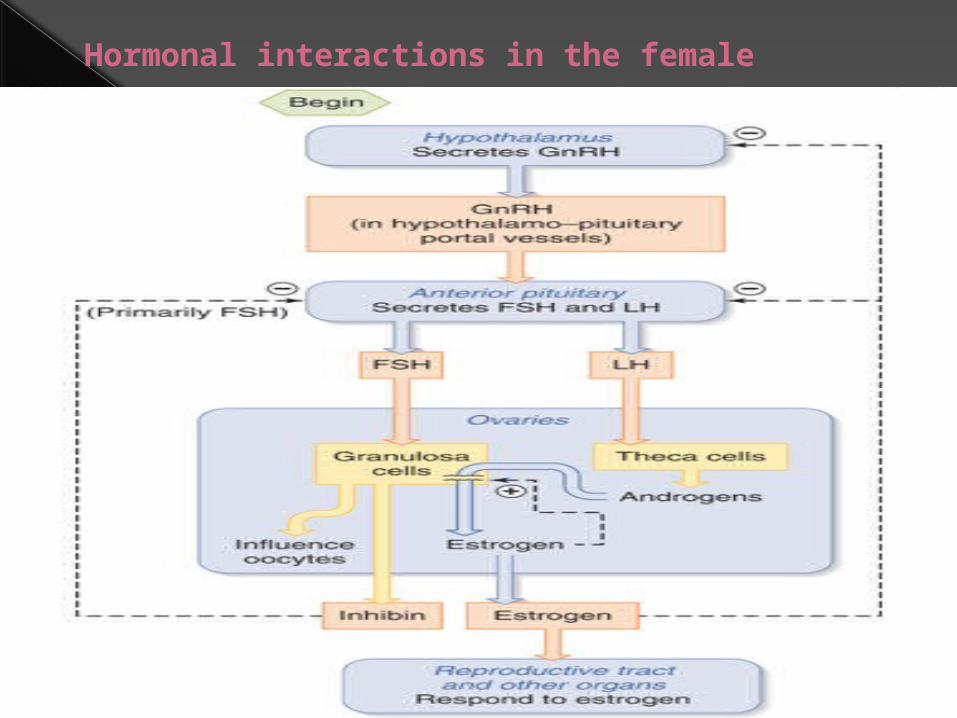
Hormonal interactions in the female

Hormonal initiation of ovulation

© 2009 The McGraw-Hill Companies, Inc. All rights reserved
31-79
Follicular cellsbecome
corpus luteumwhich secretesprogesterone
Anterior pituitaryreleases FSH
Uterine lining thickensOvarian folliclematures and secretes
estrogenThen releases LH
Triggers ovulation
Lining more vascular and glandular
Without fertilization
Corpus luteum degenerates
Estrogen and progesterone levels fall
Uterine lining breaks down – menses starts
Cycle begins again with release of FSH
Female Reproductive System: Reproductive Cycle (cont.)

Normal Menstruation Menstrual blood is predominantly arterial, with
only 25% of the blood being of venous origin.
It contains tissue debris, prostaglandins, and relatively large amounts of fibrinolysin from endometrial tissue.
The fibrinolysin lyses clots, so that menstrual blood does not normally contain clots unless the flow is excessive.

The usual duration of the menstrual flow is 3 to 5 d, but flows as short as 1 d and as long as 8 d can occur in normal women.
The amount of blood lost may range normally from slight spotting to 80 mL; the average amount lost is 30 mL. Loss of more than 80 mL is abnormal.
Obviously, the amount of flow can be affected by various factors, including the thickness of the endometrium, medication, and diseases that affect the clotting mechanism.

Vaginal Cycle Under the influence of estrogens, the vaginal
epithelium becomes cornified, and cornified epithelial cells can be identified in the vaginal smear.
Under the influence of progesterone, a thick mucus is secreted, and the epithelium proliferates and becomes infiltrated with leukocytes.
The cyclical changes in the vaginal smear in rats are relatively marked. The changes in humans and other species are similar but not so clear-cut.
Ovarian Hormones Chemistry, Biosynthesis, &
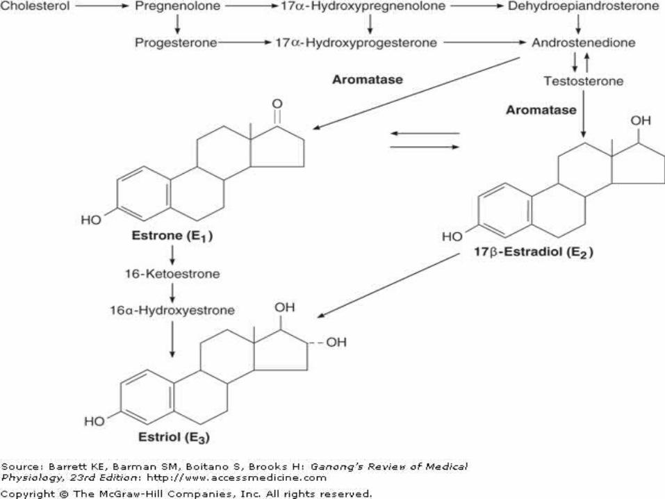
Metabolism of Estrogens The naturally occurring estrogens are 17-estradiol, estrone, and estriol.
They are C18 steroids which do not have an angular methyl group attached to the 10 position or a 4-3-keto configuration in the A ring.

Ovarian Hormones They are secreted primarily by the granulosa cells
of the ovarian follicles, the corpus luteum, and the placenta.
Their biosynthesis depends on the enzyme aromatase (CYP19), which converts testosterone to estradiol and androstenedione to estrone.
The latter reaction also occurs in fat, liver, muscle, and the brain.

Secretion The concentration of estradiol in the plasma during the
menstrual cycle.
Almost all of this estrogen comes from the ovary, and two peaks of secretion occur: one just before ovulation and one during the midluteal phase.
The estradiol secretion rate is 36 g/d (133 nmol/d) in the early follicular phase, 380 g/d just before ovulation, and 250 g/d during the midluteal phase.
After menopause, estrogen secretion declines to low levels.

Effects on the Female Genitalia
Estrogens facilitate the growth of the ovarian follicles and increase the motility of the uterine tubes.
Their role in the cyclic changes in the endometrium, cervix, and vagina has been discussed previously.
They increase uterine blood flow and have important effects on the smooth muscle of the uterus.
In immature and castrated females, the uterus is small and the myometrium atrophic and inactive.
Estrogens increase the amount of uterine muscle and its content of contractile proteins.
Under the influence of estrogens, the muscle becomes more active and excitable, and action potentials in the individual fibers become more frequent.

The "estrogen-dominated" uterus is also more sensitive to oxytocin.
Chronic treatment with estrogens causes the endometrium to hypertrophy.
When estrogen therapy is discontinued, sloughing takes place with withdrawal bleeding.
Some "breakthrough" bleeding may occur during treatment when estrogens are given for long periods.

Effects on Endocrine Organs
Estrogens decrease FSH secretion. Under some circumstances, they inhibit LH
secretion (negative feedback) In other circumstances, they increase LH
secretion (positive feedback).
Women are sometimes given large doses of estrogens for 4 to 6 d to prevent conception after coitus during the fertile period (postcoital or "morning-after" contraception).


Effects on the Central Nervous System
The estrogens are responsible for increase in libido in humans.
They apparently exert this action by a direct effect on certain neurons in the hypothalamus

Effects on the Breasts Estrogens produce duct growth in the
breasts and are largely responsible for breast enlargement at puberty in girls.
They have been called the growth hormones of the breast.
They are responsible for the pigmentation of the areolas.
Although pigmentation usually becomes more intense during the first pregnancy than it does at puberty

Female Secondary Sex Characteristics
The body changes that develop in girls at puberty—in addition to enlargement of breasts, uterus, and vagina—are due in part to estrogens, which are the "feminizing hormones," and in part simply to the absence of testicular androgens.
Women have narrow shoulders and broad hips, thighs that converge, and arms that diverge (wide carrying angle).

Female Secondary Sex Characteristics
This body configuration, plus the female distribution of fat in the breasts and buttocks, is due to action of estrogen.
Women have less body hair and more scalp hair, and the pubic hair generally has a characteristic flat-topped pattern

Mechanism of Action of Estrogen
There are two principal types of nuclear estrogen receptors:
Estrogen receptor α(ER α) encoded by a gene on chromosome 6;
Estrogen receptor β (ER β), encoded by a gene on chromosome 14.
Both are members of the nuclear receptor superfamily .

After binding estrogen, they form homodimers and bind to DNA, altering its transcription.
Some tissues contain one type or the other, but overlap also occurs, with some tissues containing both ER α and ER β.
ERα is found primarily in the ; uterus, kidneys, liver, heart,

Whereas ERβ is found primarily in the ovaries, prostate, lungs, gastrointestinal tract, hemopoietic system, central nervous system (CNS). They also form heterodimers with ERα binding
to ERβ

Most of the effects of estrogens are genomic, that is, due to actions on the nucleus,
But some are so rapid that it is difficult to believe they are mediated via production of mRNAs.
These include effects on neuronal discharge in the brain and, possibly, feedback effects on gonadotropin secretion.
Evidence is accumulating that these effects are mediated by cell membrane receptors

Chemistry, Biosynthesis, & Metabolism of Progesterone
Progesterone is a C21 steroid secreted by the corpus luteum,
the placenta, and (in small amounts) the follicle. It is an important intermediate in steroid
biosynthesis in all tissues that secrete steroid hormones, and small amounts apparently enter the circulation from the testes and adrenal cortex

Chemistry, Biosynthesis, & Metabolism of Progesterone
About 2% of the circulating progesterone is free .
whereas 80% is bound to albumin and 18% is bound to corticosteroid-binding globulin.
Progesterone has a short half-life and is converted in the liver to pregnanediol, which is conjugated to glucuronic acid and excreted in the urine


ACTIONS The principal target organs of progesterone are the
uterus, the breasts, and the brain. Progesterone is responsible for the progestational
changes in the endometrium and the cyclic changes in the cervix and vagina described above.
It has an antiestrogenic effect on the myometrial cells, decreasing their excitability, their sensitivity to oxytocin, and their spontaneous electrical activity while increasing their membrane potential.
It also decreases the number of estrogen receptors in the endometrium and increases the rate of conversion of 17-estradiol to less active estrogens.

ACTIONS The principal target organs of progesterone are the
uterus,
the breasts, and
the brain.
Progesterone is responsible for the progestational changes in the endometrium and the cyclic changes in the cervix and vagina described above.
In the breast, progesterone stimulates the development of lobules and alveoli.
It induces differentiation of estrogen-prepared ductal tissue and supports the secretory function of the breast during lactation.
The feedback effects of progesterone are complex and are exerted at both the hypothalamic and pituitary levels.

Large doses of progesterone inhibit LH secretion and potentiate the inhibitory effect of estrogens, preventing ovulation.
Progesterone is thermogenic and is probably responsible for the rise in basal body temperature at the time of ovulation

Mechanism of Action The effects of progesterone, like those of other
steroids, are brought about by an action on DNA to initiate synthesis of new mRNA.
The progesterone receptor is bound to a heat shock protein in the absence of the steroid, and progesterone binding releases the heat shock protein, exposing the DNA-binding domain of the receptor
The synthetic steroid mifepristone (RU 486) binds to the receptor but does not release the heat shock protein, and it blocks the binding of progesterone.
Because the maintenance of early pregnancy depends on the stimulatory effect of progesterone on endometrial growth and its inhibition of uterine contractility,
Mifepristone combined with a prostaglandin can be used to produce elective abortions.

There are two isoforms of the progesterone receptor—PRA and PRB—
That are produced by differential processing from a single gene.
PRA is a truncated form, but it is likely that both isoforms mediate unique subsets of progesterone action

Pregnancy Fertilization & Implantation In humans, fertilization of the ovum by the sperm
usually occurs in the ampulla of the uterine tube.
Fertilization involves
(1) chemoattraction of the sperm to the ovum by substances produced by the ovum;
(2) adherence to the zona pellucida, the membranous structure surrounding the ovum;

(3) penetration of the zona pellucida and the acrosome reaction; and
(4) adherence of the sperm head to the cell membrane of the ovum, with breakdown of the area of fusion and release of the sperm nucleus into the cytoplasm of the ovum.

Millions of sperms are deposited in the vagina during intercourse.
Eventually, 50 to 100 sperms reach the ovum, and many of them contact the zona pellucida.
Sperms bind to a sperm receptor in the zona, and this is followed by the acrosomal reaction, that is, the breakdown of the acrosome, the lysosome-like organelle on the head of the sperm.
Various enzymes are released, including the trypsin-like protease acrosin.

Acrosin facilitates but is not required for the penetration of the sperm through the zona pellucida.
When one sperm reaches the membrane of the ovum, fusion to the ovum membrane is mediated by fertilin, a protein on the surface of the sperm head that resembles the viral fusion proteins that permit some viruses to attack cells.

The fusion provides the signal that initiates development.
In addition, the fusion sets off a reduction in the membrane potential of the ovum that prevents polyspermy, the fertilization of the ovum by more than one sperm.
This transient potential change is followed by a structural change in the zona pellucida that provides protection against polyspermy on a more long-term basis.

Endocrine Changes In all mammals, the corpus luteum in the ovary at
the time of fertilization fails to regress and instead enlarges in response to stimulation by gonadotropic hormones secreted by the placenta.
The placental gonadotropin in humans is called human chorionic gonadotropin (hCG).
The enlarged corpus luteum of pregnancy secretes estrogens, progesterone, and relaxin.

The relaxin helps maintain pregnancy by inhibiting myometrial contractions.
In most species, removal of the ovaries at any time during pregnancy precipitates abortion.
In humans, however, the placenta produces sufficient estrogen and progesterone from maternal and fetal precursors to take over the function of the corpus luteum after the sixth week of pregnancy.

Ovariectomy before the sixth week leads to abortion, but ovariectomy thereafter has no effect on the pregnancy.
The function of the corpus luteum begins to decline after 8 wk of pregnancy, but it persists throughout pregnancy.
hCG secretion decreases after an initial marked rise, but estrogen and progesterone secretion increase until just before parturition

Human Chorionic Gonadotropin
hCG is a glycoprotein that contains galactose and hexosamine. It is produced by the syncytiotrophoblast.
Like the pituitary glycoprotein hormones, it is made up of and subunits.

hCG- is identical to the subunit of LH, FSH, and TSH.
The molecular weight of hCG- is 18,000, and that of hCG- is 28,000. hCG is primarily luteinizing and luteotropic and has little FSH activity.
It can be measured by radioimmunoassay and detected in the blood as early as 6 d after conception.

© 2009 The McGraw-Hill Companies, Inc. All rights reserved
31-121
Pregnancy: Hormonal Changes
Embryonic cells secrete human chorionic gonadotropin (HCG) Maintains the corpus luteum
Estrogen and progesterone Secreted by corpus luteum and placenta Functions
Stimulate uterine lining to thicken, development of mammary glands, enlargement of female reproductive organs
Inhibit release of FSH and LH from anterior pituitary gland (preventing ovulation) and uterine contractions

© 2009 The McGraw-Hill Companies, Inc. All rights reserved
31-122
Relaxin From corpus luteum Inhibits uterine contractions and relaxes
ligaments of pelvis Lactogen
From placenta Stimulates enlargements of mammary glands
Aldosterone From adrenal gland Increases sodium and water retention
Parathyroid hormone (PTH) Helps maintain high calcium levels in the blood
Pregnancy: Hormonal Changes (cont.)

Human Chorionic Somatomammotropin
The syncytiotrophoblast also secretes large amounts of a protein hormone that is lactogenic and has a small amount of growth-stimulating activity.
This hormone has been called chorionic growth hormone-prolactin (CGP) and human placental lactogen (hPL),
but it is now generally called human chorionic somatomammotropin (hCS).
The structure of hCS is very similar to that of human growth hormone

28-125
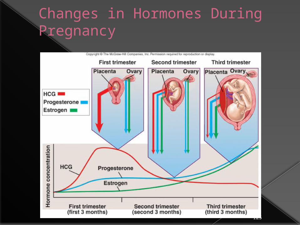
Changes in Hormones During Pregnancy

Hormone Levels in Human Maternal Blood during Normal Pregnancy
Hormone Approximate Peak Value
Time of Peak Secretion
hCG 5 mg/mL First Trimester
Relaxin 1 ng/mL First Trimester
hCS 15 mg/mL Term
Estradiol 16 ng/mL Term
Estriol 14 ng/mL Term
Progesterone 190 ng/mL Term
Prolactin 200 ng/mL Term