endodontic management of talon cusp causing apical ......talon cusp, which is also deffned as dens...
TRANSCRIPT
Remedy Publications LLC.
Journal of Dentistry and Oral Biology
2017 | Volume 2 | Issue 13 | Article 10821
IntroductionTalon cusp, which is also defined as dens evaginatus, supernumerary cusp, horn, hyperplastic
cingulum, evaginated odontome, cusped cingulum, accessory cusp, and supernumerary lingual tubercle [1-3] is a rare odontogenic developmental dental anomaly projecting from the cingulum area or the cementoenamel junction (CEJ). It is characterized by an accessory cusp as a projection of the maxillary or mandibular teeth in both deciduous and permanent dentition with normal enamel and dentin containing a varying degree of pulp tissue [2,4-13]. This cusp normally presented in the palatal or occlusal surfaces of the teeth, however, there were some reported cases of talon cusps in labial surfaces of teeth [11]. Although there is no clear etiology for this anomaly; most of authors consider that this anomaly has multifactorial etiology involving both genetic and environmental factors [6,9-11,14]. In dental anomalies frequency of occurrence of this anomaly is less than 1%. It can be observed as an isolated entity or in association with some systemic conditions such as Rubinstein-Taybi syndrome, Mohr syndrome (oral-facial-digital syndrome, type II), Sturge-Weber syndrome (enceph-alotrigeminal angiomatosis), or incontinentia pigmenti achromians [2,3,6,7,10,11,15-19]. Treatment modalities change according to the type of presentation and complications of this anomaly. According to the literature, there are many treatment options range from prophylactic sealing to endodontic treatment methods [11]. Small talon cusps usually don’t need treatment. Esthetics may be a major issue if talon cusp is present on labial aspect. On the other hand, large talon cusps frequently cause clinical problems such as; tongue injuries, caries, occlusal interferences, speech and mastication problems, pulpal necrosis, periapical pathosis and periodontal problems necessitating complicated treatment modalities. On literature review, there are several reports presenting concurrence of developmental anomalies with talon cusp in the same tooth [13,20], however only 4 cases of talon cusp teeth with periapical pathosis have been reported [21-24]. The aim of this report is to present a clinical case of the endodontic treatment of a permanent maxillary left central incisor with talon cusp causing periapical pathosis related to occlusal trauma.
Case PresentationA 45-year-old male patient was referred to the Department of Dentomaxillofacial Radiology
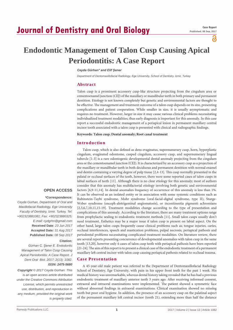
School of Dentistry, Ege University, with pain in his upper front teeth for the past 1 week. His medical history was unremarkable, whereas dental history taking revealed that he has had a previous endodontic treatment of maxillary anterior teeth 3 years ago. After receiving informed consent, extraoral and intraoral examinations were implemented. The patient showed a symmetric face without abnormal findings in extraoral examinations. Clinical examination showed no missing tooth but poor oral hygiene. In addition, the presence of an accessory cusp on the palatinal aspect of the permanent maxillary left central incisor (tooth 21), extending more than half the distance
Endodontic Management of Talon Cusp Causing Apical Periodontitis: A Case Report
OPEN ACCESS
*Correspondence:Ceyda Gürhan, Department of Oral and Maxillofacial Radiology, Ege University Faculty of Dentistry, Izmir, Turkey, Tel:
+902323881081; Fax: +902323880325;E-mail: [email protected]
Received Date: 23 Jun 2017Accepted Date: 31 Aug 2017
Published Date: 08 Sep 2017
Citation: Gürhan C, Şener E. Endodontic
Management of Talon Cusp Causing Apical Periodontitis: A Case Report. J
Dent Oral Biol. 2017; 2(13): 1082.ISSN: 2475-5680
Copyright © 2017 Ceyda Gürhan. This is an open access article distributed
under the Creative Commons Attribution License, which permits unrestricted
use, distribution, and reproduction in any medium, provided the original work
is properly cited.
Case ReportPublished: 08 Sep, 2017
AbstractTalon cusp is a prominent accessory cusp-like structure projecting from the cingulum area or cementoenamel junction (CEJ) of the maxillary or mandibular teeth in both primary and permanent dentition. Etiology is not known completely but genetic and environmental factors are thought to be effective. The management and treatment outcome of a talon cusp depends on its size, presenting complications and patient cooperation. While smaller in size, it is usually asymptomatic and requires no treatment. However, larger in size it may cause various clinical problems necessitating individualized treatment modalities; thus early diagnosis is important for this anomaly. In this case report a successful endodontic management of a periapical lesion in permanent maxillary central incisor tooth associated with a talon cusp is presented with clinical and radiographic findings.
Keywords: Talon cusp; Dental anomaly; Root canal treatment
Ceyda Gürhan* and Elif Şener
Department of Dentomaxillofacial Radiology, Ege University, School of Dentistry, Izmir, Turkey
Ceyda Gürhan, et al., Journal of Dentistry and Oral Biology
Remedy Publications LLC. 2017 | Volume 2 | Issue 13 | Article 10822
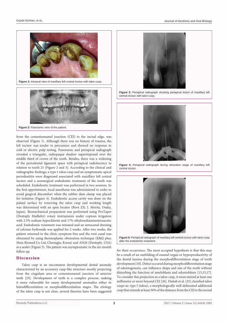
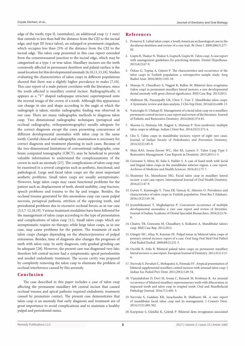
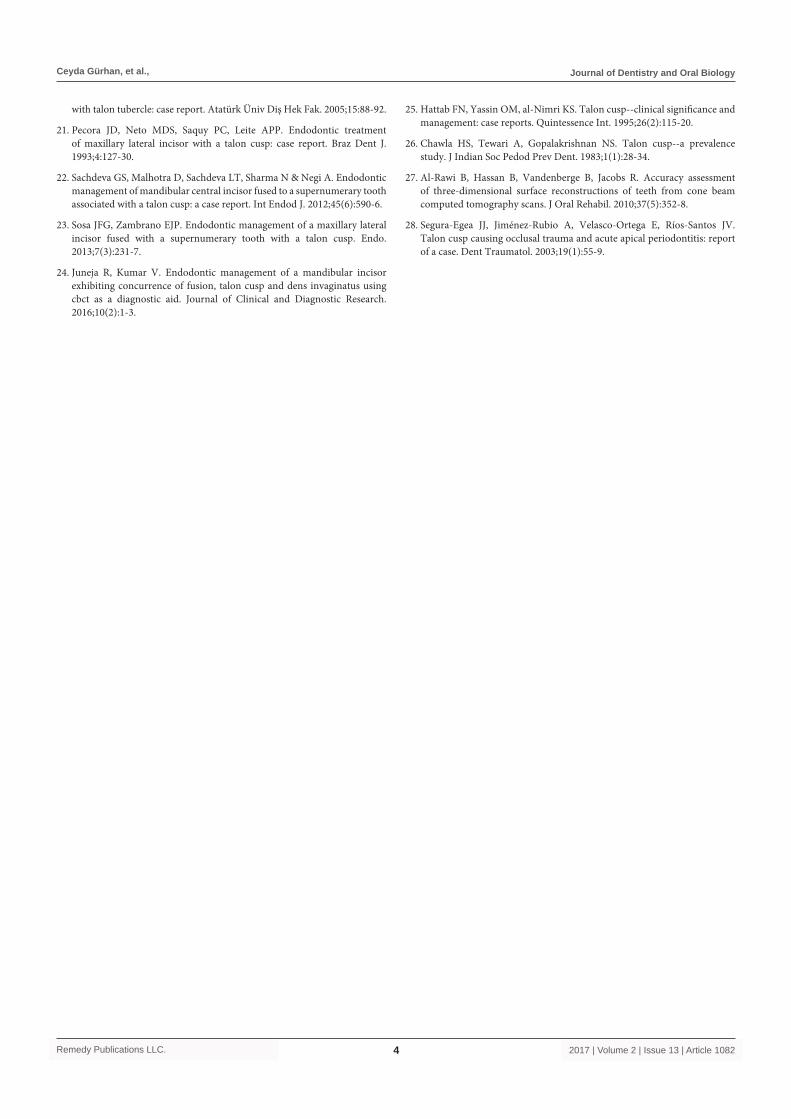
from the cementoenamel junction (CEJ) to the incisal edge, was observed (Figure 1). Although there was no history of trauma, the left incisor was tender to percussion and showed no response to cold or electric pulp testing. Panoramic and periapical radiograph revealed a triangular, radiopaque shadow superimposed over the middle third of crown of the tooth. Besides, there was a widening of the periodontal ligament space with periapical radiolucency in relation to tooth 21 (Figure 2 and 3). According to the clinical and radiographic findings, a type 1 talon cusp and an symptomatic apical periodontitis were diagnosed associated with maxillary left central incisor and a nonsurgical endodontic treatment of the tooth was scheduled. Endodontic treatmant was performed in two sessions. In the first appointment, local anesthesia was administered in order to avoid gingival discomfort when the rubber dam clamp was placed for isolation (Figure 4). Endodontic access cavity was done on the palatal surface by removing the talon cusp and working length was determined with an apex locator (Root ZX; J. Morita, Osaka, Japan). Biomechanical preparation was performed using ProTaper (Dentsply Maillefer) rotary instruments under copious irrigation with 2.5% sodium hypochlorite and 17% ethylenediaminetetraacetic acid. Endodontic treatment was initiated and an intracanal dressing of calcium hydroxide was applied for 2 weeks. After two weeks, the patient returned to the clinic symptom-free and the root canal was obturated by using thermoplastic obturation technique (E&Q plus; Meta Biomed Co Ltd, Cheongju, Korea) and AH26 (Dentsply, USA) as a sealer (Figure 5). The patient was asymptomatic in the six-month follow up.
DiscussionTalon cusp is an uncommon developmental dental anomaly
characterized by an accessory cusp-like structure mostly projecting from the cingulum area or cementoenamel junction of anterior teeth [25]. Development of teeth is a complex process, making it more vulnerable for many developmental anomalies either in histodifferentiation or morphodifferentiation stages. The etiology of the talon cusp is not clear, several theories have been suggested
for their occurrence. The most accepted hypothesis is that this may be a result of an outfolding of enamel organ or hyperproductivity of the dental lamina during the morphodifferentiation stage of tooth development [18]. Defect occured during morphodifferentiation stage of odontogenezis, can influence shape and size of the tooth without disturbing the function of ameloblasts and odontoblasts [13,15,17]. To consider this projection as a talon cusp, it must extend at least one millimetre or more beyond CEJ [26]. Hattab et al. [25] classified talon cusps as: type I (talon), a morphologically well-delineated additional cusp that extends at least 50% of the distance from the CEJ to the incisal
Figure 1: Intraoral view of maxillary left central incisor with talon cusp.
Figure 2: Panoramic view of the patient.
Figure 3: Periapical radiograph showing periapical lesion of maxillary left central incisor with talon cusp.
Figure 4: Periapical radiograph during obturation stage of maxillary left central incisor.
Figure 5: Periapical radiograph of maxillary left central incisor with talon cusp after the endodontic treatment.
Ceyda Gürhan, et al., Journal of Dentistry and Oral Biology
Remedy Publications LLC. 2017 | Volume 2 | Issue 13 | Article 10823
edge of the tooth; type II, (semitalon), an additional cusp (≥ 1 mm) that extends to less than half the distance from the CEJ to the incisal edge; and type III (trace talon), an enlarged or prominent cingulum, which occupies less than 25% of the distance from the CEJ to the incisal edge. The talon cusp presented in this case report extended from the cementoenamel junction to the incisal edge, which may be categorized as a type 1 or true talon. Maxillary incisors are the teeth commonly affected in permanent dentition and palatal surface is the usual location for this developmental anomaly [6,10,11,13,18]. Studies evaluating the characteristics of talon cusps in different populations showed that there was a slightly higher prevalence in males [7,10]. This case report of a male patient correlates with the literature, since the tooth affected is maxillary central incisor. Radiographically, it appears as a “V” shaped radiopaque structure superimposed onto the normal image of the crown of a tooth. Although this appearance can change in size and shape according to the angle at which the radiograph is taken, similar radiographic finding was observed in our case. There are many radiographic methods to diagnose talon cusp. Two dimensional radiographic techniques (periapical and occlusal radiography, orthopantomography) usually adequate for the correct diagnosis except the cases presenting concurrence of different developmental anomalies with talon cusp in the same tooth. Careful clinical and radiographic examination is necessary for correct diagnosis and treatment planning in such cases. Because of the two-dimensional limitations of conventional radiographs, cone beam computed tomography (CBCT), may be beneficial to provide valuable information to understand the complexanatomy of the crown in such an anomaly [27]. The complications of talon cusp may be examined in a several categories such as aesthetic, functional, and pathological. Large and facial talon cusps are the most important aesthetic problems. Small talon cusps are usually asymptomatic. However, large talon cusps may cause functional problems for the patient such as; displacement of teeth, dental mobility, cusp fracture, speech problems and trauma to the lip and tongue. Besides, the occlusal trauma generated by this anomalous cusp can cause pulpal necrosis, periapical pathosis, attrition of the opposing tooth, and periodontal problems due to excessive occlusal forces, as in our case [3,5,7-12,18,19]. Various treatment modalities have been followed for the management of talon cusps according to the type of presentation and complications of talon cusp [11]. Small talon cusps which are asymptomatic require no therapy; while large talon cusps, as in our case, may cause problems for the patient. The treatment of such talon cusps changes depending on the absence/presence of pulpal extensions. Besides, time of diagnosis also changes the prognosis of teeth with talon cusp. In early diagnosis, only gradual grinding can be adequate [28]. However, the present case was diagnosed very late; therefore left central incisor had a symptomatic apical periodontitis and needed endodontic treatment. The access cavity was prepared by completely removing the talon cusp to eliminate the problem of occlusal interference caused by this anomaly.
ConclusionThe case described in this paper includes a case of talon cusp
affecting the permanent maxillary left central incisor that caused occlusal trauma and apical pathosis required endodontic treatment caused by premature contact. The present case demonstrates that talon cusp is an anomaly that early diagnosis and treatment are of great importance to avoid complications and to maintain a healthy pulpal and periodontal status.
References1. Pomeroy E. Labial talon cusps: a South American archaeological case in the
deciduous dentition and review of a rare trait. Br Dent J. 2009;206(5):277-82.
2. Gupta R, Thakur N, Thakur S, Gupta B, Gupta M. Talon cusp: A case report with management guidelines for practicing dentists. Dental Hypotheses. 2013;4(2):67-9.
3. Özkan G, Toptaş A, Güneri P. The characteristics and occurrence of the talon cusps in Turkish population: a retrospective sample study. Surg Radiol Anat. 2016;38(9):1105-10.
4. Manuja N, Chaudhary S, Nagpal R, Rallan M. Bilateral dens evaginatus (talon cusp) in permanent maxillary lateral incisors: a rare developmental dental anomaly with great clinical significance. BMJ Case Rep. 2013;2013.
5. Mallineni SK, Panampally GK, Chen Y, Tian T. Mandibular talon cusps: A Systematic review and data analysis. J Clin Exp Dent. 2014;6(4):e408-13.
6. Yazicioglu O, Ulukapi H. Management of a facial talon cusp on a maxillary permanent central incisor:a case report and review of the literature. Journal of Esthetic and Restorative Dentistry. 2014;26(6):374-81.
7. Sharma G, Mutneja AR, Nagpal A, Mutneja P. Non-syndromic multiple talon cusps in siblings. Indian J Dent Res. 2014;25(2):272-4.
8. Cho S. Talon cusps in mandibular incisors: report of eight rare cases. Journal of Indian Society of Pedodontics and Preventive Dentistry. 2014;32(2):185-9.
9. Maia RAS, Souza-Zaroni WC, Mei RS, Lamers F. Talon Cusp Type I: Restorative Management. Case Reports in Dentistry. 2015;2015:1-5.
10. Goswami S, Mitra M, Saha S, Halder S. A case of fused teeth with facial and lingual talon cusps in the mandibular anterior region: a case report. Archives of Medicine and Health Sciences. 2016;4(1):75-7.
11. Shaminey SA, Manoharan MG. Facial talon cusp in maxillary lateral incisor: a rare case report. International Journal of Oral Health Dentistry. 2016;2(1):47-9.
12. Guven Y, Kasimoglu Y, Tuna EB, Gencay K, Aktoren O. Prevalence and characteristics of talon cusps in Turkish population. Dent Res J (Isfahan). 2016;13(2):145-50.
13. Jeyanthikumari T, Meghalapriya P. Concurrent occurrence of multiple developmental anomalies: a rare case report and review of literatüre. Journal of Indian Academy of Dental Specialist Researchers. 2016;2(2):76-8.
14. Chaitra TR, Goswami M, Chaudhary S, Kulkarni A. Mandibular talon's cusp. BMJ Case Rep. 2012;2012.
15. Güngör HC, Altay N, Kaymaz FF. Pulpal tissue in bilateral talon cusps of primary central incisors: report of a case. Oral Surg Oral Med Oral Pathol Oral Radiol Endod. 2000;89(2):231-5.
16. Ozcelik B, Atila B. Bilateral palatal talon cusps on permanent maxillary lateral incisors: a case report. European Journal of Dentistry. 2011;5(1):113-6.
17. Nuvvula S, Pavuluri C, Mohapatra A, Nirmala SV. Atypical presentation of bilateral supplemental maxillary central incisors with unusual talon cusp. J Indian Soc Pedod Prev Dent. 2011;29(2):149-54.
18. Vijayalakshmi D, Devi M, Sreeja C, Ramesh M, Krishnan R. An unusual occurrence of bilateral maxillary supernumerary teeth with dilaceration in impacted tooth and talon cusp in erupted tooth. Oral and Maxillofacial Pathology Journal. 2016;7(1):691-3.
19. Nuvvula S, Gaddam KR, Jayachandra B, Mallineni SK. A rare report of mandibular facial talon cusp and its management. J Conserv Dent. 2014;17(5):499-502.
20. Kayipmaz S, Gündüz K, Çelenk P. Bilateral dens invaginatus associated
Ceyda Gürhan, et al., Journal of Dentistry and Oral Biology
Remedy Publications LLC. 2017 | Volume 2 | Issue 13 | Article 10824
with talon tubercle: case report. Atatürk Üniv Diş Hek Fak. 2005;15:88-92.
21. Pecora JD, Neto MDS, Saquy PC, Leite APP. Endodontic treatment of maxillary lateral incisor with a talon cusp: case report. Braz Dent J. 1993;4:127-30.
22. Sachdeva GS, Malhotra D, Sachdeva LT, Sharma N & Negi A. Endodontic management of mandibular central incisor fused to a supernumerary tooth associated with a talon cusp: a case report. Int Endod J. 2012;45(6):590-6.
23. Sosa JFG, Zambrano EJP. Endodontic management of a maxillary lateral incisor fused with a supernumerary tooth with a talon cusp. Endo. 2013;7(3):231-7.
24. Juneja R, Kumar V. Endodontic management of a mandibular incisor exhibiting concurrence of fusion, talon cusp and dens invaginatus using cbct as a diagnostic aid. Journal of Clinical and Diagnostic Research. 2016;10(2):1-3.
25. Hattab FN, Yassin OM, al-Nimri KS. Talon cusp--clinical significance and management: case reports. Quintessence Int. 1995;26(2):115-20.
26. Chawla HS, Tewari A, Gopalakrishnan NS. Talon cusp--a prevalence study. J Indian Soc Pedod Prev Dent. 1983;1(1):28-34.
27. Al-Rawi B, Hassan B, Vandenberge B, Jacobs R. Accuracy assessment of three-dimensional surface reconstructions of teeth from cone beam computed tomography scans. J Oral Rehabil. 2010;37(5):352-8.
28. Segura-Egea JJ, Jiménez-Rubio A, Velasco-Ortega E, Ríos-Santos JV. Talon cusp causing occlusal trauma and acute apical periodontitis: report of a case. Dent Traumatol. 2003;19(1):55-9.