endoluminal therapies for gastroesophageal reflux disease
TRANSCRIPT

1
ENDOLUMINAL THERAPIES FOR
GASTROESOPHAGEAL REFLUX DISEASE, OBESITY
AND BARRETT’S ESOPHAGUS
Development of a new endoluminal device and a technique
Ph.D. Thesis
By
András Légner M.D.
Supervisor
Prof. Dr. Örs Péter Horváth
University of Pécs Faculty of Medicine St. George University Teaching Hospital
Pécs Department of Surgery, Székesfehérvár
Pécs, Hungary
2016

2
TABLE OF CONTENTS Page
1. Introduction 3-7
1.1 Physiology of GERD 3
1.2 Management of GERD 3
1.3 Physiology of obesity 5
1.4 Management of obesity 5
1.5 Endoluminal therapies for Barrett’s esophagus 7
2. Overall aims of the study 8
3. Development of the device and a new technique 9-11
3.1 Background 9
3.2 Methods 9
3.3 Results 10
3.4 Discussion 11
4. Safety and feasibility study 12-14
4.1 Background 12
4.2 Methods 12
4.3 Results 13
4.4 Discussion 14
5. Gastric submucosal fibrosis generation 15-17
5.1 Background 15
5.2 Methods 15
5.3 Results 16
5.4 Discussion 17
6. Human pilot study 18-21
6.1 Background 18
6.2 Methods 18
6.3 Results 20
6.4 Discussion 21
7. Barrett’s strip endoscopic mucosal excision 22-24
7.1 Background 22
7.2 Methods 22
7.3 Results 23
7.4 Discussion 23
8. Summary of the work 25-29
9. Acknowledgements 30
10. Publications and presentations 31-35
11. Abbreviations 36

3
1. INTRODUCTION
The number of patients with gastroesophageal reflux disease (GERD) as well as the
prevalence of obesity and Barrett’s esophagus have dramatically increased in the last two
decades particularly in the Western world. This has generated an extensive research and it
led to a better understanding of the pathophysiology of these conditions; however, their
appropriate management continues to be debated. New anti-secretory medications,
laparoscopic surgical techniques and novel endoluminal devices have been introduced. The
outcomes are encouraging but the costs continue to be high.
1.1 Physiology of GERD
The lower esophageal sphincter (LES) and the geometric profile of the cardia (the angle of
His) are factors to prevent gastroesophageal reflux (GER) and are the targets of surgical
and endoluminal GERD procedures. A decrease in pressure and/or the overall length or
just the length of the abdominal segment of the LES predisposes to reflux, as it decreases
the resistance against the high pressure gastric contents. In patients with severe GERD, the
LES or the “high-pressure zone” is virtually nonexistent or greatly reduced. At a critical
length of 1-2 cm intraabdominal LES, the LESP drops acutely and GER occurs.
Additionally, alterations in neuro regulation of the LES may lead to inappropriate transient
LES relaxations (tLESR) through vagal afferent fibers that are terminated in specialized
intra-ganglionic laminar endings (IGLEs) within the muscle fibers at the cardia and
fundus.
1.2 Management of GERD
The treatment of GERD is individualized depending on the patient’s co-morbidities,
symptom severity, response to medication and physiologic test results. The treatment
spectrum is wide; from simple life style changes to Roux-en-Y gastric bypass.
Intensive proton pump inhibitor (PPI) therapy in generally has a high success rate;
however, it has no effect on alkaline reflux and the underlying anatomical defects.
Anti-reflux surgery is recommended for patients with refractory or complicated GERD and
provides excellent symptom control in 85%-90% of cases. Nevertheless, laparoscopic anti-
reflux surgery (LARS) requires general anesthesia, hospitalization, it is expensive and it is
associated with 3%-5% failure rate as well as post-operative morbidity and even mortality.
Endoluminal intervention for GERD is a relatively new and promising concept. In light of
the above described pathophysiologic factors, endoscopic therapies should prevent reflux
in one or more of the following ways; 1. alter the compliance of the cardia and prevent
tLES shortening/relaxation; 2. increase baseline LES tone or, 3. increase baseline LES

4
length. The endoscopic anti-reflux procedures published to date can be categorized into
three groups: (1) ablation, (2) injection or implantation, (3) fixation.
The basis of ablation therapy is muscular hypertrophy, fibrosis and neurolysis at the level
of the lower esophageal sphincter and gastric cardia by means of radio frequency energy.
The Stretta procedure showed only 30-40 % the pH normalization rate at 12 month follow
up.
The goal of injection therapies is to deliver a biologically inert, injectable substance into
different depths of the LES region in order to increase the LESP. Enteryx, an ethylene
vinyl alcohol copolymer with tantalum dissolved in dimethyl sulfide. It is injected into the
muscularis propria of the LES. The Gatekeeper is a dehydrated hydrogel prosthesis
implanted into the submucosa at the level of LES. The Durasphere is a sterile,
biocompatible injectable bulking agent composed of pyrolytic carbon coated graphite
beads containing zirconium oxide, suspended in a water-based, absorbable polysaccharide
carrier gel and it is injected into the submucosa within 1 cm of the Z-line. The results are
discouraging as only about 40% pH normalization rates, early device expellation and
serious adverse events were reported.
Endoluminal fixation techniques are based on intraluminal apposition of tissue by using
either staples, suture fasteners or sutures. The procedures are visualized by use of
commercially available or by specially developed endoscopes. The EndoCinch® device
was originally developed to create a full thickness intussusception at the gastroesophageal
junction (GEJ). The sewing capsule is attached to the tip of the gastroscope. Figure of
eight sutures are created with a straight needle and then tightened by using plastic anchors.
Results of multicenter trials demonstrated only 39.7% pH normalization rate at 6 months
follow up as the tissue folds in lack of robust scar tissue were lasting a short time. The
NDO Plicator system was developed to create and fixate a gastric plication below the GEJ
in the anterior cardia with serosa to serosa apposition. The system consists of a plicator
instrument, a helical shaped “corkscrew” tissue retractor and a pretied suture insert. The
procedure is done under direct visualization by inserting a 6 mm flexible endoscope
through the device. A prospective multicenter and sham controlled randomized trials
showed distal esophageal acid normalization occurred in only 23% to 30% after one year
post-procedure follow-up. The EsophyX device is used to increase the competency of the
anti-reflux barrier by restoring the angle of His. A valve is created at the GEJ by delivering
multiple full thicknesses, non-resorbable polypropylene fasteners. The end result is an
anterio-lateral, 200-300 º, 3-5 cm long partial fundoplication. Post procedural distal
esophageal pH normalization was seen in only 37% of patients at one year follow up. The
Medigus SRS system is a special 15 mm thick endoscope that combines a surgical stapler,
video camera and sonar to create a 180º anterior fundoplication. The tip of the endoscope
retroflexes to the endoscope’s rigid 6 cm long segment and bring the proximal gastric wall
to the lower anterior esophagus, 2-3 cm above the GEJ. Tissue fixation is achieved by
firing a cartridge of 5 staplers from the rigid segment of the endoscope.

5
1.3 Physiology of obesity
The pathophysiology of obesity is complex as it is influenced by environmental,
behavioral, genetic, endocrine and neurotransmitter factors. Although obesity is simply a
result of an imbalance between energy intake and expenditure, the various combinations of
these factors results in heterogeneous clinical manifestations of obesity. Recent research
implicates that besides of gene-diet interactions, environmental and social-behavioral risk
factors including poor quality nutrients, chronic stress, pre-and postpartum environment,
sedentary lifestyle and exposure to chemical or pharmaceutical agents, such as
antipsychotics, antidepressants or corticosteroids are playing also an important role.
1.4 Management of obesity
As our knowledge of the pathophysiology of obesity increases, it is becoming clear that the
treatment of obesity is complex and must be individualized. Present treatment options
range from life style change and dieting to bariatric surgery.
The pharmaceutical options such as inhibitors of intestinal fat absorption,
sympathomimetic agents that suppress appetite, increase satiety or thermogenesis, and
antagonists of the endocannabinoid system are usually non effective because the
psychological, environmental and socioeconomic circumstances are usually not optimal.
The spectrum of bariatric surgery is wide including adjustable gastric banding, sleeve
gastrectomy, jejunoileal bypass, duodenal switch biliopancreatic bypass and Roux-en-Y
gastric bypass. The complication and cost of bariatric surgery has decreased, however,
surgery is available only for a small part of the morbidly obese population. Despite the
improved results a large proportion of patients still resist having operative intervention.
An effort to develop newer and less invasive techniques for the obese population is
imperative. Endoluminal management of obesity is challenging as it must be safe,
effective, durable and cost effective. The approach still is in its infancy but holds great
promise for providing presurgical weight loss, postsurgical revisions and even primary
intervention. The current and emerging endoscopic devices for obesity are numerous and
can be categorized as (1) space occupying devices, (2) transoral endoluminal stapling or
suturing devices, (3) prosthetic gastric/duodenal sleeves, (4) miscellaneous.
Space occupying therapy is the most preferred endoscopic bariatric procedure worldwide.
This popularity is based on its relative safety and low price compared to other techniques.
Intragastric balloon therapy related weight loss is due to mechanical and physiological
effects; however the durability of weight loss continues to be a pitfall. The most frequent
device related complications are GERD and consequent esophagitis, nausea or vomiting
which usually responds to medication. They may have a role as a bridge to bariatric
surgery to reduce perioperative complications in morbid obese population.
6
Complex platforms for transoral endoluminal stapling or suturing are designed to reduce
gastric volume or to restrict gastro-jejunal stomas. These devices deliver sutures, tissue
anchors, fasteners or even staplers in order to preserve the new geometry of the stomach.
The Incisionless Operating Platform (IOP) is used for translumenal, endoluminal or single
incision surgery. For bariatric patients it is intended for either primary or revisional
procedures. The IOP contains 4 channels, one for a flexible 4.9 mm endoscope for
visualization and the other 3 for tissue manipulation. The specially designed tissue
anchors provide durable tissue approximation because the holding force is widely
distributed. This device showed about 30% excess weight loss (EWL) in patients after
failed gastric bypass surgery. The StomaphyX is a single-use device, designed to create
large gastric tissue folds. A regular gastroscope is used through the device for direct
visualization. Tissue is drawn into the distal part of the device and five to six gastric folds
are created and stabilized one by one after delivery of non-resorbable polypropylene
fasteners. The TOGA system is an endoscopic stapling device to create a gastric sleeve
parallel to the lesser curvature. The procedure is performed under direct visualization as a
gastroscope can be advanced through the device and retroflexed. The stapler line engages
the anterior and posterior gastric walls. Six month follow up showed 45% EWL.
In summary, endoluminal suturing and stapling techniques are seemed to be safe and have
demonstrated good to moderate weight loss on short term, however long term durability is
related to sufficient scar tissue formation.
Prosthetic sleeves are tube-like plastic devices delivered and secured endoscopically in the
proximal GI tract to restrict absorption of nutrients in the small intestine and/or exclude the
stomach from the digestive process. The main advantage of the procedure that it does not
alter permanently the anatomy. Its potential disadvantages are difficulty in securing the
device with the consequent potential of migration and small bowel obstruction. The Valen
Tx gastric sleeve is designed to mimic the effect of the Roux-en-Y gastric bypass. The
device is attached by transmural anchors to the GEJ, and extends through the stomach and
into the distal duodenum or proximal jejunum. The length of the sleeve is variable
depending on the therapeutic goal. The Endobarrier is a 60 cm long, flexible sheath
composed of a nutrient-impermeable fluoropolymer that is deployed in the duodenal bulb
and extends to the jejunum. The device prevents nutrient absorption and mixing with
digestive enzymes, mimicking one of the components of the RNY gastric bypass. Short
term follow up results show about 45% EWL and significant decrease inHbA1c% was also
achieved.
Other therapies have been also investigated. The bariatric effect of electrical stimulation
is based on a series of low-energy electrical impulses delivered to the smooth muscle of
the stomach intended to create a feeling of fullness or gastroparesis. Localized
radiofrequency ablation of gastric tissue may have beneficial effects on weight loss as
structural and functional changes may happen that lead to decreased appetite and
consequent weight loss.

7
1.5 Endoluminal therapies for Barrett’s esophagus
Persistent exposure of gastric content to esophageal mucosa creates an abnormal
environment where after a cellular damage of the stratified squamous epithelium, intestinal
metaplasia can develop. This is a well-studied premalignant condition of esophageal
adenocarcinoma. In the era of minimally invasive procedures the treatment of Barrett’s
esophagus and early mucosal adenocarcinomas has been changed. Different endoscopic
treatment modalities for mucosal destruction are available such as thermal, photodynamic,
radiofrequency or endoscopic mucosal removal therapy.
Thermal therapies result in destruction of columnar esophageal epithelium achieved by
administration of heat form electrocoagulation, a heater probe, neodymium-doped yttrium
aluminum garnet laser or argon beam plasma. After elimination of esophageal mucosa
regrowth of normal squamous lining occurs. Heat depth penetration is not well controlled
and as a consequence the procedure is not devoid of complications.
Photodynamic therapy utilizes a photosensitizing drug and laser light. As a result, singlet
oxygen is generated which causes irreversible oxidation of essential cellular components.
The unquestionable disadvantage of this technique is a prolonged general photosensitivity.
Radiofrequency ablation is a relatively new therapeutic method. Energy from a controlled
radiofrequency source is applied to the Barrett’s epithelium using a balloon catheter or a
wired paddle and causes complete destruction beyond the lamina propria. The advantage
of this procedure is that the depth of epithelial damage is better controlled in contrast to
photodynamic therapy, but histological assessment is not possible.
The major advantage of endoscopic mucosal removal is that pathological examination
after tissue removal is possible. Endoscopic mucosal resection (EMR) and endoscopic
submucosal dissection (ESD) were developed to remove superficial cancerous mucosa
from the gastrointestinal tract. Endoscopic submucosal dissection is more often used to
remove gastric mucosal lesions as manipulation in the thin walled esophagus carries more
risk. Both techniques are labor intense and have a long learning curve.
8
2. OVERALL AIMS OF THE STUDY
In the last decades extensive innovational effort has been addressed to endoluminal GERD
and obesity therapies. Majority of these new devices have failed to demonstrate long-term
efficacy and/or safety mostly due to the complex anatomy of the GEJ area and
pathophysiology of these diseases. Data from previous studies suggest that mechanical
changes to restore the angle of His and LES pressure for GERD and restrictive bariatric
techniques for obesity may have the greatest success. However, these techniques often fail
due to the weakness of the fixation method.
The overall aim of the study was to develop a device and a procedure to create effective
and durable gastroplasty to treat GERD and also to create a small proximal gastric pouch
and restricted pouch outlet for obese patients. Our principal hypothesis was that gastric
mucosal excision followed by full thickness suture placement is feasible, safe and provides
long lasting tissue apposition and surgical effect. We also investigated the possibility to
safely remove esophageal mucosa for Barrett’s esophagus by using our modified device.
The aims of our work were:
To develop and test a complex device that excises gastric mucosa and places full thickness
sutures in one. We performed ex vivo and in vivo experiments to optimize device
characteristics and to develop the operative technique for both GERD and obesity.
To demonstrate feasibility of mucosal excision and full-thickness suture apposition of the
excision beds at the gastroesophageal junction by using a new generation of devices. We
measured the GEJ compliance in a survival canine study and determined the durability of
the restrictive gastric pouch outlet in the obesity model. Histologic examination was also
performed to visualize tissue healing and scar formation.
To understand more how to augment scar tissue formation in the submucosa for more
durable gastroplasties. We tested different hypertonic solutions in survival swine
experiments.
To evaluate the safety of the endoluminal gastroplasty procedure for GERD and obesity in
a human pilot trial. We studied the effect of the gastroplasty procedure on symptom scores,
quality of life, antireflux medication usage and pH monitoring for GERD patients and
excess body weight loss for obese patients.
To develop a technique and evaluate the feasibility of strip endoscopic mucosal resection
(SEMR) for Barrett’s esophagus utilizing a modified gastroplasty excision device.

9
3. DEVELOPMENT OF THE DEVICE AND A NEW TECHNIQUE
3.1 Background
In the first phase of the study a complex transoral endoscopic device was developed. This
was able to excise mucosa from both the GEJ and from the proximal stomach and also to
place full thickness sutures into the denuded areas. Two procedures were designed: one to
reduce tissue attenuation at the GEJ for GERD; another to create a small proximal gastric
pouch to treat obesity. The main focus of our initial laboratory work was feasibility, safety,
quality, and reliability of the procedures.
3.2 Methods
The device
The first generation device was a dilator shape instrument with handle, shaft and a distal
integrated operating capsule that was capable both to perform mucosal excision and suture
placement in one. The device was designed to be used with 5 mm - 6.5 mm diameter
flexible pediatric gastroscope that enters into a dedicated channel at the handle and exits
the device at the flexible transition segment. This endoscope provided direct visualization
for the procedure.
Ex vivo experiments
First ex vivo porcine tissue experiments were conducted on stomachs and esophagi to test
feasibility of mucosal excision. Then both device and operative technique refinement were
done on human esophagi and stomachs that were harvested from cadavers after organ
donation. Histologic sections of the stomach wall were used to determine excision depth
and excision overlap safety. Possible complication scenarios such as needle penetration
into adjacent tissue were also tested.
In vivo acute experiments
The protocol was approved by the Creighton University Institutional Animal Care and Use
Committee under the number (IACUC PN-0785). This in-vivo acute experiments (n=7)
were carried out on domestic pigs (Sus scrofa domesticus, ssp. Large white), weighting
between 22.5 and 26.3 kg, focusing on mucosal excision and suture actuation reliability for
GERD and obesity procedures.
In vivo survival experiments
For both GERD and obesity procedural efficacy survival studies were performed in
baboons (Papio hamadryas) at the Southwest Foundation for Biomedical Research (San
Antonio, TX, USA (IACUC PN-1161)). Total of 12 animals underwent GERD (n=6)
(mean weight 30,4 kg) or obesity (n=6) (mean weight 30,6 kg) procedures. In the case of
GERD animals the compliance of the GEJ was preoperatively evaluated. The animals were

10
fed with a hyper caloric clear liquid diet for 8 weeks. Before euthanasia the compliance of
the GEJ as well as the body weight were recorded. After euthanasia gross inspection and
histologic examinations were completed.
The procedure
The main steps of the in vivo GERD operations were the following: under general
anesthesia and endotracheal intubation the device was placed through the mouth into the
stomach. A 6 mm pediatric flexible endoscope (FG-100 RE, Fujinon, Tokyo, Japan) was
introduced through the dedicated channel of the device for direct procedural visualization.
The device was positioned at the greater curvature and three cycles of mucosal excision
and suturing were carried out after the submucosal space was injected with 15 ml of
1:200.000 adrenalin solution. The sutures were organized then tied under endoscopic
visualization by using a knotting device.
The obesity procedure consisted of 3 overlapping excision-suturing cycles on the greater
curvature side to create a vertical gastroplasty line to form a neo-esophagus with a small
gastric pouch and restrictive outlet along the lesser curvature.
3.3 Results
Ex vivo
Total of 104 excisions and 55 suture actuations were performed. The size of the operative
capsule trough and the depth of the excision blade provided reliable mucosal excision;
however, full thickness injury occurred in 3 (2.9%) cases. The suture penetration depth
was accurate but suture actuation reliability was poor as in 8 (14,5%) cases malfunctions
occurred. Adjacent tissue suture entrapment experiments showed safety of full thickness
suturing. Based on these results injection needle positions and suturing mechanism of the
device were modified.
In vivo
During the in vivo animal studies total of 82 excision-suturing cycles were carried out with
the modified device. There was no major bleeding. In the survival obesity group 1 stomach
wall perforation occurred that was successfully closed endoluminal using the suturing
device, and the animal was survived without incident. The mean operation times for the
survival GERD and obesity groups were 85 min (60-110 min) and 289 min (258-390 min)
respectively. The mean estimated blood loss in case of the survival GERD animals was 13
cc (5-50 cc) and 31 cc (25-50 cc) in the obesity group. The overall suturing depth accuracy
was low as only 65% of the sutures were full thickness resulting in satisfactory stomach
wall apposition and scar tissue formation. Total of 3 (25%) from the survival procedures
were considered to be satisfactory. Postoperative bleeding was noted in one of 12 survival
animals with a single melanotic stool being passed. The mean weight change was 1,6 kg
and 0,3 kg in the obesity and GERD groups respectively. After euthanasia no injury was
found in the abdominal cavity in any of the 19 animals.
11
3.4 Discussion
With the first laboratory results from ex vivo studies we demonstrated that gastric mucosal
excision and suturing is feasible when using the first prototypes of the excision-suturing
device.
The in vivo animal experiments showed that the transoral endoscopic gastroplasty device
is generally safe and the procedure was technically feasible with no 60-day complications
or mortality. The survival animal experiments revealed inconsistent size mucosal excisions
and poor suture penetration depth with consequent high procedural unsuccess rate. It was
also demonstrated that the three steps obesity gastroplasty procedure required significantly
more effort to accomplish than acceptable.
Our results suggested that larger excisions first, followed by suturing with a separate,
longer trough device would obtain more consistent full thickness suture penetration and a
durable gastroplasty. Changes in the obesity procedure to reduce operation time and failure
rate were also necessary.

12
4. SAFETY AND FEASIBILITY CANINE STUDY
4.1 Background
Data from the first survival study revealed that modifications on both the device and
procedure steps are necessary in order to provide a safe, effective and relatively easy
outpatient procedure in the future. To improve the procedure two separate devices were
created: one for mucosal excision and another one for suturing. The gastroplasty procedure
for obesity had been reduced in complexity as well. This survival canine study was
designed to demonstrate safety, feasibility and efficacy of the new devices and the new
gastroplasty techniques.
4.2 Methods
The new gastroplasty system
The excision and suturing devices were similar in construction each having an operational
distal tip, flexible transition piece, 60 Fr flexible shaft, dedicated 5-6 mm endoscope
channel and handle. The trough of the excision device was wider and longer comparing to
the previous combined excision-suturing device. The trough of the suturing device was
also longer and operated two circular needles 1 cm apart, each connected to a separate 2.0
Prolene suture.
Measurement of GEJ compliance
For the GERD procedure the Barostat device (G & J Electronics Inc., Toronto, Ontario,
Canada) was used to measure compliance of the GEJ pre and post-procedure. The Barostat
is a specially designed instrument used to maintain constant pressure in a closed chamber
by means of a pneumatic pump. This allows measurements of the volume of the studied
organ at different pressures.
Animals
A total of seven male domestic dogs (Canis lupus familiaris), weight ranging from 16 kg to
17.5 kg, were used for the study. The protocol was approved by the Creighton University
Institutional Animal Care and Use Committee under the number (IACUC PN-0813).
Procedure
Four dogs underwent the GERD procedure and three the obesity. The animals first were
anesthetized with intravenous propofol, then endotracheal intubated, placed in the right
lateral decubitus position and maintained under general anesthesia with 2% Isoflurane.
The GERD procedure began with the compliance assessment of the GEJ by using the
Barostat. Then the excision device was introduced and a 6 mm videoendoscope (Fujunon
EG-470N5) was inserted through a dedicated channel of the device for visualization. The

13
proximal gastric mucosa was targeted just below the GEJ on the greater curvature side and
lifted from the muscularis propria by injecting 14-21 cc 1:100,000 adrenaline solution with
40% Dextrose. The mucosa was then excised and after removed. The excision procedure
was repeated both on the anterior and posterior side of the first excision creating three,
approximately 20 mm by 45mm adjacent excision beds. The excision device was then
withdrawn.
The suturing device was inserted and again the 6 mm endoscope was used for
visualization. The proximal aspects of the two lateral excision beds were captured and full
thickness sutures were placed. The device was then withdrawn and the sutures were
reloaded. The procedure was repeated in the distal aspect of the excision beds for a total of
4 sutures. The sutures were then organized, tied and cut by using the knotter device.
The technique for obesity was similar but the gastroplasty was placed 2 cm below the
squamocolumnar junction and only two - but further separated - sutures were placed
through the anterior and posterior excisions to create a restrictive procedure.
Following the procedure, dogs received 500 ml of subcutaneous physiologic saline and
were kept without oral intake for 24 hours. From the first postoperative day to euthanasia
they received a high protein high calorie pureed diet three times a day.
All dogs were survived for 8 weeks. The end term endoscopies and Barostat procedures for
the GERD dogs were performed under general anesthesia and the animals were euthanized
with IV. sodium pentobarbital. The stomachs and distal esophagi were explanted,
examined, photographed and sectioned for histologic examination.
4.3 Results
There were no intra or postoperative complications with all dogs surviving uneventfully.
The mean operative time for GERD and obesity procedures were 197 min (155-240 min)
and 158 min (145-180 min) respectively with an average amount of blood loss of 15 cc
(10-25 cc) and 17 cc (12-30cc) respectively. The GERD dogs lost an average of 0.13 kg
and the obesity dogs gained an average of 0.5 kg at 8 weeks. No bleeding was observed,
some animals did consume less in the first 10 days post procedure but eventually regained
their weight. There was no evidence of swallowing problems such as increased salivation,
retching or regurgitation. At end term endoscopy all 7 animals had a gastroplasty in place
and there was no injury to the esophagus or stomach. The three obesity dogs had an
average 8,6 mm gastric pouch outlet. The 4 GERD dogs had an average full insufflation
gap to a 10 mm endoscope of 4 mm.

14
Barostat tracings at baseline as compared to the post procedure 8 week study showed
satisfactory results with a compliance decrease similar to that seen after Nissen
fundoplication.
At autopsy there was no sign of perforation or injury of surrounding organs in any dogs.
External fibrosis at the gastroesophageal junction was noted in 6 of the 7 animals.
Nineteen of the 20 knotters placed were present.
On histologic examination cicatrix formation was present within the submucosa, however
it was full thickness only in some areas and circumferential on the average 42% in the
obesity dogs.
4.4 Discussion
We performed endoluminal proximal gastroplasty as an antireflux procedure and created a
small proximal gastric pouch with a restricted outlet for obesity using the second
generation of our gastroplasty system on total of 7 dogs. This study demonstrated that one
step endoscopic gastric mucosal excision and suture placement at and below the GEJ is
feasible and safe. All animals survived without complications. A significant decrease in
GEJ compliance was seen in each animal after the GERD procedure. Good proximal
gastric pouch outlet restriction was achieved after the obesity procedure. Scar tissue
formation was satisfactory but more fibrosis on the lesser curvature was needed for a long
lasting obesity procedure. An injectable agent that generates more robust scarring would
contribute to durability.

15
5. GASTRIC SUBMUCOSAL FIBROSIS GENERATION
5.1 Background
Current bariatric and antireflux endoscopic treatment options that are using sutures, staples
or fasteners have demonstrated encouraging results but not competitive with existing
surgical procedures.
We hypothesized that significant submucosal fibrosis at the gastroesophageal junction is
essential for better and more durable results after the sutured gastroplasty. Submucosal
fibrosis rather than muscle or panmural fibrosis is also critical to success as the latter two
can create excessive luminal compromise that would not respond to mechanical dilation of
any kind.
Submucosal injection of hypertonic solutions can cause acute mucosal erosion with
degradation of epithelial glands and congestion of capillary blood vessels on the day of
injection. Such tissue damage may create mucosal erosion with fibrosis of the submucosal
layer resulting in permanent fibrotic deposition with luminal deformation and decreased
tissue compliance. The aim of this study was to determine if gastric submucosal injections
of hypertonic saline and dextrose solutions will produce significant submucosal fibrosis.
5.2 Methods
Preliminary study
The protocol was approved by the Creighton University Institutional Animal Care and Use
Committee under the number (IACUC MCL 0976). This preliminary pilot study involving
two female miniature swine (Sus scrofa domesticus) (21.4 and 24.4 kg) was conducted to
determine if 4.05% hypertonic saline (HTS) or 50% dextrose in water solution (D50W)
forms more submucosal fibrosis. We also investigated what volume of these solutions was
optimal for fibrosis deposition.
With the minipig under endotracheal general anesthesia a midline incision was made. A 7
cm gastrotomy along the greater curvature was created. The gastric wall submucosa was
injected using either a HTS solution with 1:1000 epinephrine or D50W solution with
1:1000 epinephrine. Three, 6, or 9 ccs of solution were injected at pre-designated sites
using a modified rigid gastroplasty excision device prototype.
For the first pig 3, 6 or 9 cc of 4.05% HTS with adrenaline was used and in case of the
second pig 3, 6, or 9cc of D50W with adrenaline. At two sites in pig #1 9cc of 4.05% HTS
was placed followed by mucosal excision. At another site in pig #1 normal saline with
1:1000 adrenaline was injected followed by mucosal excision as a control. The same was
followed in pig #2 using D50W. The injection sites were marked on the serosal surface.
16
The gastrotomy was closed and the animals recovered without incident. The animals were
survived for 8 weeks and received a normal diet. Following the euthanasia each of the
stomachs and distal esophagi were explanted. All 18 intervention sites were individually
cut from the stomachs according to the serosal marks.
The specimens were then kept in 10% formalin. Eighteen hours later the tissue was cut in 3
mm thick pieces. Each 3 mm wide block was cut in each millimeter using a microtome
resulting in three sections per block. The amount and level of fibrosis and other histologic
features were recorded for each section. An H & E stain was used for the two outer
sections of each block and a Trichrome stain for the middle section.
A hypertonic saline study
Based on the results of the preliminary pilot study, a further experiment was conducted
using 3 domestic pigs (Sus scrofa domesticus). The protocol was approved by the Indiana
University Institutional Animal Care and Use Committee under the number (IACUC PN-
3696). Each animal underwent a series of gastric submucosal injections of different
concentration HTS by using a therapeutic adult flexible endoscope (GIF 140, Olympus,
Tokyo, Japan) and endoscopic injection needles. The injections were placed every time in
the fundus, body and antrum of the stomach. Each animal received 10 ccs of 4.2% HTS
with Methylene Blue injections on three sites of the stomach. On Day 14 each animal was
injected with 15 ccs of 5% HTS in three different sites as no ulcers were observed from the
initial 4.2% HTS injections. Sites were changed to an untreated area of the stomach in a
pre-determined manner. The volume and concentration of HTS was increased to 20 ccs of
7.2% HTS on Day 30 as no animal had a healed ulcer greater than 1 cm in diameter.
Thirty days after the last procedure all 3 pigs were euthanized, the stomachs were
explanted and each was opened on the greater curvature side. Using palpation and visual
assessment, areas of fibrosis were identified and a corresponding 3-0 silk suture was
placed on the serosal side.
The specimens were submerged in 4% formalin for 5 days. The specimens were cut in 3
mm blocks but only one section from each block was examined histologically. The same
method of tissue staining, measuring and volume calculation was used a sin the
preliminary study.
Statistical analysis was performed using a t-test, Mann Whitney and ANOVA with SPSS
version 17.0 (SPSS, Inc. Chicago, IL).
5.3 Results
Preliminary pilot animal study
No intra or postoperative complications occurred. Results showed that 3 cc of HTS and
D50W caused no fibrosis. The difference in fibrosis volume between 6 and 9 cc of 4.05%
HTS and D50W was insignificant (p=0.683 and p=0.750 respectively). When comparing
17
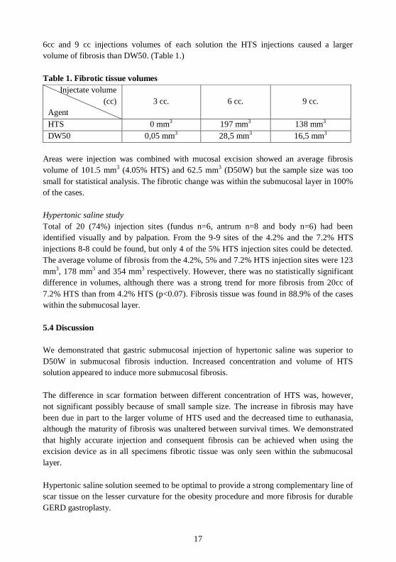
6cc and 9 cc injections volumes of each solution the HTS injections caused a larger
volume of fibrosis than DW50. (Table 1.)
Table 1. Fibrotic tissue volumes
Injectate volume
(cc)
Agent
3 cc. 6 cc. 9 cc.
HTS 0 mm3 197 mm3 138 mm3
DW50 0,05 mm3 28,5 mm3 16,5 mm3
Areas were injection was combined with mucosal excision showed an average fibrosis
volume of 101.5 mm3 (4.05% HTS) and 62.5 mm3 (D50W) but the sample size was too
small for statistical analysis. The fibrotic change was within the submucosal layer in 100%
of the cases.
Hypertonic saline study
Total of 20 (74%) injection sites (fundus n=6, antrum n=8 and body n=6) had been
identified visually and by palpation. From the 9-9 sites of the 4.2% and the 7.2% HTS
injections 8-8 could be found, but only 4 of the 5% HTS injection sites could be detected.
The average volume of fibrosis from the 4.2%, 5% and 7.2% HTS injection sites were 123
mm3, 178 mm3 and 354 mm3 respectively. However, there was no statistically significant
difference in volumes, although there was a strong trend for more fibrosis from 20cc of
7.2% HTS than from 4.2% HTS (p<0.07). Fibrosis tissue was found in 88.9% of the cases
within the submucosal layer.
5.4 Discussion
We demonstrated that gastric submucosal injection of hypertonic saline was superior to
D50W in submucosal fibrosis induction. Increased concentration and volume of HTS
solution appeared to induce more submucosal fibrosis.
The difference in scar formation between different concentration of HTS was, however,
not significant possibly because of small sample size. The increase in fibrosis may have
been due in part to the larger volume of HTS used and the decreased time to euthanasia,
although the maturity of fibrosis was unaltered between survival times. We demonstrated
that highly accurate injection and consequent fibrosis can be achieved when using the
excision device as in all specimens fibrotic tissue was only seen within the submucosal
layer.
Hypertonic saline solution seemed to be optimal to provide a strong complementary line of
scar tissue on the lesser curvature for the obesity procedure and more fibrosis for durable
GERD gastroplasty.
18
6. A HUMAN PILOT STUDY
6.1 Background
After successful animal safety and feasibility studies numerous acute animal operations
were carried out to refine technical and procedural details. Small device modifications
were completed as well. The principal aim of the first human trial was to demonstrate
safety and feasibility of the endoluminal gastroplication procedure.
6.2 Methods
The protocol was approved by the Institutional Ethics Committee (IKEB 7255/2013) and
the national Office of Health Authorization and Administrative Procedures
(35661/2011/OTIG).
Patients
Patients with symptomatic GERD and with obesity over BMI 35 kg/m2 were included in
the study. Data were collected prospectively and each subject served as their own control.
The inclusion and exclusion criteria were:
Inclusion criteria
For GERD patients: symptomatic reflux defined as a heartburn frequency score 2, when
off medication, with or without erosive esophagitis; patients who were dependent upon PPI
medication; documented acid reflux by pH monitoring (pH 4 for more than 4% for 24
hours following discontinuation of all GERD anti-secretory medications for 7 days), and
patients with an HRQL <10 on medication and HRQL >15 off medication.
For obesity: BMI > 35 kg/m2 or at least 45 kgs above ideal body weight and history of
obesity for at least 5 years.
Exclusion criteria
Pregnancy or intent to become pregnant during study participation; < 18 years of age;
patients who were not candidates for general anesthesia or whose medical history
classified them as level ASA 3 or higher; grade 2 or higher dysphagia (grade 2 - occasional
trouble swallowing - 1 or 2 times per week); grade C or D erosive esophagitis while on
medication; significant heart failure or a prosthetic heart valve; history of portal
hypertension; previous gastroesophageal surgical procedures; endoscopic GERD therapy
and/or thoracic surgical procedures; a disease state that is a general contraindication for an
endoscopic procedures; condition which general surgery is contraindicated.
Special exclusion criteria for GERD patients: less than 30mmHg of pressure at any
esophageal body level or more than 20% dropped or simultaneous waves; a hiatus hernia >

19
2 cm; if pH monitoring score is greater than 15% total time over pH of 4 or whose
DeMeester score is >50. Special exclusion criteria for obesity were: achalasia; systemic
lupus erythematosus or other autoimmune disorders and patients who are mentally
challenged or emotionally unfit as determined by standard psychological evaluation
Preoperative work up
All patients after the screening process underwent a medical history, physical examination
and upper endoscopy. Esophageal manometry and pH monitoring were also performed.
For patients with GERD, symptom scoring questionnaire and GERD-HRQL (health related
quality of life) evaluation were administered. Obese patients underwent upper endoscopy,
physical examination, laboratory testing, IWQOL (Impact of Weight on Quality of Life)
questionnaire and standard psychological evaluation.
Device
The excision device had been modified with the tip of the operating capsule flattened and
the through had a rhomboid shape for better tissue capture. The suturing device handle was
modified as well for easier manipulation.
Procedure
The procedure was carried out with the patient being in right lateral decubitus position
under general anesthesia. After upper endoscopy was performed the esophagus was dilated
to 60 Fr. We used a cautery snare to place marks along the lesser curvature for orientation.
GERD procedure
The excision device was inserted into the proximal stomach. Through the device dedicated
channel a 6 mm videoendoscope (Fujunon EG-470N5) was introduced for procedure
visualization and the stomach was insufflated. The device was positioned; the mucosa was
captured - by using -500 mmHg vacuum - and held in the trough. A 4.2% hypertonic saline
(HTS) solution with 1:100.000 adrenaline solution was injected. After desufflation of the
stomach and a short delay for appropriate vasoconstriction the excision blade was actuated.
The vacuum was discontinued and the stomach insufflated. The excised mucosal piece was
removed. The procedure was repeated two more times for a confluent 3 excision denuded
area at the GEJ.
The suturing device was than inserted and its trough positioned on the third excision bed.
The mucosa free gastric wall was captured by vacuum. The suture cycle was completed
and the device was rotated to the first excision site and the procedure was repeated and the
device was removed. The sutures were paired outside the patient's mouth. By endoscopic
visualization the sutures were tied using the knotter device. After hemostasis was checked
the outlet diameter was measured.
Obesity
In obesity patients the procedure was carried out with small modifications. The excisions
were positioned 2 cm distal to the GEJ and the sutures were more separated radially in the

20
excised area to create a tighter gastric outlet and a small proximal gastric pouch. Before
suturing the lesser curvature side was injected with the HTS solution to form a 3600
scarred submucosal ring after procedure completion for gastric pouch outlet restriction
Postoperative care
Patients were delivered to the Intensive Care Unit for a 12 hours long observation. A chest
x-ray was taken to check for stomach size and free air. Before extubating 8 mg intravenous
ondansetron was administered to reduce the possibility of retching and vomiting. On the
first postop day a gastrografin swallow study was performed to rule out esophageal or
gastric perforation.
Patients were kept on a Cleveland Clinic bariatric liquid diet and were given omeprazole
40mg two times a day for 1 month.
Follow up
Follow up data was recorded by a study nurse on postop day1, 14, and 1, 3, 6, 12, 18 and
24 months after the operation.
6.3 Results
Study population
Fourteen patients were screened and total of 8 were included in the study (5 obese and 3
with GERD). From the 8 patients originally included one obesity patient was excluded
after procedural attempt as her anatomy did not allow us to dilate her upper esophageal
sphincter safely. In two GERD patients the procedure was incomplete due to device and
technical difficulties. These patients were excluded from the study as well. Total of 5
patients underwent a complete procedure and remained for follow up.
Operative data and follow up
There was no intraoperative significant bleeding or perforation encountered. The total
procedural times were between 1h 30 minutes and 4h 45 minutes.
Patient #4 (obese): she had no intraoperative complication. Her initially 2 cm hiatal hernia
was reduced in size on 6, 12 and 18 month’s endoscopy. At 24 month follow-up 67 %
EWL plus normalization of her elevated baseline blood pressure were seen.
Patient #5 (obese): she had an uncomplicated procedure. However, on videotape review it
was evident that the gastric outlet was 8 mm in diameter after suture tying (ideal would be
6 mm). The patient did not lose weight or experience food restriction at 6 and 12 months,
but showed 12% EWL at 24 month follow-up.

21
Patient #6 (obese): she had a satisfactory intervention without any intraoperative
complication. She experienced intermittent dysphagia with decreased frequency up to the
18 month follow-up. Her blood pressure had normalized. She had no other comorbidities.
Her EWL was 34% at 24 months.
Patient #7 (obese): she had a complete procedure but vomited repeatedly in the Intensive
Care Unit before extubating her. Her initial chest X-ray was unremarkable but 12 hours
postoperatively she had free abdominal air under both hemi-diaphragms. Laparoscopy and
simultaneous upper endoscopy showed no perforation. A nasogastric tube was left in
place, and she recovered uneventfully. However, on day 9 she developed vertigo with
repeated vomiting and required re-hospitalization. At 6 month endoscopy her gastroplasty
was loose and she had no food restriction. The 18 month endoscopy showed no change and
this patient had no weight loss.
Patient #8 (GERD): she had an uneventful operation. Her pre-operative HRQOL was 19
and the DeMeester score was 44. At 6 months her DeMeester score was 16 and at 12
months the HRQOL was 10. pH monitoring was not performed at 12 months due to
equipment unavailability. No esophagitis was evident. At 18 month follow-up she
remained asymptomatic and was off all anti-secretory medications. At 2 years her
DeMeester score normalized and her HRQOL was the lowest (9) since the procedure.
6.4 Discussion
We demonstrated that endoluminal gastroplasty for GERD and obesity is feasible and safe
when using our new mucosal excision and suturing system. Initial patient outcomes
showed that the procedure has the potential to effectively treat both pathologies.
During the first human study we learned the proper way to insert the devices which was
simplified by the use of an adjustable mouth-opener and that the right lateral decubitus
position is best for the proximal greater curvature tissue manipulation. Special care to
avoid postoperative retching, gagging or vomiting is necessary to prevent tissue separation
in the operative area. The submucosal fibrosis, which was created by mucosal excision and
injection of hypertonic saline solution, served as a reinforcement to prevent expansion of
the gastroplasty.
Twenty four month patient follow-up showed promising results for both GERD and obese
patients. Longer patient follow-up and a larger study are necessary to standardize the
procedures and prove efficacy.

22
7. BARRETT’S STRIP ENDOSCOPIC MUCOSAL EXCISION
7.1 Background
Long lasting GERD may result in metaplastic changes of the esophageal mucosa and its
removal may be necessary to prevent development of malignancies or to treat in situ
superficial carcinomas. Excision of the metaplastic mucosa allows a definitive histologic
diagnosis while also potentially curative. The disadvantages of current excisional
techniques are that they are labor intensive and have a long learning curve. Based on our
experience from the gastroplasty device our intention was to create a flexible instrument to
excise and remove esophageal mucosa and muscularis mucosa safely, rapidly and with a
low complication rate.
7.2 Methods
The device
The instrument shares the common characteristics with the gastroplasty device as it
consisted of a handle and a flexible shaft with an integrated distal excision capsule. The
device has a dedicated channel for a standard 4.8 – 5.5 mm diameter flexible endoscope
with an opening on the handle and exiting on the proximal edge of the operating tip. The
device slides over the endoscope through the oropharynx and into the esophagus.
The procedure
The main steps of the procedures were the follows: first an esophageal cautery mark was
placed as a target site prior to excision device introduction. The trough was positioned on
the target point by direct endoscopic visualization. After device positioning the endoscope
was retracted into the device shaft. After suction was applied the multiple suction ports
pulled the mucosa into the capsule. The longitudinal injection needle was incrementally
forwarded above the bottom of the trough and a 1:100.000 Adrenaline solution was
injected to separate the muscularis mucosa from the muscularis propria thus increasing the
“target space” and to provoke hemostasis. After injection the horizontal guillotine blade
was advanced with a single motion and the mucosa was resected in a fixed plane. The
device was then withdrawn from the esophagus with the specimen within the capsule. This
allowed the specimens to be easily orientated for histological analysis.
Ex-vivo experiments
Preliminary ex-vivo studies were carried out with porcine, canine, baboon and human
esophagi. Human esophagi were harvested from tissue donor patients with family
informed consent under the auspices of the Nebraska Organ Recovery System. These
experiments allowed us to determine the correct device characteristics necessary for
consistent strip endoscopic esophageal mucosal resection (SEMR).

23
Additional animal tissues were used to assist in design modifications. Before in-vivo
experiments the device was studied using fresh ex-vivo non-fixed human esophagi for
completeness of mucosal resection and uniformity of excision depth. The excised human
mucosal strips were assessed histologically. The excision depth was microscopically
determined in 15 systematically separated locations within all tissue specimens.
In vivo experiments
In vivo experiments were conducted involving three animals (1 canine and 2 pigs). In
accordance with the ethical principle of “Reduction” in animal experimentation, animals
were included at the end of a different experimental protocol, which received full approval
by the Creighton University Institutional Animal Care and Use Committee under the
number (IACUC PN-0659 and PN-701). Total of 6 excisions were done to determine
device efficacy and safety. In the porcine model, esophagi were myotomized from the
gastroesophageal junction to the proximal 1/3 of the esophagus to provide a large enough
esophageal lumen for comfortable device manipulation.
7.3 Results
Ex vivo experiments
The device allowed precise localization and positioning with satisfactory excision size and
depth. A total of 10 excisions were performed on 5 ex-vivo cadaveric human esophagi.
The specimens ranged in size from 3 x 2.5 cm to 2.5 x 2.2 cm. The average thickness of
the excised specimens was 0.297 mm with the excision level within the superficial
submucosa (Sm1). One hundred and forty seven of 150 examined microscopic fields
included the muscularis mucosa.
In vivo experiments
The first non-survival canine and porcine experiments were promising in terms of safety.
The device could be introduced without trauma in both canine and porcine models and 6
mucosal excisions were performed without bleeding. The average specimen size was 2,8
cm x 2,3 cm. Target cautery mark localization and accurate capsule placement was proven.
No perforations occurred and none of the in vivo esophagi, after removal, showed
evidence of excision penetration to the muscularis propria level.
7.4 Discussion
This new flexible endoluminal mucosa excision device fulfilled requirements for a
successful endoscopic Barrett’s mucosa excision device. Large mucosal strip excisions,
using a fast and cold blade technique, without bleeding and esophageal wall perforation
were performed in acute animal experiments. Desired cutting depth and excision size was
demonstrated, in 98% of ex vivo cases. This is the first automated endoluminal mucosal
strip resection device that allows accurate deep and lateral margins and with relative ease

24
of use. Further survival experiments and clinical trials will define the role of this device for
endoscopic mucosal resection.

25
8. SUMMARY OF THE WORK
We were focused in our work on develop new techniques and devices for transoral
endoluminal treatment of gastroesophageal reflux disease, obesity and - with some
technical modifications- Barrett’s esophagus.
The treatment of GERD is individualized. The spectrum is wide from simple life style
changes to Roux-en-Y gastric bypass. Antisecretories often provide subjective and
objective resolution of GERD; however they have no effect on the underlying anatomical
defects or on the alkaline reflux. Moreover, 50% of patients continue to exhibit low intra-
gastric pH and objective evidence of acid regurgitation when reported complete
symptomatic control on PPI therapy. Despite the relative safety of these medications new
data has increased concern about the long-term effects and safety of anti-secretory drugs.
Thus many patients must commit to other therapy to provide lifelong solution for
gastroesophageal reflux. Anti-reflux surgery is recommended for patients with refractory,
medication resistant or complicated GERD and provides excellent symptom control in
85%-90% of cases. In the era of laparoscopy the number of antireflux procedures has
significantly increased. Notwithstanding of the minimally invasive nature of these
interventions they are not devoid from complications. Early or late postoperative
complications may prolong hospitalization, alter quality of life or require remedial surgical
interventions. Reoperative anti-reflux surgery is a feasible option for patients with
recurrent disease, although inferior results with a higher mortality and morbidity compared
with primary surgery are seen.
Present choices of weight reduction for the obese population are limited to life style
change, adjunct pharmaceutical therapy and bariatric surgery. The spectrum of bariatric
surgery is wide and the number of bariatric surgical procedures has significantly increased
in the recent past. Although majority of these procedures are performed laparoscopically
the complication rate is still not negligible. The cost of bariatric surgery is high and it is
available for a small part of the morbidly obese patients. Despite the improved results a
large proportion of patients still hesitate having operative intervention.
Advanced endoscopic therapy provides different treatment options for patients with
Barrett’s metaplasia. Thermal or photodynamic therapy and radiofrequency ablation
destroy the columnar epithelium allowing the regrowth of physiologic stratified squamous
epithelium. Endoscopic mucosal resection and endoscopic submucosal dissection are other
options. However, these techniques carry disadvantages; the formers do not provide tissue
for pathologic examination, their durability is questionable and the procedure related
complication rate is relatively high. The latters are time consuming and only endoscopists
with significant experience are able to perform.

26
The increasing need for effective, safe, durable and inexpensive procedures dedicated to
GERD and obesity resulted in new endoscopic treatment modalities. The numerous
different procedures published to date can be categorized as ablative,
injection/implantation, fixation, space occupying, transoral stapling, gastric sleeves and
others. The idea of endoluminal management of these conditions is relatively new and
devices providing long lasting effect have not been developed yet. Based on this new
methods and instruments novel treatment options for other pathologies of the upper
gastrointestinal tract – such as the Barrett’s esophagus – also can be developed.
We developed a transoral endoscopic flexible devices to excise gastric mucosa and to
place full thickness sutures in the excision beds to create an effective gastroplasty. With
some device modification we used this technique to excise mucosa from the esophagus for
Barrett’s disease.
Radiofrequency ablation and injection of different agents in the LES area has not fulfilled
the expectations regarding to long term results in case of GERD. Fixation methods both for
GERD and obesity are seem to be more durable but they are still along with high failure
rate. We believe that the reason for failure is the lack of strong tissue apposition at the GEJ
and the gastric fundus area where multidirectional and significant forces may arise.
In the first phase of our study we developed a multifunctional endoscopic device to excise
gastric mucosa and place full thickness sutures in the excision beds creating a gastroplasty.
We hypothesized that mucosal excision and apposition of the excision beds are necessary
to prevent tissue separation. We placed the gastroplasty at the level of GEJ for GERD and
first created a vertical gastroplasty line for obesity forming a neo-esophagus with pouch
and restrictive outlet along the lesser curvature.
The in vivo laboratory work with baboons showed gastric mucosal excision feasibility and
safety but durability of effect was lacking. From the first animal study we also learned that
for ease of device adjustment to gastric tissue as well as for ease of procedure performance
changes are required. We understood that a separate excision and suturing device would be
favorable to obtain optimal size mucosal excisions and consistent full thickness suture
penetration.
In the second phase of the study changes in both the design of the gastroplasty device and
in the procedure were done. A separate excision and suturing device with different trough
size were developed. These changes resulted in ease of use and accuracy in both excision
and suturing. Procedural changes for obesity resulted that the gastroplasty positioning was
similar to that of the external gastric band when utilizing the pars flaccida approach.
We performed endoluminal gastroplasty as an antireflux procedure and as a gastric outlet
restriction for obesity using the second generation of our gastroplasty system on 7 dogs.
This study demonstrated that endoscopic gastric mucosal excision and suture placement at
27
the GEJ is feasible, safe and easier with this devices. All animals survived without
complications. A significant decrease in GEJ compliance was seen in each animal after the
GERD procedure. Good gastric pouch outlet restriction was achieved after the obesity
procedure. Scar tissue formation after mucosal excision and full thickness suturing was
satisfactory, however we assumed that more amount and greater extension of fibrosis may
be needed. We believed that an injectable agent that generates more robust scarring would
contribute to durability. In the next step of our study we examined different solutions that
may fulfill this requirement.
Previous studies demonstrated that results of endoscopic GERD and obesity therapies
often fail even in the short term. In many of these therapeutic options the main target site is
the GEJ and the subcardial area. This is formed by a complex net of smooth muscle fibers
resulting in a highly elastic and stretchable stomach wall where significant forces arise.
This anatomy may be responsible for the high recurrence rate of GERD after endoluminal
fixation methods. We hypothesized that generation of scar tissue in this area can prevent
tissue disintegration to achieve more durable results.
We hypothesized that the use of different hypertonic solutions is suitable for robust scar
tissue generation in the submucosal layer. The level of scar tissue generation is critical as
panmural fibrosis can be resulted in excessive luminal compromise that may be resistant to
dilation of any kind. First we compared 4.2% hypertonic saline and 50% dextrose solutions
to find which is more effective in terms of scar formation. Results demonstrated that with
submucosal injection of hypertonic saline stronger fibrosis was generated than with
hypertonic dextrose. Based on these results we used more concentrated saline solutions in
larger volumes. It appeared that more intensive fibrosis after injection of more
concentrated saline solution can be achieved.
In the first human mucosal excision and suturing gastroplasty pilot study we performed
procedures to treat GERD and to reduce excess weight in obese patients. Total of 8
patients were included. Three with GERD having elevated DeMeester score without hiatal
hernia and 4 patients over BMI 35 were included. Endoluminal gastroplasty at the level of
LES and in the proximal stomach were created. The system and procedures were proven to
be feasible and safe. Patients were followed up for two years by endoscopy and functional
testing.
The mid-term results demonstrated that the procedure holds the potential either to rebuild
the barrier function of the gastric cardia or to be a restrictive obesity procedure. The key
for long term success is the scar tissue generated by mucosal excision and injection of
hypertonic saline solution. Technical and procedural refinements are necessary to improve
the results and reduce operating time.
We are planning to follow the patients and conduct a larger study to standardize the
procedures.
28
Excision of pathologic mucosa from the esophagus in case of premalignant mucosal
changes or in presence of in situ mucosal carcinomas is a treatment option. Based on our
experience from the gastroplasty device our intention was to create a flexible instrument to
excise and remove esophageal mucosa and muscularis mucosa safely, fast and with low
complication rate. Existing other techniques are along with high perforation and stricture
rate or require a significantly higher level of endoscopic skill making the procedure
operator dependent and time consuming.
We performed mucosal excisions from ex vivo human and in vivo dog and swine
esophagi. Desired cutting depth and excision size was demonstrated without perforation.
Accurate device positioning was demonstrated with relative ease of use. Further survival
experiments and clinical trials will define the role of this device for endoscopic mucosal
resection.
New statements from the study
We found that a safe gastric mucosal removal and suture placement for an endoluminal
proximal gastroplasty is feasible by using a single transoral device; however in vivo acute
and survival animal studies revealed insufficient excision size and poor full-thickness
suturing accuracy.
We demonstrated safety and efficacy of the gastroplasty technique for both GERD and
obesity by using two separate devices for tissue excision and suturing.
We found that hypertonic saline solution is an effective and safe scar tissue generator when
injecting into the gastric submucosa.
We showed safety and feasibility of the sutured gastroplasty after mucosal excisions both for GERD and obesity in humans.
We demonstrated that large esophageal mucosal pieces can be excised safe and with
relative ease in a targeted fashion by using a cold blade technique.
This work is the first to demonstrate safety and feasibility of sutured gastroplasty after
mucosal excision, suturing and submucosal hypertonic saline injection for GERD and
obesity in humans. These findings support to conduct a larger human study to evaluate procedureal efficacy and may serve to develop other effective treatment modalities for the
endoluminal management of GERD and obesity.

29
9. ACKNOWLEDGEMENTS
First and foremost I would like to express my endless appreciation to Professor Charles
Filipi for his confidence that made me possible to join to his team at the Creighton
University in Omaha, Nebraska, and to work with him on his device for many years.
Without him this work would have never been happened.
I would like to express my thanks to my supervisor Professor Péter Örs Horváth for all
the support I was given to realize this work.
Particular thanks must also be recorded to Professor Áron Altorjay – my boss in our
Department of surgery - for the opportunity I was given and also for his sustained
encouragement; these were essential.
I would also like to acknowledge the work of Szilvia Devecseri who did an outstanding
job as a study nurse in coordination and follow up of our patients.
Last but not least I would like to express my endless gratitude to my wife Krisztina Rácz
and to my family for all the renunciation, tolerance and strong support, without them this
work would have never been realized.
30
10. PUBLICATIONS AND PRESENTATIONS
Publications
1. Tsuboi K, Mittal SK, Légner A, Yano F, Filipi CJ. Relationship between
manometric findings and reported symptoms in nutcracker esophagus: insights gained from a review of 313 patients. J Gastroenterol. 2010 Oct;45(10):1033-8.
Impact factor:3.61
2. Légner A, Stadlhuber RJ, Yano F, Tsuboi K, Mittal SK, Rothstein RI, Filipi CJ. Initial experience with Barrett's strip endoscopic mucosal excision: a new
Barrett's excision device. Surg Endosc. 2011 Feb;25(2):651-4.
Impact factor:4.013
3. Légner A, Tsuboi K, Bathla L, Lee T, Morrow LE, Mittal SK. Reoperative
antireflux surgery for dysphagia. Surg Endosc. 2011 Apr;25(4):1160-7.
Impact factor:4.013
4. Bathla L, Légner A, Tsuboi K, Mittal S.Efficacy and feasibility of laparoscopic
redo fundoplication. World J Surg. 2011 Nov;35(11):2445-53.
Impact factor:2.362
5. Tsuboi K, Lee TH, Légner A, Yano F, Dworak T, Mittal SK.Identification of
risk factors for postoperative dysphagia after primary anti-reflux surgery.Surg
Endosc. 2011 Mar;25(3):923-9.
Impact factor:4.013
6. Castillo C, Légner A.The "AMID" hernia and skin stapler for Lichtenstein
hernia repair: a pilot study. Surg Innov. 2012 Sep;19(3):258-62.
Impact factor: 2.126
7. Cruz M, Filipi L, Estevez J, Marte E, Dethlefs H, Dowd R, Légner A, Kottoor
V, Filipi CJ. A diabetic hypertension treatment program for the underserved in
rural Dominican Republic. Open Journal of Internal Medicine. 2012 2, 72-79
8. Légner A., Kazuto T., Stadlhuber R., Fumiaki Y., Halvax P., Brandon H.,
Wayne P., Filipi CJ. Mucosal Excision and Suturing for Obesity and GERD.
Surg Innov 2013Dec; 20(6):586-93. Impact factor:1.338
9. Mittal SK, Légner A, Tsuboi K, Juhasz A, Bathla L, Lee TH. Roux-en-Y
reconstruction is superior to redo fundoplication in a subset of patients with failed antireflux surgery. Surg Endosc. 2013 Mar;27(3):927-35.
Impact factor:3.313
31
10. Légner A, Altorjay A, Juhasz A, Stadlhuber R, Reich V, Hunt B, Rothstein R, Filipi C. Transoral mucosal excision sutured gastroplasty: a pilot study for
GERD and obesity with two-year follow-up. Surg Innov 2014 Oct;21(5):456-63.
Impact factor:1,338
11. Liu YY, Diana M, Halvax P, Cho S, Légner A, Alzaga A, Swanström
L, Dallemagne B, Marescaux J. Flexible endoscopic single-incision
extraperitoneal implant and fixation of peritoneal dialysis catheter: proof of
concept in the porcine model. Surg Endosc. 2015 Aug;29(8):2402-6. Impact factor:3.313
12. Halvax P, Légner A, Paál B, Somogyi R, Ukös M, Altorjay A. [Laparoscopic
reconstruction in traumatic rupture of the diaphragm]. Magy Seb. 2014 Oct;67(5):304-7.
13. Diana M, Swanström LL, Halvax P, Légner A, Liu YY, Alzaga A, D'Urso
A, Marescaux J. Esophageal covered stent fixation using an endoscopic over-the-scope clip. Mechanical proof of the concept and first clinical experience.
Surg Endosc. 2015 Jan 29. [Epub ahead of print]
Impact factor:3.313
14. Halvax P, Diana M, Légner A, Lindner V, Liu YY, Nagao Y, Cho S, Marescaux
J, Swanström LL. Endoluminal full-thickness suture repair of gastrotomy: a
survival study. Surg Endosc. 2015 Jan 29. [Epub ahead of print]
Impact factor:3.313
15. Diana M, Halvax P, Mertz D, Légner A, Brulé JM, Robinet E, Mutter
D, Pessaux P, Marescaux J. Improving Echo-Guided Procedures Using an
Ultrasound-CT Image Fusion System. Surg Innov. 2015 Jun;22(3):217-22. Impact factor:1.338
16. Diana M, Halvax P, Pop R, Schlagowski I, Bour G, Liu YY, Légner
A, Diemunsch P, Geny B, Dallemagne B, Beaujeux R, Demartines N, Marescaux J. Gastric supply manipulation to modulate ghrelin production and
enhance vascularization to the cardia: proof of the concept in a porcine model.
Surg Innov. 2015 Feb;22(1):5-14
Impact factor:1,338
17. Kong SH, Diana M, Liu YY, Lee HJ, Légner A, Soares R, Swanström L,
Dallemagne B, Yang HK, Marescaux J. Novel method for hybrid endo-
laparoscopic full-thickness gastric resection using laparoscopic transgastric suture passer device. Surg Endosc. 2015 Jul 7. [Epub ahead of print]
Impact factor:3.313
18. Diana M, Noll E, Agnus V, Liu YY, Kong SH, Légner A, Diemunsch P, Marescaux J. Reply to Letter: "Enhanced Reality Fluorescence Videography to
Assess Bowel Perfusion: The Cybernetic Eye". Ann Surg 2015 May 27. [Epub
ahead of print]
Impact factor:7.188
32
19. Légner A; Diana M; Halvax P;Liu YY; Zorn L; Zanne P; Nageotte F; De Mathelin M; Dallemagne B; Marescaux J. Endoluminal surgical triangulation
2.0: a new flexible surgical robot. Preliminary pre-clinical results with colonic
submucosal dissection. Surg Endosc. [Accepted for publication]
Impact factor:3.313
Cumulative Impact factor: 52.562
Abstracts
20. Légner A., Diana M., Halvax P., Liu YY., Lindner V., Swanstom L., Marescaux
J.Creation of a realistic biocompatible phantom tumor for advanced minimally invasive procedures on a gastrointestinal tract in animal model. Gastrointest
Endosc 05/2015; 81(5):AB334
Impact factor:4.90
21. András Légner, Kazuto Tsuboi, Tommy H. Lee, Lokesh Bathla, Sumeet K.
Mittal. Evaluation of preoperative risk factors for postoperative heartburn after
reoperative anti-reflux surgery. Gastroenterology 2010, 138(5):S-871
Impact factor:12.032
Cumulative Impact factor: 16.932
Book chapter
1. András Légner, Charles J. Filipi. (2010) Endoluminal therapies for
gastroesophageal reflux disease and obesity. In Paul Alan Wetter (Ed)
Prevention and Management of Laparoscopic Surgical Complications 3rd
edition. (Chapt 22.)

33
Presentations
1. Ferencz A, Légner A, Gasz B, Rőth E
Studying NF-kB activation on intestinal ischemic preconditioning produced my
microsurgical technique 7th Congress of the International Society for Experimental Microsurgery,
Debrecen (2004)
2. Ferencz A, Légner A, Rőth E The role of NF-kB to modify the oxidative stress in small bowel
autotransplantation model
XX International Congress of the Transplantation Society, Vienna, Austria
(2004)
3. Ferencz A, Légner A, Rőth E
A citoplazmális és nukleáris NF-kB aktiváció ischémiás prekondícionálást
követő változása patkány bélszövetben Magyar Szabadgyök Kutató Társaság Kongresszusa, Budapest (2004)
4. Ferencz A, Légner A, Gasz B, Szántó Z, Rőth E
Changes of cytoplasmic and nuclear NF-kB activation in intestinal tissue following ischemic preconditioning in rats
XXXIXth Congress of the European Society for Surgical Research, Athen,
Greece (2004)
5. Légner A., Jaskó R., Szilágyi A., Madácsy L., Altorjay Á.
Szövődményes gyomordiverticulum esete
Magyar Sebész Társaság 59. kongresszusa. Debrecen (2008)
6. Légner A, Tsuboi K, Bathla L, Morrow LE, Lee T, Mittal SK.
Reoperative anti-reflux surgery for dysphagia.
SAGES 2010 Annual Meeting, 14-17th April, National Harbor, MD
7. Légner A, Mittal SK, Tsuboi K, Lee T, Morrow LE.
Can we predict the outcome of redo-fundoplication for patients with failed anti-
reflux surgery?
12th World Congress of the ISDE, Kagoshima, Japan, 2010 September 2-5
8. Légner A, Tsuboi K, Lee T, Morrow LE, Mittal SK.
Effect of esophageal dysmotility on outcomes after re-operative anti-reflux
surgery 12th World Congress of the ISDE, 2010 September 2-5 Kagoshima, Japan
9. Légner A, Tsuboi K, Makris KI, Morrow LE, Lee T, Mittal SK.
Outcomes beyond one year following re-operative intervention for failed fundoplication. American College of Surgeons 96th Annual Clinical Congress,
2010 October 3-7, Washington DC

34
10. Légner A. Altorjay Á Új utak az antireflux sebészetben
Dunántúli Sebész Társaság Kongresszusa, Sopron, 2012.03.02-03
11. Légner A, Reich V, Székely A, Altorjay Á. A gastroesophagealis reflux betegség és a kóros kövérség endoluminális
terápiájának új eszköze
Magyar Sebész Társaság Kísérletes Sebészeti Szekció XXIV. Kongresszusa,
Debrecen, 2013.06.13-15
12. Légner A.,Swanström L.,Altorjay Á.,Putz G.,Halvax P.,Devecseri Sz.,Filipi CJ.
Endoscopic proximal gastroplasty for obesity using a novel full-thickness
suturing device. SAGES 2014 Annual Meeting, 2-5 April, 2014 Salt Lake City, UT, USA
13. Légner A.
A tápcsatorna endoszkópos sebészeti lehetőségei. Balassa János nap Szekszárd, 2014 október 11
14. Légner A, Diana M,; Halvax P, Liu YY, Zorn L, Zanne P, Nageotte F, De
Mathelin M, Dallemagne B, Marescaux J. A robotized flexible endoscopic platform for colorectal interventions. EAES 23rd International Congress, 3-6
June 2015, Bucharest, Romania
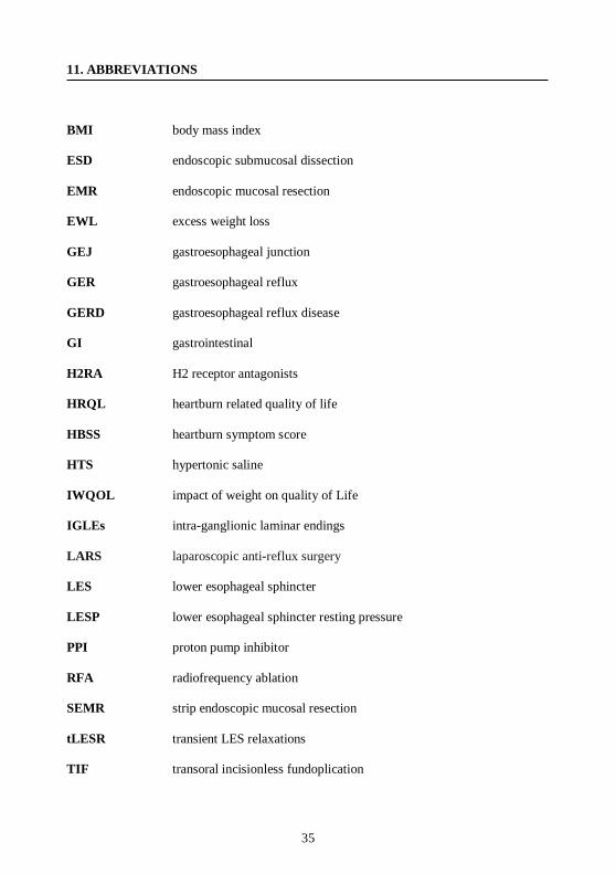
35
11. ABBREVIATIONS
BMI body mass index
ESD endoscopic submucosal dissection
EMR endoscopic mucosal resection
EWL excess weight loss
GEJ gastroesophageal junction
GER gastroesophageal reflux
GERD gastroesophageal reflux disease
GI gastrointestinal
H2RA H2 receptor antagonists
HRQL heartburn related quality of life
HBSS heartburn symptom score
HTS hypertonic saline
IWQOL impact of weight on quality of Life
IGLEs intra-ganglionic laminar endings
LARS laparoscopic anti-reflux surgery
LES lower esophageal sphincter
LESP lower esophageal sphincter resting pressure
PPI proton pump inhibitor
RFA radiofrequency ablation
SEMR strip endoscopic mucosal resection
tLESR transient LES relaxations
TIF transoral incisionless fundoplication