epithelial cadherin re-expression in metastatic...
TRANSCRIPT

EPITHELIAL CADHERIN RE-EXPRESSION IN METASTATIC BREAST CANCER AS A STRATEGY FOR METASTATIC COLONIZATION OF THE LIVER
by
Christopher Reed Shepard
Bachelor of Sciences, Wake Forest University, 2003
Submitted to the Graduate Faculty of
Cellular and Molecular Pathology in partial fulfillment
of the requirements for the degree of
Doctor of Philosophy
University of Pittsburgh
2007

ABSTRACT
Epithelial-cadherin downregulation enables cancer cells to escape from the primary mass;
however, E-cadherin has been found to be expressed on metastatic foci, bringing into question
the role of this molecule in tumor progression. We define a novel role for the cellular adhesion
molecule E-cadherin, in which the protein’s re-emergence promotes carcinoma-parenchymal
interactions in ectopic sites. Non-metastatic E-cadherin positive MCF7 breast cancer cells form
heterotypic cohesions mediated by E-cadherin, and in invasive and metastatic MDA-MB-231
cells, the E-cadherin promoter hypermethylation that prevents endogenous E-cadherin expression
is reversed when these cells are cultured with hepatocytes. The function of this re-expression is
suggested by the E-cadherin-dependent sustained activation of Erk-MAP kinase and Akt in these
breast carcinoma cells. Thus, we propose that E-cadherin expression and subsequent
heterocellular interactions direct cell fate decisions that may ultimately enable colonization of a
secondary site by an invasive cancer cell.

UNIVERSITY OF PITTSBURGH
SCHOOL OF MEDICINE
This dissertation was presented
by
Christopher Reed Shepard
It was defended on
October 25, 2007
and approved by
Adam Brufsky, MD PhD, Department of Medicine
Linda Griffith, PhD, Department of Biological Engineering, MIT
Paul Monga, MD, Department of Pathology
Committee Chair: Donna Beer-Stolz, PhD, Department of Cellular Physiology
Thesis Advisor: Alan Wells, MD DMSc, Department of Pathology
ii

Copyright © by Christopher Reed Shepard
2007
iii

EPITHELIAL CADHERIN RE-EXPRESSION IN METASTATIC BREAST CANCER AS A STRATEGY FOR METASTATIC COLONIZATION OF THE LIVER
Christopher Reed Shepard, B.Sc.
University of Pittsburgh, 2007
iv

TABLE OF CONTENTS
TABLE OF CONTENTS ............................................................................................................ V
LIST OF FIGURES ..................................................................................................................... X
PREFACE...................................................................................................................................XX
1.0 INTRODUCTION........................................................................................................ 1
1.1 BREAST CANCER ............................................................................................. 1
1.2 EPITHELIAL TO MESENCHYMAL TRANSITION IN CANCER ............ 3
1.3 CANCER CELL DISSEMINATION ................................................................ 5
1.4 E-CADHERIN IN NORMAL AND TRANSFORMED CELLS..................... 8
1.4.1 E-cadherin structure and function.............................................................. 9
1.4.2 E-cadherin in cancer progression.............................................................. 17
1.5 BREAST CANCER METASTATIC TO THE LIVER ................................. 21
2.0 PLCγ CONTRIBUTES TO METASTASIS OF IN SITU-OCCURING
MAMMARY AND PROSTATE TUMORS............................................................................. 23
2.1 ABSTRACT........................................................................................................ 24
2.2 INTRODUCTION ............................................................................................. 25
2.3 RESULTS ........................................................................................................... 27
2.3.1 Expression of the DOX-inducible PLCz transgene is restricted to
mammary and prostate epithelia.............................................................................. 27
v

2.3.2 Induction of PLCz inhibits invasiveness of the primary tumor and
metastatic tumor formation ...................................................................................... 29
2.4 DISCUSSION..................................................................................................... 35
2.5 MATERIALS AND METHODS...................................................................... 38
2.5.1 Generation of double transgenic TRAMP and MMTV-PyVmT with a
DOX-inducible PLCz transgene ............................................................................... 38
2.5.2 PLCz expression.......................................................................................... 40
2.5.3 Histologic and immunohistochemical assessments of mammary and
prostate glands............................................................................................................ 40
2.5.4 Statistical analyses ...................................................................................... 41
2.6 ACKNOWLEDGEMENTS .............................................................................. 41
3.0 EXPRESSION OF THE CYTOSOLIC TAIL OF E-CADHERIN PARTIALLY
REVERTS THE EPITHELIAL TO MESENCHYMAL TRANSITION IN AGGRESSIVE
BREAST CARCINOMA CELLS.............................................................................................. 43
3.1 ABSTRACT........................................................................................................ 44
3.2 INTRODUCTION ............................................................................................. 45
3.3 RESULTS ........................................................................................................... 46
3.3.1 Breast cancer cell lines retain epithelial elements.................................... 46
3.3.2 Epithelial markers vary quantitatively between breast cancer lines ..... 49
3.3.3 Intermediate mesenchymal-epithelial phenotypes can be engineered ... 53
3.4 DISCUSSION..................................................................................................... 55
3.5 MATERIALS AND METHODS...................................................................... 59
3.5.1 Cell lines and reagents ................................................................................ 59
vi

3.5.2 Immunofluorescence staining and analysis .............................................. 59
3.5.3 Immunoprecipitations and western blots ................................................. 60
3.5.4 Invasion assay.............................................................................................. 60
3.5.5 Statistical Analysis ...................................................................................... 61
3.6 ACKNOWLEDGEMENTS .............................................................................. 61
4.0 NORMAL EPITHELIAL CELLS OF THE METASTATIC NICHE DRIVE
FUNCTIONAL RE-EXPRESSION OF E-CADHERIN BREAST CANCER CELLS........ 62
4.1 ABSTRACT........................................................................................................ 63
4.2 INTRODUCTION ............................................................................................. 63
4.3 RESULTS ........................................................................................................... 65
4.3.1 Distant soft tissue micrometastases that originate from E-cadherin-
negative MDA-MB-231 primary tumor xenografts re-expression E-cadherin.... 65
4.3.2 E-cadherin protein and message expression in MDA-MB-231 cells is
driven by hepatocyte co-culture................................................................................ 68
4.3.3 Demethylation of the E-cadherin promoter region without global
demethylation allows for protein re-expression ...................................................... 71
4.3.4 Cancer cells undergo a proliferation-dependent demethylation of the E-
cadherin promoter ..................................................................................................... 75
4.3.5 E-cadherin on breast cancer cells can mediate functional heterotypic
adhesion to hepatocytes ............................................................................................. 76
4.3.6 E-cadherin trafficking to the surface requires EGFR attenuation but
EGFR functionality is necessary for preserved E-cadherin-dependent MAPK
activation in MDA-MB-231 cells............................................................................... 83
vii

4.3.7 Hepatocytes drive the upregulation of E-cadherin in a subset of cells
from primary breast cancer explants....................................................................... 87
4.4 DISCUSSION..................................................................................................... 90
4.5 MATERIALS AND METHODS...................................................................... 95
4.5.1 Xenografts.................................................................................................... 95
4.5.2 Cell culture and co-culture......................................................................... 96
4.5.3 Immunohistochemistry............................................................................... 96
4.5.4 SiRNA........................................................................................................... 97
4.5.5 Real-time quantitative PCR....................................................................... 97
4.5.6 Methylation specific PCR........................................................................... 97
4.5.7 Westernblots, flow cytometry, and immunofluorescence ....................... 98
4.5.8 Centrifugal assay for fluorescent cell adhesion (CAFCA) ...................... 99
4.5.9 Hepatocyte membrane assays .................................................................... 99
4.5.10 Primary explants....................................................................................... 100
4.6 ACKNOWLEDGEMENTS ............................................................................ 100
DISCUSSION ............................................................................................................................ 101
5.0 CARCINOMA DISSEMINATION FOLLOWS FROM AN EPITHELIAL-TO-
MESENCHYMAL-LIKE TRANSITION (EMT).................................................................. 102
6.0 METASTATIC SEEDING IS RATE-LIMITING FOR METASTATIC
COLONIZATION OF DISTANT ORGANS ......................................................................... 106
7.0 E-CADHERIN IN THE MESENCHYMAL-TO-EPITHELIAL REVERTING
TRANSITION ........................................................................................................................... 109
viii

8.0 A MODEL FOR THE MESENCHYMAL-EPITHELIAL REVERTING
TRANSITION ........................................................................................................................... 113
BIBLIOGRAPHY..................................................................................................................... 117
ix

LIST OF FIGURES
Figure 1. Motility, autocrine signaling, and loss of epithelial architecture are all key to
transformative and metastatic progression. Epithelial tissues consist of sheets of normal cells
(green) linked together by E-cadherin (thick black bars). This establishes a polarity that
segregates apically secreted factors, such as EGF, from their basolaterally-presented receptors
that normally are utilized by stromally derived factors, such as TGFβ. Due to genetic and
epigenetic events, E-cadherin is lost during neoplastic transition (red/orange cells), allowing for
autocrine signaling. This ‘dedifferentiation’ is the carcinoma-associated EMT. However, we
propose that during metastatic seeding to other epithelial organs E-cadherin is re-expressed
enabling linkages to normal parenchymal cells. This characterizes the mesenchymal to-epithelial
reverting transition (MErT)............................................................................................................. 7
Figure 2. E-cadherin cis- and trans- ligation is calcium dependent. Calcium coordination
rigidifies the 5 extracellular domains of the E-cadherin molecule (B). The unorganized
molecular lattice of (A) proceeds to the ordered cis-interaction in (C), and then to trans-ligation
of adjacent molecules in (D). Figure used with Open Access permission: Pertz et al. (1999). “A
new crystal structure, Ca2+ dependence and mutational analysis reveal molecular details of E-
cadherin homoassociation.” EMBO 18(7):1738-1747. ................................................................ 10
Figure 3. E-cadherin sequesters catenins and controls their signaling in addition to forming cell-
cell adhesions. A) E-cadherin sequesters β- and p120- catenins on its intracellular catenin
x

binding domains. In an untransformed cell, p120 is thought to stabilize E-cadherin at the surface,
β-catenin is sequestered from forming a complex with axin and, in this location also functions as
an adaptor protein for α-catenin, which in turn anchors E-cadherin to the cytoskeleton. B) In
many carcinomas, E-cadherin is silenced by promoter methylation allowing β-catenin to
translocate to the nucleus and p120 to promote an epithelial phenotype. However the
mechanisms of how these catenins act and whether they act individually or in concert are not
settled. C) E-cadherin dependent adhesion in itself is not a dominant stop mechanism to inhibit
invasion. In studies where the β-catenin binding domain was deleted from the E-cadherin
intracellular domain, but E-cadherin was still able to mediate adhesion through direct
crosslinking with α-catenin and therefore interaction with the actin cytoskeleton, β-catenin was
free to signal in the cell cytoplasm and led to an invasive phenotype (though this phenotype was
independent of its TCF-mediated transcriptional activity). Therefore, cytoplasmic localization of
β-catenin is thought to contribute to the mesenchymal nature of cells. D) In studies of cell that
had low levels of E-cadherin and cytoplasmic localization of p120, tyrosine phosphorylation on
p120’s amino-terminal by the pro-oncogene Src was thought to contribute to modulate its
contribution to cell migration. When p120 is knocked down, the equilibrium shift to Rho-GDP
promotes actin polymerization, stress fiber formation, a flattened morphology and less invasive
phenotype. Therefore, cytoplasmic localization of p120 is thought to contribute to the
mesenchymal nature of cells. These four scenarios provide data that show the critical role of E-
cadherin as a signal modulation molecule by sequestering catenins, primarily p120 and α-
catenin. In the absence of E-cadherin homotypic binding, this plaque is unstable and the catenins
are now free to relocalize. ............................................................................................................. 12
xi

Figure 4. E-cadherin is regulated on multiple levels. E-cadherin is regulated on the epigenetic
level by methylation of the promoter region, on the transcriptional level by the Snail/Slug
transcription factors, and on the effector level by the receptor tyrosine kinase EGFR.
Overcoming all of these repression mechanisms can result in a reversion of the EMT, or the so-
called mesenchymal-epithelial reverting transition (MErT)......................................................... 20
Figure 5. Generation of PLCz-expressing mice. The pC3(1)tPLCz bicistronic construct (a)
contains the reverse tet repressor driven by the breast and prostate specific promoter, C3(1), that
binds to the TRE (tetracycline response element) in the presence of doxycycline (DOX). VP-16
transcriptional activation domain of herpes simplex virus enhances expression of the downstream
PLCz. Thus, PLCz expression is expressed only on DOX induction in a tissue-specific manner.
To detect transmission of the pC3(1)tPLCz construct, 1 mg of genomic DNA from a tail of each
transgenic or wild-type mouse was used to perform the PCR analysis. 10 pg of transgene
fragment from pTGC3(1) was used as template for the positive control and there is no template
with the negative control. (b) The arrow head indicates the 0.8 kb PCR products from the
transgene. ...................................................................................................................................... 28
Figure 6. Inducible expression of PLCz in tissues from male and female mice. Proteins were
analysed on 10% SDS–PAGE and immunoblotted with a rabbit polyclonal antibody that
recognizes the Z region of PLCg. DOX was added to the drinking water to induce PLCz
expression. One week after DOX addition, mice were killing and tissues were processed to get
protein lysates. A selection of tissues and mice are shown to present evidence that PLCz
expression is dependent on (a) transgene presence, (a) DOX induction (b) steroid-responsive
tissues and (c) continued presence of DOX. (a) PLCz is induced by DOX in the prostate (PR)
and mammary glands (MG) of transgenic (T) mice and is not expressed in PLCz- (W) mice. (b)
xii

PLCz is expressed in the PG of male T mouse in a tissue-specific fashion and is not expressed in
the seminal vesicles (SV), liver (Liv), lungs (lun) or spleen (Spl). Nontransgenic mice were
negative in all tissues. (c) Three different PLCz-transgenic female mice were treated with DOX
for 1 week and then switched to normal drinking water and sacrificed 2 weeks later. The positive
control was killed after the DOX treatment. The inguinal MG was dissected and protein lysates
analysed. (c) is overexposed compared to (a) and (b) to detect lowlevel persistence of PLCz.... 30
Figure 7 Fisher’s exact test was used to generate P-values to determine whether induction of
PLCz had a significant influence on the extent of lung metastases. A, TRAMP and
TRAMP/PLCz mice were scored for invasiveness of the primary prostate tumors. The TRAMP
mice expressing the PLCz trangene had a significant reduction in the invasiveness of the primary
tumor (P<0.02). B, TRAMP and TRAMP/PLCz mice were scored for the extent of the lung
metastases. The TRAMP mice expressing the PLCz transgene had a significant decrease in the
extent of lung metastasis (P<0.06). C, PyVmT and PyVmT/PLCz mice were scored as in (B).
The PyVmT mice expressing the PLCz transgene had a significant decrease in the extent of lung
metastasis (P<0.03). ...................................................................................................................... 30
Figure 8. Metastatic tumors in the lungs of MMTV-PyV mice expressing PLCz. H&E sections
of dissected lung tissue from representative mice. Normal mouse lung tissue (top left). Lung
sections of transgenic PyVmT without the PLCz transgene show large metastatic tumors
throughout the lung parenchyma that crowd alveolar septa (top right). PyVmT-PLCz mice whose
PLCz expression is induced with DOX treatment markedly diminishes the number of tumors in
the lung parenchyma (bottom left). DOXtreatment has no effect on metastatic lung deposits in
PyVmT mice (bottom right).......................................................................................................... 33
xiii
Figure 9. The number of metastatic lung tumors is drastically reduced in PLCz+ mice, while the
tumor size remains unchanged. The number of tumors were counted and sized according to the
Materials and methods section. (a) The number of metastatic lung tumors in PyVmT/PLCz+
mice is fewer compared to the number of metastatic tumors in PyVmT not expressing the PLCz
transgene. (b) The PLCz transgene does not affect the size of metastatic lung tumors in
PyVmT/PLCz mice when compared to the PyVmT not expressing the transgene. ..................... 34
Figure 10. Invasive cancer cells retain epithelial characteristics. A) a, MCF7 cells grow in
clustered epithelial sheets. b, when high-E-cadherin expressing MCF7 are plated, they grow in a
very tightly adherent domes. c, four weeks after sorting out the high E-cadherin MCF7 cells,
they have returned to a flattened morphology and a more similar morphology as seen in panel a.
B) MDA-231 cells (a) and MCF7 cells (b) were kept in culture with and without varying
concentrations of TGF-β. Both cells lines, which were isolated from exudative pleural effusions
indicating that they had already metastasized, were responsive to the growth suppressive effects
of TGF-β. ...................................................................................................................................... 48
Figure 11. Invasive cancer cells exhibit a very skewed protein profile when compared with
untransformed HMECs. A) Cells were examined by immunofluorescence and the histograms of
the acquired pictures were analyzed for pixel intensity. B) 231/H2kd cells exhibit remarkably
upregulated levels of cytokeratin-18 compared to their untransformed MDA-231 counterparts. 51
Figure 12. Cancer cells can be engineered to be more epithelial by introducing a dominant
negative E-cadherin which sequesters catenins and mitigates the invasive phenotype. A) In
whole cells lysates, the H2kd fragment is detected in MDA-231 cells transfected with a dominant
negative E-cadherin (H-2kd-Ecad). β-catenin and p120 can be co-immunoprecipitated with
H2kd in 231/H2kd cells. 231/H2kd cells have a flattened morphology with punctuated
xiv

lamellipodia rather than the apparent leading edge seen in the untransformed MDA-231
counterparts (insets). B) Invasion assay comparing wild-type MDA-231 cells, epithelial MCF7
cells, and 231/H2kd cells. Asterisks denotes statistical significance by Student t-test (*p<0.08).
....................................................................................................................................................... 54
Figure 13. E-cadherin positive metastatic foci originate from E-cadherin negative primary
tumors. A) Left, human MDA-MB-231 breast cancer cell xenograft in a mouse inguinal fatpad
(H&E); middle, immunoperoxidase labeling with a human-specific E-cadherin antibody
indicates the absence of E-cadherin expression in the center of the primary tumor; right,
immunoperoxidase labeling of a field at the periphery of the tumor indicates the homogeneity of
the absence of E-cadherin in all fields of the primary tumor. B) Left, islands of micrometastases
in the lung originating from the primary xenograft in A (H&E); top adjacent, immunoperoxidase
staining using a human-specific antibody of diseased portions of the mouse lung indicate the
presence of E-cadherin positive human MDA-MB-231 cancer cells (arrows); bottom adjacent,
mouse epithelial alveolar cells in a portion of the unaffected lung do not exhibit labeling with the
human-specific E-cadherin antibody. ........................................................................................... 67
Figure 14. Hepatocytes drive the re-expression of E-cadherin in MDA-MB-231 breast cancer
cells. A) Immunoblot of proteins lysates from MDA-MB-231/hepatocyte co-cultures using a
human-specific antibody indicates that within 6 days of co-culture with hepatocytes, the cancer
cells re-express E-cadherin. B) Flow cytometry analysis of the MDA-MB-231 population.
Hepatocyte interference was excluded using a human-specific antibody as well as SS/FS gating.
On Day 0, MDA-MB-231 cells have a homogenous level of background fluorescence with a
coefficient of variation (CV) of 63.01 that falls within the first decade of the log scale, gated as
R10. The E-cadherin negative MDA-MB-231 cells on day 6 have a similar CV of 66.91, which
xv
suggests the MDA-MB-231 population has become bimodal, with E-cadherin positive cells gated
as R11............................................................................................................................................ 70
Figure 15. Re-expression of E-cadherin follows a proliferation-dependent demethylation of the
E-cadherin promoter. A) a-b, HGM does not affect the methylation status of E-cadherin positive
MCF7 cells or E-cadherin negative MDA-231 cells; c, primers that amplify only the human E-
cadherin promoter sequence indicate that hepatocytes drive the demethylation of the E-cadherin
promoter region by 6 days of co-culture with hepatocytes; d, human specific RT-PCR primers
indicate that E-cadherin message in MDA-MB-231 cells after 6 days of co-culture is comparable
to MCF7. B) human specific primers that amplify the imprinted H19 gene indicate that a global
hypomethylation phenomena is not occurring. C) a, upon addition of 50μg/ml MMC,
demethylation of the MDA-MB-231 E-cadherin promoter does not occur; b, with the addition of
MCC, E-cadherin message transcript in MDA-MB-231 cells is undetectable; c, human-specific
E-cadherin is not detectable on the protein level with the addition of MMC. Shown are mean±sd
(n=4).............................................................................................................................................. 74
Figure 16. Heterotypic adhesion between cancer cells and hepatocytes exerts an E-cadherin-
dependent functional mechanical force. A) Homotypic cohesion between MCF7-MCF7 cells
develops in a single logarithmic step (triangles); heterotypic cohesion between MCF7-
hepatocytes still develops in a single logarithmic step, though the half-maximal number of cells
bound is significantly less (squares); heterotypic cohesion between 231-hepatocytes is negligible
and indistinguishable from background levels (circles). B) Hetertypic MCF7-hepatocyte
cohesion is E-cadherin dependent and can be abolished by either calcium chelation (triangles) or
the E-cadherin function blocking antibody, SHE78 (circles). C) siRNA knock-down of E-
cadherin in MCF7 cells. D) Heterotypic adhesion between MCF7-hepatocytes can be abolished
xvi

to near-background levels with an E-cadherin-directed siRNA, but adhesion remains unaffected
with a non-targeted siRNA. Shown are mean±sd(n=4). .............................................................. 78
Figure 17. In E-cadherin positive MCF7 cells, the actin assembly complex Arp 2/3 is recruited
to points of heterotypic cohesion to actively anchor the cancer cells to the hepatocytes. DAPI,
blue; actin, yellow; Arp3, green; human specific E-cadherin, red. A) Cell interaction was
observed 90 minutes after seeding of MCF7 cells into hepatocyte cultures. At points where well-
differentiated hepatocytes and E-cadherin positive MCF7 cells are directly juxtaposed, focused
actin polymerization occurs at the periphery of the cells that colocalizes with Arp3, while absent
from the periphery not juxtaposed. B) A human specific antibody was used to show the
colocalization of E-cadherin and Arp3. Colocalization in yellow................................................ 80
Figure 18. The canonical Erk-MAPK and Akt pathways are activated in E-cadherin positive
MCF7 cells upon ligation with hepatocyte E-cadherin. A) Erk-MAPK activation peaks at 30min
after ligation and Akt activation peaks at 60min after contact; activation of Erk and Akt can be
attenuated with calcium chelation or the function blocking antibody, SHE78. B) 60min time-
course of Erk-MAPK and Akt activation. Calcium chelation completely abrogates activation of
Erk and Akt(pS473). C) Total Erk or Akt were immunoprecipitated and MBP was used as a
substrate for in vitro kinase assays. Results are shown as fractions of maximal activation by 5min
EGF or PDGF treatment. Shown are mean±sd (n=3). ................................................................. 82
Figure 19. Abrogation of EGFR signaling in MDA-MB-231 cells is sufficient to increase
heterotypic binding with hepatocytes, but EGFR signaling is necessary for E-cadherin dependent
Erk-MAPK activation. A) MDA-MB-231 cells were co-cultured with hepatocytes and subjected
to heterotypic CAFCA. MDA-MB-231 cells that were treated with the EGFR inhibitor
PE153035 increased adhesion 1.7 fold. Shown is mean±sd (n=4). B) Despite re-expression of E-
xvii

cadherin in MDA-MB-231 cells, a large amount of E-cadherin does not traffick to the cell
surface, but is rather seen in perinuclear organelles. C) Trafficking of E-cadherin to the cell
surface can be recovered by abrogation of the EGFR with PD153035. D) E-cadherin that does
traffick to the cell surface transduces the same pattern of Erk-MAPK activation as seen in MCF7
cells. Activation can be attenuated by a function blocking antibody directed towards E-cadherin.
E) Treatment with PD153035 completely attenuates E-cadherin transduced Erk-MAPK
activation. F) EGFR co-immunoprecipitates with E-cadherin in MDA-MB-231 cells that have
been subjected to hepatocyte coculture. An E-cadherin antibody that reacts with a human-
specific epitope was used for the immunoprecipitation................................................................ 86
Figure 20. A subset of primary breast carcinoma explants re-express E-cadherin when
cocultured with primary hepatocytes. A) Flow cytometry analysis of primary explants using the
human-specific E-cadherin antibody. A fluorescence unit of 1 indicates that the fluorescence
intensity was equal to the same gate performed without addition of antibody. B) Two explants,
C, positive for E-cadherin upregulation when cocultured with hepatocytes, H, identified by
confocal microscopy. In control experiments, hepatocytes were identified by endogenous
autofluorescence excited by the 488-laser line. ............................................................................ 89
Figure 21. A model for the mesenchymal-epithelial reverting transition (MErT). I, The normal
mammary epithelium (pink), which expresses E-cadherin (green bars), can become carcinoma in
situ by EMT (red), which involves hypermethylation of the E-cadherin promoter. The cancer cell
disseminates to the liver, extravasates from the sinusoid, and invades the hepatic plates. II, In the
liver, cancer cells are juxtaposed to hepatocytes and in contact with the basement-like membrane
in the liver parenchyma. An unknown signal causes E-cadherin promoter hypomethylation,
which allows E-cadherin expression. EGFR ligands, such as EGF and TGFα, either from cancer
xviii

cell autocrine loops or secreted by the hepatocytes, are present in the microenvironment. III,
Sustained tyrosine kinase activation causes proliferation, while any E-cadherin participating in
trans-ligation with hepatocyte E-cadherin transduces EGFR-dependent Erk-MAPK activation
that remains close to the cell surface resulting in motility/invasion. A primary response to the
damaged hepatic epithelium may result in release of TGFβ, which results in a stop-proliferation
signal. IV, In a dormant micrometatasis, an equilibrium has been reached between E-cadherin
signaling and TGFβ signaling. The cell has now undergone partial or complete MErT........... 116
xix

PREFACE
This thesis could not have been written without the help from many people.
Firstly, I would like to thank my PhD mentor, Alan Wells. I could not have imagined a
better advisor and mentor for my PhD, and I hope to work with him for a very long time. He
gave me the independence I needed, but at the same time, I would have been lost without him.
My thesis committee has also been particularly supportive. I have been inspired by their
intelligence, direction, and high expectations. I would be honored to work with them in the
future.
Next, I would like to thank all the Members of the Wells Lab throughout the years. They
have always been there when I needed their help, even though I can be sometimes difficult to get
along with while in pursuit of good data.
Lastly, I would like to thank my family. Even though my parents thought I was working
towards an M.D. for the first two years of my graduate work, I could not have succeeded without
their love and support. Also to Pat, through whom I have lived vicariously since graduating from
college.
xx
1.0 INTRODUCTION
As is the case with scientific investigations, a macroscopic perspective is required to understand
the significance of the individual steps taken to arrive at the conclusions of the study. Cancer
pathology, a disease of vastly heterogeneous etiology, is especially suited to such perspective, as
dissection of sequential steps of the metastatic process without context results in meaningless
segmentation of the disease. The following introduction describes the events in the
transformation of normal breast cells to their invasive counterparts and metastatic disease of the
liver, all elements essential to understanding the significance of the thesis as a whole.
1.1 BREAST CANCER
The normal mammary gland has the structure of a modified sweat gland. The breasts’ anatomy
consists of the skin, the subcutaneous adipose tissue, and the glandular tissue, comprising the
stromal and parenchymal architectures. The breasts are in systemic linkage to the rest of the
body by way of the arterial (thoracic, thoracoacromial, posterior intercostal) and venous
(axillary) blood supply and drainage, respectively. The breasts also have rich lymphatic
network, most of which flows to the axillary lymph nodes. The lumen of the lymphatic
structures are open-ended and in continuity with the extracellular tissue spaces, which allows the
1
free flow of lymph and cellular content. The relationship of structure to function of the human
female breast has been covered in much greater depth elsewhere (Hovey and Trott 2004); the
most relevant part of the breast microanatomy to the thesis herein is the epithelial parenchymal
component.
The breast is divided into six to 10 duct systems, each of which is subdivided into
lobules, the organizational unit of the mammary parenchyma. Iterative branching of the large
ducts results in the eventual terminal duct. The distal proliferation of these terminal ducts at the
beginning of female menarche results in lobules composed of ductules lined with epithelium.
This architectural unit is known as the terminal duct lobular unit. Carcinoma in situ is thought to
develop when genetic or epigenetic changes allow the epithelial cells in the terminal duct lobular
units to take on a transformed phenotype, which is antecedent to a more motile cell type.
Subsequent dissemination and colonization of distant organs throughout the body is mostly
responsible for the pathologies of invasive cancers.
The symbolic and esthetic value of the female breast has increased public awareness of
the breast cancer for the past half-century. Previous theories on breast cancer pathology
motivated the radical mastectomy procedure, which was overwhelmingly favored by physicians
for the first half of the century. This radical procedure was unsuccessful though, as most patients
relapsed in systemic disease (Fisher 1999). More modern locoregional procedures to control the
spread of breast cancer also proved deficient. The modern treatment regimen combines both
local and systemic treatment, taking advantage of new tools that allow clinicians to personalize
therapy based on the molecular variants of the disease in the individual person.
The etiology of breast cancer is heterogeneous. The greatest risk factors include
advanced age, being born in North America or Northern Europe, high premenopausal insulin-like
2
growth factor (IGF)-1 level, high postmenopausal blood estrogen level, and a familial history of
breast cancer. Genetic predisposition to the disease is responsible for 5-10% of diagnosed cases
(Oldenburg, Meijers-Heijboer et al. 2007). BRCA1 and BRCA2 account for the majority of
autosomal predisposition. Though there are other germ line mutations (p53, Cowden syndrome
10q locus, ATM) that can act as predictive criteria for the development of metastatic disease, the
precise determination of who is at risk has been impossible and therefore proactive management
has also been unachievable.
The preponderance of evidence supports that there is no single reason for the cellular
transformation to neoplastic phenotypes. For that reason, targeted molecular therapies have had
the most success for cytotoxic and cytostatic cancer treatment. This thesis investigates two
targetable molecules for potential therapeutic intervention: Phospholipase-C-γ (PLCγ), a
molecule important to cellular motility, and Epithelial-cadherin, a cell-adhesion molecule
integral to the maintenance of epithelial architecture.
1.2 EPITHELIAL TO MESENCHYMAL TRANSITION IN CANCER
The epithelial to mesenchymal transition (EMT) was recognized as early as 1908 (Lillie 1908).
It was characterized as being a critical part of embryonic development in many animal species.
It was not until 1982, when scientists found that if Madaline-Darby canine kidney cells were
treated with conditioned fibroblast media, they could be converted to a nonpolarized migratory
cell type (Greenburg and Hay 1982). The connection between EMT and cancer progression
came even later primarily because it is impossible to recognize EMT in vivo during carcinoma
progression because of the spatial and temporal complexity of metastasis (Thiery 2002).
3
Epithelial cell plasticity has been conserved and is integral to development of all
metazoans. The remodeling of epithelial sheets in higher organisms is conserved from the basic
remodeling actions of delamination, invagination, and cavitation in the most primitive
organisms. It has been reported that in most metazoans, epithelial sheets can undergo both EMT
and the mesenchymal-epithelial transition (MET) (Gilbert 1997). In humans, the MET is
particularly important in the normal developmental process of kidney ontogenesis (Stark, Vainio
et al. 1994).
The loss of the E-cadherin cell adhesion molecule is of particular importance to the EMT,
and it has even been given the name “caretaker of the epithelial phenotype” (Thiery 2002). In
normal development, E-cadherin is responsible for organizing cells into their constituent organs,
because the cell adhesion molecule allows cells of the same type to organize together. In turn, E-
cadherin foci form adherence junctions and in some instances promote the formation of
desmosomes (Kowalczyk, Bornslaeger et al. 1999).
The stability of these cohesive junctions depends on the stability of the E-cadherin
molecule, including E-cadherin’s transcription, translation, trafficking to and from the cell
surface, and effector control over the cell’s presentation on the cell surface. Function-blocking
antibodies or siRNA directed to E-cadherin will cause loss of integrity of epithelial sheets in vivo
or loss of cobblestone morphology in vitro (Imhof, Vollmers et al. 1983). In the context of EMT
and MET, E-cadherin is lost during the gastrulation of Drosophila, chick, and mouse (Edelman,
Gallin et al. 1983; Burdsal, Damsky et al. 1993; Tepass, Gruszynski-DeFeo et al. 1996); and,
importantly, E-cadherin is gained in the reciprocal transition of kidney ontogenesis mentioned
above (Kuure, Vuolteenaho et al. 2000).
4

In modeling cellular behavior in three-dimensional collagen gels, it has become well
known that if E-cadherin is abrogated, the epithelial nature of polarized cells is also
compromised, and they will invade and degrade the collagen substratum (Chen and Obrink
1991). In studies where E-cadherin expression was rescued in mesenchymal cells, the previously
motile and invasive cells will undergo partial or complete reversion to an epithelial type
(Behrens, Mareel et al. 1989; Navarro, Gomez et al. 1991; Vleminckx, Vakaet et al. 1991). In
vivo, there is a strong correlation between loss of E-cadherin in invasive cancers, number of
metastases, and short survivor rate of cancer patients (Birchmeier and Behrens 1994; Hirohashi
1998). Notably, E-cadherin deactivation is usually by epigenetic means, which are reversible,
opposed to the small minority of cancers that deactivate E-cadherin through mutation (Risinger,
Berchuck et al. 1994; Yoshiura, Kanai et al. 1995). In Chapter 4, it is reported for the first time
that cancer cells will re-expression E-cadherin in the appropriate physiological context, namely
the ectopic hepatic parenchymal environment.
1.3 CANCER CELL DISSEMINATION
As the EMT is a precursor to a more motile and invasive phenotype, the view that invasive
cancer is a disease of dysregulated cell motility has become more accepted (Kassis,
Lauffenburger et al. 2001). The important reason to focus on motility is the ability to parse
motility from proliferation, both caused by receptor tyrosine kinase activation (Turner, Chen et
al. 1996; Turner, Epps-Fung et al. 1997). Motility is a key step in the metastatic cascade because
migration and invasion into adjacent tissues is dependent on productive motility through the
basement membrane (Figure 1).
5

EGF and TGFα, both EGFR ligands, as well as HGF/Scatter factor have all been linked
to tumor cell invasiveness (Wells 2000). The amplification of EGFR and HER2 has been
documented repeatedly in cancers associated with poor prognosis, and EGFR-family member
signaling has been a particularly successful target in the clinic with the development of
Trastuzumab, Cetuximab, and other function blocking antibodies to EGFR-family members.
Downstream of receptor tyrosine kinase activation, signaling pathways must diverge into either
pro-migratory or pro-proliferatory roles. Phospholipase-C-γ (PLCγ), in conjunction with actin
modifying proteins, was initially described as being necessary for growth-factor induced cell
motility (Chen, Murphy-Ullrich et al. 1996; Gilmore and Burridge 1996). Upon receptor
tyrosine kinase activation, which activates PLCγ, phosphatidylinostitol biphosphate (PIP2) is
cleaved at the membrane causing the release of gelsolin, profiling, and cofilin that direct changes
in cytoskeletal architecture (Chen, Murphy-Ullrich et al. 1996). The cytoskeletal remodeling
results in cellular polarization, which is necessary for motility (Wells, Ware et al. 1999).
Abrogation of PLCγ activation does not block proliferation, an observation that confirms the
independence of the growth factor induced responses (Bornfeldt, Raines et al. 1994; Chen, Xie et
al. 1994).
Initial studies to determine this involved expressing an EGFR that could transmodulate
PLCγ, the wild-type EGFR, and a construct that lacked the ability to transactivate PLCγ (Xie,
Turner et al. 1995). Though both receptors were able to activate the pro-proliferatory pathway,
the cells expressing the wild-type EGFR were significantly more invasive in invasion assays and
6

Figure 1. Motility, autocrine signaling, and loss of epithelial architecture are all key to transformative and metastatic progression. Epithelial tissues consist of sheets of normal cells (green) linked together by E-cadherin (thick black bars). This establishes a polarity that segregates apically secreted factors, such as EGF, from their basolaterally-presented receptors that normally are utilized by stromally derived factors, such as TGFβ. Due to genetic and epigenetic events, E-cadherin is lost during neoplastic transition (red/orange cells), allowing for autocrine signaling. This ‘dedifferentiation’ is the carcinoma-associated EMT. However, we propose that during metastatic seeding to other epithelial organs E-cadherin is re-expressed enabling linkages to normal parenchymal cells. This characterizes the mesenchymal to-epithelial reverting transition (MErT).
7

xenograft models (Xie, Turner et al. 1995; Turner, Chen et al. 1996). In studies that directly
targeted PLCγ with the pharmacological inhibitor U73122, tumor invasiveness was inhibited
while growth remained unchanged (Turner, Chen et al. 1996; Turner, Epps-Fung et al. 1997).
The evidence above in combination with other studies on autocrine-induced PLCγ
activation and observations of PLCγ in other cancer cell lines (Kassis, Moellinger et al. 1999;
Khoshyomn, Penar et al. 1999) underlines the important of PLCγ to tumor cell invasion. As
PLCγ is also activated by the PDGF and IGF-1 receptors (Bornfeldt, Raines et al. 1994; Kundra,
Escobedo et al. 1994), PLCγ may be a convergence point to negotiate the cross-talk between a
number of pro-migratory and pro-proliferatory pathways (Kassis, Radinsky et al. 2002). In total,
there is a well-supported proof-of-concept that PLCγ signaling is fundamental to neoplastic cell
dissemination that causes the majority of cancer morbidity and mortality. In Chapter 2, the
critical in situ data validating the proof-of-concept is reported, thus substantiating the in vitro
data with its clinical presentation.
1.4 E-CADHERIN IN NORMAL AND TRANSFORMED CELLS
Epithelial-cadherin (E-cadherin) is central to the dynamic cellular and morphological changes
that occur during both normal development and the EMT. Many biological processes, such as
hemostasis, immunological response, inflammation, embryogenesis, and development of neural
tissue depend on the precise selective molecular interactions that E-cadherin orchestrates
(Petruzzelli, Takami et al. 1999). In addition, the spatiotemporal expression of E-cadherin is
fundamental to the processes of normal development and progression of EMT / MET. The
importance of such concepts to this thesis includes 1) E-cadherin has historically taken the role
8

of directing interactions between cells of the same type (homotypic interactions; and 2) the
disappearance and reappearance of E-cadherin is a continual theme throughout development and
EMT. Chapter Three of this thesis provides compelling information that the role of E-cadherin
may be greatly expanded in the process of MET-related metastatic progression (Figure 1).
1.4.1 E-cadherin structure and function
Though many cadherins have been characterized, the classic (Type I) cadherins, the group to
which E-cadherin belongs, have been best described and offer the best insight into the structure-
function relationship of these proteins. The classical cadherins are a family of transmembrane
proteins, whose extracellular domains control the adhesive function of the proteins and whose
intracellular domains allow docking of cadherin substrates to stabilize the protein at the cell
membrane and connection to the actin cytoskeleton. While the calcium dependent ligation of the
extracellular domains without involvement of the actin cytoskeleton is sufficient for adhesion,
the anchoring of the intracellular cadherin tail to cytoskeletal components significantly
strengthens the adhesion (Yap, Brieher et al. 1997). Cadherin substrate molecules including β-
catenin, α-catenin, and vinculin anchor cadherins to the actin cytoskeleton.
1.4.1.1 E-cadherin structure
The extracellular domain of E-cadherin is made up of 5 domains, labeled EC-1 through
EC-5. EC-1, the domain most distal to the cell membrane was resolved by NMR spectroscopy
and X-ray crystallography in 1995 (Overduin, Harvey et al. 1995; Shapiro, Fannon et al. 1995).
The resolved structures bared highly conserved structure to the immunoglobulin variable-like
domains. Within these structures, coordinated Ca2+ was determined to be necessary for
9

Figure 2. E-cadherin cis- and trans- ligation is calcium dependent. Calcium coordination rigidifies the 5 extracellular domains of the E-cadherin molecule (B). The unorganized molecular lattice of (A) proceeds to the ordered cis-interaction in (C), and then to trans-ligation of adjacent molecules in (D). Figure used with Open Access permission: Pertz et al. (1999). “A new crystal structure, Ca2+ dependence and mutational analysis reveal molecular details of E-cadherin homoassociation.” EMBO 18(7):1738-1747.
10

productive ligation by rigidifying domain structure and hence orientation (Nagar, Overduin et al.
1996).
Upon the appropriate engagement of adjacent cell membranes, E-cadherin undergoes
rearrangement in the membrane form a disordered structure to an ordered lattice. The formation
of the ordered lattice is also supported by Ca2+ coordination. Lattice coordination is thought to
occur in two steps: cis-dimerization of E-cadherin proteins on the cell surface and then trans-
dimerization of E-cadherin proteins on adjacent cell membranes (Shapiro, Fannon et al. 1995).
The Ca2+ fortified lattice provides substantial protection again exogenous proteases (Yoshida
and Takeichi 1982). Although dimer formation takes on a zipper appearance with interdigitated
residue adducts, the attempt to find conserved amino acid recognition sequences for the
dimerization structures that have held up to subsequent experimentation have eluded researchers
to this point (Shapiro, Fannon et al. 1995).
1.4.1.2 E-cadherin substrates determining binding efficiency
The cytoplasmic domain of E-cadherin is crucially important to stable cell-cell adhesion
(Figure 3). The intercellular domain is highly conserved across the classical cadherins and
contains a membrane-proximal domain and a series of catenin-binding domains. The C-terminal
residues of the E-cadherin cytoplasmic domain are very important for β-catenin-E-cadherin
complex formation, and these residues are highly conserved in the classical cadherin family
(Stappert and Kemler 1994). It has also been shown that phosphorylation of the E-cadherin
cytoplasmic domain strengthens the interaction between the C-terminal residues and β-catenin
(Stappert and Kemler 1994; Kinch, Clark et al. 1995). There is well documented role for β-
catenin in the Wnt growth factor signaling pathway (Brennan and Brown 2004). β-catenin in its
unactivated state and while not being sequestered on the E-cadherin binding domain, is targeted
11

A B C D
Figure 3. E-cadherin sequesters catenins and controls their signaling in addition to forming cell-cell adhesions. A) E-cadherin sequesters β- and p120- catenins on its intracellular catenin binding domains. In an untransformed cell, p120 is thought to stabilize E-cadherin at the surface, β-catenin is sequestered from forming a complex with axin and, in this location also functions as an adaptor protein for α-catenin, which in turn anchors E-cadherin to the cytoskeleton. B) In many carcinomas, E-cadherin is silenced by promoter methylation allowing β-catenin to translocate to the nucleus and p120 to promote an epithelial phenotype. However the mechanisms of how these catenins act and whether they act individually or in concert are not settled. C) E-cadherin dependent adhesion in itself is not a dominant stop mechanism to inhibit invasion. In studies where the β-catenin binding domain was deleted from the E-cadherin intracellular domain, but E-cadherin was still able to mediate adhesion through direct crosslinking with α-catenin and therefore interaction with the actin cytoskeleton, β-catenin was free to signal in the cell cytoplasm and led to an invasive phenotype (though this phenotype was independent of its TCF-mediated transcriptional activity). Therefore, cytoplasmic localization of β-catenin is thought to contribute to the mesenchymal nature of cells. D) In studies of cell that had low levels of E-cadherin and cytoplasmic localization of p120, tyrosine phosphorylation on p120’s amino-terminal by the pro-oncogene Src was thought to contribute to modulate its
12

contribution to cell migration. When p120 is knocked down, the equilibrium shift to Rho-GDP promotes actin polymerization, stress fiber formation, a flattened morphology and less invasive phenotype. Therefore, cytoplasmic localization of p120 is thought to contribute to the mesenchymal nature of cells. These four scenarios provide data that show the critical role of E-cadherin as a signal modulation molecule by sequestering catenins, primarily p120 and α- catenin. In the absence of E-cadherin homotypic binding, this plaque is unstable and the catenins are now free to relocalize.
13

for degradation by the GSK3-APC-axin complex. In the presence of a Wnt activation signal, β-
catenin acts as a transcriptional coactivator in complex with members of the Lef/Tcf family
(Behrens, von Kries et al. 1996). This equilibrium is essential for stable cell-cell adhesion. In a
transformed state, there is a large pool of free β-catenin that results in accumulation of the β-
catenin-Tcf complex, which in turn activates transcription of a number of pro-mitogenic genes.
In balanced equilibrium, most of the β-catenin is sequestered by E-cadherin and involved in
stabilizing cell-cell adhesion and only a small fraction of free β-catenin exists in a soluble
cytoplasmic pool.
Another catenin-protein capable of binding the E-cadherin cytoplasmic domain is
p120ctn, although the binding of this catenin occurs at the membrane proximal domain
(Gooding, Yap et al. 2004). Though the exact role for p120ctn has yet to be determined, p120ctn
can affect E-cadherin’s adhesiveness both negatively and positively (Ireton, Davis et al. 2002;
Gumbiner 2005). p120ctn can also modulate actin modifying proteins such as RhoA, Rac, and
Cdc42, which regulate actin clustering (Ireton, Davis et al. 2002), though there is no evidence
that p120 links directly to the actin cytoskeleton. Modulating level of p120ctn have been found
to influence levels of E-cadherin expression, though mechanisms of p120ctn adhesion
modulation independent of E-cadherin protein levels have been found (Aono, Nakagawa et al.
1999; Thoreson, Anastasiadis et al. 2000; Davis, Ireton et al. 2003). Therefore, it is unclear as
the most important role for p120ctn in the regulation of cadherin adhesive function.
1.4.1.3 E-cadherin presentation at the cell surface
Catenins control the regulation of adhesion by controlling the amount of E-cadherin
available at the cell surface. To fully appreciate E-cadherin translocation on and off the
membrane, the exocytic path of E-cadherin will be explored from the beginning. The trans-
14

Golgi network is the first place where delivery of E-cadherin is fated. The highly conserved
dileucine sorting motif in the cytoplasmic tail of E-cadherin dictates its sorting to the basolateral
surface (Miranda, Khromykh et al. 2001). β-catenin is associated with E-cadherin very early in
the exocytic pathway, and is delivered to the membrane surface in a single complex (Chen,
Stewart et al. 1999). In preconfluent or nonadherent cells, E-cadherin is trafficked constantly to
and from the cell membrane (Bryant and Stow 2004). In cells making productive contacts, E-
cadherin trafficking is minimized, and the protein is stably incorporated into adherens junctions.
Only at the cell surface does p120ctn become associated with E-cadherin, and the turnover of E-
cadherin is thought to be controlled by speed of p120ctn turnover from E-cadherin’s membrane
proximal domain (Davis, Ireton et al. 2003). Confluent cell populations have very small
intracellular pools of E-cadherin (Bryant and Stow 2004). Further studies have revealed how
contacts between cells are initiated, strengthened, compacted, and condensed (Adams, Chen et
al. 1998). At early points of contact, E-cadherin assemble in puncta and eventually form
plaques. These plaques colocalize with actin nucleating complexes, such as Arp2/3 (Kovacs,
Goodwin et al. 2002; Verma, Shewan et al. 2004). Hence, the coordinated reorganization of E-
cadherin and the actin cytoskeleton results in mature cell-cell contacts.
When cells become motile and break mature cell-cell contacts, E-cadherin is rapidly
removed from the cell surface. The rapid turnover of E-cadherin at the cell surface is also linked
to neoplastic events. In the context of most cancers, EGFR is intimately involved in the
transformation of normal cells to their neoplastic counterparts. It has been repeatedly reported
that increased receptor tyrosine kinase activity and loss of E-cadherin function are interconnected
events associated with tumor progression. EGFR activation causes subsequent tyrosine
phosphorylation of E-cadherin substrates, such as β-catenin, causing a loss of anchorage to the
15

cytoskeleton. E-cadherin is then internalized and either recycled or degraded (Pece and Gutkind
2002). Internalization occurs through clathrin mediated endocytosis, and ubiquitinated E-
cadherin is directed to the early endosome, where it can either be trafficked to the late endosome
or be recycled to the cell surface (Fujita, Krause et al. 2002).
1.4.1.4 Cadherin binding
Observations from early experiments found that different types of animal cells sort
themselves from each other when artificially mixed (Moscona and Moscona 1952; Steinberg
1963; Steinberg and Gilbert 2004). This phenomenon was presumed to play a very large role
during early ontogenesis. These results combined with previous experiments demonstrating that
cells with different cadherins would segregate from one another in vitro (Takeichi, Atsumi et al.
1981; Nose, Nagafuchi et al. 1988) had yet to be reconciled. The first direct evidence that
showed the presumable unique binding specificity of the cadherin subclasses was demonstrated
by transfection of cells lacking endogenous cadherins with the different cadherin subclasses
(Nose, Nagafuchi et al. 1988). The study was very straightforward: L-cells were transfected with
cDNAs encoding two different subclasses of cadherin. When in spheroid culture, aggregates
would form of cells only expressing similar cadherin subclass. They also found that cells
without cadherin expression would not associate with those cells that expressed cadherins. They
concluded that the homotypic aggregates overcame the affinities of the heterotypic aggregates,
thus resulting in cell sorting.
Next came a study that built on the previous report to conclude that the amount of
cadherin molecule presented on the cell surface was important for binding specificity
(Friedlander, Mege et al. 1989). This study verified that cadherins allow cells to sort from one
another, but it is a more complex process than merely subclass presentation on particular cell
16
type. The more complex view of cell sorting was verified many years in later in a modern study
using six different cadherin subclasses. By this time, the Differential Adhesion Hypothesis had
been well documented. The Differential Adhesion Hypothesis states that cell sorting
rearrangements “result from the repeated exchange of weaker for stronger adhesions by
intrinsically motile cells. The final configuration, approaching that of minimal interfacial free
energy, is achieved when total cell-cell binding strength is maximized” (Steinberg and Roth
1964). The ultimate test of the Differential Adhesion Hypothesis was to determine the extent to
which cadherin of different subclass could participate in heterocadherin bonding. The study
determined that coaggregation of similar cadherin subclasses occurred when shear forces were
low or absent, but at higher shear forces, subclasses intermixed (Duguay, Foty et al. 2003). The
conclusion was that adhesions of the classic classical cadherin subclass must be of similar
strength. The most recent study that verified the finding that the ability to sort cells that express
different cadherin subclasses is determined by other mechanisms than simply the differential
affinity of one subclass over the other (Niessen and Gumbiner 2002).
In total, these studies are important because they note the relative promiscuity of cadherin
binding. Despite the many years of study, no one has shown a physiologically relevant context
for the binding of cadherins on different types of cells. In Chapter 4, the notion is developed that
transformed breast cells may be taking advantage of the cadherin ligation to establish functional
contacts with hepatocytes.
1.4.2 E-cadherin in cancer progression
The invasive phenotype is gained upon disruption of tight epithelial cell-cell contacts, which
results in release of invasive tumor cell from the primary site (Figure 1). E-cadherin has been
17
known as a tumor suppressor since 1991 (Frixen, Behrens et al. 1991; Sommers, Thompson et al.
1991), because the molecule’s downregulation is a turning point from carcinoma in situ to a
metastatic phenotype. It is also thought that the loss of E-cadherin protein expression, in
addition to reducing coupling of epithelial cells, also solublizes β-catenin from its previously
sequestered status. β-catenin can then travel to the nucleus to activate TCF-induced genes, such
as c-myc, cyclin-D1, and matrilysin (Brennan and Brown 2004). Though mutation of the E-
cadherin gene has been reported as the cause for E-cadherin silencing, the mutation rate of E-
cadherin seems to be very low in a cancers (Kanai, Oda et al. 1994; Risinger, Berchuck et al.
1994). Epigenetic silencing due to methylation of the promoter region seems to be the most
frequent cause of E-cadherin silencing in human cancers (Graff, Herman et al. 1995; Yoshiura,
Kanai et al. 1995).
The first studies dealing with the transcriptional regulation of E-cadherin alluded to
reduced activity of the promoter using a chloramphenicol acetyltransferase assay, which
suggested negative regulation in a cis-regulatory manner (Behrens, Lowrick et al. 1991). The
promoter region was also found not to be active in the TSU-pr1 prostate cancer cell line, but
active in the PC-3 cell line (Bussemakers, Giroldi et al. 1994). The most likely cause of
transcription repression from promoter silencing is the methylation of CpG islands proximal to
the 5’ regulatory regions of genes. Altered methylation profiles of cancer cells is widely
reported (Liteplo, Frost et al. 1985; Das and Singal 2004; Issa 2004; Esteller 2005; Esteller 2005;
Jair, Bachman et al. 2006). In fact, E-cadherin promoter methylation is common to a wide
variety of cancers (Yoshiura, Kanai et al. 1995). It should be noted, however, that a methylation-
free promoter does not guarantee protein expression on the cell surface. A variety of
mechanisms including transcriptional repression (SLUG/SNAIL) and effector tyrosine kinase
18

activation (reviewed above) can prevent the positive functioning of E-cadherin (Maeda, Johnson
et al. 2005) (Figure 3). It should further be noted that methylation is a reversible epigenetic
mode of silencing, whether active or passive (Ramchandani, Bhattacharya et al. 1999; Lucifero,
Mertineit et al. 2002; Das and Singal 2004; Turek-Plewa and Jagodzinski 2005). The
demethylation of genes is a common event in the development-associated mesenchymal-
epithelial transition. Chapter Four queries whether a MET-like demethylation is occurring in
breast cancer cells in a hepatocyte microenvironment.
19

Figure 4. E-cadherin is regulated on multiple levels. E-cadherin is regulated on the epigenetic level by methylation of the promoter region, on the transcriptional level by the Snail/Slug transcription factors, and on the effector level by the receptor tyrosine kinase EGFR. Overcoming all of these repression mechanisms can result in a reversion of the EMT, or the so-called mesenchymal-epithelial reverting transition (MErT).
20

1.5 BREAST CANCER METASTATIC TO THE LIVER
The transition from in situ neoplastic growth in breast to metastatic disease is characterized by
the ability of cells to invade the basement membrane into adjacent local tissue (Figure 1).
Metastasis follows a series of sequential steps that allows for extravasation from the primary site,
dissemination throughout the body, intravasation into ectopic tissues, and metastatic colonization
of those tissues (Kassis, Lauffenburger et al. 2001). To successfully colonize an ectopic site, the
cancer cells must strategically arrest at the appropriate site by either hematogenous or lymphatic
routes of dissemination, elude immune surveillance, cope with hemostatic shear stress, reorient
metabolic needs to the ectopic environment, and overcome other metastatic inefficiencies.
The distribution of metastases is strongly correlated to those soft tissue organs that
circulate a large volume of lymph or blood. Appropriately, the liver is the second most common
organ involved in metastatic disease after the lymph nodes. The dual blood supply and
microvasculature significantly contribute to the formation of metastases. In fact, a focal liver
lesion is more likely to represent a metastatic deposit than a primary malignancy (Bail, Foultier
et al. 1994). Tumor emboli flowing through the blood stream are trapped by physical obstruction
due to the narrowness of the venous branches or by other cells in the lumen of the hepatic
environment, such as Kupffer cells. For extravasation, the normally fenestrated endothelium of
the liver allows access to the underlying basement membrane-like layer. Implantation into the
Space of Disse further allows access to diffusible nutrients from the hepatic capillaries. If a
cancer cell progresses beyond arresting in the endothelium and begins to colonize the ectopic
21

site, the liver parenchymal hepatocytes are compressed, causing atrophy and clinically presenting
as hepatolomegaly or acites. The process of colonization involves more than simply
proliferation, but is a combination of dormancy, apoptosis, novel sets of cell-cell cell-tissue
interactions, and altered responsiveness to paracrine factors (Steeg 2000).
Breast cancer metastasizes to the liver in 60.6% of all cases (O'Reilly, Richards et al.
1990); for non-nodal metastases, that frequency is second only to that of the lung. Patients
presenting with breast cancer metastatic to the liver represent a poor median survival group with
median survival rates of less than 6 months (Pentheroudakis, Fountzilas et al. 2005). Indeed,
studies have suggested that colonization of the secondary-site is rate-limiting to the pathological
nature of metastatic cancer (Steeg 2000). The most germane question remains whether
metastatic colonization of the liver is a translational target.
22
2.0 PLCγ CONTRIBUTES TO METASTASIS OF IN SITU-OCCURING
MAMMARY AND PROSTATE TUMORS
CR Shepard1, J Kassis1,4, DL Whaley2, HG Kim3,5 and A Wells1
1Department of Pathology, University of Pittsburgh School of Medicine, Pittsburgh, PA, USA; 2Department of Pathology, VeteransAffairs Medical Center Pittsburgh, Pittsburgh, PA, USA
3Department of Pathology, University of Alabama at Birmingham, Birmingham, AL, USA
23

2.1 ABSTRACT
PLCγ (phospholipase C-γ) has been implicated in tumor cell motility required for invasiveness
and metastasis. Diminished tumor dissemination has been demonstrated in xenograft models, but
studies in naturally-occurring tumors are lacking having been limited by the timing of the
interventions. Therefore, we generated mice which express a doxycycline (DOX)-inducible
dominant-negative fragment of PLCγ, PLCz; this approach avoids the in utero lethality caused
by the absence of PLCγ. As we targeted two de novo-occurring carcinomas of the mammary
(MMTV-driven polyoma middle T antigen model, PyVmT) and prostate (TRAMP model)
glands, we limited expression to these epithelial cells by driving doxycycline transactivator from
the prostatein C3 promoter. This avoids the confounding variable of potentially abrogating
motility in stromal and endothelial cells. These mice developed normally in the presence of
doxycycline, except for limited mammary development if treated before 6 weeks and immaturity
of the prostate gland if treated before 2 weeks of age. DOX-mediated induction of PLCz from
age 8 to 16 weeks in PyVmT mice decreased the number of lung metastases by >10 fold (p <
0.06) without a detectable effect on in situ tumor cell proliferation or tumor size. Lung
metastases were also significantly decreased in the TRAMP model in which the mice expressed
the PLCz fragment (p < 0.05). Doxycycline treatment itself had no effect on tumor size or
metastasis in control mice, nor did it affect tumor dissemination in nontransgenic littermates. In
conclusion, abrogation of the PLCγ signaling pathway can limit the metastatic potential of
carcinomas.
24

2.2 INTRODUCTION
Cancer morbidity and mortality results mainly from invasion and dissemination of the primary
tumor. This spread of the tumor requires cell proliferation, motility, and survival in an ectopic
environment (Wells, Kassis et al. 2002; Fidler 2003; Wang, Goswami et al. 2005). Inhibition of
any one of these steps would stop tumor dissemination; however, therapies aimed at cell
proliferation and survival have been limited by toxicity as these properties are common to
normal homeostatic mechanisms. On the other hand, the induced motility noted during tumor
invasion and metastasis appears to be a re-iteration of that noted during organogenesis and
regenerative wound repair (Wells 2000), and not molecularly analogous to the motility extant
during homeostasis. This offers a novel avenue for intervention against tumor spread that could
have minimal toxicity.
Key molecular switches have been identified during induced cell migration of tumor cells
(Wells 2000; Ridley, Schwartz et al. 2003). These are downstream of growth factor receptors
activated by autocrine and paracrine signals. These switches include phospholipase C-γ (PLCγ),
activation of which lies upstream of cytoskeletal reorganization. PLCγ appears to be at a
convergence point of various signaling pathways, and as such offers an opportunity for
differential abrogation of multiple yet distinct signaling events (Kassis, Moellinger et al. 1999;
Jones, Peak et al. 2005). In previous studies, we tested whether PLCγ signaling contributes to
tumor invasion indirectly through an examination of EGF receptor-mediated invasiveness. We
engineered EGFR constructs that were fully mitogenic, but either activated PLCγ (WT) or did
not (c’973) in DU-145 androgen-independent human prostate cancer cells. The cells expressing
the WT EGFR were significantly more invasive through Matrigel and xenograft models (Xie,
25

Turner et al. 1995; Turner, Epps-Fung et al. 1997). When this cascade was intervened at the
level of PLCγ using pharmacological inhibitors (U73122) or a dominant negative fragment
(PLCz), tumor invasiveness and dissemination was inhibited in xenograft models, but tumor
growth was unaffected. Subsequent studies confirmed the applicability of PLCγ signaling of
tumor cell invasiveness in other prostate tumor cells as well as those derived from breast, bladder
and oral mucosa (Kassis, Moellinger et al. 1999; Price, Tiganis et al. 1999; Dittman, Husemann
et al. 2002; Thomas, Coppelli et al. 2002).
Although these published reports provide a ‘proof-of-concept’ that PLCγ signaling can be
targeted to limit carcinoma invasion (Mouneimne, Soon et al. 2004), in vivo experiments have
been lacking to support this clinically relevant hypothesis that such inhibition limits tumor
metastasis. Therefore, we have developed a double-transgenic model of oncogene-induced
mammary or prostate carcinoma in which PLCγ can be conditionally regulated, thus avoiding
tumor suppressive effects during early phases of transformation. By limiting the expression of a
dominant-negative PLCγ fragment (PLCz) to the epithelial cells of the mammary and prostate
glands (Guy, Webster et al. 1992; Tehranian, Morris et al. 1996), we also avoid any questions as
to limiting stromal responsiveness or angiogenesis needed for metastases to become
macroscopic. We tested the hypothesis in spontaneous tumor models of mammary and prostate
carcinomas. In these models, induction of the dominant negative fragment PLCz clearly
decreased the number of metastases to the lung. These results not only support tumor cell
motility as a rate-limiting step in metastasis, but also are the first to limit dissemination of in situ
de novo-occurring tumors by inhibiting PLCγ mediated motility.
26
2.3 RESULTS
2.3.1 Expression of the DOX-inducible PLCz transgene is restricted to mammary and
prostate epithelia
A constitutively active PLCz transgene under the control of the C3(1) prostatein promoter (Guy,
Webster et al. 1992; Tehranian, Morris et al. 1996) was constructed. The rat C3(1) prostatein
promoter drives the expression of the reverse tetracycline transactivator (rTetR-VP16) which
binds to TRE (tetracycline response element) and activates the transcription of PLCz in the
presence of DOX (Figure 5A). We chose to utilize expression of a dominant-negative rather
than an inducible excision/deletion situation for two reasons, one being that low level leakage of
expression would lead to inadvertent deletion but not competition, and the second being the
ability to revert to a PLCγ-positive state by withdrawal of DOX. The predicted 800bp PCR
product was amplified from positive PLCz transgene founders (Figure 5B).
The expression of the transgene was confirmed by immunoblot analysis of protein from
tissues of mice using a polyclonal anti-PLCγ antiserum. We treated mice between 4 and 8 weeks
of age with DOX to determine if PLCz expression was inducible, and in which tissues it would
be expressed. After one week of DOX treatment, PLCz was found only in the prostate and
mammary glands of transgenic animals and not in the wild-type littermates. The 51kDa PLCz
band was not detected in mice in the absence of DOX treatment, nor was it found in
nontransgenic mice in the presence of DOX (Figure 6A). We did not detect PLCz in the ventral
prostate and seminal vesicles, consistent with reports of C3(1) being expressed only in the dorsal
prostate (Figure 6B) (Claessens, Celis et al. 1989; Maroulakou, Anver et al. 1994; Tehranian,
Morris et al. 1996).
27
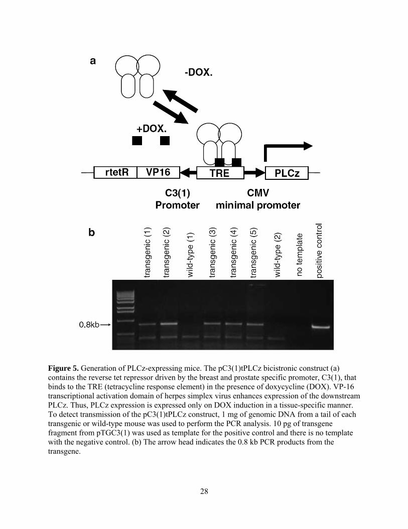
Figure 5. Generation of PLCz-expressing mice. The pC3(1)tPLCz bicistronic construct (a) contains the reverse tet repressor driven by the breast and prostate specific promoter, C3(1), that binds to the TRE (tetracycline response element) in the presence of doxycycline (DOX). VP-16 transcriptional activation domain of herpes simplex virus enhances expression of the downstream PLCz. Thus, PLCz expression is expressed only on DOX induction in a tissue-specific manner. To detect transmission of the pC3(1)tPLCz construct, 1 mg of genomic DNA from a tail of each transgenic or wild-type mouse was used to perform the PCR analysis. 10 pg of transgene fragment from pTGC3(1) was used as template for the positive control and there is no template with the negative control. (b) The arrow head indicates the 0.8 kb PCR products from the transgene.
28

The persistent expression of PLCz depended on continued treatment with DOX, as
removal of the DOX-laced drinking water resulted in dramatically reduced PLCz expression
after two weeks (Figure 6C). For both the induction and the reversion experiments, we probed
the protein rather than mRNA, as protein processing and stability might be extended and thus
alter the kinetics of biological response. As abrogation of signaling by dominant-negative
constructs requires excess expression, we confirmed that PLCz expression was significantly
higher than that of endogenous PLCγ, seen at low levels at 130kDa (Figure 6A). These results
strongly indicate that the C3(1) promoter is able to induce the transgene expression in a tissue
specific and regulatable manner in the mammary and prostate gland of transgenic mice.
2.3.2 Induction of PLCz inhibits invasiveness of the primary tumor and metastatic tumor
formation
To determine whether a PLCγ-mediated motility-associated signaling pathway is associated with
tumor cell dissemination, the pC3(1)tPLCz transgene founders were crossed with MMTV-
PyVmT and TRAMP mice to generate double transgenic MMTV-PyVmT-PLCz (PyVmT-PLCz)
mice and TRAMP-PLCz mice. MMTV-PymT transgenic mice develop mammary tumors with
an average latency of 53 days and form lung metastases with 100% penetrance by 100 days of
age (Muraoka, Dumont et al. 2002). TRAMP transgenic mice develop prostate tumors with an
average latency of 84 days and form lung metastases with 95% penetrance by 24 weeks of age
(Gingrich, Barrios et al. 1997).
In the absence of DOX, tumors arose with similar latencies to MMTV-PyVmT
and TRAMP mice. Because the average tumor latency for the PyV-PLCz mice was ~8 weeks,
29

Figure 6. Inducible expression of PLCz in tissues from male and female mice. Proteins were analysed on 10% SDS–PAGE and immunoblotted with a rabbit polyclonal antibody that recognizes the Z region of PLCg. DOX was added to the drinking water to induce PLCz expression. One week after DOX addition, mice were killing and tissues were processed to get protein lysates. A selection of tissues and mice are shown to present evidence that PLCz expression is dependent on (a) transgene presence, (a) DOX induction (b) steroid-responsive tissues and (c) continued presence of DOX. (a) PLCz is induced by DOX in the prostate (PR) and mammary glands (MG) of transgenic (T) mice and is not expressed in PLCz- (W) mice. (b) PLCz is expressed in the PG of male T mouse in a tissue-specific fashion and is not expressed in the seminal vesicles (SV), liver (Liv), lungs (lun) or spleen (Spl). Nontransgenic mice were negative in all tissues. (c) Three different PLCz-transgenic female mice were treated with DOX for 1 week and then switched to normal drinking water and sacrificed 2 weeks later. The positive control was killed after the DOX treatment. The inguinal MG was dissected and protein lysates analysed. (c) is overexposed compared to (a) and (b) to detect lowlevel persistence of PLCz.
Figure 7 Fisher’s exact test was used to generate P-values to determine whether induction of PLCz had a significant influence on the extent of lung metastases. A, TRAMP and TRAMP/PLCz mice were scored for invasiveness of the primary prostate tumors. The TRAMP mice expressing the PLCz trangene had a significant reduction in the invasiveness of the primary tumor (P<0.02). B, TRAMP and TRAMP/PLCz mice were scored for the extent of the lung metastases. The TRAMP mice expressing the PLCz transgene had a significant decrease in the extent of lung metastasis (P<0.06). C, PyVmT and PyVmT/PLCz mice were scored as in (B). The PyVmT mice expressing the PLCz transgene had a significant decrease in the extent of lung metastasis (P<0.03).
30
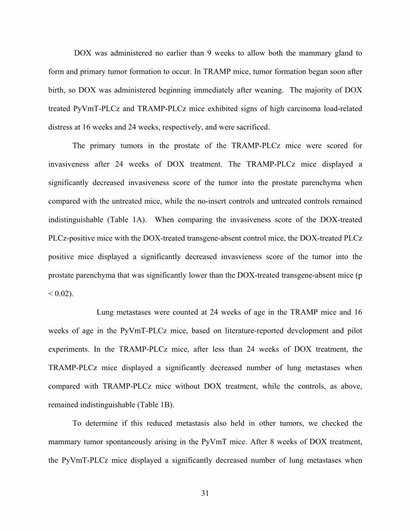
DOX was administered no earlier than 9 weeks to allow both the mammary gland to
form and primary tumor formation to occur. In TRAMP mice, tumor formation began soon after
birth, so DOX was administered beginning immediately after weaning. The majority of DOX
treated PyVmT-PLCz and TRAMP-PLCz mice exhibited signs of high carcinoma load-related
distress at 16 weeks and 24 weeks, respectively, and were sacrificed.
The primary tumors in the prostate of the TRAMP-PLCz mice were scored for
invasiveness after 24 weeks of DOX treatment. The TRAMP-PLCz mice displayed a
significantly decreased invasiveness score of the tumor into the prostate parenchyma when
compared with the untreated mice, while the no-insert controls and untreated controls remained
indistinguishable (Table 1A). When comparing the invasiveness score of the DOX-treated
PLCz-positive mice with the DOX-treated transgene-absent control mice, the DOX-treated PLCz
positive mice displayed a significantly decreased invasvieness score of the tumor into the
prostate parenchyma that was significantly lower than the DOX-treated transgene-absent mice (p
< 0.02).
Lung metastases were counted at 24 weeks of age in the TRAMP mice and 16
weeks of age in the PyVmT-PLCz mice, based on literature-reported development and pilot
experiments. In the TRAMP-PLCz mice, after less than 24 weeks of DOX treatment, the
TRAMP-PLCz mice displayed a significantly decreased number of lung metastases when
compared with TRAMP-PLCz mice without DOX treatment, while the controls, as above,
remained indistinguishable (Table 1B).
To determine if this reduced metastasis also held in other tumors, we checked the
mammary tumor spontaneously arising in the PyVmT mice. After 8 weeks of DOX treatment,
the PyVmT-PLCz mice displayed a significantly decreased number of lung metastases when
31

compared with PyVmT-PLCz mice without DOX treatment (p < 0.02), while the “no insert”
controls and positive insert/no treatment mice remained statistically indistinguishable (Table 1C;
Figure 7, Figure 8A). Primary mammary tumors from DOX-treated PyVmT and PyVmT-PLCz
mice had no differences in tumor volume due to DOX induction (Figure 8B). The number of
lung metastases in PyVmT-PLCz mice not induced to express PLCz by DOX was not different
from the number observed in MMTV-PyVmT mice.
32

Figure 8. Metastatic tumors in the lungs of MMTV-PyV mice expressing PLCz. H&E sections of dissected lung tissue from representative mice. Normal mouse lung tissue (top left). Lung sections of transgenic PyVmT without the PLCz transgene show large metastatic tumors throughout the lung parenchyma that crowd alveolar septa (top right). PyVmT-PLCz mice whose PLCz expression is induced with DOX treatment markedly diminishes the number of tumors in the lung parenchyma (bottom left). DOXtreatment has no effect on metastatic lung deposits in PyVmT mice (bottom right).
33
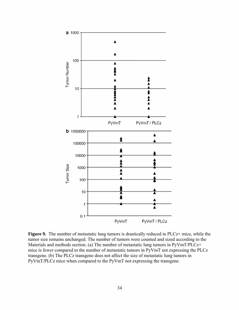
Figure 9. The number of metastatic lung tumors is drastically reduced in PLCz+ mice, while the tumor size remains unchanged. The number of tumors were counted and sized according to the Materials and methods section. (a) The number of metastatic lung tumors in PyVmT/PLCz+ mice is fewer compared to the number of metastatic tumors in PyVmT not expressing the PLCz transgene. (b) The PLCz transgene does not affect the size of metastatic lung tumors in PyVmT/PLCz mice when compared to the PyVmT not expressing the transgene.
34

2.4 DISCUSSION
Tumor invasion, both into adnexia and as the first step in dissemination, has been proposed to be
a consequence of dysregulated cell motility (Kassis, Lauffenburger et al. 2001). The
dissemination of cells from the primary tumor to metastatic sites is limiting for the tumor’s
progression. Although many of the molecules that regulate or effect cell motility have been
investigated (Ridley, Schwartz et al. 2003), the dysregulation of those same molecular events has
yet to be properly characterized in the context of the dissemination and metastatic seeding of de-
novo arising cancers. The models examined herein, are of spontaneous metastasis of in situ
carcinomas that arise secondary to oncogenic activation. We queried whether the dissemination
of such tumors can be limited by blocking a key cell motility pathway, that of PLCγ.
PLCγ signaling is an enticing target to dissect the contributions of induced cell motility to
tumor progression, as it distinguishes the cell motility response from that of proliferation (Chen,
Xie et al. 1996). Obviously, any intervention that also prevents proliferation would be falsely
scored as metastasis suppressing as metastasis requires subsequent proliferation. Despite the
extensive investigations on motility in culture systems and confirmation in xenograft models,
experimental evidence is lacking as to the relative or specific contributions of motility on the
metastatic dissemination of de novo-occuring cancer cells. This is due to the pleiotropic nature
of the signals during tumor progression, such as the autocrine stimulatory loops activating
growth factor receptors that drive both motility and proliferation. For instance, current
molecularly-targeted therapies inhibit signaling from the EGFR family members HER2 (erbB2)
and HER1 (EGFR, erbB1). Both of these receptors initiate and sustain proliferation and
migration in breast and prostate carcinoma cells. Thus, while these inhibitors limit dissemination,
whether growth factor-enhanced motility underlies metastasis remains unproven. Abrogation of
35

PLCγ signaling by pharmacological or genetic means eliminates motility but not mitogenesis
induced by EGFR, HER2 (Chen, Gupta et al. 1994; Chen, Xie et al. 1994) and other growth
factor receptors (Reddy, Wells et al. 1996). By targeting this convergent downstream effector
molecule, we defined a role for growth factor-induced cell motility in the dissemination of
primary breast and prostate cancer in vivo.
In this study, we conditionally expressed a dominant-negative fragment of PLCγ in the
mammary and prostate epithelium of TRAMP and MMTV-PyVmT. This conditional, post-natal
expression was required as constitutive ablation of PLCγ is embryonic lethal (Ji, Ermini et al.
1998). Furthermore, by limiting expression to the target epithelial cells, we avoid issues of
altering the microenvironment of the primary and metastatic tumor sites, as well as affecting
tumor-associated angiogenesis (Bodnar, Yates et al. 2006). We chose a reversible, inducible
system rather than conditional gene deletion (i.e. Cre-Lox) for the flexibility provided by the
ability to revert to a functional phenotype (see Supplemental data).
Expression of the dominant-negative PLCz construct during prenatal and immediate post-
natal periods retarded organogenesis specific to the prostate and mammary epithelium. This was
reversible upon DOX withdrawal, validating our strategy and demonstrating that low level
leakiness of the tetracycline-on system was not consequential as a dominant-negative is only
effective when in stoichiometric excess.
To study the role of PLCγ signaling in metastasis, we allowed organogenesis and
maturation to proceed, and spontaneous tumorigenesis to be initiated by the transgenic
oncogenes. Forcing expression of PLCz limited the metastases from both the breast and prostate
carcinomas; the lungs were chosen for ease and reproducibility of quantitation, though similarly
decreased metastasis was noted in other internal organs (data not shown). This decrease in
36
number and size of metastases was not due to slower tumor growth as the primary tumor sizes
were unaffected by PLCγ expression. These data confirm that cancer cell invasiveness is a
determining factor of tumor progression in vivo.
These studies confirm that PLCγ function is necessary for tumor cell dissemination in
both breast and prostate cancers. That tumorigenesis was incompletely abrogated is not
unexpected, but may be due to several reasons. First, the expression of PLCz does not
completely eliminate EGF-induced PIP2 hydrolysis in vitro (Chen, Gupta et al. 1994) and may be
similarly incomplete in in vivo systems. Second, some low level EGF-induced motility is noted
in PLCγ-1 devoid cells (Ji, Ermini et al. 1998) this is due to other pathways that regulate
motility-associated processes other than protrusion, such as ERK MAP kinase/m-calpain (Chen,
Xie et al. 1996; Glading, Chang et al. 2000) and PKCd/MLC (Iwabu, Smith et al. 2004), as well
some upregulation of PLCγ-2 expression. It should also be noted that while the targeted
inhibition of PLCγ is being discussed as a downstream effector required for EGFR-mediated
motility, we cannot be certain that the extracellular trigger for breast and prostate cancer
dissemination are the EGFR ligands. EGFR signaling is likely to be the primary trigger as it has
been implicated in the transformation, mitogenic and motogenic properties of tumor cells
(Carpenter 2000); however, it is possible that other growth factors are signaling via
PLCγ (Carpenter and Ji 1999; Jones, Peak et al. 2005). As PLCγ seems to serve as a point of
convergence for PDGF, HGF, and IGF-1 in addition to EGF in signaling motility (Bornfeldt,
Raines et al. 1994; Kundra, Escobedo et al. 1994), expression of PLCz would be expected to
diminish cell motility signaled by all of these factors. Furthermore, PLCγ has been linked to
integrin-related changes that result in cell motility in the context of invading cancer cells (Jones,
Peak et al. 2005) The progression of invasive cancers to the morbidity and mortality of
37

metastasis encompasses a series of highly coordinated events, though the literature offers a
compelling line of evidence for the general role of PLCγ throughout the metastatic process. We
have demonstrated here that signaling through PLCγ is a necessary and potentially limiting step
that may be targeted in a rational intervention.
2.5 MATERIALS AND METHODS
All animal experimentations were approved by the Institutional Animal Care and Use
Committees respectful to where the experiments occurred, including the University of
Pittsburgh, Pittsburgh VAMC, and the University of Alabama at Birmingham.
2.5.1 Generation of double transgenic TRAMP and MMTV-PyVmT with a DOX-
inducible PLCz transgene
The reverse tetracycline plasmids (tet-on) PUHD13-6 and PUHD172-1neo were kindly provided
by Dr. Hermann Bujard (Heidelberg) (Gossen, Freundlieb et al. 1995). The CMV promoter
driving the reverse tet repressor (rTetR) of PHUD172-1neo was replaced with rat C3(1)
prostatein promoter (kindly provided by Dr. Peter Barry, UC Davis (Tehranian, Morris et al.
1996)). The dominant-negative PLCγ fragment, PLCz, ((Homma and Takenawa 1992; Chen,
Gupta et al. 1994)) was placed downstream of the TRE (tetracycline response element) with the
TATA box from CMV minimal promoter of PUHD13-6. These two DNA fragments containing
C3(1)rtetR and TRE-TATA-PLCz were ligated into the pBluescript to create a single bicistronic
38

construct; this was done to increase expression levels (Hofmann, Nolan et al. 1996) and simplify
breeding schemes by physically linking the two halves of the system. The resulting plasmid,
pC3(1)tPLCz, was used in microinjection (Fig.1A)
pC3(1)tPLCz was digested with NotI and PstI and electrophoresed through a
0.7% low melting point agarose gel and purified using Quiax DNA binding beads. Purified DNA
was injected into one cell FBV zygotes by the University of Alabama at Birmingham transgenic
polyomafacility (Carl Pinkert, Director). Following injection, viable eggs were transferred to the
oviducts of pseudopregenant female mice. To identify positives, DNA was extracted from 1cm
section of tails using Quiamp DNA extraction kit (Quiagen). DNA was resuspended with 200 μl
of TE (10 mM Tris, 1 mM EDTA pH 8.0). Screening of transgenic mice was by polymerase
chain reactions with one pair of oligonucleotides annealed to the PLCz. The PCR was performed
on a DNA thermocycler (Ericomp) using following program: 94oC for 1 minutes, 56oC for 1
minute, and 72oC for 1 minute. PCR product was analyzed by electrophoresis on a 1% agarose
gel (Fig. 1B).
The PLCz mice were mated with TRAMP mice, kindly provided by Norman Greenburg
(Gingrich, Barrios et al. 1996), or MMTV-PyV mice, the gift of Dr. WJ Muller (Webster,
Martin-Soudant et al. 1998). Male PLCz mice were mated with female TRAMP mice or female
PLCz were mated with male MMTV-PyV mice. The F1 crosses were used for all experiments.
The mice lacking the PLCz transgene presented tumors at the expected rates and times.
39

2.5.2 PLCz expression
To induce expression of the PLCz transgene, doxycycline was also administered orally in
drinking water (2 mg/ml) for varying times before sacrificing mice. Various tissues from wild
type and transgenic mice were sonicated in PBS with protease inhibitors (10 μg/ml leupeptin, 20
μg/ml aprotinin, 1 mM phenylmethylsulfhnyl fluoride). The concentration of each sample was
determined by measuring the absorbance at 595 nm using Biorad Bradford assay. 50 μg of
lysates were subjected to 10% SDS-PAGE, and the resolved proteins were transferred
electrophoretically to PVDF membrane. After shaking the blot in TBST with 5% non-fat dry
milk (W/V) for 1 hour and blot was incubated with the anti-rabbit PLCγ antibody (Santa Cruz) at
4oC for overnight. After incubation with primary antibody, the blot was washed in TBST for 15
minutes twice and incubated with secondary antibody (anti rabbit IgG, 2500:1 dilution,
Promega). Protein bands were visualized with NBT and BCIP (Promega).
2.5.3 Histologic and immunohistochemical assessments of mammary and prostate glands
For histology, part of mammary gland and dorsal prostate gland were fixed in 10%
neutral buffered formalin, processed for paraffin sectioning and stained with hematoxylin and
eosin. Paraffin sections were deparaffinized, hydrated, pretreated with 2 N HCl for 20 minutes at
37oC and exposed to 0.01% trypsin at 37oC for 3 minutes, followed by staining using a mouse
monoclonal anti-PCNA antibody and biotinylated secondary antibody. 3,3' Diaminobenzidine
tetrahydrochloride was used as chromogen. Sections were also tested for apoptosis using the
Apotag kit (Oncor). After hydration, sections were treated with proteinase K (20 μg/ml) and the
40

endogenous hydrogen peroxide was quenched by incubating with 2% hydrogen peroxide in PBS.
Terminal deoxytransferase was added to sections followed by the incubation with anti-
digioxigenin antibody conjugated with HRP. Color detection was accomplished with DAB as
Chromogen.
The primary and metastatic tumors were scored for invasion of the parenchyma and were
evaluated using a method modified from Eneroth (Jakobsson, Eneroth et al. 1973). The tumor
invasion modes were scored into four histopathologic malignancy grade, given as (score), gross
description: (0.5) micrometastasis; (1) well defined border; (2) cords/Indian files, less well
defined border; (3) groups of cells, no distinct border; (4) diffuse growth.
2.5.4 Statistical analyses
Data were categorized into PLC-/No DOX, PLC+/No DOX, PLC-/DOX and PLCz+/DOX
groups. Differences in continuous parameters (size, number) were evaluated by Wilcoxon-Rank
Sum Test using the SAS statistical software package (Cary, NC). Fisher’s exact test was used to
evaluate the significance of differences in the parameters. All Ps are represented by one-tailed
tests (Kassis, Moellinger et al. 1999) and were deemed statistically significant at p < 0.10;
though levels of statistical differences are provided.
2.6 ACKNOWLEDGEMENTS
We thank Norman Greenberg for the gift of the TRAMP mice and Dr. William Muller for the
gift of the MMTV-PyV mice. Dr. Peter Barry kindly provided the prostatein promoter construct.
41

This work was supported in part by grants from the Department of Defense Congressionally
Mandated Medical Research Program on Prostate and Breast Cancer and a VA Merit Award.
42

3.0 EXPRESSION OF THE CYTOSOLIC TAIL OF E-CADHERIN PARTIALLY
REVERTS THE EPITHELIAL TO MESENCHYMAL TRANSITION IN AGGRESSIVE
BREAST CARCINOMA CELLS
Christopher R. Shepard, Alan Wells
Department of Pathology, University of Pittsburgh School of Medicine and Pittsburgh VA Medical Center, Pittsburgh, PA
43

3.1 ABSTRACT
Coculturing of the aggressive MDA-231 breast carcinoma cell and primary breast carcinoma
cells with normal parenchymal cells of a metastatic target organ, the liver, resulted in renewed
transcription of E-cadherin secondary to specific loss of promoter methylation. Despite the re-
expression of E-cadherin, the MDA-231 cells do not fully revert to an epithelial cohesive-cluster
morphology. We speculated that the MDA-231 cells that re-expressed E-cadherin may have an
intermediate phenotype, wherein the cells benefit from E-cadherin-dependent canonical pathway
activation, but also retain characteristics of invasive cancer cells. We tested this hypothesis by
using a dominant negative E-cadherin, in which an intact intracellular domain is coupled to an
extracellular domain from the class I major histocompatibility complex antigen (H-2kd). This
dominant negative construct sequesters the E-cadherin plaque-associated molecules beta-catenin
and p120, which, in the absence of E-cadherin, are conventionally thought to propagate
oncogenic signals. The cells demonstrated a level of invasiveness in vitro intermediate between
MDA-231 cells and non-aggressive MCF7 cells, and similar to that of partially-reverted MDA-
231 cells cocultured with hepatocytes. These data suggest a model in which breast cancer cells
in the metastatic environment partially downregulate catenin-driven oncogenic signaling by a
phenotypic shift in the E-cadherin expression equilibrium. In this manner, the cells remain in a
partial EMT or reversion thereof, whereby they are able to readily shift between epithelial-like
dormancy and aggressive metastatic growth as determine by other micro-environmental cues.
44

3.2 INTRODUCTION
Breast cancer is the most frequently diagnosed cancer in women, and it is the second
leading cause of cancer death in women of all ages (Punglia, Morrow et al. 2007). The majority
of these cancers originate from the epithelial cells lining the mammary ducts. Transformation of
normal ductal epithelial cells into their metastatic counterparts occurs in a series of sequential
steps, during which the cells acquire a more amoeboidal phenotype, become motile, disseminate,
and colonize distant sites of the body. The initial stages of this transformation are described as
an epithelial-to-mesenchymal transition (EMT) (Lee, Dedhar et al. 2006). The EMT, which
includes a loss of E-cadherin as a precursor to a more motile and invasive phenotype, is
necessary to break away from the primary mass and either disseminate by hematagenous or
lymphatic routes. Upon colonization of a distant ectopic organ, it is hypothesized that cancer
cells undergo a reprogramming to a more epithelial phenotype, described as a mesenchymal-to-
epithelelial reverting transition (MErT) (Yates, Shepard et al. 2007). MErT is hypothesized to
play a role in the ability of cancer cells to colonize an ectopic locale and counter many of the
metastatic inefficiencies and pharmaceutical challenges that affront the cancer cells.
A hallmark measure of malignant tumor cells is invasiveness. Invasiveness is the
result of disconnect of E-cadherin-mediated cell-cell adhesions that limit cell dispersion and
establish apical-basal polarity with segregation of signals. This phenomenon has been shown
repeatedly in simulations of the cadherin switch phenomena in which E-cadherin presence or
absence dictates the degree of epithelial-like phenotype in a cell. This has been demonstrated by
expression of a dominant negative Epithelial-cadherin (H-2kd-Ecad), formed by fusion of an
intact intracellular domain to the extracellular domain from the class I major histocompatibility
complex antigen (H-2kd), in non-metastatic epithelial-like MCF-7 breast cancer cells causing
45
them to become more invasive (Vizirianakis, Chen et al. 2002). Thus, invasiveness can be
considered an inverse measure of epithelial quality of a carcinoma cell (Heimann and Hellman
2000), and can be used to determine how restoration of the appropriate equilibriums restores the
epithelial phenotype.
E-cadherin not only established cell-cell connections but alters the localization and
signaling capacity of multifunctional signaling molecules, the catenins in particular. One
molecular equilibrium necessary for the epithlelial phenotype is the appropriate stoichiometry
between E-cadherin, β-catenin, and p120 (Aberle, Schwartz et al. 1996; Hirohashi 1998). Cell-
cell binding of E-cadherin inhibits β-catenin and p120 signaling by sequestering the proteins at
the cell surface; active β-catenin and p120 signaling have both been implicated in invasive
phenotypes (Gottardi, Wong et al. 2001; Reynolds and Roczniak-Ferguson 2004). Noting that
many cancer cell lines retain epithelial elements, we hypothesized whether epitheliality could be
incrementally gained by simple sequestration of the effector catenins. Herein, we report that
expression of the intracellular domain of E-cadherin binds the catenins, and partially reverts the
EMT, limiting tumor cell invasiveness similar. This suggests that a major role of E-cadherin
expression in breast carcinoma cells is to modulate catenin signaling.
3.3 RESULTS
3.3.1 Breast cancer cell lines retain epithelial elements
Two well-characterized model breast cancer cell lines, MCF7 and MDA-231, though
both originating as exudative pleural effusions from ductal carcinoma in situ, possess seemingly
46

polar characteristics on the EMT spectrum (Soule, Vazguez et al. 1973; Cailleau, Young et al.
1974; Cailleau, Olive et al. 1978). MCF7 cells retain many features of their epithelial
counterparts and are not invasive in vivo. MCF7 cells present cobblestone morphology when
grown in monolayer and epithelial levels of E-cadherin expression, and have even been
considered in some studies as immortalized mammary epithelial cells. MDA-231 cells, in
contrast, are highly invasive and have a mesenchymal phenotype. MDA-231 cells grow
individually and unconnected in monolayer cultures, do not express E-cadherin due to promoter
methylation (which is characteristic of E-cadherin silencing in invasive carcinoma cells), and are
highly invasive in vivo. Since the preponderance of literature suggests that carcinoma cells, in
order to metastasize, must lose their epitheliality, specifically the cell adhesion molecule E-
cadherin to decouple from the primary epithelia, we were curious as to what epithelial
characteristics each cell line endogenously retained or reacquired before being isolated from the
pleural effusion with the intention of engineering the equilibrium of these features further into
the study. In order to determine the extent to which breast cancer cells retained epithelial
elements we assayed two hallmark features: their ability to assume a mammary-specific
epithelial morphology and their responsiveness to TGF-β.
To determine how E-cadherin expression effected the morphological clustering of
MCF7 cells, we performed FACS analysis and gated the top 5% of E-cadherin expressors in a
screen of 5e6 MCF7 cells. When these cells were cultured, they grew in dome-shaped, tightly
adherent clusters (Figure 10A), in contrast to their more spread morphology prior to the sorting.
This morphology has been seen in both primary isolates and clonally derived cell lines of
mammary adenocarcinoma (Pickett, Pitelka et al. 1975; Zucchi, Bini et al. 2002), though this
morphology does not occur spontaneously in MCF7 cells (Figure 10A). After four weeks of
47

Figure 10. Invasive cancer cells retain epithelial characteristics. A) a, MCF7 cells grow in clustered epithelial sheets. b, when high-E-cadherin expressing MCF7 are plated, they grow in a very tightly adherent domes. c, four weeks after sorting out the high E-cadherin MCF7 cells, they have returned to a flattened morphology and a more similar morphology as seen in panel a. B) MDA-231 cells (a) and MCF7 cells (b) were kept in culture with and without varying concentrations of TGF-β. Both cells lines, which were isolated from exudative pleural effusions indicating that they had already metastasized, were responsive to the growth suppressive effects of TGF-β.
48
normal culture without any selective mechanism, the MCF7 cells return to their original
morphology (Figure 10A). This qualitative data suggests that MCF7 cells retain their ability to
undergo reversible shifts in differentiation. Contrary to other mammary adenocarcinoma cell
lines that require de novo protein expression for dome formation (Zucchi, Bini et al. 2002), high
E-cadherin expression is the primary phenotypic element that promotes dome-forming ability in
MCF7 cells. The return of MCF7 cells to their original phenotype after sorting suggests that
MCF7 have the plasticity to move along the spectrum of the EMT.
A hallmark biological response that is considered to distinguish normal epithelial cells
from transformed ones is the cessation of proliferation upon treatment with TGF-β (Nelson,
Vanduijn et al. 2006), though growth effects of transformed mammary epithelia upon TGF-β
treatment has been subject to much debate (Roberts and Wakefield 2003). We treated both
MCF7 and MDA-231 cells with TGF-β and saw a growth inhibitory effect at all concentrations
in both cell types (Figure 11a and 11b), which is consistent with previous literature (Mazars,
Barboule et al. 1995). Contrary to the currently accepted model, that as mammary cells progress
toward malignancy the TGF-β response is more oncogenic (Wakefield and Roberts 2002), the
response of MCF7 and MDA-231 resembled cells of the normal epithelium, where suppressor
activities dominate in response to TGF-β treatment. These data suggest mammary tumor cells
that retain elements of the epithelial lineage and are plastic in these aspects.
3.3.2 Epithelial markers vary quantitatively between breast cancer lines
The carcinoma-related EMT affects more than E-cadherin cohesiveness and TGF-β
responsiveness. Many cytoskeletal and cell adhesion proteins change levels with the acquisition
of a more mesenchymal phenotype (Gotzmann, Mikula et al. 2004). We queried how these
49
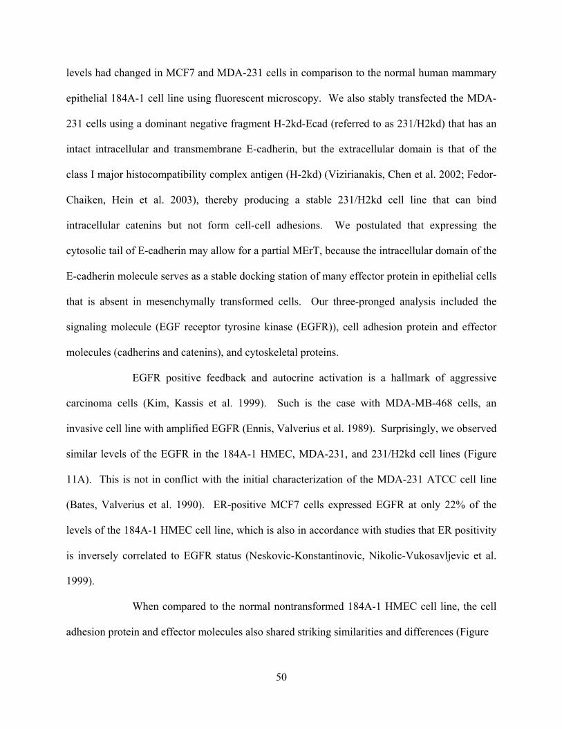
levels had changed in MCF7 and MDA-231 cells in comparison to the normal human mammary
epithelial 184A-1 cell line using fluorescent microscopy. We also stably transfected the MDA-
231 cells using a dominant negative fragment H-2kd-Ecad (referred to as 231/H2kd) that has an
intact intracellular and transmembrane E-cadherin, but the extracellular domain is that of the
class I major histocompatibility complex antigen (H-2kd) (Vizirianakis, Chen et al. 2002; Fedor-
Chaiken, Hein et al. 2003), thereby producing a stable 231/H2kd cell line that can bind
intracellular catenins but not form cell-cell adhesions. We postulated that expressing the
cytosolic tail of E-cadherin may allow for a partial MErT, because the intracellular domain of the
E-cadherin molecule serves as a stable docking station of many effector protein in epithelial cells
that is absent in mesenchymally transformed cells. Our three-pronged analysis included the
signaling molecule (EGF receptor tyrosine kinase (EGFR)), cell adhesion protein and effector
molecules (cadherins and catenins), and cytoskeletal proteins.
EGFR positive feedback and autocrine activation is a hallmark of aggressive
carcinoma cells (Kim, Kassis et al. 1999). Such is the case with MDA-MB-468 cells, an
invasive cell line with amplified EGFR (Ennis, Valverius et al. 1989). Surprisingly, we observed
similar levels of the EGFR in the 184A-1 HMEC, MDA-231, and 231/H2kd cell lines (Figure
11A). This is not in conflict with the initial characterization of the MDA-231 ATCC cell line
(Bates, Valverius et al. 1990). ER-positive MCF7 cells expressed EGFR at only 22% of the
levels of the 184A-1 HMEC cell line, which is also in accordance with studies that ER positivity
is inversely correlated to EGFR status (Neskovic-Konstantinovic, Nikolic-Vukosavljevic et al.
1999).
When compared to the normal nontransformed 184A-1 HMEC cell line, the cell
adhesion protein and effector molecules also shared striking similarities and differences (Figure
50

Figure 11. Invasive cancer cells exhibit a very skewed protein profile when compared with untransformed HMECs. A) Cells were examined by immunofluorescence and the histograms of the acquired pictures were analyzed for pixel intensity. B) 231/H2kd cells exhibit remarkably upregulated levels of cytokeratin-18 compared to their untransformed MDA-231 counterparts.
51

11A). Expectedly, E-cadherin was very similar in the MCF7 and 184A-1 HMEC cell lines,
while MDA-231 and 231/H2kd cells completely lacked the protein. The expression levels of α-
catenin, and E-cadherin effector molecule that couples the transmembrane adhesion protein to
the actin cytoskeleton, were polar opposite in MDA-231 and MCF7 cells. The more epithelial
MCF7 line had α-catenin levels over two-fold higher than in 184A-1 HMECs, while the MDA-
231 line had much lower levels. β-catenin was expressed in both MCF7 and MDA-231 lines at
levels almost 6-fold above the 184A-1 HMEC line; this is not surprising given the link between
the canonical Wnt signaling pathway and transformation. β-catenin expression in 231/H2kd
cells was similarily elevated. β-catenin’s homologue, γ-catenin, was elevated in MCF7 cells and
a little more than half that of 184A-1 HMEC expression. E-cadherin’s other effector catenin,
p120, was present in very high levels in MCF7 and at lower levels in MDA-231 and 231-derived
lines.
The EMT is also characterized by a switch from cytokeratin-18 to vimentin intermediate
filament expression. We did not observe the characteristic switch in intermediate filaments, as
MCF7 and MDA-231 cells exhibited similar amounts of vimentin below 10% that of the model
epithelial 184A-1 cells (Figure 11A), though some groups report that low vimentin expression is
indicative of high tumor grade in vivo (Willipinski-Stapelfeldt, Riethdorf et al. 2005). Our most
significant observations were those of cytokeratin-18 expression and how the expression
changed upon introduction of the dominant negative H-2kd-Ecad. Consistent with a mechanism
that restores epitheliality, MCF7 cells display 2.5-fold more cytokeratin-18 than the HMEC
counterparts, while this epithelial marker was decreased in MDA-231 cells (Figure 11A). Most
interestingly, in the 231/H2kd cells, cytokeratin-18 was restored to levels similar to MCF7
(Figure 11A and 11B), though the cellular localization of cytokeratin-18 in 231/H2kd cells was
52

starkly different, being punctate rather than as a cytosolic scaffold such as with the normal cells.
This suggests that the cytosolic tail of E-cadherin acts to affect an increase in cytokeratin-18
levels. In total, while the MCF7 cell line is much more epithelial than its MDA-231 counterpart,
there are similar mesenchymal markers that the two lines share; conversely, the MDA-231 cell
line has features shared by the normal 184A-1 HMEC line, such as untransformed levels of
EGFR. Though the dominant negative construct imparted the ability on MDA-231 cells to
upregulate cytokeratin-18, a robust epithelial marker, we observed the other proteins to stay
similar to those levels of MDA-231.
3.3.3 Intermediate mesenchymal-epithelial phenotypes can be engineered
Upon seeing the varied protein profiles of the MCF7,MDA-231, and 231/H2kd cells, we
suspected that we had engineered an epithelial to mesenchymal reverting transition in the more
invasive MDA-231 cells by controlling the localization of β-catenin and p120. Upon co-
immunoprecipitation of H-2kd from total cell lysate of 231/H2kd cells, we were able to co-
precipiate β-catenin and p120, a result of their sequestration onto the cytoplasmic domain of E-
cadherin (Figure 12A). This engineering also resulted in a change of cell morphology in the
231/H2kd cells to a more flattened morphology and the 231/H2kd cells having punctuated
lamellipodia instead of a defined leading edge as in the MDA-231 cells (Figure 12A, inset). It is
important to note that this engineering was performed without establishing cell-cell connections,
and therefore suggests that outside-in signaling by E-cadherin is not necessary for at least a
partial switch to epithelial morphology.
Testing the functional implications of H-2kd-Ecad in MDA-231 cells using the invasion
assay, we found that sequestration of E-cadherin effector catenins abrogated the invasiveness of
53

Figure 12. Cancer cells can be engineered to be more epithelial by introducing a dominant negative E-cadherin which sequesters catenins and mitigates the invasive phenotype. A) In whole cells lysates, the H2kd fragment is detected in MDA-231 cells transfected with a dominant negative E-cadherin (H-2kd-Ecad). β-catenin and p120 can be co-immunoprecipitated with H2kd in 231/H2kd cells. 231/H2kd cells have a flattened morphology with punctuated lamellipodia rather than the apparent leading edge seen in the untransformed MDA-231 counterparts (insets). B) Invasion assay comparing wild-type MDA-231 cells, epithelial MCF7 cells, and 231/H2kd cells. Asterisks denotes statistical significance by Student t-test (*p<0.08).
54

these cells by almost almost one-third when comparing the difference between the untransfected
MDA-231 cells and the basal MCF7 cells (Figure 12B). Though we were able to mitigate
invasiveness in MDA-231 cells by sequestering β-catenin and p120, we found that the baseline
invasiveness without a chemotactic signal (“serum free” in the bottom chamber) was still greater
than the MCF7 cells (p < 0.08), in which E-cadherin is expressed in high amounts both limiting
the signaling of the catenin proteins and establishing cell-cell connections. Though β-catenin
and p120 have been the most studied proteins to date regarding their ability to regulate
epitheliality with their association in the E-cadherin complex, the expression of the cytosolic tail
of E-cadherin could have sequestered or effected the signaling of other uncharacterized effector
proteins that could have contributed to these results. In total, this suggests that changes to the
localization of key signaling proteins during the mesenchymal to epithelial transition can have
profound effects mitigating the mesenchymal nature of an invasive cell.
3.4 DISCUSSION
Metastatic transformation follows a sequence of ordered events, during which normal
epithelial cells become metastatic by both the gain and loss of functions that perturb the cell from
its epithelial equilibrium. The hallmark imbalance of the epithelial to mesenchymal transition
begins at the primary tumor, where E-cadherin silenced (Foty and Steinberg 2004). The
silencing of the cell adhesion molecule not only contributes to the decoupling of the epithelial
sheet, which allows malignant cells to break away from the primary mass, but also to increased
activity of E-cadherin’s effector catenins, β-catenin and p120 (Brennan and Brown 2004; Sarrio,
Perez-Mies et al. 2004). In epithelial equilibrium, the catenin signaling is limited by
55

sequestration on the intracellular tail of the E-cadherin transmembrane protein; when E-cadherin
is no longer present, β-catenin and p120 are free to transit throughout the cytoplasm of the cell,
signaling to their downstream pathways. When this signaling is not balanced by E-cadherin
sequestration, it most likely results in a metastatic cascade, further perturbing the equilibrium
until the full mesenchymal nature of the cell is reached. The present study provides proof-of-
principle that even invasive carcinoma cells can regain some epithelial phenotypes by
modulating this catenin signaling.
In previous studies, we documented how normal epithelial cells can drive the re-
expression of E-cadherin in breast cancer cells in a process described as the mesenchymal to
epithelial reverting transition (MErT) (Yates, Shepard et al. 2007). In the prostate cancer cell
lines DU-145 and PC-3, E-cadherin expression could be induced by inhibition of the EGFR,
indicating that a post-transcriptional pathway was responsible for E-cadherin downregulation.
Several intermediate pathways linking tyrosine kinase activation to E-cadherin recycling and/or
destruction have been reported (Pece and Gutkind 2002). As EGFR-family member
amplification is a frequent occurrence in invasive cancers, increased signaling through this
receptor family is a likely cause for loss of epithelial equilibrium and progression towards a
mesenchymal phenotype. Inhibition of EGFR signaling demonstrated epithelial phenotype
plasticity in highly invasive carcinomas, leading us to query how else this equilibrium could be
affected.
In the MDA-231 breast cancer line, in which E-cadherin is silenced on the genetic level
by promoter methylation, we saw that coculturing with epithelial cells triggered a demethylation
of the promoter region and subsequent expression of E-cadherin that could functionally ligate
with juxtaposed epithelial cells and transduce signals by canonical pathway activation (Shepard
56

and Wells 2007). In these studies, we observed that the epithelial interaction altered the MET
equilibrium on the epigenetic level, allowing re-expression of E-cadherin and relocalization of β-
catenin and p120 to the cytosolic tail of E-cadherin. Our observations of mesenchymal-epithelial
equilibrium shifts raised the question of what aspect of E-cadherin expression was responsible –
the cell-cell connections or the altered signaling from relocalization of the attendant catenins.
First, by examining phenotypic and proteomic patterns in both the aggressive MDA-231
and the nearly nontumorigenic MCF7 cells, we found that there is variation in breast cancer cell
lines such that this no ‘pure’ signature for either end of the EMT spectrum; and that these
characteristics are plastic. In MCF7 cells, which are non-invasive in vivo, a highly differentiated
phenotype can be realized simply by sorting for those cells at the tail-end of the E-cadherin
expression distribution; in this way, an equilibrium shift changing the amount of E-cadherin
expression in the MCF7 population would restore the epithelial phenotype of the cell population.
We also observed biological responses, thought only present in normal epithelial cells, to be
preserved in the invasive cancer cell line MDA-231; responsiveness to TGF-β1 mediated growth
suppressive effects is a property of development-associated transitions involving branching
morphogenesis. Our data suggests that invasive cells can still be responsive to TGF-β in a
growth-suppressive manner. This is such a hallmark characteristic, one of the defining ones in
mammary bud development, that the possibility for MDA-231 cells’ ability to redifferentiate to a
pseudo-normal mammary epithelium cannot be ignored.
After observing these variations at odds with a one-way EMT theory, we hypothesized that we
could engineer a small phenotypic equilibrium shift in MDA-231 cells by sequestering the E-
cadherin-associated catenins with a non-binding E-cadherin construct. After transfecting the
MDA-231 cells with the cytosolic domain of E-cadherin linked to the MHC external domain, we
57

saw that the dominant negative protein sequestered both β- and p120- catenins. The advantage
of using this dominant negative is that the catenin signaling could be parsed from other activities
of the extracellular domain of E-cadherin including cell adhesion through trans-ligation and
EGFR cis-modulation (Fedor-Chaiken, Hein et al. 2003). We compared the transfectants with
the parental MDA-231 cells and the MCF7 cells. Not only did the 231/H2kds expression levels
of cytokeratin-18 seen in the epithelialoid MCF7 cells, but when the invasiveness was measured
by invasion chamber assay, we found that MDA-231/H2kd cells were less invasive than their
MDA-231 counterparts. The MDA-231/H2kd cells were still more invasive than the MCF7 cells
even at basal levels without a chemoattractant present. Thus, we provide proof-of-principle that
cancer cells can be engineered to approach a mesenchymal-to-epithelial reverting transition by
altering the functions of E-cadherin expression. Malignant carcinoma reversion to epitheliality
in vivo, whether in the natural history of the disease or by pharmaceutical intervention, may be a
double edged sword. The result of returning invasive cells back into their epithelial counterparts
will mitigate the pathology of invasive cells. However, this reversion may provide other
behaviors that may make the cells more likely to persist and survive attempts to exterminate the
tumor. More research into how invasive cells undergo the MErT is necessary to formulate a
pharmaceutical intervention strategy.
58

3.5 MATERIALS AND METHODS
3.5.1 Cell lines and reagents
The human breast adenocarcinoma cell lines MCF7 and MDA-231 were originally derived from
pleural effusions. Cells were maintained in RPMI-1640 with 10% FBS as previously described
(Yates, Shepard et al. 2007). 184A-1 HMECS were a kind gift from Lauffenburger et al and
cultured as previously described (Wolf-Yadlin, Kumar et al. 2006). High E-cadherin expression
MCF7 cells were sorted by FACS using a human E-cadherin-specific (67A4) antibody (Santa
Cruz Biotechnology; Santa Cruz, CA) and cultured as above. TGFβ-1 (Peprotech; Rocky Hill,
NJ) was used at stated concentrations in serum-supplemented media. MDA-231/H2kd cells were
generated using the Myc/His encoding H-2kd-E-cad dominant negative E-cadherin construct, a
kind gift from Vizirianakis et al (Vizirianakis, Chen et al. 2002). H-2kd-Ecad cells were selected
by FACS using the H-2kd (SF1-1.1) antibody (BD Pharmingen; San Jose, CA) and were
maintained in 600μg/ml G418 until used for experimentation.
3.5.2 Immunofluorescence staining and analysis
Cells were plated in Nunc Lab-Tek 8-well Chamber Slides (Fisher; Houston, TX) and allowed to
attach overnight. The next day cell were fixed in 4% paraformaldehyde then permeabilzed using
1% Triton in PBS for 4 minutes. Cells were incubated with primary antibody for 60 minutes,
washed, and then secondary antibody for 60 minutes, washed, then mounted using Vectashield
with DAPI (Vector Labs; Burlingame, CA). Images were acquired using an Olympus 1X70
inverted microscope (Center Valley, PA). Primary antibodies were purchased from Santa Cruz
59

Biotechnology, unless otherwise stated: E-cadherin (67A4), p120 (H-90), β-catenin (C-18),
vimentin (J144), cytokeratin-18 (RCK106), γ-catenin (C-20), α-catenin (H297), EGFR (Ab-3)
(Oncogene; Uniondale, NY). Raw 8-bit color depth images were analyzed using the Photoshop
(Adobe; San Jose CA) pixel histogram tool using channel-specific parameters. Enlarged insets
were sharpened using Photoshop’s “Smart Sharpen” tool; image manipulations were applied
equally to all comparable figures.
3.5.3 Immunoprecipitations and western blots
MDA-231/H2kd cells were quiesced for 24 hours and immunoprecipitation was performed using
the H-2kd antibody from sodium-dodecyl-sulfate (SDS)-sample buffer cell lysate as previously
described (PMID: 14747473). Immunoprecipiated proteins were subjected to SDS-PAGE and
transferred to polyvinylidene difluoride membrane/Immobilon-P (Millipore; Beford MA). Blots
were probed with given primary antibody above before visualizing with the Enhanced
Chemiluminescence Kit (Amersham Biosciences; Piscataway, NJ).
3.5.4 Invasion assay
Invasive potential was determined in vitro by migration through an artificial ECM (Zhou,
Grandis et al. 2006). 1.5e4 cells were challenged in growth-factor reduced matrigel invasion
chambers (Becton Dickinson/Biocoat; Bedford, MA) as previously described. Cells were plated
in 1% BSA for the first 24 hours; after, media in the top chamber was replaced with serum-free
media and media containing 10% serum was added to the lower chamber for the remainder of the
assay. After 48 hours, the remaining cells and ECM in the top chamber were removed by cotton
60

swab and the cells that invaded through the matrix and reached the bottom of the filter were
counted visually. Individual experiments were performed in triplicate. Negative controls were
performed by using serum-free media in the bottom chamber for the duration of the experiment.
Measurements were recorded as the fraction of total cells seeded at the beginning of the assay.
3.5.5 Statistical Analysis
All quantitative data are presented as mean ± sd obtained from at least three independent
experiments. P-value significance was determined using a two-tailed unpaired Student t-test.
All images were representative of at least three independent observations.
3.6 ACKNOWLEDGEMENTS
We thank Dr. Ioannis Vizirianakis for the H-2kd-Ecad, and Dr. Douglas Lauffenburger for the
184A-1 human mammary epithelial cells. These studies were supported by grants from the DoD
CDMRP on Breast Carcinoma and the VA Merit Award Program.
61

4.0 NORMAL EPITHELIAL CELLS OF THE METASTATIC NICHE DRIVE
FUNCTIONAL RE-EXPRESSION OF E-CADHERIN BREAST CANCER CELLS
CR Shepard1, C Yates2, A Wells1 1University of Pittsburgh School of Medicine, Pittsburgh, PA
2Tuskeegee University Carver Cancer Center Research Foundation, Tuskeegee, AL
62
4.1 ABSTRACT
Epithelial-cadherin downregulation enables cancer cells to escape from the primary mass;
however, E-cadherin has been found to be expressed on metastatic foci, bringing into question
the role of this molecule in tumor progression. We define a novel role for the cellular adhesion
molecule E-cadherin, in which the protein’s re-emergence promotes carcinoma-parenchymal
interactions in ectopic sites. Non-metastatic E-cadherin positive MCF7 breast cancer cells form
heterotypic cohesions mediated by E-cadherin, and in invasive and metastatic MDA-MB-231
cells, the E-cadherin promoter hypermethylation that prevents endogenous E-cadherin expression
is reversed when these cells are cultured with hepatocytes. The function of this re-expression is
suggested by the E-cadherin-dependent sustained activation of Erk-MAP kinase and Akt in these
breast carcinoma cells. Thus, we propose that E-cadherin expression and subsequent
heterocellular interactions direct cell fate decisions that may ultimately enable colonization of a
secondary site by an invasive cancer cell.
4.2 INTRODUCTION
Cadherins make up a family of adhesion molecules that mediate Ca2+-dependent cell-cell
adhesion at points of cell-cell contact (Nose, Nagafuchi et al. 1988; Takeichi 1991). Epithelial-
cadherin (E-cadherin, CDH1), the prototype classical cadherin present on the surface of most
63

epithelial cells, has a cytoplasmic domain that anchors the cell adhesion molecule to the actin
cytoskeleton via catenin-based complexes (Aberle, Schwartz et al. 1996). It is generally
considered that E-cadherin directs homotypic binding, organizing cells of the same lineage into a
functional tissue during morphogenesis (Takeichi 1991). Thus, E-cadherin is central to epithelial
cell differentiation and suppression of proliferation and migration.
Finding E-cadherin downregulated or even lost in invasive and metastatic carcinomas
buttressed this role of E-cadherin in modulating the epithelial phenotype (Hirohashi 1998). It has
been hypothesized that loss of E-cadherin allows individual tumor cells to break from the
primary tumor mass at the same time as enabling autocrine pro-proliferative and –migratory
signaling to ensue from receptors and ligands physiologically separated by cell polarity and the
E-cadherin-based tight junctions (Hazan and Norton 1998). This supported a designation as a
tumor suppressor, even placing E-cadherin at the apex of a “tumor suppressor system”
(Vleminckx, Vakaet et al. 1991). More recent reports of E-cadherin being expressed at the site of
metastatic foci in the liver, lung and lymph nodes (Kowalski, Rubin et al. 2003) have caused
reconsideration of E-cadherin downregulation as required for tumor dissemination. The key
question is whether downregulation of E-cadherin is not required for dissemination, or rather, as
we posit here, that E-cadherin expression is re-established at the metastatic site.
We previously reported that co-culture of DU145 human prostate carcinoma cells with normal
rodent hepatocytes causes an increase in E-cadherin at both the mRNA and the protein level
(Yates, Shepard et al. 2007). This re-expression could also be accomplished by LHRH receptor
negative attenuation of the stimulatory autocrine EGFR signaling loop extant in most carcinomas
(Kim, Turner et al. 1999; Wells 2000). We thus proposed that the re-expression of E-cadherin on
invasive prostate cancer cells was described by a mesenchymal-to-epithelial reverting transition
64

(MErT) (Yates, Shepard et al. 2007). In this study, we test this hypothesis in a cell line in which
the E-cadherin promoter is hypermethylated, the aggressive breast carcinoma line MDA-MB-
231. Herein, we report that hepatocytes drive the re-expression of E-cadherin in these breast
carcinoma cells and in some primary breast carcinoma explants. These are functional in that
ligation with cell heterotypic E-cadherins elicits intracellular signaling related to tumor cell
survival. Thus, we propose that a stimulus originating from the hepatocytes drives the
demethylation of the E-cadherin promoter region in breast cancer cells and results in a functional
adhesion between invasive cancer cells and the parenchymal hepatocyte, suggesting that E-
cadherin may not only be a tumor-supressor in the locale of the primary tumor, but also act as a
metastasis-specific oncogene promoting survival in ectopic organs.
4.3 RESULTS
4.3.1 Distant soft tissue micrometastases that originate from E-cadherin-negative MDA-
MB-231 primary tumor xenografts re-expression E-cadherin
Our working model proposes two changes in E-cadherin expression that promote tumor
dissemination. First, E-cadherin downregulation or loss in the primary tumor site enables
emigration, as has been shown in numerous studies (Mareel, Behrens et al. 1991; Shiozaki,
Tahara et al. 1991; Vleminckx, Vakaet et al. 1991; Bracke, Van Roy et al. 1996; Hirohashi 1998;
Hazan, Qiao et al. 2004). Second, E-cadherin expression is upregulated in metastatic sites to
provide for survival in the ectopic environments; we show evidence for the re-expression
phenomena herein. We evaluated metastases of the highly aggressive, E-cadherin deficient
65

MDA-MB-231 human breast carcinomas from orthotopic xenografts in the inguinal fat pads.
After four weeks to allow for dissemination from the primary tumor, the mice were sacrificed
and lung micrometastases examined (Figure 13). Our use of human breast cancer cells and a
mouse host allowed for a human-specific E-cadherin antibody to discern between the cancer
cells and the epithelial mouse parenchyma. The primary xenograft tumor was similar to de novo
occurring in situ human breast cancers, exemplified by nuclear atypia and cellular
disorganization. At the periphery of the tumor, cancer cells invaded the neighboring adipocytes
indicative of their invasive ability. We first confirmed that the primary xenograft transplants in
the inguinal fat pads did not express E-cadherin (Figure 13A). The lack of E-cadherin expression
was expected in the primary xenograft, since the E-cadherin promoter region in the MDA-MB-
231 cells is highly methylated (Graff, Herman et al. 1995). There was no change in E-cadherin
status of the invading cells in the primary xenograft, as we observed that in both the center and
periphery areas of the tumor that E-cadherin was not detectable by immunoperoxidase staining
(Figure 13A, middle and right). The early micrometastases in the lung showed a markedly
different pattern of E-cadherin expression; these tumor cell nodules were less than 2mm in
diameter fitting the definition of a micrometastase as stated in the AJCC staging manual (Figure
13B, left) (Huvos, Hutter et al. 1971). When immunoperoxidase labeling was performed on these
sections, we found that isolated islands expressed E-cadherin (Figure 13B, top). When the image
was captured at higher resolution (Figure 13B, top adjacent), we found that the E-cadherin
66

Figure 13. E-cadherin positive metastatic foci originate from E-cadherin negative primary tumors. A) Left, human MDA-MB-231 breast cancer cell xenograft in a mouse inguinal fatpad (H&E); middle, immunoperoxidase labeling with a human-specific E-cadherin antibody indicates the absence of E-cadherin expression in the center of the primary tumor; right, immunoperoxidase labeling of a field at the periphery of the tumor indicates the homogeneity of the absence of E-cadherin in all fields of the primary tumor. B) Left, islands of micrometastases in the lung originating from the primary xenograft in A (H&E); top adjacent, immunoperoxidase staining using a human-specific antibody of diseased portions of the mouse lung indicate the presence of E-cadherin positive human MDA-MB-231 cancer cells (arrows); bottom adjacent, mouse epithelial alveolar cells in a portion of the unaffected lung do not exhibit labeling with the human-specific E-cadherin antibody.
67
staining in the micrometastases was localized to the membrane of the cells, and that E-cadherin
positive cancer cells occurred in islands and were not disseminated throughout the entire tumor
field. Other fields of the same lung, unaffected and clear of metastatic lesions, had no staining
(Figure 13B, bottom). The heterogeneity of the secondary micrometastases was distinct from the
homogeneous solid tumor at the primary xenograft transplant, and we queried this re-expression
in the remainder of the study.
4.3.2 E-cadherin protein and message expression in MDA-MB-231 cells is driven by
hepatocyte co-culture
The initial finding of E-cadherin expression being upregulated in the early micrometastases of
MDA-MB-231 cells, while consistent with our model of re-expression at the ectopic site, leaves
open the possibility of preferential dissemination of E-cadherin-positive cells. We had earlier
shown that prostate cancer cells undergo a program of mesenchymal to epithelial reverse
transition (MErT) that includes upregulation of E-cadherin when these cells are co-cultured with
primary hepatocytes (Yates, Shepard et al. 2007). Therefore, we determined whether the
individual breast cancer cells also would undergo this reverse transition as best determined in
vitro. This is of particular interest as the E-cadherin is silenced by promoter hypermethylation in
the MDA-MB-231 cells, similar to breast cancers and most invasive carcinomas (Graff, Herman
et al. 1995), while in prostate cancer cell lines, including DU-145 and PC3, E-cadherin is
downregulated at the post-transcriptional stage (Mitchell, Abel et al. 2000).
We co-cultured MDA-MB-231 cells with freshly isolated rodent hepatocytes, as liver is a
common site of breast cancer metastasis (Goldhirsch, Gelber et al. 1988; O'Reilly, Richards et al.
68
1990; Wyld, Gutteridge et al. 2003) and isolation of highly enriched parenchymal cells is more
readily accomplished in liver than lung. Upon whole cell lysis of both hepatocytes and cancer
cells on day 6 we noted E-cadherin immuno-reactivity using a human-specific antibody (Figure
14A), suggesting that this expression occurred in the MDA-MB-231 cells. Control experiments
were performed to confirm the minimal cross-reactivity of the antibody with E-cadherin of rat
origin (data not shown). To both quantify the level of E-cadherin upregulation on individual cells
and further define it is the breast cancer cells that express E-cadherin, we determine cell
reactivity by quantitative flow-cytometry (Figure 14B). Side and forward scatter as well as
hepatocyte-specific autofluorescence gating were optimized to exclude the hepatocyte
population. MDA-MB-231 cells has a unimodal level of background fluorescence. The same
analysis of MDA-MB-231 cells after 6 days of co-culture with hepatocytes forms a bimodal
distribution, with 22.32% of cells forming a distinct population in the second decade of the log
scale. From this data, we observe that E-cadherin is translated or survives at the protein level in
only a fraction of the breast cancer cells, which we further studied below. Importantly, we did
not detect E-cadherin upregulation in MDA-MB-231 cells when cultured alone (Figure 14C).
Therefore, hepatocytes drive the expression of E-cadherin in MDA-MB-231 invasive
adenocarcinoma cell line.
69

Figure 14. Hepatocytes drive the re-expression of E-cadherin in MDA-MB-231 breast cancer cells. A) Immunoblot of proteins lysates from MDA-MB-231/hepatocyte co-cultures using a human-specific antibody indicates that within 6 days of co-culture with hepatocytes, the cancer cells re-express E-cadherin. B) Flow cytometry analysis of the MDA-MB-231 population. Hepatocyte interference was excluded using a human-specific antibody as well as SS/FS gating. On Day 0, MDA-MB-231 cells have a homogenous level of background fluorescence with a coefficient of variation (CV) of 63.01 that falls within the first decade of the log scale, gated as R10. The E-cadherin negative MDA-MB-231 cells on day 6 have a similar CV of 66.91, which suggests the MDA-MB-231 population has become bimodal, with E-cadherin positive cells gated as R11.
70

4.3.3 Demethylation of the E-cadherin promoter region without global demethylation
allows for protein re-expression
We explored an epigenetic mechanism to explain the re-expression of E-cadherin in MDA-MB-
231 cells when in the presence of ectopic parenchymal cells, since the promoter region of MDA-
MB-231 cells has been well characterized (Graff, Herman et al. 1995; Graff, Herman et al. 1997;
Graff, Gabrielson et al. 2000) as being fully methylated, with this silencing E-cadherin
transcription. We assayed a CpG island that was proximal to the E-cadherin transcription start
site, whose methylation correlates with E-cadherin expression (Kallakury, Sheehan et al. 2001).
Following the same co-culture protocol as above, we isolated genomic DNA for methylation
specific PCR (MSP) analyses. When human MDA-MB-231 cells were co-cultured with rat
hepatocytes over a period of 6 days, the methylation status of the E-cadherin promoter region
changed from a hypermethylated state to a hypomethylated state (Figure 15A.c). It was
determined in control experiments of hepatocytes alone in culture over the same period of time
that the methylation-specific primers amplified only human E-cadherin DNA and not rat (Figure
15A.c). The stability of the E-cadherin promoter in MCF7 cells, an E-cadherin-positive, non-
invasive breast epithelial cell line, was assessed over the same time-course and remained
unchanged (Figure 15A.a) and MDA-MB-231 cells remained hypermethylated in the absence of
hepatocytes (Figure 12A.b). These MSP data show that demethylation of the promoter region
was antecedent to transcription of the E-cadherin message in MDA-MB-231 cells co-cultured
with hepatocytes.
71

An open question is whether the loss of promoter methylation is specific or global in the
presence of hepatocytes. The H19 gene is a paternally imprinted gene whose methylation is
modulated during gametogenesis (Lucifero, Mertineit et al. 2002), and does not change after
terminal differentiation of a cell line. We performed bisulfite MSP analysis on MDA-MB-231
cells before and 1,3, and 6 days into co-culture with hepatocytes, examining a previously
reported CpG site of H19 (Figure 15B). Evaluation of the data revealed that the average
methylation of H19 remained unchanged at all time points. This suggests that MDA-231 cells
initiate a specific program of demethylation involving the E-cadherin gene when co-cultured
with hepatocytes, and demethylation of the E-cadherin promoter is not epiphenomenal to a
global demethylation program.
Our data show a loss of nearly all methylation in the MDA-MB-231 promoter region,
despite our flow cytometry data indicating only a subpopulation of cells expressed the protein.
To confirm that demethylation of the promoter region allowed E-cadherin transcription, we
quantified E-cadherin message in MDA-MB-231 using qRT-PCR; MCF7 cells served as the
positive control. MDA-MB-231 cells presented undetectable levels of transcript (Figure 15A.d).
However, after 6 days of coculture, the level of E-cadherin transcript was comparable to that in
MCF7 cells (95 ± 2%, n = 4).
72

73

Figure 15. Re-expression of E-cadherin follows a proliferation-dependent demethylation of the E-cadherin promoter. A) a-b, HGM does not affect the methylation status of E-cadherin positive MCF7 cells or E-cadherin negative MDA-231 cells; c, primers that amplify only the human E-cadherin promoter sequence indicate that hepatocytes drive the demethylation of the E-cadherin promoter region by 6 days of co-culture with hepatocytes; d, human specific RT-PCR primers indicate that E-cadherin message in MDA-MB-231 cells after 6 days of co-culture is comparable to MCF7. B) human specific primers that amplify the imprinted H19 gene indicate that a global hypomethylation phenomena is not occurring. C) a, upon addition of 50μg/ml MMC, demethylation of the MDA-MB-231 E-cadherin promoter does not occur; b, with the addition of MCC, E-cadherin message transcript in MDA-MB-231 cells is undetectable; c, human-specific E-cadherin is not detectable on the protein level with the addition of MMC. Shown are mean±sd (n=4).
74
4.3.4 Cancer cells undergo a proliferation-dependent demethylation of the E-cadherin
promoter
Currently, there are no well-defined DNA demethylases, leaving either a passive mechanism,
lack of maintenance methylation subsequent to mitosis, or an active mechanism, enzyme-
mediated excision, to explain the loss of methylation on the E-cadherin promoter. Our MSP
experiments suggested a passive process, reflected in both intermediate stages of promoter
methylation on Day 3 and extended time period to unmethylated status (6 days). To test directly
whether the demethylation was dependent on proliferation of the cancer cells, we inhibited
proliferation of the cancer cells using mitomycin-C (50 ug/ml). This treatment completely
prevented promoter demethylation as demonstrated by MSP (Figure 15C.a). Quantitative RT-
PCR confirmed the levels of E-cadherin transcript in co-cultured MDA-MB-231 cells treated
with mitomycin-C were undetectable (Figure 15C.b). Furthermore, this translated through to the
protein level, wherein we could not detect protein if mitomycin-C was present during the
induction period (Figure 15C.c). These findings further support E-cadherin demethylation as
occurring by reported mechanisms in the literature (Razin and Riggs 1980; Turek-Plewa and
Jagodzinski 2005).
75

4.3.5 E-cadherin on breast cancer cells can mediate functional heterotypic adhesion to
hepatocytes
As we had established that re-expression of E-cadherin occurs, we speculated that E-cadherin on
cancer cells may play the same role as on mesenchymal cells after dissemination from the neural
crest, one of mediating interactions with the ectopic environment (Pla, Moore et al. 2001) . The
primary physiological role of E-cadherin is one of cell-cell adhesion, as the molecule is the chief
contributor to the architecture of epithelial sheets. Similarly, the functioning of the molecule on
the cell surface depends on several factors not limited to the density of protein expression of the
adhering cell types, the cadherin isotype, and amount of glycosylation on the protein. In order to
show that E-cadherin on breast cancer cells can mediate an adhesive ligation with the E-cadherin
on hepatocytes, we used a centrifugal assay that had been used in previous studies to quantify the
kinetics of E-cadherin ligation in homocellular interactions; these studies have shown that E-
cadherin ligation occurs in a single logarithmic step (McClay, Wessel et al. 1981; Angres, Barth
et al. 1996; Giacomello, Neumayer et al. 1999).
The kinetics of adhesion between breast cancer cells and hepatocytes were measured over
a period of 60 minutes. To determine whether breast cancer cells can make cell heterotypic
interactions, we used the MCF7 cell line, which has high E-cadherin expression. These cells
homotypically adhered to each other in a single logarithmic step, in agreement with the literature
(Figure 8A) (Angres, Barth et al. 1996). Interestingly, MCF7 cells adhered to hepatocytes with
similar kinetics during the initial stages of strengthening of the adhesion, though the half-
maximal binding of cells in the heterotypic adhesion was slightly, though significantly less
(Figure 16A). MDA-MB-231 cells did not adhere to hepatocytes (Figure 16A). To demonstrate
that this cell adhesion was E-cadherin mediated, we targeted this mechanism specifically. The
76

CAFCA binding was abrogated by EDTA, a calcium chelator, or an E-cadherin blocking
antibody (SHE78) (Figure 16B). Both of these interventions were consistent with E-cadherin-
mediated cell-cell adhesion. However, to confirm that it is the breast cancer cell E-cadherin that
is responsible for adhesion, we treated the MCF7 cells with siRNA constructs prior to CAFCA
analysis. A human E-cadherin-specific siRNA reduced E-cadherin levels by well over half in
MCF7 cells while a different, nontargeted siRNA had no effect on E-cadherin presentation
(Figure 16C). The E-cadherin-specific siRNA reduced cell heterotypic adhesion down to
background levels (Figure 16D). These data, for the first time, hint at the relevancy of
heterotypic cohesion between two different lineages of cells, both with epithelial characteristics.
77

Figure 16. Heterotypic adhesion between cancer cells and hepatocytes exerts an E-cadherin-dependent functional mechanical force. A) Homotypic cohesion between MCF7-MCF7 cells develops in a single logarithmic step (triangles); heterotypic cohesion between MCF7-hepatocytes still develops in a single logarithmic step, though the half-maximal number of cells bound is significantly less (squares); heterotypic cohesion between 231-hepatocytes is negligible and indistinguishable from background levels (circles). B) Hetertypic MCF7-hepatocyte cohesion is E-cadherin dependent and can be abolished by either calcium chelation (triangles) or the E-cadherin function blocking antibody, SHE78 (circles). C) siRNA knock-down of E-cadherin in MCF7 cells. D) Heterotypic adhesion between MCF7-hepatocytes can be abolished to near-background levels with an E-cadherin-directed siRNA, but adhesion remains unaffected with a non-targeted siRNA. Shown are mean±sd(n=4).
78
To confirm that the E-cadherin in MCF7 cells was participating in a functional
heterotypic ligation with the E-cadherin on hepatoytes, we localized the cytoskeleton and
cytoskeleton assembly proteins to points of juxtaposed MCF7 cells and hepatocytes. That the E-
cadherin ligation was functional in the CAFCA studies indicated of involvement of the actin
cytoskeleton to anchor the heterotypic cellular-adhesion. We observed areas of focused actin
polymerization where MCF7 cells came into contact with hepatocytes at early co-culture time
points (Figure 17A). Arp2/3, a good indicator of focused actin polymerization (Machesky and
Insall 1999; Machesky, Mullins et al. 1999), co-localized with these areas. To further show
involvement of cytoskeletal anchoring, we set out to reproduce studies that determined Arp2/3
associates with E-cadherin and is necessary for efficient E-cadherin ligation (Kovacs, Goodwin
et al. 2002; Verma, Shewan et al. 2004). After co-culturing E-cadherin positive MCF7 cells with
hepatocytes for 90 minutes, we found that both Arp2 and E-cadherin localized to E-cadherin
plaques at the edge of MCF7 cells that were directly juxtaposed with hepatocytes (Figure 17B).
Hence, the adhesion machinery that mediates the contact between hepatocytes and breast cancer
cells is mechanically functional and the molecules that mediate actin-based anchoring in cell-cell
adhesion are present in plaques at places of cell juxtaposition.
79

Figure 17. In E-cadherin positive MCF7 cells, the actin assembly complex Arp 2/3 is recruited to points of heterotypic cohesion to actively anchor the cancer cells to the hepatocytes. DAPI, blue; actin, yellow; Arp3, green; human specific E-cadherin, red. A) Cell interaction was observed 90 minutes after seeding of MCF7 cells into hepatocyte cultures. At points where well-differentiated hepatocytes and E-cadherin positive MCF7 cells are directly juxtaposed, focused actin polymerization occurs at the periphery of the cells that colocalizes with Arp3, while absent from the periphery not juxtaposed. B) A human specific antibody was used to show the colocalization of E-cadherin and Arp3. Colocalization in yellow.
80

Recent reports have documented E-cadherin ligation to activate canonical signaling
pathways involved in mitogenesis, motility, and survival (Pece, Chiariello et al. 1999; Pece and
Gutkind 2000). However, as soluble factors from hepatocytes might also signal the breast cancer
cells, we isolated hepatocyte membranes using differential centrifugation and passively adsorbed
them onto culture plates coated with poly-L-lysine. The membranes were characterized by
immunoblot and shown to contain E-cadherin (data not shown). In this way, we could stringently
assay kinase activation in the breast cancer cells without assaying the activity of pathways in the
hepatocytes or those induced by soluble factors from hepatocytes. In MCF7 cells, both Erk and
Akt are activated in an E-cadherin-dependent manner (Figure 18). This activation could be
attenuated to background levels by either Ca2+ removal from the media or by an antibody that
blocks E-cadherin function (Laur, Klingelhofer et al. 2002). Immunocomplex assays confirmed
that these canonical pathways were being activated in MCF7 cells (Figure 18C). The kinase
assay showed maximal phosphorylation of Erk was 65% of EGF treatment and maximal Akt
activation was 45% of PDGF treatment. The ability of an E-cadherin functional blocking
antibody or calcium chelation to abrogate pathway activation suggests that E-cadherin is directly
propagating signals to the canonical Erk-MAPK and Akt pathways.
81

Figure 18. The canonical Erk-MAPK and Akt pathways are activated in E-cadherin positive MCF7 cells upon ligation with hepatocyte E-cadherin. A) Erk-MAPK activation peaks at 30min after ligation and Akt activation peaks at 60min after contact; activation of Erk and Akt can be attenuated with calcium chelation or the function blocking antibody, SHE78. B) 60min time-course of Erk-MAPK and Akt activation. Calcium chelation completely abrogates activation of Erk and Akt(pS473). C) Total Erk or Akt were immunoprecipitated and MBP was used as a substrate for in vitro kinase assays. Results are shown as fractions of maximal activation by 5min EGF or PDGF treatment. Shown are mean±sd (n=3).
82

4.3.6 E-cadherin trafficking to the surface requires EGFR attenuation but EGFR
functionality is necessary for preserved E-cadherin-dependent MAPK activation in MDA-
MB-231 cells
The invasive, mesenchymally-transitioned MDA-MB-231 cells did not adhere to hepatocytes in
the CAFCA assay (Figure 17A). This was expected as these cells do not express E-cadherin. We
postulated upon E-cadherin upregulation after co-culturing, these cells should adhere to
hepatocytes in an E-cadherin dependent manner. After co-culturing, MDA-MB-231 cells did
adhere to the hepatocytes, but not at the levels of the E-cadherin positive MCF7 cells (Figure
19A). This suggested that E-cadherin was present at the cell surface in sufficient amounts to
allow for adhesion, but not in amounts that allowed the robust adhesion similar to MCF7. Upon
examination by confocal microscopy, we found that the re-expressed E-cadherin was not stably
presented on the surface of MDA-MB-231 cells, but much of the protein remained in
intracellular locales (Figure 19B). This was postulated as due to autocrine EGFR signaling
leading to E-cadherin internalization (Pece and Gutkind 2002). As we can restore E-cadherin
surface expression and cell-cell adhesion in prostate carcinoma cells by inhibiting EGFR (Yates,
Wells et al. 2005; Yates, Shepard et al. 2007), we exposed these co-cultured MDA-MB-231 cells
to PD153035, an inhibitor of EGFR kinase activity. This resulted in E-cadherin re-localization to
the cell surface that corroborated with increased beta-catenin localization (Figure 19C) and
increased cell adhesion to hepatocytes (Figure 19A).
We were curious whether the E-cadherin dependent canonical activation we observed in
the MCF7 cells was preserved in MDA-MB-231 cells that re-expressed E-cadherin. MDA-MB-
83

231 cells that were co-cultured with hepatocytes for 6 days, when exposed to hepatocytes,
exhibited the same maximal Erk activation at 30 minutes and was prolonged through 60 minutes
(Figure 19D). As our previous PD153035 experiment allowed E-cadherin to the surface, we
questioned whether PD153035 treatment would either increase Erk signal activation or abrogate
it, since it has been reported that E-cadherin activates the MAPK pathway through EGFR (Pece
and Gutkind 2000). Upon treatment with PD153035, we observed a complete abrogation of Erk
signaling (Figure 19E), suggesting that E-cadherin-mediated activation occurs via transactivation
of EGFR as has been reported in the literature. To further support this data, we found that EGFR
co-immunoprecipitates with E-cadherin in MCF7 cells and MDA-MB-231 cells after co-culture
with hepatocytes. These data indicate that modulation of E-cadherin adhesion and signaling in
invasive cancer cells by EGFR receptor tyrosine kinase signaling is a delicate balance between
high EGFR signaling and complete destruction or remodeling of the receptor, or low but not
absent EGFR signaling to allow cell fate decisions transduced by E-cadherin.
84

85

Figure 19. Abrogation of EGFR signaling in MDA-MB-231 cells is sufficient to increase heterotypic binding with hepatocytes, but EGFR signaling is necessary for E-cadherin dependent Erk-MAPK activation. A) MDA-MB-231 cells were co-cultured with hepatocytes and subjected to heterotypic CAFCA. MDA-MB-231 cells that were treated with the EGFR inhibitor PE153035 increased adhesion 1.7 fold. Shown is mean±sd (n=4). B) Despite re-expression of E-cadherin in MDA-MB-231 cells, a large amount of E-cadherin does not traffick to the cell surface, but is rather seen in perinuclear organelles. C) Trafficking of E-cadherin to the cell surface can be recovered by abrogation of the EGFR with PD153035. D) E-cadherin that does traffick to the cell surface transduces the same pattern of Erk-MAPK activation as seen in MCF7 cells. Activation can be attenuated by a function blocking antibody directed towards E-cadherin. E) Treatment with PD153035 completely attenuates E-cadherin transduced Erk-MAPK activation. F) EGFR co-immunoprecipitates with E-cadherin in MDA-MB-231 cells that have been subjected to hepatocyte coculture. An E-cadherin antibody that reacts with a human-specific epitope was used for the immunoprecipitation.
86
4.3.7 Hepatocytes drive the upregulation of E-cadherin in a subset of cells from primary
breast cancer explants
To make our in vitro results more germane to clinical cases of breast adenocarcinoma, we used
explants from primary breast tumors to determine if patterns of E-cadherin expression occured
when these primary cells were cultured with primary rat hepatocytes. All primary explants were
used before the third passage. In total, we assayed four primary explants by flow cytometry and
seven primary explants by immunofluorescence. Following the same protocol as in the previous
co-culture experiments, we analyzed the cells by flow cytometry using side scatter and forward
scatter gating in addition to using a human specific antibody to exclude interference from the
hepatocytes. Our flow cytometry experiments indicated that one of the four explants we tested,
designated 1570, expressed E-cadherin at 7-fold higher levels after co-culture with hepatocytes
(Figure 20A). The delay in E-cadherin expression of the 1570 explant could be attributable to a
delay in proliferation of the cancer cells, which would be consistent with passive E-cadherin
promoter demethylation as the cause of protein expression as we observed in our in vitro
findings. We observed a decrease in E-cadherin in the 1568 and 1569 explants, and no change in
expression in the 1550 explant. (Figure 20A) We found these same patterns in a proportionate
amount in seven other primary breast cancer explants that we observed by confocal microscopy.
When we imaged two explants that were E-cadherin negative when introduced to the
hepatocyte population, after 6 days in co-culture with hepatocytes, the cancer cells expressed
robust and well localized E-cadherin (Figure 20B). Five of the explants that we examined by
confocal immunofluoresence already expressed E-cadherin that was well localized to the plasma
87

membrane in the cancer cells. We cultured these five lines with primary hepatocytes for 6 days
in HGM. Interestingly, those lines that predominately expressed E-cadherin, T2410, T2391, and
T2392, lost most of their E-cadherin expression, except for those cancer cells that were
juxtaposed to the primary hepatocytes (Figure 20B, merge). Though the limited number of
primary cells prevented us from performing quantitation of the number of cells that expressed E-
cadherin alone or in hepatocyte co-culture, we measured the median and standard deviation of
fluorescence intensity of the E-cadherin expression on the primary cells to stay the same, except
in the T2410 primary cell line, in which it decreased. We were unable to run further experiments
or controls due to the limited number and passage integrity of the primary breast cancer explants,
nonetheless, this line of evidence strongly correlates to an E-cadherin-dependent hepatocyte
interaction in clinically relevant breast carcinomas.
88
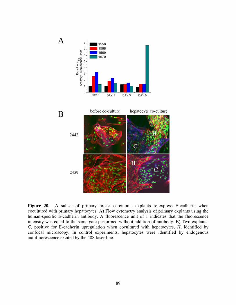
Figure 20. A subset of primary breast carcinoma explants re-express E-cadherin when cocultured with primary hepatocytes. A) Flow cytometry analysis of primary explants using the human-specific E-cadherin antibody. A fluorescence unit of 1 indicates that the fluorescence intensity was equal to the same gate performed without addition of antibody. B) Two explants, C, positive for E-cadherin upregulation when cocultured with hepatocytes, H, identified by confocal microscopy. In control experiments, hepatocytes were identified by endogenous autofluorescence excited by the 488-laser line.
89

4.4 DISCUSSION
Carcinomas are generally considered to undergo an epithelial to mesenchymal-like transition
(EMT) as a prelude to dissemination. The key marker for this phenotypic shift is the
downregulation of E-cadherin, a cell-adhesion molecule whose normal functioning is vital to the
maintenance of epithelial tissue integrity and whose loss enables tumor cells to disseminate from
the primary masszInterestingly E-cadherin expression has been noted in distant metastases of
primary tumors suggesting a possible survival advantage as is noted during development with the
reverse mesenchymal to epithelial transition (Larue and Bellacosa 2005). It is currently unknown
whether these E-cadherin-positive tumor cells disseminate from the primary tumor and form
secondary metastatic lesions, or if E-cadherin-negative cells regain E-cadherin expression in
response to the ectopic environment. The present study provides proof-of-principle that, at least
in the presence of hepatocytes, E-cadherin-negative invasive breast cancer cells undergo a
mesenchymal to epithelial reverting transtion (MErT) secondary to loss of E-cadherin promoter
hypermethylation, and that the hepatocyte-ligated E-cadherin on tumor cells can activate
canonical survival pathways in the cancer cells.
E-cadherin is downregulated in carcinomas predominantly by epigenetic
processes such as receptor tyrosine kinase signaling and promoter methylation (Becker, Atkinson
et al. 1994; Kanai, Oda et al. 1994; Risinger, Berchuck et al. 1994). This is unusual among tumor
suppressor in which loss or mutation appears to be the rule. However, it does allow for reversion
of this phenotypic change. In MDA-MB-231 cells, that are representative of invasive breast
90
cancers and in which E-cadherin silencing is due to promoter hypermethylation (Graff, Herman
et al. 1995), the CpG islands in the promoter region most proximal to the E-cadherin initiation
site are fully methylated, and this exerts a profound effect on mesenchymal nature, as
demethylation of these islands by a chemical agent causes re-expression of E-cadherin and loss
of invasive ability (Nam, Ino et al. 2004). As we had previously found that co-culture of invasive
prostate cancer cells with primary hepatocytes allowed stable re-expression of E-cadherin and
that re-expressed E-cadherin participated in a cell-cell adhesive ligation in E-cadherin on
hepatocytes (Yates, Shepard et al. 2007), we queried whether this phenomenon was relevant to
breast cancer cells. The primary distinction is that in the prostate cancer cells, E-cadherin was
downregulated only by receptor tyrosine kinase signaling (Yates et al., 2005), while in breast
cancer there is also methylation induced silencing of E-cadherin (Graff et al, 1995). Indeed,
culturing of MDA-MB-231 cells with primary hepatocytes resulted in lack of methylation of the
E-cadherin promoter and expression of E-cadherin message and protein, thought at a longer time
course than the prostate cancer cells. We observed that the loss of methylation was dependent on
the proliferation of the cancer cells. This re-expression of E-cadherin on the surface requires also
the secondary step of inhibition of receptor tyrosine kinase signaling, as hepatocyte coculture-
induced E-cadherin compartmentalized to perinuclear organelles, with mass relocation to the
membrane only upon further treatment with a tyrosine kinase inhibitor.
The consequence of this E-cadherin transcriptional upregulation is to allow for
additional signaling from the surface. Using the MCF7 cells that retain E-cadherin expresion and
epithelial characteristics (Soule, Vazguez et al. 1973), E-cadherin-mediated heterotypic
interactions with hepatocytes are functional, inducing plaque formation with Arp2/3
colocalization, which had previously only been shown to occur in epithelial homotypic
91

interactions. The ability of E-cadherin on MCF7 cells to engage in a heterotypic interaction with
hepatocytes was also documented in our centrifugal studies, the results of which showed that the
E-cadherin ligation between MCF7 cells and hepatocytes was force-bearing, which is consistent
with previous homotypical E-cadherin interaction and our own Arp2/3 localization experiments
(Angres, Barth et al. 1996). Finally, we saw that ligation of E-cadherin between MCF7 cells and
hepatocytes could transduce activation of both Erk-MAPK and Akt pathways with independent
kinetics. These signaling pathways were also actuated in MDA-MB-231 cells after reversion to
E-cadherin expression by coculture with hepatocytes. Though cadherin-mediated heterotypic
interactions have been previously noted in cells made to express various cadherins, to our
knowledge this is the first line of evidence that has shown cancer cells using heterotypic
interactions to activate canonical pathways which may very well determine their fate in a foreign
in vitro environment.
The foundation of our findings rest on the epigenetic reversion we observe when
breast cancer cells are co-cultured with primary hepatocytes. The epigenetic status of the primary
tumor and disseminated metastases is most likely important, since primary tumors that have high
E-cadherin levels have very little systemic disease (Goldstein 2002), suggesting that the
epigenetic reversion at distant secondary sites is important. These observations in connection
with clinical observations suggest that E-cadherin positive cells do not leave the tumor, but
actually regain the protein at distant sites. In addition, a similar phenomenon has been observed
in human tumor cells when cultured in either spheroid or transwell invasion chambers, during
which E-cadherin promoter methylation is unstable during in vitro simulation of metastatic
progression (Graff, Gabrielson et al. 2000). Our study brings those previous studies full circle
with relevant parenchymal cell interaction and physiologically relevant pathway activation. We
92

found it especially interesting that the loss of methylation of the E-cadherin promoter in MDA-
MB-231 cells was dependent on proliferation of the cancer cells. This dependency on
proliferation suggests a passive loss of methylation of the E-cadherin promoter region, in which
daughter strands do not participate in the maintenance methylation to which other methylated
CpG islands are subjected after DNA replication. Our cursory observations of doubling times of
MDA-MB-231 cells suggest that these cells undergo about three rounds of replication in about 6
days (unpublished data). A passive process would result in non-methylation of 87.5% of the
alleles in three rounds of replication; this is consistent with our observations of a lack of
amplification of methylated loci after 6 days of culture. PCR bias must be taken into account in
that the geometric progression of the reaction will favor the template of higher frequency in the
reaction, so the complete absence of methylated E-cadherin alleles may not be literal. In
addition, it has been demonstrated that suboptimal melting temperature may also contribute bias
to nonmethylated alleles. To ensure the efficacy of these results, we used a commercially
marketed kit to evaluate the status of the E-cadherin promoter. To our knowledge, we are the
first group to demonstrate that normal primary hepatocytes drive the demethylation of the E-
cadherin promoter in invasive cancer cells.
Our in vitro data is consistent with the storyline that cancer cells respond in a
series of tightly woven sequential steps to adapt in foreign environments. There are several
possible outcomes or combinations of outcomes after a cell extravasates into a metastatic target
tissue: 1) a single cell can succumb to metastatic inefficiencies, leading to necrosis or apoptosis;
2) dormancy has been reported, which may result in short-term remission with long-term
recurrence; or 3) the cell could receive either a mitogenic or motogenic signal. We speculate with
our current data that the re-expression of E-cadherin in invasive cancer cells may act to counter
93

apoptosis and either promote long-term malignant recurrence by cloaking the cancer cell with
epithelioid-like characteristics, or re-expressed E-cadherin may act to transmit either mitogenic
or motogenic signals resulting in proliferation and invasion of the target tissue. Indeed, the
downstream proliferatory and migratory pathways of Erk-MAPK as well as the anti-apoptotic
effects of Akt are well documented (Torii, Yamamoto et al. 2006; Dillon, White et al. 2007).
Further supporting this are quantitative studies that show during the phases of formation and
development of micrometastases in secondary organs, cells are particularly vulnerable to
apoptosis (Luzzi, MacDonald et al. 1998), yet it is still unclear what gives metastatic cells
competency to survive this phase. In the context of the cadherin-switch, evidence that E-
cadherin, and not N-cadherin, can modulate EGFR may play a role (Fedor-Chaiken, Hein et al.
2003). E-cadherin re-expression has been speculated to play a central role in establishing
metastases (Park, Karesen et al. 2007). In developmental mesenchymal to epithelial transition
associated with the dorsal-lateral pathway dissemination from the neural crest, E-cadherin re-
expression may enable melanoblasts to degrade the basement membrane locally by
metalloproteinase activity, as the cytoplasmic domain of E-cadherin can induce the expression of
stromelysin-1 (Delmas, Pla et al. 1999). This is relevant because it has been considered that
cancer is merely an emulation of development-associated pathways. Finally, E-cadherin-
dependent intercellular adhesion enhances chemoresistance (Nakamura, Kato et al. 2003). This
line of data illustrated the chemo-protectant effects of E-cadherin, which is very suggestive with
our result of canonical pathway activation. Collectively, we see the picture emerging of E-
cadherin re-expression occurring during the micrometastatic stages of cancer formation in
ectopic tissues, which not only allows cancer cells to evade metastatic inefficiencies and survive
94

in the foreign microenvironment, but re-expressed E-cadherin may also contribute to
pharmaceutically refractive disease.
We demonstrate in this study that E-cadherin demethylation in breast cancer cells is
modulated by co-culture with primary hepatocytes, and the protein expression is dependent on
proliferation of the cancer cells. We have implicated that a gene-specific program is executed in
the cellular cohesion machinery during co-culture, and E-cadherin is targeted preferentially
converse to a global hypomethylation program. Finally, E-cadherin ligation activates canonical
pathways in the cancer cells including the MAPK and Akt pathways, which may contribute to
either the mitogeneis, motogenesis, or survival in metastasized cells; these different outcomes
may be controlled by subtle regulatory factors such as cadherin affinity and clustering that
preceeds activation of specific signaling cascades. The search for therapeutic targets that act on
molecules specific to the process of metastatic colonization of distant sites, rather than reliance
on systemic cytotoxic treatments, is ongoing. Upon further investigation of these mechanisms, E-
cadherin may emerge as a molecule that is tumor-suppressant at the primary site but oncogenic at
distant metastases.
4.5 MATERIALS AND METHODS
4.5.1 Xenografts
The Institutional Animal Care and Use Committee at the Veterans Affairs Hospital in Pittsburgh
approved all animal procedures. Experiments were performed in 8 week old female athymic
nude mice. One million MDA-MB-231 cells were injected into the right mammary fat pad;
95

injection vehicle was the culture medium (0.2 mL/site). Mice were sacrificed 4-5 weeks after
tumor cell implantation and the primary xenograft and lungs removed.
4.5.2 Cell culture and co-culture
Human MDA-MB-231 and MCF7 cells were cultured as described previously (Kassis,
Moellinger et al. 1999). Primary rat hepatocytes were isolated and cultured as described
previously (Rosenberg, Strom et al. 1982; Yates, Shepard et al. 2007). Cancer cells were
separated from hepatocytes using FACS.
4.5.3 Immunohistochemistry
Xenograft and other harvested tissues were fixed in 4% buffered formalin and 4μm thick paraffin
sections underwent antigen retrieval for 5 min in 95°C 10mM citrate solution in preparation for
H&E and immunochemistry. With the use of the Mouse on Mouse Kit (Vector Labs,
Berlingame, CA), positive labeling was confirmed by comparing serial sections incubated with
the primary human-specific E-cadherin antibody (67A4 1:100; Santa Cruz Biotechnology, Santa
Cruz, CA) or the biotinylated secondary antibody alone. Labeling was visualized with the
Vectastain Elite kit (Vector Labs).
96

4.5.4 SiRNA
Silencer Validated siRNA (#161135, Ambion, Austin, TX) was transfected using Lipofectamine
2000 (Invitrogen, Carlsbad, CA). Cells were gorwn in antibiotic-free media till 60% confluent
and transfected with siRNA for 24h. The final concentration of siRNA in the experiment was
100nM. Cells were allowed to recover in media with antibiotics for 24h before further
experimentation. The control GFP DNA (5’-ACCCGCGCCGAGGUGAAGTT-3’, IDT, Skokie,
IL) was that from another species.
4.5.5 Real-time quantitative PCR
RNA was isolated from hepatocyte-cancer cell co-cultures with the PureYield RNA Midiprep
System (Promega, Madison, WI). cDNA was obtained with High Capacity cDNA RT Kit
(Applied BioSystems, Foster City, CA). TaqMan Gene Expression Assay Hs00170423_A1
CDHI probe was obtained from Applied Biosystems (Foster City, CA). Amplification and
analysis in quadruplicate was run in an Applied Biosystems 7500 Real-Time PCR System.
Relative values were normalized by using GAPDH levels as a reference using TaqMan Pre-
Developed Human GAPDH Assay Reagent by Applied Biosystems.
4.5.6 Methylation specific PCR
DNA was isolated from co-culture using the DNeasy Blood and Tissue Kit (Qiagen, Velencia,
CA). 500ng of isolated DNA was subjected to bisulfite treatment using the EZ DNA Methylation
97

Gold Kit (Zymo, San Diego, CA) per the manufacturer’s specifications. MSP was performed
using the CpG WIZ E-cadherin Amplification Kit per the manufacturer’s instructions (Millipore,
Temecula, CA). MSP of H19 after bisulfite conversion was performed using the following
primers: F 5’-TTA TAA AAT CGA AAA TTA CGC GCG A-3’ R 5’-TTT TAG ATG ATT TTT
GTG AAT TTT-3’. Cycling conditions were 95 °C for 15 min, 35 cycles of 94°C for 1 min, 55°C
for 1 min, and 72°C for 1 min with a final extension of 5 min at 72 °C. The reactions were
performed using Platinum Taq (Invitrogen).
4.5.7 Westernblots, flow cytometry, and immunofluorescence
Proteins were resolved on 7.5% SDS-PAGE and detected by Western analysis. Mouse-
monoclonal human-specific E-cadherin (67A4 1:1000), pAkt1/2/3 (Ser473 1:1000),
pAkt1/2/3(Thr308 1:1000), pErk (E-4 1:1000) were purchased from Santa Cruz Biotech (Santa
Cruz, CA). Flow cytometry was performed using a DakoCytomation CyAN High Speed
Analyzer. Co-cultures were non-ezymatically dissociated from the culture plates and vortexed
into a single-cell suspension. The cells were fixed in 2% Paraformaldehyde for 30 minutes,
permeabilized with 1% Triton for 3 minutes, and incubated with a PI-conjugated E-cadherin
antibody (67A4) for 30 minutes. The mixed hepatocyte-cancer cell suspension was gated as to
exclude hepatocytes using the appropriate SSC/FSC parameters. Data was collected on at least
106 cells in the appropriate SSC/FSC region. Immunofluorescence was performed similarly to
flow cytometry, except primary antibody incubation was performed overnight. Arp3 (G-15
1:100), E-cadherin (67A4 1:100), beta-catenin (C-18 1:100) were purchased from Santa Cruz
Biotech and Alexa 488-phalloidin was purchased from Molecular Probes (Carlsbad, CA).
Secondary fluorphores included Alexa Fluor-488, -594, and -674 (Molecular Probes).
98

Visualization was performed on an Olympus Fluoview 1000 confocal microscope (Olympus,
Center Valley, PA). Histograms of acquired pictures were widened equally on all comparable
images.
4.5.8 Centrifugal assay for fluorescent cell adhesion (CAFCA)
This assay is a modification of the McClay and Giacolmello assays (McClay, Wessel et al.
1981). Cancer cells were non-enzymatically dissociated and labelled with 5 M Calcein AM
(Molecular Probes, Carlsbad, CA, USA). Labelled cancer cells were seeded at a density of 42
000 cells well-1 in 96-well plates containing a densely confluent hepatocyte monolayer. The
plates were centrifuged for <60s at 50g to pellet the cancer cells onto the hepatic monolayer, then
incubated at 37°C. At defined times, the plates were inverted and centrifuged at 600g for 5 min
and then gently washed to remove unbound cells from the hepatocyte monolayer. Fluorescence
was measured with a 494/517 bandpass filter set-up from the bottom of the plate by a TECAN
Spectra-Fluor plate fluorometer. Absolute emission measurements were background subtracted.
4.5.9 Hepatocyte membrane assays
Heptocyte membranes were isolated as described (Forster and Kaltschmidt 1999). Culture plates
were coated with poly-L-lysine per the manufacturer (mol wt 150000-300000 Sigma, St. Louis,
MO). Hepatocyte membranes (2 mg protein/cm2) were passively adsorbed onto 6-well culture
plates using poly-L-lysine (Sigma) for 10 minutes. MCF7 and MDA-MB-231 cells were
quiesced in serum-free media for 3 hours before phosphorylation measurements were taken by
99

Western blot or in vitro kinase assay. MCF7 or MDA-MB-231 cells were seeded at 19E3
cells/cm2 onto 6-well plates coated with the membranes and briefly centrifuged at 50g for 1
minute to synchronize the cells with the membrane layer. At defined time-points, RIPA lysates
were taken and for in vitro kinase assay, total Erk and Akt proteins were immunoprecipitated
from the total protein using agarose conjugated Erk (K-23) and Akt1/2(N-19) antibodies, both
purchased from Santa Cruz Biotech. Kinase activity was assayed using the MBP 96-well Assay
Kit Chemiluminescence Detection from Upstate (Lake Placid, NY). Luminescence detection was
performed with either a Tecan SpectraFluor or a Tecan GENios Pro apparatus.
4.5.10 Primary explants
Polyclonal primary human tumor explants were obtained and cultured as previously reported
(Ochs, Fensterer et al. 2003). Immunofluorescence labeling was performed as above.
4.6 ACKNOWLEDGEMENTS
This work was supported by DoD Congressionally Directed Medical Research Program in Breast
Cancer Merit Awards and a Pre-Doctoral Fellowship. The VA provided in-kind support for these
studies. We thank the Dr. David Geller lab for the rat hepatocytes and Precision Therapeutics for
primary breast cancer explants.
100

DISCUSSION
101
5.0 CARCINOMA DISSEMINATION FOLLOWS FROM AN EPITHELIAL-TO-
MESENCHYMAL-LIKE TRANSITION (EMT)
The major part of cancer morbidity and mortality results from metastatic colonization of distant
organs by carcinomas far from their original sites of development. In tumors of epithelial origin,
a change in architecture usually denotes the first signs of neoplastic transformation. While
neoplastic transformation is usually a precursor to a more motile phenotype, carcinomas are
distinguished from their normal counterparts by a loss of tissue and cell coupling (Guarino,
Rubino et al. 2007).
In untransformed tissues, the epithelial cells are polarized and hence the apical face of the
cell interacts with the luminal compartment, and the basolateral face of the cell interacts with the
stromal compartment. Epithelial cells are often arranged in sheets with adjacent cells tightly
connected via tight junctions and gap junctions. At the apical surface in most tissues, the
epithelial cells are in contact with body fluids that are either wholly or partly produced by these
same cells. These fluids contain biologically factors, growth factors in particular, that are
usually inert to the tissue as the receptors for such factors are sequestered on the basolateral
surfaces. The tight junctions, constructed upon homotypic binding of E-cadherin, limit the
access of the apical fluids to the basolateral spaces and surfaces and the underlying stromal
compartment. Distinct and compartmentalized interactions occur solely because of the
organized structure of both the cell and the tissue as a whole.
Upon neoplastic transformation, this orderly arrangement is lost. A host of genetic and
epigenetic changes occur that lead to degradation and eventual loss of the tight junctions. What
102

follows contributes fundamentally to the change from neoplasm to carcinoma in situ. First, the
epithelial cells are now exposed to potential autocrine factors, as the localization of ligand
production and receptor expression are no longer segregated. Second, the epithelial cells are
released from physical restraints with loss of contact inhibition, allowing for cell movement and
even proliferation. And third, the stromal elements are now exposed to many epithelially derived
bioactive factors promoting a stromal response. The latter event may change the profile of
signals that derive from the stromal compartment as the composition and architectural profile of
the stromal elements change. The net sum of these tissue alterations are that the epithelial cells
are directed to assume a less differentiated state that converges towards the mesenchymal
phenotype, characterized by motility and proliferation with amoeboidal architecture that has
limited direct cell-cell communication. This is the so-called epithelial-to-mesenchymal-like
transition (EMT) of carcinomas (Peinado, Portillo et al. 2004; Guarino, Rubino et al. 2007).
Carcinoma EMT is distinguished from normal developmental EMT by the fact that the
cells do not actually reassume a fully functioning physiological role similar to an epithelially
derived cell (Peinado, Portillo et al. 2004). Rather, the attributes of the cells converge on those
noted in stromal cells, and the normal epithelial markers and functions are lost indefinitely.
Thus, the first steps in the dedifferentiation process of carcinoma progression is an EMT
process very similar to the transformation of primitive epithelial that form the primitive
trophoblastic germ layers in normal development. Despite the long-standing recognition of this
carcinoma EMT, there is no accepted molecular definition of what constitutes this process
(Peinado, Portillo et al. 2004; Thompson, Newgreen et al. 2005; Lee, Dedhar et al. 2006). The
vastly heterogeneous etiology of cancer probably contributes to this ambiguity. A picture is
emerging of common molecular findings that account for both the histopathological picture and
103

the cell biology findings. At the minimum, the carcinoma EMT is defined by a loss of normal
epithelial architecture, namely both the homogeneous physiological symmetry of the cell, cell-
cell contacts, and communications dependent on that architecture. At the molecular level, this is
reflected by downregulation or loss of specific cytokeratins and E-cadherin. The latter is of
particular importance, because it can be viewed as both a cause and effect of the EMT.
Loss of E-cadherin disrupts not only cell-cell junctions but also allows for loss of the
normal organ architecture. The transformed cell’s endowed properties allow them to move with
many more degrees of freedom within the epithelial layer, as they are no longer constrained
within a functional syncytium (Friedl, Zanker et al. 1998). This is a histopathological hallmark
of neoplastic transformation. Furthermore, the absence of apical-basal barriers between the cells
enables soluble factors secreted by the cells to reach cognate receptors normally segregated on
the basolateral surfaces. This autocrine signaling often further reinforces the EMT phenotype
because stimulation of most receptors with intrinsic tyrosine kinase activity, particularly the
ubiquitous EGF receptor system, drive E-cadherin downregulation and epithelial
dedifferentiation (Roura, Miravet et al. 1999; Cavallaro, Schaffhauser et al. 2002). Lastly, the E-
cadherin-based cell connection can control global cell signaling, as the catenins that anchor these
plaques possess signaling properties dependent upon whether that are in soluble or insoluble
complexes (Aberle, Schwartz et al. 1996; Bracke, Van Roy et al. 1996). At an albeit simplistic
level, the E-cadherin protein complex at the cell membrane sequesters α-, β- and p120-catenins
in epithelial cells and prevents transformative effects on the cellular cytoskeleton and
transcriptome. The extent of the global control on cell phenotype exerted by E-cadherin is
inherent to its designation as a tumor suppressor; forced re-expression of E-cadherin in invasive
carcinoma cells can revert the neoplastic phenotype (Chen and Obrink 1991; Frixen, Behrens et
104

al. 1991). For these reasons, E-cadherin expression on the cell surface has emerged as a
molecular hallmark of the carcinoma EMT.
105

6.0 METASTATIC SEEDING IS RATE-LIMITING FOR METASTATIC
COLONIZATION OF DISTANT ORGANS
Key to carcinoma pathology is the ability of carcinoma cells to survive and thrive in ectopic
tissues after their escape from the primary tumor mass. As described in Chapter 5, the carcinoma
EMT is critical to this initial escape by enabling individual cell migration and invasion through
barrier matrices (Wells 2000). In order to establish metastatic foci, similar transition-like events
occur (Luzzi, MacDonald et al. 1998). First, the cells arrest in the dissemination conduits
(lymph and blood vessels) by size mismatch between the tumor cells and the diameter of
capillaries or because of obstruction of rolling immune cells. The carcinoma cells then must
penetrate between the endothelial cells and migrate into the ectopic tissue. This appears to be
relatively efficient, as cells extravasate at high frequencies (Chambers, Naumov et al. 2000).
The primary rate-limiting step after initial EMT and dissemination is the ability of invasive cells
to successfully colonize the ectopic site.
Many years ago it was appreciated that tumor cells disseminate preferentially to ectopic
tissues based mainly on the ability of that host tissue to support the survival and proliferation of
those cells (Fidler 2001). This is referred to as the ‘seed and soil’ hypothesis. The stromal
component of different tissues and organs produce and present overlapping but distinct matrix
components and secreted factors that serve to hospitably accommodate the local cells.
Disseminated tumor cells must find a host environment that provides the necessary signals or
106
these cells must provide the missing factors in an autocrine fashion or other manner that results
from transformation. Some of these missing factors might be replaced by genetic mutations that
alleviate a particular requirement or are provided by the autocrine signaling inherent in the
carcinoma EMT. Still the transformation rarely allows for proliferation outside of a hospitable
micro-environment (Bissell 2007).
The survival of these carcinoma cells in ectopic tissue is complicated by the question of
tumor cell dormancy. While tumor masses need to approach a billion cells to be detectable as
metastases, this should occur relatively quickly if the tumor cells continue to proliferate at the
inherent cell-doubling rate quantifiable in most highly aggressive primary tumors. However, in
many tumors, metastases appear only years after removal of the primary mass. During this time,
the metastatic foci must remain minimal, being either in a state of balanced proliferation-necrosis
or as a dormant cancer cell. The key difference between these two options is that in the former,
the local milieu must present a full complement of factors for cell proliferation plus signals for
death, whereas in the latter, only a partial complement of factors needs to be present to enable
cell survival at a lower metabolic load. The question of ectopic site preference, survival, and
proliferation reduces to that of the ectopic microenvironment. While there are nascent efforts to
fully catalogue tumor microenvironments components, qualitative aspects can be deduced from
in situ studies. What has been surprising is that many metastatic carcinomas appear more
differentiated than the primary tumor from which they derive. In fact, it is well established that
many breast cancer metastases to the liver seem to recreate the hepatic architecture without a
desmoplastic reaction (Stessels, Van den Eynden et al. 2004). This is inconsistent with the
prevailing view that carcinomas continue down a dedifferentiation EMT cascade as they get
107

more aggressive and disseminate. Rather these reports suggest that the metastatic foci may be
more epithelialoid than the primary site.
Examining these metastases with immunohistochemistry confirmed this more
differentiated phenotypes in many carcinomas. Interestingly, a number of reports related E-
cadherin membrane staining in the metastases but not in the primary carcinoma sites (Putz,
Witter et al. 1999; Rubin MA 2001; Kowalski, Rubin et al. 2003). It has been reported in vitro
that cancer cells expressing E-cadherin are chemoresistant (Green 2004 ; Nakamura 2003). If E-
cadherin re-expression is necessary for metastatic dormancy, then these micrometastases would
escape chemotherapies that actively target proliferating cells, as is the case for cancer relapse
many years after the primary tumor has been eliminated.
108
7.0 E-CADHERIN IN THE MESENCHYMAL-TO-EPITHELIAL REVERTING
TRANSITION
The finding of membrane-expressed E-cadherin in carcinoma metastases raises two possibilities.
The first is that the distant metastases formed from E-cadherin-positive cancer cells that
disseminated from the primary mass. Even though many primaries are scored as E-cadherin-
negative (either the protein is silenced completely or robustly downregulated), this possibility
cannot be excluded as the larger primary tumors examined show heterogeneity of expression
with some areas of primary tumor expressing the epithelial marker. Still, the clinicopathological
correlation between low to absent E-cadherin and tumor dissemination is strong (Birchmeier and
Behrens 1994; Cavallaro, Schaffhauser et al. 2002), and the cellular mechanisms supporting this
model are logical and repeatedly demonstrated. Carcinoma cells that express E-cadherin show
limited migration and invasiveness in vitro, and do not form distant metastatic tumors in animal
models. However, given the rare nature of metastases despite relatively frequent shedding of
cells from the primary mass (Cristofanilli, Budd et al. 2004), these metastases could arise from
the unusual escape of an E-cadherin-positive cell in the primary mass, if E-cadherin expression
provided a survival and proliferation advantage. This question in its current form cannot be
addressed by human tumor surveys because of technical complications, but rather requires
experimental probing using clonal carcinoma cell populations. In what appears to be the first
report directly addressing this question, this thesis describes how the highly aggressive and E-
109

cadherin-negative MDA-MB-231 breast carcinoma cell line forms lung metastases from
orthotopic primary tumors in the inguinal mammary fat pads of mice; these metastases express
E-cadherin. While this is far from conclusive, it does present a proof a principle that E-cadherin-
expressing metastases might arise from E-cadherin-negative cells. Re-expression of E-cadherin
in the metastatic site, therefore, represents the second possibility. While this is consistent with
the histopathological correlations, E-cadherin re-expression would be the result of tumor cell
plasticity, that of reverting the carcinoma associated EMT that has not been demonstrated to-
date. The nature of E-cadherin downregulation mechanisms suggests how this could be
accomplished.
E-cadherin surface expression is downregulated by two known mechanisms, functioning
separately at the post-translational and the transcriptional levels. Tyrosine kinase signaling,
noted both in response to various growth factors and during neoplastic progression, can
downregulate E-cadherin secondary to phosphorylation of β-catenin on the E-cadherin complex
(Reynolds, Daniel et al. 1994; Hazan and Norton 1998). EGF and HGF/scatter factor lead to
epithelial cell migration away from cohesive masses secondary to E-cadherin-complex
dissocation (Birchmeier and Behrens 1994; Miura, Nishimura et al. 2001; Morkel, Huelsken et
al. 2003). The endocytosis of E-cadherin after dissociation results in E-cadherin internalization
and degradation. This may function in carcinomas, almost all of which have autocrine growth
factor signaling loops most often that via the EGF receptor (Wells 2000). Previous reports have
found in prostate carcinoma lines that inhibition of this autocrine EGF receptor loop (and likely
the EGFR-induced HGF/c-met autocrine loop (Mamoune, Kassis et al. 2004)), either by direct
disruption of the signaling loop or by trans-attenuation, results in E-cadherin re-expression and
110

cell-cell cohesion (Yates, Wells et al. 2005; Yates, Shepard et al. 2007; Yates, Shepard et al.
2007).
Thus, post-translational E-cadherin downregulation represents an available target for
counter-regulation by other factors that might be present in the metastatic microenvironment. In
most carcinomas, E-cadherin appears to be shut off at the transcriptional level by promoter
hypermethylation (Graff, Herman et al. 1995; Jones and Baylin 2002; Strathdee and Brown
2002). This mode of generating a null phenotype differs from other tumor suppressors that are
usually victims of other genetic anomalies such as deletions, mutations, or frame-shifts of the
coding DNA. While these latter mechanisms are irreversible by their nature, promoter
hypermethylation is readily reversible if only by maintenance methylation of the CpG site after
daughter strand synthesis. One distinction between normally irreversible tumor suppressors from
the E-cadherin tumor suppressor is that loss of the former often occurs early in neoplastic
transformation, whereas E-cadherin is relevant throughout many of the sequential steps that
result in metastatic colonization of secondary tissues. It has been noted that E-cadherin promoter
methylation is unstable (Graff, Gabrielson et al. 2000). This thesis documents that E-cadherin
promoter methylation can be selectively lost in breast cancer cells when proliferating in the
presence of normal hepatocytes. Thus, there exist signals in the hepatic microenvironment that
undo the epigenetic silencing of E-cadherin by promoter methylation.
The above discussion begs the question of what selective advantage does the re-
expression of E-cadherin impart? Nascent investigation beyond this thesis is currently answering
that question. The speculation can be easily cast that E-cadherin re-expression provides a
survival advantage for these cells in a challenging ectopic environment.
111

Two key pathways activated upon E-cadherin binding are the survival-associated ERK
MAP kinase and Akt/PKB cascades (Pece, Chiariello et al. 1999; Pece and Gutkind 2000).
Complementing the seed-and-soil hypothesis, functional E-cadherin ligation, either between the
secondary parenchymal-cancer cells or cancer cell-cancer cell, would provide necessary signals
to endure in the ectopic soil. At the start of metastatic seeding, this requires cell-heterotypic E-
cadherin binding, which is reported for the first time in this thesis. Later as the metastatic cells
proliferate, E-cadherin ligation may occur between carcinoma cells. To support the possibility of
carcinoma-parenchymal binding via E-cadherins, histopathological analyses of many tumors
suggest close associations between metastatic carcinoma cells and the neighboring parenchymal
cells (Putz, Witter et al. 1999; Rubin MA 2001; Kowalski, Rubin et al. 2003). Furthermore, in
an ex vivo model of carcinoma metastasis to the liver, close connections are observed on the
ultrastructural level (Yates, C et al. 2007a). Also, when prostate carcinoma cells are induced to
re-express E-cadherin, both DU-145 and PC3 cells can form heterotypic adhesions with rat
hepatocytes (Yates, C C et al. 2007b). These data provide proof of principle that carcinoma
cells may re-express E-cadherin in response to the ectopic organ micro-environment so as to
establish connections with the resident, non-neoplastic epithelial cells.
112

8.0 A MODEL FOR THE MESENCHYMAL-EPITHELIAL REVERTING
TRANSITION
The following is a speculative model for the mesenchymal-epithelial reverting transition, with a
focus on the results present in the thesis. It encompasses a model of tumor cell plasticity with E-
cadherin playing a central role in the phenotypic differentiation in both the primary and
metastatic sites.
At the primary site, ill-defined genetic and epigenetic changes lead to E-cadherin
downregulation, via receptor tyrosine kinase signaling and/or, as is the case with the majority of
invasive cancer, promoter hypermethylation (Figure 21, I). The loss of adherens and tight
junctions in those cells that progress from neoplastic to carcinoma in situ allows physical
freedom to move and reorganize, as well as the biological signals derived from autocrine
signaling. It is unknown whether the loss of the epithelial barrier further alters the cancer field
stroma in a manner that accelerates EMT.
Once free of the physical tethering and architectural restraints imposed by E-cadherin
binding, the now mesenchymal-like carcinoma cells can actively migrate through barriers and
enter vascular conduits for dissemination to distant sites. While the cell must counter many
metastatic inefficiencies during travel to distant sites of the body, the major impediment to
metastatic growth appears to be the survival and growth (albeit often delayed or slow) in the
ectopic site. This is likely due to the incomplete match of factors and signals in different tissues.
113

The literature reports that many metastatic carcinoma foci do express E-cadherin, whose
downregulation is considered critical if not required for dissemination. This suggests that a
subset of the disseminated carcinoma cells receive signals from the ectopic organ
microenvironment to revert the EMT process at least partially to enable E-cadherin re-expression
(Figure 21, II). As E-cadherin binding elicits canonical survival signals and prevents anoikis
(Conacci-Sorrell, Simcha et al. 2003; Reddy, Liu et al. 2005; Kang, Jenabi et al. 2007), the
selective advantage is obvious. This thesis documents the process that is the so-called
mesenchymal-epithelial reverting transition, or MErT.
Further investigations are required to establish the reverting transition can occur in ex
vivo and in vivo model systems, as important for de novo disseminating cancers. Future
investigations would include the abrogation and rescue of E-cadherin re-expression to limit
metastatic seeding and also inducible E-cadherin re-expression to counteract metastatic
inefficiency.
Still, the advantages of E-cadherin re-expression and seeming redifferentiation must be
explored. The reversion of the general carcinoma cell phenotype to a less aggressive, less
proliferative cell seems counterintuitive particularly in regards to the picture that emerges at the
primary site. While activation of ERK-MAP kinase and Akt/PKB signaling pathways promote
survival, these pathways are even more strongly elicited by other factors and external signals,
without the suppression of proliferation that ensues from E-cadherin adhesions (Simcha et al.
1996) (Figure 21, III). The thesis herein does not answer those questions, but one can speculate
that the combination of survival signals with limited proliferation and lowered metabolic
requirements are precisely the keys to metastatic seeding. By and large, fast growing cells are
most susceptible to death signals whether due to DNA damage, limited nutrients/metabolic
114

inefficiency, absent pro-survival signals, or apoptotic signals. It is well recognized in oncology
that the slowest growing tumors generally are the most resistant to standard chemotherapies. In
the ectopic metastatic environment, tumor cells not only carry damaged DNA but also face an
absence of external factors, as reflected in the seed and soil hypothesis (Fidler 2003). It is also
possible that entry of these foreign cells will elicit a localized inflammatory reaction, with the
production of pro-death signals. Thus, not only would activation of the pro-survival ERK-MAP
kinase and Akt/PKB pathways protect the cancer cell, but also proliferation and metabolic
suppression would provide added survival advantages possibly equilibrated by secretion of
TGFβ in the damaged liver (Figure 21, IV) This speculation can be tested in models of
chemotherapeutic and apoptotic challenge.
Lastly, one must consider the implications of this proposed carcinoma-related MErT.
Currently, E-cadherin is categorized as a tumor suppressor due to its protective role against the
carcinoma-related EMT at the primary site. However, interventions to prevent E-cadherin
downregulation may perversely promote seeding of disseminated cells as the re-expression
phenomena appears to be highly inefficient in the natural course of cancer. Thus, any E-cadherin
targeted therapies must be highly cognizant of carcinoma progression dynamics. Second, the
formation of E-cadherin ligation to the metastatic target organ parenchyma may result in
dormancy at the micrometastatic stage. This would result not only in resistance to chemotherapy
with tumor re-emergence, but also have implications for what may lead to delayed relapses. It is
tempting to speculate that if the proposed MErT leads to dormancy of early micrometastases,
then a secondary insult to the local environment may be what induces renewed carcinoma cell
proliferation and escape from E-cadherin-mediated contact inhibition. This, along with other
many questions raised by this proposed MErT, awaits further experimentation.
115

Figure 21. A model for the mesenchymal-epithelial reverting transition (MErT). I, The normal mammary epithelium (pink), which expresses E-cadherin (green bars), can become carcinoma in situ by EMT (red), which involves hypermethylation of the E-cadherin promoter. The cancer cell disseminates to the liver, extravasates from the sinusoid, and invades the hepatic plates. II, In the liver, cancer cells are juxtaposed to hepatocytes and in contact with the basement-like membrane in the liver parenchyma. An unknown signal causes E-cadherin promoter hypomethylation, which allows E-cadherin expression. EGFR ligands, such as EGF and TGFα, either from cancer cell autocrine loops or secreted by the hepatocytes, are present in the microenvironment. III, Sustained tyrosine kinase activation causes proliferation, while any E-cadherin participating in trans-ligation with hepatocyte E-cadherin transduces EGFR-dependent Erk-MAPK activation that remains close to the cell surface resulting in motility/invasion. A primary response to the damaged hepatic epithelium may result in release of TGFβ, which results in a stop-proliferation signal. IV, In a dormant micrometatasis, an equilibrium has been reached between E-cadherin signaling and TGFβ signaling. The cell has now undergone partial or complete MErT.
116

BIBLIOGRAPHY
Aberle, H., H. Schwartz, et al. (1996). "Cadherin-catenin complex: protein interactions and their implications for cadherin function." J Cell Biochem 61(4): 514-23.
ACS. (2007). "Learn about Breast Cancer." Retrieved September 29, 2007, from www.cancer.org.
Adams, C. L., Y. T. Chen, et al. (1998). "Mechanisms of epithelial cell-cell adhesion and cell compaction revealed by high-resolution tracking of E-cadherin-green fluorescent protein." J Cell Biol 142(4): 1105-19.
Angres, B., A. Barth, et al. (1996). "Mechanism for transition from initial to stable cell-cell adhesion: kinetic analysis of E-cadherin-mediated adhesion using a quantitative adhesion assay." J Cell Biol 134(2): 549-57.
Aono, S., S. Nakagawa, et al. (1999). "p120(ctn) acts as an inhibitory regulator of cadherin function in colon carcinoma cells." J Cell Biol 145(3): 551-62.
Bail, J. P., M. T. Foultier, et al. (1994). "Experimental model of liver metastases: adhesion and growth of cells on contact with endothelial or hepatocyte cell monolayer cultures." Res Exp Med (Berl) 194(1): 53-61.
Bates, S. E., E. M. Valverius, et al. (1990). "Expression of the transforming growth factor-alpha/epidermal growth factor receptor pathway in normal human breast epithelial cells." Endocrinology 126(1): 596-607.
Becker, K. F., M. J. Atkinson, et al. (1994). "E-cadherin gene mutations provide clues to diffuse type gastric carcinomas." Cancer Res 54(14): 3845-52.
Behrens, J., O. Lowrick, et al. (1991). "The E-cadherin promoter: functional analysis of a G.C-rich region and an epithelial cell-specific palindromic regulatory element." Proc Natl Acad Sci U S A 88(24): 11495-9.
Behrens, J., M. M. Mareel, et al. (1989). "Dissecting tumor cell invasion: epithelial cells acquire invasive properties after the loss of uvomorulin-mediated cell-cell adhesion." J Cell Biol 108(6): 2435-47.
Behrens, J., J. P. von Kries, et al. (1996). "Functional interaction of beta-catenin with the transcription factor LEF-1." Nature 382(6592): 638-42.
Birchmeier, W. and J. Behrens (1994). "Cadherin expression in carcinomas: role in the formation of cell junctions and the prevention of invasiveness." Biochim Biophys Acta 1198(1): 11-26.
117

Bissell, M. J. (2007). "Modelling molecular mechanisms of breast cancer and invasion: lessons from the normal gland." Biochem Soc Trans 35(Pt 1): 18-22.
Bodnar, R. J., C. C. Yates, et al. (2006). "IP-10 blocks vascular endothelial growth factor-induced endothelial cell motility and tube formation via inhibition of calpain." Circ Res 98(5): 617-25.
Bornfeldt, K. E., E. W. Raines, et al. (1994). "Insulin-like growth factor-I and platelet-derived growth factor-BB induce directed migration of human arterial smooth muscle cells via signaling pathways that are distinct from those of proliferation." J Clin Invest 93(3): 1266-74.
Bracke, M. E., F. M. Van Roy, et al. (1996). "The E-cadherin/catenin complex in invasion and metastasis." Curr Top Microbiol Immunol 213 (Pt 1): 123-61.
Brennan, K. R. and A. M. Brown (2004). "Wnt proteins in mammary development and cancer." J Mammary Gland Biol Neoplasia 9(2): 119-31.
Bryant, D. M. and J. L. Stow (2004). "The ins and outs of E-cadherin trafficking." Trends Cell Biol 14(8): 427-34.
Burdsal, C. A., C. H. Damsky, et al. (1993). "The role of E-cadherin and integrins in mesoderm differentiation and migration at the mammalian primitive streak." Development 118(3): 829-44.
Bussemakers, M. J., L. A. Giroldi, et al. (1994). "Transcriptional regulation of the human E-cadherin gene in human prostate cancer cell lines: characterization of the human E-cadherin gene promoter." Biochem Biophys Res Commun 203(2): 1284-90.
Cailleau, R., M. Olive, et al. (1978). "Long-term human breast carcinoma cell lines of metastatic origin: preliminary characterization." In Vitro 14(11): 911-5.
Cailleau, R., R. Young, et al. (1974). "Breast tumor cell lines from pleural effusions." J Natl Cancer Inst 53(3): 661-74.
Carpenter, G. (2000). "The EGF receptor: a nexus for trafficking and signaling." Bioessays 22(8): 697-707.
Carpenter, G. and Q. Ji (1999). "Phospholipase C-gamma as a signal-transducing element." Exp Cell Res 253(1): 15-24.
Cavallaro, U., B. Schaffhauser, et al. (2002). "Cadherins and the tumour progression: is it all in a switch?" Cancer Lett 176(2): 123-8.
Chambers, A. F., G. N. Naumov, et al. (2000). "Molecular biology of breast cancer metastasis. Clinical implications of experimental studies on metastatic inefficiency." Breast Cancer Res 2(6): 400-7.
Chen, P., K. Gupta, et al. (1994). "Cell movement elicited by epidermal growth factor receptor requires kinase and autophosphorylation but is separable from mitogenesis." J Cell Biol 124(4): 547-55.
Chen, P., J. E. Murphy-Ullrich, et al. (1996). "A role for gelsolin in actuating epidermal growth factor receptor-mediated cell motility." J Cell Biol 134(3): 689-98.
118

Chen, P., H. Xie, et al. (1994). "Epidermal growth factor receptor-mediated cell motility: phospholipase C activity is required, but mitogen-activated protein kinase activity is not sufficient for induced cell movement." J Cell Biol 127(3): 847-57.
Chen, P., H. Xie, et al. (1996). "Mitogenic signaling from the egf receptor is attenuated by a phospholipase C-gamma/protein kinase C feedback mechanism." Mol Biol Cell 7(6): 871-81.
Chen, W. C. and B. Obrink (1991). "Cell-cell contacts mediated by E-cadherin (uvomorulin) restrict invasive behavior of L-cells." J Cell Biol 114(2): 319-27.
Chen, Y. T., D. B. Stewart, et al. (1999). "Coupling assembly of the E-cadherin/beta-catenin complex to efficient endoplasmic reticulum exit and basal-lateral membrane targeting of E-cadherin in polarized MDCK cells." J Cell Biol 144(4): 687-99.
Claessens, F., L. Celis, et al. (1989). "Functional characterization of an androgen response element in the first intron of the C3(1) gene of prostatic binding protein." Biochem Biophys Res Commun 164(2): 833-40.
Conacci-Sorrell, M., I. Simcha, et al. (2003). "Autoregulation of E-cadherin expression by cadherin-cadherin interactions: the roles of beta-catenin signaling, Slug, and MAPK." J Cell Biol 163(4): 847-57.
Cristofanilli, M., G. T. Budd, et al. (2004). "Circulating tumor cells, disease progression, and survival in metastatic breast cancer." N Engl J Med 351(8): 781-91.
Das, P. M. and R. Singal (2004). "DNA methylation and cancer." J Clin Oncol 22(22): 4632-42.
Davis, M. A., R. C. Ireton, et al. (2003). "A core function for p120-catenin in cadherin turnover." J Cell Biol 163(3): 525-34.
Delmas, V., P. Pla, et al. (1999). "Expression of the cytoplasmic domain of E-cadherin induces precocious mammary epithelial alveolar formation and affects cell polarity and cell-matrix integrity." Dev Biol 216(2): 491-506.
Dillon, R. L., D. E. White, et al. (2007). "The phosphatidyl inositol 3-kinase signaling network: implications for human breast cancer." Oncogene 26(9): 1338-45.
Dittman, T., A. Husemann, et al. (2002). "Induction of cancer cell migration by epidermal growth factor is initiated by specific phosphorylation of tyrosine 1248 of c-erbB-2 receptor via EGFR." FASEB Journal 16(13): 1823-1825.
Duguay, D., R. A. Foty, et al. (2003). "Cadherin-mediated cell adhesion and tissue segregation: qualitative and quantitative determinants." Dev Biol 253(2): 309-23.
Edelman, G. M., W. J. Gallin, et al. (1983). "Early epochal maps of two different cell adhesion molecules." Proc Natl Acad Sci U S A 80(14): 4384-8.
Ennis, B. W., E. M. Valverius, et al. (1989). "Anti-epidermal growth factor receptor antibodies inhibit the autocrine-stimulated growth of MDA-468 human breast cancer cells." Mol Endocrinol 3(11): 1830-8.
Esteller, M. (2005). "Aberrant DNA methylation as a cancer-inducing mechanism." Annu Rev Pharmacol Toxicol 45: 629-56.
119

Esteller, M. (2005). "Dormant hypermethylated tumour suppressor genes: questions and answers." J Pathol 205(2): 172-80.
Fedor-Chaiken, M., P. W. Hein, et al. (2003). "E-cadherin binding modulates EGF receptor activation." Cell Commun Adhes 10(2): 105-18.
Fidler, I. J. (2001). "Seed and soil revisited: contribution of the organ microenvironment to cancer metastasis." Surg Oncol Clin N Am 10(2): 257-69, vii-viiii.
Fidler, I. J. (2003). "The pathogenesis of cancer metastasis: the 'seed and soil' hypothesis revisited." Nature Reviews - Cancer 3: 453-458.
Fisher, B. (1999). "From Halsted to prevention and beyond: advances in the management of breast cancer during the twentieth century." Eur J Cancer 35(14): 1963-73.
Forster, E. and C. Kaltschmidt (1999). "Improved protocol for a microsphere-adhesion assay on living tissue slices." Biotechniques 26(3): 466-8, 470, 472.
Foty, R. A. and M. S. Steinberg (2004). "Cadherin-mediated cell-cell adhesion and tissue segregation in relation to malignancy." Int J Dev Biol 48(5-6): 397-409.
Friedl, P., K. S. Zanker, et al. (1998). "Cell migration strategies in 3-D extracellular matrix: differences in morphology, cell matrix interactions, and integrin function." Microsc Res Tech 43(5): 369-78.
Friedlander, D. R., R. M. Mege, et al. (1989). "Cell sorting-out is modulated by both the specificity and amount of different cell adhesion molecules (CAMs) expressed on cell surfaces." Proc Natl Acad Sci U S A 86(18): 7043-7.
Frixen, U. H., J. Behrens, et al. (1991). "E-cadherin-mediated cell-cell adhesion prevents invasiveness of human carcinoma cells." J Cell Biol 113(1): 173-85.
Fujita, Y., G. Krause, et al. (2002). "Hakai, a c-Cbl-like protein, ubiquitinates and induces endocytosis of the E-cadherin complex." Nat Cell Biol 4(3): 222-31.
Giacomello, E., J. Neumayer, et al. (1999). "Centrifugal assay for fluorescence-based cell adhesion adapted to the analysis of ex vivo cells and capable of determining relative binding strengths." Biotechniques 26(4): 758-62, 764-6.
Gilbert, S. F. (1997). Developmental Biology. Sunderland, Sinauer Associates Inc.
Gilmore, A. P. and K. Burridge (1996). "Regulation of vinculin binding to talin and actin by phosphatidyl-inositol-4-5-bisphosphate." Nature 381(6582): 531-5.
Gingrich, J. R., R. J. Barrios, et al. (1997). "Androgen-independent prostate cancer progression in the TRAMP model." Cancer Res 57(21): 4687-91.
Gingrich, J. R., R. J. Barrios, et al. (1996). "Metastatic prostate cancer in a transgenic mouse." Cancer Res 56(18): 4096-102.
Glading, A., P. Chang, et al. (2000). "Epidermal growth factor receptor activation of calpain is required for fibroblast motility and occurs via an ERK/MAP kinase signaling pathway." J Biol Chem 275(4): 2390-8.
120

Goldhirsch, A., R. D. Gelber, et al. (1988). "Relapse of breast cancer after adjuvant treatment in premenopausal and perimenopausal women: patterns and prognoses." J Clin Oncol 6(1): 89-97.
Goldstein, N. S. (2002). "Does the level of E-cadherin expression correlate with the primary breast carcinoma infiltration pattern and type of systemic metastases?" Am J Clin Pathol 118(3): 425-34.
Gooding, J. M., K. L. Yap, et al. (2004). "The cadherin-catenin complex as a focal point of cell adhesion and signalling: new insights from three-dimensional structures." Bioessays 26(5): 497-511.
Gossen, M., S. Freundlieb, et al. (1995). "Transcriptional activation by tetracyclines in mammalian cells." Science 268(5218): 1766-9.
Gottardi, C. J., E. Wong, et al. (2001). "E-cadherin suppresses cellular transformation by inhibiting beta-catenin signaling in an adhesion-independent manner." J Cell Biol 153(5): 1049-60.
Gotzmann, J., M. Mikula, et al. (2004). "Molecular aspects of epithelial cell plasticity: implications for local tumor invasion and metastasis." Mutat Res 566(1): 9-20.
Graff, J. R., E. Gabrielson, et al. (2000). "Methylation patterns of the E-cadherin 5' CpG island are unstable and reflect the dynamic, heterogeneous loss of E-cadherin expression during metastatic progression." J Biol Chem 275(4): 2727-32.
Graff, J. R., J. G. Herman, et al. (1995). "E-cadherin expression is silenced by DNA hypermethylation in human breast and prostate carcinomas." Cancer Res 55(22): 5195-9.
Graff, J. R., J. G. Herman, et al. (1997). "Mapping patterns of CpG island methylation in normal and neoplastic cells implicates both upstream and downstream regions in de novo methylation." J Biol Chem 272(35): 22322-9.
Greenburg, G. and E. D. Hay (1982). "Epithelia suspended in collagen gels can lose polarity and express characteristics of migrating mesenchymal cells." J Cell Biol 95(1): 333-9.
Guarino, M., B. Rubino, et al. (2007). "The role of epithelial-mesenchymal transition in cancer pathology." Pathology 39(3): 305-18.
Gumbiner, B. M. (2005). "Regulation of cadherin-mediated adhesion in morphogenesis." Nat Rev Mol Cell Biol.
Guy, C. T., M. A. Webster, et al. (1992). "Expression of the neu protooncogene in the mammary epithelium of transgenic mice induces metastatic disease." Proceedings of the National Academy of Sciences (USA) 89: 10578-10582.
Hazan, R. B. and L. Norton (1998). "The epidermal growth factor receptor modulates the interaction of E-cadherin with the actin cytoskeleton." J Biol Chem 273(15): 9078-84.
Hazan, R. B., R. Qiao, et al. (2004). "Cadherin switch in tumor progression." Ann N Y Acad Sci 1014: 155-63.
Heimann, R. and S. Hellman (2000). "Individual characterisation of the metastatic capacity of human breast carcinoma." Eur J Cancer 36(13 Spec No): 1631-9.
121
Hirohashi, S. (1998). "Inactivation of the E-cadherin-mediated cell adhesion system in human cancers." Am J Pathol 153(2): 333-9.
Hofmann, A., G. P. Nolan, et al. (1996). "Rapid retroviral delivery of tetracycline-inducible genes in a single autoregulatory cassette." Proc Natl Acad Sci U S A 93(11): 5185-90.
Homma, Y. and T. Takenawa (1992). "Inhibitory effect of src homology (SH) 2/SH3 fragments of phospholipase C-gamma on the catalytic activity of phospholipase C isoforms. Identification of a novel phospholipase C inhibitor region." J Biol Chem 267(30): 21844-9.
Hovey, R. C. and J. F. Trott (2004). "Morphogenesis of mammary gland development." Adv Exp Med Biol 554: 219-28.
Huvos, A. G., R. V. Hutter, et al. (1971). "Significance of axillary macrometastases and micrometastases in mammary cancer." Ann Surg 173(1): 44-6.
Imhof, B. A., H. P. Vollmers, et al. (1983). "Cell-cell interaction and polarity of epithelial cells: specific perturbation using a monoclonal antibody." Cell 35(3 Pt 2): 667-75.
Ireton, R. C., M. A. Davis, et al. (2002). "A novel role for p120 catenin in E-cadherin function." J Cell Biol 159(3): 465-76.
Issa, J. P. (2004). "CpG island methylator phenotype in cancer." Nat Rev Cancer 4(12): 988-93.
Iwabu, A., K. Smith, et al. (2004). "Epidermal growth factor induces fibroblast contractility and motility via a protein kinase C delta-dependent pathway." J Biol Chem 279(15): 14551-60.
Jair, K. W., K. E. Bachman, et al. (2006). "De novo CpG Island Methylation in Human Cancer Cells." Cancer Res 66(2): 682-92.
Jakobsson, P. A., C. M. Eneroth, et al. (1973). "Histologic classification and grading of malignancy in carcinoma of the larynx." Acta Radiol Ther Phys Biol 12(1): 1-8.
Ji, Q.-S., S. Ermini, et al. (1998). "Epidermal growth factor signaling and mitogenesis in Plcg1 null mouse embryonic fibroblasts." Molecular Biology of the Cell 9(4): 749-757.
Ji, Q. S., S. Ermini, et al. (1998). "Epidermal growth factor signaling and mitogenesis in Plcg1 null mouse embryonic fibroblasts." Mol Biol Cell 9(4): 749-57.
Jones, N. P., J. Peak, et al. (2005). "PLCgamma1 is essential for early events in integrin signalling required for cell motility." J Cell Sci 118(Pt 12): 2695-706.
Jones, P. A. and S. B. Baylin (2002). "The fundamental role of epigenetic events in cancer." Nat Rev Genet 3(6): 415-28.
Kallakury, B. V., C. E. Sheehan, et al. (2001). "Decreased expression of catenins (alpha and beta), p120 CTN, and E-cadherin cell adhesion proteins and E-cadherin gene promoter methylation in prostatic adenocarcinomas." Cancer 92(11): 2786-95.
Kanai, Y., T. Oda, et al. (1994). "Point mutation of the E-cadherin gene in invasive lobular carcinoma of the breast." Jpn J Cancer Res 85(10): 1035-9.
122
Kang, H. G., J. M. Jenabi, et al. (2007). "E-cadherin cell-cell adhesion in ewing tumor cells mediates suppression of anoikis through activation of the ErbB4 tyrosine kinase." Cancer Res 67(7): 3094-105.
Kassis, J., D. A. Lauffenburger, et al. (2001). "Tumor invasion as dysregulated cell motility." Semin Cancer Biol 11(2): 105-17.
Kassis, J., J. Moellinger, et al. (1999). "A role for phospholipase C-gamma-mediated signaling in tumor cell invasion." Clin Cancer Res 5(8): 2251-60.
Kassis, J., R. Radinsky, et al. (2002). "Motility is rate-limiting for invasion of bladder carcinoma cell lines." Int J Biochem Cell Biol 34(7): 762-75.
Khoshyomn, S., P. L. Penar, et al. (1999). "Inhibition of phospholipase C-gamma1 activation blocks glioma cell motility and invasion of fetal rat brain aggregates." Neurosurgery 44(3): 568-77; discussion 577-8.
Kim, H., T. Turner, et al. (1999). "EGF receptor signaling in prostate development." Histology and Histopathology 14: 1175-1182.
Kim, H. G., J. Kassis, et al. (1999). "EGF receptor signaling in prostate morphogenesis and tumorigenesis." Histol Histopathol 14(4): 1175-82.
Kinch, M. S., G. J. Clark, et al. (1995). "Tyrosine phosphorylation regulates the adhesions of Ras-transformed breast epithelia." J Cell Biol 130(2): 461-71.
Kovacs, E. M., M. Goodwin, et al. (2002). "Cadherin-directed actin assembly: E-cadherin physically associates with the Arp2/3 complex to direct actin assembly in nascent adhesive contacts." Curr Biol 12(5): 379-82.
Kowalczyk, A. P., E. A. Bornslaeger, et al. (1999). "Desmosomes: intercellular adhesive junctions specialized for attachment of intermediate filaments." Int Rev Cytol 185: 237-302.
Kowalski, P. J., M. A. Rubin, et al. (2003). "E-cadherin expression in primary carcinomas of the breast and its distant metastases." Breast Cancer Res 5(6): R217-22.
Kundra, V., J. A. Escobedo, et al. (1994). "Regulation of chemotaxis by the platelet-derived growth factor receptor-beta." Nature 367(6462): 474-6.
Kuure, S., R. Vuolteenaho, et al. (2000). "Kidney morphogenesis: cellular and molecular regulation." Mech Dev 92(1): 31-45.
Larue, L. and A. Bellacosa (2005). "Epithelial-mesenchymal transition in development and cancer: role of phosphatidylinositol 3' kinase/AKT pathways." Oncogene 24(50): 7443-54.
Laur, O. Y., J. Klingelhofer, et al. (2002). "Both the dimerization and immunochemical properties of E-cadherin EC1 domain depend on Trp(156) residue." Arch Biochem Biophys 400(1): 141-7.
Lee, J. M., S. Dedhar, et al. (2006). "The epithelial-mesenchymal transition: new insights in signaling, development, and disease." J Cell Biol 172(7): 973-81.
Lillie, F. R. (1908). The Development of the Chick. New York City, Henry Colt and Co.
123

Liteplo, R. G., P. Frost, et al. (1985). "Genetic and epigenetic aspects of tumor progression and tumor heterogeneity." Basic Life Sci 33: 285-305.
Lucifero, D., C. Mertineit, et al. (2002). "Methylation dynamics of imprinted genes in mouse germ cells." Genomics 79(4): 530-8.
Luzzi, K. J., I. C. MacDonald, et al. (1998). "Multistep nature of metastatic inefficiency: dormancy of solitary cells after successful extravasation and limited survival of early micrometastases." Am J Pathol 153(3): 865-73.
Machesky, L. M. and R. H. Insall (1999). "Signaling to actin dynamics." J Cell Biol 146(2): 267-72.
Machesky, L. M., R. D. Mullins, et al. (1999). "Scar, a WASp-related protein, activates nucleation of actin filaments by the Arp2/3 complex." Proc Natl Acad Sci U S A 96(7): 3739-44.
Maeda, M., K. R. Johnson, et al. (2005). "Cadherin switching: essential for behavioral but not morphological changes during an epithelium-to-mesenchyme transition." J Cell Sci 118(Pt 5): 873-87.
Mamoune, A., J. Kassis, et al. (2004). "DU145 human prostate carcinoma invasiveness is modulated by urokinase receptor (uPAR) downstream of epidermal growth factor receptor (EGFR) signaling." Exp Cell Res 299(1): 91-100.
Mareel, M. M., J. Behrens, et al. (1991). "Down-regulation of E-cadherin expression in Madin Darby canine kidney (MDCK) cells inside tumors of nude mice." Int J Cancer 47(6): 922-8.
Maroulakou, I. G., M. Anver, et al. (1994). "Prostate and mammary adenocarcinoma in transgenic mice carrying a rat C3(1) simian virus 40 large tumor antigen fusion gene." Proc Natl Acad Sci U S A 91(23): 11236-40.
Mazars, P., N. Barboule, et al. (1995). "Effects of TGF-beta 1 (transforming growth factor-beta 1) on the cell cycle regulation of human breast adenocarcinoma (MCF-7) cells." FEBS Lett 362(3): 295-300.
McClay, D. R., G. M. Wessel, et al. (1981). "Intercellular recognition: quantitation of initial binding events." Proc Natl Acad Sci U S A 78(8): 4975-9.
Miranda, K. C., T. Khromykh, et al. (2001). "A dileucine motif targets E-cadherin to the basolateral cell surface in Madin-Darby canine kidney and LLC-PK1 epithelial cells." J Biol Chem 276(25): 22565-72.
Mitchell, S., P. Abel, et al. (2000). "Phenotypic and genotypic characterization of commonly used human prostatic cell lines." BJU Int 85(7): 932-44.
Miura, H., K. Nishimura, et al. (2001). "Effects of hepatocyte growth factor on E-cadherin-mediated cell-cell adhesion in DU145 prostate cancer cells." Urology 58(6): 1064-9.
Morkel, M., J. Huelsken, et al. (2003). "Beta-catenin regulates Cripto- and Wnt3-dependent gene expression programs in mouse axis and mesoderm formation." Development 130(25): 6283-94.
124

Moscona, A. and H. Moscona (1952). "The dissociation and aggregation of cells from organ rudiments of the early chick embryo." J Anat 86(3): 287-301.
Mouneimne, G., L. Soon, et al. (2004). "Phospholipase C and cofilin are required for carcinoma cell directionality in response to EGF stimulation." J Cell Biol 166(5): 697-708.
Muraoka, R. S., N. Dumont, et al. (2002). "Blockade of TGF-beta inhibits mammary tumor cell viability, migration, and metastases." J Clin Invest 109(12): 1551-9.
Nagar, B., M. Overduin, et al. (1996). "Structural basis of calcium-induced E-cadherin rigidification and dimerization." Nature 380(6572): 360-4.
Nakamura, T., Y. Kato, et al. (2003). "E-cadherin-dependent intercellular adhesion enhances chemoresistance." Int J Mol Med 12(5): 693-700.
Nam, J. S., Y. Ino, et al. (2004). "5-aza-2'-deoxycytidine restores the E-cadherin system in E-cadherin-silenced cancer cells and reduces cancer metastasis." Clin Exp Metastasis 21(1): 49-56.
Navarro, P., M. Gomez, et al. (1991). "A role for the E-cadherin cell-cell adhesion molecule during tumor progression of mouse epidermal carcinogenesis." J Cell Biol 115(2): 517-33.
Nelson, C. M., M. M. Vanduijn, et al. (2006). "Tissue geometry determines sites of mammary branching morphogenesis in organotypic cultures." Science 314(5797): 298-300.
Neskovic-Konstantinovic, Z., D. Nikolic-Vukosavljevic, et al. (1999). "Expression of epidermal growth factor receptor in breast cancer, from early stages to advanced disease." J Exp Clin Cancer Res 18(3): 347-55.
Niessen, C. M. and B. M. Gumbiner (2002). "Cadherin-mediated cell sorting not determined by binding or adhesion specificity." J Cell Biol 156(2): 389-399.
Nose, A., A. Nagafuchi, et al. (1988). "Expressed recombinant cadherins mediate cell sorting in model systems." Cell 54(7): 993-1001.
O'Reilly, S. M., M. A. Richards, et al. (1990). "Liver metastases from breast cancer: the relationship between clinical, biochemical and pathological features and survival." Eur J Cancer 26(5): 574-7.
Ochs, R. L., J. Fensterer, et al. (2003). "Evidence for the isolation, growth, and characterization of malignant cells in primary cultures of human tumors." In Vitro Cell Dev Biol Anim 39(1-2): 63-70.
Oldenburg, R. A., H. Meijers-Heijboer, et al. (2007). "Genetic susceptibility for breast cancer: how many more genes to be found?" Crit Rev Oncol Hematol 63(2): 125-49.
Overduin, M., T. S. Harvey, et al. (1995). "Solution structure of the epithelial cadherin domain responsible for selective cell adhesion." Science 267(5196): 386-9.
Park, D., R. Karesen, et al. (2007). "Expression pattern of adhesion molecules (E-cadherin, alpha-, beta-, gamma-catenin and claudin-7), their influence on survival in primary breast carcinoma, and their corresponding axillary lymph node metastasis." Apmis 115(1): 52-65.
125

Pece, S., M. Chiariello, et al. (1999). "Activation of the protein kinase Akt/PKB by the formation of E-cadherin-mediated cell-cell junctions. Evidence for the association of phosphatidylinositol 3-kinase with the E-cadherin adhesion complex." J Biol Chem 274(27): 19347-51.
Pece, S. and J. S. Gutkind (2000). "Signaling from E-cadherins to the MAPK pathway by the recruitment and activation of epidermal growth factor receptors upon cell-cell contact formation." J Biol Chem 275(52): 41227-33.
Pece, S. and J. S. Gutkind (2002). "E-cadherin and Hakai: signalling, remodeling or destruction?" Nat Cell Biol 4(4): E72-4.
Peinado, H., F. Portillo, et al. (2004). "Transcriptional regulation of cadherins during development and carcinogenesis." Int J Dev Biol 48(5-6): 365-75.
Pentheroudakis, G., G. Fountzilas, et al. (2005). "Metastatic breast cancer with liver metastases: a registry analysis of clinicopathologic, management and outcome characteristics of 500 women." Breast Cancer Res Treat: 1-8.
Petruzzelli, L., M. Takami, et al. (1999). "Structure and function of cell adhesion molecules." Am J Med 106(4): 467-76.
Pickett, P. B., D. R. Pitelka, et al. (1975). "Occluding junctions and cell behavior in primary cultures of normal and neoplastic mammary gland cells." J Cell Biol 66(2): 316-32.
Pla, P., R. Moore, et al. (2001). "Cadherins in neural crest cell development and transformation." J Cell Physiol 189(2): 121-32.
Price, J. T., T. Tiganis, et al. (1999). "Epidermal growth factor promotes MDA-MB-231 breast cancer cell migration through a phosphatidylinositol 3'-kinase and phospholipase C-dependent mechanism." Cancer Research 59(21): 5475-5478.
Punglia, R. S., M. Morrow, et al. (2007). "Local therapy and survival in breast cancer." N Engl J Med 356(23): 2399-405.
Putz, E., K. Witter, et al. (1999). "Phenotypic characteristics of cell lines derived from disseminated cancer cells in bone marrow of patients with solid epithelial tumors: establishment of working models for human micrometastases." Cancer Res 59(1): 241-8.
Ramchandani, S., S. K. Bhattacharya, et al. (1999). "DNA methylation is a reversible biological signal." Proc Natl Acad Sci U S A 96(11): 6107-12.
Razin, A. and A. D. Riggs (1980). "DNA methylation and gene function." Science 210(4470): 604-10.
Reddy, C. C., A. Wells, et al. (1996). "Receptor-mediated effects on ligand availability influence relative mitogenic potencies of epidermal growth factor and transforming growth factor alpha." J Cell Physiol 166(3): 512-22.
Reddy, P., L. Liu, et al. (2005). "Formation of E-cadherin-mediated cell-cell adhesion activates AKT and mitogen activated protein kinase via phosphatidylinositol 3 kinase and ligand-independent activation of epidermal growth factor receptor in ovarian cancer cells." Mol Endocrinol 19(10): 2564-78.
126

Reynolds, A. B., J. Daniel, et al. (1994). "Identification of a new catenin: the tyrosine kinase substrate p120cas associates with E-cadherin complexes." Mol Cell Biol 14(12): 8333-42.
Reynolds, A. B. and A. Roczniak-Ferguson (2004). "Emerging roles for p120-catenin in cell adhesion and cancer." Oncogene 23(48): 7947-56.
Ridley, A. J., M. A. Schwartz, et al. (2003). "Cell migration: integrating signals from front to back." Science 302(5651): 1704-9.
Risinger, J. I., A. Berchuck, et al. (1994). "Mutations of the E-cadherin gene in human gynecologic cancers." Nat Genet 7(1): 98-102.
Roberts, A. B. and L. M. Wakefield (2003). "The two faces of transforming growth factor beta in carcinogenesis." Proc Natl Acad Sci U S A 100(15): 8621-3.
Rosenberg, M. R., S. C. Strom, et al. (1982). "Effect of hydrocortisone and nicotinamide on gamma glutamyltransferase in primary cultures of rat hepatocytes." In Vitro 18(9): 775-82.
Roura, S., S. Miravet, et al. (1999). "Regulation of E-cadherin/Catenin association by tyrosine phosphorylation." J Biol Chem 274(51): 36734-40.
Rubin MA, M. N., Figurski J, Fecko A, Pienta KJ, Day ML. (2001). "E-cadherin expression in prostate cancer: a broad survey using high-density tissue microarray technology." Hum Pathol 32(7): 690-7.
Sarrio, D., B. Perez-Mies, et al. (2004). "Cytoplasmic localization of p120ctn and E-cadherin loss characterize lobular breast carcinoma from preinvasive to metastatic lesions." Oncogene 23(19): 3272-83.
Shapiro, L., A. M. Fannon, et al. (1995). "Structural basis of cell-cell adhesion by cadherins." Nature 374(6520): 327-37.
Shepard, C. R. and A. Wells (2007). Re-expression of E-cadherin by invasive breast cancer cells as a strategy for metastatic colonization of the liver. Experimental Biology 2007, Washington, D.C., FASEB J.
Shiozaki, H., H. Tahara, et al. (1991). "Expression of immunoreactive E-cadherin adhesion molecules in human cancers." Am J Pathol 139(1): 17-23.
Sommers, C. L., E. W. Thompson, et al. (1991). "Cell adhesion molecule uvomorulin expression in human breast cancer cell lines: relationship to morphology and invasive capacities." Cell Growth Differ 2(8): 365-72.
Soule, H. D., J. Vazguez, et al. (1973). "A human cell line from a pleural effusion derived from a breast carcinoma." J Natl Cancer Inst 51(5): 1409-16.
Stappert, J. and R. Kemler (1994). "A short core region of E-cadherin is essential for catenin binding and is highly phosphorylated." Cell Adhes Commun 2(4): 319-27.
Stark, K., S. Vainio, et al. (1994). "Epithelial transformation of metanephric mesenchyme in the developing kidney regulated by Wnt-4." Nature 372(6507): 679-83.
Steeg, P. S. (2000). "Molecular biology of breast cancer metastasis. 'Has it spread?' disarming one of the most terrifying questions." Breast Cancer Res 2(6): 396-9.
127

Steinberg, M. S. (1963). "Reconstruction of tissues by dissociated cells. Some morphogenetic tissue movements and the sorting out of embryonic cells may have a common explanation." Science 141: 401-8.
Steinberg, M. S. and S. F. Gilbert (2004). "Townes and Holtfreter (1955): directed movements and selective adhesion of embryonic amphibian cells." J Exp Zoolog A Comp Exp Biol 301(9): 701-6.
Steinberg, M. S. and S. A. Roth (1964). "Phases In Cell Aggregation And Tissue Reconstruction An Approach To The Kinetics Of Cell Aggregation." J Exp Zool 157: 327-38.
Stessels, F., G. Van den Eynden, et al. (2004). "Breast adenocarcinoma liver metastases, in contrast to colorectal cancer liver metastases, display a non-angiogenic growth pattern that preserves the stroma and lacks hypoxia." Br J Cancer 90(7): 1429-36.
Strathdee, G. and R. Brown (2002). "Aberrant DNA methylation in cancer: potential clinical interventions." Expert Rev Mol Med 4(4): 1-17.
Takeichi, M. (1991). "Cadherin cell adhesion receptors as a morphogenetic regulator." Science 251(5000): 1451-5.
Takeichi, M., T. Atsumi, et al. (1981). "Selective adhesion of embryonal carcinoma cells and differentiated cells by Ca2+-dependent sites." Dev Biol 87(2): 340-50.
Tehranian, A., D. W. Morris, et al. (1996). "Neoplastic transformation of prostatic and urogenital epithelium by the polyoma virus middle T gene." Am J Pathol 149(4): 1177-91.
Tepass, U., E. Gruszynski-DeFeo, et al. (1996). "shotgun encodes Drosophila E-cadherin and is preferentially required during cell rearrangement in the neurectoderm and other morphogenetically active epithelia." Genes Dev 10(6): 672-85.
Thiery, J. P. (2002). "Epithelial-mesenchymal transitions in tumour progression." Nat Rev Cancer 2(6): 442-54.
Thomas, S. M., F. M. Coppelli, et al. (2002). "Epidermal growth factor receptor-stimulated activation of phospholipase Cy-1 and its role in invasion of head and neck squamous cell carcinoma in vitro." Proceedings of the American Association of Cancer Research 43: 542-543.
Thompson, E. W., D. F. Newgreen, et al. (2005). "Carcinoma invasion and metastasis: a role for epithelial-mesenchymal transition?" Cancer Res 65(14): 5991-5; discussion 5995.
Thoreson, M. A., P. Z. Anastasiadis, et al. (2000). "Selective uncoupling of p120(ctn) from E-cadherin disrupts strong adhesion." J Cell Biol 148(1): 189-202.
Torii, S., T. Yamamoto, et al. (2006). "ERK MAP kinase in G cell cycle progression and cancer." Cancer Sci 97(8): 697-702.
Turek-Plewa, J. and P. P. Jagodzinski (2005). "The role of mammalian DNA methyltransferases in the regulation of gene expression." Cell Mol Biol Lett 10(4): 631-47.
Turner, T., P. Chen, et al. (1996). "EGF receptor signaling enhances in vivo invasiveness of DU-145 human prostate carcinoma cells." Clin Exp Metastasis 14(4): 409-18.
128

Turner, T., M. V. Epps-Fung, et al. (1997). "Molecular inhibition of phospholipase cgamma signaling abrogates DU-145 prostate tumor cell invasion." Clin Cancer Res 3(12 Pt 1): 2275-82.
Verma, S., A. M. Shewan, et al. (2004). "Arp2/3 activity is necessary for efficient formation of E-cadherin adhesive contacts." J Biol Chem 279(32): 34062-70.
Vizirianakis, I. S., Y. Q. Chen, et al. (2002). "Dominant-negative E-cadherin alters adhesion and reverses contact inhibition of growth in breast carcinoma cells." Int J Oncol 21(1): 135-44.
Vleminckx, K., L. Vakaet, Jr., et al. (1991). "Genetic manipulation of E-cadherin expression by epithelial tumor cells reveals an invasion suppressor role." Cell 66(1): 107-19.
Wakefield, L. M. and A. B. Roberts (2002). "TGF-beta signaling: positive and negative effects on tumorigenesis." Curr Opin Genet Dev 12(1): 22-9.
Wang, W., S. Goswami, et al. (2005). "Tumor cells caught in the act of invading: their strategy for enhanced cell motility." Trends in Cell Biology 15(3): 138-145.
Webster, M. A., N. Martin-Soudant, et al. (1998). "The induction of uterine leiomyomas and mammary tumors in transgenic mice expressing polyomavirus (PyV) large T (LT) antigen is associated with the ability of PyV LT antigen to form specific complexes with retinoblastoma and CUTL1 family members." Oncogene 16(15): 1963-72.
Wells, A. (2000). "Tumor invasion: role of growth factor-induced cell motility." Adv Cancer Res 78: 31-101.
Wells, A., J. Kassis, et al. (2002). "Growth factor-induced cell motility in tumor invasion." Acta Oncologica 41(2): 124-130.
Wells, A., M. F. Ware, et al. (1999). "Shaping up for shipping out: PLCgamma signaling of morphology changes in EGF-stimulated fibroblast migration." Cell Motil Cytoskeleton 44(4): 227-33.
Willipinski-Stapelfeldt, B., S. Riethdorf, et al. (2005). "Changes in cytoskeletal protein composition indicative of an epithelial-mesenchymal transition in human micrometastatic and primary breast carcinoma cells." Clin Cancer Res 11(22): 8006-14.
Wolf-Yadlin, A., N. Kumar, et al. (2006). "Effects of HER2 overexpression on cell signaling networks governing proliferation and migration." Mol Syst Biol 2: 54.
Wyld, L., E. Gutteridge, et al. (2003). "Prognostic factors for patients with hepatic metastases from breast cancer." Br J Cancer 89(2): 284-90.
Xie, H., T. Turner, et al. (1995). "In vitro invasiveness of DU-145 human prostate carcinoma cells is modulated by EGF receptor-mediated signals." Clin Exp Metastasis 13(6): 407-19.
Yap, A. S., W. M. Brieher, et al. (1997). "Lateral clustering of the adhesive ectodomain: a fundamental determinant of cadherin function." Curr Biol 7(5): 308-15.
Yates, C., C. R. Shepard, et al. (2007). "Novel three-dimensional organotypic liver bioreactor to directly visualize early events in metastatic progression." Adv Cancer Res 97: 225-46.
129

Yates, C., A. Wells, et al. (2005). "Luteinising hormone-releasing hormone analogue reverses the cell adhesion profile of EGFR overexpressing DU-145 human prostate carcinoma subline." Br J Cancer 92(2): 366-75.
Yates, C. C., C. R. Shepard, et al. (2007). "Co-culturing human prostate carcinoma cells with hepatocytes leads to increased expression of E-cadherin." Br J Cancer 96(8): 1246-52.
Yoshida, C. and M. Takeichi (1982). "Teratocarcinoma cell adhesion: identification of a cell-surface protein involved in calcium-dependent cell aggregation." Cell 28(2): 217-24.
Yoshiura, K., Y. Kanai, et al. (1995). "Silencing of the E-cadherin invasion-suppressor gene by CpG methylation in human carcinomas." Proc Natl Acad Sci U S A 92(16): 7416-9.
Zhou, W., J. R. Grandis, et al. (2006). "STAT3 is required but not sufficient for EGF receptor-mediated migration and invasion of human prostate carcinoma cell lines." Br J Cancer 95(2): 164-71.
Zucchi, I., L. Bini, et al. (2002). "Dome formation in cell cultures as expression of an early stage of lactogenic differentiation of the mammary gland." Proc Natl Acad Sci U S A 99(13): 8660-5.
130