erysipelothrix rhusiopathiae bacteraemia: an unusual pathogen€¦ · · 2017-03-19erysipelothrix...
TRANSCRIPT

Erysipelothrix rhusiopathiae
Bacteraemia: An Unusual
Pathogen
Kirsty Keenan
Biomedical Scientist
Royal Alexandra Hospital, Paisley

Introduction
• Principles of Blood Culture Systems.
• Bacteraemia case report.
• Clinical manifestations and bacteriology of the
occupational pathogen, Erysipelothrix
rhusiopathiae.

Principles of Common Blood
Culture Systems
• The principle of all blood culture systems is to
encourage the maximum yield of a pathogen present
in the blood in as short a time as possible in order to
have the greatest influence on patient management,
thereby generating the best outcomes.
• By using highly nutritious media and incubation at an
optimal temperature rapid growth is obtained.
Principles of Common Blood
Culture Systems
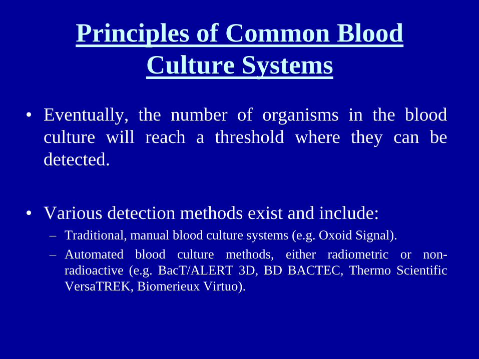
• Eventually, the number of organisms in the blood
culture will reach a threshold where they can be
detected.
• Various detection methods exist and include: – Traditional, manual blood culture systems (e.g. Oxoid Signal).
– Automated blood culture methods, either radiometric or non-
radioactive (e.g. BacT/ALERT 3D, BD BACTEC, Thermo Scientific
VersaTREK, Biomerieux Virtuo).
Automated Blood Culture Systems
Biomerieux BacT/ALERT 3D
• The BacT/ALERT is a simple, automated rapid
microbial detection system that uses colorimetric
technology.
• If microorganisms are present in the blood culture,
CO2 is produced as the microorganisms metabolise
the substrates in the culture medium.
Automated Blood Culture Systems
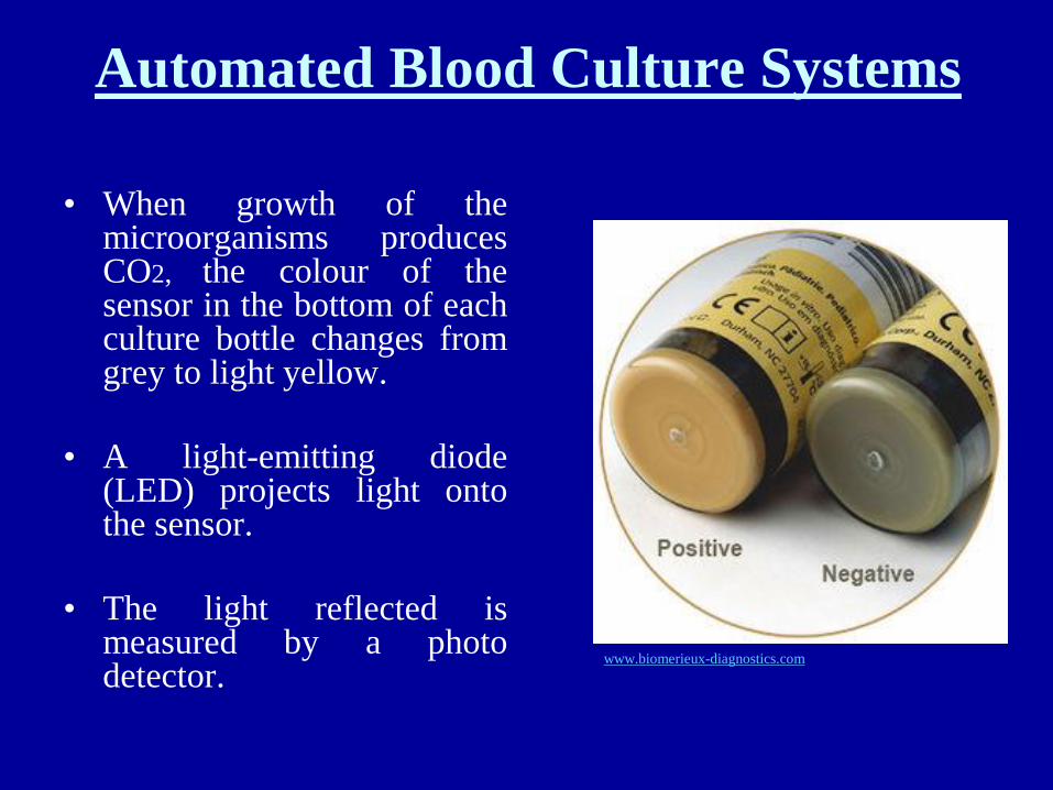
• When growth of the microorganisms produces CO2, the colour of the sensor in the bottom of each culture bottle changes from grey to light yellow.
• A light-emitting diode (LED) projects light onto the sensor.
• The light reflected is measured by a photo detector.
www.biomerieux-diagnostics.com

Automated Blood Culture Systems
• As more CO2 is generated, more light is reflected.
• This information is compared to the initial sensor
reading.
• If there is a high initial CO2 content, an unusually
high rate of CO2 production, and/or a sustained
production of CO2, the sample is determined to be
positive.
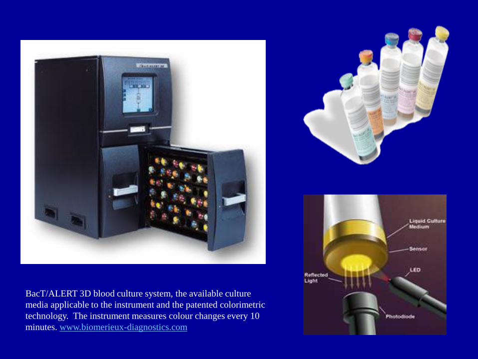
BacT/ALERT 3D blood culture system, the available culture
media applicable to the instrument and the patented colorimetric
technology. The instrument measures colour changes every 10
minutes. www.biomerieux-diagnostics.com
Bacteraemia Case Report
• 61yr old female with a past medical history of right breast cancer and hypothyroidism presented to A&E at Victoria Hospital Rothesay.
• She was admitted on Saturday 21st November, 2015 with a working diagnosis of sepsis.
• Physical examination revealed infected shingles.
• Blood cultures were drawn on admission and FBC, U&Es, CRP and Glucose were all requested.

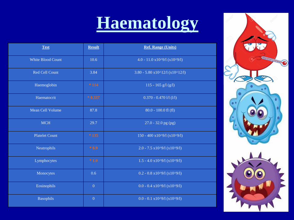
Haematology Test Result Ref. Range (Units)
White Blood Count 10.6 4.0 - 11.0 x10^9/l (x10^9/l)
Red Cell Count 3.84 3.80 - 5.80 x10^12/l (x10^12/l)
Haemoglobin * 114 115 - 165 g/l (g/l)
Haematocrit * 0.337 0.370 - 0.470 l/l (l/l)
Mean Cell Volume 87.8 80.0 - 100.0 fl (fl)
MCH 29.7 27.0 - 32.0 pg (pg)
Platelet Count * 135 150 - 400 x10^9/l (x10^9/l)
Neutrophils * 8.9 2.0 - 7.5 x10^9/l (x10^9/l)
Lymphocytes * 1.0 1.5 - 4.0 x10^9/l (x10^9/l)
Monocytes 0.6 0.2 - 0.8 x10^9/l (x10^9/l)
Eosinophils 0 0.0 - 0.4 x10^9/l (x10^9/l)
Basophils 0 0.0 - 0.1 x10^9/l (x10^9/l)

Biochemistry
Test Result Ref. Range (Units)
Sodium 135 133 - 146 mmol/L (mmol/L)
Potassium 3.8 3.5 - 5.3 mmol/L (mmol/L)
Chloride 101 95 - 108 mmol/L (mmol/L)
Urea 3.8 2.5 - 7.8 mmol/L (mmol/L)
Creatinine 73 40 - 130 umol/L (umol/L)
Estimated GFR >60 >60 ml/min (ml/min)
C Reactive Protein *17 0 - 10 mg/L (mg/L)
Glucose 5.2 3.5 - 6.0 mmol/L (mmol/L)
Microbiology
• Blood culture set incubated in the BacT/ALERT 3D system (21/11/2015).
• On Monday 23rd November, bacterial growth was detected in both O2 and AnO2 blood culture bottles.
• CBA, FHB and MHS culture plates inoculated and incubated overnight in CO2, 36°C.
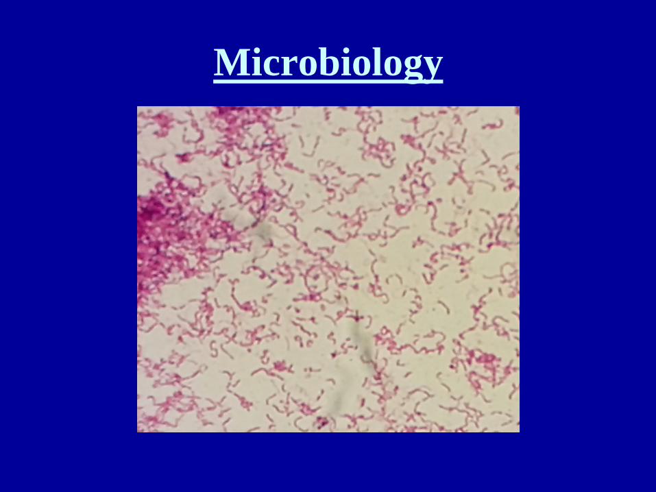
• Microscopy revealed Gram positive bacilli.
Microbiology
Microbiology
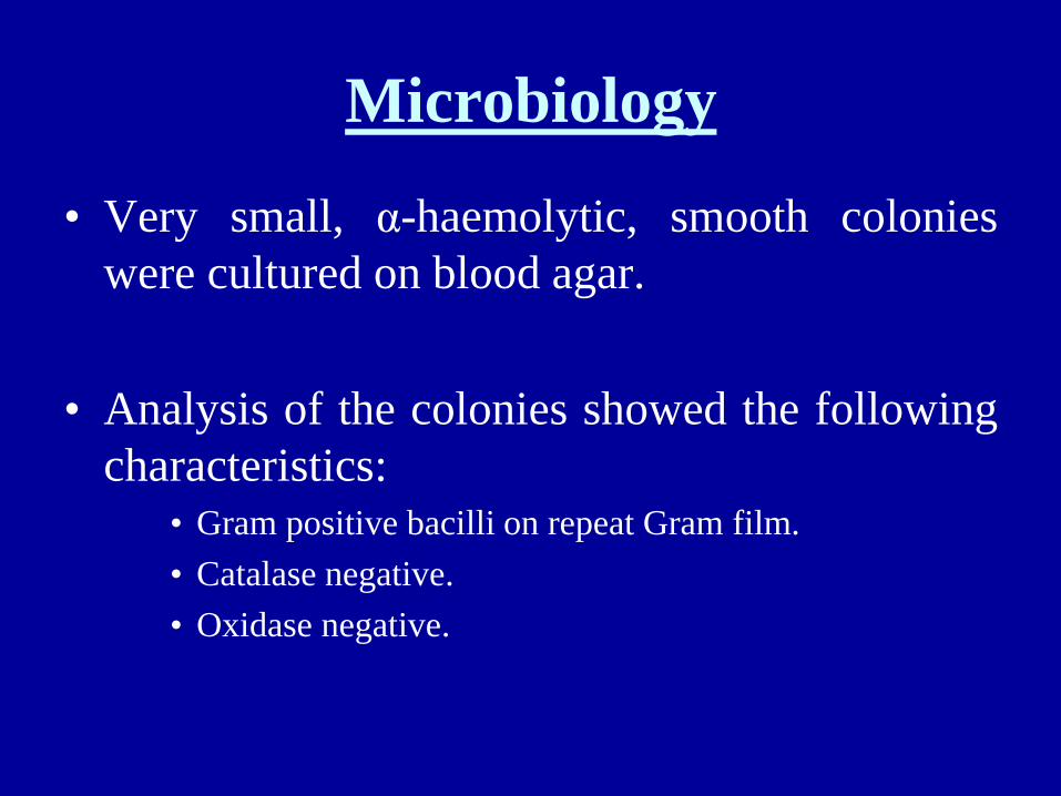
• Very small, α-haemolytic, smooth colonies
were cultured on blood agar.
• Analysis of the colonies showed the following
characteristics: • Gram positive bacilli on repeat Gram film.
• Catalase negative.
• Oxidase negative.

Microbiology
Isolate identified by Vitek MS as Erysipelothrix rhusiopathiae.

Microbiology
• As per Microbiology Consultant, Penicillin, Ciprofloxacin and Vancomycin E-tests were set up.
• To confirm the isolate identification, a Gram positive (GP) I.D. card was inoculated using the Vitek 2 system.
• Organism associated with fish and pigs – patient cooked a lot of fish and was previously a sheep farmer.
• Patient was on IV Cefuroxime and Flagyl.

Antibiotic Sensitivity Results
• Using both EUCAST and CLSI criteria the E-tests revealed the following MIC results for this organism:
• Penicillin 0.12 mg/L.
• Ciprofloxacin 0.12 mg/L.
• Vancomycin 32 mg/L.
• To supplement these results, the Microbiology Consultant also requested cephalosporin sensitivity.
• CXM disc zone size 35mm and CRO MIC 0.06 mg/L.

Antibiotic Sensitivity Results

Antibiotic Therapy
• Patient was administered IV antibiotics followed by 2-4 weeks of PO antibiotics.
• PO Cephalexin suggested for 2-4 weeks – better bioavailability.
• Risk of C. difficile with this option so advised this should be monitored and reviewed.
• Diarrhoeal stool samples negative for C. difficile infection (25/02/2015).
Antibiotic Therapy
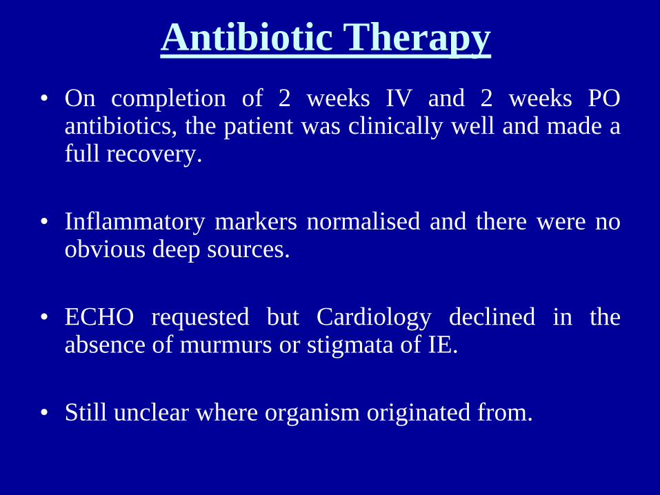
• On completion of 2 weeks IV and 2 weeks PO antibiotics, the patient was clinically well and made a full recovery.
• Inflammatory markers normalised and there were no obvious deep sources.
• ECHO requested but Cardiology declined in the absence of murmurs or stigmata of IE.
• Still unclear where organism originated from.

Isolates in NHSGGC
• 80yr old female presented to Glasgow Victoria
Infirmary with infected foot ulcer (13/05/2013).
• Wound swab of this area cultured E. rhusiopathiae.
• CRP = 246mg/L.
• WBC = 27.9x10^9/l.
• Neutrophils = 25.8x10^9/l.
• Intra-abdominal sepsis with an unrelated necrotic foot
ulcer secondary to Peripheral Vascular Disease.
Isolates in NHSGGC
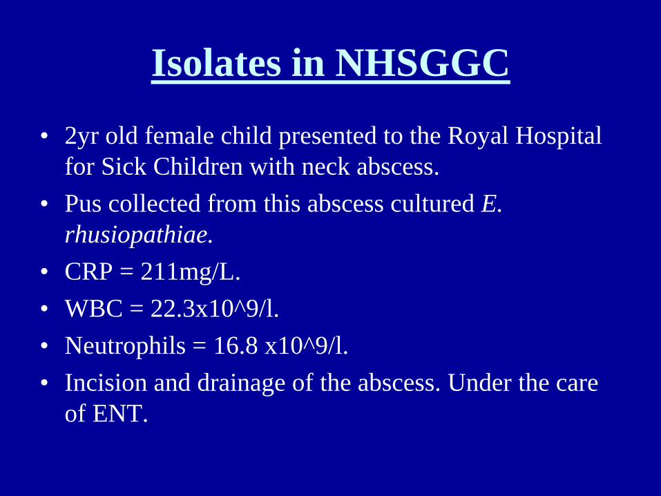
• 2yr old female child presented to the Royal Hospital
for Sick Children with neck abscess.
• Pus collected from this abscess cultured E.
rhusiopathiae.
• CRP = 211mg/L.
• WBC = 22.3x10^9/l.
• Neutrophils = 16.8 x10^9/l.
• Incision and drainage of the abscess. Under the care
of ENT.
Erysipelothrix rhusiopathiae
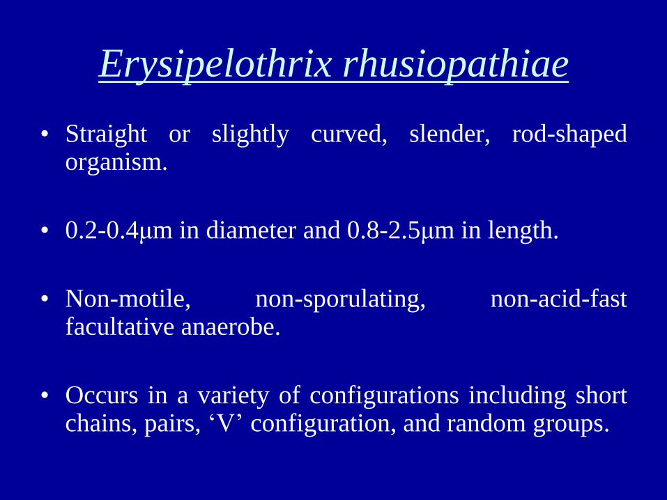
• Straight or slightly curved, slender, rod-shaped organism.
• 0.2-0.4μm in diameter and 0.8-2.5μm in length.
• Non-motile, non-sporulating, non-acid-fast facultative anaerobe.
• Occurs in a variety of configurations including short chains, pairs, ‘V’ configuration, and random groups.

Erysipelothrix rhusiopathiae
• Based on colonial appearance, Erysipelothrix morphology is described as smooth (S) or rough (R).
• S-form colonies are convex, with a smooth surface and entire edge.
• R-form colonies are larger with an irregular edge and flattened, rough surface.
• S-form morphology is typically seen in chronic infections, i.e., arthritis and endocarditis.
Erysipelothrix rhusiopathiae
• E. rhusiopathiae is found worldwide and has been reported as a commensal or pathogen in a variety of wild and domestic animals, birds and fish.
• Animal-to-human transmission occurs by direct cutaneous contact (via scratches or puncture wounds).
• Human-to-human infection has not been documented.
• Most human cases are associated with occupational exposure to contaminated meat or fish (fishmonger’s finger).
Pathogenesis & Pathology
• Very little is actually known about the pathogenesis
of E. rhusiopathiae.
• The organism produces a hyaluronidase and a
neuraminidase and it is hypothesised that the level of
these enzymes may correlate with virulence.
• IV injection of the pathogen into rabbits is fatal in 2-3
days – erysipeloid rash, lungs become haemorrhagic
and a pericardial exudate develops.
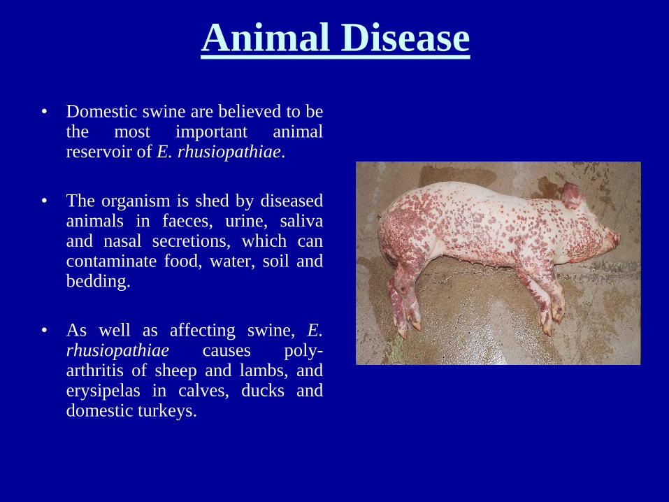
Animal Disease
• Domestic swine are believed to be the most important animal reservoir of E. rhusiopathiae.
• The organism is shed by diseased animals in faeces, urine, saliva and nasal secretions, which can contaminate food, water, soil and bedding.
• As well as affecting swine, E. rhusiopathiae causes poly-arthritis of sheep and lambs, and erysipelas in calves, ducks and domestic turkeys.
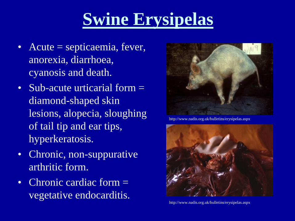
Swine Erysipelas
• Acute = septicaemia, fever,
anorexia, diarrhoea,
cyanosis and death.
• Sub-acute urticarial form =
diamond-shaped skin
lesions, alopecia, sloughing
of tail tip and ear tips,
hyperkeratosis.
• Chronic, non-suppurative
arthritic form.
• Chronic cardiac form =
vegetative endocarditis.
http://www.nadis.org.uk/bulletins/erysipelas.aspx
http://www.nadis.org.uk/bulletins/erysipelas.aspx

Marine Environments
• Associated with marine fish, molluscs and
crustaceans.
• The organism survives and grows on the exterior
mucoid slime of fish.
• Doesn’t cause disease in the fish but is thought to be
an important source of infection for man.
Clinical Manifestations in Humans
• Infection with E. rhusiopathiae can cause three forms of human disease:
• Localised cutaneous form. (also known as erysipeloid of Rosenbach).
• Generalised, diffuse cutaneous form.
• Septicaemia often associated with endocarditis.
• Erysipeloid is the most common form and is an acute localised cutaneous infection, usually cellulitis.
• Typically occurs on the hands or fingers.
Clinical Manifestations in Humans
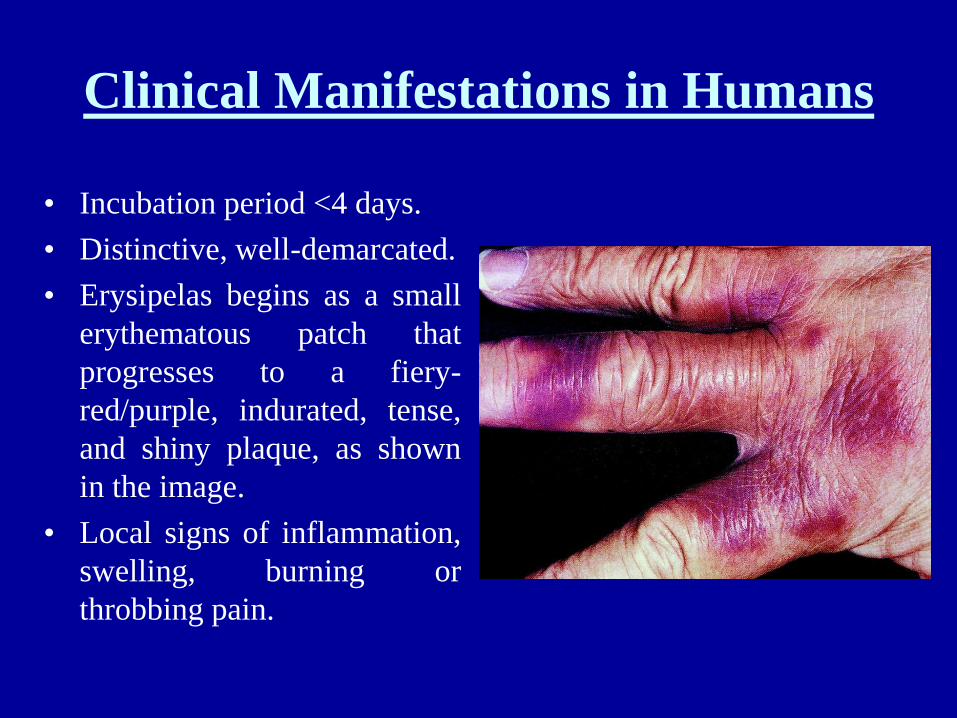
• Incubation period <4 days.
• Distinctive, well-demarcated.
• Erysipelas begins as a small
erythematous patch that
progresses to a fiery-
red/purple, indurated, tense,
and shiny plaque, as shown
in the image.
• Local signs of inflammation,
swelling, burning or
throbbing pain.
Clinical Manifestations in Humans
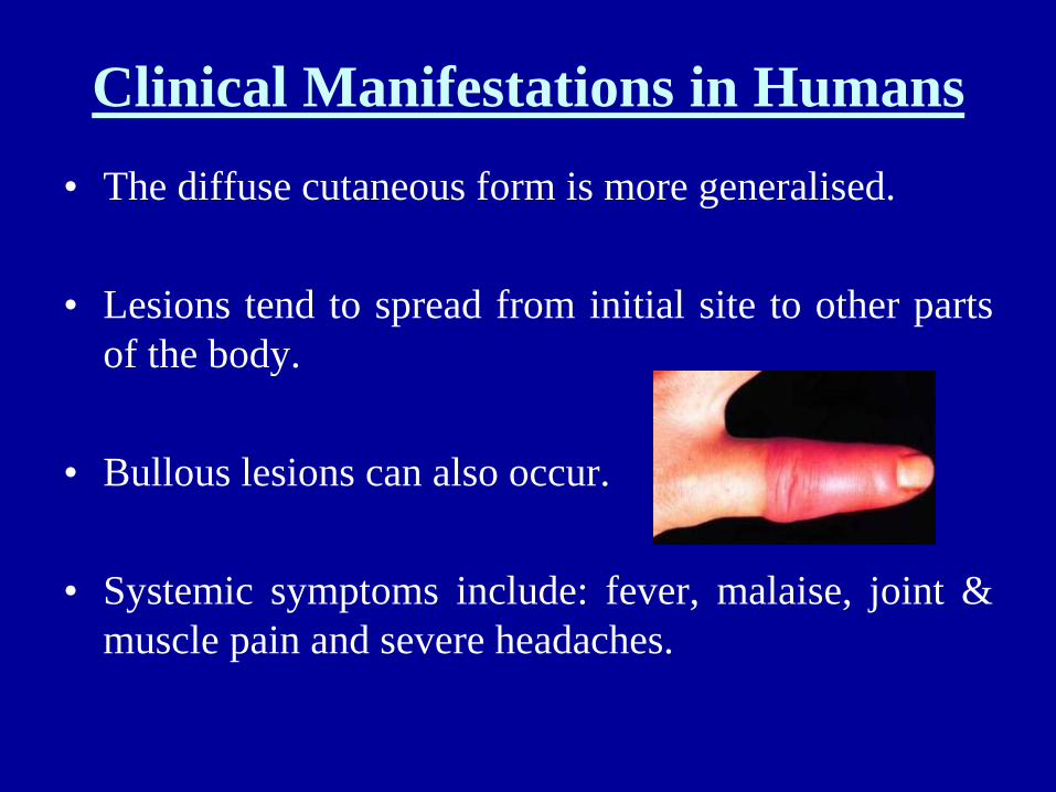
• The diffuse cutaneous form is more generalised.
• Lesions tend to spread from initial site to other parts
of the body.
• Bullous lesions can also occur.
• Systemic symptoms include: fever, malaise, joint &
muscle pain and severe headaches.
Clinical Manifestations in Humans
• Bloodstream infections with E. rhusiopathiae are not common.
• Bacterial cultures are positive in only 5% of cases.
• A strong association exists between bacteraemia and the development of IE.
• It tends to occur in immunocompromised patients, has a higher male to female ratio and can occur in patients with normal native values as well as prosthetic valves.

Treatment
• Penicillin is the drug of choice.
• Cephalosporins are suitable alternatives in patients allergic to Penicillin.
• Erysipelothrix is also highly susceptible to Clindamycin.
• Most strains are resistant to Aminoglycosides, SXT, Sulphonamides, Streptomycin and Vancomycin.
Prevention
• Containment and control.
• Cleaning and disinfection of work surfaces and tools, hand
hygiene, and use of gloves reduce the risk of infection when
working with animals or animal products.
• Protective apparel should be worn by those working in
slaughterhouses or fisheries.
• Control of animal disease – herd management, good sanitation,
immunisation.

Thank you!