experiences rat zollinger-ellison - gutgut.bmj.com/content/gutjnl/11/5/409.full.pdfexperiences with...
TRANSCRIPT

Gut, 1970, 11, 409-419
Experiences with a rat bio-assay in thediagnosis of the Zollinger-Ellison syndrome
CHRISTINE G. THOMSON, I. G. M. CLEATOR, AND W. SIRCUS'From the Teaching and Research Centre and the Gastro-Intestinal Unit, the Western General Hospital,Edinburgh, and the Departments of Clinical Chemistry, Clinical Surgery, and Medicine (WesternGeneral Hospital), University ofEdinburgh.
SUMMARY Reported here are the assay results, using the anaesthetized rat preparation ofLai (1964), of plasma from patients known or suspected to have peptide-secreting adenomataand these are compared with assays carried out on plasma from patients whose clinicalhistory excluded such pathology. It is suggested that these demonstrate that this techniqueis of diagnostic value in this condition and that it has practical advantages over other bio-assaymethods currently available and physiological advantages over immunological methods.
It may well be that identification of the presence of tumour in these patients may first bemade by immunological techniques to be followed by functional identification using a bio-assay procedure such as is described here.
In the first description of the clinical entitywhich linked recurrent peptic ulceration and'gigantic' gastric hypersecretion with islet-celltumours of the pancreas, the authors (Zollingerand Ellison, 1955) suggested that the basis of thesyndrome might be the secretion into the circula-tion of a humoral substance from the tumour.The extraction from such tumour of a gastricsecretagogue with physiological properties similarto those of antral gastrin was subsequentlyreported by Gregory, Tracy, French, and Sircus(1960). The hypothesis of Zollinger and Ellisonwas supported by the demonstration of a gastricsecretagogue in the serum and gastric juice oftwo patients with the disease (Sircus, 1964) andsubsequently by Banks and Marks (1964) in a
further case.The evidence that the substance that could be
extracted from the tumours was similar to, ifnot identical with, gastrin was supported bythe immunological studies of Monaco, Lythgoe,and Waddell (1961), and the physiologicalstudies of Jackson and his colleagues (Jackson,Blair, Dawson, Reed, and Watts, 1963). Amino-acid analysis of an acid hydrolysate by Gregory,Grossman, Tracy, and Bentley (1967) showedthat the substance elaborated by the tumour
'Member of the external scientific staff, Medical ResearchCouncil.
had an amino-acid composition similar to theantral hormone gastrin.The development of a bio-assay technique for
the recognition of circulating gastrin, using therat, by Lai (1964) was extended by us as a methodof diagnosis in patients with clinical suspicionof the disease (Thomson and Sircus, 1967).Others have used the cat (Blair and Wood,1968).Recently an immunological assay has been
developed by McGuigan and Trudeau (1968).This demonstrates the existence of an immuno-logically active protein in the circulation butdoes not establish that this material is physio-logically active and therefore the method haslimitations.We report here our experiences to date with
the modification of bio-assay by the method ofLai (1964) in the rat for use in clinical diagnosis.
Patients
Plasma from 42 patients was examined. In allbut a few cases neither the history, nor theresults of other investigations, nor the findingsat operation or necropsy were known tothe workers performing the assay. Ten of these
on 28 June 2018 by guest. Protected by copyright.
http://gut.bmj.com
/G
ut: first published as 10.1136/gut.11.5.409 on 1 May 1970. D
ownloaded from

Christine G. Thomson, I. G. M. Cleator, and W. Sircus
patients served as controls. Of these, two werenormal healthy adults, two had neoplasticdisease of the alimentary tract but with nosuspicion of the Zollinger-Ellison syndrome,one had Crohn's disease of the jejunum, andfive had benign peptic ulcer disease withoutany unusual features. The remaining 32 subjectswere patients with clinical features generatingsuspicion of the syndrome.
In arriving at definitive clinical diagnoses forthe purpose of analysis of the results, we haveused all data available to date from the physiciansand surgeons in whose care the patients were.Thus far, twelve patients have been shown byoperation, or by necropsy study, to have hadeither islet-cell tumour in the pancreas or atthe ectopic sites of that organ, or to have hadsignificant islet-cell hyperplasia. In some casesextracts of the tumours found have showngastrin-like activity. Of the remaining 20 patients,10 have been considered as a result of investiga-tion, operation, and their subsequent clinicalcourse, to be unlikely to be examples of thesyndrome. Thus, there are ten patients in whomclinical suspicion remains until operation ornecropsy supplies the evidence and in whoma definitive diagnosis cannot yet be made.The basis for clinical suspicion has been thesubject of previous reviews (Sircus, 1962 and1968).
Methods
Venous blood was withdrawn after an overnightfast, and collected in lithium-heparin tubes.It was centrifuged as rapidly as possible afterwithdrawal for 15 minutes at 1,200 g at 5°Cand the resulting plasma was equally rapidlyseparated and stored at -20°C in 2-5 ml aliquots.Approximately one hour before it was requiredfor assay, an aliquot was thawed at not higherthan 30°C and immediately recentrifuged at5°C. The resulting defibrinated plasma was thenassayed with no further manipulation. Nosample was frozen or thawed more than once.The assays for gastrin-like activity in all
preparations were carried out on the modifiedanaesthetized-rat preparation as previously de-scribed by Thomson and Sircus (1968). Thereference standards used in this study wereappropriate dilutions of Peptavlon (ICI 50123).Several techniques have been investigated in anattempt to improve the sensitivity of the pre-paration, since, using this assembly, the infusionof 1-2 gg Peptavlon (ie, 6 ,ug/kg/15 min) isrequired to produce significant acid secretionin most animals. Various manipulations of theanimal have been tried, including filling theruminant space of the stomach with a Perspexplug to reduce the dead space, but have provedunrewarding. Clamping off the ruminant portion
0 3.0 4.0 5.0ml OO1N HCL
Fig. 1 Titration curves of buffers. (0 01J54 x 10-3Mphosphate, 2.3 x 10⁻⁴M citrate,and 1.S4 x 10-1M sodium chloride. x X6.16 x 10-4Mphosphate, 9.2 x 1J-5M citrate, and1J54 x 10-1M sodium chloride. A A1J54 x 10-1M sodium chloride.)
of the stomach minimizes stasis, with its attendantbaseline fluctuation, and we believe also in-creases sensitivity somewhat. Precautions aretaken to exclude the liver and the spleen fromthe clamp. This is attached at one end to theligature on the ruminant portion and at the otherto a pin fixed in the cork mat on which the ratis resting. Occasionally, unresponsive animalsare encountered, in whom secretion can onlybe induced by the infusion of 20 ,ug Peptavlonor more per kg/15 minutes. Such animals werenot used for the studies described in this paper.
Sensitivity of the assay assembly can, however,be further improved by altering the concentrationof the perfusing buffer, and some of the analysesreported here were carried out using this modi-fication. This buffer (6.16 x 10⁻⁴M Na2HPO4:9.2 x 10⁻⁵M citric acid: 1.54 x 10⁻¹M NaCI,I = *156, pH 6.6), as with the original, permitsan approximately linear pH response to unitadditions of acid, but yields a titration curvemore responsive to small amounts of addedacid and is therefore valuable in the assayassembly for assessing low concentrations ofhumoral activity (Figure 1). Using this buffer,significant acid secretion can usually be demon-strated with 0.48 jug Peptavlon (2-4 ,ug/15minutes). Since the rat's response to gastrin IIis some four times higher than the response toPeptavlon on a weight for weight basis, secretionproduced in response to the infusion of 0.10 ,tggastrin II should therefore be readily observedin the assay when using the dilute buffer.
410
on 28 June 2018 by guest. Protected by copyright.
http://gut.bmj.com
/G
ut: first published as 10.1136/gut.11.5.409 on 1 May 1970. D
ownloaded from
Experiences with a rat bio-assay in the diagnosis of the Zollinger-Ellison syndrome
ASSAY PROTOCOLIn practice, assay of plasma preparations forgastrin-like activity was normally carried outusing the following protocol. A primer dose of0.6 ,ug Peptavlon was infused over 15 minutes.If this produced a brisk secretion of gastric acidinto the perfusing buffer, then the animal wasregarded as sufficiently sensitive for serum assay.After the baseline secretion had been re-estab-lished for 15 to 30 minutes, 10 mg hydrocortisonehemisuccinate in 0.5 ml 0.9% NaCl was injectedsubcutaneously, and immediately thereafter 2.0 mlof suitably prepared test plasma was infusedover 25 minutes. When possible, after theoriginal baseline secretion was restored, a secondsample from another patient was infused similarlyand the analytical run terminated with a furtherinfusion of 0.6 ,ug Peptavlon for 15 minutes.
Duplicate analyses were carried out on allsera when sufficient was available and where aresult was still in doubt further assays wereperformed. Dose order was reversed in theduplicate analysis, so that the plasma analysedfirst in the original assay was analysed second inthe duplicate assay. It was hoped to avoidpossible serum interaction in this way.
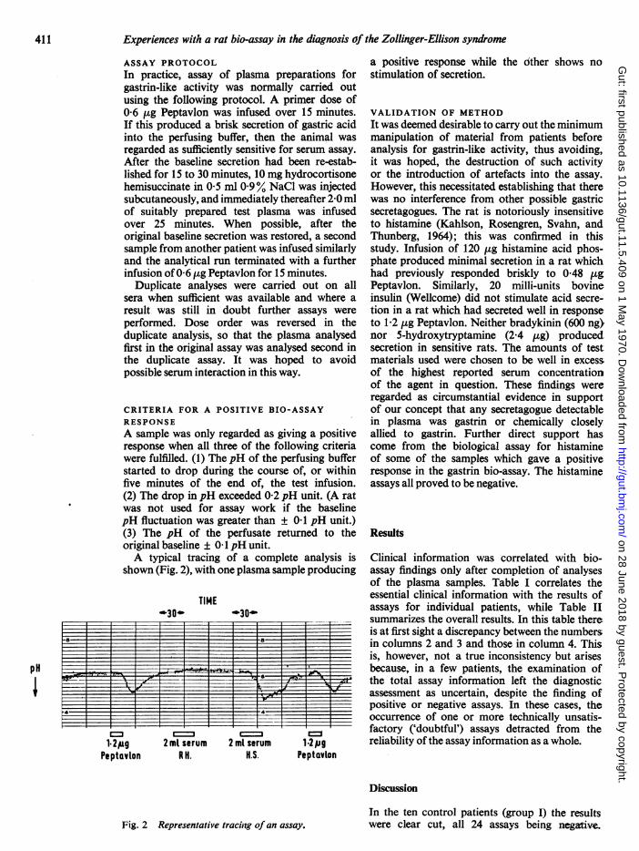
CRITERIA FOR A POSITIVE BIO-ASSAYRESPONSEA sample was only regarded as giving a positiveresponse when all three of the following criteriawere fuffilled. (1) The pH of the perfusing bufferstarted to drop during the course of, or withinfive minutes of the end of, the test infusion.(2) The drop in pH exceeded 0.2 pH unit. (A ratwas not used for assay work if the baselinepH fluctuation was greater than ± 0.1 pH unit.)(3) The pH of the perfusate returned to theoriginal baseline ± 0 1 pH unit.A typical tracing of a complete analysis is
shown (Fig. 2), with one plasma sample producing
TIME
I I 1 1 1-a _ a
4- 4HI '-:-4--F-_rl---~~~~~ _2
1.2pg
Peptuvion
2 mt serumRH.
2 ml serumH.S.
1.2pgPeptavion
a positive response while the dther shows nostimulation of secretion.
VALIDATION OF METHODIt was deemed desirable to carry out the minimummanipulation of material from patients beforeanalysis for gastrin-like activity, thus avoiding,it was hoped, the destruction of such activityor the introduction of artefacts into the assay.However, this necessitated establishing that therewas no interference from other possible gastricsecretagogues. The rat is notoriously insensitiveto histamine (Kahlson, Rosengren, Svahn, andThunberg, 1964); this was confirmed in thisstudy. Infusion of 120 gg histamine acid phos-phate produced minimal secretion in a rat whichhad previously responded briskly to 0.48 ,ugPeptavlon. Similarly, 20 milli-units bovineinsulin (Wellcome) did not stimulate acid secre-tion in a rat which had secreted well in responseto 1.2 gg Peptavlon. Neither bradykinin (600 ng)nor 5-hydroxytryptamine (2.4 ,ug) producedsecretion in sensitive rats. The amounts of testmaterials used were chosen to be well in excessof the highest reported serum concentrationof the agent in question. These findings wereregarded as circumstantial evidence in supportof our concept that any secretagogue detectablein plasma was gastrin or chemically closelyallied to gastrin. Further direct support hascome from the biological assay for histamineof some of the samples which gave a positiveresponse in the gastrin bio-assay. The histamineassays all proved to be negative.
Results
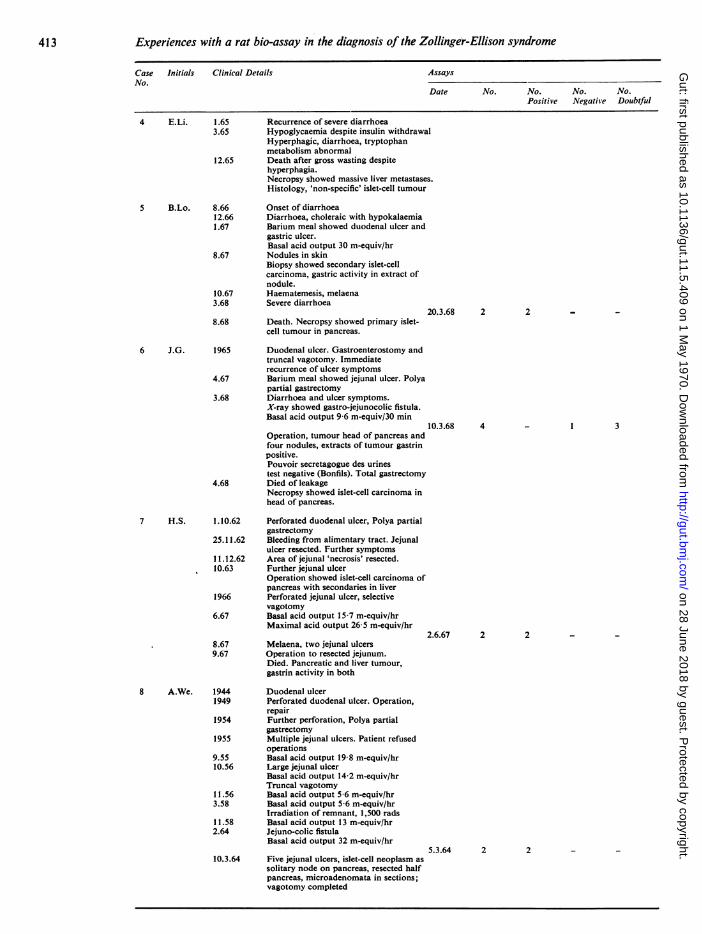
Clinical information was correlated with bio-assay findings only after completion of analysesof the plasma samples. Table I correlates theessential clinical information with the results ofassays for individual patients, while Table IIsummarizes the overall results. In this table thereis at first sight a discrepancy between the numbersin columns 2 and 3 and those in column 4. Thisis, however, not a true inconsistency but arisesbecause, in a few patients, the examination ofthe total assay information left the diagnosticassessment as uncertain, despite the finding ofpositive or negative assays. In these cases, theoccurrence of one or more technically unsatis-factory ('doubtful') assays detracted from thereliability of the assay information as a whole.
Discussion
Fig. 2 Representative tracinig of an assay.
In the ten control patients (group I) the resultswere clear cut, all 24 assays being negative.
pH
411
on 28 June 2018 by guest. Protected by copyright.
http://gut.bmj.com
/G
ut: first published as 10.1136/gut.11.5.409 on 1 May 1970. D
ownloaded from

412 Christine G. Thomson, I. G. M. Cleator, and W. Sircus
Case Initials Clinical Details AssaysNo.
Date No. No. No. No.Positive Negative Doubtful
Group II: Proven Zollinger-Ellison syndromes1 A.A. 1960 Duodenal ulcer
1962 Gastric ulcer and duodenal ulcer,perforation, metatastic neoplasia in liver.
1962-64 Dyspepsia, tetany9.64 Gastric rugal hypertrophy
Maximal acid output 42 m-equiv/hrTruncal vagotomy, and pyloroplasty forislet-cell carcinoma in pancreas.
11.64 Basal acid output 3-0 m-equivlhrMaximal acid output 4-1 m-equivlhrInsulin stimulated secretion 2-2 m-equiv/hr 9.12.64 2
18.12.64 57.65 Recurrence of ulcer dyspepsia
Basal acid output 29-0 m-equiv/hrMaximal acid output 29-4 m-equiv/hr
7.65 Total gastrectomy 5.11.65 27.6.68 2
11.68 Died. Necropsy showed masses ofwidespread tumour-extracts stronglygastrin positive.
2 R.H.
1.67
6.671.68
Family history, sibling with hyperpara-thyroidism and Zollinger-Ellisonsyndrome.Renal colic, hypercalcaemia, raised5-HIAASubtotal parathyroidectomyBasal acid output 15-4 m-equiv/hrMaximal acid output 54 m-equiv/hrPouvoir secretagogue des urines slightlyraised (Bonfils)
15.2.68 Distal pancreatectomy for multiplenon-B islet-cell tumours
_ 22 1
1 12 _
10.1.68 3
18.2.68 Gastric hypersecretion, hyperglycaemia,hypocalcaemia
24.2.68 Melaena and deathNecropsy showed six ulcers in duodenum,multiple residual tumours of pancreasand duodenum.
1.68 Onset of duodenal ulcer type dyspepsia1.9.68 Continued epigastric pain18.9.68 Barium meal showed duodenal ulcer and
hiatus hernia. DuodenitisBasal acid output 20 m-equiv/hrMaximal acid output 104 m-equiv/hr
I
320.10.68 At operation for duodenal ulcer no
obvious abnormality seen in pancreas;partial pancreatectomy, Polya partialgastrectomy and selective vagotomy.Histology, microadenomata ofpancreatic islets.
12.68 Symptomless
1959 Heartburn1961 Profuse diarrhoea with incontinence1963 Diarrhoea, flushing, colic, borborygmi,
hepatomegalyBasal acid output 50 m-equiv/hrMaximal acid output 89 m-equiv/hrGastrin activity in circulating blood
5.63 Partial pancreatectomy, seven-eighthsgastrectomy, truncal vagotomy forpancreatic islet-cell 'pseudo-carcinoid'tumour with liver metastases.
6.63 Basal acid output 3.5 m-equiv/hrMaximal acid output 6-3 m-equiv/hrSymptomless
9.64 Pigmentation12.64 Severely diabetic, fasting blood sugar
436 mg/100 ml
1.10.68 23.10.68 1
7.11.68 1
_ I
1 _
_ _ 1
10.12.64 3 3 -
18.12.64 2 2 -
Table I Summaries of the clinical histories and the bio-assay results of all patients studied in groups,II and III
2
3 C.K.
4 E.L.
1
on 28 June 2018 by guest. Protected by copyright.
http://gut.bmj.com
/G
ut: first published as 10.1136/gut.11.5.409 on 1 May 1970. D
ownloaded from
Experiences with a rat bio-assay in the diagnosis of the Zollinger-Ellison syndrome
Case Initials Clinical Details AssaysNo.
Date No. No. No. No.Positive Negative Doubtful
1.65 Recurrence of severe diarrhoea3.65 Hypoglycaemia despite insulin withdrawal
Hyperphagic, diarrhoea, tryptophanmetabolism abnormal
12.65 Death after gross wasting despitehyperphagia.Necropsy showed massive liver metastases.Histology, 'non-specific' islet-cell tumour
8.66 Onset of diarrhoea12.66 Diarrhoea, choleraic with hypokalaemia1.67 Barium meal showed duodenal ulcer and
gastric ulcer.Basal acid output 30 m-equiv/hr
8.67 Nodules in skinBiopsy showed secondary islet-cellcarcinoma, gastric activity in extract ofnodule.
10.67 Haematemesis, melaena3.68 Severe diarrhoea
20.3.68 28.68 Death. Necropsy showed primary islet-
cell tumour in pancreas.
1965 Duodenal ulcer. Gastroenterostomy andtruncal vagotomy. Immediaterecurrence of ulcer symptoms
4.67 Barium meal showed jejunal ulcer. Polyapartial gastrectomy
3.68 Diarrhoea and ulcer symptoms.X-ray showed gastro-jejunocolic fistula.Basal acid output 9-6 m-equiv/30 min
10.3.68 4Operation, tumour head of pancreas andfour nodules, extracts of tumour gastrinpositive.Pouvoir secretagogue des urinestest negative (Bonfils). Total gastrectomy
4.68 Died of leakageNecropsy showed islet-cell carcinoma inhead of pancreas.
1.10.62 Perforated duodenal ulcer, Polya partialgastrectomy
25.11.62 Bleeding from alimentary tract. Jejunalulcer resected. Further symptoms
11.12.62 Area of jejunal 'necrosis' resected.10.63 Further jejunal ulcer
Operation showed islet-cell carcinoma ofpancreas with secondaries in liver
1966 Perforated jejunal ulcer, selectivevagotomy
6.67 Basal acid output 15-7 m-equiv/hrMaximal acid output 26-5 m-equiv/hr
2.6.67 28.67 Melaena, two jejunal ulcers9.67 Operation to resected jejunum.
Died. Pancreatic and liver tumour,gastrin activity in both
1944 Duodenal ulcer1949 Perforated duodenal ulcer. Operation,
repair1954 Further perforation, Polya partial
gastrectomy1955 Multiple jejunal ulcers. Patient refused
operations9.55 Basal acid output 19-8 m-equiv/hr10.56 Large jejunal ulcer
Basal acid output 14-2 m-equiv/hrTruncal vagotomy
11.56 Basal acid output 5 6 m-equiv/hr3.58 Basal acid output 5-6 m-equiv/hr
Irradiation of remnant, 1,500 rads11.58 Basal acid output 13 m-equiv/hr2.64 Jejuno-colic fistula
Basal acid output 32 m-equiv/hr
10.3.64 Five jejunal ulcers, islet-cell neoplasm as
solitary node on pancreas, resected halfpancreas, microadenomata in sections;vagotomy completed
2 _ _
_ 1 3
2 _ _
5.3.64 2
413
4 E.Li.
5 B.Lo.
6 J.G.
7 H.S.
8 A.We.
on 28 June 2018 by guest. Protected by copyright.
http://gut.bmj.com
/G
ut: first published as 10.1136/gut.11.5.409 on 1 May 1970. D
ownloaded from

Christine G. Thomson, I. G. M. Cleator, and W. Sircus
Case Initials Clinical Details AssaysNo.
Date No. No. No. No.Positive Negative Doubtful
20.6.64 Operation, re-explored. Resection of headof pancreas, duodenectomy.Developed bacteraemia, myocardial infarct.Death. No residual tumour at necropsy
1962 Spontaneous hypoglycaemiaOperation, multiple islet-cell adenomaexcised from pancreas.
3.65 DyspepsiaBasal acid output 20 m-equiv/hrMaximal acid output 131 m-equiv/hrHypercalcaemia, hyperinsulinaemia
3.654.65 Polya partial gastrectomy and total
pancreatectomy for multiple nodes inpancreas, gastrin activity in extracts.
4.657.65 Still hypoglycaemic8.66 Hyperparathyroidism proven, hyper-
calcaemic.5.68 Operation, parathyroid adenoma excised.7.68 Still hypoglycaemic and hypocalcaemic
(calcium 5.6 mg/ml)1963-65 Watery diarrhoea4.65 Barium meal showed second partial
duodenal ulcer, later perforated.Operation, repair.
8.65 Haematemesis and melaena.Polya partial gastrectomy and truncalvagotomy for 'stomal ulcer', pancreasnormal
11.65 Diarrhoea prominent1.66 Ulcer of cardia perforated.
9.1.6Total gastrectomy. Died postoperatively.Necropsy showed islet-cell tumournodule on surface of pancreas, fivesecondary involved glands, hyperplasiaof parietal cells of gastric mucosa.
9.66 Childbirth. Two weeks later profusediarrhoea.
6.67 Water-losing, electrolyte-depleting diarrhoea,Shigella positive. Transferred to hospitalfor infectious diseases.
9.67 Extreme myasthenia, severe choleraicdiarrhoea, hypokalaemia, hypomagnesaemia,hypercalcaemiaBarium meal showed jejunitis. Gastricrugal hypertrophy
21.6.67 Basal acid output 4 m-equiv/hrMaximal acid output 24.2 m-equivlhr
5.10.67 Basal acid output 14-4 m-equiv/hr9.10.
9.10.67 Basal acid output 5.5 m-equiv/hr10.10.67 Islet-cell tumour enucleated from head
of pancreas 10.10
3 2
2
66 2
.67 3
D.67 2(duringoperation)
26.10.67 Basal acid output 27-0 m-equiv/hr26.10.67 2
7.11.67 On ProbanthineBasal acid output 10.3 m-equiv/hr 7.11.67 3
11.1.68 119.1.68 Basal acid output 3-6 m-equiv/hr4.48 Well, pregnant12.68 Childbirth, symptomless
2.63 Diarrhoea, borborygmi, flushing, nobiochemical abnormality except singlehigh 5-HIAA in urine.
12.63 Explosive diarrhoea, toxic psychosis settledspontaneously.
7.64 Diarrhoea and flushing, normal tryptophanmetabolism.Basal acid output 1-3 m-equiv/hrMaximal acid output 31-0 m-equiv/hrPartial pancreatectomy for four tumoursin pancreas palpable, seven tumours inspecimen, all atypical islet-cell 'pseudo-carcinoid'. Diarrhoea and flushdisappeared after operation.
414
_ 1
2
8 A.We.
9 S.Wo.
10 R.WI.
11 E.ST
12 J.P.
2
_ 3_ 1
on 28 June 2018 by guest. Protected by copyright.
http://gut.bmj.com
/G
ut: first published as 10.1136/gut.11.5.409 on 1 May 1970. D
ownloaded from

415 Experiences with a rat bio-assay in the diagnosis of the Zollinger-Ellison syndrome
Case Initials Clinical Details AssaysNo.
Date No. No. No. No.Positive Negative Doubtful
12 J.P. 12.65 Onset of dyspepsia9.66 Barium meal showed large duodenal ulcer.
Basal acid output 7-2 m-equiv/hrMaximal acid output 39 m-equiv/hr
3.10.66 214.6.67 116.6.67 1
7.67 Mild dyspepsia onlyBasal acid output 1.1 m-equiv/hrMaximal acid output 38-0 m-equiv/hr
10.67 Symptomless, obese10.68 Symptomless
Group III: Diagnosis not yet certaina) On clinical evidence to date probably Zollinger-Ellison cases
1 T.Co. 12.64 Haematemesis, duodenal ulcer;emergency Polya partial gastrectomy
10.66 Severe dyspepsia.Total gastrectomy. Four jejunal ulcers,one high gastric ulcer, pancreasunapproachable.
6.68 Symptomless6
7.65 Perforated duodenal ulcer repaired.5.66 Gastroenterostomy and truncal vagotomy8.66 Recurrent jejunal ulcer11.67 Polya partial gastrectomy1.68 Bleeding
Basal acid output 6-7 m-equiv/30 minMaximal acid output 59 m-equiv/hr
5.68 Symptoms recurring.Barium meal showed jejunal ulcer.Total gastrectomy for huge jejunal ulcer.Distal pancreatectomy, gastrin extractsnegative.
9.68 Symptomless
5.4.68 3
1.6.68 5
1962 Duodenal ulcer7.63 Polya partial gastrectomy12.64 Recurrence of symptoms.
Barium meal showed jejunal ulcer.Further Polya partial gastrectomy,vagotomy.
5.67 Hypercalcaemia (calcium 12.8 mg/100 ml),dyspepsia continued.
9.5.679.5.67 Operation, parathyroid adenoma excised.18.5.67 Hypocalcaemia (calcium 7.7 mg/100 ml)
18.5.676.67 Basic acid output 1-6 m-equiv/hr
Maximal acid output 8-6 m-equiv/hr10.68
10.68 Basic acid output 0.9 m-equiv/hrMaximal acid output 7.2 m-equiv/hrCalcium 9.3 mg/100 mlSymptomless
2 _ 1
3 1 1
2 _ _
7 2
2
1947 Barium meal showed duodenal ulcer.1951 Gastroenterostomy1954 Recurrence of duodenal ulcer.1955 Barium meal showed jejunal ulcer.1957 Large duodenal ulcer, vagotomy1957-65 Symptomless1966 Recurrence of duodenal ulcer. Further
truncal vagotomy and pyloroplasty,jejunal ulcer also found.
1967 Barium meal showed pyloric channel ulcer.1.68 Basic acid output 7.6 m-equiv/hr
Maximal acid output 12-5 m-equiv/hr25
2.68 Perforation suspected.Antrectomy and truncal vagotomy, noevidence of tumour in pancreas.
7.68 Pregnant and well
1966 Acute duodenal ulcerBasic acid output 25.7 m-equiv/hrMaximal acid output 42.3 m-equiv/hr.Hyperparathyroidism due to hyperplasiaof gland.Polya partial gastrectomy, solitary islet-cell adenoma near pancreas 'in gland'.
1
- 1
- 1
2 T.Cu.
3 B.H.
4 E.A.
5 A.Mc.
;.1.68 2
1
on 28 June 2018 by guest. Protected by copyright.
http://gut.bmj.com
/G
ut: first published as 10.1136/gut.11.5.409 on 1 May 1970. D
ownloaded from

Christine G. Thomson, I. G. M. Cleator, and W. Sircus
Case Initials Clinical Details AssaysNo.
Date No. No. No. No.Positive Negative Doubtful
5 A.Mc. 1968 Symptomless
Basic acid output 0.0 m-equiv/hrMaximal acid output 0.5 m-equiv/hr
6 B.S.
7 J.Sk.
8 G.D.
1961 Ulcer symptoms8.66 Barium meal showed duodenal ulcer.
Basic acid output 7 m-equiv/hrRenal calculus, hypercalcaemia
9.66 Parathyroid gland hyperplasis, three anda half glands removed.
1967 Remained hypercalcaemic (calcium12.3 mg/100 ml).
3.68 Dyspepsia, hypercalcaemia continues.7.68 Mediastinum explored, fifth parathyroid
found and excised.12.68 Clinical status not known.
1964 Onset of diarrhoea1966 15 to 20 watery stools in 24 hours,
steatorrhoea.1967 Basic acid output 158 m-equiv/hr
Operation, nothing abnormal found.1968 Basic acid output 46-6 m-equiv/hr
Barium meal showed 'duodenitis'.2
6-68 Operation, pancreas 'twice normal size',no biopsy, selective vagotomy andpyloroplasty.Basic acid output 60 m-equiv/hr (post-operatively)
6.67 -Haematemesis.Barium meal, negativeBasic acid output 7 m-equiv/hrMaximal acid output 40 m-equiv/hr
5.68 HaematemesisBasic acid output 12-6 m-equiv/hrMaximal acid output 50.3 m-equiv/hrBarium meal showed second partialduodenal ulcer and 'duodenitis'.Hypercalcaemia
7.68 Selective vagotomy and pyloroplasty,duodenitis with gastric metaplasia,pancreas 'normal'.
12.68 Recurrence of symptoms.
19.8.66 2
3.10.66 2
20.3.68 3
_ 2
_ 3
12.6.68 5
9 D.Sm. 1960 Duodenal ulcer1963 Pyloric stenosis
Operation, Billroth I1964 Two jejunal ulcers Polya partial
gastrectomy.1966 Jejunal ulcer. Further Polya partial
gastrectomy8.68 Jejunal ulcer, haematemesis
8.8.68 2Basic acid output 2-9 m-equiv/hrMaximal acid output 9-6 m-equiv/hr
12.68 Died. Necropsy, no macroscopicevidence of tumour.
10 V.P. 1959 Duodenal ulcer. Polya partialgastrectomy
3.12 (later) Jejunal ulcer. Repeat Polya partialgastrectomy and truncal vagotomy
1960-65 Continued dyspepsia, jejunal ulcer foundon investigation.Basal acid output 14-4 m-equiv/hrMaximal acid output 14-7 m-equiv/hr
11.66 Basal acid output 48-0 m-equiv/hrMaximal acid output 32-0 m-equiv/hr
6.12.66 41.67 Total gastrectomy for large recurrent
ulcer, pancreas not approachable.4.5.67 28.68 4
9.68 Symptomless
2
2 _ _
3 _ 1
_ 23 1
416
8.68 1 1
on 28 June 2018 by guest. Protected by copyright.
http://gut.bmj.com
/G
ut: first published as 10.1136/gut.11.5.409 on 1 May 1970. D
ownloaded from

Experiences with a rat bio-assay in the diagnosis of the Zollinger-Ellison syndrome
Case Initials Clinical Details AssaysNo.
Date No. No. No. No.Positive Negative Doubtful
b) On clinical evidence to date unlikely to be cases ofZoilinger-Ellison syndrome
1 D.Co. Recurrent jejunal ulcer following 3 - 3gastroenterostomy for duodenal ulcer.
2 J.Cr. Negative laparotomy for diarrhoea, 2 - 2 -
abdominal pain.Basal acid output 29 m-equiv/hr
3 M.H. Recurrent jejunal ulcer after Polya 3 - 2 1partial gastrectomy for duodenal ulcer(antral remnant).
4 J.M. Recurrent jejunal ulcer after Polya 2 - - 2partial gastrectomy for duodenal ulcer/gastric ulcer.
5 C.C. Recurrent jejunal ulcer after Polya 7 1 6 3partial gastrectomy and truncal vagotomyplus pyloroplasty.
6 E.P. Recurrent jejunal ulcer after Polya 2 - 2 -
partial gastrectomy and truncalvagotomy.
7 D.H. Multiple duodenal ulcers 3 - 2 1Basal acid output 24 m-equiv/hr
8 J.S. Multiple duodenal ulcers 3 - 3 -
Basal acid output 21 m-equiv/hr
9 D.S. Perforated duodenal ulcer 4 - 3 1Basal acid output 10-2 m-equiv/hrNodular pancreasMaximal acid output 78-6 m-equiv/hr
10 A.D. Duodenal ulcer, nodular pancreas 2 - 2
Table I Summaries of the clinical histories and the bio-assay results of all patients studied-concluded
Group Clinical Total Number of SubjectsClassification
With One With all Diagnosis Totalor More Assays Uncertain Number ofPositive Negative from Assays' AssaysAssay
I Controls 10 0 10 0 24II Confirmed by histo- 12 10 0 3 62
pathology as peptide-secreting adenomaof pancreas
III Diagnosisnotdefinod, 10 7 3 4 41on clinical dataprobably peptide-secreting adenoma
I1I Diagnosis not defined, 10 1 5 2 34on clinical dataimprobably peptide-secreting adenoma
Table II Comparison of clinical diagnosis withresults ofplasma bio-assay
'Details of the number of patients in whom the diagnosis remaineduncertain after survey of the assay results for each individual.
Sixty-two assays were carried out on the twelvepatients ultimately shown to have provenpeptide-secreting adenomata (group II, Table I).Of these assays, 25 were positive, 25 negative,and the remaining 14 were technically unsatis-factory in some way.
In their observations on bio-assays carriedout on the sera of 25 patients with the Zollinger-Ellison syndrome, in 15 patients Wilson, Mathison,
4
Schulte, and Ellison (1968) found that the resultswere consistently negative and postulated reasonsfor this. Our experience, and especially theclinical suggestion of a relationship betweenthe stage of activity and extant mode of presenta-tion of the disease and the result of a particularassay, supports the probability that theexplanationlies more likely in fluctuation of circulatinggastrin levels rather than in technical inadequaciesof the bio-assay. It appeared that unequivocalpositive assays were obtained when the patientsshowed active disease clinically, while assayswere negative or equivocal when the patientappeared to be in clinical remission, or, alterna-tively, after satisfactory removal of primarytumour, either from the pancreas or the para-thyroid (see patients A.A., A.W., S.W. andE.S. in group II, Table 1). In one of this group,(J.P.) analyses were only performed after a partialpancreatectomy had resulted in the removalof multiple tumours, since when the patient'sclinical condition has been satisfactory. Thusthis patient is classified in Table II as having anuncertain diagnosis on the basis of the assayfindings. The other two patients so classifiedin this group are R.H. and J.G.Of the remaining 20 patients who cannot
yet be diagnosed with certainty, the clinicaldevelopments suggest that the syndrome ispresent in ten patients (group Hla, Table I)
417
on 28 June 2018 by guest. Protected by copyright.
http://gut.bmj.com
/G
ut: first published as 10.1136/gut.11.5.409 on 1 May 1970. D
ownloaded from

418 Christine G. Thomson, I. G. M. Cleator, and W. Sircus
and a positive assay has been obtained on oneor more occasions in seven of these cases.Patient G.D., however, must fall into the classifi-cation 'diagnosis uncertain on the basis of assay',due to the variation in assay results. The otherpatients, B.S. and B.H., who might be expectedalso to be thus classified are, however, regarded as'assay-positive' since, before their operations,their plasmas did not give negative assays.There are in this group three patients with clinicalfeatures consistent with a probable diagnosis ofpeptide-secreting adenoma, as yet unproven,in whom assays have so far been negative. Inone case (A.Mc.) only one assay has beenperformed, while in the other two (E.A. andJ.Sk.) only single plasma specimens have beenavailable for assay.
In group IlIb, there are nine patients who areunlikely on clinical grounds to have other thanbenign peptic-ulcer disease and none of the24 assays was positive. One patient in thisgroup (C.C.) has so presented clinically thather diagnosis remains uncertain; her earlyhistory is not that usually seen in ordinarypeptic-ulcer disease, but her recent clinicalbehaviour since her second operation does notgive sufficient grounds to exclude her from thisgroup of patients. Of the 10 assays carried outon her over two years, one has been positive,six negative, and three doubtful, so that on thecriterion of assay she must also be classified asuncertain. One other patient (J.M.) must also beplaced in this category, because both attemptsat assay of his plasma resulted in death of theassay animals.We believe that this bio-assay method has a
role to play in the diagnosis of peptide secretingadenomata, in that our results suggest that apositive result on bio-assay is very stronglyindicative of presence of tumour. Out of 161assays on 42 patients only one assay has beenrecorded as positive where there is not strongcircumstantial or conclusive evidence on clinicalgrounds of secreting tumour. However, in22 patients with probable or certain evidenceof tumour, there were 22 negative assays recordedon serum samples taken when functioningtumour was probably present and 20 assayswhich had to be classified as technically doubtful.
Several explanations may be advanced for thefinding of a negative assay with plasma from apatient with a secreting tumour. Referen;e hasalready been made to the possibility of variationof concentration of circulating secretagoguereflecting the clinical state. In support of this isthe finding of the greatest concentration ofhormonal activity in our entire series in thefinal plasma sample from patient A.A. (group II,Table I) taken when metastases were widespread.Allied to this concept is the suggestion that thereis a phasic pattern in the behaviour of suchtumours (Sircus, 1968) and, for example, thattumour may secrete predominantly a gastrin-
like humoral agent at one stage in the clinicalcondition, and other agents at other times.One patient, S.Wo., in group II, Table I, may havehad such a multipotential tumour.
There are, however, additional factors implicitin the assay method that may explain the negativeassays. Variation in rat sensitivity can be con-siderable and associated with this is the variationin the slope of the dose-response curve from oneanimal to another (Lai, 1964). As each assay iscarried out on a different animal, the samespecimen of plasma may produce a positiveresponse in one animal but a negative in another(for example, patient T.Cu. in group Illa).Another factor that reduces the value of thistechnique is represented by the 'doubtful' assays.Among this group of assays are those carriedout on animals who died during infusion of thetest plasma. While hydrocortisone injectionreduced the incidence of 'plasma deaths', itdid not remove it completely and some specimensof plasma were particularly notable for causingdeath during or just subsequent to infusion(eg, patient J.G., group II, and patient J.M.,group ITlb).The question is thus posed of another animal
preparation that would be free of these limitations.Assay using the anaesthetized cat appears tooffer an increased sensitivity (Blair, Keenlyside,Newell, Reed, and Richardson, 1968) but thehigh cost of such preparations restricts their usein clinical assessment. It may be that cats pro-vided with permanent cannulated gastric fistulae(Uvnas and Emas, 1961) may offer a usefulalternative to both the rat and the anaesthetizedcat.
Since the preparation of this report increasingexperience of the method with further cases isyielding still more consistent and reproduciableresults. The method is now being applied on aservice basis to hospitals.
We should like to thank Dr H. Adam of the Depart-ment of Pharmacology for performing the histamineassays, and Professor R. A. Gregory who providedextracts from necropsy material.
References
Bank, S., and Marks, I. N. (1964). Circulating gastric secretagoguein Zollinger-Ellison syndrome. Lancet, 2,1012-1013.
Blair, E. L., Keenlyside, R. M., Newell, D. J., Reed, J. D., andRichardson, D. R. (1968). Assay of gastrin by means ofits gastric acid stimulating activity. J. Physiol. (Lond.),198,613-626.
Blair, E. L., and Wood, D. D. (1968). The estimation of gastrinactivity in blood. J. Physiol. (Lond.), 194,44-45P.
Gregory, R. A., Grossman, M. I., Tracy, H. J., and Bentley, P. H.(1967). Nature of the gastric secretagogue in Zollinger-Ellison tumours. Lancet, 2, 543-544.
Gregory, R. A., Tracy, H. J., French, J. M., and Sircus, W.(1960). Extraction of a gastrin-like substance from apancreatic tumour in a case of Zollinger-Ellison syndrome.Lancet, 1, 1045-1048.
Jackson, R. H., Blair, E. L., Dawson, P. J., Reed, J. D., andWatts, W. P. T. (1963). Gastrin activity of tumour tissuein a child with the Zollinger-Ellison syndrome. Lancet, 2,908-912.
on 28 June 2018 by guest. Protected by copyright.
http://gut.bmj.com
/G
ut: first published as 10.1136/gut.11.5.409 on 1 May 1970. D
ownloaded from
419 Experiences with a rat bio-assay in the diagnosis of the Zollinger-Ellison syndrome
Kahison, G., Rosengren, E., Svahn, D., and Thunberg, R. (1964).Mobilisation and formation of histamine in the gastricmucosa as related to acid secretion. J. Physiol. (Lond.),174,400-416.
Lai, K. S. (1964). Studies on gastrin. Gut, 5, 327-341.McGuigan, J. E., and Trudeau, W. L. (1968). Immunochemical
measurement of elevated levels of gastrin in the serum ofpatients with pancreatic tumours of the Zollinger-Ellisonvariety. New Engl. J. Med., 278,1308-1313.
Monaco, A. P., Lythgoe, J. P., and Waddell, W. R. (1961).Immunological study in the Zollinger-Ellison syndrome.Lancet, 2,1016-1017.
Sircus, W. (1962). Lessons from the Zollinger-Ellison syndrome.In Surgical Physiology of the Gastro-intestinal Tract:Proceedings of a Symposium, edited by A. N. Smith,pp. 92-98. Royal College of Surgeons, Edinburgh.
Sircus, W. (1964). Evidence for a gastric secretagogue in the
circulation and gastric juice of patients with the Zollinger-Ellison syndrome. Lancet, 2,671-672.
Sircus, W. (1968). Peptide secreting adenomata with specialreference to the Zollinger-Ellison syndrome. Postgrad.med. J., 44,742-748.
Thomson, C. G., and Sircus, W. (1967). Experiences with a bio-assay method for the diagnosis of the Zollinger-Ellisonsyndrome. Gut, 8,632.
Thomson, C. G., and Sircus, W. (1968). III. The action of the rat.Effects ofSC 15396 on gastric secretion, Gut, 9, 649-651.
Uvnas, B., and Emas, S. (1961). A method for biologic assay ofgastrin. Gastroenterology, 40,644-648.
Wilson, S. D., Mathison, J. A., Schulte, W. J., and Ellison, W. H.(1968). The role of bioassay in the diagnosis of ulcerogenictumors. Arch. Surg., 97,437-443.
Zollinger, R. M., and Ellison, E. H. (1955). Primary pepticulcerations of the jejunum associated with islet-celltumours ofthe pancreas. Arch. Surg., 142,709-728.
The April 1970 Issue
THE APRIL 1970 ISSUE CONTAINS THE FOLLOWING PAPERS
Small-intestinal mucosal abnormalities in variousskin diseases-fact or fancy? JANET MARKS ANDSAM SHUSTER
Dermatogenic enteropathy JANET MARKS ANDSAM SHUSTER
Effect of graded doses of pentagastrin in patientswith and without gastritis A. BERSTAD ANDJ. MYREN
The cricopharyngeal sphincter in gastric refluxP. S. HUNT, A. M. CONNELL, AND T. B. SMILEY
Diagnostic procedure in bleeding of obscureorigin from the alimentary canal FRANZ BARANYAND LARS H :SON NILSSON
Urinary crystalloid excretion in patients withinflammatory bowel disease RICHARD I. BREUER,EUGENE A. GELZAYD, AND JOSEPH K. KLRSNER
Observations on an unusual enzyme distributionpattern in the colon of a case of familial polyposiswith malignant changes v. MAGGI AND A. P.WYATT
Familial polyposis coli associated with extra-colonic abnormalities T. G. PARKS, H. J. R.BUSSEY, AND H. E. LOCKHART-MUMMERY
Measurement of electrical potentials of the humanrectum and pelvic colon in normal and aldo-sterone-treated patients c. J. EDMONDS AND R. C.GODFREY
Incidence and clinical significance of lactosemalabsorption in ulcerative colitis and Crohn'sdisease E. GUDMAND-H0YER AND STIG JARNUM
Inhibition of peptic activity by carbenoxoloneand glycyrrhetinic acid F. D. HENMAN
Gastric anthrax with massive ascites W. DUTZ,F. SAIDI, AND E. KOHOUT
Variation in the pH of faeces in disease N.MADANAGOPALAN, S. ARUMUGAN NADAR, AND R.SUBRAMANIAM
Radiological anatomy after pyloroplasty D. K. M.TOYE, J. F. K. HUTTON, AND J. ALEXANDER WILLIAMS
Progress report On antibodies to gastrin: con-cerning their production, behavioural charac-teristics, and uses JAMES E. MCGUIGAN
The British Society of Gastroenterology
Notes and Activities
Copies are still available and may be obtained from the PUBLISHING MANAGER,BRITISH MEDICAL ASSOCIATION, TAVISTOCK SQUARE, w.c.1. price 17s. 6D.
4*
on 28 June 2018 by guest. Protected by copyright.
http://gut.bmj.com
/G
ut: first published as 10.1136/gut.11.5.409 on 1 May 1970. D
ownloaded from