exposure to high frequency electromagnetic fields...
TRANSCRIPT

Exposure to high frequency electromagnetic fields, biological effects and health consequences (100 kHz-300 GHz)


Exposure to high frequency electromagnetic fields, biological effects and health consequences (100 kHz-300 GHz)
Review of
the scientific evidence on dosimetry, biological effects, epidemiological observations, and health
consequences concerning exposure to high frequency electromagnetic fields (100 kHz to 300 GHz)
Editors: Paolo Vecchia, Rüdiger Matthes, Gunde Ziegelberger
James Lin, Richard Saunders, Anthony Swerdlow
ICNIRP 16/2009
International Commission on Non-Ionizing Radiation Protection
ICNIRP Cataloguing in Publication Data Review of the scientific evidence on dosimetry, biological effects, epidemiological observations, and health consequences concerning exposure to high frequency electromagnetic fields (100 kHz to 300 GHz). ICNIRP 16/2009 1. Electromagnetic fields 2. Biological effects 3. Non-Ionizing Radiation 4. RF I. Review ISBN 978-3-934994-10-2 The International Commission on Non-Ionizing Radiation Protection welcomes requests for permission to reproduce or translate its publications, in part or full. Applications and enquiries should be addressed to the Scientific Secretariat, which will be glad to provide the latest information on any changes made to the text, plans for new editions, and reprints and translations already available. © International Commission on Non-Ionizing Radiation Protection 2009 Publications of the International Commission on Non-Ionizing Radiation Protection enjoy copyright protection in accordance with the provisions of Protocol 2 of the Universal Copyright Convention. All rights reserved. ICNIRP Scientific Secretary Dr. G. Ziegelberger Bundesamt für Strahlenschutz Ingolstädter Landstraße 1 85764 Oberschleißheim Germany Tel: (+ 49) 3018 333 2156 Fax: (+49) 3018 333 2155 E-mail: [email protected] www.icnirp.org

Preface
International Commission on Non-Ionizing Radiation Protection
The International Commission on Non-Ionizing Radiation Protection (ICNIRP) is an
independent scientific organization whose aims are to provide guidance and advice on the health
hazards of non-ionizing radiation exposure.
ICNIRP was established to advance non-ionizing radiation protection for the benefit of people
and the environment. It develops international guidelines on limits of exposure to non-ionizing
radiations which are independent and science based; provides science based guidance and
recommendations on protection from non-ionizing radiation exposure; establishes principles of
non-ionizing radiation protection for formulating international and national protection programs.
ICNIRP is a non-governmental organization in non-ionizing radiation in formal relations with
the World Health Organization and the International Labour Office. It maintains a close liaison
and working relationship with all international bodies engaged in the field of non-ionizing
radiation protection, and interacts with radiation protection professionals worldwide through its
close collaboration with the International Radiation Protection Association and its national
societies.
Work is conducted in four standing committees - on Epidemiology, Biology, Physics and Optical
Radiation - and in conjunction with appropriate international and national health and research
organizations as well as universities and other academic institutions.

Preface
During the preparation of this document, the composition of the ICNIRP was as follows:
2004-2008
Vecchia P, Chairperson (Italy)
Hietanen M, Vice-Chairperson (Finland)
Ahlbom A (Sweden)
Breitbart E (Germany)
De Gruijl F (Netherlands)
Lin J (USA)
Matthes R (Germany)
Peralta A (Philippines)
Saunders R (United Kingdom)
Söderberg P (Sweden)
Stuck B (USA)
Swerdlow A (UK)
Taki M (Japan)
Veyret B (France)
Ziegelberger G, Scientific Secretary (Germany)
Since 2008
Vecchia P, Chairman (Italy)
Matthes R, Vice-Chairman (Germany)
Feychting M (Sweden)
Green A (Australia)
Jokela K (Finland)
Lin J (USA)
Peralta A (Philippines)
Saunders R (United Kingdom)
Schulmeister K (Austria)
Söderberg P (Sweden)
Stuck B (USA)
Swerdlow A (UK)
Veyret B (France)
Ziegelberger G, Scientific Secretary (Germany)

Preface
FOREWORD
This document addresses the current scientific evidence concerning exposure to high frequency
electromagnetic fields (EMF) and the resulting consequences for health. The following review was
conducted by the ICNIRP Standing Committees in cooperation with its Consulting Members. It covers all
scientific aspects relevant in this area which include numerical dosimetry, measurements, biological
laboratory investigations in vitro and in vivo, as well as epidemiological findings. This review was
motivated by the needs of the World Health Organization’s International EMF Project and ICNIRP’s own
agenda of reviewing its guidance and advice on the health hazards of EMF exposure. Since the 1998
publication of the ICNIRP guidelines on limiting exposure to electromagnetic fields, there have been
important studies published, that need detailed analysis and discussion to determine their implications for
health.
This review only addresses high frequency EMFs from 100 kHz to 300 GHz. It aims at providing input to
the respective health risk assessment currently undertaken by the World Health Organization (WHO). A
similar review of the scientific evidence in the static and low frequency fields was published by ICNIRP
in 2003.
Both reviews will form the basis for a thorough reevaluation of ICNIRP’s science-based guidance on
limiting exposure to electromagnetic fields.
The effort put into this review by the ICNIRP Standing Committees was supported by many external
experts who provided very helpful comments. ICNIRP wishes to thank these scientists sincerely for their
support.
The Editors

Preface
ACKNOWLEDGEMENT
The support received from the World Health Organization for the development and production of
this review “Exposure to High Frequency Fields, Biological Effects and Health Consequences
(100 kHz -300 GHz)” is gratefully acknowledged.
The comments received during the review process by the following experts are gratefully
acknowledged: Jørgen Bach-Andersen, Aalborg University, Denmark; Jürgen H Bernhardt,
ICNIRP Consulting Expert; Simon Bouffler, HPA, United Kingdom; Jutta Brix, Bavarian
Ministry for Environment, Germany; Lawrie Challis, University of Nottingham, United
Kingdom; Rodney Croft, Swinburne University of Technology, Australia; Peter Dimbylow,
HPA, United Kingdom; Patrick Haggard, University College London, United Kingdom; Päivi
Heikkinen, National Public Health Institute, Finland; Wolfgang Kainz, Food and Drug
Administration, United States of America; Dariusz Leszczynski, STUK, Finland; Maria Lönn,
Karolinska Institute, Sweden; Carmela Marino, ENEA, Italy; Kenneth McLeod, Binghamton
University (SUNY), United States of America, Georg Neubauer, Austrian Institute of
Technology, Austria; Maria Rosaria Scarfi, CNR-IREA, Italy; Gernot Schmid, Austrian Institute
of Technology, Austria; Murielle Taxille, IMS Laboratory, France; Evi Vogel, Bavarian Ministry
for Environment, Germany; Joe Wiart, France Telecom Orange Labs RD, France; Zenon
Sienkiewicz, HPA, United Kingdom.
The support received from Mona Bittar, Karolinska Institute, Sweden is gratefully
acknowledged.

Content
PREFACE ICNIRP Foreword Acknowledgement I. Dosimetry of high frequency electromagnetic fields (100 kHz to 300 GHz) ..................................... 1 Allen S, Bassen H, D´Inzeo G, Hirata A, Jokela K, Lin J, Mann S, Matthes R, Roy C, Taki M, Wang J, and Watanabe S ........................................................................................................................... 1
I.1. SUMMARY.......................................................................................................................................................... 3 I.1.1. Sources........................................................................................................................................................... 3 I.1.2. Measurement.................................................................................................................................................. 3 I.1.3. Interaction mechanisms ................................................................................................................................. 5 I.1.4. Dosimetry....................................................................................................................................................... 6
I.2. PHYSICAL CHARACTERISTICS ..................................................................................................................... 8 I.2.1. Introduction.................................................................................................................................................... 8 I.2.2. Quantities and units ....................................................................................................................................... 8
I.3. SOURCES AND EXPOSURES........................................................................................................................... 9 I.3.1. Introduction.................................................................................................................................................... 9 I.3.2. Natural high frequency fields ........................................................................................................................ 9 I.3.3. Man-made fields .......................................................................................................................................... 11 I.3.4. Exposure systems for laboratory studies ..................................................................................................... 29
I.4. RF MEASUREMENT ........................................................................................................................................ 34 I.4.1. Introduction.................................................................................................................................................. 34 I.4.2. Principles of measurements ......................................................................................................................... 34 I.4.3. Characteristics of Electromagnetic Fields ................................................................................................... 35 I.4.4. Instrumentation ............................................................................................................................................ 39 I.4.5. Calibration of external field measurement equipment ................................................................................ 44
I.5. MECHANISMS OF INTERACTION................................................................................................................ 46 I.5.1. RF exposure and coupling into biological systems ..................................................................................... 46 I.5.2. Biophysical mechanisms of interaction....................................................................................................... 48
I.6. DOSIMETRY ..................................................................................................................................................... 52 I.6.1. Introduction.................................................................................................................................................. 52 I.6.2. Biological models and materials.................................................................................................................. 53 I.6.3. Dosimetry of contact and induced currents ................................................................................................. 55 I.6.4. Specific absorption rates (SAR) .................................................................................................................. 57 I.6.5. Temperature elevation ................................................................................................................................. 60 I.6.6. Uncertainties of RF dosimetry..................................................................................................................... 62 I.6.7. Other topics.................................................................................................................................................. 63
I.7. REFERENCES ................................................................................................................................................... 70 II. Review of Experimental Studies of RF Biological Effects (100 kHz – 300 GHz) .......................... 90 Juutilainen J, Lagroye I, Miyakoshi J, van Rongen E, Saunders R, de Seze R, Tenforde T, Verschaeve L, Veyret B, and Xu Z.......................................................................................................... 90
II.1. INTRODUCTION............................................................................................................................................. 91
II.2. BIOLOGICAL EVIDENCE FOR INTERACTION MECHANISMS............................................................. 92 II.2.1. Biophysical studies..................................................................................................................................... 93
Content
II.2.2. Biochemical studies.................................................................................................................................... 94 II.2.3. Summary on mechanisms........................................................................................................................... 96
II.3. CELLULAR STUDIES..................................................................................................................................... 96 II.3.1. Introduction ................................................................................................................................................ 96 II.3.2. Genotoxicity ............................................................................................................................................... 98 II.3.3. Studies on non genotoxic cellular effects................................................................................................. 113 II.3.4. Cell transformation................................................................................................................................... 146 II.3.5. Summary on cellular studies .................................................................................................................... 148
II.4. ANIMAL STUDIES........................................................................................................................................ 149 II.4.1. Genotoxicity ............................................................................................................................................. 149 II.4.2. Cancer....................................................................................................................................................... 153 II.4.3. Reproduction and development................................................................................................................ 175 II.4.4. Nervous system ........................................................................................................................................ 180 II.4.5. Auditory system ....................................................................................................................................... 196 II.4.6. Endocrine system ..................................................................................................................................... 197 II.4.7. Cardiovascular system.............................................................................................................................. 199 II.4.8. Immunology and hematology................................................................................................................... 203 II.4.9. Skin........................................................................................................................................................... 206 II.4.10. Eye.......................................................................................................................................................... 208 II.4.11. Summary on animal studies ................................................................................................................... 211
II.5. HUMAN STUDIES......................................................................................................................................... 212 II.5.1. Nervous system ........................................................................................................................................ 213 II.5.2. Endocrine system ..................................................................................................................................... 245 II.5.3. Cardiovascular function and thermoregulation ........................................................................................ 249 II.5.4. Summary on human studies ..................................................................................................................... 257
II.6. SUMMARY AND CONCLUSIONS.............................................................................................................. 259 II.6.1. Summary................................................................................................................................................... 259 II.6.2. Conclusions .............................................................................................................................................. 260
II.7. REFERENCES................................................................................................................................................ 262 III. Epidemiology .................................................................................................................................... 305 A. Epidemiology of health effects of radiofrequency exposure........................................................... 305 B. Epidemiologic evidence on mobile phone and tumor risk .............................................................. 305 Ahlbom A, Feychting M, Green A, Kheifets L, Savitz D, and Swerdlow A....................................... 305
EPIDEMIOLOGY OF HEALTH EFFECTS OF RADIOFREQUENCY EXPOSURE ............................... 307
III.A.1. INTRODUCTION...................................................................................................................................... 308
III.A.2. EXPOSURE ............................................................................................................................................... 308 III.A.2.1. Sources of exposure............................................................................................................................ 308 III.A.2.2. Distribution of exposure in the population......................................................................................... 309 III.A.2.3. Epidemiological considerations in exposure assessment ................................................................... 310
III.A.3. MECHANISMS ......................................................................................................................................... 311
III.A.4. OUTCOMES.............................................................................................................................................. 312
III.A.5. REVIEW OF STUDIES ON OCCUPATIONAL EXPOSURE................................................................ 312 III.A.5.1. Cancer.................................................................................................................................................. 312 III.A.5.2. Other outcomes ................................................................................................................................... 314
III.A.6. REVIEW OF STUDIES ON ENVIRONMENTAL EXPOSURE FROM TRANSMITTERS................. 316
III.A.7. REVIEW OF STUDIES ON MOBILE PHONE USE............................................................................... 319
III.A.8. GENERAL CONCLUSIONS AND RECOMMENDATIONS................................................................. 321
Content
III.A.9. REFERENCES........................................................................................................................................... 337
EPIDEMIOLOGIC EVIDENCE ON MOBILE PHONES AND TUMOR RISK: A REVIEW................... 343
III.B.1. METHODOLOGIC CONSIDERATIONS ................................................................................................ 344 III.B.1.1. Exposure Characteristics ..................................................................................................................... 344 III.B.1.2. Tumor location and laterality of tumor in relation to habitual side of phone use............................... 345 III.B.1.3. Induction and latency periods ............................................................................................................. 345 III.B.1.4. Definition of Cases.............................................................................................................................. 346 III.B.1.5. Selection of Controls ........................................................................................................................... 346 III.B.1.6. Response rates ..................................................................................................................................... 346 III.B.1.7. Precision of risk estimates................................................................................................................... 347
III.B.2. METHODS OF STUDIES ......................................................................................................................... 347
III.B.3. GLIOMA: RESULTS AND INTERPRETATION.................................................................................... 350
III.B.4. MENINGIOMA: RESULTS AND INTERPRETATION ......................................................................... 351
III.B.5. ACOUSTIC NEUROMA: RESULTS AND INTERPRETATION .......................................................... 352
III.B.6. SALIVARY GLAND TUMORS: RESULTS AND INTERPRETATION............................................... 353
III.B.7. CONCLUSIONS ........................................................................................................................................ 353
III.B.8. REFERENCES........................................................................................................................................... 355


I. Dosimetry of high frequency electromagnetic fields (100 kHz to 300 GHz)
ICNIRP Standing Committee III and Task Group – Physics and Engineering
Allen S, Bassen H, D´Inzeo G, Hirata A, Jokela K, Lin J, Mann S, Matthes R, Roy C, Taki M, Wang J, and Watanabe S


Exposure to high frequency electromagnetic fields, biological effects and health consequences (100 kHz-300 GHz)
3
I.1. SUMMARY
I.1.1. Sources
The electromagnetic environment consists of natural radiation and man-made electromagnetic fields that are produced either intentionally or as by-products of the use of electrical devices and systems.
The natural electromagnetic environment originates from terrestrial and extraterrestrial sources such as electrical discharges in the earth’s atmosphere and radiation from sun and space. Characteristic of natural fields is a very broadband spectrum where random high peak transients or bursts arise over the noise-like continuum background. This natural background is orders of magnitude below local field levels produced by man-made RF-sources considered here. The everyday use of devices and systems emitting radio frequency (RF) electromagnetic fields is continuously increasing. Sources generating high levels of electromagnetic fields are typically found in medical applications and at certain workplaces. Medical devices used for magnetic resonance imaging, diathermy, hyperthermia, various kinds of RF ablation, surgery, and diagnoses may cause high levels of electromagnetic fields at the patients position or locally inside the patient’s body. In addition, some of these medical applications may produce high fields at certain workspaces.
For broadcasting high RF power is generally required to maximize the area of coverage. Close to the antennas electric field strengths can reach several hundred volts per meter. Even higher values can be found close to occupational sources used for processing of various materials by heating and sometimes by formation of plasma discharge in the material. In many such applications RF-safety problems arise because RF- power is high and it may be difficult to enclose the field-generating electrodes and processing space inside a good electromagnetic shield. Sources used by the general public e.g. for wireless communication, data transmission or food processing generate comparably much lower fields at the position of the user. But this may also depend on the behavior of the user especially concerning the distance to the source.
Cellular mobile communication networks cause on average low levels of electromagnetic fields in areas accessible to the general public. Handsets and cell phones, however, might cause significantly higher peak levels of exposure during use.
Electronic article surveillance (EAS) systems and radio frequency identification devices (RFID) operate at many different frequencies within the RF band. Inside some EAS gates electromagnetic fields could get close to the existing exposure limits. In general these systems cause only low fields in the environment.
Radars produce high power main beams only a few degrees wide and usually not accessible during operation. In addition radar antennas typically rotate and signals are pulsed, leading to a reduction in average exposure.
In recent years specialized exposure systems have been designed for laboratory studies. The main purpose of exposure systems is to provide a highly defined electromagnetic exposure to the study subject. This includes all exposure parameters and their variation over time and space. In addition exposure systems for laboratory studies need to fulfill certain criteria in order to prevent or at least minimize any non electromagnetic fields (EMF) exposure related interference of the system itself with the study subject.
I.1.2. Measurement
Given the disparity in the type and nature of the sources, a wide range of approaches is used to evaluate exposure. There are many factors that affect instrumentation and its use in evaluating exposure for a variety of purposes; consequently, there will be particular needs associated with specific tasks.

Exposure to high frequency electromagnetic fields, biological effects and health consequences (100 kHz-300 GHz)
4
Both narrow-band (frequency selective) and broad-band instruments can be used for assessing exposure to RF fields. In selecting instrumentation it is necessary to consider a number of key factors that include the response time of the instrument, peak power limitations of the sensor, polarization aspects of the field, dynamic range, response to the characteristics of the signal(s) being measured, including the detailed frequency spectrum content and aspects of time variations, modulation and harmonics and the capability to measure in near and far-fields depending on the circumstances of the field measurement. Moreover, appropriate calibration of the instruments using realistic signals as reference should be performed, i.e. using actual modulation rather than continuous wave (CW) signals for devices intended to measure modulated signals. Potential interference from out of band signals should also be considered.
For external measurements there are essentially three methods that are used to measure electric and magnetic fields and these are portable survey instrumentation, spectrum analyzers and personal exposure monitors.
Portable RF measurement instrumentation provide a relatively simple and convenient means for measuring electric and magnetic field strengths to assess compliance with exposure guidelines. In most cases only instruments with shaped frequency response should be used for that purpose. (It is a type of broadband instrument that is specially designed to have RF field sensors with detection sensitivity that varies as a function of frequency.) The limitations inherent in broadband instrumentation of relative spectral insensitivity, slow response time, and the lack of information on the frequencies of measured fields can be overcome by narrowband measurements, such as spectrum analyzers. There are many parameters that have to be set carefully when using a spectrum analyzer in order to obtain a reading of the desired signal.
In recent years, telecommunications systems have been developed that separate different transmitted signals on the basis of waveform orthogonality rather than in terms of frequency and/or time. Many signals are therefore transmitted at the same time within the same bandwidth meaning that even a spectrum analyzer cannot separate them. Such systems include the existing 3G cellular systems, which use CDMA (Code Division Multiple Access). In order to identify the individual signals associated with such systems, it is necessary to use specialized equipment able to correlate with all of the possible signal patterns and thereby identify the power level and source of each individual signal present.
For studies of health effects on people exposed to RF fields it is clearly important to have meaningful estimates of exposure over time. In the past, personal exposure assessments have been made using exposure data obtained from spot measurements. More recently, instruments have been developed to enable exposure estimates to be made using personal exposure monitors worn on the body. The type of monitor has been dependent on the environment in which people are exposed. Workers on antenna sites have worn pocket-sized devices that are relatively inexpensive whereas more sensitive instruments have been developed to capture relatively low level exposures of the general population over a range of frequency bands used for telecommunications. The characteristics of these types of device is to carry out data logging over periods of activity that sample field strength periodically and store the results for subsequent downloading. While personal monitoring may be very useful for categorizing exposure of groups of people for epidemiological studies, the perturbation of the impressed field by the body may result in considerable uncertainty. The field strength recorded by a body worn instrument may differ from that recorded by the same instrument in the same position with the body absent by up to 10-15 dB close to body resonance frequencies (few 10s of MHz), depending on the direction of incidence and the polarization of the radiation. The accuracy of personal monitors will also be limited in situations where the field strengths are non-uniform over the body.
In addition to the measurement of external electric and magnetic fields, in some circumstances it is possible to measure currents induced as a result of exposure to RF fields. There are two main types of body current meter. Transformer clamps measure the currents flowing through limbs while foot current meters measure the current flowing through the feet to the ground. Meters are also available for measuring contact current as a result of a person contacting conducting objects.
There are various factors that contribute to the derivation of the expanded uncertainty budget of any of the described measurement procedures. In addition to the uncertainty in the calibration procedures, there are other measurement factors that will affect the overall uncertainty when using RF field instrumentation in

Exposure to high frequency electromagnetic fields, biological effects and health consequences (100 kHz-300 GHz)
5
particular situations. These will include temperature and drift effects, resolution of the display, issues related to the relative location of the RF source and the measurement probe, positioning of the sensor, nature of polarization, perturbation of measurement by people and the degree of repeatability. The overall uncertainty may be much larger than the calibration uncertainty but may be reduced by adopting approaches to minimize the uncertainty on some of the foregoing factors.
Computational techniques are appropriate in some circumstances and discussion and references are provided.
I.1.3. Interaction mechanisms
Radio-frequency exposure of biological systems is usually specified in terms of such physical characteristics as modulation (continuous wave or pulsed), incident electric-field and magnetic-field strengths, incident power density (when appropriate), source frequency, type and zone of exposure (near or far field), and duration of exposure. The coupling of RF energy into biological systems may be quantified by the induced electric and magnetic fields, power deposition, energy absorption, and the distribution and penetration into biological tissues. These quantities are all functions of its relationship to the physical configuration and dimension of the biological body. A complicating factor is that exposure of the whole body to a given field strength could have outcomes far different for partial body or localized exposure at the same strength. The spatially averaged field strength, depending on the region of space over which the fields are averaged, may vary widely for a given body. Current understanding is that induced fields are the primary cause for biological effect of RF exposure, regardless of the mechanism. Thus, to achieve a quantitative understanding of biological response, dosimetric quantities such as SAR, induced electric field, and current density, must be quantified and correlated with the observed phenomenon. It is noteworthy that dosimetric quantities and their determinations are tissue-type dependent, and require a region of specific tissue mass for averaging, and for correlation with any induced biological response. Thus, a smaller averaging region is scientifically more relevant and precise. It is emphasized that the sensitivity and resolution of present-day computational algorithms and resources, and experimental measurement devices and techniques, can provide accurate dosimetric values with a spatial resolution on the order of 1-mm in dimension or better.
The established biophysical mechanisms underlying the interaction of RF radiation with cells, tissues and entire bodies include ionization potential, induced charge and dipole relaxation, enhanced attraction between cells for pearl-chains formation and other RF-induced force effects, microwave auditory phenomenon, and thermal effects as manifested in tissue temperature elevations. It should be noted that the low energy photons of RF radiation are too weak to affect ionization or cause significant damage to biological molecules such as DNA, under ordinary circumstances.
Polar molecules such as water and other cellular components of biological materials can translate and rotate in response to an applied sinusoidal electric field. The translation and rotation is impeded by inertia and by viscous forces. Since reorientation of polar molecules does not occur instantaneously, this gives rise to a time-dependent behavior known as the relaxation process in biological tissues. Under the influence of RF electric fields at frequencies up to 100 MHz molecules and cells would rearrange and form chains along the direction of the field. A threshold electric field strength between 2 and 10 kV·m-1 is needed to produce the non-thermal effect which depends on frequency, cell or particle size, and pulsing parameters of the applied field. Both pulsed and CW fields are known to produce the pearl-chain effect, with a time constant that appears to be proportional to E-2. In addition to alignment of cells and larger molecules, other RF fields-induced effects such as shape changes and electroporation or permeabilization of cells have been documented. However, the reversible and irreversible changes in membranes require much stronger fields.
Exposure to high frequency electromagnetic fields, biological effects and health consequences (100 kHz-300 GHz)
6
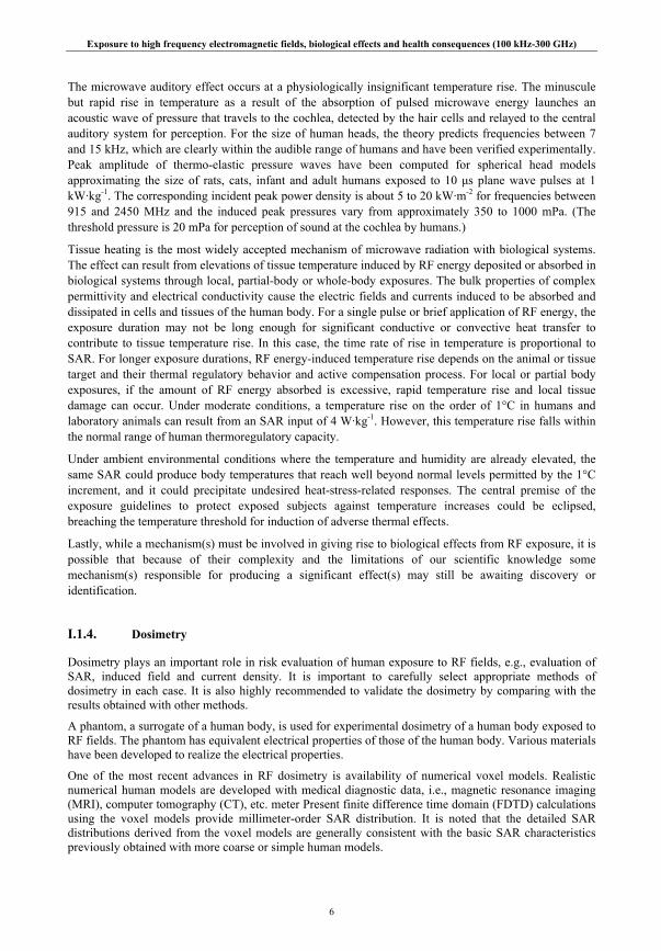
The microwave auditory effect occurs at a physiologically insignificant temperature rise. The minuscule but rapid rise in temperature as a result of the absorption of pulsed microwave energy launches an acoustic wave of pressure that travels to the cochlea, detected by the hair cells and relayed to the central auditory system for perception. For the size of human heads, the theory predicts frequencies between 7 and 15 kHz, which are clearly within the audible range of humans and have been verified experimentally. Peak amplitude of thermo-elastic pressure waves have been computed for spherical head models approximating the size of rats, cats, infant and adult humans exposed to 10 μs plane wave pulses at 1 kW·kg-1. The corresponding incident peak power density is about 5 to 20 kW·m-2 for frequencies between 915 and 2450 MHz and the induced peak pressures vary from approximately 350 to 1000 mPa. (The threshold pressure is 20 mPa for perception of sound at the cochlea by humans.)
Tissue heating is the most widely accepted mechanism of microwave radiation with biological systems. The effect can result from elevations of tissue temperature induced by RF energy deposited or absorbed in biological systems through local, partial-body or whole-body exposures. The bulk properties of complex permittivity and electrical conductivity cause the electric fields and currents induced to be absorbed and dissipated in cells and tissues of the human body. For a single pulse or brief application of RF energy, the exposure duration may not be long enough for significant conductive or convective heat transfer to contribute to tissue temperature rise. In this case, the time rate of rise in temperature is proportional to SAR. For longer exposure durations, RF energy-induced temperature rise depends on the animal or tissue target and their thermal regulatory behavior and active compensation process. For local or partial body exposures, if the amount of RF energy absorbed is excessive, rapid temperature rise and local tissue damage can occur. Under moderate conditions, a temperature rise on the order of 1°C in humans and laboratory animals can result from an SAR input of 4 W·kg-1. However, this temperature rise falls within the normal range of human thermoregulatory capacity.
Under ambient environmental conditions where the temperature and humidity are already elevated, the same SAR could produce body temperatures that reach well beyond normal levels permitted by the 1°C increment, and it could precipitate undesired heat-stress-related responses. The central premise of the exposure guidelines to protect exposed subjects against temperature increases could be eclipsed, breaching the temperature threshold for induction of adverse thermal effects.
Lastly, while a mechanism(s) must be involved in giving rise to biological effects from RF exposure, it is possible that because of their complexity and the limitations of our scientific knowledge some mechanism(s) responsible for producing a significant effect(s) may still be awaiting discovery or identification.
I.1.4. Dosimetry
Dosimetry plays an important role in risk evaluation of human exposure to RF fields, e.g., evaluation of SAR, induced field and current density. It is important to carefully select appropriate methods of dosimetry in each case. It is also highly recommended to validate the dosimetry by comparing with the results obtained with other methods.
A phantom, a surrogate of a human body, is used for experimental dosimetry of a human body exposed to RF fields. The phantom has equivalent electrical properties of those of the human body. Various materials have been developed to realize the electrical properties.
One of the most recent advances in RF dosimetry is availability of numerical voxel models. Realistic numerical human models are developed with medical diagnostic data, i.e., magnetic resonance imaging (MRI), computer tomography (CT), etc. meter Present finite difference time domain (FDTD) calculations using the voxel models provide millimeter-order SAR distribution. It is noted that the detailed SAR distributions derived from the voxel models are generally consistent with the basic SAR characteristics previously obtained with more coarse or simple human models.

Exposure to high frequency electromagnetic fields, biological effects and health consequences (100 kHz-300 GHz)
7
In the frequency range from 100 kHz to 110 MHz, induced electric field and current, and contact current should be quantified in order to evaluate the effects of shocks and burns. Several numerical methods have been used to evaluate the detailed information in the voxel human models. It is however noted that the procedure of the spatial averaging can significantly affect the evaluation.
Theoretical analysis using simple human models, such as a dielectric spheroid, shows general characteristics of SAR inside the human body, including whole-body resonance. From the 1970s, method of moments (MoM) calculations with relatively coarse block human models demonstrated various characteristics of human-body SAR and helped to establish the rationale of the reference levels of RF safety guidelines. Since the 1990s, FDTD calculations with millimeter resolution block models have contributed towards the development of RF dosimetry. These FDTD calculations show whole-body SAR characteristics similar to those obtained from MoM calculations but with wider variations of spatial averaged local SAR. The differences of the shape and structure of the voxel models and of the procedure of spatial averaging of the local SAR over 1 g or 10 g are important causes of this variation. Also the finite element method is used extensively in commercially available software to resolve sub millimeter induced currents, electric and magnetic fields and SAR at lower frequencies.
Detailed SAR distribution in a human head exposed to the near-field of a cellular phone has been derived from FDTD calculations. It is found that the antenna current distribution is one of the important factors to determine the SAR distribution and the position of the maximum local SAR.
SAR distribution inside a human body or a laboratory animal has also been evaluated experimentally. Phantoms have usually been used for experimental dosimetry of human exposure while animal cadavers have been used for dosimetry in laboratory studies. Measurement procedures with an electric field probe have been standardized for compliance tests of cellular phones to RF safety guidelines requiring high reproducibility. Experimental dosimetry based on temperature measurement has also been conducted.
Temperature elevation has been evaluated as a factor in inducing adverse health effects due to exposure to RF fields. Numerical simulation techniques using voxel human models have been developed to include complex thermal properties of a human body. Time constants of temperature elevation at locally-exposed region depend on the blood-flow convection and heat conduction while the time constant of body-core temperature due to the whole-body exposure is also affected by thermoregulatory response which results in longer time constants compared with those of partial-body exposure.
Temperature elevation of tissues associated with the localized exposure of the human head to near field of a cellular phone has been studied. The eye has been extensively investigated using various models for the temperature simulation. It has been found that tissue thermal properties influence greatly temperature elevation inside the eye. Temperature elevation in other organs of the head is an issue of equal importance. Indeed there exists good correlation between peak spatial-average SAR and maximum temperature elevation in the head. It is also clear that the presence of the handset and the battery causes temperature elevation in the skin greater than that from RF energy.
The age dependence aspect is also of relevance for dosimetry and risk assessment. It is found that the permittivity and conductivity of tissues are higher for young rats than for adult ones. Recent studies using realistic whole-body voxel models of children suggest that the whole-body averaged SAR can be higher for children than for adults. However, significant differences in SAR average over 10 g due to a cellular phone have not been found between child and adult head models in a multi-laboratory collaboration study, although some research suggest the possibility of significant increase of the child head SAR. It remains possible that the distribution of absorption within the child and adult head may be different. Pregnant female voxel models have also been developed recently. Although most of the calculated SAR of the fetus or embryo models are similar or lower than that of the mother, temperature simulation is required for a more comprehensive risk assessment of RF exposure of fetuses and embryos.
Metal objects implanted in a human body can cause enhancement of local SAR around the objects although RF exposure guidelines often do not address such situations as well as malfunction of medical implanted devices. Numerical dosimetry has revealed that the enhancement of the SAR due to the metal objects is limited to a very small area around the tip or corner of the metal objects.

Exposure to high frequency electromagnetic fields, biological effects and health consequences (100 kHz-300 GHz)
8
Above 10 GHz, a direct relationship exists between the temperature elevation and the incident power density. The power absorption is localized within the skin and some thresholds of thermal sensation have been estimated based on present data. However, more detailed dosimetry as well as the measurement of electrical properties at millimeter-wave frequencies is needed to better evaluate safety of millimeter-wave exposure.
Micro-dosimetry is the quantitative study of the spatial and temporal distributions of electromagnetic fields imparted in cellular and sub-cellular biological structures and their relationship to biological effects. Although marked field discontinuities exist at microscopic level of cell membrane, micro-thermal heating due to RF exposure is negligible. Methodologies for micro-dosimetry have been developed for microscopic dielectric theory and biochemical process, as well as the interaction of fields with biological materials, e.g., electric field manipulation of cells and electroporation.
An evaluation of uncertainty in RF dosimetry is necessary for appropriate risk assessment. While international standards exist for the evaluation of uncertainty in the maximum local SAR values for compliance tests of cellular phones, procedures to evaluate the uncertainty of the numerical dosimetry have not been established. The representativeness of the human anatomic voxel models in use is also a limitation for risk assessment. Accurate and repeatable dosimetry is essential in developing laboratory exposure systems.
I.2. PHYSICAL CHARACTERISTICS
I.2.1. Introduction
High frequency electromagnetic fields are parts of the electromagnetic spectrum between the low frequency and the optical part of the spectrum. As this part of the spectrum is used for broadcasting and telecommunication, it is termed radio frequency (RF). The RF spectrum is defined in the frequency range between 9 kHz and 300 GHz. In this review only frequencies above 100 kHz are considered.
Electromagnetic fields in this frequency range have natural or man made origin. They may have a continuous sinusoidal waveform, but more often they have a complex amplitude distribution over time. For broadcast or telecommunication purposes for example they are modulated or pulsed.
I.2.2. Quantities and units
High frequency electromagnetic fields are quantified in terms of the electric field strength E, expressed as volts per meter (V·m-1) and magnetic field strengths H, expressed as amperes per meter (A·m-1). E and H are vector fields1. In the far field of an antenna, the high frequency electromagnetic field is often quantified in terms of power flux density S, expressed in units of watt per square meter (W·m-2).
For the purpose of radiation protection physical quantities to describe sources and field properties as well as the interaction of such fields with biological systems are needed to quantify the exposure of the human body to non-ionizing radiation and to estimate the absorbed energy and its distribution inside the body (dosimetric quantities).
A dosimetric measure that has been widely adopted is the specific absorption rate (SAR), defined as the time derivative of the incremental energy δW, absorbed by or dissipated in an incremental mass, δm, contained in a volume element, δV, of a given density ρ:
1 The ratio E/H is called the intrinsic impedance and for free space it has the value of 377 ohms.

Exposure to high frequency electromagnetic fields, biological effects and health consequences (100 kHz-300 GHz)
9
⎟⎟⎠
⎞⎜⎜⎝
⎛=⎟
⎠⎞
⎜⎝⎛=
VW
tmW
tSAR
ρδδ
δδ
δδ
δδ
Eqn. 2.2.1
The SAR is expressed in watt per kilogram (W·kg-1).
Table I. 2.1.: Quantities and units used in the radiofrequency band
Quantity Symbol Unit Symbol
Conductivity σ Siemens per meter S·m-1
Permittivity ε Farad per meter F·m-1
Current I Ampere A
Current density J Ampere per square meter A·m-2
Electric field strength E Volt per meter V·m-1
Power density S Watt per square meter W·m-2
Frequency f Hertz Hz
Impedance Z Ohm Ω
Magnetic field strength H Ampere per meter A·m-1
Propagation constant k per meter m-1
Specific absorption SA Joule per kilogram J·kg-1
Specific absorption rate SAR Watt per kilogram W·kg-1
Wavelength λ Meter m
I.3. SOURCES AND EXPOSURES
I.3.1. Introduction
The man-made electromagnetic environment consists of electromagnetic fields that are produced either intentionally or as by-products of the use of electric devices. Man-made RF-sources considered here produce local field levels many orders of magnitude above the natural background. For all practical purposes of hazard assessment, therefore, the electromagnetic fields on the earth's surface arise from man-made sources.
Exposure quantities used in this chapter depend upon the exposure conditions. In the near field of a source, field strengths are quoted, whereas in the far field, where the plane wave model applies, power densities are quoted.
I.3.2. Natural high frequency fields
The natural electromagnetic environment originates from terrestrial and extraterrestrial sources such as electrical discharges in the earth’s atmosphere and radiation from sun and space (Figure I.3.1). Compared to man-made fields, natural fields are extremely small at radio-frequencies (RF). Characteristic of natural fields is a very broadband spectrum where random high peak transients or bursts arise over the noise-like continuum background.

Exposure to high frequency electromagnetic fields, biological effects and health consequences (100 kHz-300 GHz)
10
Figure I.3.1. : Terrestrial and extraterrestrial sources of radio-frequency radiation.
At lower radio frequencies, below 30 MHz, the background electromagnetic radiation is mainly due to lightning discharges during thunderstorms. In most cases it is a cloud to cloud flash but also more dangerous cloud to ground flashes are common. Satellite observations show that over land areas the annual number of lightning flashes varies from 2 to 50 km-2, the maximum arising in the tropics (Cooray 2003). The intense current pulse (up to 100 kA) associated with the discharge generates a broadband electromagnetic pulse which propagates long distances in the waveguide composed of the conducting ionosphere and the surface of the earth. The intensity and spectrum of the pulse depends on the current of the lightning discharge, distance and electric properties of the earth. At a distance of a few hundred kilometers, typical peak electric field strength and the width of the main peak of the pulse may vary from 1 to 5 V·m-1 and 10 to 50 μs respectively. At a range of 30 km the typical peak value may range from 5 to 20 V·m-1 (Willett et al 1990). At short distances less than 100 m to the ground flash the peak electric field strength may exceed 10 kV·m-1. The main part of the spectral energy of lightning pulses is distributed below 100 kHz. In the frequency band from 0.2 MHz to 20 MHz the spectral energy decays as 1/f2 and much faster above 20 MHz (Willett et al 1990).
At high radio frequencies, above 30 MHz, the natural EM-fields originate from very broadband blackbody radiation from the warm earth and from extraterrestrial processes, mainly from the sun and the extraterrestrial microwave background radiation from the whole sky (Kraus 1986; Burke and Graham-Smith 1997). It should be noted that only at frequencies above 30 MHz and below 30 GHz do electromagnetic waves penetrate the atmosphere efficiently. Below 30 MHz the ionosphere reflects the radiation back to the space and above 30 GHz attenuation is high except in narrow frequency windows. The power density of the radiation component emitted by the warm surface of the ground at 300 K temperature (27 oC) is a few mW·m-2. The extraterrestrial radiation is approximately 1000 times smaller. It is of interest to note that the blackbody radiation from a person in the RF-band is approximately 3 mW·m-2.
Exposure to high frequency electromagnetic fields, biological effects and health consequences (100 kHz-300 GHz)
11
I.3.3. Man-made fields
I.3.3.1. Telecommunications/Broadcasting
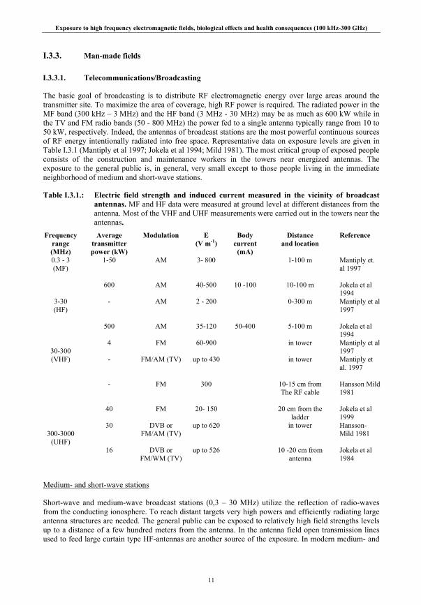
The basic goal of broadcasting is to distribute RF electromagnetic energy over large areas around the transmitter site. To maximize the area of coverage, high RF power is required. The radiated power in the MF band (300 kHz – 3 MHz) and the HF band (3 MHz - 30 MHz) may be as much as 600 kW while in the TV and FM radio bands (50 - 800 MHz) the power fed to a single antenna typically range from 10 to 50 kW, respectively. Indeed, the antennas of broadcast stations are the most powerful continuous sources of RF energy intentionally radiated into free space. Representative data on exposure levels are given in Table I.3.1 (Mantiply et al 1997; Jokela et al 1994; Mild 1981). The most critical group of exposed people consists of the construction and maintenance workers in the towers near energized antennas. The exposure to the general public is, in general, very small except to those people living in the immediate neighborhood of medium and short-wave stations.
Table I.3.1.: Electric field strength and induced current measured in the vicinity of broadcast antennas. MF and HF data were measured at ground level at different distances from the antenna. Most of the VHF and UHF measurements were carried out in the towers near the antennas.
Frequency range (MHz)
Average transmitter power (kW)
Modulation E (V m-1)
Body current
(mA)
Distance and location
Reference
0.3 - 3 (MF)
1-50
600
AM
AM
3- 800
40-500
10 -100
1-100 m
10-100 m
Mantiply et. al 1997 Jokela et al 1994
3-30 (HF)
-
500
AM
AM
2 - 200
35-120
50-400
0-300 m
5-100 m
Mantiply et al 1997 Jokela et al 1994
30-300 (VHF)
4 - -
40
FM
FM/AM (TV)
FM
FM
60-900
up to 430
300
20- 150
in tower
in tower
10-15 cm from The RF cable
20 cm from the
ladder
Mantiply et al 1997 Mantiply et al. 1997 Hansson Mild 1981 Jokela et al 1999
300-3000
(UHF)
30
16
DVB or FM/AM (TV)
DVB or
FM/WM (TV)
up to 620
up to 526
in tower
10 -20 cm from antenna
Hansson- Mild 1981 Jokela et al 1984
Medium- and short-wave stations
Short-wave and medium-wave broadcast stations (0,3 – 30 MHz) utilize the reflection of radio-waves from the conducting ionosphere. To reach distant targets very high powers and efficiently radiating large antenna structures are needed. The general public can be exposed to relatively high field strengths levels up to a distance of a few hundred meters from the antenna. In the antenna field open transmission lines used to feed large curtain type HF-antennas are another source of the exposure. In modern medium- and
Exposure to high frequency electromagnetic fields, biological effects and health consequences (100 kHz-300 GHz)
12
short-wave broadcast stations the transmitter building as well as transmitters and transmission lines are normally well shielded against electromagnetic interference and leakage fields such that RF-exposure is not a problem inside the buildings.
A typical example of exposure conditions in medium and short-wave broadcast stations is data measured in the Pori (Finland) broadcasting station (Jokela et al 1994). The MF-antenna is a vertical monopole antenna with a height of 185 m, input power of 600 kW and frequency 963 kHz. The electric field measured at a height of 1 m was 500 V·m-1 at a distance of 10 m from the antenna decreasing to 90 V·m-1 at 40 m. At the same distances the total current flowing from the feet of a grounded person decreased from 140 mA to 30 mA.
For HF -transmission the most popular antenna is a large dipole curtain antenna which is comprised of an array of half-wavelength dipoles installed in front of a reflecting mesh. As a typical example consider the exposure environment in front of the 500 kW HF-curtain antenna operating at 21.55 MHz at the Pori broadcasting station. The maximal measured electric field and total current from a grounded person are found at a distance of 30 m from the antenna where the electric field strength is 90 V·m-1 (at 1 m height) and current is 400 mA. At a distance of 100 m there is a second maximum 35 V·m-1 and 75 mA. The electric field in front of large curtain antennas does not drop below 20 V m-1 until a distance of 150 -200 m is reached. On the other hand, the field strength in the immediate vicinity of the antenna is not extremely large because the transmitter power is distributed over a large antenna area and the radiated power is not effectively concentrated into the main lobe in the reactive near field.
FM and TV
People working in FM/TV towers near high power FM/TV broadcast antennas are exposed to intense electromagnetic fields in the frequency range of 50 to 800 MHz (Jokela and Puranen 1999; Hansson-Mild 1981). Even though the power to the antenna under work may be switched off the workers may need to climb through energized antennas because the broadcast distribution companies try to minimize breaks in the transmissions. The antennas consist typically of three or four vertical dipole array antennas installed on three or four sides of the tower. Input power to the whole antenna varies typically from 10 to 50 kW and the input power to one dipole from 50-500 W even though in USA as high power as 5 kW is not uncommon (Mantiply et al 1997). The nearest dipoles are the primary source of the exposure. The secondary source of the exposure comprises of currents induced in the metallic structures of the mast. Part of that current may also couple directly to the hands and legs which are in contact with ladders and other tower structures.
Because the FM and TV antennas have been designed to radiate a disc-like beam pointed slightly below the horizon, radiation towards vertical direction along the tower is much smaller than towards the main beam which is normally inaccessible. Typically the most hazardous area is confined to a distance of about 15 m from the dipoles. In USA, however, relatively high electric field strengths from 2 to 200 V·m-1 have been measured at ground level (Mantiply et al 1997). High levels are explained by the relatively low height of the antenna in the tower and down directed side-lobe of the antenna.
In the FM band measured fields varied from 60 to 900 V·m-1 (Hansson-Mild 1981; Mantiply et al 1997; Jokela and Puranen 1999.). In the VHF TV band the exposure is generally slightly lower than in the FM band, the order of 60 V·m-1, but close to the dipoles and metallic parts of the tower high values from 400 to 900 V·m-1 have been reported. Near UHF-TV antenna elements maximum electric field may exceed 600 V·m-1. It is, however, not clear how relevant these highest field strength values are for the assessment of exposure because they may have been measured too close to the metallic parts of the tower where the fields are very non-uniform. For realistic exposure analysis the fields should be measured at a distance greater than 20 cm and averaged in terms of E2 or H2 (Jokela 2007). When the distance is 30 - 50 cm the maximal field strengths seem to remain below 300 V·m-1 and 0.8 A·m-1. The averaged electric field, measured at a realistic distance, however, may still exceed 60 V·m-1 (10 W·m-2) at 100 MHz.
In many countries terrestrial digital video (DVB-T) and audio broadcast (DAB) have or are about to replace the existing analogue broadcast systems. Schubert et al (2007) have made measurements, at more than 300 identical points, in a ‘before’ and ‘after’ switchover in parts of Germany. Statistical analysis of

Exposure to high frequency electromagnetic fields, biological effects and health consequences (100 kHz-300 GHz)
13
the measurement showed an increase in mean exposure in the center of the DVB-T starting areas which was mainly based on the increase in the radiated power at the transmitter stations. The maximal exposure value for analogue TV in the ‘before’ measurement was 0.9 mWm-2 and 6.5 mWm-2 in the ‘after’ measurement for DVB-T. A comparison of analogue FM radio and DAB showed that FM exposure was more than a factor of 10 higher. However, planned increase of DAB transmitter power to improve DAB indoor coverage will reduce this difference. Relatively high body average electric field up to 200 V·m-1 (100 W·m-2) has been measured in Finland inside a relatively small digital TV antenna. The increase is explained by the high power and small size of the antenna. If the size of the antenna remains the same as for analog UHF antennas the exposure is expected to remain the same (Jokela 2007).
Mobile and wireless communication technologies
The cellular mobile telephone industry has undergone rapid growth; in many countries the take-up rate is approaching and sometimes exceeding 100%. Wireless communication devices are used widely in all parts of modern society. Cellular mobile communication technologies have developed markedly since the early 1980s when analogue cellular radio systems were introduced in Europe. The development has proceeded through the generations described below.
1G Systems
The first generation of mobile telephones consisted of analog systems - typically operating at 450 MHz or 800/900 MHz - using frequency modulation. The Advanced Mobile Phone Standard (AMPS) was developed in the USA in the 1970s. The analog systems deployed during the 1980s in various part of the world were slightly different, namely, Nordic Mobile Telephony (NMT) mainly in the North European countries, Total Access Communication System (TACS) in some European countries, AMPS in the USA, and the Nippon Telegraph and Telephone (NTT) system in Japan. At present, the service has either stopped or is running at a low level of traffic, in most parts of the world. Apart from mobile handsets and base stations, analog systems also are used for cordless telephones. 1G provided mostly voice services.
2G Systems
2G refers to development of digital mobile communication systems (GSM or Global System for Mobile Communication) in the early 1990s. Globally, there are currently more than 1 billion users. There are a number of different systems. In Europe and parts of Asia and the Americas the GSM system is dominating. It features carrier frequencies at 900 and 1800 MHz (850 and 1900 MHz in USA). The bandwidth of each frequency channel is around 200 kHz, and a 9.6 Kbits/s data rate for encoded speech. It uses a time division multiple access (TDMA) technique - each user is ‘on’ for 4.615/8 = 0.58 milliseconds - then comes back periodically at a frequency of 217 Hz. The remaining 7/8 of the time is used for other users. So from the RF point of view it is a burst type of transmission. Apart from the access frequency of 217 Hz and its harmonics, there are various control and system signals giving rise to power variations at the frequency of 2 and 8 Hz. Japan developed its own TDMA system operating in the 1.5 GHz band. North American developed a version of a code division multiple access (CDMA) standard. This version is a so-called direct-sequence spread spectrum system where the users are ‘on’ simultaneously, but separated by different codes, which are ‘spread’ on the carrier to a wider bandwidth than dictated by the un-spread scheme. These systems carry voice, data and enable the sending of text messages
2.5G Systems
The popularity of the Internet and of personal computers created a need for higher data rates on wireless networks than available with 2G systems, which were designed mainly for voice applications. One of the systems that evolved was the general packet radio service (GPRS). The GPRS supports a data rate of up to 140.8kbit/s and is packet based rather than connection oriented. It is deployed in many places where GSM is used. GPRS achieves the higher data rates by combining several timeslots. Another system, Enhanced Data rates for GSM Evolution (EDGE) is an add-on enhancement for 2.5G GSM and GPRS networks and can carry data speeds up to 236.8 kbit/s for 4 timeslots with a theoretical maximum of 473.6 kbits/s for 8 timeslots. It meets the definition of a 3G system.
Exposure to high frequency electromagnetic fields, biological effects and health consequences (100 kHz-300 GHz)
14
3G Systems
3G is the newest digital mobile communications technology, and is also known as UMTS in Europe. It operates at frequencies between 1900 and 2200 MHz. Mobile phones are no longer used simply for voice communications, users now require video games and playback, email access, internet browsing, video telephony, high speed data access and music downloads. Hence the requirement for 3G is higher data rates, which can be as high as 384 Kbits/s and up to 2 Mbits/s in indoor environments. The global standard for 3G wireless communications, IMT-2000, is a family of 3G standards adopted by of the International Telecommunications Union (ITU). It includes the universal mobile telecommunications system (UMTS) and wideband CDMA, or W-CDMA. The common feature is the use of spread spectrum as the dominant access scheme for multiple users. The first W-CDMA system was developed in Japan under the name FOMA (freedom of mobile multimedia access) however it is currently incompatible with standard UMTS.
CDMA-2000 is the North American version of the 3G system. It differs from UMTS mainly in the network architecture. CDMA-2000 uses one or more 1.25 MHz channels for each direction of transmissions. The specific frequency bands are 1885-2025 MHz and 2110-2200 MHz, for uplink (from user to base station) and downlink, respectively. W-CDMA (UMTS) uses a pair of 5-MHz channels, one in the 1900 MHz range for uplink and one in the 2100 MHz range for downlink. Thus, UMTS has wider bandwidth requirements. UMTS supports up to 2 Mbit/s data transfer rates, although rates can drop markedly in a heavily loaded site.
Beyond 3G
4G (or beyond 3G) is the tentative descriptor for the next system in the technology and for which research is already underway. For this generation the ITU has set goals of 100 Mbits/s for general environments and 1 Gbits/s (1000 Mbits/s) for indoors. IEEE 802.16 has been engaged in developing an air interface for combined fixed and mobile broadband wireless access to support platforms moving at vehicular speeds. The system is specified to operate in the 2 and 6 GHz licensed bands suitable for mobility.
Mobile telephony networks
The mobile phone network consists of a system of adjoining zones called 'cells'. Each cell has its own base station that sends and receives radio signals throughout its specified zone. Macrocells provide the main structure for the network and the base stations have power outputs of tens of watts and communicate with phones up to a few tens of kilometers distant (35 km in the case of GSM). Microcells are used to infill and improve the main network, especially where the volume of calls is high. The microcell base stations emit less power (a few watts) and have an effective range of a few hundred meters. Picocell base stations have a lower power again (typically a fraction of a watt) and provide very short-range communication, often being sited inside buildings. The RF wave used for communication is referred to as a carrier wave. The information it carries – speech, data, photos etc – is added to the carrier wave in a process known as modulation. The change from analog to digital technology, as described above, is to meet the demand for more data and faster transmission.
Henderson and Bangay (2006) reported the results of an exposure level survey of radiofrequency electromagnetic energy originating from mobile telephone base station antennas. Measurements of CDMA800, GSM900, GSM1800 and 3G (UMTS) signals were performed at distances ranging over 50m to 500m from sixty base stations in five Australian cities. The exposure levels from these mobile telecommunications base stations were very low. The highest recorded level from a single base station was 8.1 · 10-4 W·m-2, (see Table I.3.2.).

Exposure to high frequency electromagnetic fields, biological effects and health consequences (100 kHz-300 GHz)
15
Table I.3.2.: Measurements made at nominal distances from base station tower. Measurements units are W m-2.
Measured powerflux density levels
Technology 50 m
200 m
500 m
Maximum1
CDMA (29 towers)
2.7·10-5
3.3·10-5
5.9·10-6
8.1·10-5
GSM900 (51 towers)
3.3·10-4
2.6·10-4
2.3·10-5
7.1·10-4
GSM1800 (12 towers)
3.1·10-4
4.1·10-5
4.7·10-6
4.3·10-4
3G (35 towers)
4.1·10-5
5.6·10-5
7.6·10-6
1.4·10-4
All mobile 3.8·10-4 2.8·10-4 2.8·10-5 8.1·10-4
1Maximum occurred at distances varying between 50 and 200 m.
Power density measurements were made in the vicinity of 20 randomly selected GSM microcells and picocells by Cooper et al (2006). The base stations employed a single antenna and between one and four transmitters. The antenna heights ranged between 2.5 m and 9 m and the total radiated power was in the range 1-5 W. Ninety-five percent of the data fell within two ‘tramlines’ separated by 21 dB. The average power density at a distance of 1m was about 2·10-2 Wm-2 which decreased to about 3·10-3 Wm-2 at 10 m and 2·10-6 Wm-2 at 100 m. The ‘tramlines’ had a gradient of -10 dB up to a distance of 20 m and a gradient of -40 dB per decade to longer distances.
Mobile transmitters
Mobile transmitters are usually vehicle mounted and there are no physical restrictions to prevent the public approaching even to within touching distance of them. Passengers inside vehicles with roof mounted antennas will be partially shielded from the fields and in the case of antennas mounted at the rear of a car, separations from rear passengers are likely to exceed 60 cm. The far-field distances are only between about 2 and 4.3 cm, allowing field strengths calculations for exposure assessments at all but the closest distances.
Very close to the antenna of mobile telephones very high field strengths can be measured. It is important to note that although these field strengths are high, they are highly non-uniform reactive fields which do not give rise to the same level of induced currents and heating effects as equivalent plane waves. They also only give rise to exposure over very small regions of the body.
Handsets
3G mobile phones operate at lower power levels than both GSM and CDMA handsets. The maximum power from a 3G phone (2100 MHz) is 0.125 watts produced over a 5 MHz bandwidth, whereas GSM phones (900 and 1800 MHz) emit an average power of 0.25 and 0.125 watts over a 0.2 MHz bandwidth and CDMA handsets (800 MHz) have a maximum power of 1 watt. With adaptive power control technology, handsets operate at the lowest power necessary for good radio communications. Handsets are held against the head while a call is made. Typically, the distance from the antenna to the head is only about 2 cm or less. Therefore, the user is in the near-field of the source and simple field calculations are not appropriate to assess exposure.
Terrestrial trunked radio
Terrestrial trunked radio (TETRA) is a digital mobile radio standard, with some similarities to GSM, especially designed for professional users who need high reliability and security (i.e. emergency services and commercial organizations with mobile workforces or large vehicle fleets). The standard defines four
Exposure to high frequency electromagnetic fields, biological effects and health consequences (100 kHz-300 GHz)
16
basic power classes – 1, 3, 10, and 30 W. The frequency bands recommended for use in Europe are 380-400, 410-430, 450-470 and 870-933 MHz. Vehicle mounted transmitters and hand portables have output powers of 3W and 1W respectively. Voice data are in timeslots 14.2 ms long and occur every 56.7 ms. This corresponds to a duty factor of 0.25 and a pulse frequency of 17.6 Hz. With this duty factor the average output powers will be 0.75 and 0.25 W.
Citizens band radio
Citizens band (CB) radio in the 27 MHz and 477 MHz band is used in some countries. Antennas are often mounted upon the bumpers of cars, on poles outside houses or on mobile handsets which are held close to the heads of users. Transmitters are permitted a maximum power of 4 W into a 50 Ω load. At close distances, the fields depend upon the precise length and structure of the antenna. Loading coils have a very great effect upon the near-fields of CB antennas with much stronger electric fields close to the shorter antennas. E-field strengths of 200 to 1350 V·m-1 have been measured 2 cm from low power mobile antennas (27-450 MHz, Allen, 1991). Although the field strengths are high, the relevance of such localized reactive fields for radiation protection is limited. In general the use of CB radio has fallen dramatically in recent years as the use of mobile phones and related technologies has increased.
Microwave communication links
Pairs of highly directive microwave dish antennas are used to provide line of sight communications links in a variety of applications including cellular telephony, public telecommunications, private business communications, and digital data links. Systems can usually transmit over large distances using only low power levels.
The frequencies used for microwave links are usually in the range 5 to 40 GHz and power levels range from less than 1 to a 8 W. Highly directive dish antennas are used; however, they also have many side lobes which may be the more significant in relation to public exposure but the power is usually at least 20 dB below that in the main beam.
The antennas are mounted upon towers or the tops of buildings with heights of at least 20 m, thus a typical main beam normally does not intercept the ground at distances of less than 230 m. With a radiated power of 8 W and a gain of 50 dB, the power density would be 2.4 W·m-2. Assuming a gain of 10 dB for a side lobe traveling directly downwards, the power density at 20 m from an 8 W antenna will be 0.064 W·m-2, under far-field conditions.
Satellite uplinks
Powerful and highly directive transmission systems are used to communicate between Earth stations and satellites which are usually in geostationary orbits. The antennas have very high gains ranging from 50 to 70 dB corresponding to very narrow main beam widths and operate at typical equivalent isotropic radiated powers from 50 MW to 350 GW. Therefore, in the main beam it would be possible to be exposed to power densities of a few hundred W·m-2. A 225 kW EIRP station at 2.38 GHz using a 64 m dish antenna gives a power density of 2.77 W·m-2 even at 100 km. However, the antennas are directed at satellites and of necessity nearby buildings and features have to be avoided; consequently exposure in the main lobe is most unlikely to arise under normal circumstances.
I.3.3.2. Medical applications
Diathermy and hyperthermia
The earliest therapeutic application of radiofrequency electromagnetic fields was in diathermy. Two types of diathermy are commonly used, short-wave (at 13.56 or 27.12 MHz) and microwave. Only a part of the patient's body is exposed to RF energy and exposure duration is limited (typically 15-30 minutes). However, exposure intensity is high and sufficient to cause the intended sustained increase in tissue temperature. Exposures to operators of short-wave diathermy devices may exceed 60 V·m-1 and/or 0.16

Exposure to high frequency electromagnetic fields, biological effects and health consequences (100 kHz-300 GHz)
17
A·m-1 for operators standing in their normal positions (in front of the diathermy console) for some treatment regimes. Stronger fields are encountered close to the electrodes and cables (Stuchly et al 1982). In the "worst case", high exposure of staff may occur at distances less than 1.5-2 m (27.12 MHz) or 1 m (433 MHz and 2.45 GHz, Veit and Bernhardt, 1984). However, more information is needed to fully characterize RF exposures encountered by staff in the therapeutic environment (Shah and Farrow 2007). Electromagnetic energy has also been used in inducing hyperthermia for cancer therapy where the tumor temperature is elevated to the range of 43-45 oC (Falk and Issels 2001). The procedure is mostly used in conjunction with radiotherapy and chemotherapy since the ability of ionizing radiation to kill tumor cells and the anticancer action of drugs are enhanced by hyperthermia. Systems designed for local or regional hyperthermia operating at 13.56, 27,12, 433, 915 or 2450 MHz employ induction coils, interstitial antennas, dipole arrays or waveguide applicators (Lin 1999a, 2004; Pisa et al 2003). As in diathermy, the patient is exposed to intense fields for about 30 to 60 min during hyperthermia with 20 to 100 W of RF power. While the most significant side effect is a thermal burn on the skin or subcutaneous tissue, there is relatively little information on operator exposure.
Magnetic Resonance Imaging (MRI)
MRI is an imaging technique that employs strong static, gradient, and radiofrequency magnetic fields. It can image soft tissues - unobstructed by bone - with enhanced contrast. Moreover, the ability to provide images in numerous planes without requiring the repositioning of the patient has rendered MRI a very effective and important tool for soft tissue imaging. Indeed, it has become the radiological modality of choice for a great number of diagnostic procedures.
In a clinical MRI system operating at 1.5 T, because of its design, it is unlikely that radiological staff would be exposed to significant RF fields. Some newer open 0.7 T MRI systems allow medical personnel to perform interventional procedures on patients under MRI guidance. It is possible that their hands, heads or torsos may receive significant exposure under such conditions, especially for gradient fields (ICNIRP 2004; 2008). The gradient field is pulsed rapidly in time and is a function of the imaging technique and design of the MRI system. It is significant to note that the time rate of change of the gradient magnetic field is closely related to the strength of electric field induced inside the body.
Recently, the demand for increased spatial resolution and high signal-to-noise ratio (SNR) from MRI instruments has prompted the development of systems using much higher static magnetic fields (greater than 11 T). This development has led to the use of higher RF frequencies for MRI, which, in principle, not only can augment the amount of RF power deposition inside the patient’s body, but also increases the EMF exposure for workers using MRI equipment in the hospital environment and workers employed for supporting, servicing, developing and manufacturing this equipment. There has been particular interest in the exposure of the head, torso, and limbs to the gradient fields, which may be substantial under certain operational environments.
Typical exposure levels from electromagnetic fields for medical applications are summarized in Table I.3.3.

Exposure to high frequency electromagnetic fields, biological effects and health consequences (100 kHz-300 GHz)
18
Table I.3.3.: Typical exposure levels from electromagnetic fields from medical applications
Source Frequency Distance Exposure Remarks Shortwave diathermy Microwave treatment Magnetic Resonance Imaging (MRI)
27.12 MHz 433 MHz 2450 MHz 433 MHz 2450 MHz 42-300 MHz
0.2 m 0.5 m 1 m 0.5 m 1 m 0.3-3 m Within system
<1000 V·m-1
<500 W·m-2
<140 V·m-1
100-1000 V·m-1
25 W·m-2
10 W·m-2
50-200 V·m-1
20-140 W·m-2
up to 2 W kg-1
Staff exposed Patient, untreated body parts Staff Patient, untreated body parts Whole body average Frequency depending on the static field SAR refers to normal operational mode
RF ablation
Radiofrequency ablation is a technique that uses contact electrodes to deliver low frequency (500 – 750) voltages for a wide variety of medical therapies. For over a half century, an electrosurgical knife (electro surgery) has been used by surgeons to cut and cauterize tissues as a replacement for the scalpel.
Cardiac ablation uses a catheter electrode, inserted through a vein, in the heart, without requiring opening of the chest wall or heart. An RF generator with a power of about 50 watts is used to creating lesions on the inner wall of the heart for the treatment of various cardiac rhythm disorders. These disorders are due to abnormal cardiac rhythms (arrhythmias) as a result of abnormal electrical pathways in the heart muscle (Huang and Wilber 2000; Lin 2000a; Bernardi et al 2004).
Radiofrequency ablation (RFA) for cancer therapy is a new technique that uses heat to destroy tumors deep within the body. A small needle electrode is placed directly into the tumor. The electrode's high frequency voltages create intense heat that can reach the boiling point of water, killing cancerous cells. This technique has been used to destroy liver tumors as well as renal and breast tumors (Garbey et al 2008; Gervais et al 2009; Hui et al 2008). Similarly, small interstitial microwave antennas have been used in minimally invasive medical ablation techniques (Lin 2003).
RF Telemetry
RF telemetry transmitters encapsulated in a small pill have been used to monitor internal body temperature and other physiological parameters. In addition, pills with imaging cameras have been discussed and may be developed. These devices transmit at a variety of frequencies. Since the receiver is a few meters away (outside the body) total radiated power from the pills does not exceed a few milliwatts.
Devices that are planned for use in patients must pass the safety requirements of the countries where they are sold.
I.3.3.3. Industrial and domestic applications
Intense electric and magnetic fields are used for processing of various materials by heating and sometimes by formation of plasma discharge in the material. In many applications RF-safety problems are unavoidable because RF-power is high and it may be difficult to enclose the field-generating electrodes

Exposure to high frequency electromagnetic fields, biological effects and health consequences (100 kHz-300 GHz)
19
and processing space inside a good electromagnetic shield. Consequently relatively intense stray fields and leakage radiation may arise in the vicinity of the electrodes. The manually operated older appliances in the workplaces are frequently more problematic than the new automatic appliances where the operator can control the device remotely. Consumer products such as microwave ovens are nowadays of little concern because as a rule the heating process is well shielded and the units have to meet product standards.
The main objective of this chapter is to present a brief review of high power RF sources used for material processing in industrial and domestic environments Those RF sources which produce high exposure are chosen for closer inspection. Illustrative data from various exposure situations are presented and problems with exposure assessment addressed.
Dielectric heating
High-Frequency dielectric heating is potentially one of the most important RF-exposure sources in the workplace (Mantiply et al 1997, AGNIR 2003). Dielectric heaters and sealers are intended to heat wood (glue dryers) or weld plastics (plastic sealers) by applying a strong radio-frequency electric field between two capacitive metal electrodes (ILO 1998). Plastic sealers operate at 27.12 MHz and less frequently at 40.68 MHz frequency, while glue dryers are generally operated at 13.56 MHz. The RF power varies from less than one kilowatt to tens of kilowatts for typical heat sealers, while for glue-dryers the maximum power may exceed 100 kW (ILO 1998). Most of this power is absorbed in the material to be processed, but some of the power is absorbed by the operator of the heater. The electric and magnetic fields are highly non-uniform, concentrating around the electrode.
HF dielectric heaters are used for other industrial applications such as food processing and paper making (Jones 1987). The RF power may be very high, for example 500 kW in paper making. Radiofrequency exposure is generally not as relevant as in the case of HF-sealers because the machines are well shielded and the presence of operators in the vicinity of the machine is not required due to automatic operation.
Absorption from plastic sealers and glue dryers is determined by many factors related to the appliance and work practices, such as RF-power, shielding of the electrode, thickness and dielectric properties of the material, grounding of the electrode, distance of the worker from the electrode and the duty factor. Duty factor DF ( ton/(ton + toff)) varies typically from 0.07 to 0.5 for plastic sealers and from 0.3 to 0.8 for glue dryers. The distance to the electrodes is particularly critical because the reactive electric and magnetic near-field of the dielectric heaters decays rapidly as a function of distance. Many plastic sealers continue to be operated manually by a person standing or sitting during the heating. Semi-automatic or automatic sealers, where the operator has no need to be close to the electrode during the heating, are generally less problematic. Glue dryers are one example of this category.
Several surveys (Bini et al 1986; Joyner 1986; Conover 1992) show that the spatial maximum of the peak electric field produced by some plastic sealers in the position of the operator may exceed 1000 V·m-1, particularly at the position of hands. Values in excess of 100 V·m-1 are not uncommon (Wilen et al 2004).
Induction heating
Induction heaters use strong magnetic fields at power frequencies (50/60 Hz) and radio-frequencies for heating of conducting bodies. Heating is due to ohmic and magnetic losses. The former are associated with strong currents induced by the field in the work piece and the latter with direct interaction of the field with magnetic dipoles in the material. When the frequency increases, the current concentrates due to the skin effect on the surface of the work piece. Therefore RF induction heaters are most suitable for surface processing of relatively small work pieces. The frequency and power of RF heaters vary typically from 100 kHz to 3 MHz and from 1 to 100 kW, respectively. Depending on the localization of the heated volume, the field-generating coils may vary from small single-turn devices to larger multi-turn systems. In addition to magnetic fields, electric fields may also be relevant for the exposure at frequencies above several hundred kHz because the impedance of the coil increases as a function of frequency thereby generating high voltages along the coil.

Exposure to high frequency electromagnetic fields, biological effects and health consequences (100 kHz-300 GHz)
20
Table 3.4 shows some representative exposure data in the position of the operator of RF induction heaters (AGNIR 2003; Cooper 2002). Magnetic field strength varied from 0.2 to 20 A·m-1 and electric field from 10 V·m-1 to 1600 V·m-1 in the position of head and torso.
Table I.3.4.: Measurements of electric and magnetic fields at the position of the operator of an induction heater (Cooper 2002, AGNIR 2003).
Magnetic field strength (A·m -1) Electric field strength (V m-1)
Frequency (kHz) Head Hands Abdomen Head Hands Abdomen
484 1,44 - 1,68 650 8175 500
743 0,88 0,72 0,40 160 213 32
394 1,52 12,88 5,44 168 840 70
300 0,24 0,24 0,24 16 16 8
630 1,28 0,80 0,80 35 35 23
785 14,64 9,92 0,72 929 310 36
715 18,00 - 6,72 1583 - 326
790 7,04 8,64 1,2 413 722 16
434 20,48 20,48 14,64 1192 1828 646
500 8,48 - 3,52 192 - 64
These values are in general agreement with the previous exposure surveys which showed that the exposure varied from 2 to 8000 V·m-1 and from 0.1 to 20 A·m-1 as Mantiply et al (1997) have reviewed.
Floderus et al (2002) measured relatively low values 2 A·m-1 and 0.3 A·m-1 in the vicinity of a 900 kHz hardening machine and a 1.25 MHz brazing machine, respectively. These were spot measurements at a distance of 0.5 m from the machine. The corresponding electric field strengths were 20 and 40 V·m-1. For a 400 kHz surface treatment machine they measured 4.8 A·m-1 and 160 V·m-1 at the same distance.
Estimated on the basis of electric and magnetic field strength alone, RF induction heating seems to produce exposures comparable to the exposures from dielectric RF heating (Mantiply et al 1997). However, based on exact dosimetry, the exposure is clearly lower because the coupling of the human body to the external fields is not as efficient as at higher frequencies. At the same external field level the current density and SAR arising from induction heater exposure are typically lower by a factor of 10 (current density) and 100 (SAR) than for the dielectric heater case.
Domestic induction heating
Domestic induction heating hobs (stoves or cook tops) have recently gained some popularity in Japan and European countries, even though they were introduced into the market some time ago. When electrically conducting materials are immersed in an alternating magnetic field, they can be heated as a result of eddy current losses (Joule effect). This heating technique has been applied mainly for industrial purposes, such as in metal furnaces, but it can also be used as a cooking tool. Aside from high-power (5-10 kW) equipment for commercial catering use, low-power (1-3 kW) induction heating hobs are produced as domestic kitchen appliances. Induction heating hobs operate at the intermediate frequencies of 20 to 50 kHz to take advantage of efficient energy usage and avoiding audible noise created by cooking utensils (pots, pans, and other containers) made of cast iron and stainless steel having high magnetic permeability (ICNIRP 2003; Litvak et al 2002; Wennberg 2001). More recent developments in induction heating hobs have enabled the use of aluminum cookware at higher frequencies (over 60 kHz) (Suzuki and Taki 2005).
Exposure to high frequency electromagnetic fields, biological effects and health consequences (100 kHz-300 GHz)
21
The strength of the electric field in the vicinity of induction heating hobs is much lower (a few tens of volts per meter at a distance of 10 cm from the stove edge) than the strength of the magnetic field (Stuchly and Lecuyer 1987). A typical waveform of the magnetic field consists of a carrier wave (26.1 kHz ), amplitude modulated at a frequency of 100 Hz (for 50 Hz power) or 120 Hz (for 60 Hz power). In general, the harmonic content of the amplitude modulation extends significantly higher, and the operating frequency depends on the output power setting. For a given power setting, the magnetic field strengths around the hob depend on the material and size of the utensils. The magnetic fields decrease rapidly with distance, and are characterized by the magnetic field distributions of a magnetic dipole or a current loop (Yamazaki 2004).
In practice, the magnetic field strength experienced by the user depends on the user’s position, i.e., where the operator is likely to stand (IEC 62233), or whether a person is leaning over the top of the hob or not (Stuchly and Lecuyer 1987). Numerical calculations of induced current showed that only a part of the body of the operator, in particular the hands, are significantly exposed (Burais 1998; Suzuki and Taki 2005).
Plasma discharge equipment
Very intensive RF electric fields produce plasma discharge, which are used in semiconductor fabrication processes such as etching and sputtering. The operating frequency of the plasma discharge appliances is most commonly 13.56 MHz and the power ranges from a few hundred watts to kilowatts. Measurements (Cooper 2002) indicate that the exposure of the operator is relatively low, less than 10 V m-1 for distances greater than 10 cm and 0.07 A m-1 for distances greater than 30 cm from the discharge electrode. Higher field strengths were measured at shorter distances, but these much localized fields are not very relevant for the exposure assessment. In these conditions SAR and induced current density are much lower than in the case of uniform fields. Some units were found to operate at lower frequencies (0.38 and 0.14 MHz) but, taking into account the lower frequency, the exposure does not exceed the exposure from 13.56 MHz devices.
Microwave heating and drying
Microwave energy is used for heating and drying of many materials such as foods, building materials, paper, rubber, cloths, medical supplies and chemical mixtures (Osepchuk 2002). Generation of plasma in UV-curing is a novel rapidly expanding application. The most popular and well known use of microwave energy is the cooking and heating of food at home and in restaurants and cafe’s. Most microwave heating devices operate at the frequency of 2.450 MHz but in some countries 915 MHz is also used.
Industrial microwave systems are most commonly compact batch ovens or large conveyer belts where the microwave power varies from 1 to 600 kW. Despite the large power, most systems are well shielded meeting the requirements of the product performance standards for microwave ovens (leakage radiation 50 W m-2 at 5 cm distance). Additionally, due to automatic or semiautomatic operations, operators need not stay in the vicinity of the microwave source.
Microwave levels are more likely to be a problem in mobile applications where high power microwaves are guided to the material to be heated through open applicators pressed toward the material surface, or by using small coaxial antennas drilled into the material. Asphalt processing and moisture-drying of buildings are a few examples. In moisture-drying the power density may well exceed 1000 W m-2 on the back surface of the wall being dried.
Microwave ovens
In the western world, up to 90 percent of the households own a microwave oven (Bangay and Zombolas 2003). Due to high microwave power, which typically varies from 500 to 1500 W, this consumer product is potentially hazardous. The present day domestic microwave ovens, however, have been designed and manufactured to satisfy stringent requirements set out in internationally approved product standard. The safety design of these standards aim is to reduce the leakage radiation well below 50 W·m-2 at a 5 cm distance and prevent generation of the microwave power when the door is open. Additional protection is
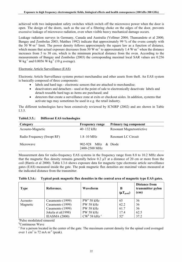
Exposure to high frequency electromagnetic fields, biological effects and health consequences (100 kHz-300 GHz)
22
achieved with two independent safety switches which switch off the microwave power when the door is open. The design of the doors, such as the use of a filtering choke on the edges of the door, prevents excessive leakage of microwave radiation, even when visible heavy mechanical damage occurs.
Leakage radiation surveys in Germany, Canada and Australia (Vollmer 2004; Thansandote et al 2000; Bangay and Zombolas 2003; Matthes 1992) indicate that approximately 99 % of the ovens comply with the 50 W·m-2 limit. The power density follows approximately the square law as a function of distance, which means that actual exposure decreases from 50 W·m-2 to approximately 1.4 W·m-2 when the distance increases from 5 to 30 cm, which is the minimum practical distance from the oven. According to the measurements of Bangay and Zombolas (2003) the corresponding maximal local SAR values are 0.256 W·kg-1 and 0.0056 W·kg-1 (10 g average).
Electronic Article Surveillance (EAS)
Electronic Article Surveillance systems protect merchandise and other assets from theft. An EAS system is basically composed of three components:
• labels and hard tags - electronic sensors that are attached to merchandise; • deactivators and detachers - used at the point of sale to electronically deactivate labels and
detach reusable hard tags as items are purchased; and • detectors that create a surveillance zone at exits or checkout aisles. In addition, systems that
activate tags may sometimes be used in e.g. the retail industry.
The different technologies have been extensively reviewed by ICNIRP (2002) and are shown in Table I.3.5.
TableI.3.5.: Different EAS technologies
Category Frequency range Primary tag component Acousto-Magnetic 40–132 kHz Resonant Magnetostrictive
Radio Frequency (Swept RF) 1.8–10 MHz Resonant LC Circuit
Microwave 902-928 MHz & 2400-2500 MHz
Diode
Measurement data for radio-frequency EAS systems in the frequency range from 8.8 to 10.2 MHz show that the magnetic flux density remains generally below 0.2 μT at a distance of 20 cm or more from the coil (Harris et al 2000). Table I.3.6 shows exposure data for magnetic type electronic article surveillance gates (EAS) measured inside the gate. The peak magnetic flux densities are maximal values measured at the indicated distance from the transmitter.
Table I.3.6.: Typical peak magnetic flux densities in the central area of magnetic type EAS gates.
Type
Reference.
Waveform
B (μTpeak)
Distance from transmitter pylon (cm)
Acousto- Magnetic
Casamento (1999) Casamento (1999) Casamento (1999) Jokela et al(1998) IEASMA (2000)
PWa 58 kHz PW 58 kHz PW 58 kHz PW 58 kHz CWb 58 kHz c
65 62.2 61.7 17.4 52c
36 36 36 62.5 37.2
aPulse modulated sinusoid bContinuous Wave c For a person located in the center of the gate. The maximum current density for the spinal cord averaged over 1 cm2 is 72 mA m-2 (peak).

Exposure to high frequency electromagnetic fields, biological effects and health consequences (100 kHz-300 GHz)
23
Radiofrequency identification (RFID)
The object of any RFID system is to carry data in suitable transponders, generally known as “tags,” and to retrieve those data by machine-readable means at a suitable time and place to satisfy particular application needs. Data within a tag may provide identification for an item in manufacture, goods in transit, a location, a vehicle, an animal or an individual.
A system requires, in addition to tags, “readers” for interrogating the tags and some means of communicating the data to a host computer or information management system.
Interrogator and reader units may be handheld, fixed or mounted on vehicles. Likewise, tags/transponders may be attached or embedded into various objects, or fixed to the ground.
Portable Data Capture systems are characterized by the use of portable data terminals with integral RFID readers. The hand-held readers/portable data terminals capture data that are then either transmitted to a host information management system via a radio frequency data communication (RFDC) link or held for delivery by line-linkage to the host on a batch processing basis.
Transfer of data between tags and a reader is by wireless communication. Two methods distinguish and categorize RFID systems, one based upon close proximity electromagnetic or inductive coupling and one based upon propagating electromagnetic waves.
The technology varies according to the required application: • low frequency 124-135 kHz tags have been accepted for near-contact reading for applications
such as access control, item identification and animal identification; • high frequency (13.56 MHz) RFID originated from smart card technology. It offers a longer
read range, typically one meter, and is being used more extensively in item management systems e.g. library systems;
• active tag technology uses 433 MHz for read ranges of up to 100 meters. The tags are used for asset tracking, with the tag signaling its presence by transmitting an identifier signal;
• the 860-960 MHz range is used for supply chain and logistics purposes. The actual band used is much narrower e.g. the European spectrum is 865-868 MHz;
• Microwave frequency (2.45 GHz) is used for logistics purposes, for factory automation applications and for active tag technology; and
• Microwave frequency (5.8 GHz) is for road traffic and road tolling systems, where active tag technology provides the range and the frequency provides fast data transfer rates.
I.3.3.4. Safety applications/navigation/radar
Radar
Radar systems mainly use microwave frequencies from 500 MHz up to around 15 GHz, although there are some systems operating up to 100 GHz. The signals produced differ from those of the other sources described in this chapter in that they are pulsed with very short duty cycles that give average powers relevant for radiation protection which are several orders of magnitude less than the peak powers.
The antennas used for radars produce main beams only a few degrees wide. In addition, many of the systems feature antennas whose direction is continuously varied by either rotating them in azimuth or varying their elevation by a nodding motion. Typically this rotation or nodding will reduce mean power by a factor of at least 100 and thus reduce root-mean-square (RMS) fields by a factor of 10. These considerations further reduce the likelihood of excessive exposure.
With stationary antennas, which represent the worst case, peak power flux densities of 10 MW m-2 may occur on the antenna axis up to a few meters from the source.
Exposure to high frequency electromagnetic fields, biological effects and health consequences (100 kHz-300 GHz)
24
Acquisition and tracking
These antennas can either rotate to perform a scan or, if they lock on to a target, point in a particular direction for an appreciable length of time. Certain tracking radar systems can produce mean power densities greater than 100 W·m-2 at distances in excess of a kilometer, even after duty cycle correction. In the case of acquisition radar systems which rotate, the effect of rotation reduces the average power density by a factor of around 100.
Air traffic control
Air traffic control (ATC) radars are scanning devices which are used to track aircraft flights and control their landings at airports. They rotate through a full 360° arc and therefore produce relatively low mean power densities in any one direction. Also, the powers used tend to be slightly lower than with tracking radars. Measurements made in the vicinity of an ATC radar operating at 2.8 GHz with a peak output power of 650 kW gave power densities with the antenna stationary of less than 0.5 W·m-2 at 60 m and 20 W·m-2 at 19 m. With a rotating antenna, the mean power densities would be lower.
In an exposure survey of civilian airport radar workers in Australia it was found that, unless working on open waveguide slots, or within transmitter cabinets when high voltage arcing was occurring, personnel were, in general, not exposed to levels of radiation exceeding the specified limits (Joyner and Bangay 1986).
Ground penetrating radar (GPR)
Ground Penetrating Radar (GPR), surface penetrating radar, or subsurface radar are all names which refer to the same technique used to locate objects and (or) interfaces situated in a region not penetrable to the eyes. GPR is similar to the conventional free space radar used to detect backscattered radiation from a target to evaluate its position and velocity. GPR systems are made of a transmitting part (source and antenna) which transmits electromagnetic power to the region under investigation, and a receiving part which collects the reflected power and, through signal processing techniques, elaborates it to extract the requested information. The presence of the interface between the air where the antenna is located and the region under investigation, and its influence on the reflected signal, are the fundamental differences between GPR and conventional radar. GPR is used as an alternative technique to seismic methods, sonar, or other specific techniques, its main advantage over those techniques being the general purpose principles of operation and the use of remote, non-contacting transducers to radiate and receive the electromagnetic energy. Moreover, it has the highest resolution in subsurface imaging of any geophysical method, approaching centimeters under the right conditions (Leon et al 1994).
The design of GPR systems is largely applications-oriented and the overall design philosophy, as well as the details, depends on the target type and the background medium. The bandwidth of the received signal is directly linked to the number of features (geological strata or buried objects) which will be resolved. Since penetration depth decreases with frequency, usually GPR systems work with frequencies less than 1 GHz. In long range investigations, frequencies as low as a few tens of MHz have been also used. On the other hand, resolution is higher for higher frequencies. Consequently, low frequency antennas (10-120 MHz) radiate long wave-length electromagnetic fields that can penetrate up to 50 meters or more in certain conditions, but are capable of resolving only very large subsurface features. In contrast, the penetration depth of a 900 MHz electromagnetic field is about one meter, and often less in typical ground conditions, but the generated reflections can resolve features down to a few centimeters in diameter (Carin 2001; Daniels 1996).
Generally, GPR systems use very narrow pulses (e.g. pulse duration of 1 ns) with low mean power (e.g. peak pulse power 50 W, mean pulse power 50 mW) and the received power is at least one order of magnitude below that transmitted. It should be noted that GPR systems, on the basis of the Federal Communications Commission (FCC) classification (FCC 2002), belong to the imaging system class; as a consequence, their transmitted power should conform to Table I.3.12, at least in the USA. Since antenna frequency, radiation pattern and radiated power strongly depend on the application, it is very difficult to define general exposure conditions with reference to GPR systems as a whole. In particular, to evaluate

Exposure to high frequency electromagnetic fields, biological effects and health consequences (100 kHz-300 GHz)
25
the operator exposure to the GPR electromagnetic field, it must be considered that the operator will be in the near-field of the transmitting antenna, so that the exposure evaluation should be conducted considering the SAR according to the formula for multiple frequency exposure (ICNIRP 1998).
Marine radar
Marine radar equipment ranges from large installations on super tankers to the smaller mast mounted equipment used by yachts. Utilization of the systems is also variable with the larger installations of cross-channel ferries being operated continuously while the battery-powered equipment of small-boat radars is used only intermittently. Generally the powers are rather lower than other radar systems with peak powers of up to 30 kW and mean powers ranging from around 1 to 25 W. Under normal operating conditions with the antenna rotating, the average power density of the higher power systems within a meter of the turning circle of the radar system can be calculated to be less than 10 W·m-2.
I.3.3.5. New and emerging technologies
Wireless LANs
Simultaneous with cellular mobile communication, significant developments have taken place in the area of Wireless Local Area Networks (WLAN), with rather short range communication between an access point (a base station) and one or several users. WLANs are ad-hoc systems set up in the home, hotels, cafes, office buildings, airports, city parks, corporate and university campuses as hotspots, and usually are connected to the Internet. It allows mobility of data terminals in a well-defined area.
WLANs have been standardized through different standards such as the IEEE Standard family, (IEEE 802.11), or the European HIPERLAN standard (HIPERLAN2). The main features of the different extensions of the IEEE 802.11 Standard are summarized in Table I.3.7.
Table I.3.7.: Summary of the WLAN Standards family IEEE 802.11 and extensions
Standard Description Frequency Data rate Year
IEEE 802.11 Original standard, exploiting the ISM band 2.4 GHz 2 Mb/s 1997
IEEE 802.11b Enhanced data rate in the ISM band 2.4 GHz 11 Mb/s 1999
IEEE 802.11a Fastest version of the standard, exploiting the UNII band 5.7 GHz 54 Mb/s 1999
IEEE 802.11g Same 802.11a speed, but in the ISM band 2.4 GHz 54 Mb/s 2003
IEEE 802.11h Modification of 802.11a to ensure usability in Europe 5.7 GHz 54 Mb/s 2003
The IEEE 802.11 standard does not impose any limit on the maximum radiated power, because such limits, together with the available frequency bands, are decided by different regulatory bodies, such as FCC (2005) in the US and CEPT (2002, 2004) in Europe. The assigned frequency bands and allowed maximum radiated powers are summarized in Table I.3.8. The modulation schemes employed by WLANs include frequency hopping and direct sequence spread spectrum in the 2.4 GHz band and orthogonal frequency division multiplexing in both the 2.4 and 5 GHz band. WLAN transmissions are intermittent, which lead to power fluctuations at the stated data rates or higher. Therefore, time-averaged powers are lower and depend on the quantity of data being transmitted.
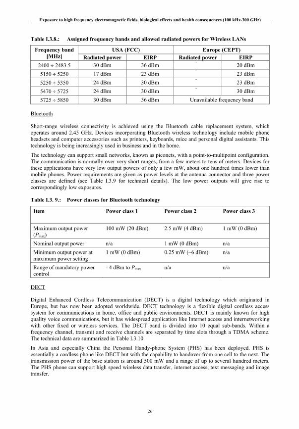
Exposure to high frequency electromagnetic fields, biological effects and health consequences (100 kHz-300 GHz)
26
Table I.3.8.: Assigned frequency bands and allowed radiated powers for Wireless LANs
USA (FCC) Europe (CEPT) Frequency band [MHz] Radiated power EIRP Radiated power EIRP
2400 ÷ 2483.5 30 dBm 36 dBm 20 dBm 5150 ÷ 5250 17 dBm 23 dBm 23 dBm 5250 ÷ 5350 24 dBm 30 dBm 23 dBm 5470 ÷ 5725 24 dBm 30 dBm 30 dBm 5725 ÷ 5850 30 dBm 36 dBm Unavailable frequency band
Bluetooth
Short-range wireless connectivity is achieved using the Bluetooth cable replacement system, which operates around 2.45 GHz. Devices incorporating Bluetooth wireless technology include mobile phone headsets and computer accessories such as printers, keyboards, mice and personal digital assistants. This technology is being increasingly used in business and in the home.
The technology can support small networks, known as piconets, with a point-to-multipoint configuration. The communication is normally over very short ranges, from a few meters to tens of meters. Devices for these applications have very low output powers of only a few mW, about one hundred times lower than mobile phones. Power requirements are given as power levels at the antenna connector and three power classes are defined (see Table I.3.9 for technical details). The low power outputs will give rise to correspondingly low exposures.
Table I.3. 9.: Power classes for Bluetooth technology
Item
Power class 1 Power class 2 Power class 3
Maximum output power (Pmax)
100 mW (20 dBm) 2.5 mW (4 dBm) 1 mW (0 dBm)
Nominal output power n/a 1 mW (0 dBm) n/a Minimum output power at maximum power setting
1 mW (0 dBm) 0.25 mW (–6 dBm) n/a
Range of mandatory power control
- 4 dBm to Pmax n/a n/a
DECT
Digital Enhanced Cordless Telecommunication (DECT) is a digital technology which originated in Europe, but has now been adopted worldwide. DECT technology is a flexible digital cordless access system for communications in home, office and public environments. DECT is mainly known for high quality voice communications, but it has widespread application like Internet access and internetworking with other fixed or wireless services. The DECT band is divided into 10 equal sub-bands. Within a frequency channel, transmit and receive channels are separated by time slots through a TDMA scheme. The technical data are summarized in Table I.3.10.
In Asia and especially China the Personal Handy-phone System (PHS) has been deployed. PHS is essentially a cordless phone like DECT but with the capability to handover from one cell to the next. The transmission power of the base station is around 500 mW and a range of up to several hundred meters. The PHS phone can support high speed wireless data transfer, internet access, text messaging and image transfer.
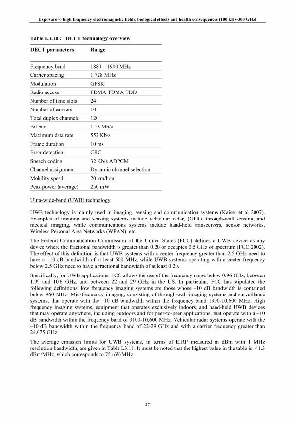
Exposure to high frequency electromagnetic fields, biological effects and health consequences (100 kHz-300 GHz)
27
Table I.3.10.: DECT technology overview
DECT parameters
Range
Frequency band 1880 – 1900 MHz Carrier spacing 1.728 MHz Modulation GFSK Radio access FDMA TDMA TDD Number of time slots 24 Number of carriers 10 Total duplex channels 120 Bit rate 1.15 Mb/s Maximum data rate 552 Kb/s Frame duration 10 ms Error detection CRC Speech coding 32 Kb/s ADPCM Channel assignment Dynamic channel selection Mobility speed 20 km/hour Peak power (average) 250 mW
Ultra-wide-band (UWB) technology
UWB technology is mainly used in imaging, sensing and communication systems (Kaiser et al 2007). Examples of imaging and sensing systems include vehicular radar, (GPR), through-wall sensing, and medical imaging, while communications systems include hand-held transceivers, sensor networks, Wireless Personal Area Networks (WPAN), etc.
The Federal Communication Commission of the United States (FCC) defines a UWB device as any device where the fractional bandwidth is greater than 0.20 or occupies 0.5 GHz of spectrum (FCC 2002). The effect of this definition is that UWB systems with a center frequency greater than 2.5 GHz need to have a –10 dB bandwidth of at least 500 MHz, while UWB systems operating with a center frequency below 2.5 GHz need to have a fractional bandwidth of at least 0.20.
Specifically, for UWB applications, FCC allows the use of the frequency range below 0.96 GHz, between 1.99 and 10.6 GHz, and between 22 and 29 GHz in the US. In particular, FCC has stipulated the following definitions: low frequency imaging systems are those whose –10 dB bandwidth is contained below 960 MHz. Mid-frequency imaging, consisting of through-wall imaging systems and surveillance systems, that operate with the –10 dB bandwidth within the frequency band 1990-10,600 MHz. High frequency imaging systems, equipment that operates exclusively indoors, and hand-held UWB devices that may operate anywhere, including outdoors and for peer-to-peer applications, that operate with a –10 dB bandwidth within the frequency band of 3100-10,600 MHz. Vehicular radar systems operate with the –10 dB bandwidth within the frequency band of 22-29 GHz and with a carrier frequency greater than 24.075 GHz.
The average emission limits for UWB systems, in terms of EIRP measured in dBm with 1 MHz resolution bandwidth, are given in Table I.3.11. It must be noted that the highest value in the table is -41.3 dBm/MHz, which corresponds to 75 nW/MHz.
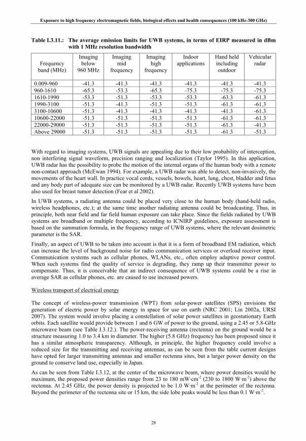
Exposure to high frequency electromagnetic fields, biological effects and health consequences (100 kHz-300 GHz)
28
Table I.3.11.: The average emission limits for UWB systems, in terms of EIRP measured in dBm with 1 MHz resolution bandwidth
Frequency
band (MHz)
Imaging below
960 MHz
Imaging mid
frequency
Imaging high
frequency
Indoor applications
Hand held including outdoor
Vehicular radar
0.009-960 -41.3 -41.3 -41.3 -41.3 -41.3 -41.3 960-1610 -65.3 -53.3 -65.3 -75.3 -75.3 -75.3 1610-1990 -53.3 -51.3 -53.3 -53.3 -63.3 -61.3 1990-3100 -51.3 -41.3 -51.3 -51.3 -61.3 -61.3 3100-10600 -51.3 -41.3 -41.3 -41.3 -41.3 -61.3 10600-22000 -51.3 -51.3 -51.3 -51.3 -61.3 -61.3 22000-29000 -51.3 -51.3 -51.3 -51.3 -61.3 -41.3 Above 29000 -51.3 -51.3 -51.3 -51.3 -61.3 -51.3
With regard to imaging systems, UWB signals are appealing due to their low probability of interception, non interfering signal waveform, precision ranging and localization (Taylor 1995). In this application, UWB radar has the possibility to probe the motion of the internal organs of the human body with a remote non-contact approach (McEwan 1994). For example, a UWB radar was able to detect, non-invasively, the movements of the heart wall. In practice vocal cords, vessels, bowels, heart, lung, chest, bladder and fetus and any body part of adequate size can be monitored by a UWB radar. Recently UWB systems have been also used for breast tumor detection (Fear et al 2002).
In UWB systems, a radiating antenna could be placed very close to the human body (hand-held radio, wireless headphones, etc.); at the same time another radiating antenna could be broadcasting. Thus, in principle, both near field and far field human exposure can take place. Since the fields radiated by UWB systems are broadband or multiple frequency, according to ICNIRP guidelines, exposure assessment is based on the summation formula, in the frequency range of UWB systems, where the relevant dosimetric parameter is the SAR.
Finally, an aspect of UWB to be taken into account is that it is a form of broadband EM radiation, which can increase the level of background noise for radio communication services or overload receiver input. Communication systems such as cellular phones, WLANs, etc., often employ adaptive power control. When such systems find the quality of service is degrading, they ramp up their transmitter power to compensate. Thus, it is conceivable that an indirect consequence of UWB systems could be a rise in average SAR as cellular phones, etc. are caused to use increased powers.
Wireless transport of electrical energy
The concept of wireless-power transmission (WPT) from solar-power satellites (SPS) envisions the generation of electric power by solar energy in space for use on earth (NRC 2001; Lin 2002a, URSI 2007). The system would involve placing a constellation of solar power satellites in geostationary Earth orbits. Each satellite would provide between 1 and 6 GW of power to the ground, using a 2.45 or 5.8-GHz microwave beam (see Table I.3.12.). The power-receiving antenna (rectenna) on the ground would be a structure measuring 1.0 to 3.4 km in diameter. The higher (5.8 GHz) frequency has been proposed since it has a similar atmospheric transparency. Although, in principle, the higher frequency could involve a reduced size for the transmitting and receiving antennas, as can be seen from the table current designs have opted for larger transmitting antennas and smaller rectenna sites, but a larger power density on the ground to conserve land use, especially in Japan.
As can be seen from Table I.3.12, at the center of the microwave beam, where power densities would be maximum, the proposed power densities range from 23 to 180 mW·cm-2 (230 to 1800 W·m-2) above the rectenna. At 2.45 GHz, the power density is projected to be 1.0 W·m-2 at the perimeter of the rectenna. Beyond the perimeter of the rectenna site or 15 km, the side lobe peaks would be less than 0.1 W·m-2.
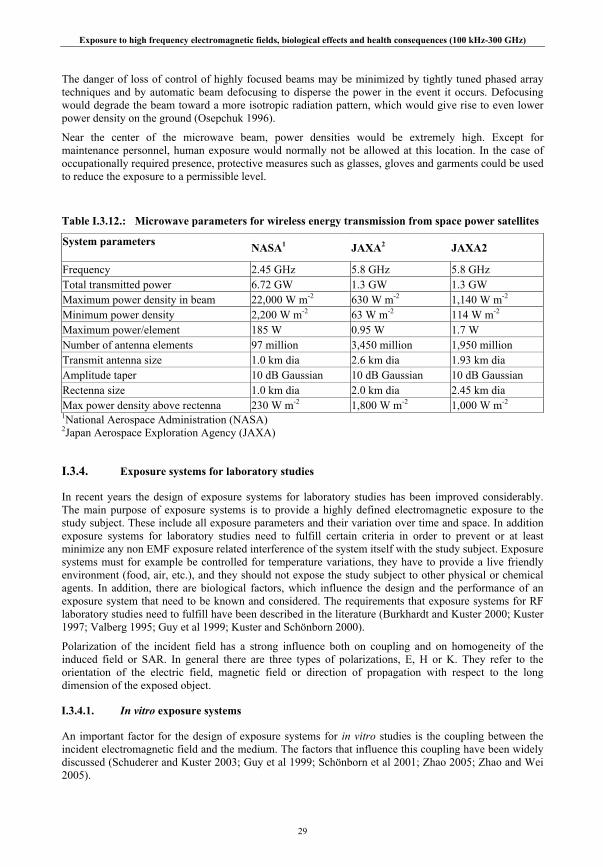
Exposure to high frequency electromagnetic fields, biological effects and health consequences (100 kHz-300 GHz)
29
The danger of loss of control of highly focused beams may be minimized by tightly tuned phased array techniques and by automatic beam defocusing to disperse the power in the event it occurs. Defocusing would degrade the beam toward a more isotropic radiation pattern, which would give rise to even lower power density on the ground (Osepchuk 1996).
Near the center of the microwave beam, power densities would be extremely high. Except for maintenance personnel, human exposure would normally not be allowed at this location. In the case of occupationally required presence, protective measures such as glasses, gloves and garments could be used to reduce the exposure to a permissible level.
Table I.3.12.: Microwave parameters for wireless energy transmission from space power satellites
System parameters NASA1 JAXA2 JAXA2
Frequency 2.45 GHz 5.8 GHz 5.8 GHz Total transmitted power 6.72 GW 1.3 GW 1.3 GW Maximum power density in beam 22,000 W m-2 630 W m-2 1,140 W m-2
Minimum power density 2,200 W m-2 63 W m-2 114 W m-2
Maximum power/element 185 W 0.95 W 1.7 W Number of antenna elements 97 million 3,450 million 1,950 million Transmit antenna size 1.0 km dia 2.6 km dia 1.93 km dia Amplitude taper 10 dB Gaussian 10 dB Gaussian 10 dB Gaussian Rectenna size 1.0 km dia 2.0 km dia 2.45 km dia Max power density above rectenna 230 W m-2 1,800 W m-2 1,000 W m-2
1National Aerospace Administration (NASA) 2Japan Aerospace Exploration Agency (JAXA)
I.3.4. Exposure systems for laboratory studies
In recent years the design of exposure systems for laboratory studies has been improved considerably. The main purpose of exposure systems is to provide a highly defined electromagnetic exposure to the study subject. These include all exposure parameters and their variation over time and space. In addition exposure systems for laboratory studies need to fulfill certain criteria in order to prevent or at least minimize any non EMF exposure related interference of the system itself with the study subject. Exposure systems must for example be controlled for temperature variations, they have to provide a live friendly environment (food, air, etc.), and they should not expose the study subject to other physical or chemical agents. In addition, there are biological factors, which influence the design and the performance of an exposure system that need to be known and considered. The requirements that exposure systems for RF laboratory studies need to fulfill have been described in the literature (Burkhardt and Kuster 2000; Kuster 1997; Valberg 1995; Guy et al 1999; Kuster and Schönborn 2000).
Polarization of the incident field has a strong influence both on coupling and on homogeneity of the induced field or SAR. In general there are three types of polarizations, E, H or K. They refer to the orientation of the electric field, magnetic field or direction of propagation with respect to the long dimension of the exposed object.
I.3.4.1. In vitro exposure systems
An important factor for the design of exposure systems for in vitro studies is the coupling between the incident electromagnetic field and the medium. The factors that influence this coupling have been widely discussed (Schuderer and Kuster 2003; Guy et al 1999; Schönborn et al 2001; Zhao 2005; Zhao and Wei 2005).

Exposure to high frequency electromagnetic fields, biological effects and health consequences (100 kHz-300 GHz)
30
In this case E-polarization provides the weakest coupling. It has been calculated that for a 60 mm Petri dish the coupling factor increases by approximately one order of magnitude between 1 GHz and 2.5 GHz. It also increases by approximately one order of magnitude when medium height increases from 2 to 5 mm. Depending on the height of the medium, coupling efficiency for an E-polarized standing wave can be up to a factor of four higher than for a plane wave. SAR is highest at the bottom of the dish. Homogeneity of the spatial SAR distribution with respect to a cell monolayer at the bottom of the dish or flask is very good. For a 60 mm Petri dish a standard deviation of approximately 20% was calculated with only little variation with medium height or frequency. For cell suspensions, however, homogeneity is poor. H-polarization or K-polarization provides a much higher coupling efficiency, but homogeneity of SAR distribution is very weak.
In addition the meniscus, which forms at the walls of dishes or flasks at the air-liquid boundary, can significantly influence the SAR distribution in the medium (Schuderer and Kuster 2003; Guy et al 1999). For a cell monolayer at the bottom of the dish, SAR values are underestimated if the meniscus is not taken into account. The magnitude of this effect depends on medium height in the dish and on frequency. In a 35 mm Petri dish with a medium height of 2 mm the error of ignoring the meniscus at 1800MHz can be approx. 60 %. At much higher frequencies this error is reduced (Zhao and Wei 2005). If cells do not settle from the meniscus in cell suspensions very high inhomogeneity in the SAR distribution of more than 100% may result.
Generally, the placement of flasks within the scattering field of other flasks can result in significant changes of the conditions, even in cases in which the magnitude of the scattering field is small. The material used in the vicinity of the flasks can significantly alter the coupling as well.
Special attention must be paid to temperature control. For plastic flasks surrounded mainly by air, the thermal coupling between the medium and the temperature controlled environment is poor, and even SAR values much below 2 W kg-1 may result in an unacceptable temperature rises (Pickard et al 2000; Schönborn et al 2000).
For in vitro exposure systems different technical solutions have been chosen. They include wave guides, transverse electromagnetic (TEM) cells, RF chambers or wire patch cells. The characteristics of these exposure systems are quite different (Schönborn et al 2001).
TEM Cell
The most commonly used system in the past has been the TEM cell, since it is small, self-contained, and can easily be placed in commercial incubators. In addition the incident field is similar to a plane wave with only transversal electric and magnetic components. If only few dishes are used per cell, the homogeneity is excellent. In an improved design Nikoloski et al (2005) modified a TEM cell to hold four T25 flasks. They achieved an overall average SAR within the medium of 6.0 W kg-1 at 1 W input power with a standard deviation of less than 52%.
However, for larger numbers of dishes the inhomogeneity increases drastically, since the E field amplitude decreases rapidly toward the wall of the cell. Therefore, the TEM cell can only be recommended for studies with a very low number of dishes.
When using TEM cells for ultra wide band exposure, it should be considered that the flasks containing the cells may disturb the field due to refraction and distortion of the incident wave combined with the excitation of resonant modes within the flasks (Ji et al 2006).
RF Anechoic Chamber
In a RF chamber an array of flasks with a dimension of several wavelengths can be simultaneously exposed. K polarization is normally employed, due to the immense power requirements for E polarization.
In a more recent design (Lyama et al 2004) for a large scale in vitro study, up to 49 Petri dishes were exposed employing a horn antenna, a dielectric lens, and a culture case in an anechoic chamber. The average SAR was 0.175 W kg-1 per 1 W antenna input power with a standard deviation of 59%. There are

Exposure to high frequency electromagnetic fields, biological effects and health consequences (100 kHz-300 GHz)
31
solutions to reduce the inhomogeneity of this design, for example by surrounding the flask with a matching box filled with liquid to the same level as the medium in the flasks.
Radial Transmission Line
The Radial Transmission Line (RTL) exposure system consists of a circular parallel plate applicator, driven at its center by a conical antenna and terminated radially by microwave absorbers or a matching load (Pickard et al 2000). An interesting feature of this system is that several dishes can be exposed at the same time and that it can be used for a wide frequency band. Moros et al (1999) positioned the flasks either directly on the metal bottom or on an aluminum oxide layer in the RTL.
Waveguide System
Waveguide systems have also been widely used in the past (Czerska et al 1992; Joyner et al1989). The flasks can be oriented in E, H or K polarization. If E polarization is employed to achieve low inhomogeneity for plated cells at the bottom of the flasks, it may be necessary to overcome the poor efficiency due to the weak coupling. One possibility that increases the efficiency by a factor of almost four is to terminate one end of the waveguide with a short circuiting plate as described in Schönborn et al (2000).
Optimized systems have been described (Schönborn 2000; Schuderer et al 2004a; Schuderer et al 2004b; Calabrese et al 2006). Depending on the type of cell culture, the frequency used and other factors, up to ten 35 mm Petri dishes are located inside a standard L-band waveguide (selected for the frequency used). The efficiency in a non resonant waveguide setting at 1.62 GHz was 1.6 W kg-1 per 1 W input power and an inhomogeneity of approximately ±30%. In a resonant design an efficiency of 50 W kg-1 per 1 W input power could be achieved with an inhomogeneity of again ±30%. In general, the efficiency of a tuned resonant system is higher than a non resonant one.
Another approach was to expose cells in a 60 mm Petri dish at the open end of a waveguide (Gajda et al 2002). Temperature was controlled by placing the Petri dish inside a 150 mm dish with circulating coolant water. The efficiency was 8.55 W kg-1 per 1 W input power and a standard deviation of SAR at the bottom of the Petri dish of 24 %.
Wire Patch Cell
The wire patch cell is basically a parallel plate resonator fed in the center of the plate, resulting in large E fields between the plates (Laval et al 2000). To reduce the inhomogeneity caused by the tangential E field, the Petri dishes with medium are placed inside larger Petri dishes filled with medium to the same height as in the smaller dishes. The efficiency reported in Laval et al (2000) is 0.6 W kg-1 per W input power at 900 MHz. The deviations from the mean value were within 12% when the evaluation was restricted to the area more than 3 mm away from the edge of each 35 mm Petri dish. This exposure system has been modified (Ardoino et al 2004) for experimental evaluations at 1800 MHz. The mean power efficiency was 1.25 W kg-1 per 1 W input power with a standard deviation of 15,2%.
I.3.4.2. In vivo exposure systems
In vivo exposure systems should in principle fulfill the same criteria as in vitro systems but with special consideration of the needs of animals, which may cause additional problems like animal movement for example. Free movement may have a huge impact on exposure homogeneity. Restraining of the animals may increase exposure homogeneity but cause unacceptable stress for the animal. In addition, animals can move even in the restraining holder, resulting in variation of exposure. It is important to evaluate inhomogeneity of exposure during the experiment taking into account the many factors affecting exposure (Kuster 2000). This includes limitations due to the animals’ body and its associated dependence on coupling mechanisms of the electromagnetic fields. Details of dosimetric differences between laboratory animals and humans are described in II.6.7. Exposure systems need to provide a clearly defined SAR distribution within the experimental animal (Kuster et al 2006). Average SAR may be misleading in cases

Exposure to high frequency electromagnetic fields, biological effects and health consequences (100 kHz-300 GHz)
32
where organ specific reactions are tested. Exposure systems for whole body and for partial body exposure have been developed and used to investigate biological effects from near field and far field exposure.
Whole-body exposure systems
TEM cells
TEM cells have been used for animal exposure (Ardoino et al 2005). The animals are usually restrained in holders. These systems operating at 900 MHz provide a mean whole body SAR in mice (24 g) of 0,38 W kg-1 per 1 W input power with a standard deviation of approximately 25 %.
Radial waveguide
Radial waveguides have been designed for whole-body exposure of different laboratory animals (Hansen 2003). Depending on the design up to 120 animals can be exposed simultaneously. Animals are typically not restrained, but can move freely in a small volume. In hamsters the typical medium whole-body SAR per 1 W input power was 1,7 mW kg-1 (±20%) at 383 MHz, 27,6 mW kg-1 (±30%) at 900 MHz, and 24,2 mW kg-1 (±30%) at 1800 MHz. The shielding factor of the system is better than 75 dB.
In a classical waveguide system (Chou et al 1984; Chao et al 1985; Chou and Guy 1987) with circular polarization at 24,50 MHz, a whole-body average SAR in mice of 3.6 W kg-1 per 1 W was reported. The absorption in animals varies considerably with body mass and orientation.
Ferris wheel
The Ferris wheel design consists of two parallel circular plates shorted around the perimeter to form a radial electromagnetic cavity fed at the center in order to excite a cylindrical TEM wave. This is a resonant system and needs an appropriate tuning. These systems have been characterized and optimized for the in vivo whole-body exposure of laboratory animals (e.g. mice) (Balzano et al 2000; Faraone 2006). The average whole-body SAR in mice in this system was 0.79 W kg-1 per 1 Watt. Over the selected range of body mass from 23 to 36 g and varying locations of the animals in the exposure system, the peak SAR variation was about 29%.
Reverberation chamber
Reverberation chamber exposure systems have been developed to overcome the body restraining to allow free movement (Kainz 2006; Jung et al 2008). This is a multimode resonant cavity exposure system. In this case the dosimetry is based on stochastic SAR values varying over time and space in a random manner.
Anechoic exposure chamber
Anechoic exposure chambers have been used for free moving as well as for restrained animals (Chou et al 1985; Chou and Guy 1987). Calorimetric measurements showed an efficiency of one system with respect to the whole body average SAR in mice ranging from 0.11 to 0.17 W kg-1 per 1 W with a standard error of 0.01 W kg-1 per 1 W depending on the orientation of the animal with respect to the electric field vector.
A system for the exposure of 100 free moving animals in a multi-generation study (Schelkshorn et al 2007; Tejero et al 2005) used a parabolic reflector with a diameter of 320 cm, to obtain a plane wave at a relative short distance. Results from a numerical simulation show that the plane wave condition has been fulfilled with a maximum phase deviation of 12° compared to an ideal plane wave. The standard deviation of the power density within the whole exposure volume, was 14.9% and 15,5 % for GSM and UMTS systems, respectively. Whole body SAR in the rats was 0002.3 mW kg-1 per 1 W with a standard deviation of 41 % at 900 MHz and 0002.5 W kg-1 per 1 W with a standard deviation of 45% at 1966 MHz.
A similar system (Wilson et al 2002) used a flared parallel plate waveguide to produce a TEM wave exposure to 18 animal cages located at the aperture plane. Average SAR efficiency with respect to whole body SAR of the free moving animals was 2 mW kg-1 per 1 W at 1.6 GHz.

Exposure to high frequency electromagnetic fields, biological effects and health consequences (100 kHz-300 GHz)
33
In mice experiments an exposure chamber attached to a horn antenna (Wang et al 2002) was used. Mice could freely move inside a plastic container. This system had an efficiency with respect to the whole-body average SAR of 0.36 W kg-1 per 1 W and a variation of ± 0.09 W kg-1 at 2.45 GHz.
Partial-body exposure systems
Carousel systems
One example of local exposure systems is the carousel-type head exposure systems, although some body exposure occurs also (Schönborn et al 2004; Wake et al 2007a; Swicord et al 1999). A dipole or monopole antenna is located in the center of circularly arranged animal holders, like a carousel with his head toward the antenna. Target organ is rat brain. The ratio of brain average to whole-body average SAR is reported to be 5 and 9 in these systems operated at 900 MHz and 1.5 GHz, respectively. The ratio is much less than that of actual mobile phone exposures in humans. The efficiency of such exposure systems varies with animal mass and ranges from 5.3 W kg-1 per 1 W for animals weighting 70-120 g to 2,8 W kg-1 per 1 W for animals weighting more than 180 g, at a frequency of 1.6 GHz. SAR varied with animal movement in the restrainers by +15% to - 30% (Schönborn et al 2004). SAR efficiency also varies with frequency.
In another carousel design (Moros et al 1998; Moros et al 1999) average SAR in the brain of small rats as measured thermo-metrically was 0.85 ± 0.34 W kg-1 per 1 W at a frequency of 835.6 MHz.
Loop antenna
Several exposure systems using a tuned loop antenna close to the head of a restrained animal have been designed (Chou et al 1999; Leveque et al 2004; Lopresto et al 2007). They can provide peak SAR values inside the skull of well above 10 W kg-1 for 1 W. Simulations showed that the ratio of the maximum local SAR in the brain of a rat exposed with a loop antenna versus a human exposed by a GSM cell phone was 1.3 ±0.6. In the human head, 20% of the brain absorbs approximately 60% of the total power deposited in the brain, compared to approximately 35% of the total power absorbed by the same percentage of rat brain. Additional exposure data obtained by such systems are summarized in Table I.3.13.
Table I.3.13.
System Frequency MHz Target Average W kg-1 per 1W
SD W kg--1 per 1 W
Ref
Loop antenna 837 brain 23.8 14.4 Chou et al 1999
whole body 1.2 4.6
1957 brain 22.6 11.3
whole body 1.1 4.6
900 brain 6,8 Leveque et al 2004
1800 cochlea 4.5 1.3 Lopresto et al 2007
I.3.4.3. Human exposure systems
Important characteristics that exposure systems for human laboratory studies have to fulfill, include well defined exposure parameters, blinded exposure, and no emission of other physical or chemical agents.
Partial-body exposure systems
There are several exposure systems used for human studies (Boutry 2008; Haarala et al 2007, Krause et al 2007; Loughran 2005; Regel et al 2006). Some studies employed modified commercial products of

Exposure to high frequency electromagnetic fields, biological effects and health consequences (100 kHz-300 GHz)
34
mobile phones. The dosimetric analysis is based on a numerical approach which has been validated by measurements with phantoms. SAR distributions in brain are estimated for each exposure system in detail. The results show that the highly exposed part is limited and the location is different from phone to phone. It has been shown that the SAR distribution from different types of cell phones could vary by more than 15 dB (Kuster et al 2004).
As an alternative to the use of cell phones, antenna systems were developed that can comfortably be worn on the head all day long and even through the night (Bahr et al 2006). Those systems simulate the exposure from a standard mobile phone with integrated antenna. The efficiency at 900 MHz (GSM signal) was estimated to be 7.66 W kg-1 per 1 W, at 1966 MHz (WCDMA signal) it was 13.3 W kg-1 per 1 W. Similar dosimetric results were reported from integrated mobile phone antennas (Manteuffel et al 2001; Kivekäs et al 2004).
Whole-body exposure systems
In two studies on human well-being and cognitive performances (Health Council of the Netherlands 2004, Regel et al 2006) a far-field, whole-body exposure system was used emitting GSM- and UMTS-like signals of 1 V m-1 incident electric field strength. A base station antenna was located at a distance of 3 m from the subject sitting in an anechoic room. Numerical calculations by FDTD method revealed that the whole-body average SAR was 6.2 μW kg-1, and that the average and peak (1 g average) SAR in brain was 11 and 73 μW kg-1, respectively for 1 V m-1 incident electric field strength of UMTS signal at 2.1 GHz (Regel et al 2006).
I.4. RF MEASUREMENT
I.4.1. Introduction
RF sources give rise to electric and magnetic fields which can directly couple into people, inducing fields and currents in their bodies. The fields from sources can also couple into objects, which can then give rise to indirect exposure when people touch the objects and currents flow into their body at points of contact.
The presentation here is concerned only with measurements performed outside the body. The external measurable quantities include electric and magnetic field strength, induced current and, on occasion, temperature. Measurements made with instruments inside the body are discussed in Chapter I.6.
Given the disparity in the type and nature of the sources, a wide range of approaches is used to evaluate exposure. There are many factors that affect instrumentation and its use in evaluating exposure for a variety of purposes; consequently, there will be particular needs associated with specific tasks. However, there are some commonalities in approach that will be highlighted here.
The electric and magnetic field components of an electromagnetic field can vary throughout space and over time in terms of their magnitude and direction. A measurement aims to gain information about these quantities that is needed for a particular purpose. The aim here is to indicate the approaches that can be used to assess exposure to RF fields to evaluate compliance with guidelines, standards and regulations, or for personal exposure assessment for health related studies.
While it is often convenient to describe the time-domain characteristics of fields, the diversity of sources requires the assessment of sinusoidal, non-sinusoidal, pulse-modulated and wideband signals. The implications for measurements of these aspects of fields are also considered here.
I.4.2. Principles of measurements
The measurement equipment must suitably record the quantities to be measured with sufficient accuracy and precision with regard to the signal characteristics and the conditions under which the measurements
Exposure to high frequency electromagnetic fields, biological effects and health consequences (100 kHz-300 GHz)
35
are made. The equipment must have a sensitivity and a frequency range suitable for the application and the measurement uncertainty must be considered.
The measurement results may be affected by environmental parameters such as temperature and humidity, the equipment itself, or interference. The latter may arise due to interactions with the operator, inadequate immunity of the equipment, including pick-up in its connecting cables, and the effect of other fields including the effect of the magnetic component in the measurement of an electric field and vice versa.
It is important that the behavior of the instrument as an entity is known insofar as its response to the characteristics of the signal(s) is being measured. The detailed frequency spectrum content and aspects of modulation and harmonics in the measured fields/currents must be taken into account. The calibration of an instrument should take into account the purpose for which it is to be used, e.g. calibration should be done using a GSM signal if an instrument is to be used for measuring GSM signals
Both narrow-band (frequency selective) and broad-band instruments can be used for assessing exposure to RF fields (Chapter I.4.4). In selecting instrumentation it is necessary to consider a number of key factors that include the response time of the instrument, peak power limitations of the sensor, polarization aspects of the field, dynamic range and the capability to measure in near- and far-fields depending on the circumstances of the field measurement.
Standardization bodies such as the International Electrotechnical Commission (IEC), the European Committee for Electrotechnical Standardization (CENELEC), and the Institute of Electrical and Electronics Engineers (IEEE) have devoted considerable efforts into developing technical standards for the assessment of EMF exposure. This has been to satisfy various needs, including product safety certification, occupational exposure legislation, and the desire to standardize the methods for making environmental measurements of electromagnetic fields. The documents are too numerous to list here and this remains a rapidly developing area. The reader is advised to consult the work of the above bodies to gain further perspective.
Guidance and suggestions for evaluating compliance with exposure guidelines have been given by the FCC in OET Bulletin 65 (FCC 1997a). The Bulletin provides advice in predicting and measuring field strengths. A supplement to the Bulletin has been published providing additional detailed information relevant to radio and television broadcast stations (FCC 1997b).
The US National Council on Radiation Protection and Measurements (NCRP) has published a report containing a practical guide to the determination of exposure to RF fields (NCRP 1993). The report outlines procedures for evaluating exposure. It also describes methods for performing practical measurements and computations of exposure specific to a number of different types of RF source.
In addition to the reports mentioned above, there are a number of monographs and technical notes produced by instrumentation manufacturers that provide advice on making measurements and using commercial products (Bitzer and Keller 1999; Kitchen 2001).
I.4.3. Characteristics of Electromagnetic Fields
I.4.3.1. General Considerations
The measurement of EM fields must account for numerous parameters including the following: • The power of each field source and the field strength it produces at the location of interest.
Relevant considerations are whether the source uses adaptive power control, produces intermittent transmissions, and whether it can produce multiple carriers.
• The modulation of the signal; that is, the time-dependent amplitude and frequency (or phase) changes of each carrier.
• Multipath propagation, wave contributions from the same source arriving at the measurement position via different reflected paths and adding constructively or destructively according to the path length in relation to the wavelength.
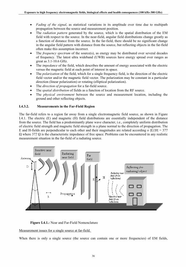
Exposure to high frequency electromagnetic fields, biological effects and health consequences (100 kHz-300 GHz)
36
• Fading of the signal, as statistical variations in its amplitude over time due to multipath propagation between the source and measurement position.
• The radiation pattern generated by the source, which is the spatial distribution of the EM field with respect to the source. In the near-field, angular field distributions change greatly as a function of distance from the source. In the far-field, there should be no significant change in the angular field pattern with distance from the source, but reflecting objects in the far-field often make this assumption incorrect.
• The frequency spectrum of the source(s), as energy may be distributed over several decades of frequency. The latest ultra wideband (UWB) sources have energy spread over ranges as great as 3.1-10.6 GHz.
• The impedance of the field, which describes the amount of energy associated with the electric versus the magnetic field at each point of interest in space.
• The polarization of the field, which for a single frequency field, is the direction of the electric field vector and/or the magnetic field vector. The polarization may be constant in a particular direction (linear polarization) or rotating (elliptical polarization).
• The direction of propagation for a far-field source. • The spatial distribution of fields as a function of location from the RF source. • The physical environment between the source and measurement location, including the
ground and other reflecting objects.
I.4.3.2. Measurements in the Far-Field Region
The far-field refers to a region far away from a single electromagnetic field source, as shown in Figure I.4.1. The electric (E) and magnetic (H) field distributions are essentially independent of the distance from the source. The field has a predominantly plane wave character, i.e., completely uniform distribution of electric field strength and magnetic field strength in a plane normal to the direction of propagation. The E and H-fields are perpendicular to each other and their magnitudes are related according o |E|/|H| = 377 Ω where 377 Ω is the characteristic impedance of free space. Problems can be encountered in any realistic measurement situation in the far-field of a radiating source.
Figure I.4.1.: Near and Far-Field Nomenclature
Measurement issues for a single source at far-field.
When there is only a single source (the source can contain one or more frequencies) of EM fields,
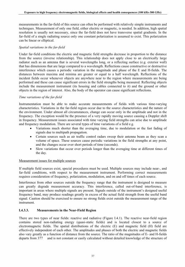
Exposure to high frequency electromagnetic fields, biological effects and health consequences (100 kHz-300 GHz)
37
measurements in the far-field of this source can often be performed with relatively simple instruments and techniques. Measurement of only one field, either electric or magnetic, is needed. In addition, high spatial resolution is usually not necessary, since the far-field does not have transverse spatial gradients. In the far-field of a single radiating source only one constant polarization is assumed to exist. This polarization can be linear or elliptical.
Spatial variations in the far-field
Under far-field conditions the electric and magnetic field strengths decrease in proportion to the distance from the source (inverse relationship). This relationship does not apply close to an electrically large radiator such as an antenna that is several wavelengths long, or a reflecting surface (e.g. exterior wall) that has dimensions that are large compared to a wavelength. Reflections cause constructive or destructive interference which causes a periodic variation in the magnitude and phase of the E and H-fields. The distances between maxima and minima are greater or equal to a half wavelength. Reflections of the incident fields occur whenever objects are anywhere near to the region where measurements are being performed and these can cause significant errors in the field strengths being measured. Reflecting objects include the measurement instrument (its housing and cables connected to it) and the ground or other objects in the region of interest. Also, the body of the operator can cause significant reflections.
Time variations of the far-field
Instrumentation must be able to make accurate measurements of fields with various time-varying characteristics. Variations in the far-field region occur due to the source characteristics and the nature of the environment. Under almost all circumstances, change can occur only in the amplitude and not in the frequency. The exception would be the presence of a very rapidly moving source causing a Doppler shift in frequency. Measurement issues associated with time varying field strengths can arise due to amplitude and frequency modulation. There are several types of time variations of a field e.g.
• Variations much shorter than the averaging time, due to modulation or the fast fading of signals due to multipath propagation.
• Certain sources such as air traffic control radars sweep their antenna beam as they scan a volume of space. These sources cause periodic variations in the field strengths at any point, and the changes occur over short periods of time (seconds).
• Slow variations that occur over periods longer than the averaging time at different times of the day.
Measurement issues for multiple sources
If multiple field sources exist, special procedures must be used. Multiple sources may include near-, and far-field conditions, with respect to the measurement instrument. Performing correct measurements requires consideration of frequency, polarization, modulation, and on and off times of each source.
Interference from other sources outside the frequency range that the instrument is designed to measure can greatly degrade measurement accuracy. This interference, called out-of-band interference, is important in areas where multiple signals are present. Signals outside of the instrument’s designed useful frequency band, may produce readings greatly in excess of the actual field strength from the useful band signal. Caution should be exercised to ensure no strong fields exist outside the measurement range of the instrument.
I.4.3.3. Measurements in the Near-Field Region
There are two types of near fields: reactive and radiative (Figure I.4.1). The reactive near-field region contains stored non-radiating energy (quasi-static fields) and is located closest to a source of electromagnetic fields. The spatial distributions of the electric (E) and magnetic field (H) field are effectively independent of each other. The amplitudes and phases of both the electric and magnetic fields also vary greatly as a function of distance from the source. The ratio of the magnitudes of E- and H-fields departs from 377 and is not constant or easily calculated without detailed knowledge of the structure of

Exposure to high frequency electromagnetic fields, biological effects and health consequences (100 kHz-300 GHz)
38
the EM source. Therefore, both E and H must be measured at every point of interest. The transition from a region where the spatial distributions for E and H are independent to one where they are correlated is gradual with increasing distance. For radiators that are small compared to a wavelength, the reactive near-field is taken as extending to
πλ 2/=r Eqn. 4.3.1
For radiators that are not small with respect to a wavelength, it is taken as extending to
)/(62.0 3 λDr = Eqn. 4.3.2
The radiating near-field region is farther away from the source. The spatial distributions of E- and H-fields are well correlated in the radiating near-field region, but the far-field radiation pattern of a source is not yet fully formed and there are changes in the angular distribution of the E- and H-fields with increasing distance. This region is defined as beyond the reactive near-field region and extends to
λ22 Dr = Eqn. 4.3.3
In the above three equations:
r = distance from the geometric center of the radiating object λ = wavelength D = the largest linear dimension of the radiator
Issues of near-field measurement do not only apply close to a traditional radiating object, such as a transmitting antenna or mobile phone handset. A reflecting object that is in the far-field of a transmitter produces near-field “radiation”. For example, a metallic object such as a structural beam in a building, an electrical cable behind a wall, or the ground is a re-radiating object that produces near-fields.
Measurement issues for a single source of near-fields
Spatial variations in the near-field
In the near-field the field strength (E or H) does not diminish in direct proportion to increasing distance from the source but more rapidly, as shown in equations 4.3.4 to 4.3.7 for a very small (infinitesimal) electric dipole. The electric field component varies with distance cubed very close to the dipole (source). Therefore, measurements of near-fields must be made at very frequent spatial intervals. Preliminary measurements must be made to estimate the spatial gradients that exist in the region of interest. Then final measurements can be performed to obtain accurate data.
θωε
ηπ
cos224 32
00⎟⎟⎠
⎞⎜⎜⎝
⎛+= −
djdehIE jkr
r Eqn. 4.3.4
θηωε
ωμπθ sin1
4 20
30
⎟⎟⎠
⎞⎜⎜⎝
⎛++= −
ddjdjehIE jkr Eqn. 4.3.5
θπφ sin1
4 20 ⎟
⎠⎞
⎜⎝⎛ += −
ddjkehIH jkr Eqn. 4.3.6

Exposure to high frequency electromagnetic fields, biological effects and health consequences (100 kHz-300 GHz)
39
where
)(m2 1−=λπk Eqn. 4.3.7
η0 = impedance of free space (377 Ω)
ε = permittivity of free space (F m−1)
μ = permeability of free space (H m−1)
h = length of the dipole (m)
I0 = antenna current (A)
ω = angular frequency (rad s−1)
λ = wavelength (m)
d = distance from center of dipole to the location of interest (m)
θ = angle between the axis of the dipole and the vector from the center of the dipole to the point r
Time variations in the near-field
When measuring time varying E- and H-fields, the factors that must be considered are identical to the factors discussed in this chapter for time variations in the far-field. These factors include changes in amplitude and frequency.
Perturbations of the near-field
Measurement instruments or other objects in the reactive near-field of a source can alter the field strengths and phases of E and H. For example, the presence of measurement personnel or an instrument at an arbitrary location in the reactive near-field of a source may change the E- and H-fields at any other nearby locations. Therefore, sensors that are used to measure fields in this region must be very small compared not only to the wavelength, but also to the field gradients.
I.4.4. Instrumentation
Traditionally, there have been two categories of instruments, namely broad- and narrowband (or frequency selective). The band refers to the frequency range that the instrument measures at a particular instant. A narrow bandwidth is one that is small with respect to the frequencies being measured and is such that two different sources can be distinguishly resolved.
Modern telecommunications systems have been developed that separate different transmitted signals on the basis of orthogonality of signals instead of frequency and/or time. Such systems include the current 3G cellular systems, which use CDMA (Code Division Multiple Access). Many signals are transmitted at the same time within the same bandwidth meaning that even a spectrum analyzer cannot separate them. In order to identify the individual signals associated with such systems, it is therefore necessary to use specialized equipment able to detect all possible signal patterns and thereby identify the power level and source of each individual signal.
There are essentially three classes of instruments used to measure external electric and magnetic fields, namely survey instrumentation, spectrum analyzers and personal exposure monitors. These types of equipment are described below, as are the instruments used to measure body current.
I.4.4.1. Broadband instrumentation for electric and magnetic fields
Portable RF measurement instrumentation, or “hazard survey meters”, provides a relatively simple and convenient means for measuring electric and magnetic field strength to assess compliance with exposure

Exposure to high frequency electromagnetic fields, biological effects and health consequences (100 kHz-300 GHz)
40
guidelines. The desired characteristics of the meters, the principles of operation of different types of probe and calibration methods have been described in the literature (see Chapter I.4.2).
Most commercially available RF survey meters are broadband devices. A broadband electromagnetic field instrument is one that ideally measures the total field (both near- and far-fields) impinging on the instrument’s sensors simultaneously regardless of modulation (amplitude and/or frequency) within the range specified by the manufacturer.
For the specific purpose of checking compliance with exposure standards, shaped frequency response instruments have been developed. They are specially designed to have RF field sensors with detection sensitivity that varies as a function of frequency. The displayed output from the instrument is a single number that is expressed as a percent of the limit from a specific frequency-dependent standard.
The major components of a broadband instrument, as shown in Figure I.4.2, are: • Field Sensor – an antenna and detection device that produces a low frequency signal
proportional to the magnitude of the total field strengths or the square of field strengths being measured. Usually the antenna is a dipole or loop that is small compared to the shortest wavelength of the field being measured.
• Data link – a resistive or metallic wire, or a fiber optic cable that carries the output of the field sensor to a display and data collection unit. The link cable usually is designed to prevent RF currents from flowing from the sensor to the display/data collection unit. Ideally the link is “transparent” to the RF field by being highly resistive at RF frequencies or being a fiber optic cable. Some broadband units do not have this data link, since the field sensor, signal conditioning and display parts are integrated into one unit that is small compared to a wavelength.
• Data processing and display – provides the signal processing, which can include filtering, amplification, summation, digitization, and a display to show the field strength, field-strength-squared, and other data. This unit may also perform signal averaging and storage and data transmission to a computer or other external computing device.
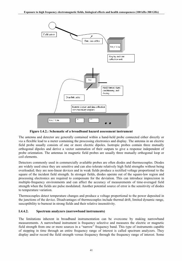
Exposure to high frequency electromagnetic fields, biological effects and health consequences (100 kHz-300 GHz)
41
Figure I.4.2.: Schematic of a broadband hazard assessment instrument
The antenna and detector are generally contained within a hand-held probe connected either directly or via a flexible lead to a meter containing the processing electronics and display. The antenna in an electric field probe usually consists of one or more electric dipoles. Isotropic probes contain three mutually orthogonal dipoles and derive a vector summation of their outputs to give a response independent of probe orientation. The antennas in magnetic field probes are usually three mutually orthogonal loop or coil elements.
Detectors commonly used in commercially available probes are often diodes and thermocouples. Diodes are widely used since they are sensitive and can also tolerate relatively high field strengths without being overloaded; they are non-linear devices and in weak fields produce a rectified voltage proportional to the square of the incident field strength. In stronger fields, diodes operate out of the square-law region and processing electronics are required to compensate for the deviation. This can introduce imprecision in multiple-frequency environments and can affect the accuracy of measurements of time-averaged field strength when the fields are pulse modulated. Another potential source of error is the sensitivity of diodes to temperature variation.
Thermocouples detect temperature changes and produce a voltage proportional to the power deposited in the junctions of the device. Disadvantages of thermocouples include thermal drift, limited dynamic range, susceptibility to burnout in strong fields and their relative insensitivity.
I.4.4.2. Spectrum analyzers (narrowband instruments)
The limitations inherent in broadband instrumentation can be overcome by making narrowband measurements. A narrowband instrument is frequency selective and measures the electric or magnetic field strength from one or more sources in a “narrow” frequency band. This type of instruments capable of stepping in time through an entire frequency range of interest is called spectrum analyzers. They display and/or record the field strength versus frequency through the frequency range of interest. Some
Exposure to high frequency electromagnetic fields, biological effects and health consequences (100 kHz-300 GHz)
42
spectrum analyzers are designed to display the field strength at each frequency as a percentage of the exposure limit in a specific frequency-dependent standard.
The instrument is designed to measure near-fields, the dimensions of the probe sensor should be a small fraction of a wavelength at the highest operating frequency. The sensor and other instrument components should not produce significant scattering of the incident electromagnetic fields.
The sensor response should be isotropic (independent of orientation), non directional, and not sensitive to the polarization of the fields to be measured. A sensor with a non isotropic response is useful if the polarization of the measured quantity (E or H) is known or if the sensor can be rotated to find the direction of polarization. The leads from the sensor to the meter should not interact significantly with the field or conduct RF current from the field to the sensor.
A spectrum analyzer generally employs a broadband antenna in conjunction with a narrowband tunable receiver that provides the frequency and amplitude of the signal to which it is tuned. Spectrum analyzers are tunable over a wide frequency range and they can be used to display the variation of amplitude over a specified portion of the spectrum. An example of the equipment showing antenna, spectrum analyzer and computer control is shown in Figure I.4.3. Most types of antennas used in conjunction with spectrum analyzers for narrowband measurements are not isotropic. Therefore, three measurements are required to determine the vector-summed resultant field strength if the direction of propagation and/or the frequency are unknown. The antennas also tend to be large, since they contain resonant elements, and this may give rise to perturbation in the near-field and prohibit measurements with high spatial resolution. Moreover, the antennas may couple with nearby dielectric bodies, including the operator, which complicates the measurement.
Figure I.4.3.: Typical spectrum analyzer measurement kit
There are many parameters that have to be carefully set when using a spectrum analyzer in order to obtain a reading of the signal, e.g. the RMS field strength. Some of these are as follows:
• Frequency span – this is the bandwidth over which the analyzer sweeps. The sweep is not continuous, but made in discrete frequency steps.
• Resolution bandwidth – the bandwidth with which the analyzer measures the field strength at a particular frequency. The measurement is typically made with a Gaussian filter. Insufficient resolution bandwidth may result in under-reading of the field strength.
• Number of points – the number of discrete frequencies at which measurements are made over the frequency span.
• Dwell time – the dwell time at any particular frequency is defined by the sweep time divided by the number of points.
• Detector –several types of detectors are provided in most spectrum analyzers for average or peak measurement.
A new generation of frequency-selective instruments has been developed for easy-to-perform frequency-selective measurements. These portable spectrum analyzers with tailor-made software measure multiple signals at different frequencies and then sum the results in the context of a given set of exposure

Exposure to high frequency electromagnetic fields, biological effects and health consequences (100 kHz-300 GHz)
43
guidelines and assigning percentage contributions to different signals. In some cases the measuring antennas are placed on tripods in order to minimize interaction with the operator, while in other cases the entire instrument (probe and spectrum analyzer) is in a single hand-held unit.
I.4.4.3. Personal exposure monitors
For studies of human exposure it is important to have meaningful estimates of exposure over time. Personal exposure assessments have been made using exposure data obtained from spot measurements, taken at a point in time and space where a person may be present. Measurements are generally made of the electric field strengths and plane-wave equivalent power densities, and exposures are estimated based on time and motion investigations. More recently, instruments using personal exposure monitors worn on the body have been developed to enable exposure estimates. The type of monitor varies depending on the exposure environment. Workers on antenna sites have worn pocket-sized devices and more sensitive instruments have been developed to capture relatively low level exposures of the general population over a range of frequency bands used for telecommunications. The characteristics of these types of devices allow data logging over extended periods of time and activity.
While personal monitoring may be very useful for categorizing exposure of groups of people for epidemiological studies, the perturbation of the incident field by the body may result in considerable uncertainty.
I.4.4.4. Body current measurements
In addition to the measurement of external electric and magnetic fields, in some circumstances it is possible to measure currents induced as a result of exposure to RF fields. There are two main types of body current meter. Transformer clamps measure the currents flowing through limbs while foot current meters measure the current flowing through the feet to the ground. Meters are also available for measuring contact current as a result of a person contacting conducting objects.
Foot current meters
Current flowing between the feet and the ground can be measured using two parallel conducting plates, separated by a slab of dielectric material and short circuited via a small resistance. The individual stands on the upper plate and the lower plate is placed on the ground. The induced current is calculated by Ohm’s law from the potential difference measured across the resistor using a voltmeter incorporating e.g. a diode detector. Alternatively the resistor and detector could be replaced by a thermocouple RF milliammeter connected in series with the two plates. Foot current meters may be appropriate for measurements at ground level but are of limited use if carrying out measurements above ground.
Current transformers
Clamp-on current transformers have the advantage over foot current meters in that they can be used in a greater range of environments. The clamp consists of a solenoid wound around a ferrite core and the current induced in the coil provides a direct measurement of current flowing through the region of interest in the body. Clamp-on instruments have been developed that can be worn and are generally placed around the wrist, ankle or neck (Blackwell 1990).
The meter display unit can be mounted either directly on the transformer or connected through a fiber-optic link to indicate the current flowing through the clamped limb. Current sensing in these units may be accomplished using either narrowband techniques such as spectrum analyzers or tuned receivers or broadband techniques using diode detection or thermal conversion. Instruments have been designed to provide true RMS indications.
The upper frequency response of ferrite-cored current transformers is around 250 MHz. Lighter air-cored transformers have been used to extend the upper frequency response of these instruments but they are significantly less sensitive than ferrite-cored devices.
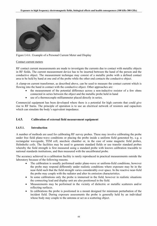
Exposure to high frequency electromagnetic fields, biological effects and health consequences (100 kHz-300 GHz)
44
Figure I.4.6.: Example of a Personal Current Meter and Display
Contact current meters
RF contact current measurements are made to investigate the currents due to contact with metallic objects in RF fields, The current measurement device has to be inserted between the hand of the person and the conductive object. The measurement technique may consist of a metallic probe with a defined contact area to be held by hand at one end of the probe while the other end contacts the conductive object.
A clamp-on current transformer, as described above, can be used to measure the contact current which is flowing into the hand in contact with the conductive object. Other approaches are:
• the measurement of the potential difference across a non-inductive resistor of a few ohms connected in series between the object and the metallic probe held in hand
• use of a thermocouple milliammeter placed directly in series.
Commercial equipment has been developed where there is a potential for high currents that could give rise to RF burns. The principle of operation is to use an electrical network of resistors and capacitors which can simulate the body’s equivalent impedance.
I.4.5. Calibration of external field measurement equipment
I.4.5.1. Introduction
A number of methods are used for calibrating RF survey probes. These may involve calibrating the probe under free field plane-wave conditions or placing the probe inside a uniform field generated by, e.g. a rectangular waveguide, TEM cell, anechoic chamber or, in the case of some magnetic field probes, Helmholtz coils. The facilities may be used to generate standard fields or use transfer standard probes whereby the field strength is first measured using a standard probe with known calibration traceable to national standards institutions, and then measured with the uncalibrated probe.
The accuracy achieved in a calibration facility is rarely reproduced in practical measurements outside the laboratory because of the following reasons.
• The calibration is usually performed under plane-wave or uniform-field conditions, however the probe may respond differently under realistic conditions where exposure may be in the near-field such that the field strength varies considerably over space. In the reactive near-field the probe may couple with the radiator and alter its emission characteristics.
• In some calibrations only the probe is immersed in the field, however in realistic situations the connecting lead and display unit are also positioned in the field.
• Measurements may be performed in the vicinity of dielectric or metallic scatterers and/or reflecting surfaces.
• In calibrations the probe is positioned in a mount designed for minimum perturbation of the incident field. During exposure assessments the probe is generally held by an individual whose body may couple to the antenna or act as a scattering object.

Exposure to high frequency electromagnetic fields, biological effects and health consequences (100 kHz-300 GHz)
45
I.4.5.2. Factors for consideration
Apart from the effects of temperature and instrument stability drift over time there are a number of factors that can materially affect the accuracy of RF instruments.
• Frequency response – calibrations are ideally carried out at frequencies over which the instruments are to be used and at field strengths that are comparable with mid-range readings or above. The response should be reasonably flat over the design frequency range and in the range 1-3 dB.
• Linearity – a range of levels between 25% and 100% of full scale on each range should permit a good assessment of linearity.
• Out of band response – the potential effect of signals outside of the specified frequency response of an instrument need to be considered. Such signals which could originate from multiple sources or harmonics and can potentially affect any element of the instruments construction e.g. the sensor/detector, connecting cables and readout.
• Near-field response – where the instrument may be used in either high or low impedance fields encountered in near-fields, the response of an instrument designed for E or H should be appropriately evaluated to examine the response to the H or E-fields respectively. This can be achieved using mismatched TEM cells up to about 300 MHz.
• Modulation – the modulation characteristics of sources are important considerations particularly where pulsed modulation associated with digital equipment or with radar signals where peak to mean power duty factors may be in the order of 0.001.
• Isotropy – instruments using orthogonal arrays of sensors should be insensitive to the direction of propagation of the incident field however there will be some uncertainty in the sensitivity of individual antenna/detector elements that can be ascertained by rotation of the probe about the handle axis. Another aspect of the isotropic nature of probes can be assessed by rotating the probe handle through the electric field plane.
• Interference – the possibility for RF interference occurring with some component of the instrument should be considered, particularly if calibrations are carried out without all components of the equipment in the field.
I.4.5.3. Uncertainty budgets
In carrying out calibrations in facilities such as those referred to above, the effect of scattering objects and the conducting parts of the RF instrumentation being calibrated will disturb the incident field. In general it would be expected that the uncertainty should not exceed 2 dB and in some circumstances may be less. Uncertainty for TEM cell calibrations may be as little as 5%, but 10% is more typical. For GTEM cells, where the field strength cannot be simply calculated from the power and cell geometry, it is likely that a transfer standard field sensor will provide the lowest uncertainty for calibration.
In addition to the uncertainty in the calibration procedures, there are other measurement factors that will affect the overall uncertainty when using RF field instrumentation in particular situations. These will include temperature and drift effects, resolution of the display, issues related to the relative location of the RF source and the measurement probe, positioning of the sensor, nature of polarization, perturbation of measurement by people and the degree of repeatability. All of these will contribute to the derivation of the expanded uncertainty budget which may be much larger than the calibration uncertainty but may be reduced by adopting approaches to minimize the uncertainty on some of the foregoing factors.
Exposure to high frequency electromagnetic fields, biological effects and health consequences (100 kHz-300 GHz)
46
I.5. MECHANISMS OF INTERACTION
I.5.1. RF exposure and coupling into biological systems
When a radio frequency electromagnetic field in air impinges on a biological body it is reflected, transmitted, refracted or scattered by the biological body; the refracted and scattered fields may proceed in directions different from that of the incident RF field. These phenomena are described and governed by the well-known Maxwell’s equations of electromagnetic theory. The transmitted and refracted fields from the RF exposure induce electric and magnetic fields in the biological systems that interact with cells and tissues in a variety of ways, depending on the frequency, waveform, and strength of the induced fields and the energy deposited or absorbed in the biological systems. Thus, to achieve a biological response, the electric, magnetic or electromagnetic field must couple into and exert its influence on the biological system in some manner, regardless of what mechanism(s) may be accountable for the response.
Nevertheless, knowledge of the specific mechanism responsible for a given observed biological effect is of scientific interest because: (1) they facilitate understanding of the phenomenon, (2) they help in analyzing relationships among various observed biological effects in different experimental models and subjects, and (3) they serve as guides for comparison and extrapolation of experimental results from tissue to tissue, from tissue to animal, from animal to animal, from animal to human, and from human to human undergoing RF exposure. Therefore, it is important in assessing the health and safety risk of RF energy to determine not only the fields induced in biological tissues, but also the mechanisms underlying its biological interactions with cells, tissues and the human body. However, while a mechanism(s) must be involved in giving rise to biological effects from RF exposure, it is possible that because of their complexity and the limitations of our scientific knowledge some mechanism(s) responsible for producing a significant effect(s) may still be awaiting discovery or identification.
As mentioned in chapter I.4.3., radiation of RF electromagnetic energy is accomplished through the use of antennas, applicators, or radiators. The spatial distribution of RF energy from an antenna is directional and varies with distance from the antenna. At distances sufficiently far from an antenna so that the RF field distribution changes only with distance, not angle or orientation, the region is called a far field or radiation zone. At lesser distances, the energy distribution in the near field or zone is a function of both angle and distance. Moreover, the behavior of RF fields and their coupling and interaction with biological systems are very different in the near and far zones.
The demarcating boundary between near and far zones occurs at a conservative distance of R = 2 D2 λ-1, where D is the largest dimension of the antenna. Furthermore, the near zone can be divided into two subregions: the radiative region and the reactive region. In the radiative region, the region close to and within 2 D2 λ-1, the radiated power varies with distance from the antenna. The vicinity of the antenna where the reactive components predominate is known as the reactive region. The precise extent of these regions varies for different antennas. For most antennas, the transition point between reactive and radiative regions occurs from 0.2 to 0.4 D2 λ-1. For a short dipole antenna, the reactive component predominates to a distance of approximately λ/2π, where the radiative and reactive components are equal to each other. However, the outer limit is on the order of a few wavelengths or less in most cases (Lin 2000b; 2007).
At the lower radio frequency of 100 kHz, the wavelength in air is 3 km and the λ/2π distance is about 477 m for the reactive and radiation fields to have equal amplitudes. In contrast, at 900 MHz, the wavelength in air is 33 cm and the λ/2π distance is 5.3 cm, which comes very close to the 2 D2 λ-1 distance of 6 cm for a 10-cm RF antenna operating at 900 MHz in air. Clearly, both near-zone reactive and far-zone radiative interactions are encountered in the vicinity of personal wireless telecommunication systems.
Some of the salient features of near zone field are: (1) RF electric and magnetic fields are decoupled, quasi-static, and are not uniform, (2) wave impedance varies from point to point, (3) beam width from the antenna is divergent and is small compared with the head or human body, especially for a small antenna, (4) the power varies less with distance from the antenna and (5) the power transfers back and forth may be nearly constant between the antenna and its surrounding medium.
Exposure to high frequency electromagnetic fields, biological effects and health consequences (100 kHz-300 GHz)
47
In the far zone, RF fields are characterized as follows: (1) they have plane wave fronts and are independent of source configurations, (2) the radiated power decreases monotonically with distance from the antenna, and (3) the electric and magnetic fields are uniquely defined through the intrinsic impedance of the medium. Thus, a determination of the electric or magnetic field behavior is sufficient to characterize the exposure in terms of power density.
An important consideration in RF exposure is the coupling of RF fields and their distribution inside the body. This association is also valuable in human epidemiological investigations on the health effects of RF field usage. The coupling of RF electromagnetic energy into biological systems may be quantified by the induced electric and magnetic fields, power deposition, energy absorption, and their distribution and penetration into biological tissues. These quantities are all functions of the source and its frequency or wavelength, and their relationship to the physical configuration and dimension of the biological body. Furthermore, the coupling is more complicated in that the same exposure or incident field does not necessarily provide the same field inside biological systems of different species, size, or constitution. Additionally, the interaction of RF energy with biological systems depends on electric field polarization, especially for elongated bodies with a large height-to-width ratio.
It is emphasized that the quantity of induced field is the primary driving force underlying the interaction of electromagnetic energy with biological systems. The induced field in biological tissue is a function of body geometry, tissue property, and the exposure conditions. Moreover, determination of the induced field is important because: (1) it relates the field to specific responses of the body, (2) it facilitates understanding of biological phenomena, and (3) it applies to all mechanism of interaction. Once the induced field is known, quantities such as current density (J) and specific energy absorption rate (SAR) are related to it by simple conversion formulas. In this case, for an induced electric field E in V·m-1, the induced current density is given by
z)y,z)E(x,y,(x, z)y,J(x, σ= Eqn. 5.3.1
where σ is the electrical conductivity (S m-1) of biological tissue and SAR is given,
⎥⎦
⎤⎢⎣
⎡=
kgW
zyxzyxEzyx
zyxSAR rms
),,(),,(),,(
),,(2
ρσ
Eqn. 5.3.2
where ρ is the mass density of the tissue (kg m-3).
At lower frequencies, e.g., 100 kHz or 10 MHz, where the wavelength of RF radiation is at least an order of magnitude longer than the dimensions of the human body, field behavior inside the body is characterized by near-zone reactive field and is quasi-static in character. The electric and magnetic fields become decoupled, and they act separately and additively inside tissue medium (Lin et al 1973; Lin 2000b; 2007). For all practical purposes, the induced fields can be obtained by combining the two independent quasi-static electric and magnetic solutions of the electromagnetic field theory. For example, an externally applied uniform electric field gives rise to a uniform induced electric field inside the body that is in the same direction, but reduced in strength by a factor inversely proportional to the dielectric constant and is independent of body size. The magnetically induced electric field amplitude inside the body is given by
),,(2),,(),,( zyxHfrrzyxBzyxE μπω == Eqn. 5.3.3
where f = ω/2π is the frequency, µ is magnetic permeability, r is the equivalent radius of a region with homogeneous electrical conductivity, B is magnetic flux density, and H is the strength of the magnetic field component. A uniform magnetic field produces an internal electric field that increases in proportion with distance away from the body center. Thus, magnetically induced electric field, i.e., inductive coupling, would dominate inside a biological body except for tissue bodies that are 1 mm or less in size. A similar scenario exists in the near-zone-reactive region of all antennas and radiating systems. A case in point, the interaction of a cellular mobile telephone handset with the user’s head is quasi-static in nature and inductive coupling of antenna-current-generated magnetic field dominates power deposition in the head.

Exposure to high frequency electromagnetic fields, biological effects and health consequences (100 kHz-300 GHz)
48
I.5.2. Biophysical mechanisms of interaction
I.5.2.1. Ionization potential of RF fields
Electromagnetic energy may be thought of as being carried by photons or quanta. In this case, the energy (E) of a photon is given by E = hf, where h is the Planck’s constant = 6.625·10-34 J·s, and f is frequency in Hz. Note that 1 eV (electron volt) is equal to 1.602·10-19 J, and the frequency of 1 eV photon is equal to 2.418·1014 Hz. Therefore, the higher the frequency, the higher the energy per photon. A definite amount of photon energy is required to produce ionization by ejection or promotion of orbital electrons from atoms of the material through which an electromagnetic wave propagates. The minimum photon energies capable of producing ionization in water and in atomic carbon, hydrogen, nitrogen, and oxygen are between 10 and 25 eV. Inasmuch as these atoms constitute the basic elements of living organisms, 10 eV may be considered as the lower limit for ionization in biological systems.
A single photon of RF radiation has relatively low energy levels, less than 1.24·10-5 eV; therefore it is not capable of ionization. Accordingly, electromagnetic radiation in the RF spectrum is regarded as non-ionizing radiation. The deleterious biological effects of such ionizing radiations as gamma- and x-rays that largely result from ionization taking place in biological cells and tissues are not produced by a single photon of RF radiation (Lin 1978). It is noted that for strong RF fields, simultaneous absorption of 8.06·105 or more low energy RF photons, could potentially produce ionization in biological materials, but the probability is small. The point is that RF radiation has low energy photons, therefore under ordinary circumstances, RF radiation is too weak to affect ionization or cause significant damage to biological molecules such as DNA, which is especially renowned for its repair mechanism.
I.5.2.2. Induced charge and dipole relaxation
Polar molecules such as water and other cellular components of biological materials can translate and rotate in response to an applied sinusoidal electric field. The translation and rotation is impeded by inertia and by viscous forces. Therefore, the orientation of polar molecules does not occur instantaneously, giving rise to a time-dependent behavior known as the relaxation process. Moreover, cells and tissue structures carry different electric charges. When subjected to a sudden electrical stimulation they require a finite time for charges to accumulate at the interfaces and to equilibrate. The accumulation of charges at the interfaces continues until a condition of equilibrium is re-established, leading to the relaxation phenomenon. Many types of relaxation processes can take place in biological tissues, owing to polar molecules and membrane charges.
When a dipole distribution is uniform, the positive charges of one dipole cancel the effect of the negative charges from another adjacent dipole. However, when the dipole distribution varies from point to point, this complete cancellation cannot occur. At an interface especially, the ends of the dipoles leave an uncancelled charge on the surface, which becomes an equivalent bound charge in the material. The relaxation process may therefore be illustrated by considering the response of bound charges to an applied electric field (Lin 2000b; Michaelson and Lin 1987). In this case, the dynamic force balance equation is given by
dtdxmxmqE
dtxdm s νω −−= 22
2
Eqn. 5.4.4
where x is the displacement of a charged particle, E the applied electric field, ωs is the characteristic frequency of the elastic, spring-mass system, ν is the particle collision frequency, and m and q are the mass and charge of the particle, respectively. The force exerted on the particle -- mass multiplied by particle acceleration on the left-hand side of equation (5.4.4), results from an electric driving force qE, an elastic restoring force in proportion to displacement x with elastic constant denoted as m ωs
2, and a retarding damping force proportional to velocity dx/dt with damping coefficient, mν.

Exposure to high frequency electromagnetic fields, biological effects and health consequences (100 kHz-300 GHz)
49
After Fourier transformation and rearranging terms, equation (5.4.4) becomes
)][])[()( 22 ωνωωω jEmqx s +−= Eqn. 5.4.5
Note that the equilibrium position for the charge (x = 0) represents local charge neutrality within the medium. When the charge is displaced from its equilibrium position, a dipole is established between the charge itself and the „hole” that is left behind and bound in the molecular and membrane structure. A dipole moment p is formed by the charge q times the displacement x. For a medium with volume-bound charge density ρ, the total dipole moment per unit volume or polarization P is
)][])([ 222 ωνωωρρ jEmqpP s +−== Eqn. 5.4.6
The electric flux density D may be expressed in terms of the electric field E and polarization P as
PEDrrr
+= 0ε Eqn. 5.4.7
For isotropic media, the permittivity may be related to D by the expression D = εE. These relations together with equation (5.4.6), give an equation for the permittivity,
)]()(1[)( 2220 ωνωωωεωε jsp +−+= Eqn. 5.4.8
Where
022 ερω mqp = Eqn. 5.4.9
and ε0 is the vacuum or free-space permittivity. Clearly, ε is a complex quantity and can be denoted by
εεε ′′−′= j Eqn. 5.4.10
where ε′ and ε″ are the real and imaginary parts of the permittivity and can be obtained by equating the real and imaginary parts of equations (5.4.8) and (5.4.10). The relationship between electrical conductivity σ and ε″ is derived from Maxwell’s equations and it is
εωσ ′′= Eqn. 5.4.11
The velocity of bound charge motion v = dx/dt can be obtained from equation (5.4.5), such that
])([])[()( 22 ωωωνω −−= sjEmqv Eqn. 5.4.12
The finite velocity of charge motion in the material media indicates that the particle cannot respond instantaneously to a suddenly applied electric field. This time-delay phenomenon gives rise to a frequency-dependent behavior of charge displacement leading to changes in permittivity with frequency or the relaxation mechanism of interaction of electromagnetic radiation with biological systems. It is noteworthy that the same conclusions are reached by performing the inverse Fourier transforms of equations (5.4.8) and (5.4.12) and examining the phenomenon in the time domain. Note that the dependence of permittivity on source and characteristic frequencies ω, ωp, and ωs suggests that the charge displacement and motion given by equations (5.4.5) and (5.4.12), respectively, can also be resonant in nature.
I.5.2.3. Enhanced attraction between cells for pearl-chain formation
Molecules and cells under the influence of RF electric fields at frequencies up to 100 MHz would rearrange and form chains along the direction of the field. This phenomenon has been observed by many investigators and often referred to as the pearl-chain effect (Schwan 1982; Takashima and Schwan 1985). Pearl chains have been formed with biological materials such as erythrocytes or bacterial suspensions.
Exposure to high frequency electromagnetic fields, biological effects and health consequences (100 kHz-300 GHz)
50
Under the influence of RF electric fields, electrical charges tend to accumulate on opposite cell surfaces to form induced dipoles, whose orientation changes with oscillations of the field. A dipole–dipole attraction occurs in the process. The attractive forces between the dipoles are enhanced when the cells are in close proximity to each other. The dipoles then align in the direction of the applied electric field and form chains of many cells or molecules. These chains are mostly single-stranded but they can be multi-stranded as well.
The pearl-chain effect has been extensively investigated, both experimentally and theoretically (Sher et al 1970; Schwan 1982; Takashima and Schwan 1985). It has been shown that, for frequencies up to about 100 MHz, the threshold electric field strength needed to produce the effect depends on frequency, cell or particle size, and pulsing parameters of the applied field. At higher frequencies, the induced dipoles have insufficient time to follow the oscillating field to change their directions. At low frequencies, the threshold is proportional to the 0.5 power of frequency, but it is nearly independent of frequency above 1 MHz. At 70 MHz, the threshold is around 10 kV·m-1 and it decreases markedly below 100 kHz to about 2 kV·m-1 at 500 Hz for an approximately 2.2 μm albumin coated silicon particle. The threshold field using a variety of particles with different sizes, shapes, and compositions indicates that particle properties do not significantly influence the threshold field strength. However, the threshold field has been demonstrated to be proportional to R-1.5 on the particle size, where R is the radius of the particle.
Both pulsed (single or multiple pulses) and continuous wave (CW) fields are known to produce the pearl-chain effect, with a time constant that appears to be proportional to E-2, where E is the field strength. A minimum amount of energy -- proportional to τE2, where τ and E are the minimum pulse width and threshold field strength, respectively, is required to overcome the Brownian forces associated with random motion. Note that the minimum average field strength required of pulsed fields to produce pearl chains is equal to the minimum average field strength for CW fields, suggesting that pulsed field is no more effective than CW fields in inducing the pearl-chain effect. On the basis that the pearl-chain effect can be produced by a single pulse without a significant temperature rise, the pearl-chain effect is regarded as being caused by forces induced by RF electric field, not by a biologically significant temperature elevation (Sher et al 1970; Takashima and Schwan 1985).
The rotation of non-spherical cells - typical biological cells in a circularly polarized electric field - is a related electric-field induced, nonthermal effect with a high threshold field strength about 10 kV·m-1, depending on the cell and at frequencies up to the GHz range (Holzapfel et al 1982; Saito et al 1966).
I.5.2.4. Other RF fields-induced force effects
In addition to alignment of cells and larger molecules, other RF fields-induced effects such as shape changes and electroporation or permeabilization of cells have been documented (Gehl 2003; Weaver 1993). However, the mechanisms responsible for reversible and irreversible changes in membranes require much stronger fields. For example, millisecond wide pulses of up to 100 kV m-1 are required for permeabilization of cells using frequencies from 50 to 500 kHz.
I.5.2.5. Microwave auditory phenomenon
The microwave auditory phenomenon or microwave hearing effect pertains to the hearing of short-pulse, modulated microwave energy at high peak power by humans and laboratory animals (Lin 1980; 1990; 2007b). It involves electromagnetic waves whose frequency ranges from hundreds of MHz to tens of GHz. Experimental and theoretical studies have shown that the microwave auditory phenomenon does not arise from an interaction of microwave pulses directly with the auditory nerves or neurons along the auditory neurophysiological pathways of the central nervous system. Instead, the microwave pulse, upon absorption by soft tissues in the head, launches a thermoelastic wave of acoustic pressure that travels by bone conduction to the inner ear. There, it activates the cochlear receptors via the same process involved for normal hearing. The effect can arise, for example, at an incident energy density threshold of 400 mJ·m-2 for a single, 10-μs-wide pulse of 2450 MHz microwave energy, incident on the head of a human subject at an SAR threshold of 1.6 kW·kg-1. A single microwave pulse can be perceived as an acoustic

Exposure to high frequency electromagnetic fields, biological effects and health consequences (100 kHz-300 GHz)
51
click or knocking sound, and a train of microwave pulses to the head can be sensed as a buzz or audible tune, with a pitch corresponding to the pulse repetition rate.
The microwave auditory effect is a biological effect of microwave radiation that occurs at a physiologically insignificant temperature rise with a known mechanism of interaction: the thermoelastic theory. Analyzes have shown that the minuscule, but rapid (~µs) rise in temperature (~10-6 °C) as a result of the absorption of pulsed microwave energy, creates a thermoelastic expansion of the soft tissue matter, which then launches an acoustic wave of pressure that travels to the cochlea, detected by the hair cells and relayed to the central auditory system for perception. In addition to the expected dependence of sound pressure on the strength of microwave pulses, the theoretical prediction and experimental measurements have shown a sound pressure or loudness that initially increases with pulse width and after reaching a peak value, and then, with further increases in pulse width, it starts to oscillate toward a lower pressure. Moreover, the induced sound frequency exhibits an acoustically resonant behavior and depends on head size. For example, the fundamental sound frequency or pitch varies inversely with the head radius: the smaller the radius, the higher the frequency. For rat-size heads, it predicts acoustic frequencies of 25 to 35 kHz in the ultrasonic range, which rats can easily hear. For the size of human heads, the theory predicts frequencies between 7 and 15 kHz, which are clearly within the audible range of humans and have been verified experimentally. Peak amplitudes of thermoelastic pressure waves have been computed for spherical head models approximating the size of rats, cats, infant and adult humans and exposed to 10 μs plane wave pulses at 1 kW·kg-1. The corresponding incident peak power density is about 5 to 20 kW·m-2 for frequencies between 915 and 2450 MHz and the induced peak pressures were found to vary from approximately 350 to 1000 mPa. (The threshold pressure is 20 mPa for perception of sound at the cochlea by humans.)
I.5.2.6. Thermal effect and temperature elevation
Tissue heating is the most widely accepted mechanism of microwave radiation with biological systems. Obviously, RF energy is the driving force for any temperature elevation associated biological response. These effects can result from elevations of tissue temperature induced by RF energy deposited or absorbed in biological systems through local, partial-body or whole-body exposures.
As mentioned previously, the bulk RF properties of biological materials are characterized by complex permittivity and electrical conductivity. These bulk properties cause the electric fields and currents induced to be absorbed and dissipated in cells and tissues of the human body with thermal consequences. The extent of tissue temperature rise depends on the various pathways through which heat is transferred and removed from the tissue inside the body, heat exchange between the body surface (namely, the skin) and the external environment, and the thermoregulatory process, besides RF energy.
The temperature distribution, T = T(r,t), as a function of location and time inside the body may be modeled using the so-called Bioheat equation for RF exposures, where Qv(r) is the SAR distribution in W·m-3 or SAR divided by the volume density, ρ(r).
( ) ( ) ][W/mtT) )C(TT,T)B()RL()(QT)A(T)K( Bv
3(,∂∂
=−−−++∇⋅∇ rrrrrrr ρ Eqn. 5.4.13
The other terms on the left side of equation (5.4.13) represent heat transfer through passive conduction, where K [W/(m°C)] is the tissue thermal conductivity; A (W·m-3) is metabolic heat production; RL (W·m-
3) is respiratory heat losses from the lungs; and the last term is heat exchange due to capillary blood perfusion, which is proportional to blood flow, and is represented by the parameter B [W/(°C m-3)], and the difference between blood and tissue temperature (TB T). Note that TB is a function of time [i.e., TB = TB(t)]. The right side of equation (13) denotes the temperature increase (or decrease) per unit time. The thermal capacitance per unit volume is given by the product between the tissue specific heat, C [J/(kg °C)] and density, ρ (kg m-3) (Lin and Bernardi 2007c).
Exposure to high frequency electromagnetic fields, biological effects and health consequences (100 kHz-300 GHz)
52
For pulsed or brief applications of RF energy, the exposure duration is not long enough for significant conductive or convective heat transfer to contribute to tissue temperature rise. In this case, the time rate of initial rise in temperature (slope of transient temperature response curve) is related to SAR through,
tTcSAR
ΔΔ
= Eqn. 5.4.14
where ΔT is the temperature increment (°C), c is the specific heat capacity of tissue (J/kg°C), and Δt is the pulse width or duration of RF exposure. Thus, the rise in tissue temperature during the initial transient period of RF energy absorption is linearly proportional to SAR and inversely proportional to the specific heat capacity of tissue. As mentioned before, only a minuscule (~10-6 °C), physiologically insignificant temperature rise would result from the absorption of brief (~10 µs) but high peak pulse of RF energy, as in the case of microwave auditory effect.
For longer durations and especially at sufficiently high intensities, RF energy can produce temperature rises that can result in thermal effects and adversely impact functioning of the human body. As suggested by equation (5.4.13), the nature of temperature rise depends on the animal or tissue target and their thermal regulatory behavior and active compensation process. For local or partial body exposures, if the amount of RF energy absorbed is excessive, rapid temperature rise and local tissue damage can occur. Under moderate conditions, a temperature rise on the order of 1°C in humans and laboratory animals can result from an SAR input of 4 W·kg-1. However, this value falls within the normal range of human thermoregulatory capacity. Above this temperature or SAR value, disruption of work in trained rodents and primates has been reported for normal environmental conditions (ICNIRP 1998).
A major consideration of existing guidelines is the prevention of adverse biological effects resulting from either partial-body or whole-body exposures that could bring about temperature rises on the order of 1°C in humans and laboratory animals. Under ambient environmental conditions where the temperature and humidity are already elevated, the same SAR could produce body temperatures that reach well beyond normal levels permitted by the 1°C increment, and it could precipitate undesired heat-stress-related responses. The central premise of the exposure guidelines to protect exposed subjects against temperature increases could be eclipsed, breaching the temperature threshold for induction of adverse thermal effects. Thus, attention to temperature as a basic restriction may be a necessity in developing RF exposure guidelines. It should be noted that an increasing number of investigations are beginning to address the problem of human exposure to RF fields with a thermal analysis to estimate the temperature increment induced inside an exposed subject (Lin and Bernardi 2007c). It is emphasized that tissue heating during RF exposure is strongly influenced not only by the power dissipated in the local tissue mass, but also by how the absorption is distributed in the surrounding volume, by the thermal characteristics of the tissue and its unexposed neighboring tissues and, finally, by the heat exchange with the external environment.
I.6. DOSIMETRY
I.6.1. Introduction
Dosimetry is a term to represent “evaluation of dose”. It is therefore necessary to identify the dose metric or the quantity that is closely related to the effect of concern. Internal field in tissue is the primary cause for biological effect of RF fields regardless of the mechanism (Lin 2007a). Thus the induced electric field or the derived dosimetric quantities of specific absorption rate (SAR) and current density must be evaluated and correlated with the observed phenomenon. This is the role of dosimetry.
The thermal effect is the dominant established mechanism of biological and health effects of RF exposures. The current guidelines of human exposure are based on thermal effects. Elevation of deep-body temperature is closely related to the energy absorption rate in the whole body, or whole-body average SAR, when the exposure duration is more than the thermal time constant of the body (> 6 minutes) (ICNIRP 1998). Thus dosimetry of RF exposure is generally equivalent to the determination of

Exposure to high frequency electromagnetic fields, biological effects and health consequences (100 kHz-300 GHz)
53
SAR in the body exposed to RF fields. It is noted that nerve and muscle stimulation are dominant at lower frequencies and are relevant for health effects up to approximately 10 MHz. This effect is related to electric currents inside the human body. Studies on contact and induced currents are summarized in Chapter I.6.3.
In the case of extremely localized exposures on some body part, significant temperature rise could occur around the exposed part resulting in thermal injury of the tissue regardless of the deep-body temperature elevation. Local SAR in the part of the body should be considered in this case. Temperature elevation in the body part, however, is not necessarily proportional to the local SAR because of the heat conduction. Thus dosimetry of RF exposure sometimes includes measurement or estimation of temperature as an adjunctive dose metric since it is more directly related to thermal injury.
The RF exposure guidelines are derived from the threshold of thermal effects in terms of SAR. A set of basic restrictions have been recommended in terms of SAR (ICNIRP 1998, IEEE Std. C95.1-2005). Local SAR limits are defined up to 10 GHz for ICNIRP guidelines while different applicable frequency region for local SAR limits are defined for different guidelines, e.g., up to 6 GHz for IEEE Std. C95.1-2005. The SAR is a quantity that is not directly measurable. Reference levels are provided in order to be used in practical assessment of compliance with the basic restrictions in actual exposure situations. Dosimetry plays an important role in the implementation of guidelines especially in the derivation of the reference levels. Dosimetry is also important when the exposure exceeds the reference levels. In this case it is necessary to examine whether the exposure actually exceeds the basic restriction or not by means of dosimetry.
Dosimetry is also important in scientific researches. The well-defined exposure conditions for biological experiments are required for adequate interpretation and reproducibility of the result. Since the International EMF project of WHO started, the importance of dosimetry has become much more recognized than before (Repacholi 1998). Minimum requirements for exposure systems were proposed for biological experiments addressing health concern of RF exposure due to wireless communications systems (Kuster and Schönborn 2000).
Various procedures are available for dosimetry. Those are classified into theoretical manner and experimental one. Each method has advantages and disadvantages. For example, theoretical dosimetry using realistic biological models can provide very fine spatial distribution of SAR, induced current density, and so on. Actual exposure conditions can be assumed in experimental dosimetry. Details are described in the following subchapters. It is highly recommended to select relevant dosimetry techniques for each purpose and to validate the evaluated dose by comparing between theoretical dosimetry and experimental dosimetry.
I.6.2. Biological models and materials
I.6.2.1. Physical phantom
For health risk assessment, it is necessary to evaluate SAR or induced current density in a human body exposed to high-frequency electromagnetic fields. It is very difficult to measure the internal E-field strength or temperature elevation in the actual human body using non-invasive methods (See Chapter I.6.4.2.). Therefore a surrogate of the human body, a so called “phantom” is used.
Phantoms for RF dosimetry are required to simulate the electrical properties equivalent to those of the human body. Various types of materials have been developed for phantoms and their references may be found in international standards on RF dosimetry (IEEE Std. C95.3-2002; IEC 62209-1-2005). In this subchapter, important characteristics of the phantoms are summarized.
Liquid, gel or jelly phantoms have widely been used for RF dosimetry because it is easy to prepare these materials and to adjust their electrical properties. Another advantage is easy to scan E-field sensors in these phantoms. One of the disadvantages of these materials is poor stability of the electrical properties due to water evaporation. Although dry phantoms with fine stability have also been developed, they require complex and skilled procedures and high cost (Kobayashi 1993; Nikawa 1996).

Exposure to high frequency electromagnetic fields, biological effects and health consequences (100 kHz-300 GHz)
54
Because electrical properties depend on the type of tissues and organs and on frequency, the phantom must be fabricated for each condition. High-water-content tissues such as muscle are easily simulated with wet material although low-water-content tissues such as fat and bone are usually realized with dry material. A phantom recipe, optimized for each tissue or organ, can generally simulate the electrical properties from several hundred MHz to several GHz (Hartsgrove 1987; Okano 2000). It is however difficult to adjust the electrical properties of the phantom within small deviation, e.g., 5 %, from those of the actual biological tissues over broad frequency region. Different recipes optimized to the target electrical properties at each frequency are therefore used for strict measurements such as compliance tests (Chou et al 1984b; IEC 62209-1 2005). Nevertheless many studies to develop broad-band phantoms are now undertaken (Youngs 2002; Lazebnik 2005; Gabriel 2007a).
There are some difficulties for preparation and maintenance of the phantom: • It is not easy to adjust both real part and imaginary part of the complex permittivity of the
phantom to the target values simultaneously. • The uncertainty of the electrical properties measured by commercially available systems is
sometime considerable. • Temperature change and water evaporation also affect the electrical properties of the phantom
materials.
In order to overcome above difficulties, detailed investigations are necessary although it requires tedious work (Fukunaga 2004).
Generally homogeneous tissue is used for physical phantom, e.g., a standard head phantom for compliance tests of cellular phones (IEC 62209-1-2005, IEEE Std 1528-2003), or full-size models of the human body (Olsen 1979; Olsen and Giner 1989) because it is difficult to develop heterogeneous structure with liquid or jelly materials. However some heterogeneous phantoms were developed. For example, Stuchly et al (1987a) developed a whole-body phantom which simulates heterogeneous structure with solid material for bone within liquid phantom for high-water content tissues such as muscle. They measured E-field distributions by scanning with an E-field probe in the heterogeneous phantom. Several heterogeneous head phantoms have also been developed for SAR evaluation during use of a mobile wireless handset (Cleveland 1989, Okano 2000). Actual bones have been used in some heterogeneous phantoms.
I.6.2.2. Numerical model
Basic characteristics of the RF energy absorption in a human body have been established by simple models such as a sphere and a spheroid. Those have been systematically summarized (Durney 1986) and used for the rationale of RF safety guidelines.
One of the most important recent dosimetric techniques is the development of voxel based anatomical human-body models. A voxel is a small volume element or cube with a few millimeters on each side and identified with corresponding tissues and organs. A whole-body human voxel model can consist of several million voxels.
Various whole-body human models and laboratory animal models have been developed (Dimbylow 1997, Dimbylow 2005a, Dimbylow 2005b, Mason 2000b, Nagaoka 2004, Gandhi 1995, Dawson 1997, Lee 2006)). The voxel model developed by Brooks AFB Laboratory based on the database of the Visible Human Project (VHP), has been most used in RF dosimetry. Various dosimetric characteristics have been investigated with the VHP Man (Mason 2000a). However the disadvantage of VHP Man, i.e., significant deviation of the size and weight from the averaged values, has promoted development of other whole-body human voxel models with average height and weight which are specified in ICRP and other standards (Dimbylow 1997, Dimbylow 2005a, Nagaoka 2004). Recent investigation suggests that the calculated SAR values of those whole-body human voxel models are generally within the variation of the calculation results of the simple human models.
Other whole-body human voxel models such as various postured ones, children, fetuses and embryos, have also been developed and described in Chapter I.6.7.2. Most of those models have been developed by deforming the up-right standing adult human models (Dimbylow 2006a, Findlay 2005, Wang 2006c,

Exposure to high frequency electromagnetic fields, biological effects and health consequences (100 kHz-300 GHz)
55
Cech 2007, Nagaoaka 2007, Kainz 2003). On the other hand, recently, whole-body child models have been developed based on MRI or CT database of children (Lee 2006, Kainz 2007, Christ et al 2008).
An anatomically based human voxel model is essential for FDTD calculation (See Chapter I.6.4.1.). Such a numerical model is developed commonly from MRI or CT scans. MRI or CT provides gray-scale image data as many transverse slices, at a designated spacing, from the head to feet of the human body; the resolution in each slice is on the order of several millimeters. MRI data are generally superior to CT data in identifying interior tissues because of high contrast images of soft tissues. Consequently MRI data are used more often in the development of numerical models. In order to develop a voxel model for FDTD calculation, original gray-scale data are interpreted into tissue types, referred to as segmentation. Since the gray scales in MR images do not correspond to tissue types directly, the tissue- and organ-identification processing has to be performed manually to a large extent. Even if software for automatic identification is applied, manual verification or correction is required. The highest complexity used in contemporary models of the whole human body is about 50 tissue types, and the finest resolution is about 1 mm.
Furthermore CAD-based human models have also been developed. The CAD models can easily move and rotate in any direction with 3-D CAD software and no limitation of their spatial resolution (Kainz 2007). The surfaces of the model can be readily deformed but care must be taken for the joints of the body to be correctly articulated. CAD models are usually segmented with voxels when applied to FDTD calculations.
Numerical dosimetry using these novel numerical models is now underway. Although it is important to pay attention to the results of the numerical dosimetry using the novel numerical models, it is also noted that most results of the realistic voxel models have generally agreed with those of the simple anatomical models of the whole body.
I.6.3. Dosimetry of contact and induced currents
In many industrial operations, RF current is induced in the body of operators, for example, plastic sealers. The magnitude of induced currents dependents on many factors, such as the electric and magnetic field strength, the polarization of the field, and the grounding conditions. In deed, operators of RF plastic sealers represent an occupational category that is highly exposed to RF electromagnetic fields (Wilen et al 2004).
The induced current flowing from the feet to the ground may reach values up to 600 mA. Table I.3.4 shows some representative values measured for typical polyvinylchloride (PVC) welders. Often, operators report of a heating sensation in their arms during the heating period, which clearly indicates that currents of several hundred milliamperes are induced per arm.
In general, the coupling of the body to the electric field is stronger than the coupling to the magnetic field. High electric fields around the electrode induce RF-currents flowing along the legs and torso. Absorption is at its maximum in the limbs where the current density increases considerably due to a small cross-section and high amount of low conductivity bone. Recent estimates strongly suggest that the local 10 g average SAR is about 10 W·kg-1 for the arm and 5-8 W·kg-1 for the foot with 100 mA current through that limb (Dimbylow 2001; Findlay and Dimbylow 2005). More than 70 percent of the power absorbed in the body is absorbed in the limbs. Good galvanic and capacitive contact to the ground increases considerably the current in the lower legs, and the current maximum shifts to the ankles. Additionally, the whole body average (WBA) SAR may increase by a factor of 2 as Chen and Gandhi (1991) have reported. Extending the hands over the electrode increased SAR by an additional factor of 2. Screening the room and adopting a sitting posture further increases SAR (Gandhi et al 1997). Therefore, it is important to isolate the feet, to keep the hands far away from the electrode and to avoid metallic structures near the HF heater. The effective blocking of the ankle current requires at least 10 cm of insulation between the feet and ground.
Measurements with a big body current transformer (Jokela et al 1999) indicate that the induced current is less sensitive to the variation in the electric field as a function of the distance. This is to be expected because the whole body integrates capacitive displacement current. For increasing distance the electric

Exposure to high frequency electromagnetic fields, biological effects and health consequences (100 kHz-300 GHz)
56
field becomes more uniform which partly compensates for the decrease of the peak field at the electrode plane. Induced current is a very useful indicator of the exposure at frequencies below 30 MHz. Recent study (Kännälä et al 2008) indicates that the induced current even in the torso is mainly longitudinal when the distance from the electrode is greater than 30 cm. This suggests there exists a simple relationship between the current and both local and whole body average SAR and consequently non-invasive SAR assessment on the site may be possible by measuring the current induced by the electric field in the operator. There exists a local hot spot in the surface of the torso region closest to the applicator but this hot spot becomes critical only when the distance is less than 30 cm.
Any assessment of the exposure from dielectric heaters must take into account the effect of the off-time on the exposure. To determine the rms (root-mean-square) value of the electric field strength and current, the short-time peak value must be multiplied by the square root of the duty factor. Additionally, the electric field strength should be averaged spatially over the whole body (see Table I.3.4.).
Table I.3.4.: Electric and magnetic field strengths and currents induced in operators seated at PVC welding machines (AGNIR 2003).
Frequency (MHz)
Power (kW)
Ground
Emax (V m-
1)
Hmax (A m-
1)
Duty factor (DF)
Operating ankle current (mA) DF corrected
Non-operating ankle current (mA) DF corrected
27 27 27 27 27 28 29 36 45 50 51
- 3 3 1 1,6 15 3 1,6 7 3 7
Rubber/concrete Concrete Concrete Concrete Concrete Concrete Wood/concrete Rubber/concrete Concrete Concrete Wood
280 280 212 40 150 316 37 30 100 113 100
- 0,45 - - 0,16 - - - - - -
0,3 0,3 0,3 0,5 0,3 0,5 0,2 0,5 0,16 0,13 0,2
84 270 85 18 100 70 27 17 60 190 37
46 148 47 13 55 49 12 12 24 69 17
79 280 55 17 - 70 19 13 60 120 37
43 153 30 12 - 49 8 9 24 43 17
1Rubber; 2Concrete
In the frequency range of 100 kHz-110 MHz, shocks and burns can result either from an individual touching an ungrounded metal object that has accumulated electric charges or from contact between a charged individual and a grounded metal object. Human body impedance is essential to estimate induced current in human. Kanai et al (1984) measured the contact body impedance and then developed a human-equivalent circuit model in the frequency range between 10 kHz and 3 MHz for limited number of human subjects. In the study of Chatterjee et al (1986), measurement has been conducted for 367 adult volunteers. Two contact areas are considered in the same frequency band, and the body impedance of a human is found to be inversely proportional to the body dimensions, i.e., the height for the case of finger-touching to the metal object by a human standing on the ground plane. They also showed that the threshold current is proportional to the cross section of the body. Kamimura et al (2005) proposed a simple equivalent circuit model in the frequency from 75 kHz to 15 MHz based on measured data for Japanese adults. Unlike the human-equivalent circuits proposed by Kanai et al (1984) and Chatterjee et al (1986), this model does not need to consider the circuit time constant for muscle.
Gandhi et al (1985) investigated current induced in the human body for plane wave exposure in the range between 3 and 30 MHz. They then found that vertically polarized electromagnetic waves induced high SAR around the ankle of a barefoot human standing on the ground plane. Foot currents were proportional to the frequency of incident wave in the frequency rage of 0.63-27.4 MHz, suggesting that the quasi-static approximation is roughly applicable up to this frequency region (Lin et al 1973), as also indicated by a

Exposure to high frequency electromagnetic fields, biological effects and health consequences (100 kHz-300 GHz)
57
simple analytical model derived by Jokela et al (1994). Gandhi et al (1986) extended their investigation to the frequency region up to 50 MHz. Measured foot current was found to become maximal at 40 MHz, and the value was 780 mA for the incident power density of 10 W m-2. This phenomenon is known as the whole-body resonance. Further studies have been conducted in the whole-body resonance frequency region (See Chapter I.6.4.1.).
From the studies by Gandhi et al (1986) and Chen and Gandhi (1989), quasi-static approximation is reasonably applicable to a few tens megahertz despite lack of detailed discussion. This implies that the human body is considered as good conductor in such frequency region, and thus computational techniques developed in the extremely low frequency (ELF) region is applicable in this frequency region. Several computational techniques were proposed and successfully applied to human body – ELF interactions. A review of computational methods based on quasi-static approximation can be found in the study by Stuchly and Dawson (2000).
In the 1980s, the impedance method was used for calculating induced current due to magnetic field (Gandhi et al 1984). In this method, the human is represented as impedance mesh. For each face of voxel, Kirchhoff voltages are equated to the electromotive force produced by the rate of change of magnetic field flux normal to the loop surface. The system of equations for loop currents is solved with the successive over-relaxation method. This method was applied for the calculation of induced current for 450-kHz induction heaters (Gandhi and Deford 1988) and electronic article surveillance (Li and Gandhi 2005) and for the calculation of body-equivalent impedance (Kamimura et al 2005).
In the 1990s, the scalar potential finite difference method (SPFD) was developed by Dawson et al, (1996). In this method, the equations for the electric field components in each voxel are derived from Maxwell’s equation. The set of equations is solved using the conjugate gradient method. A main difference between the impedance method and the SPFD method is that the first is based on vectors and the second on scalars. Then, the computational cost for the latter is more reasonable than the former.
The Finite-Difference Time-Domain (FDTD) method was proposed by Yee in 1966 and well reviewed in the book by Taflove and Hagness (2005). Although this method has mainly been used for higher frequencies such as VHF and UHF bands, it is often applied for the analysis in the ELF and the intermediate frequency (IF) regions (300 Hz to 10 MHz) using frequency scaling techniques (Furse and Gandhi 1998; Gustrau et al 1999). Namely, actual simulation is performed at a frequency of several megahertz, and then the results are scaled down linearly by the ratio of the target frequency to the frequency assumed at the FDTD calculation. A quasi-static FDTD method has been proposed for proper evaluation of induced current due to electric or magnetic field separately. In this method, two plane waves propagating in the opposite directions are excited for cancellation of electric or magnetic field (Dawson et al 1996).
As mentioned above, there are various calculation methods for dosimetry in IF region. The best method would be different for specific applications. It is also noted that the post processing of the calculation, i.e., the spatial average of the induced current density, significantly affects the results. The detailed description of the procedure is necessary to hold repeatability of the numerical calculation (Dimbylow 2005a; Hirata and Fujiwara 2007).
I.6.4. Specific absorption rates (SAR)
I.6.4.1. Numerical calculation
Numerical methods
Early dosimetry calculations were mainly focused on dielectric spheres, circular cylinders and prolate spheroid bodies (Durney 1980, Durney et al 1986; Lin 1986), which were considered as a highly simplified human head or human body model. For a homogeneous or stratiform structure, an analytical solution is possible to these models under the condition of plane-wave exposure. Although the analytical

Exposure to high frequency electromagnetic fields, biological effects and health consequences (100 kHz-300 GHz)
58
solutions do not provide detailed dosimetry information for actual human bodies, they contribute to qualitative analyzes, especially for the resonance in the whole body.
From the end of 1970s, numerical calculation methods have attracted more attentions due to their advantage in modeling the anatomy of a human body. The most notable one is the method of moments (MoM) in which the human body was divided into many blocks and the corresponding dielectric properties were assigned in each block to model the anatomical structure (Liversy and Chen 1974; Chen and Guru 1977; Hagmann et al 1979, Gandhi 1980). The MoM is based on solving linear simultaneous equations for unknown electric fields in the blocks so that its computational scale is proportional to the number of blocks squared. Furthermore, the block number is inversely proportional to the size of the blocks. This limits its application at frequencies higher than several hundred MHz because smaller size blocks are required for higher frequencies, i.e., shorter wavelength. The MoM was mainly used in 1980s for numerical calculations of the whole-body average SAR. In fact, the reference levels of incident electric field or power density in various guidelines were derived mainly from the MoM calculations of the whole-body average SAR. It was demonstrated that an incident electric field or power density under the reference levels never yield a whole-body average SAR larger than the basic restrictions.
Since 1990s, the finite-difference time-domain (FDTD) method (Taflove and Hagness 2005) becomes the most widely accepted means for SAR calculation. The FDTD method is based on Maxwell's time-domain equations. The discretization of the Maxwell’s equations is based on a Yee cell approach (Yee 1966). A special feature of the Yee cell is that the electric field (E) and magnetic field (H) components are staggered one half space-cell apart. That is , the E field is assigned at the edges of the Yee cell and the H field is assigned on the faces of the Yee cell to facilitate the differencing scheme. The computational scale of FDTD method is proportional to the number of cells, which enables to apply to millimeter-resolution human models with several millions of cells (See Chapter I.6.2.2.). The fine block models, that is, voxel models, can be used for electromagnetic simulations over 1 GHz.
In applying the FDTD method for numerical dosimetry calculation, the Yee cells correspond completely to the voxels in biological models. By assigning the corresponding permittivity and conductivity to each voxel, one can easily model the anatomical tissues and organs, and calculate the internal electric and magnetic fields. As for the permittivity and conductivity values of each tissue, the parametric models using 4-Cole-Cole equations based on measured data from 10 Hz to 20 GHz by Gabriel constitute the most widely accepted database (Gabriel 1996).
Since the FDTD method requires discretization of the entire domain over which the solution is to be calculated, it is impossible to discretize an infinite space because of the finite memory capability of computers. The calculation domain, therefore, must be truncated to a finite size. Once the infinite space is truncated to a finite size, absorbing boundary conditions must be applied to the outside boundaries of the calculation domain in order to simulate the non-reflective nature of open space. One of the most popular and effective absorbing boundary conditions is known as the perfectly matched layers (PML) (Berenger 1994). The basic concept of PML is based on impedance matching to minimize reflections. Theoretically speaking, semi-infinite PML provides a perfect absorption for traveling waves with any angle of incidence. However, in practice, the PML must be terminated, because of finite computer memory. Typically termination is accomplished using a perfect electric conductor, which introduces a reflection back into the calculation domain. The performance of PML therefore is characterized by three parameters: (1) thickness, (2) conductivity profile, and (3) the reflection coefficient at normal incidence.
In addition to the FDTD method, some hybrid methods have also been developed for SAR calculation. A typical one is the combination of the ray-tracing method and the FDTD method (Bernardi et al 2000b), in which the ray-tracing method is used to calculate the incident electric field, e.g., base station environment, and the FDTD method is used to calculate the SAR. Such an approach avoids the huge calculation burden in modeling the actual electromagnetic environment with the Yee cells. Another typical one is the combination of the MoM and the FDTD method (Mangoud et al 2000, Mochizuki et al 2004). Such an approach is commonly used in the SAR calculation of a helical antenna next to a human head because the FDTD method is not suited for modeling a curved wire.

Exposure to high frequency electromagnetic fields, biological effects and health consequences (100 kHz-300 GHz)
59
Whole-body average SAR
In the 2000’s Dimbylow ( 2002; 2005), Mason et al (2000a), Nagaoka et al (2004), and (Wang 2006c) conducted whole-body SAR calculation by the FDTD method together with anatomically based high-resolution models of the human body human. The whole-body average SAR for adult voxel models exposed to plane wave at 1 W/m-2 are equal or less than 0.04 W kg-1 at the whole-body resonance frequency, e.g., about 70 MHz for adult male in free space, and 0.008 W kg-1 at 2 GHz. However, for children, nearly 40-% increases in the whole-body SAR have been reported at the body resonance frequency and around 2 GHz (See Chapter I.6.7.2.).
Spatial peak SAR
The numerical calculation in the human head for various wireless communication devices has become an area of active research since the 1990s (Dimbylow and Mann 1994; Gandhi et al 1996; Watanabe et al 1996; Schönborn et al 1998; Lazzi and Gandhi 1998; Bernardi et al 2000; Wang and Fujiwara 2002a; Wang et al 2004a). The main efforts were focused on calculation of the spatial peak SAR as averaged over one-gram or ten-grams of tissue.
In order to investigate causes of the differences in the evaluated spatial peak SARs among different FDTD calculations with different head models, it is essential to use a common procedure to derive such a spatial-averaged SAR. Otherwise unnecessary confusion will occur especially in the case of complex tissue structure. ICNIRP guidelines define the spatial peak SAR as a contiguous 10-g tissue (ICNIRP 1998) while IEEE defines 10-g cubic tissue (IEEE Std. C95.1-2005). IEEE has also defined procedures to evaluate spatial average SAR for voxel human models (IEEE Std. C95.3-2002).
Recent inter-laboratory comparison using the same human head models and the mobile phone models reported that the maximum 10-g SARs for an adult head model with a mobile phone model at the cheek position for 1-W output power are 3.92 W kg-1 (+/- 0.35 W kg-1 STD) and 5.12 W kg-1 (+/- 1.78 W kg-1 STD) at 835 MHz and 1900 MHz, respectively (Beard et al 2006). For mobile antennas, the maximum electromagnetic absorption is found at the superficial tissues and the SAR decreases with depth into the head. No maximum local SAR occurs in the deep region of the head below 6 GHz (Dimbylow and Mann 1994; Gandhi et al 1996; Watanabe et al 1996; Schönborn et al 1998; Lazzi and Gandhi 1998; Bernardi et al 2000a; Wang and Fujiwara 2002a; Wang et al 2004a) while it occurs in a homogeneous sphere model exposed to plane wave (Kritikos 1975).
For a cellular telephone, the spatial peak SAR is strongly dependent on the antenna types. Previous studies suggest that the maximum one-gram or ten-gram averaged spatial peak SAR is induced by a helical antenna with a metal box, and this is followed by the 1/4-wavelength monopole antenna, the 3/8- or 5/8-wavelength monopole antenna, the 1/2-wavelength dipole, and the back-mounted planar-inverted-F antenna. It is noted that the actual cellular phones generally cause lower SAR than the half-wavelength dipole antenna (Ali 2007). These findings can be explained by the current distribution along the antenna and box, and the distance between the antenna and box and the head, because the current on the antenna and box, or the incident magnetic field, is directly related to the spatial peak SAR (Kuster 1992).
The ankle SAR is also an important index for the whole-body resonance region, especially for the case where a human stands on the ground plane. Limb current can be measured easily and linked to the ankle SAR. Dimbylow conducted some numerical simulations with voxel human models for investigating the relationship between the ankle SAR and limb current (Dimbylow 1988; Dimbylow 1991; Dimbylow 2001; Dimbylow 2006b).
I.6.4.2. Measurement
In order to evaluate the SAR and induced current density inside of the human body exposed to EMF, various measurement methods have been developed (IEEE C95.3-2002). For the measurements, human-body phantoms are frequently used (See Chapter I.6.2.1.) while in other cases, volunteers or cadavers have been used (Conover et al 1992; Hill 1984; Swicord et al 1999). In order to keep the repeatability of the measurement, human-body phantoms are preferable although the human-body phantoms are usually

Exposure to high frequency electromagnetic fields, biological effects and health consequences (100 kHz-300 GHz)
60
homogeneous or very simple heterogeneous structure such as bones and high-water content material which has similar permittivity to those of muscle and brain.
For local SAR measurement, there are two methods. One is E-field measurement and another is temperature measurement. E-field measurement is used for compliance tests of mobile phones because the sensitivity is relatively high and 3-D measurement is available if liquid-type phantom is used. The procedures of the compliance tests of wireless terminals such as mobile phones based on E-field measurement have been standardized internationally between 300 MHz and 3 GHz (IEC 62209-1-2005; IEEE Std. 1528-2003).
It is also noted that E-field probes must be calibrated at each frequency and in phantom materials with the electrical properties adjusted to those of the biological tissues at the frequency of interest. Thus an E-field probe which is only calibrated in free space cannot be used to measure E-field strength in phantoms that have electrical properties different from free space. Various calibration systems for E-field probes have been developed (Hill 1982; Meier 1996; Jokela 1998) and summarized in the international standards (IEC 62209-1-2005; IEEE Std. 1528-2003; IEEE Std C95.3-2002).
For the temperature method, SAR is derived from the following equation (see Chapter I.5.2.6.).
0→
=tdt
dTcSAR Eqn. 6.4.1
where c is specific heat, T is temperature, and t is duration of exposure. This equation means that temperature elevation is proportional to SAR if conduction and other thermal diffusion mechanism can be ignored during a brief RF exposure. The advantage of the temperature method is non/low-invasiveness because infra-red cameras or very-small temperature probes such as fiber-optic probes or small thermistor probes are available (Guy and Chou 1986; Okano 2000). Liquid-crystal has also been used for non-invasive temperature measurement in a phantom (Suzuki 2006).
The temperature-measurement methods are very effective for dosimetry in small laboratory animals such as rats and mice for in vivo studies (Lin et al 1977; Swicord et al 1999; Wake et al 2007a) and of in vitro studies (Pickard 2000; Schuderer 2004c). Whole-body averaged SAR can also be evaluated with calorimeters (Padilla and Bixby 1986; Olsen and Griner 1989).
I.6.5. Temperature elevation
Temperature elevation is one of the dominant factors to induce adverse health effects. The temperature elevation inside the human body, however, cannot be measured directly. In order to overcome this difficulty, computational schemes for calculating temperature variations have become very useful . A well-known bioheat equation was proposed by Pennes (1948) for following the time variation of temperatures in a human body (see Eqn. 5.4.13). When discretized, this formula has the capability of handling inhomogeneous media, and takes into account the heat conduction, basal metabolism, blood flow, heat production due to RF heating, and heat transfer between body and air. Increased blood flow and perspiration rate with the temperature elevations were also incorporated into the equation (Spiegel 1984; Hoque and Gandhi 1988). The effectiveness of the bioheat equation is discussed by Wissler (1998). The bioheat equation did not account for thermoregulatory response until later. Thermal responses were first modeled by Stolwijk and Hardy (1977) with highly-simplified human bodies . The effectiveness of this thermal response model was verified by Foster and Adair (2004) on the basis of experimental data with human volunteers (Adair et al 2003). This thermoregulatory model was incorporated into the bioheat equation by Bernardi et al (2003). This combined formula enables computation of the temperature elevation in an anatomically-based human body model in the time domain. As a drawback, the computational cost of this scheme was large. Recently, alternating direction implicit (ADI) finite-difference formulation was successfully applied to the bioheat equation for reducing the computational cost (Pisa et al 2003; Ibrahiem et al 2005).
Exposure to high frequency electromagnetic fields, biological effects and health consequences (100 kHz-300 GHz)
61
For localized exposure, the thermal time constants in human tissues are mainly determined by the balance of the rate of RF power deposition and a time constant for heat convection by blood flow and for heat conduction. Due to tissue inhomogeneity and the frequency-dependent penetration depth of EM waves, thermal time constants cannot be estimated in a straightforward manner. A 1-D model analysis is discussed by Johnson and Guy (1972) and Foster et al (1998). For whole-body and intense-localized exposure, the absorbed EM energy compared with basal metabolism leads to body-core temperature elevation (Guy et al 1975; Adair et al 2003). Body-core temperature elevation is caused by EM energy absorbed in different body parts and then transferred to body core via blood flow. Due to body-core temperature elevation, some thermoregulatory responses activate to maintain body temperature (Adair and Black 2003). Due to these factors, the thermal time constant of the body core would be somewhat larger than that of temperature elevation in a body part due to localized exposure. It is also noted that some tissues can increase their blood flow even when body core temperature is not significantly increased, and this mechanism can play an important role in limiting temperature rises for intense localized exposure (Wainwright 2003).
When considering the temperature elevation due to whole-body exposure, the temporal variation of blood temperature should be taken into account. This factor was ignored in the original bioheat equation; Bernardi et al (2003) incorporated the blood temperature into the bioheat equation and found that for plane-wave exposures at 40 MHz with a power density of 2 W m-2, the maximum steady-state temperature elevation at the ankle reached 0.7 oC where whole-body resonance occurs in a man on the perfect ground. An additional finding was that the presence of the thermoregulatory response reduces temperature elevations especially in the body core. Hirata et al (2007b) investigated elevation in body-core temperature for far-field exposures at a whole-body resonance frequency (65 MHz) and 2 GHz. In particular, they discuss the effect of perspiration on body-core temperature elevation. The variability of temperature elevation caused by sweating was found to be 30%. A whole-body average SAR of 4.5 W kg-
1 was required for a body-core temperature elevation of 1 oC after 60-min exposure in the model of human with the lower sweating coefficients. The thermal time constant in the body core was 20 min, which was shown to be almost the same at frequencies of 65 MHz and 2 GHz. In these studies, however, the effect of clothing on temperature elevation was not taken into account. Nelson et al (2005) proposed a scheme for determining heat transfer coefficient of garments suitable for high-resolution computations. Further research would be required to quantify the effect of clothing on temperature elevation.
The temperature elevation in the eye is often singled out since intense localized exposure on the eye was shown to induce a variety of effects, including cataract formation. One of the key studies was conducted by Guy et al (1975), in which microwave-induced cataract formation was reported in rabbit eyes. To computationally predict temperature elevation, Emery et al (1975) developed a heat transfer model for the rabbit eye. In this early model, the eye was assumed to be an object thermally isolated from the rest of head on the basis of l high blood flow rates in the choroids and tissues surrounding the eyeball. Lagendijk (1982) employed improved heat transfer coefficients between the eye and air and that between the eye and the rest of the head in anatomically-based human models to quantify the temperature elevation in the eye.
Bernardi et al (1998) investigated the temperature elevation at millimeter frequency bands used in WLAN applications. For frequencies above 6 GHz, the maximum temperature elevation (0.04 oC for the incident power density of 10 W m-2) appears near the surface of the eye due to small penetration depth of EM waves. For the same reason, the maximum temperature elevation in the lens decreases with increasing frequency. Hirata et al (2000) obtained a maximum temperature elevation of 0.06 oC in the lens for the same incident power density at 0.6 – 6 GHz. The temperature elevations estimated by Bernardi et al (1998) and Hirata et al (2000) were comparable at 6 GHz.
The results obtained using improved heat transfer models that take into account blood flow in the choroidal and retinal tissues and heat transfer in the whole head showed a maximum temperature elevation of 0.3oC for an eye-average SAR of 2 W kg-1 (Hirata 2005; Buccella et al 2007; Wainwright 2007). As expected, a correlation was observed between the average eye SAR and the maximum temperature elevation in the lens. However, a lower temperature elevation was reported in Flyckt et al (2007) using a heat transfer model involving discrete vasculatures (DIVA). (This is more of a side issue)With the rapid progress of wireless communications system, considerable attention has been
Exposure to high frequency electromagnetic fields, biological effects and health consequences (100 kHz-300 GHz)
62
devoted to the temperature elevation due to handset antennas. Several studies with anatomically-based head models have been published on this issue (Wang and Fujiwara 1999; Van Leeuwen et al 1999; Bernardi et al 2000a, 2001; Wainwright 2000; Gandhi et al 2001; Hirata and Shiozawa 2003; Hirata et al 2003, 2006c; Ibrahiem et al 2005). In these studies, the blood temperature in humans is assumed to be constant, since the output power of handset antenna is on the order of a few hundred mW which is much lower than the basal metabolic rate of an adult male of 100 W or more. However, using DIVA modeling, Van Leeuwen et al (1999) t showed that the local temperature elevation around the blood vessel could be lower due to the cooling effect of blood flow in the vessel. Wainwright (2000) applied the finite-element method which can better simulate surface curvatures of the human head to calculate SAR and temperature elevation. The results obtained were comparable to those reported in the above mentioned works. The issue of overestimating surface areas in the FDTD voxel models, which could potentially result in excessive heat transfer from human head to air was investigated by Samaras et al (2006) to help improve the accuracy for FDTD modeling of the bioheat equation (Neufeld et al 2007). Nevertheless, the thermal time constant of temperature elevation of 6-8 min was consistent with other studies (Wang and Fujiwara 1999; Bernardi et al 2000a). Note that it takes 30 min or more to reach a thermal steady state in human head models.
A direct comparison of the maximum temperature elevations reported in different papers is difficult since different handset antennas and head models were used. In addition, different average schemes, masses, and algorithms are used for the computation of peak spatial-average SAR and temperature. An analysis of the correlation between peak spatial-average SAR and maximum temperature elevations in the head was conducted by Hirata and Shiozawa (2003) for different frequencies, polarizations, feeding positions, and antennas. They showed fairly good correlations between peak spatial-average SAR and maximum temperature elevation in the head excluding the pinna. In addition, Hirata et al (2006c) investigated the correlation of maximum temperature elevation in the head with peak SAR calculated by different average schemes and masses. Under steady state conditions for exposure times of 60 min or longer, or, the maximum temperature elevation in the head reached 2.4 or 1.4 oC, respectively depending on whether the pinna is included or excluded from the head, at a peak SAR of 10 W kg-1 for 10g of contiguous tissue. At a peak SAR of 10 W kg-1 for averages over a 10g cubic volume, the maximum temperature elevation in a head without the pinna was 2 °C, which is higher than that for contiguous tissues (Bernardi et al 2000a; Wainwright 2000; Hirata and Shiozawa 2003; Hirata et al 2008b; Razmadze et al 2009).
It should be noted that a high degree of spatial correlation between peak SAR and maximum steady state temperature elevation for durations of 60 min or longer is not expected, especially for exposures of large biological bodies with efficient thermal transfer characteristics (Hirata 2006b,c). Heat transfer by passive diffusion and active blood flow convection in biological tissues have the averaging effect of flattening the temperature elevations even though RF heat deposition from SAR is local and instantaneous. It is also worth noting that the proximity of mobile phone handset and battery to the head allows them to behave as heat sources to cause temperature elevation (Bernardi et al 2001; Gandhi et al 2001; Ibrahim et al 2005). The temperature elevations have been shown to rise by 1oC or more, comparable to that caused by RF energy deposition.
I.6.6. Uncertainties of RF dosimetry
t is important for risk assessment to investigate the uncertainty associated with dosimetry. Uncertainty is defined as the amount by which the estimated value may depart from the true value. The expanded uncertainty with a coverage factor of k=2 means the confidence interval is nearly 95 %. The general concept and evaluation procedure are described in ISO/IEC Guide to the Expression of Uncertainty in Measurement (ISO/IEC 1995).
The expanded uncertainty (k=2) of the SAR measurement for compliance tests of mobile phones has been reported to be within 30 % (IEC 62209-1-2005; IEEE Std. 1528-2003). The dominant factors are probe calibration, boundary effect, test sample positioning and device holder (IEEE Std. 1528-2003).
However, the uncertainty of SAR calculations has not been established. One of the important factors to cause uncertainty in FDTD calculations is “staircase modeling” (Holland 1993). It has been reported that

Exposure to high frequency electromagnetic fields, biological effects and health consequences (100 kHz-300 GHz)
63
the numerical calculation of temperature elevation with voxel models is also significantly affected by the staircase modeling (Samaras 2006). Boundary conditions that require truncating the region used for FDTD calculations is also a source of uncertainty although reported significance of this effect (PML boundaries) on the whole-body SAR has not been consistent among related studies (Wang et al 2006c; Findlay et al 2006; Laakso et al 2007). Some standard organizations have undertaken inter-laboratory comparison for evaluating the uncertainty of SAR calculations. A recent study reported that a standard deviation of 30 % was found in 12 separate SAR calculations of heads exposed to the near field of a mobile phone with the same voxel models and exposure conditions (Beard et al 2006).
For risk assessment, additional uncertainty factors, i.e., the generality and/or worst-case situation of human models and exposure conditions should be taken into consideration. This is especially important when realistic voxel human models are used. Simple models have long been considered as typical worst-case models, some reports of comparison of various realistic voxel models have been published (Kainz et al 2005a). It is however noted that the simple models frequently may provide considerably higher doses (SAR or induced current density) or artificial phenomena such as the appearance of maximum local SAR in the deep region of the model (Lin 2002b). It has been reported that the standard deviation of whole-body average SARs from 20 MHz to 2.4 GHz for six adult voxel models can reach up to 40% (Conil et al 2008). An inter-laboratory comparison of whole-body SAR calculations and the uncertainty of the calculations are given in Dimbylow et al (2008).
For in vivo animal studies, there have been several investigations on dosimetric uncertainties (Wang et al 2004b; Wang et al 2006b; Kuster et al 2006; Wake et al 2007). Specifically, the estimated uncertainty of SAR was within 15 %, i.e., 0.6 dB, for a large-scale in vivo study involving 300 rats during the 2-year exposure period (Wake 2007a). However, in another systematic uncertainty evaluation the expanded uncertainty (k=2) was found to be greater than 2 dB (Kuster et al 2006). Thus, the expanded uncertainty (k=2) dosimetry in animal studies is between 1 to 2 dB if the models and exposure conditions are strictly defined although careful consideration of additional uncertainty factors of the models and exposure conditions are necessary for risk assessment.
I.6.7. Other topics
I.6.7.1. Dosimetry for biological and epidemiological studies
In vivo studies
There are two general types of exposure situations in experiments designed to investigate effects of RF exposures in vivo: near-field and far field exposures. The near-field or local body exposure is used to simulate exposures by a mobile phone handset held near the head of a user. The far-field or whole-body exposure is used to simulate exposures to the RF fields radiated from broadcasting stations or mobile phone base stations.
In the near-field exposure situation, the exposure is localized so that the local SAR is significantly higher than the whole body average SAR. The ratio of the maximum local SAR to the whole body average SAR can exceed 100 in the actual human exposure to mobile phones. It is therefore required that the exposure system should provide such the localized exposure condition in animals. This condition is not easy to achieve in animals as the body size of animals is much smaller than humans while the antenna size is determined by the wavelength. In recent studies localization of exposure has been provided by sophisticated exposure system design with appropriate dosimetry. The dosimetry has made use of anatomically realistic numerical animal models with different body sizes which takes into account animal growth during long term exposure studies.
One example of near field systems is carousel-type exposure systems for rats or mice (Swicord et al 1999; Schönborn et al 2004; Wake et al 2007a). A dipole or monopole antenna is located in the center of circularly arranged animal holders, like in a carousel, with the animal’s head toward the antenna. The reported ratio of brain average to whole-body average SAR is 5 – 9 in these systems when operating at

Exposure to high frequency electromagnetic fields, biological effects and health consequences (100 kHz-300 GHz)
64
900 MHz and 1.5 GHz, respectively. The ratio is much less than that of actual mobile phone exposures in humans and is also less than the ratio of maximum local SAR to whole-body SAR in the basic restrictions of the current exposure guidelines (ICNIRP 1998; IEEE C95.1-2005). Another example of near-field exposure concerns effects on the eyes of rabbits (Guy et al 1975; Kramar et al 1975; Wake et al 2007b) and primates (Kues et al 1985; Kamimura et al 1994). In these studies waveguide antennas or applicators for microwave hyperthermia treatment were used. Dosimetry on these studies was based on temperature measurements made with a probe inserted in the animal eye (Guy et al 1975; Kramar et al 1975; Kues et al 1985). More recently numerical calculations have provided more detailed data on SAR and temperature elevation in and around the eye (Hirata 2007b) (See Chapter I.6.5.).
It should be noted that near-field, localized exposure systems usually require constrain of animals to keep constant the relative position of body to the radiating structure. This allows better-defined exposure conditions for more precise dosimetry. On the other hand it causes restriction in the experimental design. In some cases, animals can move even in the holder for constraint, resulting in variation of exposure. Thus, many factors could affect the actual exposure during the experiment (Kuster 2000).
The far-field or whole-body exposure systems allow movement of animals without or with minimal restraint. A whole-body exposure apparatus used in an experiment involving long term exposure of transgenic mice reported an elevated risk of lymphoma at a whole body SAR ranged from 0.008 – 4.2 W kg-1(Repacholi et al 1997). The large exposure uncertainty was mainly attributed to unconstrained condition of exposure of the animals . The exposure was improved in the subsequent replication studies using Ferris wheel type exposure systems with animal holders located on the perimeter of the wheel excited by a loop antenna in the center (Utteridge et al 2002; Oberto et al 2007).
The Ferris wheel exposure system consists of a radial electromagnetic cavity formed by parallel circular plates mounted on a polycarbonate frame. A tunable transition from a 50-Ω coaxial feed line excites a cylindrical TEM wave that propagates in a carousel of symmetrically arranged mice, equidistant from the excitation. The mice, restrained in plastic tubes inserted through circular holes in the plates, are held co-polarized with the incident electric field to maximize the absorption of RF energy (Balzano et al 2000). While the Ferris wheel system allows more accurate dosimetry, constraining the animals causes stress on the animals during long term exposure experiment and limits the exposure duration per day and constraint. In addition the design makes impedance matching more sensitive to the cavity load, e.g., the size of mice.
Reverberation chamber exposure systems have been developed to overcome some of the identified limitations restrictions, principally with more extensive computer simulation of exposure scenarios (Kainz 2006). However, the dosimetry provides SAR values characterized by stochastic properties as the SAR varies in a random manner. A wide variability of exposure is expected for individual animals, akin to those associated with the Repacholi et al (1997) experiment.
In vitro studies
In vitro biological experiments usually involve cells contained within flasks or Petri dishes and are exposed to a well-defined EM field. Several types of exposure systems have been developed for in vitro studies. Many of them are closed systems based on a waveguide or a TEM cell (Schönborn 2001; De Prisco et al 2008). The coupling with field depends on the polarization, or direction of electric field relative to the surface of the medium. The E-polarization has a weak coupling, i.e. low efficiency to provide RF energy to the medium with cells and the perturbation of the field in the presence of the culture dish is small. An efficiency of 0.04 W kg-1 per 1 W input power was reported for TEM cell system with E-polarization (Schönborn et al 2001). Standing waves are sometimes utilized to improve the efficiency. It should be noted that standing waves have minimum H-field at the location of maximum E field, and vice versa. The exposure condition in vitro could be different from that in the free space because of this fact.
A far field or anechoic chamber exposure system has also been used in some experiments (Iyama et al 2004). The signal of IMT-2000 (2.14 GHz) is radiated from a horn antenna and led through a dielectric
Exposure to high frequency electromagnetic fields, biological effects and health consequences (100 kHz-300 GHz)
65
lens to be focused on a plate on which culture dishes are arranged. This system aims to expose many dishes at a time with fairly homogeneous and efficient conditions.
Dosimetry for RF in vitro experiments characterizes the SAR distribution in the medium containing the cell specimen. The energy absorbed by cells is taken to be the same as absorption in the medium. Thus the meaning of dosimetry is different from the dosimetry for in vivo or human volunteer experiments in this case.
In general, exposure should be uniform for the entire cell population to achieve a well-defined condition of exposure. It is difficult, however, to realize uniform exposures throughout the whole culture dishes especially for high frequency RF fields with the wavelengths in the medium comparable to the dimension of the dishes (Kuster et al 2000). Moreover, the SAR distribution is sensitive to the presence of meniscus at the perimeter of the culture dish. Numerical simulation of SAR in a Petri dish with meniscus revealed that it not only affects the distributions but also the average values of SAR in the dish (Schuderer et al 2003).
Non-uniform SAR can cause significant temperature gradients in the medium. Temperature gradient can then cause convective transfer in the medium, resulting in changes in temperature of the cell. The movement of fluid can also cause shear stress on the cells. These phenomena make the experimental results difficult to interpret. Toroidal convection has been observed when a culture dish was exposed to millimeter waves resulting in periodical fluctuations of temperature in the medium (Khizhnyak 1996).
Human studies
Human volunteer studies can provide important data for risk assessment as they directly assess the effects on humans. Exposure levels are low in these experiments due to ethical reasons and subtle effects on neurological functions are of principal interest. Thus, the target organ is the central nervous systems (CNS). A particular hypothesis in human studies is that the site of interaction is localized. Results of detailed dosimetry have been reported recently for several exposure systems used in human studies (Boutry et al 2008). The exposure systems examined include those used in Turku (Haarala et al 2007, Krause et al 2007), Swinburne (Loughran et al 2005), and Zurich (Regel et al 2006). The Turku and Swinburne studies employed modified commercial mobile phones. The dosimetric analysis is based on numerical approach which has been validated by comparing with measurements in phantoms. SAR distributions in the brain are estimated for each exposure system in detail. The results show that the highly exposed part is limited and the location is different from phone to phone. In fact, the peak spatial SAR within the human cortex can vary by more than a factor of 20 from phone to phone (Kuster et al 2004).
In some studies a base station antenna located at a distance of 3 m from the subject sitting in an anechoic room was employed to simulate far-field, whole-body exposure humans. Numerical calculations by FDTD method revealed that the whole-body average SAR is 6.2 μ W kg-1, and that the average and peak (1 g average) SAR in brain is 11 and 73μ W kg-1, respectively for 1 V m-1 incident power density of UMTS signal at 2.1 GHz (Regel et al 2006).
Epidemiological studies
Assessment of exposure plays a crucial role in epidemiology investigations. However, the exposure metric for this assessment is not easily defined. A plausible hypothesis is that the tissue which experiences the stronger and the more prolonged exposure could have more risk of diseases. Thus an exposure metric could be defined as the energy absorbed at a point due to the exposure as
iN
i i ttSARDose ⋅= ∑ =)(
1
where ti is the time interval of the exposure with dose rate SAR (ti) (Balzano 1999). This metric is a function of tissue or the location exposed to RF fields. It is necessary for dosimetry in this case to identify and quantify SAR distribution in tissue, radiation source, duration of exposure, characteristics of the field, output power, incident field distribution, etc. for individuals. It is important to take into account a priori the exposure contributions from all relevant sources and not to restrict the evaluation to one source so

Exposure to high frequency electromagnetic fields, biological effects and health consequences (100 kHz-300 GHz)
66
long as it is not demonstrated that this source is dominant with respect to others (Neubauer et al 2007). However, data accuracy and reliability are difficult to ascertain because data acquisition in many of the RF epidemiology research are from self reporting. In addition the radiated power varies significantly, as it is controlled by the wireless systems which depend on the status of the communication signal (Wiart 2000). Thus there are many uncertainties in the dosimetry for epidemiological study of RF exposure.
I.6.7.2. Dosimetry for children, fetuses, and embryos
The dosimetry for children and the unborn from RF exposure has gained considerable attention given their special status during human development and growth. Aside from the physical size, the variation of tissue electromagnetic properties as a function of age may have significant influence on RF energy absorption and distribution.
Dielectric properties
The most widely accepted database of dielectric property for biological tissues lacks data for children (Gabriel 1996). The dielectric properties, i.e., permittivity and conductivity, are considered to decrease with age due to the changes of water content and organic composition of tissues. This consideration has been demonstrated in Peyman et al (2001) i.e., compared to adult rats, at 900 MHz, 16% and 43% higher conductivity were found for the brain and skull of new-born rats, respectively, which suggests a possibility of SAR increase due to the higher tissue conductivity. They also reported relatively lower increase of permittivity, i.e., 9.9% and 33%. Recently, they have reported a significant dependence of the dielectric properties of the white matter and spinal cord on age while no age-related variation was found for the gray matter (Peyman et al 2007). The establishment of a database for children’s dielectric properties should be an essential and urgent task.
Spatial peak SAR for cellular telephones
In 1996, Gandhi et al reported a deeper penetration and considerable increase in the spatial peak SAR in children’s heads for cellular telephones by using linearly scaled child head models with adult dielectric properties (Gandhi et al 1996; Gandhi and Kang 2002). An increase up to 50% in the one-gram averaged spatial peak SAR was found in a child head model. On the other hand, Kuster et al developed two child head models from magnetic resonance imaging (MRI) data and used them to conduct similar calculations (Schönborn et al 1998). Their results revealed no significant differences in the peak SARs between adults and children, and also for children approximated as scaled adults. To clarify, Wang and Fujiwara (2003) repeated Gandhi’s and Kuster’s calculations using a scaled Japanese head model. The scaling was conducted based on a statistical database of child heads in order to get a better approximation. They were able to reproduce both Gandhi’s and Kuster’s calculation results, suggesting that the contradictory conclusions drawn are due to differences in their calculation conditions, specifically, whether the results were normalized with the output power or with the antenna current. Moreover, the authors pointed out the need of further studies on standardization of the averaging procedure used for spatial peak SAR calculations. The same conclusion was reached based on a statistical approach from another study (Bit-Babik et al 2005).
A multi-laboratory collaboration (Beard et al 2006) for computational comparison of spatial peak SAR was conducted by an international task force comprising 14 groups from government, academic and industrial research institutions. The study protocol specified the use of the Specific Anthropomorphic Mannequin (SAM) head (without a pinna) model designed for mobile phone compliance measurement (IEC 62209-1-2005; IEEE Std 1528-2003), an anatomically correct adult head model and a scaled 7-year-old head model. Each institution used a different FDTD code and independently positioned the cellular telephone and head models following the protocol. Each participant ran twelve simulations to fill a data sheet comprising the three head models, two frequencies (835 and 1900 MHz), and two phone positions (cheek and tilt). The spatial peak SARs for one- and ten-grams averages were required according to the IEEE C95.3-2002 averaging procedure, and tissues considered in the SAR averaging volume included all tissues, head only tissues, and pinna only tissues. In addition, the SAR values were normalized to both the antenna input power and feed-point current. The results were very different for the two frequencies and

Exposure to high frequency electromagnetic fields, biological effects and health consequences (100 kHz-300 GHz)
67
phone positions. For 1900 MHz cell phones, the peak 1 and 10 g SAR values in the head, pinna and average tissue of the adult model were consistently higher than those for the child model, either normalized to the antenna current or power for the cheek and tilt positions. However, a majority of the SARs were higher in the child than the adult model, especially for the 835 MHz phone in tilt position when normalized to antenna current.
A study by Hadjem et al (2005) using child-sized (CS) and child-like (CL) head models showed that since the brain is closer to the mobile phone in the case of the CS or CL heads, the SAR in the child brain models is slightly higher than that of the adult. The difference between the heads of 5 and 10 year olds and between the CS head and the CL head are very small, except for brain tissues at 900 MHz. More recently Wiart et al (2008) reported that exposure of the cerebral cortex of children is higher than in adults.
It should be note that Wang et al (2006) derived an empirical formula for dielectric properties in children according to Lichtenecker’s exponential law for the complex permittivity of various tissues as a function of the total body water (TBW). Following validation by comparing with the measured data for rats (Peyman et al 2001), they showed that the adjusted dielectric properties of children do not affect significantly the spatial peak SAR or the penetration depth. The finding can be qualitatively explained as cancellation of the increased conductivity and decreased electric field penetrating into tissue because of the same degree of increase between the conductivity and permittivity in children compared to the adults.
Whole-body average SAR
Some published studies (Wang et al 2006, Dimbylow and Bolch 2007, Conil et al 2008, Nagaoka et al 2008, Kuehn et al 2009) showed that in the frequency ranges of body resonance (~100 MHz) and from 1 to 4 GHz for bodies shorter than 1.3 m in height (corresponding approximately to a child of 8 years or younger) at the recommended ICNIRP reference level the induced SARs could be up to 40% higher than the current basic restriction under worst case conditions. Since the shape and tissue properties of child models can influence whole body SAR in children, there have been several efforts in developing more realistic child models based on actual anatomy (Lee 2006; Kainz 2007, Christ et al 2008, Nagaoka 2008).
Dosimetry of fetuses
Numerical dosimetry of fetuses was mainly conducted for metal detectors at several MHz (Kainz et al 2003) and MRI equipment at several 10’s of MHz (Wu 2006; Hand 2006). By modeling only the abdomen region and using nine different pregnancy stages, Wu et al showed significant increase of SAR and temperature elevation in patients at late pregnancy stage. Recently, whole-body pregnant female models have been developed. Dimbylow (2007) developed a pregnant female model by combining a non-pregnant female model and a mathematical fetus model. Nagaoka et al (2007) reported a more realistic whole-body pregnant female model by embedding a MRI-based voxel fetal model inside a non-pregnant female model. The induced current and SAR of fetuses are shown to be generally similar or lower than those of the mother. At microwave frequencies, the electromagnetic fields attenuate more rapid in the pregnant body so that the energy reaching the fetus is insignificant. Using Nagaoka’s pregnant female model, SAR calculations for the fetus in a mother holding a cellular phone around her lower abdomen showed that the averaged SAR in the fetus is lower than that in the mother (Togashi et al 2008).
More detailed calculations, it would require more knowledge of dosimetric parameters of pregnant females and fetuses. Note that Kawai et al (2006) reported that the conductivity of rabbit fetuses is 1.3 times of that of muscle at 150 MHz. The same situation is true for temperature simulation because the thresholds of thermal effects in fetuses and embryos have not been established in terms of SAR.
I.6.7.3. Dosimetry for implant issues
ICNIRP and other exposure guidelines (ICNIRP 1998; IEEE Std C95.1-2005) do not address human bodies with implanted metal objects. The guidelines do not consider the enhancement of SAR around the objects and the malfunction of medical implant equipments such as cardiac pacemakers. The number of the persons who have such implant objects within their bodies however rapidly increasing. Therefore the
Exposure to high frequency electromagnetic fields, biological effects and health consequences (100 kHz-300 GHz)
68
dosimetry of a human body with implant objects is an important topic. The enhancement of SAR due to metal implant objects are described in this chapter while the malfunction, i.e., electromagnetic interference, of medical implant devices and equipments may be found elsewhere (Hayes et al 1997; Irnich 2002; Kainz et al 2005b; Silny 2007).
Implant objects with metal parts generally cause enhancement of SAR in a human body exposed to RF fields. The enhancement depends on various factors. One of the important factors is geometrical resonance. That is, excessive enhancement of local SAR may occur when the size of a metal object in the body is comparable to wavelength in tissues. McIntosh et al (2005) also reported on resonance like multi-reflection between the metal object embedded in the cranial bone and the skin surface. The size, shape, location and orientation of a metal object implanted in a human body can affect the enhancement of the SAR and SAR distribution. These dependencies are summarized in a recent review paper (Virtanen et al 2006).
Although significant enhancement of local SAR can occur around an implanted metal object, the impact on SAR values is limited if the local SAR is averaged over 1 g and 10 g of tissues. A study with realistic heterogeneous head model exposed to a dipole antenna at 900, 1800, and 2450 MHz showed that the factor of enhancement of the maximum SAR is 3 and 2 for 1-g SAR and 10-g SAR, respectively (Virtanen 2007). Temperature simulation demonstrated that the temperature elevation due to enhanced SAR around a metal object is not higher than 1 oC for exposure of RF safety guidelines (McIntosh et al 2005).
It has been suggested that temperature elevation would be a more appropriate measure to evaluate the safety of the thermal effects due to implant metal objects in a human body exposed to RF fields instead of 1-g SAR or 10-g SAR (Virtanen et al 2006). Experimental investigation is also highly recommended, although there may be many difficulties. Detailed knowledge of thermal and physiological parameters should be considered in the thermal simulation. A very high-resolution and low-perturbation sensor is required to experimentally evaluate the effect of the implanted metal object.
I.6.7.4. Dosimetry for millimeter and THz wave exposure
Above 30 GHz, the power absorption of EMF waves becomes increasingly superficial, where the penetration depth, i.e., the distance from the boundary of a medium to the point at which the field strength or induced current density have been reduced to 1/e of their values at the boundary, is 1 mm or less. Absorption of these high-frequency EMF waves takes place in a very shallow region and depends strongly on the incident power density, thus the basic restriction of the safety guidelines are more appropriately set in term of the incident power density instead of SAR.
An important issue in millimeter and THz-wave exposures is the paucity of available data on dielectrical properties. Recent measurements of the dielectrical properties of the human and mouse skin in the 37-100 GHz frequency range showed good agreement among various reports (Alekseev and Ziskin 2007; Alekseev et al 2008a; Gabriel et al 2007b).
Dosimetric calculations of power density, penetration depth, and SAR using a single layer and multilayer models of skin showed that. Alekseev et al 2008b) the thin stratum corneum (SC), has little influence on the interaction of mm waves with skin. In contrast, the thick SC in the palm played the role of a matching layer and significantly increased power deposition. In addition, the palmar skin manifested a broad peak in reflection within the 83-277 GHz range. The viable epidermis plus dermis, containing a large amount of free water, greatly attenuated mm wave energy. Therefore, the deeper fat layer had little effect on the power density and SAR profiles. The appearance of a moderate SAR peak in the 42-62 GHz frequency range within the skin at a depth of 0.3-0.4 mm. Millimeter waves penetrate into the human skin deep enough (0.65 mm at 42 GHz) to affect most skin structures located in the epidermis and dermis (Alekseev et al 2008b). Moreover, in murine models, mm waves penetrate deep enough into tissue to reach muscle. However, in human skin, mm waves are mostly absorbed within the skin. Therefore, when extrapolating the effects of mm waves found in animals to humans, it is important to take into account the possible involvement of muscle in animal effects (Alekseev et al 2008a).
Exposure to high frequency electromagnetic fields, biological effects and health consequences (100 kHz-300 GHz)
69
A recent report showed that the Pennes bioheat equation is not adequate to quantify mm wave heating of the skin at high blood flow rates (Alekseev and Ziskin 2009). It was necessary to incorporate an “effective” thermal conductivity to obtain a hybrid bioheat equation. The presence of the fat layer (non-specific tissue) resulted in the appearance of a significant temperature gradient (up to a few oC) between the dermis and muscle layer which increased with the fat layer thickness.
It should be noted that other models have been used for millimeter wave dosimetry (Riu et al 1997; Walters 2000). However, because the voxel size of realistic human models is comparable to the wavelength in tissue, significant errors can occur in numerical calculation of EMF power absorption. Nevertheless, simple models have been used to predict a threshold of the thermal sensation due to temperature elevation (about 0.06 oC) at the skin surface. Foster and Glaser (2007) calculated the corresponding threshold in terms of incident power density from 10 to 94 GHz. They found that the threshold of incident power density decreases as frequency increases, i.e., 200 W m-2 at 10 GHz to 50 W m-2 at 94 GHz, and that the threshold at 94 GHz is the same level of the IR (50 THz) radiation. The above calculations generally assumed short-term and large-area exposures. Short-term exposures can ignore the effects due to thermo-physiological response of the human body. For large-area exposure conditions, very simple 1-D human surface models can be used because the thermal diffusion at the tangential direction can be ignored. Further dosimetric investigations considering actual complex conditions are therefore necessary. Gustrau and Bahr have reported detailed dosimetry of human skin and eye exposed to 77 GHz millimeter-wave which is used for radar systems for adaptive cruise control (Gustrau and Bahr 2002).
I.6.7.5. Microdosimetry
Microdosimetry refers to the determination of the microscopic distribution of absorbed energy. It deals with the quantitative study of the distributions of EM fields imparted in cellular and subcellular biological structures and their relationship to biological effects.
Recently a growing attention has been devoted to RF-microdosimetry. WHO has considered such item in the research priorities agenda (http://www.who.int/peh-emf/research/children/en/index1.html) and it has been argument of discussion in many workshops, one of them specifically devoted to this topic (Physical Effects of Pulsed RF Fields at Microscopic and Molecular Dimensions – Microdosimetry, Dresden 2001).
Supposing that RF bioeffects could manifest under exposure conditions that do not present detectable levels of heating of the body, then the search for biophysical mechanisms involving energy transfer over molecular dimensions and the field strength knowledge at this level is needed to establish a quantification of the effect (Schwan 1999; Apollonio et al 2000; Valberg et al 2007). In order to achieve this result it is crucial to relate the average field absorbed by the whole system, organ or tissue, obtained through macroscopic dosimetry, to the local field induced inside cells and their compartments. While the EM field distribution inside the exposed biological system can be determined via macroscopic dosimetry (Chapter I.6.7.) the problem must at the single cell level be solved considering λ>>d, where λ is the EM field wavelength and d the maximum dimension of the cell. It can be worthwhile to recall that, in the frequency range of interest, λ is around tens of centimeters and d is of the order of μm. This assumption implies quasi-static conditions, where the EM wave has a constant phase in all the points of the cell (Liu and Cleary 1995; Postow and Swicord 1996; Kotnik and Miklavcic 2000; Simeonova and Gimsa 2006).
Induced field at the microscopic level may in part be instantaneous (due to electronic and atomic polarizability) and also may have proper time delays (due to the polarization phenomena involved in the specific structures). A fist example has been suggested (Liu and Cleary 1995; Kotnik and Miklavcic 2000) in a dielectric model of a cell. The different microscopic structures imply differentiation in the polarization phenomena involved. As a consequence there will be different time (frequency) responses from some parts of the dielectric model in local EM field absorption (Kotnik and Miklavcic 2000).
Some attention has been devoted to quantifying the differences in absorption at microscopic and/or molecular levels and determining if these differences, or associated temperature gradients and energy transfer, could influence biological functions. However, there seems to be a general consensus (Schwan 1999; Foster 2000; Pickard and Moros 2001) that microthermal heating has to be considered negligible (Schäfer and Schwan 1943). The cell, cell membrane, and structures of molecular size, that may absorb
Exposure to high frequency electromagnetic fields, biological effects and health consequences (100 kHz-300 GHz)
70
more energy than surrounding matter, nonetheless are likely to remain essentially at the same temperature as the surrounding matter.
Moreover, marked field discontinuities at microscopic level of cell membrane (up to 20 fold (Kotnik and Miklavcic 2000; Munoz et al 2003)) are plausible due to differences in dielectric properties (e.g., between protein and lipid regions in the cell membrane and cytoplasm and extracellular medium) (Liu and Cleary 1995; Kotnik and Miklavcic 2000; Apollonio et al 2000). For such reason, in approaching microdosimetric studies, membrane dielectric models, valid through a wide frequency range, seem to be particularly appropriate (Kotnik and Miklavcic 2000; Simeonova and Gimsa 2006). The molecular structure and dynamics of lipid membranes and of protein domains in membranes have been extensively explored, both theoretically (Klosgen et al 1996; Simeonova and Gimsa 2006; Hu et al 2006) and experimentally (Bordi 1993; Chan et al 1997; Asami 2002; Feldman et al 2003; Bonincontro and Cametti 2004), although much more work remains to be accomplished. Membranes exhibit a complex anisotropic, frequency-dependent structure and proteins (in both membranes and cytoplasm) can have markedly different dielectric permittivity and conductivity with respect to those of the surrounding media (Bordi et al 1993; Klosgen et al 1996; Simeonova and Gimsa 2006).
The EM field solution can be held by two principal approaches: the first considers analytical methods applied to simplified cell shapes: spherical and spheroidal multi-shell models (Liu and Cleary1995; Apollonio et al 2000; Kotnik and Miklavcic 2000, 2006; Gimsa and Wachner 2001; Simeonova and Gimsa 2006). The main advantages of this approach are the simplicity of the technique (Stratton 1941) and the possibility to furnish simplified formula to evaluate influence of different parameters (Postow and Swicord 1996; Wachner et al 2002; Maswiwat et al 2007). The second way is through numerical techniques that allow irregular shape of the cells and the inhomogeneous spatial distribution of the fields due to realistic shapes (Sebastian et al 2001; 2004, Munoz 2003, 2004; Stewart et al 2005; Smith et al 2006; Pucihar et al 2006). With such techniques some authors have also approached the mesoscopic problem of cells assemblies (Pavlin et al 2002).
Further research on microdosimetry applying dielectric theory to cells and subcellular entities is needed to achieve a better understanding of the possibility that in the absence of overall temperature change, RF radiation might influence biochemical processes over microscopic dimensions and sub-microsecond times.
Microdosimetry is of interest also in all cases where the interaction of fields with biological materials at the microscopic level is studied for biomedical reasons. This is the case in the rapidly evolving field related to electric field manipulation of cells, electroporation, and a variety of possible laboratory diagnostic techniques based on dielectric spectroscopy (Pucihar et al 2001, 2007; Stewart et al 2004; Hu et al 2005; Frey et al 2006; Vasilkoski et al 2006; Gowrishankar et al 2006; Munoz et al 2006).
I.7. REFERENCES
Adair ER, Black DR. Thermoregulatory responses to RF energy absorption, Bioelectromagnetics Supplement 6:17-38; 2003.
Adair ER, Mylacraine KS, Allen SJ. Thermophysiological consequences of whole-body resonant RF exposure (100 MHz) in human volunteers. Bioelectromagnetics 24:489-501; 2003.
AGNIR. Health effects from radiofrequency electromagnetic fields. Documents of the NRPB 14(2). Report of an independent Advisory Group on Non-Ionising Radiation. National Radiological Protection Board. Chilton, Didcot, Oxfordshire OX11 0RQ; 2003.
Ali M. Threshold power of canonical antennas for inducing SAR at compliance limits in the 300-3000 MHz frequency range. IEEE Trans Electromagnetic Compatibility 49:143-152; 2007.
Alekseev SI, Ziskin MC. Human skin permittivity determined by millimeter wave reflection measurements. Bioelectromagnetics 28:331-339; 2007.
Exposure to high frequency electromagnetic fields, biological effects and health consequences (100 kHz-300 GHz)
71
Alekseev SI, Ziskin MC. Influence of blood flow and millimeter wave exposure on skin temperature in different thermal models. Bioelectromagnetics 30:52-58; 2009.
Alekseev SI, Gordiienko OV, Ziskin MC. Reflection and penetration depth of millimeter waves in murine skin. Bioelectromagnetics 29:340-344; 2008a.
Alekseev SI, Radzievsky AA, Logani MK, Ziskin MC. Millimeter wave dosimetry of human skin. Bioelectromagnetics 29:65-70; 2008b.
Anderson V. Comparisons of peak SAR levels in concentric sphere head models of children and adults for irradiation by a dipole at 900 MHz. Phys Med Biol 48:3263-3275; 2003.
Apollonio F, Liberti M, Tarricone L, D’Inzeo G. Integrated models for the analysis of biological effects of EM fields used for mobile communications. IEEE Transactions on Microwave Theory and Techniques 48(11):2082–2094; Nov. 2000.
Ardoino L, Lopresto V, Mancini S, Pinto R, Lovisolo GA. 1800 MHz in vitro exposure device for experimental studies on the effects of mobile communication systems. Radiation Protection Dosimetry 112(3):419–428; 2004.
Ardoino L, Lopresto V, Mancini S, Marino C, Pinto R, Lovisolo GA. A radio-frequency system for in vivo pilot experiments aimed at the studies on biological effects of electromagnetic fields. Phys Med Biol 50:3643–3654; 2005.
Asami K. Characterization of heterogeneous systems by dielectric spectroscopy. Progress Polym Sci; 2002.
Bahr A, Dorn H, Bolz T. Dosimetric assessment of an exposure system for simulating GSM and WCDMA mobile phone usage. Bioelectromagnetics 27:320-327; 2006.
Balzano Q, Chou CK, Cicchetti R, Faraone A, Tay RY-S. An efficient RF exposure system with precise whole-body average SAR determination for in vivo animal studies at 900 MHz. IEEE Transactions on Microwave Theory and Techniques 48(11):2040-2049; 2000.
Balzano Q. Exposure Metrics for RF Epidemiology: Cellular phone handsets. Radiation Protection Dosimetry 83:165-169; 1999.
Bangay M, Zombolas C. Radiation Protection in Australasia 20:47-51; 2003.
Beard B, Kainz W, Onishi T, Iyama T, Watanabe S, Fujiwara O, Wang J, Bit-Babik G, Faraone A, Wiart J, Christ A, Kuster N, Lee A-K, Kroeze H, Siegbahn M, Keshvari J, Abrishamkar H, Simon W, Manteuffel D, Nikoloski N. Comparisons of computed mobile phone induced SAR in the SAM phantom to that in anatomically correct models of the human head. IEEE Trans Electromagn Compat 48:397-407; 2006.
Berenger JP. A perfectly matched layer for the absorption of electromagnetic waves. J Comput Phys 114:185-200; 1994. Bernardi P, Cavagnaro M, Pisa S, Piuzzi E. SAR distribution and temperature increase in an anatomical model of the human eye exposed to the field radiated by the user antenna in a wireless LAN. IEEE Trans Microwave Theory & Tech 46:2074-2082; 1998.
Bernardi P, Cavagnaro M, Pisa S, Piuzzi E. Specific absorption rate and temperature increases in the head of a cellular-phone user. IEEE Trans Microwave Theory and Tech 48:1118-1126; 2000a.
Bernardi P, Cavagnaro M, Pisa S, Piuzzi E. Human exposure to radio base-station antennas in urban environment. IEEE Trans Microwave Theory Tech 48(11):1996-2002; 2000b.
Bernardi P, Cavagnaro M, Pisa S, Piuzzi E. Power absorption and temperature elevations induced in the human head by a dual-band monopole-helix antenna phone. IEEE Trans Microwave Theory and Tech 49(12):2539-2546; 2001.
Bernardi P, Cavagnaro M, Pisa S, Piuzzi E. Specific absorption rate and temperature elevation in a subject exposed in the far-field of radio-frequency sources operating in the 10-900-MHz range. IEEE Trans Biomed Eng 50:295-304; 2003.
Exposure to high frequency electromagnetic fields, biological effects and health consequences (100 kHz-300 GHz)
72
Bernardi P, Cavagnaro M, Lin JC, Pisa S, Piuzzi E. Distribution of SAR and temperature elevation induced in a phantom by a microwave cardiac ablation catheter. IEEE Trans Microwave Theory and Techniques. 52:978-1986; 2004.
Bini M, Checucci A, Ignesti A, Millanta L, Olmi R, Rubino N, Vanni R. Exposure of workers to intense RF electric fields that leak from plastic sealers. J Microwave Power 21:33-40; 1986.
Bit-Babik G, Guy AW, Chou CK, Faraone A, Kanda M, Gessner A, Wang J, Fujiwara O. Simulation of exposure and SAR estimation for adult and child heads exposed to radiofrequency energy from portable communication devices. Radiation Research 163(5):580–590; 2005.
Bitzer R, Keller H. Improved technique for simplifying standards-compliant tests of non-ionizing electromagnetic radiation. Pfullingen, Germany, Narda Safety Test Solutions; 1999.
Blackwell RP. The personal current meter – a novel ankle-worn device for the measurement of RF body current in a mobile subject. J Radiol Prot 10(2):109-114; 1990.
Boninncontro A, Cametti C. Interfacial characterization of mesoscopic particles suspensions by means of radiowave dielectric spectroscopy: a minireview. Colloids Surf. A: Physico-chem Eng Aspects 246:115-120; 2004.
Bordi F, Cametti C, Rosi A, Calcabrini A. Frequency domain electrical conductivity measurements of the passive electrical properties of human lymphocytes. Biochim Biophys Acta 1153:77-88; 1993.
Boutry CM, Kuehn S, Achermann P, Romann A, Keshvari J, Kuster N. Dosimetric evaluation and comparison of different RF exposure apparatuses used in human volunteer studies. Bioelectromagnetics 29(1):11-19; 2008. (See also, Loughran SP, McKenzie RJ, Anderson V, McIntosh RL, Croft RJ. Comment on dosimetric evaluation and comparison of different RF exposure apparatuses used in human volunteer studies. Bioelectromagnetics 29(3):242-243; 2008).
Buccella C, De Santis V, Feliziani M. Prediction of temperature increase in human eyes due to RF sources. IEEE Trans Electromagnet Compat 49:825–833; 2007.
Budd R. Burns associated with the use of microwave ovens. J Microwave Power and Electromagnetic Energy 27:160-163; 1992.
Burke F, Graham-Smith F. An introduction to radio astronomy. Cambridge university press, Cambridge; 1997.
Calabrese ML, d’Ambrosio G, Massa R, Petraglia G. A high-efficiency waveguide applicator for in vitro exposure of mammalian cells at 1.95 GHz. IEEE Transactions on Microwave Theory and Techniques 54(5):2256-2264; 2006.
Carin L (ed). Special issue on new advances in subsurface sensing: Systems, modeling, and signal processing. IEEE Trans Geosci Remote Sens 39 (6):1107–1339; June 2001.
Casamento JP. Characterizing electromagnetic fields of common electronic article surveillance systems. Compliance Engineering European Edition. September/October 1999.
Cech R, Leitgeb N, Pediaditis M. Field exposure to low frequency electric and magnetic field. Phys Med Biol 52:879-888; 2007.
Chan KL, Gascoyne PRC, Becker FP, Pething R. Electrorotation of liposome: verification of dielectric multi-shell model for cells. Biochimica et Biophysica Acta 1349:182-196; 1997.
Chatterjee I, Wu D, Gandhi OP. Human body impedance and threshold currents for perception and pain for contact hazards analysis in the VLF-MF band. IEEE Trans Biomed Eng 33:486-494; 1986.
Chen JY, Gandhi OP. RF currents induced in an anatomically based model of a human for plane-wave exposures 20-100 MHz. Health Phys 57:89-98; 1989.
Chen JY, Gandhi OP, Conover DL. SAR and induced current distributions for operator exposure to RF dielectric sealers. IEEE Trans on Electromag Compat EMC-33:252-261; 1991.
Exposure to high frequency electromagnetic fields, biological effects and health consequences (100 kHz-300 GHz)
73
Chen KM, Guru BS. Internal EM field and absorbed power density in human torsos induced by 1-500 MHz EM waves, IEEE Trans, Microwave Theory Tech 25:746; 1977.
Chou CK, Guy AW. Systems for exposing mice to 2,450-MHz electromagnetic fields. Bioelectromagnetics 3:401-412; 1982.
Chou CK, Guy AW, Johnson RB. SAR in rats exposed in 2,450-MHz circularly polarized waveguides. Bioelectromagnetics 5:389-398; 1984a.
Chou CK, Chen GW, Guy AW, Luk KH. Formulas for preparing phantom muscle tissue at various radiofrequencies. Bioelectromagnetics 5:435–441; 1984b.
Chou CK, Guy AW, McDougall JA, Lai H. Specific absorption rate in rats exposed to 2,450-MHz microwaves under seven exposure conditions. Bioelectromagnetics 6:73-88; 1985.
Chou CK, Chan KW, McDougall JA, Guy AW. Development of a rat head exposure system for simulating human exposure to RF fields from handheld wireless telephones. Bioelectromagnetics 20:75–92; 1999.
Christ A, Schmid G, Bouterfas M, Zefferer M, Benkler S, Überbacher R, Neufeld E, Kuster N. Development of anatomical CAD models of children for the assessment of EM field exposure. 2008 Annual Meeting of the Bioelectromagnetics Society:223-224. San Diego, CA, USA; 2008.
Cleveland RF Jr, Athey TW. Specific absorption rate (SAR) in models of the human head exposed to hand-held UHF portable radios. Bioelectromagnetics 10:173-186; 1989.
Conil E, Hadjem A, Lacroux F, Wong MF, Wiart J. Variability analysis of SAR from 20 MHz to 2.4 GHz for different adult and child models using finite-difference time-domain. Phys Med Biol 53:1511-1525; 2008.
Conover DL, Moss E, Murray E, Edwards RM, Clinton C, Grajewski B, Werren DM Smith JM. Foot currents and ankle SARs induced by dielectric heaters. Bioelectromagnetics 13:103-110; 1992.
Cooper TG. Occupational exposure to electric and magnetic fields in the context of the ICNIRP guidelines. NRPB-W24, National Radiological Protection Board; 2002.
Cooper TG, Mann SM, Khalid M, Blackwell RP. Public exposure to radio waves near GSM microcell and picocell base stations. J Radiol Prot 26:199-211; 2006.
Cooray V (ed). The lightning flash. IEE Power & Energy Series. The Institution of Electrical Engineers, London, United Kingdom; 2003.
Czerska EM, Elson EC, Davis CC, Swicord ML, Czerski P. Effects of continuous and pulsed 2450-MHz radiation on spontaneous lymphoblastoid transformation of human lymphocytes in vitro. Bioelectromagnetics 13(4):247-59; 1992.
Daniels DJ. Surface-penetrating Radar, Radar, Sonar, Navigation and Avionics Series 6, The Institution of Electrical Engineers (ed). London, UK; 1996.
Dawson TW, DeMoerloose J, Stuchly MA. Comparison of magnetically induced ELF fields in humans computed by FDTD and scalar potential FD codes. Appl Comput Electromag Soc J 11:63-71; 1996.
Dawson TW, Caputa K, Stuchly MA. A comparison of 60 Hz uniform magnetic and electric induction in the human body. Phys Med Biol 42:2319-2329; 1997.
De Prisco G, d'Ambrosio G, Calabrese ML, Massa R, Juutilainen J. SAR and efficiency evaluation of a 900 MHz waveguide chamber for cell exposure Bioelectromagnetics 29:429-438; 2008.
Dimbylow PJ. The calculation of induced currents and absorbed power in a realistic, heterogeneous model of a lower leg for applied electric fields from 60 Hz to 30 MHz. Phys Med Biol 33(12):1453-1468; 1988.
Dimbylow PJ. Finite-Difference Time-Domain calculations of absorbed power in the ankle for 10-100 MHz plane wave exposure. IEEE Trans Biomed Eng 38(5):423-8; 1991.

Exposure to high frequency electromagnetic fields, biological effects and health consequences (100 kHz-300 GHz)
74
Dimbylow PJ, Mann SM. SAR calculations in an anatomically realistic model of the head for mobile communication transceivers at 900 MHz and 1.8 GHz. Phys Med Biol 39:1537-1553; 1994.
Dimbylow PJ. FDTD calculations of the whole-body averaged SAR in an anatomically realistic voxel model of the human body from 1 MHz to 1 GHz. Phys Med Biol 42:479–490; 1997.
Dimbylow PJ. The relationship between localized SAR in the arm and wrist current. Radiation Protection Dosimetry 95:177-179; 2001.
Dimbylow PJ. Fine resolution calculations of SAR in the human body for frequencies up to 3 GHz. Phys Med Biol 47:2835-2846; 2002.
Dimbylow PJ. Development of the female voxel phantom, NAOMI, and its application to calculations of induced current densities and electric field from applied low frequency magnetic and electric fields. Phys Med Biol 50:1047-1070; 2005a.
Dimbylow PJ. Resonance behavior of whole-body averaged specific energy absorption rate (SAR) in the female voxel model, NAOMI. Phys Med Biol 50:4053-4063; 2005b.
Dimbylow PJ. Development of pregnant female, hybrid voxel-mathematical models and their application to the dosimetry of applied magnetic and electric fields at 50 Hz. Phys Med Biol 51:2383-2394; 2006a.
Dimbylow PJ. The calculation of SAR from limb current in the female voxel phantom, NAOMI. Radiat Prot Dosim 121(3):236-239; 2006b.
Dimbylow PJ. SAR in the mother and foetus for RF plane wave irradiation. Phys Med Biol 52:3791-3802; 2007.
Dimbylow PJ, Bolch WE. Whole-body averaged SAR from 50 MHz to 4 GHz in the University of Florida child voxel phantoms. Phys Med Biol 52:6639-6649; 2007.
Dimbylow PJ, Hirata A, Nagaoka T. Intercomparison of whole-body averaged SAR in European and Japanese voxel phantoms. Phys Med Biol 53:5883-97; 2008.
Douglas A Hill. Waveguide technique for the calibration of miniature implantable electric-field probes for use in microwave-bioeffects studies. IEEE Trans Microwave Theory and Tech MTT-30:92-99; 1982.
Durney CH. Electromagnetic dosimetry for models of humans and animals: A review of theoretical and numerical techniques. Proc IEEE 68:33-40; 1980.
Durney CH, Massoudi H, Iskander MF. Radiofrequency radiation dosimetry handbook (Fourth Edition), USAFSAM-TR-85-73, USAF School of Aerospace Medicine, Brooks Air Force Base, TX 78235; 1986.
Emery AF, Kramar P, Guy AW, Lin JC. Microwave induced temperature rises in rabbit eyes in cataract research. J Heat Transfer 97:123-128; 1975.
Falk MH, Issels RD. Hyperthermia in oncology. International Journal of Hyperthermia 17:1–18; 2001.
Faraone A, Luengas W, Chebrolu S, Ballen M, Bit-Babik G, Gessner AV, Kanda MY, Babij T, Swicord ML, Chou CK. Radiofrequency dosimetry for the ferris-wheel mouse exposure system. Rad Res 165:105–112; 2006.
FCC. Federal Communications Commission. Evaluating compliance with FCC guidelines for human exposure to radiofrequency electromagnetic fields. OET Bulletin 65, Edition 97-01. Washington, DC, USA. FCC; 1997a.
FCC. Federal Communications Commission. Evaluating compliance with FCC guidelines for human exposure to radiofrequency electromagnetic fields: additional information for radio and television broadcast stations. OET Bulletin 65, Supplement A, Edition 97-01. Washington, DC, USA; 1997b.

Exposure to high frequency electromagnetic fields, biological effects and health consequences (100 kHz-300 GHz)
75
FCC. Revision of part 15 of the Commission’s rules regarding ultra-wideband transmission systems: First report and order. Technical Report FCC 02-48; adopted February, 14 2002; released April 22, 2002.
FCC. Revision of Part 15 Rules; Washington, DC, USA, June 2005.
Fear EC, Hagness SC, Meaney P, Okoniewski M, Stuchly MA. Enhancing breast tumor detection with near field imaging. IEEE Microwave Magazine; March 2002.
Feldman Y, Ermolina I, Hayashi Y. Time-domain dielectric spectroscopy study of biological systems. IEEE Trans Dielectric Insulation 10:729-753; 2003.
Findlay RP, Dimbylow PJ. Effects of posture on FDTD calculations of specific absorption rate in a voxel model of the human body. Phys Med Biol 50:3825-3835; 2005.
Findlay RP, Dimbylow PJ. Variations in calculated SAR with distance to the perfectly matched layer boundary for a human voxel model. Phys Med Biol 51:N411-N415; 2006.
Floderus B, Stenlund C, Carlgren F. Occupational exposures to high frequency electromagnetic fields in the intermediate range (>300 Hz-10 MHz). Bioelectromagnetics 23:568-577; 2002.
Flyckt VMM, Raaymakers BW, Kroeze H, Lagendijk JJW. Calculation of SAR and temperature rise in a high-resolution vascularized model of the human eye and orbit when exposed to a dipole antenna at 900, 1500 and 1800 MHz. Phys Med Biol 52:2691-701; 2007.
Foster KR, Lozano-Nieto A, Riu PJ. Heating of tissues by microwaves: a model analysis. Bioelectromagnetics 19:420-428; 1998.
Foster KR. Thermal and non-thermal mechanisms of interaction of radio-frequency energy with biological systems. IEEE Transactions on Plasma Science 28(1); 2000.
Foster K R, Adair ER. Modeling thermal responses in human subjects following extended exposure to radiofrequency energy. Biomed Eng Online 3(4); 2004.
Foster KR, Glaser R. Thermal mechanisms of interaction of radiofrequency energy with biological systems with relevance to exposure guidelines. Health Phys 92:609-620; 2007.
Frey W, White JA, Price RO, Blackmore PF, Joshi RP, Nuccitelli R, Beebe SJ, Schoenbach KH, Kolby JF. Plasma membrane voltage changes during nanosecond pulsed electric field exposure. Biophysical Journal 90:3608–3615; 2006.
Fujimoto M, Hirata A, Wang J, Fujiwara O, Shiozawa T. FDTD-derived correlation of maximum temperature increase and peak SAR in child and adult head models due to dipole antenna. IEEE Trans Electromagn Compat 48(1):240-247; 2006.
Fukunaga K, Watanabe S; Yamanaka Y. Dielectric properties of tissue-equivalent liquids and their effects on specific absorption rate. IEEE Trans Electromagnetic Compatibility 46:126-129; 2004.
Furse CM, Gandhi OP. Calculation of electric fields and currents induced in a mm-resolution human model at 60 Hz using the FDTD method. Bioelectromagnetics 19:293-299; 1998.
Gabriel C. Compilation of the dielectric properties of body tissues at RF and microwave frequencies. Brooks Air Force Technical Report AL/OE-TR-1996-0037; 1996.
Gabriel C. Tissue equivalent material for hand phantoms. Phys Med Biol 52:4205-4210; 2007a.
Gabriel C. Dielectric properties of biological materials. Bioengineering and Biophysical Aspects of Electromagnetic Fields. Barnes FS, Greenebaum B (eds), CRC Press; 2007b.
Gajda GB, McNamee JP, Thansandote A, Boonpanyarak S, Lemay E, Bellier PV. Cylindrical waveguide applicator for in vitro exposure of cell culture samples to 1.9-GHz radiofrequency fields. Bioelectromagnetics 23:592-598; 2002.
Gandhi OP. State of the knowledge for electromagnetic absorbed dose in man and animals. Proc IEEE 68(1):24-32; 1980.
Exposure to high frequency electromagnetic fields, biological effects and health consequences (100 kHz-300 GHz)
76
Gandhi OP, Deford JF, Kanai H. Impedance method for calculation of power deposition patterns in magnetically induced hyperthermia. IEEE Trans Biomed Eng 31:644-651; 1984.
Gandhi OP, Chatterjee I, Wu D, Guy G. Likelihood of high rates of energy deposition in the human legs at the ANSI recommended 3-30 MHz RF safety levels. Proc IEEE 73:1145-1147; 1985.
Gandhi OP, Chen J-Y, Riazi A. Currents induced in a human being for plane-wave exposure conditions 0-50 MHz and for RF sealers. IEEE Trans Biomed Eng 33(8):757-767; 1986.
Gandhi OP, Deford JF. Calculation of EM power deposition for operator exposure to RF induction heaters. IEEE Trans Electromagnet Compat 30(1):63-68; 1988.
Gandhi OP, Furse CM. Millimeter-resolution MRI-based models of the human body for electromagnetic dosimetry from ELF to microwave frequencies. Proc Int Workshop on Voxel Phantom Development (6-7 July 1995), Chilton, UK, National Radiological Protection Board:24-31; 1995.
Gandhi OP, Lazzi G, Furse CM. Electromagnetic absorption in the human head and neck for mobile telephones at 835 and 1900 MHz. IEEE Trans Microwave Theory Tech 44(10):1884-1896; 1996.
Gandhi OP, Wu D, Chen JY, Conover DL. Induced current and SAR distributions for a worker model exposed to a RF dielectric heater under simulated workplace conditions. Health Phys 72:236-242; 1997.
Gandhi OP, Li Q-X, Kang G. Temperature rise for the human head for cellular telephones and for peak SARs prescribed in safety guidelines. IEEE Trans Microwave Theory and Tech 49(9):1607-1613; 2001.
Gandhi OP, Kang G. Some present problems and a proposed experimental phantom for SAR compliance testing of cellular telephones at 835 and 1900 MHz. Phys Med Biol 47:1501-1518; 2002.
Garbay JR, Mathieu MC, Lamuraglia M, Lassau N, Balleyguier C, Rouzier R. Radiofrequency thermal ablation of breast cancer local recurrence: a phase II clinical trial. Ann Surg Oncol 15(11):3222-3226; 2008.
Gaspard JY. Assessment of the use of intermediate frequency range. In: Proceedings of the 3rd COST 244bis workshop on “Intermediate frequency range”. Paris; 1998.
Gehl J. Electroporation: theory and methods, perspectives for drug delivery, gene therapy and research. Acta Physiol Scand 177:437–447; 2003.
Gervais DA, Goldberg SN, Brown DB, Soulen MC, Millward SF, Rajan DK; Interventional Oncology Task Force and Standards Division, Society of Interventional Radiology. Society of Interventional Radiology position statement on percutaneous radiofrequency ablation for the treatment of liver tumors. J Vasc Interv Radiol 20(1):3-8; 2009.
Gimsa J, Wachner D. Analytical description of the transmembrane voltage induced on arbitrarily oriented ellipsoidal and cylindrical cells. Biophysical Journal 81:1888-1896; 2001.
Gowrishankar TR, Esser AT, Vasilkoski Z, Smith KC, Weaver JC. Microdosimetry for conventional and supra-electroporation in cells with organelles. Biochemical and Biophysical Research Communications 341:1266–1276; 2006.
Gustrau F, Bahr A, Rittweger M, Goltz S, Eggert S. Simulation of induced current densities in the human body at industrial induction heating frequencies. IEEE Trans Electromagnet Compat 41(4):480-486; 1999.
Gustrau F, Bahr A. W-band investigation of material parameters, SAR distribution, and thermal response in human tissue. IEEE Trans Microwave Theory and Tech 50:2393-2400; 2002.
Guy AW, Lin JC, Kramar PO, Emery A. Effect of 2450-MHz radiation on the rabbit eye. IEEE Trans Microwave Theory and Tech 23:492-498; 1975.
Guy AW, Chou CK. Specific absorption rates of energy in man models exposed to cellular UHF mobile-antenna fields. IEEE Trans Microwave Theory and Tech MTT-34:671-680; 1986.
Exposure to high frequency electromagnetic fields, biological effects and health consequences (100 kHz-300 GHz)
77
Guy AW, Chou CK, McDugall JA. A quarter century of in vitro research: A new look at exposure methods. Bioelectromagnetics 20:21-39; 1999.
Haarala C, Takio F, Rintee T, Laine M, Koivisto M, Revonsuo A, Hamalainen H. Pulsed and continuous wave mobile phone exposure over left versus right hemisphere: Effects on human cognitive function. Bioelectromagnetics 28:289-295; 2007.
Hadjem A, Lautru D, Dale C, Wong MF, Hanna VF, Wiart J. Study of specific absorption rate (SAR) induced in two child head models and in adult heads using mobile phones. IEEE Trans Microwave Theory Tech 53(1):4-11; 2005.
Hagmann MJ, Gandhi OP, Durney CH. Numerical calculation of electromagnetic energy deposition for a realistic model of man. IEEE Trans Microwave Theory Tech 27:804-809; 1979.
Hand JW, Li Y, Thomas EL, Rutherford MA, Hajnal JV. Prediction of specific absorption rate in mother and fetus associated with MRI examinations during pregnancy. Magn Reson Med; 2006.
Hansen V. Hochfrequenz-Expositionsanlagen für Untersuchungen an Zellmembranen, Zellkulturen, Kleintieren und Menschen. In: Athermische Wirkungen hochfrequenter elektromagnetischer Felder, Herausgeber: Berufsgenossenschaft der Feinmechanik und Elektrotechnik, Gustav-Heinemann-Ufer 130, 50968 Köln; 2003 (in German).
Hansson-Mild K. Radiofrequency electromagnetic fields in Swedish radio stations and tall FM/TV towers. Bioelectromagnetics 2:61-69; 1981.
Hartsgrove G, Kraszewski A, Surowiec A. Simulated biological materials for electromagnetic radiation absorption studies. Bioelectromagnetics 8:29–36; 1987.
Hayes DL, Wang PJ, Reynolds DW, Estes M 3rd, Griffith JL, Steffens RA, Carlo GL, Findlay GK, Johnson CM. Interference with cardiac pacemaker by cellular telephones. New England Journal of Medicine 336:1473-1479; 1997.
Henderson SI, Bangay MJ. Survey of exposure levels from mobile telephone base stations in Australia. Biolelectromagnetics 27:73-76; 2006.
Hill DA. Waveguide Technique for the Calibration of Miniature Implantable Electric-Field Probes for Use in Microwave-Bioeffects Studies. IEEE Trans. Microwave Theory and Tech. MTT-30:92-99; 1982.
Hill D.A. The effect of frequency and grounding on whole-body absorption of humans in E-polarized radiofrequency fields. Bioelectromagnetics 5:131-146; 1984.
HIPERLAN2. “Broadband Radio Access Networks (BRAN): HIPERLAN Type 2; System Overview,” ETSI, Sophia Antipolis, France, Technical Report TR 101 683; 2000.
Hirata A, Matsuyama S, Shiozawa T. Temperature rises in the human eye exposed to EM waves in the frequency range 0.6 - 6 GHz. IEEE Trans Electromagnetic Compt 42:386-393; 2000.
Hirata A, Morita M, Shiozawa T. Temperature increase in the human head due to a dipole antenna at microwave frequencies. IEEE Trans Electromagnet Compat 45(1):109-116; Feb 2003.
Hirata A, Shiozawa T. Correlation of maximum temperature increase and peak SAR in the human head due to handset antennas. IEEE Trans Microwave Theory & Tech 51(7):1834-1841; July 2003.
Hirata A. Temperature Increase in Human eyes due to near-field and far-field exposures at 900 MHz, 1.5 GHz, and 1.9 GHz. IEEE Trans Electromagnetic Compt 47:68-76; 2005.
Hirata A, Watanabe S, Kojima M, Hata I, Wake K, Taki M, Sasaki K, Fujiwara O. Computational verification of anesthesia effect on temperature variations in rabbit eyes exposed to 2.45-GHz microwave energy. Bioelectromagnetics 27:602- 612; 2006a.
Hirata A, Fujiwara O, Shiozawa T. Correlation between peak spatial-average SAR and temperature increase due to antennas attached to human trunk. IEEE Trans Biomed Eng 53:1658-1664; 2006b.
Exposure to high frequency electromagnetic fields, biological effects and health consequences (100 kHz-300 GHz)
78
Hirata A, Fujimoto M, Asano T, Wang J, Fujiwara O, Shiozawa T. Correlation between maximum temperature increase and peak SAR with different average schemes and masses. IEEE Trans Electromagnet Compat 48:569-578; 2006c.
Hirata A, Watanabe S, Fujiwara O, Kojima M, Sasaki K, Shiozawa T. Temperature elevation in the eye of anatomically based human models for plane-wave exposure. Physics in Medicine and Biology 52:6389-6399; 2007a.
Hirata A, Asano T, Fujiwara O. FDTD analysis of human body-core temperature elevation due to RF far-field energy prescribed in ICNIRP guidelines. Phys Med Biol 52:5013-5023; 2007b.
Hirata A, Fujiwara O. Dosimetry in Japanese male and female models for a low-frequency electric field. Phys Med Biol 52:N339-N343; 2007.
Hirata A, Watanabe S, Taki M, Fujiwara O, Kojima M, Sasaki K. Computation of temperature elevation in rabbit eye irradiated by 2.45-GHz exposure systems. Health Physics 94 (2):134 – 144; 2008a.
Hirata A, Shirai K, Fujiwara O. On averaging mass of SAR correlating with temperature elevation due to a dipole antenna. Progress in Electromagnet Res 84:221-37; 2008b.
Holland R. Pitfalls of Staircase Meshing. IEEE Trans Electromagnetic Compat 35:434-439; 1993.
Holzapfel C, Vienken J, Zimmermann U. Rotation of cells in an alternating electric field: theory and experimental proof. J Membr Biol 67:13-26; 1982.
Hoque M, Gandhi OP. Temperature distributions in the human leg for VLF-VHF exposures at the ANSI recommended safety levels. IEEE Trans Biomed Eng 35:442-449; 1988.
Hu Q, Viswanadham S Joshi RP, Kolb JF, Schoenbach KH, Beebe SJ, Blackmore PF. Simulations of transient membrane behavior in cells subjected to a high-intensity ultrashort electric pulse. Physical Review E 71, 031914; 2005.
Hu Q, Viswanadham S, Joshi RP, Kolb JF, Schoenbach KH. Molecular dynamics analysis of high electric pulse effects on bilayer membranes containing DPPC and DPPS. IEEE Transactions on Plasma Science 34(4):1405-1411; August 2006.
Huang SKS, Wilber DJ (eds). Radiofrequency catheter ablation of cardiac arrhythmias: basic concepts and clinical applications, 2nd ed, Futura, Armonk, New York; 2000.
Hui GC, Tuncali K, Tatli S, Morrison PR, Silverman SG. Comparison of percutaneous and surgical approaches to renal tumor ablation: metaanalysis of effectiveness and complication rates. J Vasc Interv Radiol 19(9):1311-1320; 2008.
Ibrahim A, Dale C, Tabbara W, Wiart J. Analysis of the temperature increase linked to the power induced by RF source. Progress in Electromagnetic Res 52:23-46; 2005.
ICRP. Report of the Task Group on Reference Man N°23. ICRP; 1975.
ICNIRP. Guidelines for limiting exposure to time-varying electric, magnetic, and electromagnetic fields (Up to 300 GHz). Health Phys 74(4):494-522; 1998.
ICNIRP. Guidance on determining compliance of exposure to pulsed and complex nonsinusoidal waveforms below 100 kHz with ICNIRP guidelines. Health Phys 84(3):383-387; 2002.
ICNIRP. Possible Health Risks to the General Public from the Use of Security and Similar Devices. Munich: International Commission on Non-Ionizing Radiation Protection; 2002.
ICNIRP. Exposure to static and low frequency electromagnetic fields, biological effects and health consequences (0-100 kHz) - Review of the scientific evidence and health consequences. Munich: International Commission on Non-Ionizing Radiation Protection; 2003.
ICNIRP. Statement on medical magnetic resonance (MR) procedures: Protection of Patients. Health Physics 87(2): 197-216; 2004.
ICNIRP. Statement on EMF emitting new technologies. Health Phys 94(4):376-392; 2008.

Exposure to high frequency electromagnetic fields, biological effects and health consequences (100 kHz-300 GHz)
79
IEASMA. Submission of the International Article Surveillance Manufacturers Association. Representative studies of how EAS detectors and deactivators comply with ICNIRP guidelines. September 2000.
IEC. IEC 62233. Measurement methods for electromagnetic fields of household appliances and similar apparatus with regard to human exposure (in press).
IEC. IEC 62209-1-2005. Human exposure to radio frequency fields from hand-held and body-mounted wireless communication devices – Human models, instrumentation, and procedures – Part 1: Procedure to determine the specific absorption rate (SAR) for hand-held devices used in close proximity to the ear (frequency range of 300 MHz to 3 GHz). IEC; 2005.
IEEE. Standard 1528-2003. IEEE recommended practice for determining the peak spatial-average specific absorption rate (SAR) in the human head from wireless communications devices: measurement techniques. IEEE; 2003.
IEEE. Standard C95.1-2005. IEEE standard for safety levels with respect to human exposure to radio frequency electromagnetic fields, 3 kHz to 300 GHz. IEEE; 2005.
IEEE. Standard C95.3-2002. IEEE recommended practice for measurements and computations of radio frequency electromagnetic fields with respect to human exposure to such fields, 100 kHz-300 GHz. IEEE; 2002.
IEEE. Standard 802.11. Information technology – Telecommunications and information exchange between systems – local and metropolitan area networks – Specific requirements – Part 11: Wireless LAN medium access control (MAC) and physical layer (PHY) specifications; 1999.
ILO/ICNIRP. Safety in the use of radiofrequency dielectric heaters and sealers. ILO Occupational Health and safety series 69. International Labour Office, Geneva; 1998.
Irnich W. Electronic security systems and active implantable medical devices. Pacing Clin Electrophysiol 25:1235-1258; 2002.
ISO/IEC. Guide to the expression of uncertainty in measurement. Geneva: International Organization for Standardization; 1995.
Iyama T, Ebara H, Tarusawa Y, Uebayashi S, Sekijima M, Nojima T, Miyakoshi J. Large scale in vitro experiment system for 2 GHz exposure. Bioelectromagnetics 25:599-606; 2004.
Ji Z, Hagness SC, Bookse JH, Mathur S, Meltz ML. FDTD analysis of a gigahertz TEM cell for ultra-wideband pulse exposure studies of biological specimens. IEEE Transactions on Biomedical Engineering 53(5):780-789; 2006.
Jokela K, Keskinen K, Toivo T. Measurements of magnetic field exposure from electronic article surveillance (EAS) devices. Measurement report, STUK, Radiation and Safety Authority (Finland); 1988.
Jokela K, Puranen L, Gandhi OP. Radio frequency currents induced in the human body for medium-frequency/high-frequency broadcast antennas. Health Phys 66:237-244; 1994.
Jokela K, Hyysalo P, Puranen L. Calibration of specific absorption rate (SAR) probes in waveguide at 900 MHz. IEEE Trans Instrumentation and Measurement 47:432-438; 1998.
Jokela K, Puranen L. Occupational RF exposures. Rad Prot Dosim 83:119-124; 1999.
Jokela K. Assessment of complex EMF exposure situations including inhomogeneous field distribution. Health Phys 92:531- 540; 2007.
Jones PL. Radio frequency processing in Europe. J. Microwave power and electromagnetic energy 22:143-153; 1987.
Johnson CC, Guy AW. Nonionizing electromagnetic wave effects in biological materials and systems. Proc. IEEE, 60, 692, 1972.

Exposure to high frequency electromagnetic fields, biological effects and health consequences (100 kHz-300 GHz)
80
Joyner KJ Bangay MJ. Exposure survey of operators of radiofrequency dielectric heaters in Australia. Health Phys 50:333-344; 1986.
Jung KB, Kim TH, Kim JL, Doh HJ, Chung YC, Choi JH, Pack JK. Development and validation of reverberation-chamber type whole-body exposure system for mobile-phone frequency. Electromagn Biol Med 27(1):73-82; 2008.
Kännälä S, Puranen L, Sihvonen A-P, Jokela K. Measured and computed body currents in front of an experimental RF dielectric heater. Health Phys 94:161-169; 2008.
Kainz W, Chan DD, Casamento JP, Bassen HI. Calculation of induced current densities and specific absorption rates (SAR) for pregnant women exposed to hand-held metal detectors. Phys in Med Biol 48:2551-2560; 2003.
Kainz W, Christ A, Kellom T, Seidman S, Nikoloski N, Beard B, Kuster N. Dosimetric comparison of the specific anthropomorphic mannequin (SAM) to 14 anatomical head models using a novel definition for the mobile phone positioning. Physics in Medicine and Biology 50:3423-3445; 2005a.
Kainz W, Casamento JP, Ruggera PS, Chan DD, Witters DM. Implantable cardiac pacemaker electromagnetic compatibility testing in a novel security system simulator. IEEE Trans Biomedical Eng 52:520-530; 2005b.
Kainz W, Nikoloski N, Oesch W, Berdinas-Torres V, Frohlich J, Neubauer G, Kuster N. Development of novel whole-body exposure setups for rats providing high efficiency, National Toxicology Program (NTP) compatibility and well-characterized exposure. Phys Med Biol 51:5211-5229; 2006.
Kainz W, Christ A, Honegger K, Hahn E, Shen J, Rascher W, Janka R, Bautz W, Kiefer B, Schmitt P, Hollenbach HP, Chen J, Kam A, Neufeld E, Oberle M, Kuster N. The "Virtual Family" - Novel CAD based anatomical models of two adults and two children for dosimetry and implant evaluations 2007. Annual Meeting of the Bioelectromagnetics Society:38-40; Kanazawa, Japan; 2007.
Kaiser TS, Senger J, Schroeder S, Geller E, Dimitrov M, El-Hadidy, Tau Sieskul B. Ultra-wideband wireless systems: a broad review. Radio Science Bulletin 320:25-40; 2007.
Kamimura Y, Saito K, Shiga T, Amemiya Y. Effect of 2.45 GHz microwave irradiation on monkey eyes. IEICE Trans Commun. E77-B:762-765; 1994.
Kamimura Y, Komori K, Shoji M, Yamada Y, Watanabe S, Yamanaka Y. Human body impedance for contact current measurement in Japan. IEICE Trans Communications E88-B(8):3263-3267; 2005.
Kanai H, Chatterjee I, Gandhi OP. Human body impedance for electromagnetic hazard analysis in the VLF to MF band. IEEE Trans Microwave Theory & Tech 32(8):763-772; 1984.
Kawai H, Ito K, Takahashi M, Saito K, Ueda T, Saito M, Ito H, Osada H, Koyanagi Y and Ogawa K. Simple modeling of an abdomen of pregnant women and its application to SAR estimation. IEICE Trans Commun E89-B:3401-3410; 2006.
Khizhnyak EP, Ziskin MC. Temperature oscillations in liquid media caused by continuous (nonmodulated) millimeter wavelength electromagnetic irradiation. Bioelectromagnetics 17:223-229; 1996.
Kitchen R. RF and microwave radiation safety handbook. 2nd edition, Oxford, UK, Newnes; 2001.
Kivekäs O, Ollikainen J, Lehtiniemi T, Vainikainen P. Bandwidth, SAR, and efficiency of internal mobile phone antennas. IEEE Trans Electromagn Compat 46(1):71–86; 2004.
Klosgen B, Reichle C, Kohlsmann S, Kramer KD. Dielectric spectroscopy as a sensor of membrane headgroup mobility and hydration. Biophys J 71:3251-3260; 1996.
Kobayashi T, Nojima T, Yamada K, Uebayashi S. Dry phantom composed of ceramics and its application to SAR estimation. IEEE Trans Microwave Theory and Tech 41:136-140; 1993.

Exposure to high frequency electromagnetic fields, biological effects and health consequences (100 kHz-300 GHz)
81
Kojima M, Hata I, Wake K, Watanabe S, Yamanaka Y, Kamimura Y, Taki M, Sasaki K. Influence of anesthesia on ocular effects and temperature in rabbit eyes exposed to microwaves. Bioelectromagnetics 25:228-233; 2004.
Kotnik T, Miklavcic D. Theoretical evaluation of distributed power dissipation in biological cells exposed to electric fields. Bioelectromagnetics 21:385-394; 2000.
Kotnik T, Miklavcic D. Theoretical evaluation of voltage inducement on internal membranes of biological cells exposed to electric fields. Biophysical Journal 90:480-491; 2006.
Kramar PO, Emery AF, Guy AW, Lin JC. The Ocular Effects of Microwaves on Hypothermic Rabbits: A Study of Microwave Cataractogenic Mechanisms, Annals of New York Academy of Sciences 247:155-165; 1975.
Kraus JD. Radio Astronomy. Second edition, Cygnus-Quasar Books, Powell; 1986.
Krause CM, Pesonen M, Haarala Bjornberg C, Hamalainen H. Effects of pulsed and continuous wave 902 MHz mobile phone exposure on brain oscillatory activity during cognitive processing. Bioelectromagnetics 28:296 – 308; 2007.
Kritikos HN, Schwan HP. The distribution of heating potential inside lossy spheres. IEEE Trans Biomed Eng BME-22:457463; Nov 1975.
Kues HA Hirst LW, Lutty GA, D'Anna SA, Dunkelberger GR. l. Effects of 2.45-GHz microwaves on primate corneal endothelium. Bioelectromagnetics 6:177-188; 1985.
Kuster N, Balzano Q. Energy absorption mechanism by biological bodies in the near field of dipole antennas above 300 MHz. IEEE Trans. Vehicular Tech 41:17-23; 1992.
Kuster N, Schönborn F. Recommended minimal requirements and development guidelines for exposure setups of bio-experiments addressing the health risk concern of wireless communications. Bioelectromagnetics 21:508-514; 2000.
Kuster N, Schuderer J, Christ A, Futter P, Ebert S. Guidance for exposure design of human studies addressing health risk evaluations of mobile phones. Bioelectromagnetics 25:524-529; 2004.
Kuster N, Torres VB, Nikoloski N, Frauscher M, Kainz W. Methodology of detailed dosimetry and treatment of uncertainty and variations for in vivo studies. Bioelectromagnetics 27:378-391; 2006.
Laakso I, Ilvonen S, Uusitupa T. Performance of convolutional PML absorbing boundary conditions in finite-difference time-domain SAR calculations. Phys Med Biol 52:7183-7192; 2007.
Lagendijk JJ. A mathematical model to calculate temperature distributions in human and rabbit eyes during hyperthermic treatment. Phys Med Biol 27:1301-1311; 1982.
Laval L, Leveque Ph, Jecko B. A new in vitro exposure device for the mobile frequency of 900 MHz. Bioelectromagnetics 21:255-263; 2000.
Lazebnik M, Madsen EL, Frank GR, Hagness SC. Tissue-mimicking phantom materials for narrowband and ultrawideband microwave applications. Phys Med Biol 50:4245-4258; 2005.
Lazzi G, Gandhi OP. On modeling and personal dosimetry of cellular telephone helical antennas with the FDTD code. IEEE Trans Antennas Propagat 46(4):525-529; April 1998.
Lee A, Choi WY, Chung MS, Choi HD, Choi JI. Development of Korean male body model for computational dosimetry. ETRI Journal 2(1); 2006.
Lee C, Lee C, Williams JL, Bolch WE. Whole-body voxel phantoms of paediatric patients – UF Series B. Phys Med Biol 51:4649-4661; 2006.
Leon P Jr, Daniels JJ, Young JD. Ground penetrating radar as a subsurface environmental sensing tool, IEEE Proc 82(12):1802-1822; 1994.
Leveque P, Dale Ch, Veyret B, Wiart J. Dosimetric analysis of a 900-MHz rat head exposure system. IEEE Trans Microwave Theor Tech 52(8):2076-2083; 2004.

Exposure to high frequency electromagnetic fields, biological effects and health consequences (100 kHz-300 GHz)
82
Li Q, Gandhi OP. Calculation of magnetic field-induced current densities for humans from EAS countertop activation/deactivation devices that use ferromagnetic cores. Phys Med Biol 50:373-385; 2005.
Lin JC. Microwave auditory effects and applications. Charles C Thomas Publisher, Springfield, IL, USA; 1978.
Lin JC. Computer methods for field intensity predictions. In: CRC Handbook of Biological Effects of Electromagnetic Fields. Polk C and Postow E (eds), CRC Press, Boca Raton, FL:273-313; 1986.
Lin JC. The microwave auditory phenomenon. Proceedings of IEEE 68:67-73; 1980.
Lin JC. Biological aspects of mobile communication fields. Wireless Networks 3:439-453; 1997.
Lin JC. Hyperthermia therapy. In: Encyclopedia of Electrical and Electronics Engineering, Volume 9. Webster JG (ed), Wiley, New York: 450-460; 1999a.
Lin JC. Biological effects of microwave radiation. In: Electricity and Magnetism in Biology and Medicine. Bersani F (ed), Kluwer/Plenum, New York:165-169; 1999b.
Lin JC. Catheter microwave ablation therapy for cardiac arrhythmias. Bioelectromagnetics. 20(4):120-132; 1999c.
Lin JC. Biophysics of radiofrequency ablation. In: Huang SKS and Wilber DJ (eds). Radiofrequency catheter ablation of cardiac arrhythmias: basic concepts and clinical applications, 2nd ed:13-24. Futura, Armonk, New York; 2000a.
Lin JC. Mechanisms of field coupling into biological systems at ELF and RF frequencies. In: Advances in electromagnetic fields in living systems. Kluwer/Plenum, New York, 3:1-38; 2000b.
Lin JC. Space solar-power stations, wireless power transmissions, and biological implications. IEEE Microwave Magazine:3(1):36-42; 2002a.
Lin JC. Cellular mobile telephones and children. IEEE Antennas and Propagation Magazine 44(5):142-145; 2002b.
Lin JC. Minimally invasive medical microwave ablation technology. In: Hwang NHC and Woo SLY (eds), New frontiers in biomedical engineering required. Chapter 36:545-562, Kluwer/Plenum, New York; 2003.
Lin JC. Studies on microwaves in medicine and biology: from snails to humans. Bioelectromagnetics. 25:146-159; 2004.
Lin JC. Dosimetric comparison between different quantities for limiting exposure in the RF band: rationale and implications for guidelines. Health Phys 92:547-553; 2007a.
Lin JC, Wang ZW. Hearing of microwave pulses by humans and animals: effects, mechanism, and thresholds. Health Phys 92(6):621-628; 2007b.
Lin JC, Bernardi P. Computer methods for predicting field intensity and temperature change. In: Barnes F and Greenebaum B (eds). Bioengineering and biophysical aspects of electromagnetic fields. CRC Press, Boca Raton:275-342; 2007c.
Lin, JC, Guy, AW, Johnson CC. Power deposition in a spherical model of man exposed to 1-20 MHz EM Fields, IEEE Trans. Microwave Theory and Techniques 21(12):791-797; 1973.
Lin JC. Guy AW, Caldwell LR. Thermographic and behavioral studies of rats in the near field of 918 MHz radiations. IEEE Trans Microwave Theory Tech 25:833-836; 1977.
Liversy DE, Chen KM. Electromagnetic fields induced inside arbitrary shaped biological bodies. IEEE Trans Microwave Theory Tech 22:1273; 1974.
Litvak E, Foster KR, Repacholi MH. Health and safety implications of exposure to electromagnetic fields in the frequency range 300 Hz to 10 MHz. Bioelectromagnetics 23:68-82; 2002.

Exposure to high frequency electromagnetic fields, biological effects and health consequences (100 kHz-300 GHz)
83
Liu LM, Cleary SF. Absorbed energy distribution from radiofrequency electromagnetic radiation in a mammalian cell model: effect of membrane-bound water. Bioelectromagnetics 16:160-171; 1995.
Lopresto V, Pinto R, De Vita A, Mancini S, Galloni P, Marino C, Ravazzani P, Lovisolo GA. Exposure setup to study potential adverse effects at GSM 1800 and UMTS frequencies on the auditory systems of rats. Radiation Protection Dosimetry 123(4):473–482; 2007.
Loughran SP, Wood AW, Barton JM, Croft RJ, Thompson B, Stough C. The effect of electromagnetic fields emitted by mobile phones on human sleep. Neuroreport 16:1973-1976; 2005.
Mangoud MA, Abd-Alhameed RA, Excell PS. Simulation of human interaction with mobile telephones using hybrid techniques over coupled domains. IEEE Trans Microwave Theory Tech 48(11):2014-2021; Nov 2000.
Manteuffel D, Bahr A, Waldow P, Wolff I. Numerical analysis of absorption mechanisms for mobile phones with integrated multiband antennas. AP-S–International Symposium on Antennas and Propagation. Boston Mass, USA; 2001
Mantiply ED, Pohl KR, Poppell SW, Murphy JA. Summary of measured radiofrequency electric and magnetic fields (10 kHz to 30 GHz) in the general and work environment. Bioelectromagnetics 18:563-577; 1997.
Mason A, Hurt WD, Walters TJ, D'Andrea JA, Gajsek P, Ryan KL, Nelson DA, Smith KI, Ziriax JM. Effects of Frequency, permittivity, and voxel size on predicted specific absorption rate values in biological tissue during electromagnetic-field exposure. IEEE Trans Microwave Theory Tech 48(11):2050-2058; Nov 2000a.
Mason PA, Ziriax JM, Hurt WD, Walter TJ, Ryan KI, Nelson DA, Smith KI, D’Andrea JA. Recent advancements in dosimetry measurements and modeling. Radio Frequency Radiation Dosimetry. Klauenberg BJ, Miklavcic D (eds). Dordrecht, Kluwer:141-155; 2000b.
Maswiwat K, Wachner D, Warnke R and Gimsa J. Simplified equations for the transmembrane potential induced in ellipsoidal cells of rotational symmetry. J Phys D Appl Phys 40:914-923; 2007.
Matthes R. Radiation emission from microwave ovens. J Radiol Prot 12:167-72; 1992.
McEwan TE. Ultra-wideband radar motion sensor. US Patent 5,361,070; November 1, 1994.
McIntosh RL, Anderson V, McKenzie RJ. A numerical evaluation of SAR distribution and temperature changes around a metallic plate in the head of a RF exposed worker. Bioelectromagnetics 26:377-388; 2005.
Meier K, Burkhardt M, Schmid T, Kuster N. Broadband calibration of E-field probes in lossy media. IEEE Trans Microwave Theory and Tech 44:1954-1962; 1996.
Michaelkson SM, Lin JC. Biological effects and health implications of radio frequency radiation. Plenum, New York; 1987.
Mochizuki S, Watanabe S, Taki M, Yamanaka Y, Shirai H. A new iterative MoM/FDTD formulation for simulating human exposure to electromagnetic waves. IEICE Trans Electron E87-C(9):1540-1547; Sept 2004.
Moros EG, Straube WL, Pickard WF. Compact shielded exposure system for the simultaneous long-term UHF irradiation of forty small mammals. I. Electromagnetic and Environmental Design. Bioelectromagnetics 19:459–468; 1998.
Moros EG, Straube WL, Pickard WF. Compact shielded exposure system for the simultaneous long-term UHF irradiation of forty small mammals. II. Dosimetry. Bioelectromagnetics 20:81–93; 1999.
Munoz San Martın S, Sebastian JL, Sancho M, Miranda JM. A study of the electric field distribution in erythrocyte and rod shape cells from direct RF exposure. Phys Med Biol 48:1649–1659; 2003.
Munoz San Martın S, Sebastian JL, Sancho M, Miranda JM. Transmembrane voltage induced on altered erythrocyte shapes exposed to RF fields. Bioelectromagnetics 25:631-633; 2004.
Exposure to high frequency electromagnetic fields, biological effects and health consequences (100 kHz-300 GHz)
84
Munoz San Martın S, Sebastian JL, Sancho M, Alvarez G. Modeling normal and altered human erythrocyte shapes by a new parametric equation: application to the calculation of induced transmembrane potentials. Bioelectromagnetics; 2006.
Nagaoka T, Togashi T, Saito K, Takahashi M, Ito K, Watanabe S. An anatomically realistic whole-body pregnant-woman model and specific absorption rates for pregnant-woman exposure to electromagnetic plane waves from 10 MHz to 2 GHz. Phys Med Biol 52:6731-6745; 2007.
NCRP. National Council on Radiological Protection and Measurements. A practical guide to the determination of human exposure to radiofrequency fields. NCRP Report 119, Bethesda, Maryland, USA, NCRP; 1993.
Nagaoka T, Watanabe S, Saurai K, Kunieda E, Watanabe S, Taki M, Yamanaka Y. Development of realistic high-resolution whole- body voxel models of Japanese adult males and females of average height and weight, and application of models to radio-frequency electromagnetic-field dosimetry. Phys Med Biol 49:1-15; 2004.
Nagaoka T, Kunieda E, Watanabe S. Proportion-corrected scaled voxel models for Japanese children and their application to the numerical dosimetry of specific absorption rate for frequencies from 30 MHz to 3 GHz. Phys Med Biol 53:6695-6711; 2008.
Nelson DA, Curlee JS, Curran AR, Ziriax JM, Mason PA. Determining localized garment insulation values from manikin studies: computational method and results. European J Applied Physiology 95(5-6):464-473; 2005.
Neubauer G, Feychting M, Hamnerius Y, Kheifets L, Kuster N, Ruiz I, Schüz J, Überbacher R, Wiart J, Röösli M. Feasibility of future epidemiological studies on possible health effects of mobile phone base stations. Bioelectromagnetics 28 (3):224-230; 2007.
Neufeld E, Chavannes N, Samaras T, Kuster N. Novel conformal technique to reduce staircasing artifacts at material boundaries for FDTD modeling of the bioheat equation. Phys Med Biol 52:4371-4381; 2007.
Nikawa Y, Chino M, Kikuchi K. Soft and dry phantom modeling material using silicone rubber with carbon fiber. IEEE Trans Microwave Theory and Tech 44:1949-1953; 1996.
Nikoloski N, Fröhlich J, Samaras T, Schuderer J, Kuster N. Reevaluation and improved design of the TEM cell in vitro exposure unit for replication studies. Bioelectromagnetics 26:215-224; 2005.
NRC. Laying the foundation for space solar power: an assessment of NASA's space solar power investment strategy. National Research Council, Washington DC; 2001.
Oberto G, Rolfo K, Yu P, Carbonatto M, Peano S, Kuster N, Ebert S, Tofani S. Carcinogenicity study of 217 Hz pulsed 900 MHz electromagnetic fields in Pim1 transgenic mice. Radiat Res 168:316–326; 2007.
Okano Y, Ito K, Ida I, Takahashi M. The SAR evaluation method by a combination of thermographic experiments and biological tissue-equivalent phantoms. IEEE Trans Microwave Theory and Tech 48:2094-2103; 2000.
Olsen RG. Outdoor measurements of SAR preliminary studies: far-field microwave dosimetry of a full-scale model of man. J Microwave Power 14(4) 383–388; 1979.
Olsen RG, Griner TA. Outdoor measurements of SAR in a full-size human model exposed to 29.2 MHz near-field irradiation. Bioelectromagnetics 10:162–171; 1989.
Osepchuk JM. Health and safety issues for microwave power transmission. Solar Energy 56:53-60; 1996.
Osepchuk JM. Microwave power applications. IEEE Trans Microwave Theor Tech 50:975-985; 2002.
Padilla JM, Bixby RR. Using Dewar-flask calorimetry and rectal temperatures to determine the specific absorption rates of small rodents. USAF School of Aerospace Medicine. Report USAFSAM-TP-86-3; 1986.
Exposure to high frequency electromagnetic fields, biological effects and health consequences (100 kHz-300 GHz)
85
Pavlin M, Pavselj N, Miklavcic D. Dependence of induced transmembrane potential on cell density, arrangement, and cell position inside a cell system. IEEE Transactions on Biomedical Engineering 49(6):605-612; June 2002.
Pennes HH. Analysis of tissue and arterial blood temperature in resting forearm. J Appl Physiol 1:93-122; 1948.
Peyman A, Rezazadeh AA, Gabriel C. Changes in the dielectric properties of rat tissue as a function of age at microwave frequencies. Phys Med Biol 46(6):1617-1629; 2001.
Peyman A, Holden SJ, Watts S, Perrott R, Gabriel C. Dielectric properties of porcine cerebrospinal tissues at microwave frequencies: in vivo, in vitro and systematic variation with age. Phys Med Biol 52:2229-2245; 2007.
Pickard WF, Straube WL, Moros EG. Experimental and numerical determination of SAR distributions within culture flasks in a dielectric loaded radial transmission line. IEEE Trans Biomedical Eng 47:202-208; 2000.
Pickard WF, Moros EG. Energy Deposition processes in biological tissue: non-thermal biohazards seem unlikely in the ultra-high frequency range. Bioelectromagnetics 22:97-105; 2001.
Pisa S, Cavagnaro M, Piuzzi E, Bernardi P, Lin JC. Power density and temperature distributions produced by interstitial arrays of sleeved-slot antennas for hyperthermic cancer therapy. IEEE Trans Microwave Theory & Tech 51(12):2418-2426; 2003.
Postow E, Swicord ML. CRC Handbook of biological effects of electromagnetic fields (2nd edition). CRC, Boca Raton; 1996.
Pucihar G, Kotnik T, Kanduser M, Miklavcic D. The influence of medium conductivity on electropermeabilization and survival of cells in vitro. Bioelectrochemistry 54:107-115; 2001.
Pucihar G, Kotnik T, Valic B, Miklavcic D. Numerical determination of transmembrane voltage induced on irregularly shaped cells. Annals of Biomedical Engineering 34(4):642–652; 2006.
Pucihar G, Kotnik T, Teissie J, Miklavcic D. Electropermeabilization of dense cell suspensions. Eur Biophys J 36:173–185; 2007.
Razmadze A, Shoshiashvili L, Kakulia D, Zaridze R, Bit-Babik G, Faraone A. Influence of specific absorption rate averaging schemes on correlation between mass-averaged specific absorption rate and temperature rise. Electromagnetics 29:77-90; 2009.
Regel SJ, Negovetic S, Röösli M, Berdiñas V, Schuderer J, Huss A, Lott U, Kuster N, Achermann P. UMTS base station-like exposure, well-being, and cognitive performance. Environmental Health Perspective 114:1270-1275; 2006.
Repacholi MH. Low-level exposure to radiofrequency electromagnetic fields: Health effects and research needs. Bioelectromagnetics 19:1-19; 1998.
Repacholi MH, Basten A, Gebski V, Noonan D, Finnie J, Harris AW. Lymphomas in Eu-Pim1 transgenic mice exposed to pulsed 900 MHz electromagnetic fields. Radiation Research 147:631-640; 1997.
Riu PJ, Foster KR, Blick DW, Adair ER. A thermal model for human thresholds of microwave-evoked warmth sensations. Bioelectromagnetics 18:578-583; 1997.
Rossi HH. Specification of radiation quantity. Radiat Res 10:522; 1959.
Rossi HH, Zaider M. Microdosimetry and its Applications Springer-Verlag, Berlin, New York; 1996.
Saito M, Schwan HP, Schwarz G. Response of nonspherical biological particles to alternating electric fields. Biophysical J 6:313–327; 1966.
Samaras T, Christ A, Kuster N. Effects of geometry discretization aspects on the numerical solution of the bioheat transfer equation with the FDTD technique. Phys Med Biol 51:N221-N229; 2006.

Exposure to high frequency electromagnetic fields, biological effects and health consequences (100 kHz-300 GHz)
86
Schäfer H, Schwan HP. Zur Frage der selektiven Erhitzung kleiner Teilchen im Ultrakurzwellen-Kondensatorfeld. Annalen der Physik 43:99-135; 1943.
Shah SG, Farrow A. Investigation of practices and procedures in the use of therapeutic diathermy: a study from the physiotherapist’s health and safety perspective. Physiother Res Int 12(4):228-241; 2007.
Schelkshorn S, Tejero S, Detlefsen J. Exposure setup for animal experiments using a parabolic reflector. Radiation Protection Dosimetry; 2007.
Schönborn F, Burkhardt M, Kuster N. Differences in energy absorption between heads of adults and children in the near field of sources. Health Phys 74(2):160-168; 1998.
Schönborn F, Pokovic K, Wobus AM, Kuster N. Design, optimization, realization, and analysis of an in vitro system for the exposure of embryonic stem cells at 1.71GHz. Bioelectromagnetics 21:372-384; 2000.
Schönborn F, Pokovic K, Burkhardt M, Kuster N. Basis for optimization of in vitro exposure apparatus for health hazard evaluations of mobile communications. Bioelectromagnetics 22:547-559; 2001.
Schönborn F, Pokovic K, Kuster N. Dosimetric analysis of the carousel setup for the exposure of rats at 1.62 GHz. Bioelectromagnetics 25:16-26; 2004.
Schubert M, Bornkessel C, Wuschek M, Schimdt P. Exposure of the general public to digital broadcast transmitters compared to analogue ones. Rad Protect Dosim 124(1):53-57; 2007.
Schuderer J, Kuster N. Effect of the meniscus at the solid/liquid interface on the SAR distribution in Petri dishes and flasks. Bioelectromagnetics 24:103-108; 2003.
Schuderer J, Samaras T, Oesch W, Spät D, Kuster N. High peak SAR exposure unit with tight exposure and environmental control for in vitro experiments at 1800 MHz. IEEE Transactions on Microwave Theory and Techniques 52(8); 2004a.
Schuderer J, Spät D, Samaras T, Oesch W, Kuster N. In vitro exposure systems for RF exposures at 900 MHz. IEEE Transactions on Microwave Theory and Techniques 52(8):2067-2075; 2004b.
Schuderer J, Schmid T, Urban G, Samaras T, Kuster N. Novel high-resolution temperature probe for radiofrequency dosimetry. Phys Med Biol 49:N83-N92; 2004c.
Schwan HP. Non-thermal cellular effects of electromagnetic fields: ac-field induced ponderomotoric forces. Br J Cancer 45 (Suppl. V):220-224; 1982.
Schwan HP. EM-field induced effects. In: Chiabrera A, Nicolini C, Schwan HP (eds). Interactions between electromagnetic fields and cells. Plenum Press, New York:371–390; 1985.
Schwan HP. Carl Durney and dosimetry: some historical remarks. Bioelectromagnetics 20:3-8; 1999.
Sebastian JL, Munoz San Martın S, Sancho M, Miranda JM. Analysis of the influence of the cell geometry, orientation and cell proximity effects on the electric field distribution from direct RF exposure. Phys Med Biol 46:213–225; 2001.
Sebastian JL, Munoz San Martın S, Sancho M, Miranda JM. Modeling the internal field distribution in human erythrocytes exposed to MW radiation. Bioelectrochemistry 64:39– 45; 2004.
Sher LD, Kresch E, Schwan HP. On the possibility of non-thermal biological effects of pulsed electromagnetic radiation. Biophysical J 10:970-979; 1970.
Silny J. Demodulation in tissue, the relevant parameters and the implications for limiting exposure. Health Phys 92:604-608; 2007.
Simeonova M, Gimsa J. The influence of the molecular structure of lipid membranes on the electric field distribution and energy absorption. Bioelectromagnetics 27:652-666; 2006.
Smith KC, Gowrishankar TR, Esser AT, Stewart DA, Weaver JC. The spatially distributed dynamic transmembrane voltage of cells and organelles due to 10-ns Pulses: meshed transport networks. IEEE Transactions on Plasma Science 34(4):1394- 1404; 2006.

Exposure to high frequency electromagnetic fields, biological effects and health consequences (100 kHz-300 GHz)
87
Spiegel RJ. A review of numerical models for predicting the energy deposition and resultant thermal responses. IEEE Trans Microwave Theory & Tech 32:730-746; 1984.
Stewart DA, Gowrishankar TR, Weaver JC. Transport lattice approach to describing cell electroporation: use of a local asymptotic model. IEEE Transactions On Plasma Science 32(4):1696-1708; August 2004.
Stewart DA, Gowrishankar TR, Smith KC, Weaver JC. Cylindrical cell membranes in uniform applied electric fields: validation of a transport lattice method. IEEE Transactions On Biomedical Engineering. 52(10):1643-1650; 2005.
Stolwjk JAJ, Hardy JD. Control of body temperature. Sec 9, Handbook of Physiology, Reactions to environmental agents, Edited by H. K. Douglas, Bethesda, MD: American Physiology Society:45-69; 1977.
Stratton JA. Electromagnetic theory. McGraw Hill, New York; 1941.
Stuchly MA. Exposure to the operator and patient during short wave diathermy treatments. Health Phys 42:341 -366; 1982.
Stuchly MA, Kraszewski A, Stuchly SS, Hartsgrove GW, Spiegel RJ. RF energy deposition in a heterogeneous model of man: near-field exposures. IEEE Trans Biomedical Eng BME-34:944-950; 1987.
Stuchly MA, Lecuyer DW. Electromagnetic fields around induction heating stoves. J Microwave Power:63-69; 1987.
Stuchly MA, Dawson TW. Interaction of low-frequency electric and magnetic fields with the human body. Proc IEEE 88(5):643-663; 2000.
Suzuki Y, Taki M. Measurement of magnetic field from an induction heating hob and estimation of induced current density in human body. IEEJ Trans FM 125(5):427-433; 2005 (in Japanese).
Suzuki Y, Baba M, Taki M, Fukunaga K, Watanabe S. Imaging the 3D temperature distributions caused by exposure of dielectric phantoms to high-frequency electromagnetic fields. IEEE Transactions on Dielectrics and Electrical Insulation 13:744-750; 2006.
Swicord M, Morrissey J, Zakharia D, Ballen M, Balzano Q. Dosimetry in mice exposed to 1.6 GHz microwaves in a carousel irradiator. Bioelectromagnetics 20:42–47; 1999.
Taflove A, Hagness SC. Computational electrodynamics: the finite-difference time-domain method, 3rd edition, Norwood, MA. Artech House; 2005.
Takashima S, Schwan HP. Alignment of microscopic particles in electric fields and its biological implications. Biophysical J 47:513-518; 1985.
Taylor JD (ed). Introduction to ultra-wideband radar systems. CRC Press, Boca Raton, FL; 1995.
Tejero S, Schelkshorn S, Detlefsen J. Concept for the controlled plane wave exposure for animal experiments using a parabolic reflector. Advances in Radio Science 3:233-238; 2005.
Thansandote A, Lequyer DW, Gajda GB. Radiation leakage of before-sale and used microwave ovens. Microwave world 21:4-8; 2000.
Health Council of the Netherlands. TNO study on the effects of GSM and UMTS signals on well-being and cognition. The Hague: Health Council of the Netherlands; publication no. 2004/13E; 2004.
Togashi T, Nagaoka T, Kikuchi S, Saito K, Watanabe S, Takahashi M, Ito K. FDTD calculations of specific absorption rate in fetus caused by electromagnetic waves from mobile radio terminal using pregnant woman model. IEEE Trans Microwave Theory and Tech 56 2:554 – 559; 2008.
Utteridge TD, Gebski V, Finnie JW, Vernon-Roberts B, Kuchel TR. Long-term exposure of Eμ-Pim1 transgenic mice to 898.4 MHz microwaves does not increase lymphoma incidence. Radiat Res 158:357–364; 2002.

Exposure to high frequency electromagnetic fields, biological effects and health consequences (100 kHz-300 GHz)
88
URSI. White paper on solar-power satellite (SPS) systems. Radio Science Bulletin 321:13-27; 2007.
Valberg PA, van Deventer TE, Repacholi MH. Workgroup report: base stations and wireless networks – radiofrequency (RF) exposures and health consequences. Environmental Health Perspectives 115(3):416-424; 2007.
Van Leeuwen GMJ, Lagendijk JJW, Van Leersum BJAM, Zwamborn APM, Hornsleth SN, Kotte ANT. Calculation of change in brain temperatures due to exposure to a mobile phone. Phys Med Biol 44:2367-2379; 1999.
Vasilkoski Z, Esser AT, Gowrishankar TR, Weaver JC. Membrane electroporation: the absolute rate equation and nanosecond time scale pore creation. Physical Review E 74, 021904; 2006.
Veit R, Bernhardt JH. Strayfield measurements around microwaves and shortwave-diathermy devices in the Federal Republic of Germany. In: Kaul A et al (eds). Radiation-Risk-Protection. Proc 6th Intern Congr IRPA III:1323-1326. Verlag TÜV Rheinland; 1984.
Virtanen H, Keshvari J, Lappalainen R. Interaction of radio frequency electromagnetic fields and passive metallic implants - Brief review. Bioelectromagnetics 27:431-439; 2006.
Virtanen H, Keshvari J, Lappalainen R. The effect of authentic metallic implants on the SAR distribution of the head exposed to 900, 1800 and 2450 MHz dipole near field. Phys Med Biol 52:1221-1236; 2007.
Vollmer M. Physics of the microwave oven. Physics Education 39:74-81; 2004.
Wachner D, Simeonova M, Gimsa J. Estimating the subcellular absorption of electric field energy: equations for an ellipsoidal single shell model. Bioelectrochemistry 56:211- 213; 2002.
Wainwright P. Thermal effects of radiation from cellular telephones. Phys Med Biol 45:2363-2372; 2000.
Wainwright PR. The relationship of temperature rise to specific absorption rate and current in the human leg for exposure to electromagnetic radiation in the high frequency band. Phys Med Biol 48(19):3143-55; 2003.
Wake K, Mukoyama A, Watanabe S, Yamanaka Y, Uno T, Taki M. An exposure system for long-term and large-scale animal bioassay of 1.5-GHz digital cellular phones. IEEE Trans Microwave Theory and Tech 55(2):343-350; 2007a.
Wake K, Hongo H, Watanabe S, Taki M, Kamimura Y, Yamanaka Y, Uno T, Kojima M, Hata I, Sasaki K. Development of a 2.45-GHz local exposure system for in vivo study on ocular effects. IEEE Trans Microwave Theory and Tech 55:588-596; 2007b.
Walters TJ, Blick DW, Johnson LR, Adair ER, Foster KR. Heating and pain sensation produced in human skin by millimeter waves: comparison to a simple thermal model. Health Phys 78:259-267; 2000.
Wang J, Fujiwara O. FDTD computation of temperature rise in the human head for portable telephones. IEEE Trans Microwave Theory and Tech 47:1528-1534; 1999.
Wang J, Fujiwara O. Dosimetry in the human head for portable telephones. In: The Review of Radio Science 1999–2002. WR Stone (ed). New York: Wiley:51–63; 2002a.
Wang J, Fujiwara O, Ono T. Dosimetry evaluation of a whole body exposure setup for small animals at 2.45 GHz. IEICE Trans Commun E-85-B(12):2963-2965; 2002b.
Wang J, Fujiwara O. Comparison and evaluation of electromagnetic absorption characteristics in realistic human head models of adult and children for 900-MHz mobile telephones. IEEE Trans Microwave Theory Tech 51(3):966-971; 2003.
Wang J, Fujiwara O, Watanabe S, Yamanaka Y. Computation with a parallel FDTD system of human-body effect on electromagnetic absorption for portable telephones. IEEE Trans Microwave Theory Tech 52(1):53-58; 2004a.
Wang J, Saito T, Fujiwara O. Uncertainty evaluation of dosimetry due to plastic holder for restraining small animal in in vivo near field exposure setup. IEEE Trans EMC 46:263-267; 2004b.

Exposure to high frequency electromagnetic fields, biological effects and health consequences (100 kHz-300 GHz)
89
Wang J, Fujiwara O, Watanabe S. An approximation of aging effect on dielectric tissue properties for SAR assessment of mobile telephones. IEEE Trans Electromagn Compat 48(2):408-413; May 2006a.
Wang J, Fujita M, Fujiwara O, Wake K, Watanabe S. Uncertainty evaluation of an in vivo near-field exposure setup for testing biological effects of cellular phones. IEEE Trans EMC 48:545-551; 2006b.
Wang J, Fujiwara O, Kodera S, Watanabe S. FDTD calculation of whole-body average SAR in adult and child models for frequencies from 30 MHz to 3 GHz. Phys Med Biol 51:4119-4127; 2006c.
Watanabe S, Taki M, Nojima T, Fujiwara O. Characteristics of the SAR distributions in a head exposed to electromagnetic fields radiated by a hand-held portable radio. IEEE Trans Microwave Theory Tech 44(10):1874-1883; Oct 1996.
Weaver JC. Electroporation: A general phenomenon for manipulating cells and tissues. J Cellular Biochem 51:426-435; 1993.
Wennberg A (ed). COST 244bis Final report. Biomedical effects of electromagnetic field; 2001.
Wiart J, Dale C.; Bosisio AV, Le Cornec A. Analysis of the influence of the power control and discontinuous transmission on RF exposure with GSM mobile phones. IEEE Trans Electromagnetic Compatibility 42:376-385; 2000.
Wiart J, Hadjem A, Wong MF, Bloch I. Analysis of RF exposure in the head tissues of children and adults. Phys Med Biol 53:3681–3695; 2008.
Wilen J, Hörnsten R, Sandström M, Bjerle B, Wiklund U, Stensson O, Lyskov E, Hansson-Mild K. Electromagnetic field exposure and health among RF plastic sealer operators. Bioelectromagnetics 25:5-15; 2004.
Willett JC, Bailey JC, Leteinturier C, Krider EP. Lightning electromagnetic radiation field spectra in the interval from 0.2 to 20 MHz. Journal of Geophysical Research 95:20367 – 20387; 1990.
Wilson BW, Faraone A, Sheen D, Swicord M, Park W, Morrissey J, Creim J, Anderson LE. Space efficient system for small animal, whole body microwave exposure at 1.6 GHz. Bioelectromagnetics 23:127-131; 2002.
Wissler EH. Pennes’ 1948 paper revisited. J Appl Physiol 85:36-42; 1998.
Wu D, Shamsi S, Chen J, Kainz, W. Evaluations of specific absorption rate and temperature increase within pregnant female models in magnetic resonance imaging birdcage coils. IEEE Trans Microwave Theory and Tech. 54:4472-4478; 2006.
Yamazaki K, Kawamoto T, Fujinami H, Shigemitsu T. Equivalent dipole moment method to characterize magnetic fields generated by electric appliances: extension to intermediate frequency of up to 100 kHz. IEEE Trans EMC 46:115-120; 2004.
Yee KS. Numerical solution of initial boundary value problems involving Maxwell’s equations in isotropic media. IEEE Trans Antennas Propagat 14(4):302-307; 1966.
Youngs IJ, Treen AS, Fixter G, Holden S. Design of solid broadband human tissue simulant materials. IEE Proc Science, Measurement and Technology 149:323-328; 2002.
Zhao JX. Numerical dosimetry for cells under millimeter-wave irradiation using Petri dish exposure set-ups. Phys Med Biol 50:3405–3421; 2005.
Zhao JX, Wei Z. Numerical modeling and dosimetry of the 35mm Petri dish under 46 GHz millimeter wave exposure. Bioelectromagnetics 26:481-488; 2005.
II. Review of Experimental Studies of RF Biological Effects
(100 kHz – 300 GHz)
ICNIRP Standing Committee II – Biology
Juutilainen J, Lagroye I, Miyakoshi J, van Rongen E, Saunders R, de Seze R, Tenforde T, Verschaeve L, Veyret B, and Xu Z

Exposure to high frequency electromagnetic fields, biological effects and health consequences (100 kHz-300 GHz)
91
II.1. INTRODUCTION
This report reviews the results of biological studies of the effects of exposure to radiofrequency (RF) radiation published after the World Health Organization (WHO) Environmental Health Criteria monograph on electromagnetic fields in the range 300 Hz – 300 GHz (WHO 1993). Biological studies are taken here to include laboratory experiments using volunteers, as well as those using various animal species such as rats or mice and those using cultured cells. The report focuses on individual volunteer, animal and in vitro experimental studies published after 1993, but it also takes account of the numerous national and international reviews of RF studies published since that date. Of particular note are those published by the International Commission on Non-Ionizing Radiation Protection (ICNIRP 1997, 2001), the Health Council of the Netherlands (HCN 2004-2009), the UK Independent Expert Group on Mobile Phones (IEGMP 2000), the Royal Society of Canada Expert Panel on RF (Krewski et al 2001a, b; 2007), the UK independent Advisory Group on Non-Ionizing Radiation Protection (AGNIR 2001, 2003), the US National Council for Radiation Protection (NCRP 2003), the French Agency for Environmental Health Safety (AFSSE 2003, 2005) and the Swedish Radiation Protection Authority (SSI 2004-2008). Papers written in languages other than English have been included in the present review.
WHO (1993) reviewed a large number of biological studies of the effects of RF radiation. The studies often used levels sufficient to induce considerable heating at frequencies commonly used for industrial, scientific and medical purposes, most commonly 915 and 2450 MHz. The RF radiation was usually continuous wave (CW), sometimes pulse-modulated and occasionally amplitude-modulated at extremely low frequencies (ELF). In subsequent years, the rapid increase in wireless telecommunications, particularly those used in mobile telephony resulted in public health concerns regarding the increasingly ubiquitous exposure to the complex but generally low-level RF signals emitted by such devices. A number of large, well-coordinated programs of biological research have been undertaken, often at frequencies of around 900 and 1800 MHz which are typical of GSM (Global System for Mobile Telecommunications) signals. More recently frequencies at around 2100 MHz, typical of the third generation systems (e.g. UTMS, Universal Mobile Telecommunication System) have been investigated. Much of this effort has been centered in Europe. Past and ongoing multi-laboratory European biological research programs have included: CEMFEC, GUARD, PERFORM, RAMP 2001, and REFLEX. These projects comprise volunteer studies of physiological effects; animal studies of cancer, reproduction and other end-points; and in vitro studies of genotoxicity, gene expression, etc. In addition, many countries support their own biological research programs, e.g. Australia, China, Denmark, Finland, France, Germany, Italy, Japan, the Netherlands, South Korea, Sweden, Switzerland and the UK.
These research programs have to varying degrees been coordinated through regular meetings, workshops and conferences; an approach strongly supported by WHO through the publication of its Research Agenda (www.who.int/emf/research/en/). Such an approach favors the coordinated replication of notable study outcomes that are of some concern. These studies include, for example, the report of lower levels of well-being in volunteers following UTMS exposure (Zwamborn et al 2003), reports of increased permeability of the blood-brain barrier and the number of dark-staining neurons, thought to indicate degenerating neurons, in rat brains following exposure to GSM-type signals (Salford et al 2003), and reports by several groups of increased levels of heat shock proteins (hsps) and of increased DNA strand breaks in cultured human fibroblasts following exposure to low level GSM-type radiation (e.g. Leszczynski et al 2002; Kwee et al 2001; Diem et al 2005). In addition, an increase in the number of single- and double-strand DNA breaks in rats exposed to pulsed and CW 2.45 GHz fields was reported by Lai and Singh (1995, 1996a,b), and a two-fold increase in the incidence of lymphoblastic lymphomas in transgenic mice was reported by Repacholi et al (1997). Replications of some of these and other studies are published and discussed in this review; other replication studies are currently in progress.
Different types of laboratory study contribute to the evaluation of possible risks to human health in different ways. Studies using volunteers can give valuable insight into the transient, physiological effects of acute exposure of human populations. Animal studies provide the opportunity to investigate possible effects of prolonged exposure on reproductive outcome for example, or on the incidence of cancer, that cannot be conducted using volunteers. They play an essential role in evaluating the integrated responses of the systems of the body, particularly the nervous, endocrine and immune systems. However, the direct

Exposure to high frequency electromagnetic fields, biological effects and health consequences (100 kHz-300 GHz)
92
extrapolation of the outcome of such studies to human populations may be limited because of species differences such as lifespan or tumor susceptibility. Finally, experimental observations on cultured cells, tissue samples and biological molecules can, in principle, give insight into the basic mechanisms by which effects might be induced in more complex organisms. Again however, anomalous cellular behavior generated by the culture conditions and other factors may limit the extrapolation of such data to humans.
Criteria for assessing the strength of these experimental data include the adequacy of experimental design, the statistical analysis of the data, and the avoidance of possible confounding that might otherwise result in a misleading or erroneous conclusion (Repacholi and Cardis 1997). In this respect, it is a fundamental principle of scientific investigation that effects described in one laboratory can be repeated in the same and in other laboratories, providing the appropriate procedures and protocols are followed. Thus replication of an effect by an independent laboratory considerably strengthens the view that any effect represents a true response. In addition, the identification of a dose-response relationship would clearly strengthen the view that an agent such as RF interacts in a systematic way with a biological process. Finally, a lack of conflict with current scientific understanding further strengthens the plausibility of any effect. However, these criteria can, of course, only serve as a guide to judgment.
This review specifically examines the biological evidence for different proposed RF interaction mechanisms (Chapter II.2.), the evidence for genotoxic and non-genotoxic effects in cultured cells (Chapter II.3.) and the evidence for genotoxicity and effects on cancer, reproduction and development, the nervous, endocrine, cardiovascular, immune and hematological systems in animals (Chapter II.4.). A review of human laboratory studies (Chapter II.5.), which covers effects on the nervous system and behavior, and the endocrine and cardiovascular systems is followed by a summary and conclusions (Chapter II.6.).
II.2. BIOLOGICAL EVIDENCE FOR INTERACTION MECHANISMS
In this chapter the biological evidence for interaction mechanisms is reviewed. Physical aspects are addressed in Chapter I.5. In addition, this chapter focuses on non-thermal interactions; RF-induced heating and thermal dosimetry are also addressed in Chapter I.5. and I.6. The distinction between thermal and non-thermal interactions is rather important, particularly in the interpretation of biological studies, and has been discussed recently by Glaser (2005) and Foster and Glaser (2007). RF-induced heating is well understood, resulting from the dielectric relaxation of water and other molecules and the translational motion of ions. From a biophysical point of view, Glaser (2005) notes that a mechanism is non-thermal if the interaction of the electric or magnetic vector of the RF field leads to specific effects other than heating. Pragmatically, however, experimental effects are often termed non-thermal when they are not accompanied by a predictable or measurable temperature increase. In practice it is difficult to ensure that small localized temperature increases, in a cell culture for example, have not occurred during RF exposure. In addition, Foster and Glaser (2007) note that cells possess various temperature sensitive molecules that can activate cellular responses to small changes in temperature, sometimes of less than 0.1oC. One class of thermally sensitive molecules are the temperature-dependent ‘riboswitches’, RNA sensors that direct gene expression through changes in RNA conformation (e.g. Serganov and Patel 2007) and are, for example, involved in the heat-shock response and the expression of hsps (e.g. Chowdhury et al 2003; Shamovsky et al 2006). Another class are the transient receptor potential (TRP) family of membrane ion channel proteins which respond to a variety of changes in their local environment including temperature (e.g. Moran et al 2004; Bandell et al 2007); both warm and cold sensitive TRP ion channels have been described (Benham et al 2003; Patapoutian et al 2003). As noted by Glaser (2005), subtle temperature effects may occur following low level RF exposure that would be part of the normal repertoire of cellular responses to the small temperature changes encountered in everyday life and are therefore unlikely to be of any health significance.
Another important consideration is the plausibility of any proposed non-thermal mechanism of interaction. In terms of energy per RF photon for example, the available energy of 4x10⁻⁵ eV at 1 GHz is much lower than the average thermal energy of molecules at room temperature (3x10⁻² eV), and very much lower than the strength of a weak chemical bond (around 1 eV) or a threshold for ionization

Exposure to high frequency electromagnetic fields, biological effects and health consequences (100 kHz-300 GHz)
93
(around 10 eV), suggesting that RF will be unable to cause direct damage to biological molecules through chemical bond disruption or ionization.
However, because of the ubiquity of RF exposure and the remaining uncertainties regarding possible low-level effects, it is crucial to perform theoretical analyzes and biophysical investigations in order to yield information on plausible interaction mechanisms and suggest further research.
II.2.1. Biophysical studies
Several processes have been considered that could lead to biological effects. They have been reviewed (Adair 2003; Challis 2005; Foster and Glaser 2007) and are summarized below.
II.2.1.1. Dielectric properties
All living matter contains electrical charges (ions, electrically polarized molecules such as water, etc) and insulating materials such as lipids; it is therefore a weakly conducting medium (called a dielectric). The dielectric properties of tissues determine the net electromagnetic energy absorbed (specific absorption or SA; J kg-1) which is ultimately converted into heat due to an increase in molecular translational and rotational kinetic energy (see Chapter I.5.). Above about 500 MHz, macroscopic loss mechanisms shift from predominantly ionic conduction to more equal contributions from ionic conduction and dielectric relaxation (Pickard and Moros 2001). Increased knowledge of the dielectric properties of biological tissues has enabled a more accurate derivation of the dosimetric relationship between exposure, specific energy absorption rate (SAR; W kg-1) and the elevation of tissue temperature (see Chapter I.6.).
II.2.1.2. Magnetite
Magnetite (Fe3O4), found in magnetosomes that are present in the human body, including brain tissue, is a strong absorber of RF radiation between 500 MHz and 10 GHz (Kirschvink 1996). However, it is present at very low concentrations (5-100 ppb) in human tissues and the resultant heating should be biologically unimportant at localized SARs below guideline levels (Adair 1994; Kirschvink 1996; Pickard and Moros 2001).
A preliminary study by Cranfield and co-workers of the effect of exposure of the magnetite-containing bacterium Magnetospirillum magnetotacticum using a GSM 900 MHz handset reported that exposure increased the proportion of cell deaths (Cranfield et al 2003a). However, the experimental protocol was only briefly described and dosimetry was inadequate, although the exposure was presumably below guideline levels so the results were potentially of interest. In later work in which cells were exposed inside a waveguide with proper dosimetry (GSM-1800 MHz, with an SAR of up to 2 W kg-1), there was no effect on cell viability (Cranfield et al 2003b). The ELF magnetic fields produced by the handset were not present in the later study and the authors speculated that this might have accounted for the difference in study outcome. However, there is no clear evidence that low-level ELF magnetic fields are associated with increased cell death (ICNIRP 2003; WHO 2007).
The presence of magnetite in human tissues is not associated with any known function as it is in birds and other species and is unlikely to result in increased heating under RF exposure or in non-thermal biological effects.
II.2.1.3. Demodulation
The possibility that biological tissue can demodulate an RF signal through the non-linear conversion of RF energy, generating a signal within the tissue at the modulation frequency, is of considerable interest (Foster and Repacholi 2004). Generally, RF signals are modulated at low frequencies to which neurons and neuronal networks such as those in the CNS are particularly sensitive, and so even weak demodulation could be significant. Ionic conduction through membrane ion channels results in demodulation but only at frequencies below about 10-20 MHz (Pickard and Barsoum 1981; Pickard and Moros 2001). So demodulation at higher frequencies would need to involve other biological components

Exposure to high frequency electromagnetic fields, biological effects and health consequences (100 kHz-300 GHz)
94
and an investigation to look for these is currently underway in the UK using a setup composed of a doubly resonant cavity (Balzano 2002, 2003; Balzano and Sheppard 2003; Balzano et al 2008). In this study, the CW RF test signal at 900 MHz irradiates a sample of cultured cells held in the resonant cavity tuned to this frequency; any non-linear processes will generate a second harmonic at 1800 MHz, to which the cavity is also tuned. The sensitivity of the doubly resonant cavity system is such that it should allow the detection of one or two non-linear oscillators per 1000 cells (Balzano et al 2008).
II.2.1.4. Radical pair mechanism
The “radical pair mechanism” is one of the most plausible hypotheses for explaining the biological effects of low-level (< 1 mT) static and ELF magnetic fields (see Brocklehurst and McLauchlan 1996; Timmel and Henbest 2004). Scission of a covalent bond in biological molecules results in the formation of a radical pair, usually as an intermediate stage in some metabolic reaction. If the radical pair lives long enough, a magnetic field can affect the probability of radical recombination and thereby change the reaction yield. There is ample experimental evidence for this mechanism in biochemical systems but less so for biological processes, although some support has been given by the evidence from studies on animal navigation mechanisms (Ritz et al 2004).
For RF fields, transitions between non-degenerate states should be induced when the transition frequency equals the RF frequency. For most biomolecules, these frequencies are below 100 MHz, although molecules containing transition metal ions can have hyperfine splitting of 1000 MHz or more (Challis 2005). Generally, this phenomenon is unlikely to take place in normal solvents in living tissues such as water because of the very short lifetime of the radical pair, typically tens of nanoseconds. However, conditions are more favorable in membranes and/or bound to an enzyme where the radical pair may be held in close proximity for longer periods, possibly microseconds, increasing the possibility of singlet-triplet mixing (Brocklehurst and McLauchlan 1996; Eveson et al 2000). Ritz and colleagues have reported that an RF field of 1.315 MHz can disorient the magnetic compass orientation of the migratory bird species Erithicus rubecula in agreement with theoretical predictions based on a radical pair mechanism (Thalau et al 2005). Further experimental work should explore possible biological effects in mammalian cells and animal models.
II.2.2. Biochemical studies
Biochemical studies are carried out using cell free systems such as proteins, membranes, liposomes, etc. Such investigations can yield useful information concerning the validity of hypotheses made at the physical or biophysical level and about the way RF exposure might trigger biological effects, possibly leading to health effects.
II.2.2.1. Biological macromolecules
A few studies have addressed the effects of RF exposure on the structure and function of biological macromolecules such as proteins or DNA. The hypothesis being investigated is that absorption of RF energy by these molecules could modify their structure and/or perhaps their behavior, as first noted by Frölich (1968).
Bohr and Bohr have performed a series of experiments on globular proteins, particularly β-lactoglobulin. RF was applied for 5 s in a microwave oven at 2.45 GHz and 800 W, causing a ~0.3°C temperature increase in the protein solution. In the first publication (Bohr and Bohr 2000a) using optical rotational dispersion, the authors showed that exposure accelerated conformational changes of the protein and in a second paper (Bohr and Bohr 2000b) they reported an enhancement of folding and denaturation of the protein. These observations were interpreted as evidence of coherent RF excitation of vibrational or torsional modes leading to altered conformation of the protein molecules. However, their discussion did not consider the difficulty of direct excitation of vibrational modes by RF nor the effects of damping (Adair 2002; Challis 2005).
Exposure to high frequency electromagnetic fields, biological effects and health consequences (100 kHz-300 GHz)
95
The hypothesis of an alteration of the conformation of proteins through transient heating resulting from pulsed RF exposure was suggested by Laurence et al (2000). However, the maximum temperature rise produced by the RF heating depends on the heat capacity of the heated volume and the rate at which it diffuses away. The authors had used an incorrect value of the heat capacity and the temperature rises became extremely small when the correct figure was used (Laurence et al 2003).
D’Ambrosio and colleagues have investigated the effects of RF exposure on protein model systems for several years. In earlier work (La Cara et al 1999), they had compared the effects of RF and conventional heating on the activity of a thermophilic beta-galactosidase. This thermostable enzyme was exposed at 70°C at SAR levels of 1100 and 1700 W kg-1 for 15, 30, 45, or 60 min and its activity compared to that of a sample heated in a water bath at the same temperature. Enzyme activity was reduced to 10% at the highest SAR level while water-bath heating did not affect activity. In further work by the same group (Bismuto et al 2003) solutions of the myoglobin protein were exposed at 1.95 GHz in a thermostatically-controlled waveguide for 2.5 h at 30°C (rising from 25°C at the start of exposure at an SAR of 51 W kg-
1). Absorption spectroscopy, circular dichroism, and fluorescence emission decay in the frequency domain were used to assess the influence of RF exposure on the native structural state of the protein. Under those exposure conditions, the structural organization of myoglobin molecule, its internal dynamics and CO binding affinity were not affected. As a follow-up to the work on myoglobin, Mancinelli et al (2004), under identical exposure conditions, used an acidic solution at pH 3 to look at the kinetics of protein refolding. The kinetics of the exposed samples was slower than that of the sham-exposed one. This was interpreted as an effect of RF on the propensity of myoglobin to populate specific conformational substates among which it fluctuates at acidic pH, possibly leading to protein misfolding. However, the observations of these small-amplitude effects are prone to artefacts caused by small variations in the temperature control of the samples and the results await confirmation.
More evidence that RF exposure can alter protein conformation without bulk heating comes from the work of de Pomerai et al (2003). The exposure of solutions of bovine serum albumin at 1 GHz (15-20 mW kg-1, exposure lasting from 3 to 48 h and temperature from 25 to 45°C) enhanced the aggregation of the protein in a time- and temperature-dependent manner.
More recently, Copty et al reported on some specific effects on a solution of green fluorescent protein exposed at 8.5 GHz (Copty et al 2006). Samples were either exposed to RF or heated using resistive heating. [At maximum RF power, the calculated SAR was 4 kW kg-1 and ∆T was 3°C.] In both cases, heating produced a decrease in the protein fluorescence intensity and the spectrum became red-shifted. It was noted though that, for a similar temperature rise, the alteration of fluorescence was larger in the RF-exposed samples, which was interpreted as evidence of a specific nonthermal effect of RF exposure. However, the theoretical and experimental determination of ∆T under RF exposure is very uncertain but critical for any conclusion regarding nonthermal effects.
There have been a few investigations on isolated DNA in solution, all dating back to the 1980s. Initially there were some reports showing a frequency-specific absorption in DNA from plasmids or DNA breakage due to RF exposure in solution (Swicord and Davis 1982; Edwards et al 1984, 1985, and Sagripanti and Swicord 1986). However, follow-up studies showed that this was incorrect (Foster et al 1987; Gabriel et al 1987). DNA breakage was most likely to have been the result of free radical formation due to the use of copper electrodes and hence the presence of copper ions in solution, but not the result of a direct action of RF absorption (Sagripanti et al 1987). Further theoretical calculations by Foster and Baish (2000), Adair (2002) and Prohofsky (2004) support the view that viscous damping would be sufficient to make any ‘resonant’ behavior of DNA molecules in solution very unlikely.
Vanderstraeten and Vander Vorst (2004) have evaluated the dielectric properties of DNA in the nucleus and estimated that the local SAR in the layers of condensed counterions and bound water molecules is one and two orders of magnitude above that in muscle tissue. However, the authors conclude that the increased local RF absorption will not generate appreciable rises in temperature in those regions because of the high levels of thermal conductivity of the surrounding fluid medium.
In summary, the search for nonthermal effects of RF on biological macromolecules such as proteins and DNA has not been very active in recent years and to date there is no good evidence to suggest that such effects exist.

Exposure to high frequency electromagnetic fields, biological effects and health consequences (100 kHz-300 GHz)
96
II.2.2.2. Liposomes and membranes
Liposomes are artificial phospholipid vesicles, constructed in the laboratory, which have often been used as models for studies of membrane properties. Early work by Liburdy & Magin (1985) reported an enhanced release of drugs trapped in the liposomes under exposure at 2.45 GHz, 60 W kg-1. The effect occurred at temperatures below the membrane phase transition temperature of 41°C.
Following initial work on liposomes exposed to RF (Ramundo-Orlando et al 1993), Ramundo-Orlando and colleagues have more recently used liposomes entrapping glycoenzyme ascorbate oxidase (Ramundo-Orlando et al 2004). Exposure was performed at 2.45 GHz, at SAR levels up to 5.6 W kg-1. Exposure at the maximum SAR level reduced enzyme activity, although the conformation of the enzyme was not affected. The authors suggested that RF interactions with the oligosaccharide chains of the enzyme were critical in eliciting this effect. Further work by the same group at 130 GHz using the carbonic anhydrase enzyme led to increased liposome permeability under pulsed exposure, but only when modulation was at 7 Hz and not 5 or 10 Hz (Ramundo-Orlando et al 2007).
Overall, there is limited evidence to date that nonthermal RF effects occur in model liposomes although the biological significance of such effects is not clear.
II.2.3. Summary on mechanisms
There are several theoretical hypotheses describing potential nonthermal mechanisms for low-level RF biological effects. Some have been tested experimentally, but so far there has been no compelling evidence that they might plausibly account for any such effects. From a biophysical point of view, the most plausible include the possibility that RF can affect metabolic reactions involving a radical-pair mechanism, and that biological tissue can somehow demodulate an RF signal through the non-linear conversion of RF energy. Both are of interest with regard to potential health effects but there is as yet no convincing evidence that such interactions occur in mammalian systems.
Whilst biological effects resulting from low level RF exposure are usually taken to indicate evidence for a non-thermal interaction, it is important to note that cells possess various thermally sensitive molecules such as the TRP family of ion channels and RNA ‘riboswitches’ that are able to initiate cellular responses to temperature changes possibly as small as 0.1oC. The implication is that low level RF exposure might result in subtle thermal effects that would be part of the normal physiological cellular response and are therefore unlikely to be of any health significance.
In conclusion, whilst it is in principle impossible to disprove the possible existence of nonthermal interaction, the plausibility of the nonthermal mechanisms discussed above is very low.
II.3. CELLULAR STUDIES
II.3.1. Introduction
Cell-based assays are used extensively in toxicological investigations. This is because they can provide essential information about the potential effects of chemicals and other agents such as radiation on specific cell properties, and provide a more rapid and cost-effective approach to molecular and mechanistic studies than can conventional laboratory animal models. A wide variety of cell types, ranging from stem cells via undifferentiated fibroblast-like or epithelial-like cells to highly differentiated tissue-specific cells, can be isolated from many tissues in various species and cultured over extended periods of time and/or cryopreserved for future use. They are therefore interesting tools in toxicity studies and preferred above the many organotypic preparations that have limited in vitro longevities. One important cell type is the human lymphocyte, precisely because of its human origin and the ease with which they may be obtained (e.g., by venipuncture). Human blood lymphocytes can easily be cultured for at least 72

Exposure to high frequency electromagnetic fields, biological effects and health consequences (100 kHz-300 GHz)
97
hours which is, for example, sufficient for cytogenetic investigations after in vitro or in vivo exposures to pollutants or radiation. However, these white blood cells do not necessarily respond to chemicals or radiations in the same way as other cells; the choice of the cell system may greatly influence the results of an experiment. Lymphocytes obtained by venipuncture and stimulated by phytohaemagglutinin are a diploid, partially synchronized cell population but many cell lines may be transformed or have an abnormal chromosome count (= aneuploïdy) or have other genetic lesions and may therefore show an abnormal behavior. The cell cycle and DNA repair capacities may be different from one cell type to another, as may the presence or absence of particular membrane receptors and xenobiotic activator systems. Also the choice of the culture medium can play an important role in modulating the effects of environmental factors. It was for example shown that the concentration of folic acid in the medium greatly influences the baseline frequency of micronucleated cells in the culture (Fenech 2000; Wang and Fenech 2003).
Studies in vitro have proved to be useful in elucidating mechanisms of action and are predictive for some health hazards and illnesses. Increased frequencies of structural chromosome aberrations and micronuclei in human peripheral blood lymphocytes from a given population were, for example, indicative of an increased cancer risk, not at the individual level, but at least at the level of the study population (Bonassi et al 1995, 2007; Hagmar et al 1994). However, when using simplistic cell-based systems to assess toxicity, it is important to recognize that cells are finely-balanced homeostatic machines that respond to external stimuli through complex pathways. As toxicity can be the result of a multitude of cellular events, and because cell culture systems often lack essential systemic contributors to overall absorption, distribution, metabolism and excretion, as well as to the complex interactions and effects of the immune, endocrine and nervous system, it is clear that no in vitro assays can completely mimic the in situ condition in animals and humans of complex interactions between stem cells, proliferating progenitor cells and terminally differentiated cells within a tissue and between tissues (Bhogal et al 2005). In vitro investigations therefore only contribute to toxicity testing and risk assessment but, standing alone, they are insufficient predictors of toxicity and hazard. This is certainly also true with respect to investigations of cellular effects from RF radiation and this should be kept in mind when evaluating these data.
The possibility that exposure to RF radiation affects DNA has, particularly since the introduction of wireless communication systems, been the subject of much debate. If it were shown that low-level exposure to RF electromagnetic fields induces genetic damage, this would certainly be indicative of a potentially serious public health risk. Yet, the assumption that genetic effects are exclusively and in all cases predictive for cancer is certainly an overstatement. It is now apparent that many chemicals can contribute to the carcinogenic process without inducing mutations. Such chemicals can induce intracellular signaling, alter gap junctional intercellular communication and alter patterns of gene expression, for example by modifications of methylation and acetylation of DNA and histones. They may contribute to cancer by an ‘epigenetic’ mechanism rather than by mutation (Trosko and Upham 2005).
Non-genotoxic studies reviewed here focus on the effects of RF exposure on intracellular and intercellular signaling, gene and/or protein expression, cellular metabolism, cell cycle progression, proliferation, differentiation, apoptosis, and transformation. Cell signaling controls cellular metabolism, gene transcription, protein expression and modification, and finally cell behavior. Therefore, an effect of RF exposure on these pathways could be expected to produce biological effects in cells. If RF radiation does act as an external signal, however, the mechanism by which the external physical signal is transduced into a biological signal remains elusive. Studies on gene transcription, protein expression and modification, and cellular metabolism are thought to provide data for understanding the mechanism of RF radiation interaction, and may provide new biomarkers for further animal or even epidemiological studies. Exploring the effects of RF radiation on cell behaviors, including cell cycle progression, cell proliferation, cell differentiation, apoptosis, and cell transformation, could provide information regarding possible impacts of RF exposure on development, tumorigenesis, and other physiological or pathological processes.
With respect to in vitro investigations of RF radiation it should also be emphasized that the way RF exposure is done and hence proper dosimetry are crucial. Major improvements have been made in the quality of the exposure systems and their dosimetry. The various designs (waveguides, wire-patch cells, radial transmission lines, transverse electromagnetic cells, horns, etc.) with their respective advantages

Exposure to high frequency electromagnetic fields, biological effects and health consequences (100 kHz-300 GHz)
98
are described elsewhere (see Chapter I.3.4.). The average SAR value is a weak substitute for the real and rather complex exposure distribution in the Petri dishes or tissue culture vessels used. For a given exposure setup, cells can be exposed to SAR values that vary by several fold within a Petri dish. In addition, it is difficult to specify temperature distribution accurately within the cell culture.
II.3.2. Genotoxicity
There have been a number of reviews on genotoxicity of RF radiation, all of them reaching the conclusion that the existing data suggest that RF radiation is not directly mutagenic and that it probably does not enhance the genotoxicity of physical or chemical genotoxic agents (e.g., Brusick et al 1998; Verschaeve and Maes 1998; Meltz 2003; Vijayalaxmi and Obe 2004; Verschaeve 2005; McNamee and Bellier 2007). Positive results have been reported but these were usually attributed to hyperthermia, to possible methodological errors or to misinterpretation of the data. However, following low-level (non-thermal) exposure conditions, there may be some subtle indirect effects on, for example, the replication and/or transcription of genes under relative restricted exposure conditions, and some new studies (e.g., REFLEX 2004) have re-opened this discussion. Hence, a final consensus among investigators has not yet been obtained.
Although studies in this area have been performed at a variety of levels of biological complexity, the majority of them were cytogenetic investigations in which the frequencies of chromosomal aberrations, sister chromatid exchanges and micronuclei were investigated. This is due to ‘historical’ reasons and because it is known that increased levels of chromosomal aberrations in human peripheral blood lymphocytes are predictive for cancer risk (Hagmar et al 1994; Bonassi et al 1995). Recently, evidence was presented indicating that this is most probably also true for increased levels of micronuclei in cytokinesis-blocked lymphocytes (Mateuca et al 2006; Bonassi et al 2007). Thus, these markers can be used to identify potential cancer risk well before the clinical onset of disease. However, cytogenetic methods essentially reveal severe genetic damage and are not able to detect most of the subtle indirect effects that may be induced. Improved methods or new technologies that may be more sensitive are therefore of great importance.
The (alkaline) comet assay was introduced some twenty years ago (Singh et al 1988). For this technique cells are mixed with agarose gel and spread onto a microscope slide. The cells are lysed with high salt concentrations and detergents and the remaining nuclear DNA is then denatured and electrophoresed in a buffer solution. DNA fragments and ‘loops’ migrate out of the nucleus towards the positive pole. Hence a ‘comet like’ figure is formed that can be visualized after staining with a fluorochrome. An image analysis system can be used to measure several damage parameters, for example ‘comet tail length’ and ‘tail DNA content’. The major advantages of this assay are that the test can indeed be considered more sensitive than the cytogenetic methods and that it can be performed on virtually all cells containing DNA (including non dividing cells). Furthermore, individual cells can be analyzed and this is an advantage in terms of identifying subpopulations that respond differently to cytotoxic treatment or exposures. The comet assay is usually performed in one of two variations. The alkaline comet assay can be used to detect the combination of DNA single-strand breaks (SSBs), double-strand breaks (DSBs) and alkali-labile sites in the DNA. The second procedure is performed under neutral conditions and detects predominantly DNA double-strand breaks (Olive and Banáth 2006). The comet assay has, despite several advantages over other technologies, also a number of limitations that may hamper the interpretation of the results. The method is for example not yet completely validated. Also, a sample size of only 50 analyzed “comets” was initially recommended and therefore no more cells were investigated in most studies; however, this may not be adequate if there is significant heterogeneity in DNA damage within a population. Furthermore, if samples contain predominantly necrotic or apoptotic cells, accurate information on the presence of specific lesions like strand breaks or base damage cannot be obtained. Also, tissue disaggregation methods need to be developed to minimize any DNA damage produced by the preparation procedures. The possibility that there may be preferential loss of heavily damaged cells during single cell preparation should also be considered (Olive and Banáth 2006). As indicated by Olive and Banáth (2006) the interpretation of comet test results is complicated by the fact that there is no simple relationship between the amount of DNA damage caused by a specific agent and the biological impact of that damage.

Exposure to high frequency electromagnetic fields, biological effects and health consequences (100 kHz-300 GHz)
99
Each agent can differ in terms of the number of DNA breaks that are associated with a given biological effect. Comparing the results of the comet assay with other measures of DNA damage is necessary to interpret the biological relevance of the damage. In other words, the comet assay has become an important tool to assess DNA damage but the interpretation of the results is not always easy, and standing alone, the results can be misleading.
For this reason further new technologies might gain importance in the coming years. One such technology may be the detection of γ-H2AX phosphorylated histone (Huang X et al 2005). One of the earliest marks of a DNA double-strand break in eukaryotes is phosphorylation of the histone variant H2AX to create γ-H2AX–containing nucleosomes (Rogakou et al 1998). γ-H2AX is essential for the efficient recognition and/or repair of DNA double-strand breaks and many molecules, often thousands, of H2AX become rapidly phosphorylated at the site of each nascent double-strand break. The phosphorylated γ-H2AX can be visualized as discrete foci with the use of specific antibodies with fluorescent tags and directly counted using a fluorescent microscope. Detection of the number of DNA double-strand breaks is made possible via polyclonal antibodies to γ-H2AX. The γ-H2AX assay was found capable of detecting DNA damage at levels 100-fold below the detection limit of the alkaline comet assay. It was shown that this simple method was suitable to monitor response to radiation or other DNA-damaging agents (e.g., Nazarov et al 2003; Ismail et al 2007) and to measure cellular radiosensitivity that is potentially applicable in the clinic (Klokov et al 2006). However, it also detects intermediates in repair, or double-strand breaks induced by replication and so there is always a residual background level seen in untreated cells which can vary depending on a number of factors including cell type and, in proliferating cells, the stage of the cell cycle.
So far, this technique has been used by Markova, Belyaev and colleagues (e.g. Markova et al 2005; Belyaev et al 2005, see Table II.3.2.). Markova et al (2005), for example, carried out an analysis of the γ-H2AX protein together with an analysis of 53BP1 protein that binds with γ-H2AX to form a DNA repair complex and also examined an index of chromatin condensation termed anomalous viscosity time dependence (AVTD), developed by the authors, in lymphocytes from ‘normal’ and self reported electromagnetic hypersensitive subjects exposed in vitro to GSM-type RF radiation at 905 and 915 MHz at a mean SAR of 37 mW kg-1. The authors reported that exposure to 915 MHz resulted in a distinct reduction in the number of 53BP1/γ-H2AX DNA repair foci from both normal and hypersensitive subjects, whereas the response to 905 MHz was not consistent amongst subjects, with both increases, decreases or no effect seen, whereas exposure to 3 Gy γ rays increased the number of foci. In addition, a significant effect on chromatin condensation was reported. However, as no replication studies were performed and as it is known that many confounding factors may influence the results, it is at present difficult to assess the significance of this study in the evaluation of possible RF-induced genetic effects.
Another group (Zhang et al 2006) explored the effect of GSM 1800 on DNA damage in Chinese hamster lung (CHL) cells using γ-H2AX focus formation. The cells were intermittently exposed or sham-exposed to GSM 1800 RF (5 min on/10 min off) at an SAR of 3.0 W kg-1 for 1 or 24 h. A cell was classified as positive when more than five foci were detected in it. The data revealed that exposure to 1800 MHz RF at 3.0 W kg-1 for 24 h caused more γ-H2AX focus formation, but 1 h did not.
II.3.2.1. Studies of RF-effects alone in vitro
Genotoxicity in prokaryotes
In an experiment on Escherichia coli bacteria carrying the plasmid puc9, Daşdağ et al (1999a) found that the number of plasmid copies per cell was not changed by exposure to 9450 and 2450 MHz RF radiation for up to one hour and up to thermal exposure conditions. In an experiment on the safety of magnetic resonance imaging (MRI) using Salmonella tester strains TA98 and TA7001-7006, Mineta et al (1999) did not find any increase of point and frameshift mutations following RF exposure under a 6.3 teslas magnetic field. Belloni et al (2005) used a transmission line to investigate the effects of 900 MHz RF fields on DNA mutability and repair in E. coli. They did not find induced DNA damage following RF exposure (up to 66 V m-1 and 260 nT) but, on the contrary, they observed a protective effect that they ascribed to an improved efficiency of the mismatch repair system. Chang et al (2005) concluded from

Exposure to high frequency electromagnetic fields, biological effects and health consequences (100 kHz-300 GHz)
100
their experiments on Salmonella typhimurium and E. coli bacteria that 835 MHz CDMA RF exposure at 4 W kg-1 during 48 h neither affected reverse mutation frequencies nor accelerated DNA degradation in vitro. However, Belyaev et al (1997) did find effects from millimeter waves (51.64-51.84 GHz) on the chromatin conformation in E. coli cells using the method of anomalous viscosity time dependencies (AVTD, see above).
Genotoxicity in mammalian cells
A series of reports from Lai and Singh have caused a lot of controversy and discussion within the scientific community and the general public. These studies were conducted on rats and will therefore be discussed later on (see Chapter II.4.1.). However, they also generated a lot of in vitro investigations using the single-cell-gel-electrophoresis assay (comet test) on rodent or human cells (see Table II.3.1.). Most investigations have shown negative results. In one investigation on human Molt-4T-lymphoblastoïd cells, Phillips et al (1998) found that TDMA signals caused a significant decrease in SSBs as was also observed for the iDEN signal (frequency/modulation form specially designed for use in vehicles) at the lowest exposure. However, a 2 hour exposure at the higher exposure level increased the DNA damage as measured by the alkaline comet assay. These results may point to different biological effects of the two signals and a possible activation of DNA repair mechanisms and hence a protective effect at low exposure in contrast to the Lai and Singh papers on DNA SSBs and DSBs following in vivo exposure of rats.
Other positive findings were reported by Diem et al (2005) and Schwarz et al (2008). Both studies were from the same research group. In the first study cultured human skin fibroblasts and SV-40 transformed rat granulosa cells were examined following exposure to 1800 MHz radiofrequency radiation. Exposure was either continuous or intermittent. The authors concluded that both continuous and intermittent exposures induced SSBs and DSBs with the greatest effect found with intermittent exposure. In the second study of the effects of UMTS, 1950 MHz electromagnetic fields, exposure was found to increase DNA damage assessed using the alkaline comet assay and frequency of centromere-negative micronuclei in human cultured fibroblasts, and this occurred in a dose- and time-dependent way. However, the qualitative technique used to evaluate the “DNA comets”, especially the method by which the comets were transformed into an ‘objective’ tail factor, has been highly criticized. In a ‘letter to the editor’ Vijayalaxmi et al (2006) listed a number of arguments that they consider refute or at least question the conclusions of the first study and recommend waiting for data from confirmation/replication investigations before drawing any conclusions. Such a repeat study, on 1800 MHz continuous or intermittent exposure, has already been performed in part by Speit et al (2007). These investigators used the same (ES1 human fibroblast) cells, the same equipment and the same exposure conditions and found no effects. They also performed the same experiments with V79 cells, a sensitive Chinese hamster cell line, and did not observe any genotoxic effect in the comet assay or micronucleus test.
The criticisms mentioned above also hold true for the second study. Further objections have been formulated by Lerchl (2008) who performed a critical analysis of the data and reported unusually low levels of variability in critical data. Although one of the authors replied to the points raised (Rüdiger 2008), it is clear that these results at least need further confirmation before they can be seriously taken into consideration.
Table II.3.1.: Study of RF-induced DNA damage using the single cell electrophoresis assay
Assay endpoint Exposure Conditions Response Comment References
SSB in human white blood cells, sampled immediately after exposure
GSM signal, 935.2 MHz; SAR: 0.3-0.4 W kg-1; 2 h exposure
No effect Maes et al 1997
SSB in human glioblastoma cells, sampled immediately and up to 4 h after exposure
2450 MHz CW; SAR: 0.7 and 1.9 W kg-1; 2, 4 and 24 h exposure
No effect Malyapa et al 1997a
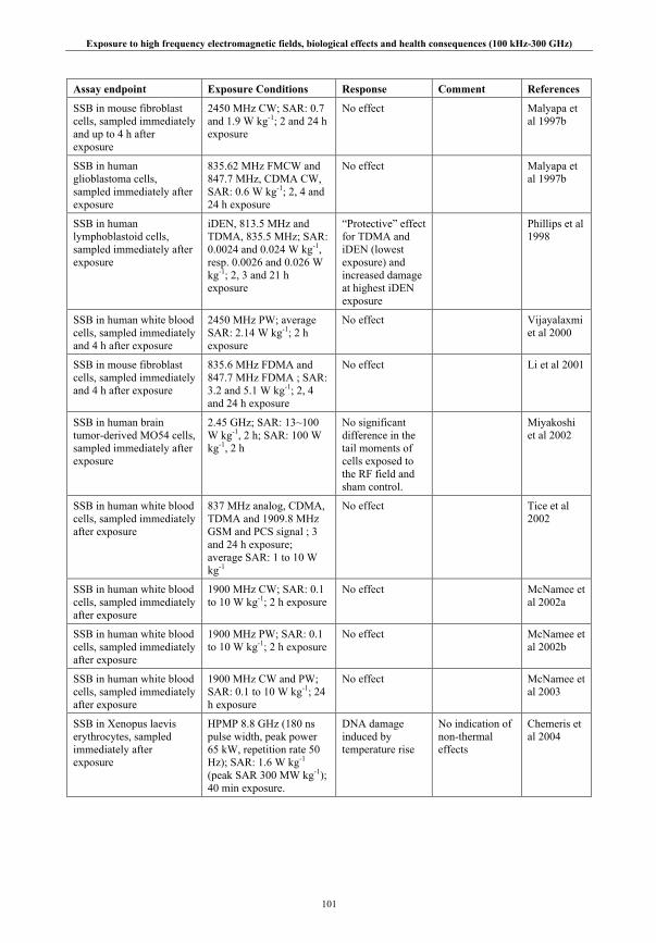
Exposure to high frequency electromagnetic fields, biological effects and health consequences (100 kHz-300 GHz)
101
Assay endpoint Exposure Conditions Response Comment References
SSB in mouse fibroblast cells, sampled immediately and up to 4 h after exposure
2450 MHz CW; SAR: 0.7 and 1.9 W kg-1; 2 and 24 h exposure
No effect Malyapa et al 1997b
SSB in human glioblastoma cells, sampled immediately after exposure
835.62 MHz FMCW and 847.7 MHz, CDMA CW, SAR: 0.6 W kg-1; 2, 4 and 24 h exposure
No effect Malyapa et al 1997b
SSB in human lymphoblastoid cells, sampled immediately after exposure
iDEN, 813.5 MHz and TDMA, 835.5 MHz; SAR: 0.0024 and 0.024 W kg-1, resp. 0.0026 and 0.026 W kg-1; 2, 3 and 21 h exposure
“Protective” effect for TDMA and iDEN (lowest exposure) and increased damage at highest iDEN exposure
Phillips et al 1998
SSB in human white blood cells, sampled immediately and 4 h after exposure
2450 MHz PW; average SAR: 2.14 W kg-1; 2 h exposure
No effect Vijayalaxmi et al 2000
SSB in mouse fibroblast cells, sampled immediately and 4 h after exposure
835.6 MHz FDMA and 847.7 MHz FDMA ; SAR: 3.2 and 5.1 W kg-1; 2, 4 and 24 h exposure
No effect Li et al 2001
SSB in human brain tumor-derived MO54 cells, sampled immediately after exposure
2.45 GHz; SAR: 13~100 W kg-1, 2 h; SAR: 100 W kg-1, 2 h
No significant difference in the tail moments of cells exposed to the RF field and sham control.
Miyakoshi et al 2002
SSB in human white blood cells, sampled immediately after exposure
837 MHz analog, CDMA, TDMA and 1909.8 MHz GSM and PCS signal ; 3 and 24 h exposure; average SAR: 1 to 10 W kg-1
No effect Tice et al 2002
SSB in human white blood cells, sampled immediately after exposure
1900 MHz CW; SAR: 0.1 to 10 W kg-1; 2 h exposure
No effect McNamee et al 2002a
SSB in human white blood cells, sampled immediately after exposure
1900 MHz PW; SAR: 0.1 to 10 W kg-1; 2 h exposure
No effect McNamee et al 2002b
SSB in human white blood cells, sampled immediately after exposure
1900 MHz CW and PW; SAR: 0.1 to 10 W kg-1; 24 h exposure
No effect McNamee et al 2003
SSB in Xenopus laevis erythrocytes, sampled immediately after exposure
HPMP 8.8 GHz (180 ns pulse width, peak power 65 kW, repetition rate 50 Hz); SAR: 1.6 W kg-1 (peak SAR 300 MW kg-1); 40 min exposure.
DNA damage induced by temperature rise
No indication of non-thermal effects
Chemeris et al 2004

Exposure to high frequency electromagnetic fields, biological effects and health consequences (100 kHz-300 GHz)
102
Assay endpoint Exposure Conditions Response Comment References
SSB in MOLT-4T lymphoblastoid cells, sampled immediately after exposure
847.74 MHz CDMA, 835.6 MHz FDMA, 813.6 MHz iDEN, 836.6 MHz TDMA; SAR: 3.2 W kg-1 (CDMA, FDMA), 0.0024 or 0.024 W kg-1 (iDEN), 0.0026 or 0.026 W kg-1 (TDMA); exposure for up to 24 h.
No effect Hook et al 2004a
SSB and DSB in human diploid fibroblasts and rat granulosa cells, sampled immediately after exposure
1800 MHz; CW or modulated; continuous and intermittent (5 min on, 10 min off) exposure during 4, 16 and 24 h; SAR: 2 W kg-1
Induction of DNA single- and double strand breaks after 16h intermittent exposure in both cell types and at different mobile phone modulations
Some objections have been raised concerning the analysis of the data – see text above.
Diem et al 2005
SSB in human glioblastoma A 172 cells and normal human fibroblasts, sampled immediately after exposure
A172 cells: W-CDMA at 80, 250, 800 mW kg-1 and CW at 80 mW kg-1 for 2 and 24 h.
IMR-90 cells: W-CDMA and CW at SAR: 80 mW kg-1 for 2 and 24 h.
No effect Sakuma et al 2006
SSB in human white blood cells, sampled immediately after exposure
8.8 GHz, HPMP (180 ns pulse width); Average SAR 1.6 W kg-1 (peak SAR 300 MW kg-1); for 40 min
No significant change to the percentage of DNA content in the comet tail compared to the respective negative and temperature controls.
Chemeris et al 2006
SSB in human white blood cells
1950 MHz UMTS signal at SAR: 0.5 and 2.0 W kg-
1; 24 h exposure
No genotoxicity and cytotoxicity at both SAR levels
Cytotoxicity assessed by the trypan blue exclusion test
Sannino et al 2006
SSB in human lens epithelial cells, sampled at 0, 30, 60, 120 and 240 min after exposure
1.8 GHz (217 Hz AM); 2 h exposure at SAR: 1, 2 and 3 W kg-1
DNA damage at 3 W kg-1 at 0 and 30 min following exposure
Exposure for 2h at 2 and 3 W kg-
1 also exhibited significantly increased hsp 70 protein expression.
Sun et al 2006
SSB in human diploid fibroblasts and Chinese hamster V79 cells, sampled immediately after exposure
1800 MHz; CW or modulated; continuous and intermittent (5 min on, 10 min off) exposure varied between 1 and 24h; SAR: 2W kg-1
No effect This study was aimed at replicating earlier findings (REFLEX 2004; Diem et al 2005) – Results were not in accordance with these.
Speit et al 2007

Exposure to high frequency electromagnetic fields, biological effects and health consequences (100 kHz-300 GHz)
103
Assay endpoint Exposure Conditions Response Comment References
Alkaline comet assay in human leukocytes
1950 MHz UMTS (intermittent exposure for 24 – 6 min on, 2 h off) in TEM cell; SAR: 2.2 W kg-
1.
No effect Also no effect on micronucleus frequency.
Zeni et al 2008
SSB in human cultured fibroblasts and white blood cells
1950 MHz UMTS; 24 h exposure and (intermittent exposure for 16h – 5min on, 10 min off and 10 min on, 20 min off); SAR <2 W kg-1
Increased “Comet Tail Factor” in a dose and time-dependent way
Some objections have been raised concerning the analysis of the data – see text above.
Schwarz et al 2008
HPMP: high power microwave pulses; FMCW: Frequency Modulated Continuous Wave; AM: amplitude modulated; CDMA = code-division multiple-access; FDMA = frequency-division multiple-access; TDMA = time-division multiple-access ; iDEN = iDEN(R) frequency/modulation form specially designed for use in vehicles.
Besides studies on DNA damage as assessed by the comet assay, many other cytogenetic studies have investigated possible RF-genetic effects in mammalian cells. Most have been performed on human lymphocytes but other cells were also studied. An overview of studies carried out after 1990 is given in Table II.3.2. (some also include data on the comet assay that were not included in Table II.3.1.). It is clear from the table that the results are more mixed than the comet assay results. However, positive findings were most often found when the exposure level was high (e.g., Garaj-Vrhovac et al 1992; Maes et al 1993; Tice et al 2002) resulting in an overall or localized thermal effect. In many other (mainly positive) investigations (e.g., Garaj-Vrhovac et al 1996) insufficient data were provided to judge the validity of the findings or interpretation of the results (see discussion in Vijayalaxmi and Obe 2004).
In recent years the observation by Tice et al (2002) of an RF-induced increase in micronucleus frequency in resting lymphocytes following a 24 h exposure attracted a lot of attention, essentially because no effects were found in the same (or related) investigations with regard to other genetic endpoints (e.g., absence of induced SSBs) and because increased micronucleus frequencies may, in the absence of chromosome aberrations, point towards an aneugenic effect (an abnormal chromosome number). It should be remembered, apart from structural chromosome aberrations, micronuclei may also originate from abnormal chromosome segregation during cell division (e.g., Fenech 2000) which may give rise to aneuploïd daughter cells. Only a few studies have so far investigated aneuploïdy with regard to RF. Mashevich et al (2003) reported increased aneuploïdy of chromosome 17, which may be seen as a corroboration of the possible aneuploïdy-inducing potency of RF radiation. These authors applied a fluorescence in situ hybridization technique to determine the incidence of aneuploidy of chromosome 17. The same group later confirmed these findings in a further investigation on aneuploïdy of the chromosomes 1, 10, 11 and 17 (Mazor et al 2008). As discussed elsewhere (Vijayalaxmi and Obe 2004) these data are intriguing and certainly need to be replicated and confirmed by others before any firm conclusion can be reached. It should furthermore be stated that there are also many investigations that do not show any micronucleus-inducing potency of RF. However, the experimental protocols used varied (e.g., 24 h exposure time vs. 2 h exposure) and these differences may eventually account for the contradictory results.
Other recent studies have also failed to reach a consensus view. Stronati et al (2006) did not find cytogenetic effects in RF exposed cells, and no co-operative effect was found with X-rays. However, Diem et al (2005) reported that RF possesses genotoxic properties, provided exposures are intermittent according to a particular protocol (5 min on/10 min off), depending on cell type. RF exposure at a SAR level below 2 W kg-1, for example, induced SSBs and DSBs and micronuclei and chromosomal aberrations in human fibroblast cells, HL-60 cells and/or rat granulosa cells. Blood lymphocytes were apparently unresponsive (REFLEX 2004). According to the authors the effects were possibly caused by an RF-induced increase in free oxygen radicals. As human lymphocytes were not responding it was believed that this cell type is not sensitive to RF and that previous negative findings that were obtained in lymphocytes (cf. Table II.3.2.) were simply due to the wrong choice of cells in these experiments. However, as mentioned above, the methodology and analysis employed in these investigations has been
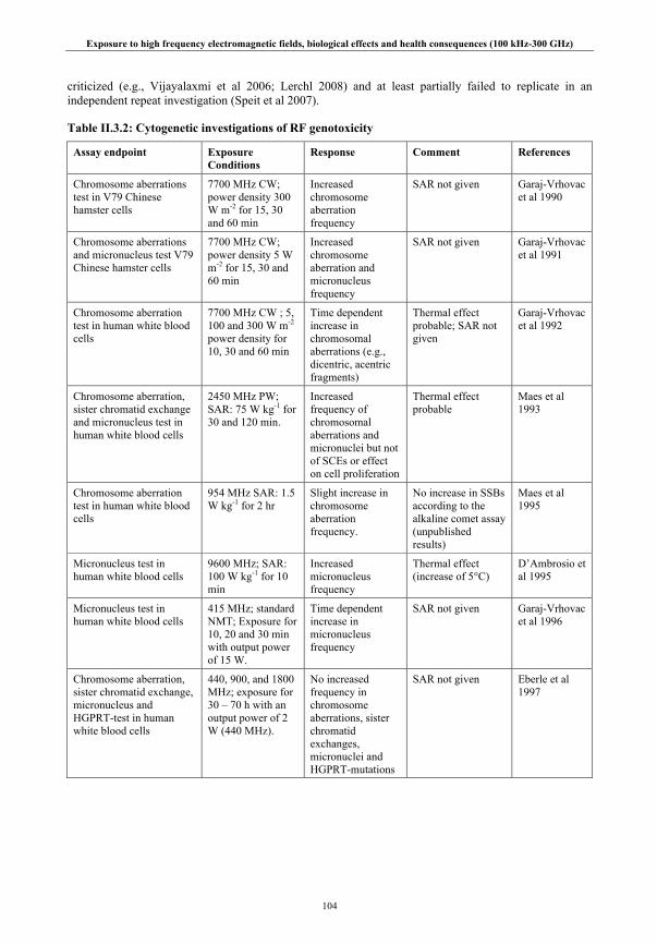
Exposure to high frequency electromagnetic fields, biological effects and health consequences (100 kHz-300 GHz)
104
criticized (e.g., Vijayalaxmi et al 2006; Lerchl 2008) and at least partially failed to replicate in an independent repeat investigation (Speit et al 2007).
Table II.3.2: Cytogenetic investigations of RF genotoxicity
Assay endpoint Exposure Conditions
Response Comment References
Chromosome aberrations test in V79 Chinese hamster cells
7700 MHz CW; power density 300 W m-2 for 15, 30 and 60 min
Increased chromosome aberration frequency
SAR not given Garaj-Vrhovac et al 1990
Chromosome aberrations and micronucleus test V79 Chinese hamster cells
7700 MHz CW; power density 5 W m-2 for 15, 30 and 60 min
Increased chromosome aberration and micronucleus frequency
SAR not given Garaj-Vrhovac et al 1991
Chromosome aberration test in human white blood cells
7700 MHz CW ; 5, 100 and 300 W m-2 power density for 10, 30 and 60 min
Time dependent increase in chromosomal aberrations (e.g., dicentric, acentric fragments)
Thermal effect probable; SAR not given
Garaj-Vrhovac et al 1992
Chromosome aberration, sister chromatid exchange and micronucleus test in human white blood cells
2450 MHz PW; SAR: 75 W kg-1 for 30 and 120 min.
Increased frequency of chromosomal aberrations and micronuclei but not of SCEs or effect on cell proliferation
Thermal effect probable
Maes et al 1993
Chromosome aberration test in human white blood cells
954 MHz SAR: 1.5 W kg-1 for 2 hr
Slight increase in chromosome aberration frequency.
No increase in SSBs according to the alkaline comet assay (unpublished results)
Maes et al 1995
Micronucleus test in human white blood cells
9600 MHz; SAR: 100 W kg-1 for 10 min
Increased micronucleus frequency
Thermal effect (increase of 5°C)
D’Ambrosio et al 1995
Micronucleus test in human white blood cells
415 MHz; standard NMT; Exposure for 10, 20 and 30 min with output power of 15 W.
Time dependent increase in micronucleus frequency
SAR not given Garaj-Vrhovac et al 1996
Chromosome aberration, sister chromatid exchange, micronucleus and HGPRT-test in human white blood cells
440, 900, and 1800 MHz; exposure for 30 – 70 h with an output power of 2 W (440 MHz).
No increased frequency in chromosome aberrations, sister chromatid exchanges, micronuclei and HGPRT-mutations
SAR not given Eberle et al 1997

Exposure to high frequency electromagnetic fields, biological effects and health consequences (100 kHz-300 GHz)
105
Assay endpoint Exposure Conditions
Response Comment References
Chromosome aberration and sister chromatid exchange test in human white blood cells
380 MHz PW; TETRA; SAR: 0.08 W kg-1
Waveguide, 900 MHz DCS; SAR: 0.2 W kg-1
Wave guide; 1800 MHz PW (GSM); exposure for up to 68 h
No increased frequency in chromosomal aberrations and sister chromatid exchanges
Antonopoulos et al 1997
Chromosome aberration and micronucleus test in human white blood cells
2450 MHz CW; SAR: 12.5 W kg-1; continuous or intermittent exposure for a total of 90 min
No increased frequency in chromosomal aberrations and micronuclei following continuous or intermittent RF exposures
Vijayalaxmi et al 1997a
Micronucleus test in human white blood cells
2450 and 7700 MHz and power density of 100, 200 and 300 W m-2; exposure for 15, 30 or 60 min
Increased frequency of micronuclei at a power density of 30mW/cm2 and after an exposure time of 30 and 60min (not at 10min)
SAR not given Zotti-Martelli et al 2000
Micronucleus test in human white blood cells
1800 MHz, CW at power densities of 5, 10 and 200 W m-
2 for 60, 120 and 180 min.
Microwaves were shown to be able to induce micronuclei in short-term exposure to medium power density fields
SAR not given Zotti-Martelli et al 2005
Chromosome aberration and micronucleus test in human white blood cells
847.7 MHz CW; CDMA; SAR: 4.9 and 5.5 W kg-1; 24 h exposure
No increased frequency in chromosomal aberrations and micronuclei
Vijayalaxmi et al 2001b
Chromosome aberration and micronucleus test in human white blood cells
835.6 MHz CW; FDMA; SAR: 4.4 and 5.0 W kg-1; exposure for 24 h.
No increased frequency in chromosomal aberrations and micronuclei
Vijayalaxmi et al 2001c
Micronucleus test in C3H 10T1/2 mouse fibroblast cells
835.6 MHz CW, FDMA and 847.7 MHz CW, CDMA; SAR: 3.2 and 5.1 W kg-1; exposure for 3, 8, 16 or 24 h
No increased frequency of micronuclei
Bisht et al 2002
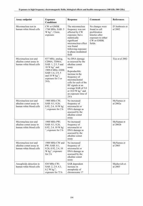
Exposure to high frequency electromagnetic fields, biological effects and health consequences (100 kHz-300 GHz)
106
Assay endpoint Exposure Conditions
Response Comment References
Micronucleus test in human white blood cells
CW or GMSK 1748 MHz; SAR: 5 W kg-1; 15min. exposure
The micronucleus frequency was not affected by CW exposure; but a statistically significant micronucleus effect was found following exposure to phase modulated field
No changes were found in cell proliferation kinetics after exposure to either CW or GMSK fields.
D’Ambrosio et al 2002
Micronucleus test and alkaline comet assay in human white blood cells
837 MHz, analog, CDMA, TDMA; SAR: 1, 2.5, 5 and 10 W kg-1 and 1909.8 MHz, GSM; SAR=1.6, 2.9, 5 and 10 W kg-1; exposure for 3 or 24 h.
No DNA damage as assessed by the alkaline comet assay
Reproducible increase in the frequency of micronucleated cells for each of the RF signals at an average SAR of 5.0 or 10.0 W kg-1 and an exposure time of 24 h
Tice et al 2002
Micronucleus test and alkaline comet assay in human white blood cells
1900 MHz CW; SAR: 0.1, 0.26, 0.92, 2.4, 10 W kg-
1; exposure for 2 h
No increased frequency of micronuclei or DNA damage as assessed by the alkaline comet assay
McNamee et al 2002a
Micronucleus test and alkaline comet assay in human white blood cells
1900 MHz PW; SAR: 0.1, 0.26, 0.92, 2.4, 10 W kg-
1; exposure for 2 h
No increased frequency of micronuclei or DNA damage as assessed by the alkaline comet assay
McNamee et al 2002b
Micronucleus test and alkaline comet assay in human white blood cells
1900 MHz CW and PW; SAR: 0.1, 0.26, 0.92, 2.4, 10 W kg-1; exposure for 2 h
No increased frequency of micronuclei or DNA damage as assessed by the alkaline comet assay
McNamee et al 2003
Aneuploidy detection in human white blood cells
830 MHz CW; SAR :2, 2.9, 4.3, 8.2 W kg-1; exposure for 72 h
SAR dependent increase in aneuploidy of chromosome 17
Mashevich et al 2003
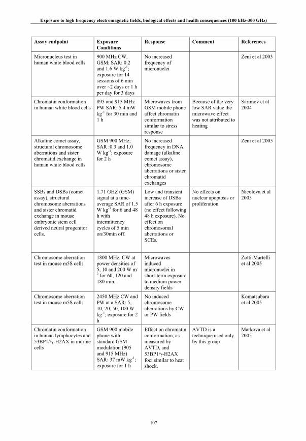
Exposure to high frequency electromagnetic fields, biological effects and health consequences (100 kHz-300 GHz)
107
Assay endpoint Exposure Conditions
Response Comment References
Micronucleus test in human white blood cells
900 MHz CW, GSM; SAR: 0.2 and 1.6 W kg-1; exposure for 14 sessions of 6 min over ~2 days or 1 h per day for 3 days
No increased frequency of micronuclei
Zeni et al 2003
Chromatin conformation in human white blood cells
895 and 915 MHz PW SAR: 5.4 mW kg-1 for 30 min and 1 h
Microwaves from GSM mobile phone affect chromatin conformation similar to stress response
Because of the very low SAR value the microwave effect was not attributed to heating
Sarimov et al 2004
Alkaline comet assay, structural chromosome aberrations and sister chromatid exchange in human white blood cells
GSM 900 MHz; SAR :0.3 and 1.0 W kg-1; exposure for 2 h
No increased frequency in DNA damage (alkaline comet assay), chromosome aberrations or sister chromatid exchanges
Zeni et al 2005
SSBs and DSBs (comet assay), structural chromosome aberrations and sister chromatid exchange in mouse embryonic stem cell derived neural progenitor cells.
1.71 GHZ (GSM) signal at a time-average SAR of 1.5 W kg-1 for 6 and 48 h with intermittency cycles of 5 min on/30min off.
Low and transient increase of DSBs after 6 h exposure (no effect following 48 h exposure). No effect on chromosomal aberrations or SCEs.
No effects on nuclear apoptosis or proliferation.
Nicolova et al 2005
Chromosome aberration test in mouse m5S cells
1800 MHz, CW at power densities of 5, 10 and 200 W m-
2 for 60, 120 and 180 min.
Microwaves induced micronuclei in short-term exposure to medium power density fields
Zotti-Martelli et al 2005
Chromosome aberration test in mouse m5S cells
2450 MHz CW and PW at a SAR: 5, 10, 20, 50, 100 W kg-1; exposure for 2 h
No induced chromosome aberrations by CW or PW fields
Komatsubara et al 2005
Chromatin conformation in human lymphocytes and 53BP1//γ-H2AX in murine cells
GSM 900 mobile phone with standard GSM modulation (905 and 915 MHz) SAR: 37 mW kg-1; exposure for 1 h
Effect on chromatin conformation, as measured by AVTD, and 53BP1/γ-H2AX foci similar to heat shock.
AVTD is a technique used only by this group
Markova et al 2005

Exposure to high frequency electromagnetic fields, biological effects and health consequences (100 kHz-300 GHz)
108
Assay endpoint Exposure Conditions
Response Comment References
Chromatin conformation in human lymphocytes
915 MHz; SAR: 37 mW kg-1
Significant condensation of chromatin found as measured by AVTD. No induction of apoptosis.
AVTD is a technique used only by this group
Belyaev et al 2005
Micronucleus test in human lymphocytes
900 MHz GSM signal, SAR: 1, 5 and 10 W kg-1, 24 h exposure
No evidence for genotoxic (micronucleus test) or cytotoxic effects
Scarfi et al 2006
Chromosome aberrations and micronuclei in human lymphocytes
2.45 GHz, 8.2 GHz, 21W, 60W; 50 W/m2 or 100 W/m2 ; SAR: 2.13 W kg-1 or 20.71 W kg-1 2 h
No adverse effects on the kinetics of cell proliferation or on the amount of chromosomal damage.
Vijayalaxmi 2006
Human diploid fibroblasts and Chinese hamster V79 cells, sampled immediately after exposure
GSM 1800 MHz; SAR: 3 W kg-1; intermittent exposure (5min on, 10min off) for 1 and 24 h
No difference with sham exposed cells after 1h; however increased DNA damage after 24h exposure
Zhang et al 2006
Alkaline comet assay and micronucleus test in human ES1 diploid fibroblast cells and in Chinese hamster V79 cells
1800 MHz; CW or modulated; continuous and intermittent (5min on, 10 min off) exposure varied between 1 and 24 h; SAR: 2W kg-1
No induction of micronucleated cells in independently repeated experiments
This study was aimed at replicating earlier findings (REFLEX 2004 and Diem et al 2005) – Results were not in accordance with these (see also comet assay results)
Speit et al 2007
Aneuploidy studies in human lymphocytes
800 MHz, CW, SAR: 2.9 and 4.1 W kg-1 for 72 h
Induced aneuploidy as determined by interphase FISH using semi-automated image analysis.
Findings were attributed to an athermal RF-effect
Mazor et al 2008
Micronucleus test and alkaline comet assay in human white blood cells
1950 MHz UMTS signal; intermittent exposure (6 min on, 2 h off) for 14 and 68 h; SAR: 2.2 W kg-1
No increased frequency of micronuclei
Also no effect on (alkaline) comet assay
Zeni et al 2008
GMSK = Gaussian minimum shift-keying; AVTD = Anomalous Viscosity Time Dependence
Cytogenetic effects in plants
Only a few cytogenetic studies on RF bioeffects have been performed in plants since 1993 and are briefly summarized here for completeness (see discussion in Chapter II.3.3.2.). Haider et al (1994) used the Tradescantia-micronucleus test in an in situ experiment to find out whether short-wave electromagnetic fields (10-21 MHz) are genotoxic. Plant cuttings bearing young flower buds were exposed during 30 h on both sides of a slewable curtain antenna (40-170 and 90 V m-1), a vertical cage antenna (70 V m-1) and at
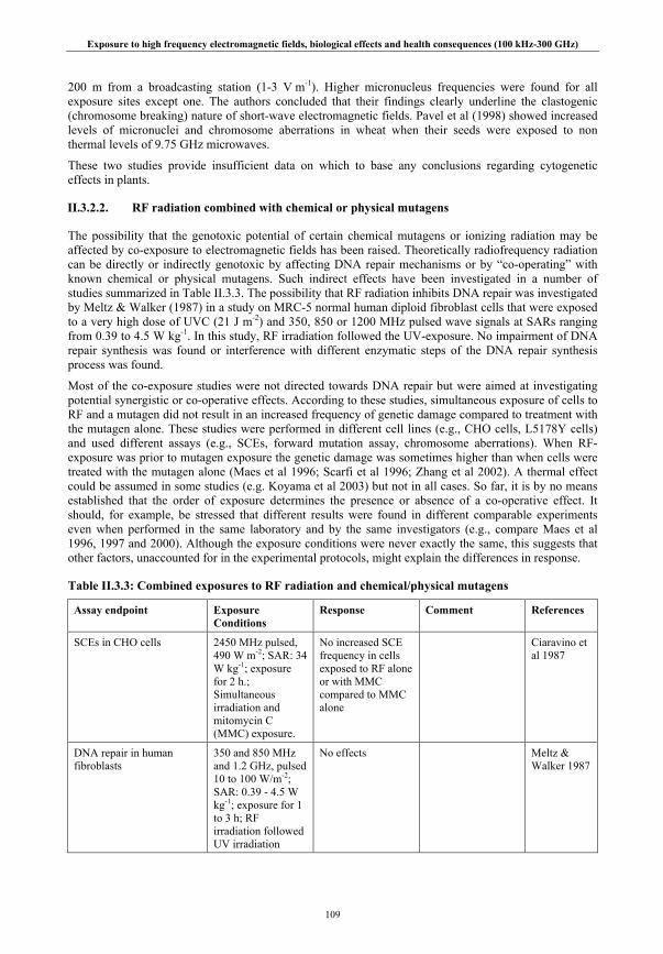
Exposure to high frequency electromagnetic fields, biological effects and health consequences (100 kHz-300 GHz)
109
200 m from a broadcasting station (1-3 V m-1). Higher micronucleus frequencies were found for all exposure sites except one. The authors concluded that their findings clearly underline the clastogenic (chromosome breaking) nature of short-wave electromagnetic fields. Pavel et al (1998) showed increased levels of micronuclei and chromosome aberrations in wheat when their seeds were exposed to non thermal levels of 9.75 GHz microwaves.
These two studies provide insufficient data on which to base any conclusions regarding cytogenetic effects in plants.
II.3.2.2. RF radiation combined with chemical or physical mutagens
The possibility that the genotoxic potential of certain chemical mutagens or ionizing radiation may be affected by co-exposure to electromagnetic fields has been raised. Theoretically radiofrequency radiation can be directly or indirectly genotoxic by affecting DNA repair mechanisms or by “co-operating” with known chemical or physical mutagens. Such indirect effects have been investigated in a number of studies summarized in Table II.3.3. The possibility that RF radiation inhibits DNA repair was investigated by Meltz & Walker (1987) in a study on MRC-5 normal human diploid fibroblast cells that were exposed to a very high dose of UVC (21 J m-2) and 350, 850 or 1200 MHz pulsed wave signals at SARs ranging from 0.39 to 4.5 W kg-1. In this study, RF irradiation followed the UV-exposure. No impairment of DNA repair synthesis was found or interference with different enzymatic steps of the DNA repair synthesis process was found.
Most of the co-exposure studies were not directed towards DNA repair but were aimed at investigating potential synergistic or co-operative effects. According to these studies, simultaneous exposure of cells to RF and a mutagen did not result in an increased frequency of genetic damage compared to treatment with the mutagen alone. These studies were performed in different cell lines (e.g., CHO cells, L5178Y cells) and used different assays (e.g., SCEs, forward mutation assay, chromosome aberrations). When RF-exposure was prior to mutagen exposure the genetic damage was sometimes higher than when cells were treated with the mutagen alone (Maes et al 1996; Scarfi et al 1996; Zhang et al 2002). A thermal effect could be assumed in some studies (e.g. Koyama et al 2003) but not in all cases. So far, it is by no means established that the order of exposure determines the presence or absence of a co-operative effect. It should, for example, be stressed that different results were found in different comparable experiments even when performed in the same laboratory and by the same investigators (e.g., compare Maes et al 1996, 1997 and 2000). Although the exposure conditions were never exactly the same, this suggests that other factors, unaccounted for in the experimental protocols, might explain the differences in response.
Table II.3.3: Combined exposures to RF radiation and chemical/physical mutagens
Assay endpoint Exposure Conditions
Response Comment References
SCEs in CHO cells 2450 MHz pulsed, 490 W m-2; SAR: 34 W kg-1; exposure for 2 h.; Simultaneous irradiation and mitomycin C (MMC) exposure.
No increased SCE frequency in cells exposed to RF alone or with MMC compared to MMC alone
Ciaravino et al 1987
DNA repair in human fibroblasts
350 and 850 MHz and 1.2 GHz, pulsed 10 to 100 W/m-2; SAR: 0.39 - 4.5 W kg-1; exposure for 1 to 3 h; RF irradiation followed UV irradiation
No effects Meltz & Walker 1987

Exposure to high frequency electromagnetic fields, biological effects and health consequences (100 kHz-300 GHz)
110
Assay endpoint Exposure Conditions
Response Comment References
Forwards mutation assay (thymidine kinase (TK) locus) in L5178Y mouse leukemia cells
2450 MHz, pulsed, 488 W m-2; SAR: 30 W kg-1; exposure for 4 h; Simultaneous irradiation and MMC exposure
RF exposure alone is not mutagenic
RF does not affect either the inhibition of cell growth or the extent of mutagenesis resulting from treatment with MMC alone
Meltz et al 1989
Forward mutation at TK-locus in L5178Y mouse leukemia cells
2.45 GHz, pulsed; SAR ~ 40 W kg-1; exposure for 4 h: Cells are simultaneously exposed to proflavin
RF alone is not mutagenic. No increased mutation frequency for the combined treatment compared to proflavin alone. No difference in colony size distribution of the mutant colonies
Meltz et al 1990
Chromosome aberrations in CHO cells
2450 MHz, pulsed 490 W m-2; SAR: 33.8 W kg-1; exposure for 2 h; Cells are simultaneously irradiated and exposed to MMC and adriamycin.
RF alone does not enhance the chromosome aberration frequency
No alteration in the extent of chromosome aberrations for the combined treatment compared to the chemicals alone
Kerbacher et al 1990
Cell cycle progression and SCEs in CHO cells.
2450 MHz pulsed, 490 W m-2; SAR: 33.8 W kg-1; exposure for 2 h; Cells are simultaneously irradiated and adriamycin exposed
RF does not affect changes in cell progression caused by adriamycin, nor change the number of SCEs that were induced by adriamycin.
Ciaravino et al 1991
SCE and cell proliferation in human lymphocytes
954 MHz (GSM), SAR ~ 1.5 W kg-1; cells exposed to MMC following a 2 h RF exposure
RF alone is not mutagenic. Increased incidence of SCEs in cells exposed to RF + MMC compared to MMC alone.
No influence on the cell proliferation for RF alone and for RF+MMC compared to MMC alone
Maes et al 1996
Micronucleus induction in bovine lymphocytes
9 GHz, SAR: 70 W kg-1 (CW); cells exposed to MMC following a 10 min RF exposure
Cooperative effect of microwaves and MMC
Scarfi et al 1996
SCE and cell proliferation, chromosome aberrations and DNA damage (comet assay) in human lymphocytes
935.2 MHz (CW and GSM), SAR ~ 0.3-0.4 W kg-1 for 2 h (TEM cell); cells exposed to MMC following a 2 h RF exposure
No effect of RF alone on DNA and chromosomes. Weak synergy with MMC (SCEs)
Maes et al 1997

Exposure to high frequency electromagnetic fields, biological effects and health consequences (100 kHz-300 GHz)
111
Assay endpoint Exposure Conditions
Response Comment References
Forward mutation, petite formation and recombination in Saccharomyces cerevisiae
900 MHz pulse modulated at SAR 0.13 and 1.3 W kg-1 + methyl methane sulfonate (MMS); exposure for 1 h and 36 h respectively
No effect on any of the genetic endpoints for the RF alone or combined with MMS
Gos et al 2000
Chromosome aberrations, SCEs in human lymphocytes
455.7 MHz, SAR: 6.5 W kg-1; 2 h exposure. Cells exposed to MMC and X-rays following RF-exposure
No consistent results (MMC); no synergy with X-rays
Maes et al 2000
Chromosome aberrations, SCEs and DNA damage (comet assay) in human lymphocytes
900 MHz (GSM; CW, dummy burst and pseudo-random); SAR: 0 - 10 W kg-1; MMC following a 2 h RF exposure, or RF exposure immediately followed by a 1 Gy X-ray exposure
No effect of the RF field alone. No evidence of a synergistic effect with MMC or X-rays
Maes et al 2001
DNA damage (comet assay); micronucleus test in human lymphocytes
2450 MHz, 50 W m-
2; exposure for 2 h; followed by MMC
Cooperative effect of RF with MMC
SAR not given Zhang et al 2002
Micronucleus test in CHO-K1 cells
2450 MHz; SAR: 13, 39, 50, 78 and 100 W kg-1; RF + bleomycin; 18 h exposure
Increased micronucleus frequency at SARs of 78 W kg-1 and higher. Potentiation of MN formation induced by bleomycin was found at SARs of 78 W kg-1 and higher.
Koyama et al 2003
Micronucleus test in CHO-K1 cells
2450 MHz; SAR: 5, 10, 20, 50, 100, 200 W kg-1; exposure for 2 h; RF + bleomycin combined treatment
Only increased micronucleus frequency at SARs of 100 and 200 W kg-1. No combined effect of RF and bleomycin
Koyama et al 2004

Exposure to high frequency electromagnetic fields, biological effects and health consequences (100 kHz-300 GHz)
112
Assay endpoint Exposure Conditions
Response Comment References
Comet assay in C3H 10T1/2 cells
2450 MHz CW at SAR: 1.9 W kg-1, 2h exposure followed by 4 Gy γ-irradiation
No DNA damage induced by 2450 MHz RF alone. 2450 MHz microwaves did not impede the DNA migration induced by γ-irradiation. No evidence for induction of DNA-protein crosslinks or changes in amount of protein associated with DNA by 2450 MHz CW microwave exposure
Lagroye et al 2004aa
Chromosome aberrations in human lymphocytes
2.5 GHz and 10.5 GHz + γ-radiation exposure for 40 sec. at 3 W for 2.5 MHz and 5 min at 15 mW for 10.5 GHz). SAR estimated at 626 W kg-1 and 0.25 W kg-1 resp.
No induction of chromosome aberrations from the microwaves alone.
No combined effects in cells pre-treated with the RF fields followed by gamma-irradiation, but increased cell mortality was observed.
Figueiredo et al 2004
Comet assay performed at 0 and 21h following exposure in human lymphocytes
1.8 GHz, SAR: 3 W kg-1 + MMS, 4-NQO, MMC and bleomycin. Exposure for 2 and 3 h in three exposure combinations
No effect of RF alone but combined treatments with MMC and 4-NQO were significantly different from the chemical exposures alone.
Wang et al 2005
Chromosomal aberrations, SCEs, micronuclei, DNA damage (comet assay), nuclear division index in human lymphocytes
935 MHz GSM signal, SAR: 1 and 2 W kg-1, 24 h continuous exposure + 1 min. 1 Gy 250 kVp X-rays
In all instances no effect from the RF signal alone or in combination with X-rays was observed
Stronati et al 2006
DNA damage (comet assay) in human white blood cells
1.8 GHz at SAR 3 W kg-1 for 0, 1.5 and 4 h following UVC exposure at 0.25, 0.5, 0.75, 1.0, 1.5 and 2.0 J m-2.
The microwaves were shown to reduce UV induced DNA damage after 1.5h of exposure and increased UV induced DNA damage at 4 h.
No effect of 1.8 GHz exposure alone.
Wang et al 2007
II.3.2.3. Summary on genotoxicity
To date many studies have investigated RF-induced genetic effects in human and other cell types. Most, but not all, studies have found no evidence of in vitro RF-induced genetic damage at non-thermal exposure regimes and indicate that RF has no marked synergistic or additive effect together with other environmental agents (mutagens/carcinogens). However, many studies were devoted to genetic endpoints
Exposure to high frequency electromagnetic fields, biological effects and health consequences (100 kHz-300 GHz)
113
that correspond to gross structural chromosome anomalies and hence possible subtle indirect effects on, for example the replication of genes under relatively restricted exposure conditions, could not be seen. Therefore more sensitive methods are probably necessary to determine whether such effects might exist. The comet assay was introduced as a method that might meet this demand. Our present-day knowledge of the comet assay shows that this is only partially true. Novel methods, for example the determination of γ-H2AX protein intranuclear foci and mini/microsatellite mutation analysis, may prove more valuable in RF genetic risk assessment.
These conclusions are supported by a recent meta-analysis of RF-genetic toxicity (Vijayalaxmi and Prihoda 2008). The authors quantitatively analyzed the results from 63 papers published between 1990 and 2005, deriving indices and 95% confidence intervals for various genetic endpoints in relation to frequency, SAR and CW or pulsed exposure. The overall genotoxicity indices obtained in from the RF exposed and control groups were similar. Also, the mean indices for chromosome aberrations and micronuclei in RF-radiation exposed and sham or unexposed controls were within spontaneous levels reported in the historical database.
II.3.3. Studies on non genotoxic cellular effects
In addition to evaluating RF radiation effects on the integrity of DNA, numerous studies have addressed other effects on cellular functions that could potentially influence development of disease in humans.
One major class of non-genotoxic cellular studies comprises studies of cell signaling, the means by which cells respond to extracellular signals such as cytokines, neurotransmitters and hormones via receptors located on the cell surface and to intracellular signals, which may be generated for example by the activation of an intracellular signaling cascade. A number of RF studies have been carried out on both intercellular and extracellular signaling processes.
Cellular responses depend on production of proteins (enzymes), key regulators of cell metabolic activity and behavior. Protein structures are encoded in DNA (genes) and are produced by the transcription of genes into mRNA and the translation of the mRNA into protein. This activity is called gene expression and RF effects on gene expression are, more precisely, classified as either effects on mRNA at the transcriptional level or on protein production. A large body of RF research has been conducted on gene and protein expression in mammalian and other cell types.
Other non-genotoxic studies address the ability of RF to affect the production of reactive oxygen species and on cell behaviors such as proliferation, differentiation, and apoptosis. In addition, studies on cell transformation, which examine the ‘transforming’ or carcinogenetic potential of RF radiation are also described.
II.3.3.1. Intracellular and intercellular signaling
The transduction of signals or ‘signal transduction’ is the integration of intra- and/or extracellular messages to or within a cell. The transduction of extracellular signals for example is common to the endocrine, immune, and nervous systems of mammals. Cells produce mediators such as ligands and hormones that can be detected by other cells via specific receptors located at the periphery of the cell on the plasma membrane and which induce responses in these cells. The signal can be transmitted inside the cell via a change of conformation of the receptor. This transconformation may initiate various intracellular pathways via secondary messengers: either the ligand penetrates into the cell to bind to a cytoplasmic receptor, which will then generally act at the nuclear level as a transcription factor, or it binds to the extracellular part of a transmembrane receptor, thus modifying its conformation including the intracellular part of the receptor. Then the receptor will have an increased enzymatic activity or bind to other proteins (e.g., kinases or G proteins). These reactions induce intracellular signaling such as protein phosphorylation cascades, which trigger specific cellular responses of the cell: expression of certain genes, exocytosis, etc. Signal transduction has thus three stages: communication, transduction, and signaling. The last two have been extensively studied in cell models exposed to RF radiation.
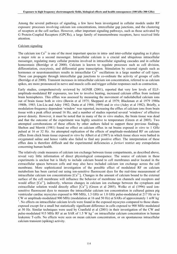
Exposure to high frequency electromagnetic fields, biological effects and health consequences (100 kHz-300 GHz)
114
Among the several pathways of signaling, a few have been investigated in cellular models under RF exposure: processes involving calcium ion concentrations, intercellular gap junctions, and the clustering of receptors at the cell surface. However, other important signaling pathways, such as those activated by G Protein-Coupled Receptors (GPCRs), a large family of transmembrane receptors, have received little attention.
Calcium signaling
The calcium ion Ca2+ is one of the most important species in intra- and inter-cellular signaling as it plays a major role as a second messenger. Intracellular calcium is a crucial and ubiquitous intracellular messenger, regulating many cellular proteins involved in intracellular signaling cascades and in cellular homeostasis (Berridge et al 2000). Calcium is known to regulate processes such as cell division, differentiation, exocytosis, and differential gene transcription. Stimulation by external signals such as hormones or neurotransmitters results in intracellular Ca2+ oscillations in a large number of cell types. These can propagate through intercellular gap junctions to co-ordinate the activity of groups of cells (Berridge et al 2000). Transient increases in intracellular calcium ion concentration, referred to as calcium spikes, are more pronounced in nerve and muscle cells and trigger cellular responses such as contraction.
Early studies, comprehensively reviewed by AGNIR (2001), reported that very low levels of ELF-amplitude-modulated RF exposures, too low to involve heating, increased calcium efflux from isolated brain hemispheres. This efflux was assessed by measuring the movement of radiolabelled calcium ions out of brain tissue both in vitro (Bawin et al 1975; Sheppard et al 1979; Blackman et al 1979 1980a 1980b, 1985; Lin-Liu and Adey 1982; Dutta et al 1984; 1989) and in vivo (Adey et al 1982). Briefly, a modulation-frequency dependent ‘window’ was reported, increasing the efflux of calcium ions from brain tissue with a peak effect around 16 Hz, and a number of studies reported several ‘windows’ of effective power density. However, it must be noted that in many of the in vitro studies, the brain tissue was dead and that the outcome of the experiment was highly sensitive to temperature (Green et al 2005). Two attempted corroborations of these effects by other authors failed to support these previous findings. Shelton and Merritt (1981) found no effect on calcium efflux in rat brains exposed in vitro to 1 GHz pulsed at 16 or 32 Hz. An attempted replication of the effects of amplitude-modulated RF on calcium efflux from chick brain tissue exposed in vitro by Albert et al (1987) in which tissue slices were bathed in oxygenated saline and hence viable also failed to find any positive effect. The interpretation of these efflux data is therefore difficult and the experimental deficiencies a fortiori restrict any extrapolation concerning human health.
The relatively crude measures of calcium ion exchange between tissue compartments, as described above, reveal very little information of direct physiological consequence. The source of calcium in these experiments is unclear but is likely to include calcium bound to cell membranes and/or located in the extracellular spaces between cells and may also have included calcium ion exchange across the cell membrane. More sophisticated investigation of the possible effect of modulated RF on calcium metabolism has been carried out using ion-sensitive fluorescent dyes for the real-time measurement of intracellular calcium ion concentrations [Ca2+]i. Changes in the amount of calcium bound to the external surface of the cell membrane will influence the behavior of membrane ion channels and receptors and would affect [Ca2+]i indirectly, whereas changes in calcium ion exchange between the cytoplasm and extracellular solution would directly affect [Ca2+]i (Green et al 2005). Wolke et al (1996) used ion-sensitive fluorescent dyes to measure the intracellular calcium ion concentration in cultured guinea pig ventricular cardiac myocytes exposed to 900 MHz, 1.3 GHz or 1.8 GHz pulse-modulated at 217 Hz or to CW or amplitude-modulated 900 MHz (modulation at 16 and 50 Hz) at SARs of approximately 1 mW kg-
1. No effects on intracellular calcium levels were found in the exposed myocytes compared to those sham-exposed except for a small but statistically significant difference in cells exposed to 900 MHz modulated at 50 Hz. Similar techniques were used by Cranfield et al (2001) in their investigation of the CW and pulse-modulated 915 MHz RF at an SAR of 1.5 W kg-1 on intracellular calcium concentration in human leukemic T-cells. No effects were seen on mean calcium concentration, or on spontaneous intracellular calcium transient (spiking) activity.
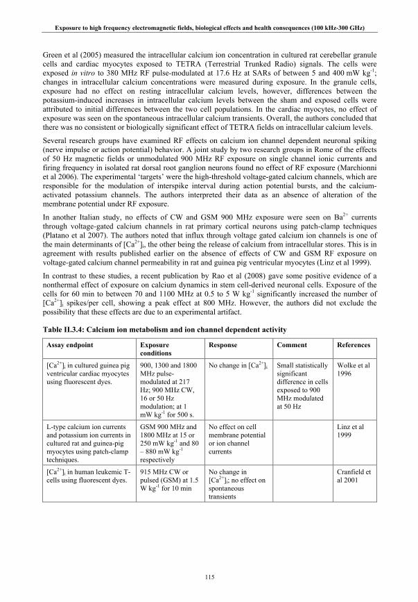
Exposure to high frequency electromagnetic fields, biological effects and health consequences (100 kHz-300 GHz)
115
Green et al (2005) measured the intracellular calcium ion concentration in cultured rat cerebellar granule cells and cardiac myocytes exposed to TETRA (Terrestrial Trunked Radio) signals. The cells were exposed in vitro to 380 MHz RF pulse-modulated at 17.6 Hz at SARs of between 5 and 400 mW kg-1; changes in intracellular calcium concentrations were measured during exposure. In the granule cells, exposure had no effect on resting intracellular calcium levels, however, differences between the potassium-induced increases in intracellular calcium levels between the sham and exposed cells were attributed to initial differences between the two cell populations. In the cardiac myocytes, no effect of exposure was seen on the spontaneous intracellular calcium transients. Overall, the authors concluded that there was no consistent or biologically significant effect of TETRA fields on intracellular calcium levels.
Several research groups have examined RF effects on calcium ion channel dependent neuronal spiking (nerve impulse or action potential) behavior. A joint study by two research groups in Rome of the effects of 50 Hz magnetic fields or unmodulated 900 MHz RF exposure on single channel ionic currents and firing frequency in isolated rat dorsal root ganglion neurons found no effect of RF exposure (Marchionni et al 2006). The experimental ‘targets’ were the high-threshold voltage-gated calcium channels, which are responsible for the modulation of interspike interval during action potential bursts, and the calcium-activated potassium channels. The authors interpreted their data as an absence of alteration of the membrane potential under RF exposure.
In another Italian study, no effects of CW and GSM 900 MHz exposure were seen on Ba2+ currents through voltage-gated calcium channels in rat primary cortical neurons using patch-clamp techniques (Platano et al 2007). The authors noted that influx through voltage gated calcium ion channels is one of the main determinants of [Ca2+]i, the other being the release of calcium from intracellular stores. This is in agreement with results published earlier on the absence of effects of CW and GSM RF exposure on voltage-gated calcium channel permeability in rat and guinea pig ventricular myocytes (Linz et al 1999).
In contrast to these studies, a recent publication by Rao et al (2008) gave some positive evidence of a nonthermal effect of exposure on calcium dynamics in stem cell-derived neuronal cells. Exposure of the cells for 60 min to between 70 and 1100 MHz at 0.5 to 5 W kg-1 significantly increased the number of [Ca2+]i spikes/per cell, showing a peak effect at 800 MHz. However, the authors did not exclude the possibility that these effects are due to an experimental artifact.
Table II.3.4: Calcium ion metabolism and ion channel dependent activity
Assay endpoint Exposure conditions
Response Comment References
[Ca2+]i in cultured guinea pig ventricular cardiac myocytes using fluorescent dyes.
900, 1300 and 1800 MHz pulse-modulated at 217 Hz; 900 MHz CW, 16 or 50 Hz modulation; at 1 mW kg-1 for 500 s.
No change in [Ca2+]i Small statistically significant difference in cells exposed to 900 MHz modulated at 50 Hz
Wolke et al 1996
L-type calcium ion currents and potassium ion currents in cultured rat and guinea-pig myocytes using patch-clamp techniques.
GSM 900 MHz and 1800 MHz at 15 or 250 mW kg-1 and 80 – 880 mW kg-1 respectively
No effect on cell membrane potential or ion channel currents
Linz et al 1999
[Ca2+]i in human leukemic T-cells using fluorescent dyes.
915 MHz CW or pulsed (GSM) at 1.5 W kg-1 for 10 min
No change in [Ca2+]i; no effect on spontaneous transients
Cranfield et al 2001
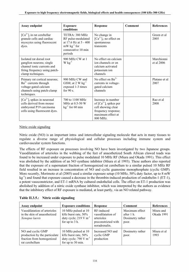
Exposure to high frequency electromagnetic fields, biological effects and health consequences (100 kHz-300 GHz)
116
Assay endpoint Exposure conditions
Response Comment References
[Ca2+]i in rat cerebellar granule cells and cardiac myocytes using fluorescent dyes.
TETRA: 380 MHz RF pulse-modulated at 17.6 Hz at 5 - 400 mW kg-1 for consecutive 10 min periods
No change in [Ca2+]i; no effect on spontaneous transients
Green et al 2005
Isolated rat dorsal root ganglion neurons; single channel ionic currents and firing frequency using patch-clamp techniques.
900 MHz CW at 1 W kg-1
No effect on calcium ion channels or on calcium activated potassium ion channels
Marchionni et al 2006
Primary rat cortical neurons; Ba2+ currents through voltage-gated calcium channels using patch-clamp techniques.
900 MHz CW and GSM; at 2 W kg-1 exposed 1-3 times for 90 s.
No effect on Ba2+
currents in voltage-gated calcium channels
Platano et al 2007
[Ca2+]i spikes in neuronal cells derived from mouse embryonal P19 carcinoma cells using fluorescent dyes.
700 to 1100 MHz MHz at 0.5-50 W kg-1 for 60 min
Increase in number of [Ca2+]i spikes per cell showing clear frequency response; maximum effect at 800 MHz.
Rao et al 2008
Nitric oxide signaling
Nitric oxide (NO) is an important intra- and intercellular signaling molecule that acts in many tissues to regulate a diverse range of physiological and cellular processes including immune system and cardiovascular system functions.
The effects of RF exposure on processes involving NO have been investigated by two Japanese groups. Vasodilatation of arterioles in the webbing of the feet of anaesthetized South African clawed toads was found to be increased under exposure to pulse modulated 10 MHz RF (Miura and Okada 1991). This effect was abolished by the addition of an NO synthase inhibitor (Miura et al 1993). These authors also reported that the exposure of a supernatant fraction of homogenized rat cerebellum to a similar pulsed 10 MHz RF field resulted in an increase in concentration of NO and cyclic guanosine monophosphate (cyclic GMP). More recently, Morimoto et al (2005) used a similar exposure setup (10 MHz, 50% duty factor, up to 8 mW kg-1) and found that exposure caused a decrease in the thrombin-induced production of endothelin-1 (ET-1), a potent vasoconstrictor, and ET-1 mRNA by cultured endothelial cells. The effect on ET-1 production was abolished by addition of a nitric oxide synthase inhibitor, which was interpreted by the authors as evidence that the inhibitory effect of RF exposure is mediated, at least partly, via an NO related pathway.
Table II.3.5.: Nitric oxide signaling
Assay endpoint Exposure conditions Response Comment References Vasodilatation of arterioles in the skin of anaesthetized Xenopus laevis
10 MHz pulsed at 10 kHz burst rate, 50% duty cycle; 219 V m-1 for up to 3 h.
RF-induced vasodilatation of arterioles preconstricted with noradrenalin.
Maximum effect after 1 h. Dosimetry rather poor.
Miura and Okada 1991
NO and cyclic GMP production by the particulate fraction from homogenized rat cerebellum
10 MHz pulsed at 10 kHz burst rate, 50% duty cycle; 790 V m-1 for up to 30 min.
Increased NO and cyclic GMP production
Dosimetry rather poor.
Miura et al 1993

Exposure to high frequency electromagnetic fields, biological effects and health consequences (100 kHz-300 GHz)
117
Assay endpoint Exposure conditions Response Comment References Thrombin-induced ET-1 and ET-1 mRNA production in bovine aortic endothelial cells, and human umbilical, aortic and microvascular endothelial cells
10 MHz pulsed at 10 kHz burst rate, 50% duty cycle; SAR: 1-8 mW kg-1 for up to 24 hr.
Decrease in thrombin-induced ET-1 and ET-1 mRNA production in RF-exposed cells.
Effect abolished by NG-monomethyl-L-arginine. Dosimetry rather poor.
Morimoto et al 2005
Gap junctions
Gap junctions are clusters of channels formed by proteins known as connexins, and permit the transfer of ions and small molecules between contiguous cells. Gap junction intercellular communication (GJIC), one component of the cell signaling system, is very important for cell homeostasis. A defect in GJIC is considered to be an important step during the multistage process of carcinogenesis, and disruption of GJIC has been recognized as one of the non-genotoxic mechanisms of carcinogenesis. Therefore, GJIC could be used as a biomarker to evaluate the possible health impact of RF exposure.
There are few reports about RF effects on GJIC. Ye et al (2002) exposed the heads of rabbits to 2450 MHz at 50 and 100 W m-2 for 3 h. One open eye was continuously exposed; the other eye of the same rabbit was covered tightly by copper grid cloth and served as control. Anterior lens capsules were obtained for a study of GJIC function using fluorescence recovery analysis after photobleaching (FRAP) technique and the localization analysis of connexin 43 using indirect immunofluorescence histochemical analysis. The results showed that the GJICs of rabbit lens epithelial cells were inhibited in a dose-dependent manner in response to RF exposure. This also caused a reduction in the amount of membrane-located gap-junction protein connexin 43. Unfortunately, the authors did not record the temperature changes during the exposure nor provided any estimate of the SAR.
Chen et al (2004a,b) studied the effect of 30.16 GHz millimeter wave RF exposure at 10 and 35 W m-2 on GJIC in cultured HaCaT keratinocytes using the FRAP approach. The results showed that RF exposure alone for 1 h at either 10 or 35 W m-2 did not affect GJIC. However, RF exposure in combination with treatment with the chemical promoter phorbol-12-myristate-13-acetate (PMA) reversed the PMA-induced suppression of GJIC. Exposure at 10 W m-2 resulted in a partial reversal, and exposure at 35 W m-2 resulted in a full reversal of the PMA-induced suppression. Temperatures during exposure were measured in this study, but no estimate of SAR was given.
Table II.3.6.: Gap junctions
Assay endpoint Exposure Conditions Response Comment References GJIC and connexin 43 in lens epithelial cells of rabbits
2450 MHz RF at 50 W m-2 and 100 W m-2 for 3 h
RF- induced inhibition of GJIC and damage to connexin 43.
No measure of temperature or SAR.
Ye et al 2002
GJIC of human (HaCaTs) keratinocytes
30.16 GHz RF exposure at 10 and 35 W m-2 for 1 h
GJIC suppression induced by PMA could be eliminated or diminished by exposure to RF.
Chen et al 2004a, b
Receptor clustering
Receptors are cellular membrane proteins that can bind specific signal molecules (ligands) and initiate a response in the cell. The response often starts with the clustering of receptors after binding its ligand, then activating certain signal pathway(s), changing cellular biological and/or biochemical processes, and resulting in the alteration of cell behaviors such as proliferation, apoptosis, invasion, or metastasis. Such receptors may be present on the outer cell membrane, the nuclear envelope or other membrane structures.

Exposure to high frequency electromagnetic fields, biological effects and health consequences (100 kHz-300 GHz)
118
In recent years, the effects of EMFs on the clustering of cellular membrane surface receptors, such as epidermal growth factor receptor (EGFR) or tumor necrosis factor receptor (TNFR) have been investigated. While there are many reports focusing on 50 Hz magnetic fields, only two studies have specifically addressed RF. Xie et al (2006) exposed Chinese hamster lung (CHL) cells to 1800 MHz fields at SARs of 0.1, 0.5, 1.0, 2.0 and 4.0 W kg-1. The results showed that clustering of EGFR was induced by exposure to 217 Hz modulated RF for 15 min at the lowest SAR of 0.5 W kg-1. Unmodulated RF did not induce this phenomenon, and superposition of 2 μT 50 Hz noise inhibited EGF receptor-clustering induced by RF. However, functional significance of this effect could only be determined by the investigation of RF effects on the activation of the normal EGFR signal pathway and resulting changes in cellular physiology.
Xu et al (2006) investigated glutamate receptor clustering and synaptic activity in rat brain cells. Glutamate receptor channels play key roles in excitatory synaptic transmission and are involved in many physiological and pathological processes. The authors exposed cultured hippocampal neurons of rats for 15 min per day for 8 days to GSM-1800 MHz signals at an average SAR of 2.4 W kg-1. Whole-cell patch clamp techniques were used to assess the miniature excitatory postsynaptic currents (mEPSCs) in NMDA (N-methyl-D-aspartate) and AMPA (α-amino-3-hydroxy-5-methyl-4-soxazole propionic acid) glutamate receptor channels. Synaptic density on the distal neuronal dendrites of the hippocampal cells was assessed using immunohistochemical staining. The authors reported that RF exposure reduced excitatory synaptic activity in AMPA receptors. Neither AMPA nor NMDA receptor clustering affected; however, the expression of post-synaptic density 95 (PSD95), which is involved in orchestrating excitatory synapse maturation and synaptic plasticity, was decreased after RF exposure. These observations require experimental confirmation.
Table II.3.7.: Receptor clustering
Assay endpoint Exposure Conditions
Response Comment References
Epidermal growth factor (EGF) receptor clustering in Chinese hamster lung fibroblasts.
1800 MHz GSM RF or CW; SAR: 0.1, 0.5, 1, 2, and 4 W kg-
1 for 15 min.
Clustering induced after exposure to GSM 1800 MHz RF modulated by 217 Hz at SAR ≤ 4 W kg-1.
Superposition of a 2 μT 50 Hz MF inhibited RF-induced EGF receptor clustering
Xie et al 2006
Synaptic activity and receptor density in cultured rat neurons using patch clamp and immunohistochemical staining.
1800 MHz GSM RF at SAR of 2.4 W kg-1 for 15 min per day for 8 days
Decreased the amplitude of AMPA mEPSCs; no change in NMDA mEPSCs amplitude.
The expression of postsynaptic density 95 (PSD95) in neuronal dendrites was decreased.
Xu et al 2006
Summary on signaling, gap junctions and receptor clustering
Overall, the evidence of effects on calcium signaling from recent, well conducted studies, particularly those using functionally significant measures of calcium ion concentration, do not support the earlier reports suggesting that low-level amplitude modulated RF can modify intracellular calcium ion metabolism, particularly with regard to its role as an intracellular messenger. In addition, no compelling effects have been seen on the spiking activity of neurons dependent on calcium ion channel properties.
The evidence for any effects of RF exposure on nitric oxide signaling processes is rather weak and insubstantial. No definite conclusion can be drawn based on the few data on effects of RF exposure on cellular gap junctions or receptor clustering.

Exposure to high frequency electromagnetic fields, biological effects and health consequences (100 kHz-300 GHz)
119
II.3.3.2. Gene and protein expression
Older studies focused on the response of small numbers of genes and/or proteins, so greatly restricting the ability of any individual study to determine the potential cellular responsiveness to RF. More recently, technological advances have facilitated the ability to screen for RF responsive gene(s) or protein(s) on a large scale using for example DNA microarray and proteomic technologies. Such automation provides the means for greatly increasing the amount of information that may be derived from a single experiment but at a cost, namely the increased difficulty in identifying biologically significant responses from experimental ‘noise’. Interpretation of the results relies heavily on complex statistical analysis that is very sensitive to the applied level of stringency with which meaningful responses are identified (Mayo, et al 2006). The various strengths and pitfalls of some of these high throughput technologies for screening for RF-induced ‘epigenetic’ changes have been discussed in detail by Leszczynski and Meltz (2006) who concluded that the techniques are at present useful primarily as experimental research tools. However, they may eventually be used to identify endpoints suitable for screening for animal, volunteer and epidemiological investigation, leading to a better understanding of the potential health effects, if any, of environmental levels of EMF exposure.
There are a number of different conventions for distinguishing the mRNAs that result from gene expression and the proteins which they encode. In this report, the following convention is used: genes are italicized and proteins are not. The prefix c- (as in c-fos) is sometimes used to indicate a gene of cellular origin, as opposed to viral (v) origin.
Gene expression
The conventional method for analysis of gene expression is Northern blotting. In this method total cellular RNA or mRNAs are separated by alkaline agarose gel electrophoresis and transferred (blotted) to membranes. Specific RNA transcripts are identified by hybridization of gene-specific probes, usually radioactively labeled, to membranes. Transcript levels are assessed by the relative strength of the signal of the radioactively labeled probe to a specific sized gene fragment. The method is at best semi-quantitative.
More recently, reverse transcriptase polymerase chain reaction (RT-PCR) methods have been introduced. In this method RNA is converted to complementary DNA (cDNA) sequences by the enzyme reverse transcriptase and gene-specific fragments are amplified by successive rounds of DNA synthesis using thermostable DNA polymerase enzymes. Gene specificity is achieved by using specifically synthesized primers that are unique to the gene being analyzed. In its simplest form RT-PCR is not highly quantitative. However, several systems such as real-time RT-PCR have been developed that allow highly precise quantification through the use of fluorescence measurements of specific gene products. This method is generally referred to as quantitative RT-PCR (QRT-PCR or Q-PCR). The Q-PCR methods are the most sensitive methods available for quantification of transcript levels and can detect differences in transcript levels over several orders of magnitude.
Transcriptomics describes the study of global gene expression; the genome in human and other mammalian cells comprises typically 20,000 – 30,000 genes. The transcriptome comprises the RNAs produced from the genome of a cell or tissue. Techniques using oligonucleotide chips, cDNA glass microarrays or microbead array systems rely on the binding of fluorescence labeled cDNA from the cells of interest to a set of complementary sequences on the chip or array and measuring the fluorescence intensity at each site. In this way the quantitative measures of gene expression within the entire genome in cells from two populations can be compared. However, it is widely acknowledged that there is a need to verify any ensuing changes in gene expression through other techniques such as Q-PCR. In particular, the sensitivity of array systems and their ‘dynamic’ range are considerably less than those of Q-PCR. Alternative sensitive techniques such as HICEP (high coverage gene expression profile) in which all RNA transcripts are amplified and separated by capillary electrophoresis for subsequent sequencing are also becoming available.
It is now becoming clear that there are additional levels of regulation of gene expression; these include the expression of regulatory micro RNAs (miRNAs), DNA methylation and a variety of modifications of chromatin-associated proteins. Methods to analyze such changes have not yet been applied to investigate

Exposure to high frequency electromagnetic fields, biological effects and health consequences (100 kHz-300 GHz)
120
the possible impact of RF, but EMF studies have, however, utilized gene-specific approaches such as Northern blotting, PCR and array-based genomic approaches to examine RF-induced changes in gene expression. Studies on mammalian cells are summarized and reviewed in the following two chapters; a third chapter addresses studies looking specifically at gene expression in plants.
Gene-specific approaches in mammalian cells
RF studies of gene expression have focused typically on early response genes, otherwise known as proto-oncogenes, such as c-fos, c-myc and c-jun, tumor suppressor genes such as p53, and stress responsive genes such as the family of heat shock protein (hsp) genes.
Czyz et al (2004) exposed pluripotent wild-type and p53-deficient mouse embryonic stem cells to pulse-modulated 1.71 GHz GSM RF field at SARs of up to 2 W kg-1. The authors reported that hsp70 m-RNA was significantly upregulated and transient slight increases were also found in c-jun, c-myc and p21 expression. However, such changes were not found in wild-type cells which have the normal p53 gene, suggesting that cellular responses to RF were determined by the genetic background of the cells, including the expression of p53.
Exponentially growing human lymphoblastoma cells were exposed to 1900 MHz pulse-modulated RF fields at average SARs of 1 and 10 W kg-1 (Chauhan et al 2006). The authors reported that the expression of the proto-oncogenes c-fos, c-jun, c-myc and the stress protein genes hsp27 and inducible hsp70 in RF-exposed cells, assessed through the use of Q-PCR, were similar to those in sham-exposed cells. Thus, the study found no evidence that the 1900 MHz RF-field exposure caused a general stress response in these cells, while a heat shock (43oC for 1 h) positive control increased the transcript levels of hsp27, hsp70, c-fos and c-jun.
Rat pheochromocytoma (PC-12) cells treated with nerve growth factor (NGF) were exposed to 836.55 MHz (TDMA) for 20 to 60 min at 0.09 to 9 W m-2, and expression levels of c-jun and c-fos were determined using Northern blot analysis (Ivaschuk et al 1997). The mRNA level for c-fos was not changed. However, expression of c-jun in cells that were exposed for 20 min at 90 W m-2 was lower than that of the sham group. Additionally, in cells that were exposed for 40 to 60 min, the expression of c-jun did not differ from sham-exposure, perhaps implying a rather variable response.
In the logarithmic growth phase, the phase transiting to the plateau phase, and the plateau phase, mouse-derived C3H10T1/2 cells were exposed to two kinds of RF fields (835.62 MHz (FMCW)) or 847.74 MHz (FDMA) for 4 days at SAR of 0.6 W kg-1 (Goswami et al 1999). In all the RNA that was isolated from the cells, mRNAs of c-fos, c-jun and c-myc were synthesized using the RT-PCR method and verified using gel electrophoresis. No differences from the sham-exposed group were found. In addition, there was no difference in DNA binding capacity of the AP1, AP2, and NF-κB transcription factors. However, in the FMCW-exposed group in both the phase transiting to plateau level and the plateau phase, mRNA of c-fos was increased about 2-fold. A similar increase (approximately 1.4 fold) mRNA of c-fos also was observed following CDMA RF exposure.
The RF effect on expression of genes other than hsps and oncogenes has been examined in several studies. The effect on egr-1 gene expression of a modulated RF field of 900 MHz generated by a wire patch cell antenna exposure system was studied as a function of time in human SH-SY5Y neuroblastoma cells. Short-term exposure induced a transient increase in the egr-1 mRNA level paralleled with activation of the MAPK subtypes ERK1/2 and SAPK/JNK (Buttiglione et al 2007). The results suggest that exposure to 900 MHz modulated RF field affects both egr-1 gene expression and cell regulatory functions involving apoptosis inhibitors such as bcl-2 and survivin.
Intermittent exposure of human Mono Mac 6 (MM6) cells to ultra-wideband (UWB) pulses for a total of 90 min, (Natarajan et al 2006), revealed no difference in NF-κB-dependent gene expression profiles at 8 or 24 h post-exposure, indicating that activated NF-κB does not lead to differential expression of κB-dependent target genes.
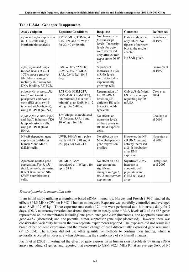
Exposure to high frequency electromagnetic fields, biological effects and health consequences (100 kHz-300 GHz)
121
Table II.3.8.: Gene specific approaches
Assay endpoint Exposure Conditions Response Comment References
c-jun and c-fos expression in PC12 cells using Northern blot analysis
836.55 MHz, TDMA, at 0.09, 0.9, and 90 W m-2 for 20, 40 or 60 min
No change in c-fos transcript levels. Transcript levels for c-jun were decreased only after 20 min exposure to 90 W m-2.
Data are shown in only tables. No figures of northern blot in the results chapter.
No SAR given.
Ivaschuk et al 1997
c-fos, c-jun and c-myc mRNA levels in C3H 10T½ mouse embryo fibroblasts using gel mobility shift assay for DNA-binding, RT-PCR.
FMCW, 835.62 MHz, FDMA, 847.74 MHz, SAR: 0.6 W kg-1 for 4 days
Significant increases in c-fos mRNA levels were detected in exponentially growing cells.
Goswami et al 1999
c-jun, c-fos, c-myc, p53, hsp27 and hsp70 in pluripotent embryonic stem (ES) cells, (wild-type and p53-deficient), using RT-PCR (mRNA)
1.71 GHz (GSM-217, GSM-Talk, GSM-DTX), intermittent (5 min on/30 min off) at an SAR: 0.11-2 W kg-1 for 6-48 hr.
Upregulation of hsp70 mRNA levels in p53-deficient ES cells, but not in wild-type cells.
Only p53-deficient ES cells were up-regulating hsp70 mRNA.
Czyz et al 2004
c-jun, c-fos, c-myc, hsp27 and hsp70 in human TK6 lymphoblastoma cells, using RT-PCR (total RNA).
1.9 GHz pulse-modulated RF fields at SAR: 1 and 10 W kg-1, for 6 hr.
No effects on transcript levels of these genes in RF-field-exposed cells.
Chauhan et al 2006
NF-κB-dependent gene expression profiles in human Mono Mac 6 (MM6) cells.
UWB, 100 kV m-1, pulse width = 0.79±0.01 ns, at 250 pps. for 8 or 24 h
No effect on the NF-κB-dependent gene expression profiles
However, the NF-κB DNA binding activity increased at 24 h incubation after EMF exposure.
Natarajan et al 2006
Apoptosis-related gene expression: Egr-1, p53, Bcl-2, survivin, etc) using RT-PCR in human SH-SY5Y neuroblastoma cells.
900 MHz, GSM modulated at 1 W kg-1, for up to 24 hr.
No effect on p53 expression but significant changes in Egr-1, Bcl-2 and survivin expression.
Significant 2.3% increase in apoptotic cell population and G2/M cell cycle arrest
Buttiglione et al 2007
Transcriptomics in mammalian cells
In an initial study utilizing a membrane-based cDNA microarray, Harvey and French (1999) studied the effects 864.3 MHz (CW) on HMC-1 human monocytes. Exposure was carefully controlled and averaged at an SAR of 7 W kg-1. Three exposure runs each of 20 min were performed at 4-h intervals daily for 7 days. cDNA microarray revealed consistent alterations in steady-state mRNA levels of 3 of the 558 genes represented on the membranes including one proto-oncogene c-kit (increased), one apoptosis-associated gene dad-1 (decreased) and one potential tumor suppressor gene ndpk (decreased). However, there was considerable variability between the two separate experiments reported. The exposure did not result in a broad effect on gene expression and the relative change of each differentially expressed gene was small (< 1.5 fold). The authors did not use other quantitative methods to confirm their finding, which is generally accepted as necessary when determining the significance of such small changes.
Pacini et al (2002) investigated the effect of gene expression in human skin fibroblasts by using cDNA arrays including 82 genes, and reported that exposure to GSM 902.4 MHz RF at an average SAR of 0.6
Exposure to high frequency electromagnetic fields, biological effects and health consequences (100 kHz-300 GHz)
122
W kg-1 for 1 h increased the expression of 14 genes which function in mitogenic signal transduction, cell growth and apoptosis. The authors further demonstrated a significant increase in DNA synthesis and intracellular mitogenic second messenger formation which matched with the high expression of MAP kinase family genes. The authors suggested that the RF exposure has significant biological effects on human skin fibroblasts. However, only one experiment was performed with array analysis and no further experiment was made by the authors to confirm the array data.
Using a cDNA microarray to examine the expression of 3600 genes, Leszczynski et al (2004) reported that exposure to GSM 900 MHz RF at an average SAR of 2.4 W kg-1 for 1 h changed expression of a number of genes, including down-regulated genes involved in forming the Fas/TNFa apoptotic pathway in human endothelial cell line EA.hy926. The authors performed three separate experiments by array analysis, but no confirmation experiments were conducted to validate the array result. More recently, Nylund and Leszczynski (2006) compared the global gene response of two human endothelial cells, EA.hy926 and its variant EA.hy926v1, to RF and reported that the same genes were differently affected by the exposure to GSM 900 MHz RF at an average SAR of 2.8 W kg-1 for 1 h in each of the cell lines. However, the differentially expressed genes in this study were not confirmed using other methods.
Lee et al (2005) used the Serial Analysis of Gene Expression (SAGE) method to measure the RF effect on genome scale gene expression in HL 60 cells. The cells were exposed to 2.45 GHz RF at an average SAR of 10 W kg-1 for 2 h and 6 h. The authors observed that, after 2 h and 6 h exposure, 221 and 759 genes altered their expression, respectively. Functional classification of the affected genes revealed that apoptosis-related genes were among the upregulated ones and the cell cycle genes among the downregulated ones, but no significant increases in the expression of heat shock genes were found. However, the SAGE experiment was repeated only once and only one control with a 2 h sham exposure was used and no confirmation experiment was reported to validate these results.
Huang et al (2006) investigated the effect of 1763 MHz RF on gene expression in Jurkat cells using Applied Biosystems 1700 full genome expression microarray. The authors found that 68 genes were differentially expressed in the cells after exposure to RF at SAR of 10 W kg-1 for 1 h and harvested immediately or after 5 h. The authors repeated the sets of runs five times to collect biological triplicates in every sample. However, the results were not confirmed by other methods.
Whitehead et al (2006a; 2006b) have performed in vitro experiments with C3H 10T(1/2) mouse cells to determine whether FDMA or CDMA modulated RF radiations can induce changes in gene expression using the Affymetrix U74Av2 GeneChip. The data showed the number of probe sets with an expression change greater than 1.3-fold was less than or equal to the expected number of false positives in C3H 10T(1/2) mouse cells after 835.62 MHz FDMA or 847.74 MHz CDMA modulated RF exposure at SAR of 5 W kg-1 for 24 h. The authors concluded that the exposures had no statistically significant effect on gene expression. Leszczynski (2007) raised the criticism that false positives had not been validated as ‘false’ using non-transcriptomic methods, but this view was challenged by Whitehead et al (2007).
In the study by Gurisik et al (2006), human neuroblastoma cells (SK-N-SH) were exposed to GSM 900 signals at SAR of 0.2 W kg-1 for 2 h and recovered without field for 2 h post-exposure. Gene expression were examined by Affymetrix Human Focus Gene Arrays including 8400 genes and followed by real-time RT-PCR of the genes of interest. Only six genes were found to be slightly down-regulated in response to RF exposure comparing with sham-exposed cells, but this response could not be confirmed by real-time RT-PCR analysis. Thus, the authors concluded that the RF exposure applied in this study did not change gene expression in SK-N-SH cells. However, the array experiment was repeated only once and only one array was used for each exposure or sham exposure group.
Qutob et al (2006) have assessed the ability of exposure to a 1.9 GHz pulse-modulated RF field to affect global gene expression in U87MG glioblastoma cells by application of Agilent Human 1A (v1) oligonucleotide 22K microarray slides. The cells were exposed to pulse-modulated (50 Hz, 1/3 duty cycle) RF fields at an SAR of 0.1, 1.0 and 10.0 W kg-1 for 4 hours, and incubated for an additional 6 hours. The authors found no evidence that exposure to RF fields under different exposure conditions can affect gene expression in the cells. In this study, the authors performed five experiments, each containing a single replicate and some of genes were confirmed as real “unaffected genes”.

Exposure to high frequency electromagnetic fields, biological effects and health consequences (100 kHz-300 GHz)
123
As a follow-up to this study, Chauhan et al, (2007b) examined the effect of RF field exposure on the possible expression of late onset genes in U87MG cells after a 24 h RF exposure period and found no changes of gene expression. They also tested immediately and 18 h post-exposure the gene expression of a human monocyte-derived cell-line (Mono-Mac-6, MM6) in response to intermittent exposure (5 min on/10 min off) for 6 h, and found again a negative effect.
Zeng et al (2006) have investigated gene expression profile in MCF-7 cells after exposing to GSM 1800 RF using Affymetrix Genechip U133A. The results showed that gene expression did not change consistently following intermittent exposure (5 min on/10 min off) at an average SAR of 2.0 W kg-1 for 24 h but the expression of five genes was changed consistently after exposure at SAR of 3.5 W kg-1. However, this result could not be further confirmed by real-time RT-PCR assay.
Remondini et al (2006) investigated the effect of RF on gene expression profile in six different cell lines or primary cells, and found that various types of cell reacted differently in RF exposure. Gene expression changed in 900 MHz-exposed EA.hy926 endothelial cells (22 up-regulated, 10 down-regulated), 900 MHz-exposed U937 lymphoblastoma cells (32 up-regulated, two down-regulated), and 1800 MHz-exposed HL-60 leukemia cells (11 up-regulated, one down-regulated), while NB69 neuroblastoma cells, T-lymphocytes, and CHME5 microglial cells did not show significant changes in gene expression. The authors concluded that there were alterations in gene expression in some human cells types exposed to RF but these changes depended on the type of cells and RF signal. However, these RF-responsive candidate genes in different types of cells were not confirmed by other methods. In addition, the RF exposures were different for the different types of cells, so a simple comparison of the effects of RF exposure on gene expression in these cells was not possible.
Zhao R et al (2007) investigated the effects of RF EMF on gene expression of cultured rat neuron with Affymetrix Rat Neurobiology U34 array. Among 1200 candidate genes, 24 up-regulated genes and 10 down-regulated genes associated with multiple cellular functions were identified after 24-h intermittent exposure (5 min on/10 min off) at an average SAR of 2.0 W kg-1. The changes of most of the genes were successfully validated by real-time RT-PCR; these included genes involved in cytoskeleton, signal transduction pathway, and metabolism.
Adopting a similar research strategy, Zhao TY et al (2007) investigated whether expression of genes related to cell death pathways are dysregulated in primary cultured neurons and astrocytes by exposure to a working GSM cell phone rated at a frequency of 1900 MHz for 2 h. Array analysis and real-time RT-PCR showed up-regulation of caspase-2, caspase-6 and Asc (apoptosis associated speck-like protein containing a caspase recruitment domain or ‘card’) gene expression occurred in both “on and “stand-by” modes in neurons, but only in “on” mode in astrocytes. Additionally, astrocytes showed up-regulation of the bax gene. The authors concluded that even relatively short-term exposure to cell phone radiofrequency emissions can up-regulate elements of apoptotic pathways in cells derived from the brain, and that neurons appear to be more sensitive to this effect than astrocytes. However, the authors used a working mobile phone as the source of RF signal, and thus the exposures were not well defined or controlled.
Table II.3.9.: Transcriptomics
Assay endpoint Exposure Conditions Response Comment References Gene expression in human mast cell line
864.3 MHz CW; SAR: 7.0 W kg-1, three 20 min exposures at 4-h intervals daily for 7 days
Changes in the expression of c-kit, nucleoside diphosphate kinase B, and DAD-1 genes by less than 1.5 fold.
Only two separate experiments, and no confirmation experiments for differentially expressed genes
Harvey and French 1999
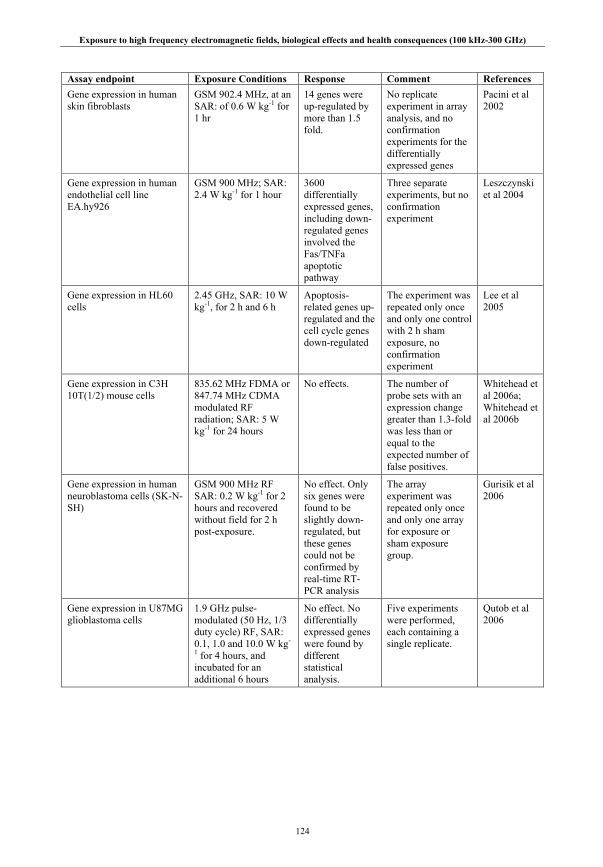
Exposure to high frequency electromagnetic fields, biological effects and health consequences (100 kHz-300 GHz)
124
Assay endpoint Exposure Conditions Response Comment References Gene expression in human skin fibroblasts
GSM 902.4 MHz, at an SAR: of 0.6 W kg-1 for 1 hr
14 genes were up-regulated by more than 1.5 fold.
No replicate experiment in array analysis, and no confirmation experiments for the differentially expressed genes
Pacini et al 2002
Gene expression in human endothelial cell line EA.hy926
GSM 900 MHz; SAR: 2.4 W kg-1 for 1 hour
3600 differentially expressed genes, including down-regulated genes involved the Fas/TNFa apoptotic pathway
Three separate experiments, but no confirmation experiment
Leszczynski et al 2004
Gene expression in HL60 cells
2.45 GHz, SAR: 10 W kg-1, for 2 h and 6 h
Apoptosis-related genes up-regulated and the cell cycle genes down-regulated
The experiment was repeated only once and only one control with 2 h sham exposure, no confirmation experiment
Lee et al 2005
Gene expression in C3H 10T(1/2) mouse cells
835.62 MHz FDMA or 847.74 MHz CDMA modulated RF radiation; SAR: 5 W kg-1 for 24 hours
No effects.
The number of probe sets with an expression change greater than 1.3-fold was less than or equal to the expected number of false positives.
Whitehead et al 2006a; Whitehead et al 2006b
Gene expression in human neuroblastoma cells (SK-N-SH)
GSM 900 MHz RF SAR: 0.2 W kg-1 for 2 hours and recovered without field for 2 h post-exposure.
No effect. Only six genes were found to be slightly down-regulated, but these genes could not be confirmed by real-time RT-PCR analysis
The array experiment was repeated only once and only one array for exposure or sham exposure group.
Gurisik et al 2006
Gene expression in U87MG glioblastoma cells
1.9 GHz pulse-modulated (50 Hz, 1/3 duty cycle) RF, SAR: 0.1, 1.0 and 10.0 W kg-
1 for 4 hours, and incubated for an additional 6 hours
No effect. No differentially expressed genes were found by different statistical analysis.
Five experiments were performed, each containing a single replicate.
Qutob et al 2006

Exposure to high frequency electromagnetic fields, biological effects and health consequences (100 kHz-300 GHz)
125
Assay endpoint Exposure Conditions Response Comment References Gene expression in Jurkat cell
1763 MHz, SAR: 10 W kg-1 for 1 hour and harvested immediately or after five hours
68 genes were differentially expressed after exposure
The authors repeated sets of experiment five times to collect biological triplicates in every sample. But the differentially expressed genes were not confirmed by other methods.
Huang et al 2006
Gene expression in EA.hy926 and EA.hy926v1
GSM 900 MHz, SAR: 2.8 W kg-1 for 1 hour
Four up-regulated genes and 89 down-regulated genes were found in EA.hy926 cell line while 61 up-regulated genes and one down-regulated gene were found in EA.hy926v1 cell line.
Each array experiment was repeated three times (n=3) for each cell line using three different cultures of cells. But no attempt was made to confirm the differentially expressed genes by other methods.
Nylund and Leszczynski 2006
Gene expression in MCF-7 cells
GSM 1800 MHz at an SAR: 2 W kg-1, 3.5 W kg-1, intermittent exposure (5 min on/ 10 min off) for 24 hours
No consistently changed genes at 2 W kg-1. 3.5 W kg-1 exposure produced five consistently changed genes, but these genes could not be confirmed by real-time RT-PCR.
Duplicate arrays were Applied to two independent exposure or sham exposure groups.
Zeng et al 2006
Gene expression in six types of cells, including NB69 neuroblastoma cells, T lymphocytes, CHME5 microglial cells, EA.hy926 endothelial cells, U937 lymphoblastoma cells, and HL-60 leukemia cells
900 and 1800 MHz RF EMF with different exposure patterns, SAR: 1-2.5 W kg-1 for up to 44 h,
22 up-regulated and 10 down-regulated genes in GSM 900-RF exposed EA.hy926 cells. 32 up-regulated, 2 down-regulated genes in U937 cells. 11 upregulated, 1 downregulated genes in HL-60 cells.
RF-responsive candidate genes in different types of cells were not confirmed by other methods.
Remondini et al 2006
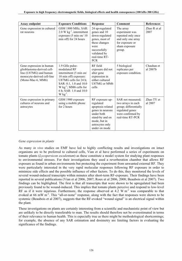
Exposure to high frequency electromagnetic fields, biological effects and health consequences (100 kHz-300 GHz)
126
Assay endpoint Exposure Conditions Response Comment References Gene expression in cultured rat neurons
GSM 1800 MHz, SAR: 2.0 W kg-1, intermittent exposure (5 min on/ 10 min off) for 24 hours
24 up-regulated genes and 10 down-regulated genes, most of these changes were successfully validated by real-time RT-PCR
The array experiment was repeated only once and only one array for exposure or sham exposure group.
Zhao R et al 2007
Gene expression in human glioblastoma-derived cell-line (U87MG) and human monocyte-derived cell-line (Mono-Mac-6, MM6)
1.9 GHz pulse-modulated RF intermittent (5 min on/ 10 min off) exposure: U87MG cells for 24 h; SAR: 0.1, 1.0 and 10.0 W kg-1; MM6 cells for 6 h; SAR: 1.0 and 10.0 W kg-1.
RF field exposure did not alter gene expression in either cultured U87MG or MM6 cells
5 biological replicates per exposure condition.
Chauhan et al 2007b
Gene expression in primary cultures of neurons and astrocytes
GSM 1900 exposure using a mobile phone for 2 hours
RF exposure up-regulated apoptosis related genes in neurons under both stand-by and on mode, but in astrocytes only under on mode
SAR not measured; two arrays in each group; differentially regulated genes were confirmed by real-time RT-PCR
Zhao TY et al 2007
Gene expression in plants
As many in vivo studies on EMF have led to highly conflicting results and investigations on intact organisms are to be preferred to cultured cells, Vian et al have performed a series of experiments on tomato plants (Lycopersicon esculentum) as these constitute a model system for studying plant responses to environmental stresses. For their investigations they used a reverberation chamber that allows RF exposure as found in urban environments but protecting the experiment from unwanted external RF. They were particularly interested in the very rapid molecular responses following RF exposure in order to minimize side effects and the possible influence of other factors. To do this, they monitored the levels of several wound-induced transcripts within minutes after short-term RF-exposure. Their findings have been reported in several publications (Vian et al 2006, 2007; Roux et al 2006, 2008; Beaubois et al 2007). Two findings can be highlighted. The first is that all transcripts that were shown to be upregulated had been previously found to be wound-induced. This implies that tomato plants perceive and respond to low-level RF as if it were injurious. Furthermore, the response observed at 4.2 W m-2 was comparable to that evoked at 66 mW m-2. This “all-or-none” response, along with the fact that responses were shown to be systemic (Beaubois et al 2007), suggests that the RF-evoked “wound signal” is an electrical signal within the plant.
These investigations on plants are certainly interesting from a scientific and mechanistic point of view but are unlikely to be directly transferable to man. The results should therefore not be overestimated in terms of their relevance to human health. This is especially true as there might be methodological shortcomings; for example, the absence of any SAR estimation and dosimetry are limiting factors in evaluating the significance of the findings.

Exposure to high frequency electromagnetic fields, biological effects and health consequences (100 kHz-300 GHz)
127
Protein expression
Conventional methods of protein analysis depend upon methods such as Western blotting and traditional biochemistry. In Western blotting, proteins are separated using acrylamide gels and transferred to membranes. The membranes are subsequently stained with antibodies to specific proteins of interest. The presence or absence of specific proteins and crude indications of relative abundance can be determined. Proteins can also be visualized in histological or cellular preparations using immunocytochemistry.
Proteomics is the term applied to the global analysis of the protein complement of a cell. The ‘proteome’ is complex consisting of tens of thousands of proteins each of which may be subject to post-translational modification. Such modifications can be important for determining the enzymatic activity half-life and location of a protein or its propensity to interact with other molecules, following phosphorylation for example. Typically, analysis is by 2 dimensional (2D) gel electrophoresis, separating individual proteins on the basis of size and electric charge. These methods have been greatly improved in recent years by the development of standardized protocols and sophisticated image analysis software. These 2D gel systems may also be able to detect different post-translationally modified forms of individual proteins. Various mass spectrometry techniques can be used to identify individual proteins. In addition, protein microarrays and chips, often based on monoclonal antibodies, are being developed that will provide quantitative information regarding the expression of a series of functionally linked proteins. These techniques can also be applied to measure the functional state of proteins by examining their phosphorylation status.
EMF studies have generally taken advantage of protein specific approaches such as Western blotting and 2D gel approaches for studying exposure-induced effects on the proteome; few groups have examined such effects using array-based proteomic approaches. However, the proteome of higher eukaryotes is far from being completely understood and it must be recognized that the techniques currently available are unable to describe all effects of toxins on the proteome. The various studies are summarized and reviewed in the following two chapters.
Protein-specific approaches
Many recent studies of RF effects at the cellular level have investigated possible effects on heat shock proteins (hsps), the expression of which is induced by various environmental stresses and forms part of a general cellular stress response, increasing stress tolerance and cytoprotection against stress-induced molecular damage. However, it is not always clear in these studies whether hsp expression has been induced by RF heating or results from a non-thermal RF field-specific stress. Such a distinction requires studies to be conducted under rigorously controlled conditions.
A few biological experiments have been designed and performed to test the hypothesis of nonthermal induction of hsp as a mechanism for RF bioeffects. One such investigation was carried out by De Pomerai et al (2000) who reported increased expression of hsp16 in the nematode Caenorhabditis elegans. These nematodes were exposed in a TEM cell at 750 MHz (CW, SAR estimated as 1 mW kg-1). However, the same group reported that a small temperature rise may have contributed to the elicitation of the effect, as losses in the TEM cell induced temperature elevation in the exposed samples of ~0.2°C (Dawe et al 2006); these authors also revised the previous estimate of SAR up to 4-40 mW kg-1. This implies that, in the initial report, at least part of the cause was thermal. More recently, Dawe et al (2006, 2007) have reported that exposure to CW or GSM 1800 RF at an SAR of 1.8 W kg-1 did not induce hsp16 in this experimental model.
Kwee et al (2001) had reported that the expression of hsp70, but not hsp27, was induced when transformed human epithelial amnion cells were exposed to a GSM 900 signal at a SAR of 2.1 mW kg-1 for 20 min.
Miyakoshi and colleagues have also investigated hsp expression. Using an exposure dish with 3 sections, human brain tumor derived MO54 cells were exposed to 2450 MHz RF fields (SAR: 5, 20, 50, and 100 W kg-1) and cell survival rates and hsp70 expression were determined. At SAR below 20 W kg-1, no effect on hsp70 expression was observed using Western blotting, but, at 20 W kg-1 and higher, hsp70 expression was increased in an SAR and exposure-duration dependent manner (Tian, et al 2002). They also examined the effects 2450 MHz exposure on hsp expression in A172 cells, using a wide range of SARs.
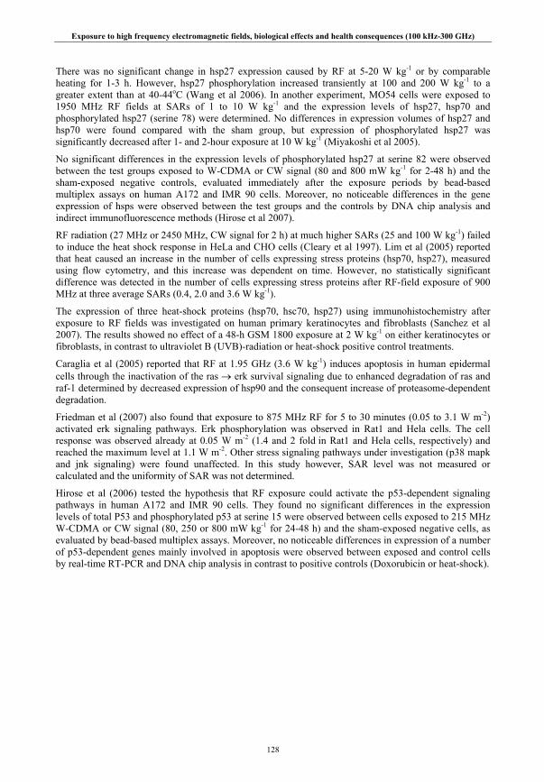
Exposure to high frequency electromagnetic fields, biological effects and health consequences (100 kHz-300 GHz)
128
There was no significant change in hsp27 expression caused by RF at 5-20 W kg-1 or by comparable heating for 1-3 h. However, hsp27 phosphorylation increased transiently at 100 and 200 W kg-1 to a greater extent than at 40-44oC (Wang et al 2006). In another experiment, MO54 cells were exposed to 1950 MHz RF fields at SARs of 1 to 10 W kg-1 and the expression levels of hsp27, hsp70 and phosphorylated hsp27 (serine 78) were determined. No differences in expression volumes of hsp27 and hsp70 were found compared with the sham group, but expression of phosphorylated hsp27 was significantly decreased after 1- and 2-hour exposure at 10 W kg-1 (Miyakoshi et al 2005).
No significant differences in the expression levels of phosphorylated hsp27 at serine 82 were observed between the test groups exposed to W-CDMA or CW signal (80 and 800 mW kg-1 for 2-48 h) and the sham-exposed negative controls, evaluated immediately after the exposure periods by bead-based multiplex assays on human A172 and IMR 90 cells. Moreover, no noticeable differences in the gene expression of hsps were observed between the test groups and the controls by DNA chip analysis and indirect immunofluorescence methods (Hirose et al 2007).
RF radiation (27 MHz or 2450 MHz, CW signal for 2 h) at much higher SARs (25 and 100 W kg-1) failed to induce the heat shock response in HeLa and CHO cells (Cleary et al 1997). Lim et al (2005) reported that heat caused an increase in the number of cells expressing stress proteins (hsp70, hsp27), measured using flow cytometry, and this increase was dependent on time. However, no statistically significant difference was detected in the number of cells expressing stress proteins after RF-field exposure of 900 MHz at three average SARs (0.4, 2.0 and 3.6 W kg-1).
The expression of three heat-shock proteins (hsp70, hsc70, hsp27) using immunohistochemistry after exposure to RF fields was investigated on human primary keratinocytes and fibroblasts (Sanchez et al 2007). The results showed no effect of a 48-h GSM 1800 exposure at 2 W kg-1 on either keratinocytes or fibroblasts, in contrast to ultraviolet B (UVB)-radiation or heat-shock positive control treatments.
Caraglia et al (2005) reported that RF at 1.95 GHz (3.6 W kg-1) induces apoptosis in human epidermal cells through the inactivation of the ras → erk survival signaling due to enhanced degradation of ras and raf-1 determined by decreased expression of hsp90 and the consequent increase of proteasome-dependent degradation.
Friedman et al (2007) also found that exposure to 875 MHz RF for 5 to 30 minutes (0.05 to 3.1 W m-2) activated erk signaling pathways. Erk phosphorylation was observed in Rat1 and Hela cells. The cell response was observed already at 0.05 W m-2 (1.4 and 2 fold in Rat1 and Hela cells, respectively) and reached the maximum level at 1.1 W m-2. Other stress signaling pathways under investigation (p38 mapk and jnk signaling) were found unaffected. In this study however, SAR level was not measured or calculated and the uniformity of SAR was not determined.
Hirose et al (2006) tested the hypothesis that RF exposure could activate the p53-dependent signaling pathways in human A172 and IMR 90 cells. They found no significant differences in the expression levels of total P53 and phosphorylated p53 at serine 15 were observed between cells exposed to 215 MHz W-CDMA or CW signal (80, 250 or 800 mW kg-1 for 24-48 h) and the sham-exposed negative cells, as evaluated by bead-based multiplex assays. Moreover, no noticeable differences in expression of a number of p53-dependent genes mainly involved in apoptosis were observed between exposed and control cells by real-time RT-PCR and DNA chip analysis in contrast to positive controls (Doxorubicin or heat-shock).

Exposure to high frequency electromagnetic fields, biological effects and health consequences (100 kHz-300 GHz)
129
Table II.3.10.: Protein-specific approaches
Assay endpoint Exposure Conditions Response Comment Reference
Electrophoresis of whole cell extract with [35S] methionine protein labeling in HeLa and CHO cells.
27 and 2450 MHz, SAR: 25W kg-1, for 2 h (HeLa cells); 27 MHz, SAR: 100 W kg-1, for 2 h (CHO cells)
No detectable effect on ‘stress protein’ induction.
Only molecular weight was used to determine if the proteins examined were ‘stress proteins’; no other evidence such as Western blotting was given.
Cleary et al 1997
Immunofluorescence staining of AMA (transformed human epithelial amnion) cells.
960 MHz (GDM), SAR: 2.1 mW kg-1, for 20 min
Higher amounts of hsp70 were present in the cells exposed RF-field at 35 and 37oC than in sham-exposed cells.
The induction of hsp70 by RF was not confirmed by other methods.
Kwee et al 2001
Western blotting of human malignant glioma (MO54) cells.
2450 MHz; SAR: ≤ 100 W kg-1 for up to 24 hr.
Increased expression of hsp70 was observed at 20 W kg-1 and higher SARs.
Annular culture plate was used for RF exposure. The difference in SAR distribution is relatively high even in the same ring.
Tian et al 2002
Flow cytometry analysis for detection of hsp70 in human blood mononuclear cells.
1.8 GHz (GSM, GSM-DTX, GSM-Talk); SAR: 2 W kg-1 (GSM, GSM-Talk) or 1.4 W kg-1 (GSM-DTX) intermittently (10 min on/20 min off) for 44 hr
RF exposure did not induce apoptosis, or affect mitochondrial function or hsp70 expression.
Detection of hsp70 was done only by flow cytometry.
Capri et al 2004b
Immunocytochemistry and Western blotting in human malignant glioma (MO54) cells.
1950 MHz; SAR: ≤ 10 W kg-1 for up to 2 hr.
No effect on hsp27 and hsp70 expression. However, phosphorylated hsp27 level decreased after RF exposure at 10W kg-1.
A slight decrease in p-hsp27 (Ser78) expression. Other phosphorylation sites at Ser15 and Ser82 were not examined.
Miyakoshi et al 2005
Flow cytometry analysis for detection of hsp70 and hsp27 in human leukocytes (lymphocytes, monocytes) from healthy volunteers.
900 MHz (CW and GSM), SAR ≤ 3.6 W kg-
1 for up to 4 hr.
No statistically significant differences were detected in the number of cells expressing hsp70 and hsp27 after RF-field exposure.
Expression of hsp70 and hsp27 was observed only by flow cytometry analysis.
Lim et al 2005
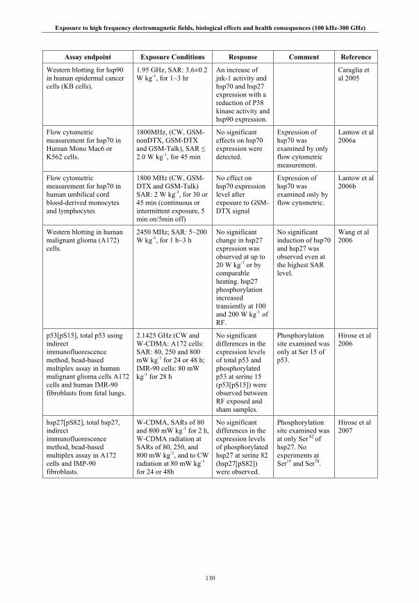
Exposure to high frequency electromagnetic fields, biological effects and health consequences (100 kHz-300 GHz)
130
Assay endpoint Exposure Conditions Response Comment Reference
Western blotting for hsp90 in human epidermal cancer cells (KB cells).
1.95 GHz, SAR: 3.6±0.2 W kg-1, for 1~3 hr
An increase of jnk-1 activity and hsp70 and hsp27 expression with a reduction of P38 kinase activity and hsp90 expression.
Caraglia et al 2005
Flow cytometric measurement for hsp70 in Human Mono Mac6 or K562 cells.
1800MHz, (CW, GSM-nonDTX, GSM-DTX and GSM-Talk), SAR ≤ 2.0 W kg-1, for 45 min
No significant effects on hsp70 expression were detected.
Expression of hsp70 was examined by only flow cytometric measurement.
Lantow et al 2006a
Flow cytometric measurement for hsp70 in human umbilical cord blood-derived monocytes and lymphocytes
1800 MHz (CW, GSM-DTX and GSM-Talk) SAR: 2 W kg-1, for 30 or 45 min (continuous or intermittent exposure, 5 min on/5min off)
No effect on hsp70 expression level after exposure to GSM-DTX signal
Expression of hsp70 was examined only by flow cytometric.
Lantow et al 2006b
Western blotting in human malignant glioma (A172) cells.
2450 MHz; SAR: 5~200 W kg-1, for 1 h~3 h
No significant change in hsp27 expression was observed at up to 20 W kg-1 or by comparable heating. hsp27 phosphorylation increased transiently at 100 and 200 W kg-1 of RF.
No significant induction of hsp70 and hsp27 was observed even at the highest SAR level.
Wang et al 2006
p53[pS15], total p53 using indirect immunofluorescence method, bead-based multiplex assay in human malignant glioma cells A172 cells and human IMR-90 fibroblasts from fetal lungs.
2.1425 GHz (CW and W-CDMA: A172 cells: SAR: 80, 250 and 800 mW kg-1 for 24 or 48 h; IMR-90 cells: 80 mW kg-1 for 28 h
No significant differences in the expression levels of total p53 and phosphorylated p53 at serine 15 (p53[pS15]) were observed between RF exposed and sham samples.
Phosphorylation site examined was only at Ser 15 of p53.
Hirose et al 2006
hsp27[pS82], total hsp27, indirect immunofluorescence method, bead-based multiplex assay in A172 cells and IMP-90 fibroblasts.
W-CDMA, SARs of 80 and 800 mW kg-1 for 2 h, W-CDMA radiation at SARs of 80, 250, and 800 mW kg-1, and to CW radiation at 80 mW kg-1 for 24 or 48h
No significant differences in the expression levels of phosphorylated hsp27 at serine 82 (hsp27[pS82]) were observed.
Phosphorylation site examined was at only Ser 82 of hsp27. No experiments at Ser15 and Ser78.
Hirose et al 2007

Exposure to high frequency electromagnetic fields, biological effects and health consequences (100 kHz-300 GHz)
131
Assay endpoint Exposure Conditions Response Comment Reference
Total ERK expression and phosphorylation, p38-MAPK and JNK phosphorylation, release of Hb-EGF, NADH oxidation using the western-blotting method, NADH oxidase enzyme activity in human Hela carcinoma cells and rat Rat-1 fibroblasts.
875 MHz (800 and 950 MHz also tested); 0.05 to 3.44 W m-2 for 5 to 30 min.
RF exposure increased ERK phosphorylation but not total ERK expression. P38-MAPK and JNK were not found activated after exposure.
The use of multiple exposure conditions, lack of statistical analysis, lack of evidence that the study was blind, and lack of state-of-the-art dosimetry weaken the study.
Friedman et al 2007
hsp70, hsc70, hsp2 using fluorescence level in human normal epidermal (NHEK) keratinocytes and dermal (NHDF) fibroblasts
GSM-1800, 1800 MHz, with a 217 Hz modulation, 48 h; SAR: 2 W kg-1,
The GSM-1800 signal does not act as a stress factor on human primary skin cells
No Western blot. Sanchez et al 2007
Proteomics
Leszczynski and co-workers (Leszczynski et al 2002; Nylund and Leszczynski 2004, 2006) have conducted several in vitro studies on the effects of GSM 900 RF exposure. In their first study (Leszczynski et al 2002), human endothelial (EA.hy926) cells were exposed to RF at an SAR of 2.0 W kg-1 for one-hour and RF exposure changed the overall pattern of protein phosphorylation, upregulated the level of the hsp27 protein and induced its hyper-phosphorylation. This induction was revealed by a variety of independent protein analytical techniques including Western blotting and indirect immunofluorescence methods. The data also revealed that activation of p38 mitogen-activated kinase (MAPK) was partially responsible for the phosphorylation of hsp27. Nylund and Leszczynski (2004) reported that 38 proteins had statistically significant alteration in expression levels in the same cell line after exposure to GSM 900 at SAR of 2.4 W kg-1 for 1 h. Western blotting and indirect immunofluorescence methods were used to confirm that one isoform of vimentin was expressed in the cells after exposure. The authors also suggested that the cytoskeleton might be one of the mobile phone radiation-responding cytoplasmic structures. Nylund and Leszczynski (2006) compared in vitro response to GSM 900 RF in EA.hy926 with its variant EA.hy926v1 by examination of protein expression using 2-D gel electrophoresis. The results showed that protein expression profiles were altered in both cell lines after RF exposure: 38 and 45 differentially expressed proteins were found in EA.hy926 and EA.hy926v1, respectively. However, the affected proteins were altered differently in each of the cell lines. Several differentially expressed proteins in EA.hy926 cells were confirmed by other methods, but differentially expressed protein in EA.hy926v1 cells was not confirmed by Western blotting (Nylund and Leszczynski 2006). Based on the proteome analysis data, the authors concluded that the response might be genome- and proteome-dependent.
Zeng et al (2006) systematically explored the effects of 1800 MHz RF on protein expression in MCF-7 cells by 2-D gel electrophoresis, and found that a few proteins were differentially expressed under continuous or intermittent RF exposure at 3.5 W kg-1 for 24 h or less, implying that the observed effects might have occurred by chance. This study combined proteomics and transcriptomics data, and did not provide convincing evidence that RF exposure could produce distinct effects on gene and protein expression in the MCF-7 cells. The authors suggested that the MCF-7 cells may be insensitive to RF exposure.

Exposure to high frequency electromagnetic fields, biological effects and health consequences (100 kHz-300 GHz)
132
Table II.3.11.: Proteomics
Assay endpoint Exposure Conditions Response Comment References Protein expression in human endothelial (EA.hy926) cell line
GSM 900 MHz, SAR: 2.0 W kg-1 for 1 hour
RF exposure changed protein phosphorylation pattern, up-regulated the levels of hsp27 protein and induced its hyper-phosphorylation.
Confirmed by independent protein analytical techniques including Western blotting and indirect immunofluorescence method
Leszczynski et al 2002
Protein expression in human endothelial (EA.hy926) cell line
GSM 900 MHz, SAR: 2.4 W kg-1 for 1 hour
Up to 38 various proteins have statistically significantly altered their expression levels after RF exposure. Increased expression of vimentin in RF exposed cells.
10 replicates in 2-DE analysis were performed, Western blotting and indirect immunofluorescence method were conducted as confirmation.
Nylund and Leszczynski 2004
Protein expression in EA.hy926 and EA.hy926v1 cells
GSM 900 MHz, SAR: 2.8 W kg-1 for 1 hour
38 and 45 differentially expressed proteins were found in EA.hy926 and EA.hy926v1 respectively. The changes observed in the two cell lines were different
10 replicates in 2-DE analysis were performed, no differentially expressed protein was confirmed by Western blotting.
Nylund and Leszczynski 2006
Protein expression in MCF-7 cells
GSM 1800 MHz, SAR: 3.5 W kg-1, continuous or intermittent exposure (5 min on/ 10 min off) for 1-24 hours
No effects. A small number of different proteins were differentially expressed under different exposure conditions, possibly by chance.
Triplicate gels were performed in each exposure condition.
Zeng et al 2006
Summary on gene and protein expression
The effects of RF exposure on the expression of cancer-related genes (proto-oncogenes and tumor suppressor gene) are considered to be very weak or absent. Some studies, however, reported proto-oncogene expression in p53-deficient cells and a transient effect on the increase in egr-1 gene expression. Although negative reports predominate in this gene-specific approach in mammalian cells, the few positive effects cannot be ignored and further studies should be carried out before reaching a final conclusion.
High-throughput studies of gene expression in various cell types have yielded a variety of results, including a lack of effect, and the up-regulation and down-regulation of various genes. Many studies however are technically incomplete in that they lack sufficient experimental repetition and replication and further validation through the use of more precise quantitative measures of gene expression. In addition, the magnitude of the changes is small, and may be of limited functional significance. However, to date, insufficient research has been carried out to allow definitive conclusions to be drawn.

Exposure to high frequency electromagnetic fields, biological effects and health consequences (100 kHz-300 GHz)
133
Many studies have examined the effect of RF exposure on stress proteins, especially hsps. However, the results of most of these studies are inconsistent, although mostly negative outcomes have been reported in vitro. Some experiments suggest that some of the positive findings might result from heating alone. Among the few signaling pathways that have been investigated, the ERK pathway was found altered but again the studies gave inconsistent data. Further studies should be conducted to evaluate the influence of RF exposure on major stress signaling pathways (MAPK, ERK, etc.). Protein-specific approaches may provide more information in studies of these pathways, which are driven mainly through phosphorylation cascades, than monitoring protein expression itself.
High-throughput studies of protein expression by one group have reported changes in protein expression and phosphorylation in two cell lines, whereas another group attributed the small changes observed in another cell type to chance. No clear patterns of response emerged. At present, the available data don’t allow valid conclusions to be drawn.
II.3.3.3. Reactive oxygen species and oxidative stress
Ageing, exercise, UV and many other forms of stress are known to increase the production of reactive oxygen species (ROS). These are generally very small and highly reactive species and include O2
.–, free radicals and both inorganic and organic peroxides. The harmful cellular effects of ROS include (i) damage to DNA, (ii) oxidation of polyunsaturated fatty acids in lipids, and (iii) oxidation of amino acid residues in proteins. Therefore, cellular damage is increased by elevated ROS levels. In addition, oxidative stress has been implicated in the initiation and promotion of carcinogenesis. Only a few studies have examined the effects of RF fields on spontaneous or induced ROS production, mostly in cells of the immune system that generate ROS as part of their function. In addition some studies have monitored more general assessment of oxidative stress, including intracellular oxidant and antioxidant levels, antioxidant defense, and heat-shock protein levels, the latter function being as molecular chaperones to protect cells from various types of stresses. [Heat shock protein expression is further discussed in the previous chapter (II.3.3.2.).]
Zmyślony et al (2004) examined the effects of 930 MHz CW RF on ROS levels in rat lymphocytes. Some of the lymphocyte samples were treated with Fe2+ ions to induce oxidative processes. The results showed that acute (up to 15 min) exposure at around 1.5 W kg-1 did not affect ROS production. However, the addition of FeCl2 to the lymphocyte suspensions significantly increased the magnitude of fluorescence, used to measure intracellular ROS levels, by ~ 15% in the exposed lymphocytes.
Hook et al (2004b) investigated the effects of FMCW-modulated 835 MHz and CDMA-modulated 847 MHz RF on the production of oxygen radicals, the enhancement of radicals produced by oxidative stress, the resulting oxidative damage and the induction of an oxidative stress response, in a mouse J774.16 macrophage cell line. Oxidative stress was induced prior to exposure using γ-interferon (IFN) and bacterial lipopolysaccharide (LPS), both of which activate cellular oxidases producing reactive nitrogen and oxygen species. No effects of RF exposure were seen on any of the endpoints, in unstimulated or in IFN/LPS stimulated macrophages.
Simko and colleagues (Lantow et al 2006a; Lantow et al 2006b) have examined the effect of 1800 MHz RF CW or various GSM modes (DTX and Talk) at up to 2 W kg-1 for 45 min on hsp70 and ROS production in human Mono Mac 6 cells (a monocyte leukemia cell line) and K562 cells (an erythroid leukemia cell line). No significant difference in free radical production was detected after RF exposure compared with their respective controls, and no additional effects on the production of superoxide radical anions was detected in cells after co-exposure to RF plus the phorbol ester PMA (phorbol-12-myristate-13-acetate) or RF plus LPS treatment (Lantow et al 2006a), both of which known to increase ROS production in monocytes and other cells of the immune system. In addition, no significant effects of RF exposure on hsp70 expression were found. The same group (Lantow et al 2006b) also used human umbilical cord blood-derived monocytes and lymphocytes to examine ROS release after continuous or intermittent (5 min on/5 min off) exposure to CW or the various GSM 1800 modes listed above at 2 W kg-1 for 30 or 45 min. No effects of RF exposure on ROS production in PMA-stimulated human monocytes or lymphocytes were seen once a correction had been made for the reduced production of ROS in the sham-exposed cells compared to incubator controls. In addition, no significant effects of RF exposure on hsp70 expression were found.

Exposure to high frequency electromagnetic fields, biological effects and health consequences (100 kHz-300 GHz)
134
As part of the CEMFEC program, Scarfi et al investigated the induction of ROS in murine L929 fibrosarcoma cells exposed to a GSM 900 RF field, with or without co-exposure to 3-chloro-4-(dichloromethyl)-5-hydroxy-2(5H)-furanone (MX), a potent environmental carcinogen produced during the chlorination of drinking water (Zeni et al 2007). Treatment with MX was found to significantly increase ROS production, with a concomitant decrease in levels of the antioxidant glutathione; however, RF exposure, either alone or in combination with MX, did not induce formation of ROS under any of the experimental conditions investigated.
Overall, the data are consistent and suggest that RF exposure has no effect on ROS production in several different cell lines.
Table II.3.12.: Reactive oxygen species and oxidative stress
Assay endpoint Exposure Conditions Response Comment Reference
ROS (fluorescent probe assay) in rat lymphocytes. Positive control: FeCℓ2
930 MHz, CW, SAR: 1.5 W kg-1 for 5 or 15 min.
No ROS induction by RF alone, but RF exposure enhanced ROS production induced by the addition of FeCℓ2.
Zmyślony et al 2004
Cell viabiliy (typan blue exclusion), oxidant and antioxidant levels, oxidative damage and nitric oxide production in mouse J774.16 macrophage cells stimulated with IFN and LPS before exposure.
835.62 MHz, FMCW modulation and 847.74 MHz CDMA modulation; at 0.8 W kg-1 for 20-22 hr
No effects of RF on cell viability, intracellular oxidants, oxidative damage or antioxidant defenses in IFN or LPS stimulated cells.
Hook et al 2004b
Measurement of superoxide radical anions, ROS and hsp70 in human Mono Mac6 or K562 cells.
Positive controls: PMA, LPS and heat.
1800 MHz, (CW, GSM-nonDTX, GSM-DTX and GSM-Talk) SAR ≤ 2.0 W kg-1 for 45 min
No RF effects on free radical production were detected, and no RF effects on superoxide radical anion production were detected after co-exposure with PMA or LPS.
Lantow et al 2006a
ROS measurement by flow cytometry in human umbilical cord blood-derived monocytes and lymphocytes.
Positive control: PMA
1800 MHz (CW, GSM-DTX and GSM-Talk) continuous or intermittent, (5 min on/5min off) SAR: 2 W kg-1 for 30 or 45 min.
No effect on ROS production of RF alone or in combination with PMA in either cell type.
ROS production was significantly different in RF exposed human monocytes compared to sham-exposed controls, possibly due to lowered value in the sham-exposed cells.
Lantow et al 2006b
ROS production (fluorescent intensity of 2’7’-dichlorofluorescein) in mouse L929 fibrosarcoma cells.
Positive control: MX
900 MHz, SAR: 0.3 or 1 W kg-1 for 10 or 30 min; with or without co-exposure to the carcinogen MX.
No effect on ROS production of RF alone or in combination with MX.
Zeni et al 2007
Exposure to high frequency electromagnetic fields, biological effects and health consequences (100 kHz-300 GHz)
135
II.3.3.4. Cell proliferation, differentiation and cell cycle control
Cancer develops when cells acquire specific growth advantages through the stepwise accumulation of heritable changes in gene function. Basically, this process is directed by changes in two different classes of genes: tumor suppressor genes that inhibit cell growth and survival, and oncogenes that promote cell growth and survival. At the cellular level, the development of cancer is associated with sustained proliferation, dedifferentiation, angiogenesis, invasion and resistance to apoptosis. This chapter reviews in vitro studies related to these phenomena under RF exposure.
Studies published since 1993 on cell proliferation after RF exposure are reviewed which show a mixture of responses including either no effect, or increases, or decreases in these various end-points. The difficulty comes, as often, from the variety of exposure conditions, exposure setups and cell types. Adequate temperature control and dosimetry in particular are critical to the evaluation of any non-thermal effects. Taken together however, some common features arise from these studies.
A number of studies showed no effects on cellular proliferation as determined by cell count, DNA synthesis and cell cycle distribution in cells exposed to RF. In primary cells, proliferation is usually unaffected by RF exposure (Stagg et al 1997; Capri et al 2004a; Nikolova et al 2005; Sanchez et al 2006a; Sun et al 2006). Stagg et al (1997) showed no effects of 836.55 MHz, TDMA RF on rat primary glial cells exposed at very low level SARs ranging from 0.15 to 59 mW kg-1 for up to 24 hr. Sanchez et al (2006a) exposed human reconstructed epidermis using keratinocytes to GSM 900, 2 W kg-1 for 48 hr and found no increase in the number of Ki67 positive cells, a marker for cell proliferation. Sun et al (2006) found no effect of a 2 h GSM-1800 exposure at 1, 2, and 3 W kg-1 on the proliferation of human lens epithelial cells. Yao et al (2004) however found a decrease in cell proliferation correlated to a repressed P27KIP1 protein expression in rabbit lens epithelial cells exposed to 2450 MHz, CW signal from 5 to 20 W m-2 for 8 hr, although the exposure conditions were not well described. By contrast, Pacini et al (2002) showed an increased proliferation in human normal fibroblasts, but the use of a genuine mobile phone for RF exposure does not enable reliable exposure measurement and dosimetry and this study is thus difficult to evaluate. Interestingly, studies using the cytokinesis-blocked micronucleus assay, mostly in human peripheral blood mononucleated cells, usually failed to detect cytotoxicity and changes in cell proliferation as determined by the mitotic index or the frequency of binucleates (see Chapter II.3.2.), even, in some cases, when increased micronucleated binucleated cells were detected (Maes et al 1993; Zotti-Martelli 2000; 2005; D’Ambrosio et al 2002; Tice et al 2002).
Proliferation and cell cycle distribution were unaffected in a number of cancer or transformed cell lines (Higashikubo et al 2001; Merola et al 2006; Gurisik et al 2006; Lantow et al 2006c; Takashima et al 2006; Chauhan et al 2007a). In fact, Takashima et al (2006) showed that the threshold for an effect on proliferation (decrease) was 200 W kg-1 CW when Chinese hamster ovary CHO-K1 cells and human glioma MO54cells were exposed for 2 hr at 2450 MHz.
Earlier however, Cao et al (1995) showed that 27-MHz RF (5 and 25 W kg-1) altered the cell cycle of Chinese hamster ovary CHO cells in an SAR-dependent way. The same group (Cleary et al 1996) found that the effect of 2450-MHz RF (5 to 50 W kg-1) was highly dependent on the concentration of the mitogen IL2 in CTLL2 mouse cytolytic T lymphocytes, and hypothesized that the effect was dependent on the presence of high affinity-IL2 receptors, suggesting that the effect is cell-type dependent. Donnellan et al (1997) found an increased proliferation of rat mast cells repeatedly exposed at 850 MHz (81 W m-2, 3 times a day for 7 days) suggesting that cells lost their contact inhibition. Unfortunately, these studies have not been independently confirmed.
There have been several studies of ornithine decarboxylase (ODC) activity after RF exposure. ODC is an enzyme involved in cell growth and ODC overexpression has been consistently reported to lead to neoplastic cellular transformation (Kubota et al 1997; Dhalluin et al 1998; Tabib and Bachrach 1999), and may thus be involved in cancer cell invasiveness. An increased ODC enzyme activity was consistently reported in murine L929 fibroblasts after an 8-hour in vitro exposure to modulated 835-840 and 915 MHz RF at 2.5 W kg-1 (Litovitz et al 1993, 1997; Penafiel et al 1997). Results from two independent groups did not confirm such effect in the same cell type exposed at a similar SAR (Desta et al 2003; Höytö et al 2006, 2007a). Both groups also reported that a temperature increase resulting from either RF exposure or

Exposure to high frequency electromagnetic fields, biological effects and health consequences (100 kHz-300 GHz)
136
conventional heating of about 1°C decreased ODC enzyme activity. When cells were isothermally exposed at higher SARs (up to 15 W kg-1) however, different outcomes in ODC activity were found according to the type of exposure system and temperature control method used. No clear explanation could be given, which suggests that temperature control is critical in the interpretation of possible non-thermal effects of RF exposure, at least in ODC experiments. Höytö et al (2007b) also exposed L929 murine fibroblasts and other cell lines (rat C6 glioblastoma cells, human SH-SY5Y neuroblastoma cells) and rat primary astrocytes to 815 MHz, CW and GSM-modulated. They found essentially no effect in secondary cell lines but a consistent significant decrease in ODC activity in primary astrocytes. While increased ODC activity has been considered as an indication of potentially harmful health effects, the health relevance of decreased ODC activity is not known.
It is known that the malignancy of a cancer is directly related to the degree of de-differentiation of tumor cells, related to their rate of growth. Differentiation under in vitro RF exposure has been sparsely studied. Nikolova et al (2005) found an effect of intermittent GSM 1800 signal (1.5 W kg-1 for up to 48 hours) in mouse pluripotent embryonic stem cells; while neural-specific Nurr-1 mRNA expression was decreased, no change in neural-specific proteins could be detected. Merola et al (2006) showed that exposure to GSM-900 MHz at 1 W kg-1 for up to 72 hours did not affect spontaneous or retinoic acid–induced differentiation of LAN-5 human neuroblastoma cells.
Finally, in yeast (Saccharomyces cerevisiae) cells, Gos et al (1997) investigated possible non-thermal effects on cell division rate in exponentially growing cells that were exposed to RF in the millimeter frequency range around 41.7 GHz at low power densities (5 and 500 mW m-2). No significant differences were seen between exposed and unexposed cells for value of S-phase and G1-phase at two different power levels. Pakhomov et al (2002) investigated the effects on the density of yeast cells, achieved after a 6 h growth period in a solid nutrient medium (agarose gel) during EHPP (extremely high power pulses) or CW exposure. They reported that CW and EHPP exposures produced highly non-uniform but identical heating patterns in exposed samples. Cell density was strongly affected by irradiation, and the changes correlated well with the local temperature rise. However, the data revealed no statistically significant difference between CW and EHPP samples across the entire studied range of SAR levels (over six orders of magnitude). A trend (p < 0.1) for such a difference was observed in gel slices that were exposed at a time averaged SAR of 100 W kg-1 and higher, which corresponded to a peak SAR above 20 MW kg-1 for the EHPP condition.
In summary, many studies have been published that suggest there are no effects of RF exposure on cell proliferation and cell cycle control. A few early studies have been published that suggest that there are effects of RF exposure below 100 W kg-1, but these should be confirmed using improved exposure equipment, temperature control and dosimetry. The very few studies on the effects of RF exposure on differentiation in vitro do not suggest any effect.
Table II.3.13.: Proliferation, differentiation and cell cycle control
Assay endpoint Exposure Conditions Response Comment References ODC activity (14CO2 generation) in L929 murine fibroblasts
915 MHz; CW, 55, 60 & 65 Hz AM 915 MHz, switched between AM frequencies at different intervals; SAR: 2.5 W kg-1 for 2 - 24 hr
No effect of CW RF. Doubling of ODC activity at 8 hr of modulated-RF exposure applied for periods exceeding 10 s.
SARs averaged over exposure flask; variable ODC activities in controls
Litovitz et al 1993
Cell cycle distribution (flow cytometry) in Chinese hamster ovary (CHO) cells exposed in different phases of the cell cycle
27 MHz, CW, SAR: 5 or 25 W kg-1 for 2 hr
SAR-dependent alterations in cell cycle progression with a maximum effect 3 days after exposure at 25 W kg-1.
Data showed considerable interexperimental variability. Cells exposed in phases G0/G1 and S phase were most sensitive.
Cao et al 1995
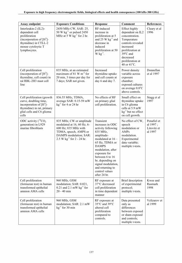
Exposure to high frequency electromagnetic fields, biological effects and health consequences (100 kHz-300 GHz)
137
Assay endpoint Exposure Conditions Response Comment References Interleukin-2 (IL2)-dependent cell proliferation (incorporation of [H3]-thymidine) in CTLL-2 mouse cytoloytic T lymphocytes.
2450 MHz CW, SAR: 25, 50 W kg-1 or pulsed 2450 MHz at 5 W kg-1 for 2 hr.
RF-induced increase in proliferation at 5 and 25 W kg-1 and decrease in induced proliferation at 50 W kg-1.
Effect highly dependent on IL2 concentration. Temperature controls revealed increased proliferation at 39oC and decreased proliferation at 40 or 41oC.
Cleary et al 1996
Cell proliferation (incorporation of [H3]-thymidine, cell count) in rat RBL-2H3 mast cell line
835 MHz, at an estimated maximum of 81 W m-2 for 20 min, 3 times per day for 7 days; SAR not given.
Increased thymidine uptake and cell counts at day 6 and day 7.
Power density variable across exposure chamber; exposed cultures on average 0.8oC above controls.
Donnellan et al 1997
Cell proliferation (growth curve, doubling time, incorporation of [H3]-thymidine) in rat, primary glial cells and C6 glioma cells
836.55 MHz, TDMA, average SAR: 0.15-59 mW kg-1 for 4 or 24 hr
No effects of RF on primary glial cell proliferation.
Small effect on thymidine uptake in C6 glioma cells at 5.9 mW kg-1 but no effect on cell growth.
Stagg et al 1997
ODC activity (14CO2 generation) in L929 murine fibroblasts
835 MHz, CW or amplitude modulated at 16, 60 Hz, 6-600 Hz; 835 MHz with TDMA, speech, AMPS or DAMPS modulation; SAR: 2.5 W kg-1 for 2 - 24 hr.
Transient increases in ODC activity following 835 MHz, amplitude-modulated at 16 – 65 Hz, TDMA or DAMPS modulation, after exposure for between 6 to 16 hr, depending on signal modulation, and returning to control values after 24 hr.
No effect of CW, speech modulation or AMPs modulation. Experimental data variable; multiple t-tests.
Penafiel et al 1997 ; Litovitz et al 1997
Cell proliferation (formazan test) in human transformed epithelial amnion AMA cells
960 MHz, GSM modulation; SAR: 0.021, 0.21 and 2.1 mW kg-1 for 20 - 40 min
RF exposure at 37°C decreased cell proliferation in time dependent manner
Brief description of experimental protocol; multiple t-tests.
Kwee and Rasmark 1998
Cell proliferation (formazan test) in human transformed epithelial amnion AMA cells
960 MHz, GSM modulation; SAR: 2.1 mW kg-1 for 30 min
RF exposure at 35°C and 39°C altered cell proliferation compared to controls.
Data presented only as differences between exposed or sham exposed and controls; multiple t-tests.
Velizarov et al 1999
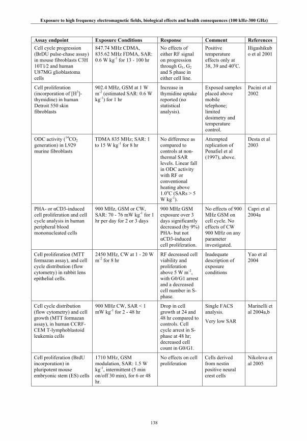
Exposure to high frequency electromagnetic fields, biological effects and health consequences (100 kHz-300 GHz)
138
Assay endpoint Exposure Conditions Response Comment References Cell cycle progression (BrDU pulse-chase assay) in mouse fibroblasts C3H 10T1⁄2 and human U87MG glioblastoma cells
847.74 MHz CDMA, 835.62 MHz FDMA, SAR: 0.6 W kg-1 for 13 - 100 hr
No effects of either RF signal on progression through G1, G2 and S phase in either cell line.
Positive temperature effects only at 38, 39 and 40oC.
Higashikubo et al 2001
Cell proliferation (incorporation of [H3]-thymidine) in human Detroit 550 skin fibroblasts
902.4 MHz, GSM at 1 W m-2 (estimated SAR: 0.6 W kg-1) for 1 hr
Increase in thymidine uptake reported (no statistical analysis).
Exposed samples placed above mobile telephone; limited dosimetry and temperature control.
Pacini et al 2002
ODC activity (14CO2 generation) in L929 murine fibroblasts
TDMA 835 MHz; SAR: 1 to 15 W kg-1 for 8 hr
No difference as compared to controls at non-thermal SAR levels. Linear fall in ODC activity with RF or conventional heating above 1.0oC (SARs > 5 W kg-1).
Attempted replication of Penafiel et al (1997), above.
Desta et al 2003
PHA- or αCD3-induced cell proliferation and cell cycle analysis in human peripheral blood mononucleated cells
900 MHz, GSM or CW, SAR: 70 - 76 mW kg-1 for 1 hr per day for 2 or 3 days
900 MHz GSM exposure over 3 days significantly decreased (by 9%) PHA- but not αCD3-induced cell proliferation.
No effects of 900 MHz GSM on cell cycle. No effects of CW 900 MHz on any parameter investigated.
Capri et al 2004a
Cell proliferation (MTT formazan assay), and cell cycle distribution (flow cytometry) in rabbit lens epithelial cells.
2450 MHz, CW at 1 - 20 W m-2 for 8 hr
RF decreased cell viability and proliferation above 5 W m-2, with G0/G1 arrest and a decreased cell number in S-phase.
Inadequate description of exposure conditions
Yao et al 2004
Cell cycle distribution (flow cytometry) and cell growth (MTT formazan assay), in human CCRF-CEM T-lymphoblastoid leukemia cells
900 MHz CW, SAR < 1 mW kg-1 for 2 - 48 hr
Drop in cell growth at 24 and 48 hr compared to controls. Cell cycle arrest in S-phase at 48 hr; decreased cell count in G0/G1.
Single FACS analysis.
Very low SAR
Marinelli et al 2004a,b
Cell proliferation (BrdU incorporation) in pluripotent mouse embryonic stem (ES) cells
1710 MHz, GSM modulation, SAR: 1.5 W kg-1, intermittent (5 min on/off 30 min), for 6 or 48 hr.
No effects on cell proliferation
Cells derived from nestin positive neural crest cells
Nikolova et al 2005
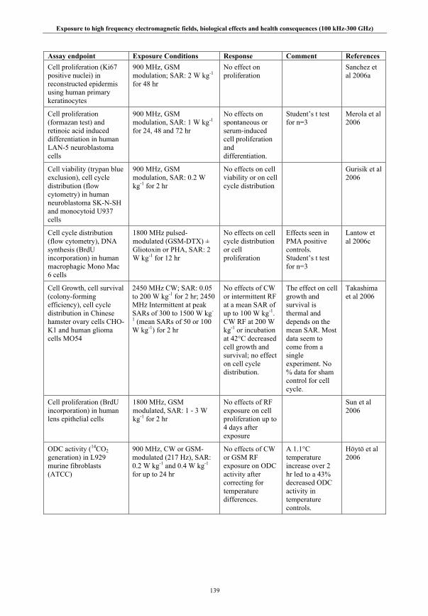
Exposure to high frequency electromagnetic fields, biological effects and health consequences (100 kHz-300 GHz)
139
Assay endpoint Exposure Conditions Response Comment References Cell proliferation (Ki67 positive nuclei) in reconstructed epidermis using human primary keratinocytes
900 MHz, GSM modulation; SAR: 2 W kg-1 for 48 hr
No effect on proliferation
Sanchez et al 2006a
Cell proliferation (formazan test) and retinoic acid induced differentiation in human LAN-5 neuroblastoma cells
900 MHz, GSM modulation, SAR: 1 W kg-1 for 24, 48 and 72 hr
No effects on spontaneous or serum-induced cell proliferation and differentiation.
Student’s t test for n=3
Merola et al 2006
Cell viability (trypan blue exclusion), cell cycle distribution (flow cytometry) in human neuroblastoma SK-N-SH and monocytoid U937 cells
900 MHz, GSM modulation, SAR: 0.2 W kg-1 for 2 hr
No effects on cell viability or on cell cycle distribution
Gurisik et al 2006
Cell cycle distribution (flow cytometry), DNA synthesis (BrdU incorporation) in human macrophagic Mono Mac 6 cells
1800 MHz pulsed-modulated (GSM-DTX) ± Gliotoxin or PHA, SAR: 2 W kg-1 for 12 hr
No effects on cell cycle distribution or cell proliferation
Effects seen in PMA positive controls. Student’s t test for n=3
Lantow et al 2006c
Cell Growth, cell survival (colony-forming efficiency), cell cycle distribution in Chinese hamster ovary cells CHO-K1 and human glioma cells MO54
2450 MHz CW; SAR: 0.05 to 200 W kg-1 for 2 hr; 2450 MHz Intermittent at peak SARs of 300 to 1500 W kg-
1 (mean SARs of 50 or 100 W kg-1) for 2 hr
No effects of CW or intermittent RF at a mean SAR of up to 100 W kg-1. CW RF at 200 W kg-1 or incubation at 42°C decreased cell growth and survival; no effect on cell cycle distribution.
The effect on cell growth and survival is thermal and depends on the mean SAR. Most data seem to come from a single experiment. No % data for sham control for cell cycle.
Takashima et al 2006
Cell proliferation (BrdU incorporation) in human lens epithelial cells
1800 MHz, GSM modulated, SAR: 1 - 3 W kg-1 for 2 hr
No effects of RF exposure on cell proliferation up to 4 days after exposure
Sun et al 2006
ODC activity (14CO2 generation) in L929 murine fibroblasts (ATCC)
900 MHz, CW or GSM-modulated (217 Hz), SAR: 0.2 W kg-1 and 0.4 W kg-1 for up to 24 hr
No effects of CW or GSM RF exposure on ODC activity after correcting for temperature differences.
A 1.1°C temperature increase over 2 hr led to a 43% decreased ODC activity in temperature controls.
Höytö et al 2006

Exposure to high frequency electromagnetic fields, biological effects and health consequences (100 kHz-300 GHz)
140
Assay endpoint Exposure Conditions Response Comment References Cell viability, cell cycle distribution in human lymphoblastoid TK6, lymphoblastic HL60 and myeloid Mono-Mac-6 cells
1900 MHz pulse-modulated, intermittent exposure (5 min on/10 min off); SAR: 1 and 10 W kg-1 for 6 hr,
No effects of RF exposure on cell viability or cell cycle progression
Heat shock (43oC) controls showed decreased viability and G2/M block
Chauhan et al 2007a
ODC activity (14CO2 generation) in L929 murine fibroblasts (ATCC).
835 and 872 MHz, CW or DAMPS-modulated (50 Hz); SAR: 2.5 or 6 W kg-1 for up to 24 hr. Two exposure systems - Crawford cell (CC – 835 MHz) and waveguide (WG – 872 MHz) were used with different cooling methods.
No effects of CW or DAMPS on ODC activity at 2.5 W kg-1. Significant decrease of ODC activity after CC exposure for 2 hr at 6 W kg-1 but not after 8 or 24 hr. Significant increase in activity after WG exposure for 8 hr at 6 W kg-1.
Unable to replicate the study of Penafiel et al (1997). However, there were discrepancies in the present study outcome at 6 W kg-1 when using two different exposure and temperature control systems
Höytö et al 2007a
ODC activity (14CO2 generation) in rat primary astrocytes and in L929 murine fibroblasts (ECACC), rat C6 glioblastoma cells and human SH-SY5Y neuroblastoma cells.
835 MHz, CW or GSM-modulated, SAR: 1.5, 2.5 and 6 W kg-1 for up to 24 hr.
Significant decrease in ODC activity in rat primary astrocytes at 1.5 and 6 W kg-
1 although after different exposure times at different SARs. No overall effects on ODC activity in rat gioblastoma and human neuroblastome cell lines.
No effect of CW or GSM RF at 2.5 W kg-1 on ODC activity in L929 murine fibroblasts used by Penafiel et al (1997), but significant reductions at 1.5 and 6.9 W kg-1. However, these results in L929 cells were affected by temperature differences between the two exposure chambers
Höytö et al 2007b
II.3.3.5. Apoptosis
Apoptosis is a “suicide” process of cells in multicellular organisms. It is one of the main types of programmed cell death (PCD), and involves an orchestrated series of biochemical events leading to a characteristic cell morphology and death. The apoptotic process is executed in such a way as to safely dispose of cellular debris. Apoptosis is initiated for various reasons, such as when a cell is no longer needed within the body (i.e. in embryonic development) or when it becomes a threat to the health of the organism (i.e. with high level of DNA damage). Severe pathological consequences, such as autoimmune disorders, neurodegenerative diseases, and cancer can arise from abnormal rates of apoptosis.
There is no single parameter that defines programmed cell death, and therefore a combination of techniques is recommended for the reliable detection of apoptosis. Using timed inductions and comparing relationships between cell populations expressing multiple markers, it is possible to estimate within a given model the relative order in which the different components of an apoptotic process become evident;

Exposure to high frequency electromagnetic fields, biological effects and health consequences (100 kHz-300 GHz)
141
these range from the externalization of the phosphatidyl-serines at an early stage to the ladder-type DNA fragmentation and the loss of membrane integrity at late stages of PCD.
PCD is activated by different apoptotic signaling pathways that can be investigated through the expression of apoptosis-related genes and proteins. The "extrinsic" pathway is activated by the binding of death-activator proteins to the cell surface. The "intrinsic" pathway is launched by signals inside the cell, such as damage caused by radiation or toxins, the withdrawal of critical survival factors (growth factors, hormones), or disturbances in the cell cycle. Both pathways converge inside the cell, turning on a central effector family of proteins: caspases. Recently, a caspase-independent pathway has also been described driven through the Apoptosis-inducing Factor (AIF).
A number of studies have been published on the effects of RF exposure, from 800 to 2450 MHz, on cellular apoptosis in vitro.
Using normal cells, ranging from yeast to mouse embryonic stem cells, primary rat neurons, and primary human fibroblasts and blood cells, most studies found no evidence that low-level RF exposure could induce apoptosis (Markkanen et al 2004; Capri et al 2004a, 2004b; Nikolova et al 2005; Joubert et al 2006; Sanchez et al 2007). However, Joubert et al (2008) recently reported an increase in AIF-dependent apoptosis in rat primary neurons 24 h after a 24-h exposure to CW-900 MHz RF at 2 W kg-1, while GSM-900 (1 W kg-1, up to 48 h) was ineffective to induce apoptosis in the same cells (Joubert et al 2007).
Contradictory data have been published on RF-induced apoptosis in tumor and mutant cells. Many tumor cell types have been used, showing no apoptotic response after exposure to RF (Peinnequin et al 2000; Hook et al 2004a; Merola et al 2006; Gurisik et al 2006; Lantow et al 2006c; Chauhan et al 2007a; Joubert et al 2007). In these studies, exposure to RF lasted from 1 to 72 hours and SAR ranged from 0.07 to 4 W kg-1. Chauhan et al (2007a) for instance, exposed three human cell lines (lymphoblastoid TK6, lymphoblastic HL60 and myeloid Mono-Mac-6 cells) to intermittent (5 min on/10 min off) PW 1900 MHz at SAR of 1 and 10 W kg-1 for 6 h. They observed no pro-apoptotic effect of RF exposure immediately and 18 h after exposure in either cell line. Hirose et al (2006) exposed a transformed (A172) and a non-transformed (IMR90) human cell lines to 2142.5 MHz RF (0.08 to 0.8 W kg-1, up to 48 h) and observed no apoptotic response.
By contrast, some authors reported an effect of RF exposure on tumor cell apoptosis (Marinelli et al 2004a,b; Caraglia et al 2005; Buttiglione et al 2007). In these investigations, exposure to RF lasted from 2 to 48 hours and SARs ranged from 0.001 to ~ 4 W kg-1. Obviously, SAR levels and exposure duration are unlikely to account for the discrepancy. In human SH-5Y-5H neuroblastoma cells, contradictory data have published despite experiments using the same exposure set-up, although slightly different exposure conditions. Joubert et al (2006) showed no apoptosis induction after GSM-900 exposure (0.25 W kg-1 for 24 h, where the maximum temperature increase was reported to be 0.3°C) while Buttiglione et al (2007) showed a small 2.3% statistically significant increase in apoptosis 30 h after exposure to GSM-900 (1 W kg-1 for 24 h, where the maximum temperature increase was reported to be 1°C); whether the difference is due a temperature increase in the culture medium is unclear at this stage.
Based on these data, the use of low-level RF exposure as a therapeutic tool for inducing apoptosis in tumor cells such as neuroblastoma cells has been suggested. However, the amplitude of the effect was highly variable (i.e. < 2% to 40% apoptotic cell population) and different signaling pathways were reported to be activated, although most indicated an inactivation of cell survival pathways such as the ras-erk and Bcl2 survival pathways.
Interactions of RF exposure with pro-apoptotic agents have been considered (Peinnequin et al 2000; Markkanen et al 2004; Capri et al 2004a,b). Markkanen et al (2004) suggested that differences in genomic background might affect the response to RF. These authors showed that for yeasts mutant for the cell-cycle dependent cyclin 48, but not their normal wild-type counterparts, apoptosis was increased by exposure to UV and RF (872 or 900 MHz GSM, at 3.0 or 0.4 W kg-1 respectively for 1 h) in combination with incubation at 37oC. However, whilst incubation at this temperature induced apoptosis in the mutant yeast strain, it did not do so in the wild type, hence an effect on apoptosis in this strain could not be tested. RF potentiation of induced apoptosis has also been shown in mammalian cells at 900 MHz GSM

Exposure to high frequency electromagnetic fields, biological effects and health consequences (100 kHz-300 GHz)
142
(Capri et al 2004a) and 2450 MHz CW (Peinnequin et al 2000), but not at 1800 MHz GSM (Capri et al 2004b). In general however, the RF-induced potentiation of apoptosis was of modest amplitude (≤ 3%).
Among genomic studies, some found changes in apoptosis-related genes (Lee et al 2005; Nikolova et al 2005; Zhao TY et al 2007). Lee et al (2005) for example observed in HL-60 cells altered expression of 221 and 759 genes, 2 and 6 h, respectively, after exposure to 2450 MHz RF (10 W kg-1). Apoptosis-related gene expression was found to be up-regulated, while down-regulation was observed for cell-cycle gene expression. It is however noteworthy that although pro-apoptotic gene expression was found to be up-regulated in mouse embryonic stem cells by Nikolova et al (2005), apoptosis induction was not observed at the cellular level. Hirose et al (2006) found no effect of RF exposure on the expression of about 20 p53-dependent genes involved in apoptosis.
In summary, results on RF effects on cellular apoptosis do not suggest any deleterious consequences. There is a need for testing other primary cell types and RF exposure schedules to confirm the lack of pro-apoptotic effects of low-level RF exposure in non-tumoral cells as suggested by all but one of the published studies. More investigations on the pro-apoptotic effect of RF in tumoral cells are necessary with regards to possible therapeutic applications. Finally, more investigation on the existence of interactions between low-level RF and physical or chemical agents may be useful for health risk assessment.
Table II.3.14.: Apoptosis
Assay endpoint Exposure Conditions Response Comment References Cell proliferation (alarmaBlue assay) using apoptosis inducers: Fas, butyrate, or ceramide for 16 hr after RF exposure of human Jurkat T-lymphocytes.
2450 MHz, CW, at 50 W m-2, SAR evaluated calorimetrically at 4 W kg-1, for 48 h
RF pre-exposure significantly decreased (+ 0.6%) Fas-induced but not butyrate and ceramide-induced cell proliferation
Not a test of apoptosis per se. Questionable use of Student t test for 3 runs (100 points/run)
Peinnequin et al 2000
Apoptosis (Annexin V affinity) measured 12 hr after UV-B ± RF exposure and elevated temperature (+37oC) in yeast S. Cerevisiae Cdc-48 wild-type or Cdc-48 mutant. Cdc-48 mutant yeasts undergo apoptosis at +37°C in contrast to the Cdc-48 wild-type.
872 MHz GSM or CW SAR: 3.0 W kg-1; 900 MHz GSM or CW at ca. 0.4 W kg-1 ; for 1 h. UVB exposure at 250 J m-2
No effect of GSM or CW RF exposure on the apoptosis rate in either yeast strain. Significant increase in UV-induced apoptosis in mutant yeasts after GSM exposure (about 2.1 fold at 872 MHz and 3 W kg-
1
Small numbers of samples (2-4)
Markkanen et al 2004
Apoptosis assay: annexin V affinity in human T lymphoblastic leukemia Molt-4 cells
847.74 MHz CDMA, SAR: 3.2 W kg-1; 835,62 MHz FDMA, SAR: 3.2 W kg-1; 813.56 MHz iDEN®, SAR: 2.4 and 24 mW kg-1 ; 836.55 MHz TDMA, SAR: 2.6 and 26 mW kg-1; for up to 24 h
No effect of exposure to any RF signal on apoptosis
Hook et al 2004a

Exposure to high frequency electromagnetic fields, biological effects and health consequences (100 kHz-300 GHz)
143
Assay endpoint Exposure Conditions Response Comment References Apoptosis assay: flow cytometry - sub-G1 peak of the cell cycle - and DNA fragmentation on gel electrophoresis; pro- and anti-apoptotic protein expression in human CCRF-CEM T-lymphoblastoid leukemia cells
900 MHz CW, SAR < 1 mW kg-1 for up to 48 hr.
Time-dependent increase in apoptosis: 15% at 2 hr to 2% at 48 hr. Early pro-apoptotic (bax, p53, p21) proteins over-expressed.
Single FACs analysis and single blots (no quantification) reported.
Marinelli et al 2004a,b
Spontaneous and induced apoptosis - assay: Annexin V affinity and mitochondrial membrane potential - in human peripheral blood mononucleated cells from 8 to 25 healthy donors per condition.
900 MHz, GSM or CW, SAR: 70- 76 mW kg-1, at 1 h per day for 2 or 3 days.
No effects of GSM-900 MHz or CW signal on spontaneous apoptosis and mitochondrial membrane potential. However, 3% increase of dRib-induced Annexin V positive cells after GSM exposure for 3 days.
Annexin V positivity usually taken to be an early marker of apoptosis, but no concomitant increase in late apoptotic cells, or any variation in mitochondrial membrane potential.
Capri et al 2004a
Apoptosis - assay: Annexin V affinity and mitochondrial transmembrane potential in human peripheral blood mononucleated cells from young and elderly healthy donors.
1800 MHz, GSM modulation: GSM-Basic at 2 W kg-1; GSM Talk at 2.0 W kg-1 and GSM-DTX at 1.4 W kg-1, intermittent exposure (5 min on/30 min off) for up to 44 hr.
No effect on apoptosis in PBMC of both young (27 ± 5 years) and elderly (88 ± 1 year) donors
Capri et al 2004b
Apoptosis assays: Internucleosomal DNA fragmentation (ladder) and Annexin V affinity plus PI staining in human oropharyngeal epidermoid carcinoma KB cells
1950 MHz, GSM modulation, SAR: 3.6 W kg-1 for up to 3 h
Time-dependent significant increase in cell apoptosis: about 20, 32 and 45% after 1, 2 and 3 hours of RF exposure, respectively as compared to 8% in sham-exposed cells.
Means and SEM were not given, but only values from a single experiment.
Caraglia et al 2005
Serial analysis of gene expression (SAGE) method (mRNA) in human HL-60 cells.
2450 MHz, SAR: 10 W kg-1, for 2 or 6 hr
Some apoptosis-related genes were up-regulated and cell cycle genes down-regulated immediately after RF exposure
Lee et al 2005

Exposure to high frequency electromagnetic fields, biological effects and health consequences (100 kHz-300 GHz)
144
Assay endpoint Exposure Conditions Response Comment References Apoptosis assays: sub-G1 peak of the cell cycle, apoptosis-related gene expression (QRT-PCR) in mouse pluripotent embryonic stem cells.
1710 MHz, GSM, SAR: 1.5 W kg-1 intermittent (5 min on/30 min off) for 6 and 48 hr.
No effects on apoptosis. Up-regulation of apoptosis related bax and gadd45 mRNA levels during the ESC differentiation process.
No effects on nuclear apoptosis or cell proliferation suggesting compensation at the translational or post-translational level.
Nikolova et al 2005
Apoptosis (assay: Annexin V affinity plus PI staining), expression of p53-dependent genes in human malignant glioma cells A172 cells and IMR-90 fetal lung fibroblasts.
2142.5 MHz; CW and W-CDMA; SAR ≤ 800 mW kg-1 for 24 or 48 h.
No effect on apoptosis or on the expression of p53-dependent genes involved in apoptosis.
Hirose et al 2006
Apoptosis (assays: TUNEL, Caspase 3 activation, DAPI staining) in human SH-SY5Y neuroblastoma cells.
900 MHz, CW and GSM at 0.25 W kg-1 (GSM), or 2 W kg-1 (CW) for 24 h
No effects of 900 MHz CW or GSM-modulated on apoptosis.
2oC rise after 2 h at 2 W kg-1 900 MHz CW necessitated the use of a 39oC temperature control.
Joubert et al 2006
Spontaneous and camptothecin-induced apoptosis (assays: Caspase 3 activation, PARP cleavage) in human LAN-5 neuroblastoma cells.
900 MHz, GSM modulation, SAR: 1 W kg-1 for up to 72 h
No effects on spontaneous and/or induced cell apoptosis, proliferation, and differentiation
Statistics: validity of the use of the Student t test with n=3.
Merola et al 2006
Apoptosis (assay: YOPRO and/or PI exclusion) in human neuroblastoma SK-N-SH and monocytoid U937 cells.
900 MHz, GSM modulation, SAR: 0.2 W kg-1 for 2 h
No effects on cell viability and apoptosis when evaluated 24 hours post-exposure.
No positive control
Gurisik et al 2006
Spontaneour and induced apoptosis (assay: Annexin V affinity and 7-AAD staining) in human macrophagic Mono Mac 6 cells.
1800 MHz pulsed-modulated (GSM-DTX) SAR: 2 W kg-1 for 12 h
No effects on spontaneous or chemically induced cell apoptosis evaluated immediately after exposure or up to 72 hr after exposure.
Lantow et al 2006c
Apoptosis (neutral comet assay) in human lymphoblastoid TK6, lymphoblastic HL60 and myeloid Mono-Mac-6 cells.
1900 MHz pulsed-modulated SAR: 1 and 10 W kg-1 intermittant (5 min on/10 min off) for 6 h
No effects on cell viability and apoptosis when evaluated immediately after exposure and 18 hr post-exposure.
Chauhan et al 2007a
Apoptosis (assays: TUNEL, caspase-3 activation, DAPI staining) in primary cultured neurons from rat cortices.
900 MHz, GSM modulation; SAR: 0.25 W kg-1 for 24 h.
No effects on apoptosis when evaluated immediately after exposure and 24 hr post-exposure.
Joubert et al 2007
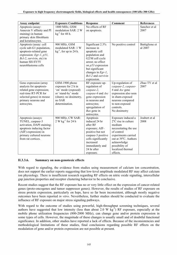
Exposure to high frequency electromagnetic fields, biological effects and health consequences (100 kHz-300 GHz)
145
Assay endpoint Exposure Conditions Response Comment References Apoptosis (assay: Annexin V affinity and PI staining) in human primary skin fibroblasts and keratinocytes.
1800 MHz, GSM modulation SAR: 2 W kg-1 for 48 h.
No effects of RF on apoptosis.
Sanchez et al 2007
Apoptosis (assay: cell cycle sub-G1 population, apoptosis-related gene expression: Egr-1, p53, Bcl-2, survivin, etc) in human SH-SY5Y neuroblastoma cells
900 MHz, GSM modulated SAR: 1 W kg-1, for up to 24 h.
Significant 2.3% increase in apoptotic cell population and G2/M cell cycle arrest; no effect on p53 expression but significant changes in Egr-1, Bcl-2 and survivin expression.
No positive control
Buttiglione et al 2007
Gene expression (array analysis for apoptosis-related gene expression; real-time RT-PCR for selected genes) in mouse primary neurons and astrocytes.
GSM-1900 phone exposure for 2 h in ‘on’ mode (exposed) or ‘stand-by’ mode (sham); no dosimetry, no SAR determination.
RF exposure up-regulation of caspase-2, caspase-6 and Asc gene expression in neurons and astrocytes; upregulation of Bax gene in astrocytes.
Up-regulation of caspase-2, caspase-6 and Asc gene expression also seen in sham-exposed neurons compared to non-exposed controls. No dosimetry
Zhao TY et al 2007
Apoptosis (assays: TUNEL, caspase-3 activation, DAPI staining; apoptosis inducing factor (AIF) expression) in primary cultured neurons from rat cortices.
900 MHz, CW SAR: 2 W kg-1 for 24 h
Apoptosis induced 24 hr after RF exposure; AIF-positive but not caspase-3 positive cells significantly increased immediately and 24 hr after exposure.
Exposure induced a 2oC rise in culture medium necessitating the use of control experiments carried out at 39oC. Authors acknowledge possibility of localized thermal effects.
Joubert et al 2008
II.3.3.6. Summary on non-genotoxic effects
With regard to signaling, the evidence from studies using measurement of calcium ion concentration, does not support the earlier reports suggesting that low-level amplitude modulated RF may affect calcium ion physiology. There is insufficient research regarding RF effects on nitric oxide signaling, intercellular gap junction properties and receptor clustering behavior to be conclusive.
Recent studies suggest that the RF exposure has no or very little effect on the expression of cancer-related genes (proto-oncogenes and tumor suppressor genes). However, the results of studies of RF exposure on stress protein expression, particularly on hsps, have so far been inconsistent, although mostly negative outcomes have been reported in vitro. Nevertheless, further studies should be conducted to evaluate the influence of RF exposure on major stress signaling pathways.
With regard to the outcome of studies using powerful, high-throughput screening techniques, several authors have suggested that low intensity (less than about 2.0 W kg-1) RF exposure, especially at the mobile phone utilization frequencies (800-2000 MHz), can change gene and/or protein expression in some types of cells. However, the magnitude of these changes is usually small and of doubtful functional significance. In addition, other studies have reported a lack of effects. Because of the inconsistencies and methodological limitations of these studies, final conclusions regarding possible RF effects on the modulation of gene and/or protein expression are not possible at present.
Exposure to high frequency electromagnetic fields, biological effects and health consequences (100 kHz-300 GHz)
146
Many studies have been published that suggest there are no effects of RF exposure on ROS production, cell proliferation, cell cycle control or on cellular apoptosis.
Ensuring adequate temperature control has proved difficult in many of these studies and heating may account for some of the positive effects reported.
II.3.4. Cell transformation
The neoplastic cell transformation assay is an integrative assay which is used to test carcinogenic and co-carcinogenic effects of chemical and physical agents. Its main advantage is that it reveals the carcinogenic potential of both genotoxic and non-genotoxic compounds. Several research groups have used this assay to determine whether RF exposure acts as an inducer, a promoter, or a co-carcinogen; most have used the chromosomally highly abnormal mouse fibroblast C3H/10T½ cell line.
In a series of experiments, Balcer-Kubiczek & Harrison (1985, 1989, 1991) exposed C3H10T½ cells to 2450 MHz RF (24 h), alone or in combination with known tumor initiators (X-rays or benzo(a)pyrene (B(a)P)), or the chemical promoter phorbol-12-myristate-13-acetate (PMA). No neoplastic transformation occurred with RF treatment alone at an SAR of up to 4.4 W kg-1 but Balcer-Kubiczek and Harrison (1991) reported that RF interacted with the promoter PMA in an SAR-dependent manner by increasing the transformation efficiency. However, unusually for in vitro RF studies, the authors exposed the cells in culture flasks situated in a waterbath situated in the far field of an anechoic chamber - dosimetry and temperature control may well be questionable. The data regarding effects on plating efficiency and the effect of RF exposure on neoplastic transformation induced by X-rays in presence of PMA were different in different experiments
Cain et al (1997) used the model of UV-TDT10e mutant cells in co-culture with parental C3H/10T½ murine fibroblasts to determine whether intermittent RF exposure (TDMA, 836.55 MHz) could influence the PMA dose-dependent promotion of focus formation. Cells were intermittently exposed (20 min on/20 min off) at SARs of 0.15, 1.5, and 15 mW kg-1, 24 h per day for 28 days. No influence of RF exposure at any SAR level was seen on PMA-induced focus formation.
Roti Roti et al (2001) investigated the neoplastic transformation potential of mobile phone signals (CDMA, 847.74 MHz; FDMA, 835.62 MHz) at an SAR of 0.6 W kg-1 in mouse C3H10T½ cells. Exposure to RF lasted 7 days and combination treatments included X-rays and PMA. RF exposure did not affect neoplastic transformation whatever treatment combination.
Wang et al (2005) exposed C3H10T½ cells to 2450 MHz CW RF at SAR levels of 5 to 200 W kg-1 for 2 hours, sufficient to raise culture medium temperatures to ~ 40 and ~ 44oC at 100 and 200 W kg-1, respectively. Cells were exposed to RF, either alone or in combination with 3-methylcholanthrene (MCA), PMA or MCA+PMA. RF alone and in combination with PMA did not affect the background neoplastic transformation. No significant differences were observed in the malignant transformation frequency in other combined treatments at SARs of up to 50 W kg-1. However, RF at 100 and 200 W kg-1 increased the transformation frequency induced by MCA or MCA plus PMA. The authors reported that the transformation assay was negative when cells were exposed at corresponding temperatures (up to 44°C), although the heating profiles may have differed.
Hirose et al (2008) used the mouse BALB/3T3 cell transformation model to evaluate the effect of a continuous 6-week RF exposure in an anechoic chamber to 2140 MHz (W-CDMA) at 80 and 800 mW kg-
1 on spontaneous and MCA±PMA-driven neoplastic transformation. No significant difference in neoplastic transformation was observed between groups.
All studies detailed above clearly show that RF exposure at SARs of up to 200 W kg-1 did not induce cell transformation. RF exposure did not promote the neoplastic transformation potential of either physical (X-rays) or chemical (B(a)P, MCA) inducers at SARs below 100 W kg-1. In one study, a promoter effect of RF was found with MCA alone and combined with PMA, but at SARs sufficient to significantly increase culture medium temperature. An interaction of RF with the promoter PMA was also reported in another study at lower SAR levels, but discrepancies within the same group were reported for RF
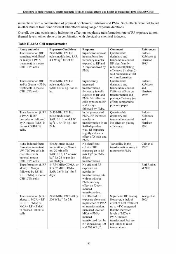
Exposure to high frequency electromagnetic fields, biological effects and health consequences (100 kHz-300 GHz)
147
interactions with a combination of physical or chemical initiators and PMA. Such effects were not found in other studies from four different laboratories using longer exposure durations.
Overall, the data consistently indicate no effect on neoplastic transformation rate of RF exposure at non-thermal levels, either alone or in combination with physical or chemical inducers.
Table II.3.15.: Cell transformation
Assay endpoint Exposure Conditions Response Comment References Transformation (RF combined with B(a)P or X-rays ± PMA treatment) in mouse C3H10T½ cells
2450 MHz, 120 Hz pulse modulation, SAR: 4.4 W kg-1 for 24 hr.
Significant increase in transformation frequency in cells exposed to RF and X-rays followed by PMA
Questionable dosimetry and temperature control. RF significantly reduced cell plating efficiency by about 2-fold but had no effect on transformation.
Balcer-Kubiczek & Harrison 1985
Transformation (RF and/or X-rays ± PMA treatment) in mouse C3H10T½ cells
2450 MHz, 120 Hz pulse modulation SAR: 4.4 W kg-1 for 24 hr.
Significantly increased transformation frequency in cells exposed to RF and PMA. No effect in cells exposed to RF and X-rays followed by PMA.
Questionable dosimetry and temperature control. Different effects on transformation and plating efficiency (no effect) compared to previous paper.
Balcer-Kubiczek and Harrison 1989
Transformation (i. RF ± PMA; ii. RF preceded or followed by X-rays ± PMA) in mouse C3H10T½ cells.
2450 MHz, 120 Hz pulse modulation SAR: 0.1, 1, or 4.4 W kg-1; ii. 4.4 W kg-1; for 24 hr.
In the presence of PMA, RF increased neoplastic transformation in an SAR-dependent way. RF exposure slightly enhances effect of X-rays and PMA.
Questionable dosimetry and temperature control. No effect on plating efficiency.
Balcer-Kubiczek and Harrison 1991
PMA-induced focus formation in mutant UV-TDT10e cells in co-culture with parental mouse C3H10T½ cells.
836.55 MHz TDMA intermittently (20 min on/ 20 min off) SAR: 0.15, 1.5 or mW kg-1 for 24 hr per day for 28 days.
No significant effect of RF exposure up to 15 mW kg-1 on PMA–driven transformation
Variability in the transformation assay in response to PMA
Cain et al 1997
Transformation (i. RF alone; ii. X-rays followed by RF; iii. RF + PMA) in mouse C3H10T½ cells.
847.74 MHz CDMA, or 835.62 MHz FDMA SAR: 0.6 W kg-1 for 7 days.
No effect of RF exposure on neoplastic transformation rate with or without PMA, nor any effect on X-ray-induced transformation.
Roti Roti et al 2001
Transformation (i. RF alone; ii. MCA + RF; iii. RF + PMA; iv. MCA+ RF + PMA) in mouse C3H10T½ cells
2450 MHz, CW SAR ≤ 200 W kg-1 for 2 h.
No effect of RF exposure alone and in presence of PMA on transformation. Increased level of MCA ± PMA-induced transformed foci by RF exposure at 100 and 200 W kg-1.
Significant RF heating. However, a lack of effect of heat treatment up to 44°C suggested that the increased levels of MCA ± PMA-induced transformed foci are not linked to raise temperatures.
Wang et al 2005
Exposure to high frequency electromagnetic fields, biological effects and health consequences (100 kHz-300 GHz)
148
Assay endpoint Exposure Conditions Response Comment References Transformation (i. RF alone; ii. MCA+ RF; iii. MCA+PMA+RF) in mouse BALB/3T3 cells assay.
2142.5 MHz, W-CDMA modulation SAR: 80 or 800 mW kg-1 for 6 weeks.
No induction, promotional or co-carcinogenic effect of RF exposure on transformation.
Hirose et al 2008
II.3.5. Summary on cellular studies
Over the last 30 years there have been many in vitro studies on potential cellular effects of RF. These studies gave insight into the basic mechanisms by which effects might be induced in more complex animal or human organisms. Interpretation is, however, limited by anomalous cell behavior generated by the culture conditions and other factors which limit the extrapolation to humans. The studies conducted so far have not provided consistent evidence of biological effects under non-thermal RF exposure conditions. In the case of genetic effects, for example, most results were negative and some of the few positive findings may be attributable to a thermal insult rather than to the RF-exposure as such. The same holds true for other endpoints. With regard to signaling, studies done using measurements of calcium ion concentration related to cellular function do not support earlier positive reports on calcium ion physiology. There is insufficient research regarding RF effects on nitric oxide signaling, gap junctions and receptor clustering to be conclusive, but the results of studies on cell proliferation and differentiation, apoptosis and cell transformation are mostly negative.
Changes in cell physiology and function imply changes in gene and protein expression. An early publication on heat shock gene expression in the nematode C. elegans initiated further investigation of various genes known to be stress-responsive. However, this positive finding was later shown to have resulted from inadvertent heating, due to lack of rigorous dosimetry. Recent studies suggest that RF exposure has no or very little effect on the expression of cancer-related genes (e.g., proto-oncogenes and tumor suppressor genes). However, the results of studies of the effects of RF exposure on stress protein expression, particularly hsps, have so far been inconsistent, although mostly negative outcomes have been reported in vitro. Heating remains a potential confounder, and probably accounts for some of the positive effects reported. Nevertheless, further studies should be conducted to evaluate the influence of RF exposure on major stress signaling pathways
More recently, studies have been carried out using powerful high-throughput screening techniques capable of examining changes in the expression of very large numbers of genes and proteins. Such studies often showed a limited number of alterations where some genes were up- and others down-regulated. Apoptosis-related genes were amongst the up-regulated ones, and cell cycle genes amongst the down-regulated ones, but this was not always the case. High-throughput techniques have thus so far not provided any evidence of an RF ‘signature’. Overall, it should be noted that:
• Quantitative methods have not always been used to confirm the initial findings; such a step is generally accepted as necessary for confirmation.
• Repeat experiments in array analysis have not often been conducted, which prevents confirmation of the earlier results.
• Changes have frequently been found in only a few genes out of several hundreds investigated, which might have occurred by chance.
• The changes that have been reported are usually very small compared to those induced from eg known carcinogens such as UVR, and may be of little functional significance.
• Ensuring adequate dosimetry and temperature control has proved difficult in many of these studies and heating may account for some of the positive effects reported.
These advances in molecular studies are promising, but not yet decisive in risk evaluation. The microarray technology, for example, can be very important in confirming results obtained by more conventional scientific methods and helping elucidate mechanisms of action, but, on their own, results

Exposure to high frequency electromagnetic fields, biological effects and health consequences (100 kHz-300 GHz)
149
from such studies are not yet sufficiently understood and the methodologies not sufficiently standardized and validated to provide decisive data on RF (and other) health effects. However, if a gene or a protein is identified as an RF-responsive molecule, the possibility that the change has a physiological or pathological consequence should be further explored with both in vitro and in vivo studies.
II.4. ANIMAL STUDIES
Animal studies are frequently based on experiments using laboratory strains of mice or rats. The advantage of such studies is that they provide information concerning the interaction of RF radiation with living systems which display the full repertoire of body functions, such as immune response, cardiovascular changes, and behavior, in a way that cannot be achieved with cellular studies. Transgenic or gene knockout animal models of certain diseases have further increased the value of animal studies to reveal potential adverse health effects. Animal studies are thus usually a more powerful experimental tool than cellular studies in this context. However, extrapolation to humans is not straightforward since there are obvious differences in physiology and metabolism between species as well as differences in life expectancy and many other variables. Nevertheless, at a molecular level, there are many similarities between processes in animals and humans and such studies have been very useful in helping unravel the sequence of genetic events in the development of a number of human cancers (e.g., Balmain and Harris 2000; Anisimov et al 2005).
Generally, animal studies can be expected to provide qualitative information regarding potential outcomes, but the data cannot be extrapolated quantitatively to give reliable estimates of human risk for the reasons outlined above. In addition, differences in body size, which are particularly marked in laboratory rodents compared to humans, means that dosimetric interaction will be different, small animals showing body resonance to RF radiation at higher frequencies than humans, with a comparatively greater depth of penetration relative to body size. Major improvements in exposure systems for animals have been achieved in the recent years. Several types of setups are being used depending on the type of exposure needed (such as head-only or whole-body). The various systems in common use (such as loop antennas, carousels, Ferris wheels, radial transmission lines and reverberation chambers) are described in Chapter I.3.4. The selection of RF exposure systems used in animal studies is often a compromise between restraint-related stress and the accuracy of RF dosimetry. If animals are allowed to move freely during RF exposure, they change their position and orientation relative to the electromagnetic wave and may also be shielded by other animals, which results in large uncertainties in dosimetry. Therefore, immobilization of animals has been used in many animal studies to achieve well-defined dosimetry. However, immobilization can cause restraint-related stress that might affect the outcome of the experiment unless appropriate steps, such as the habituation of animals to restraint, are taken.
II.4.1. Genotoxicity
Several studies have been conducted over the past 30 years using Drosophila melanogaster as the test organism. They all yielded negative results (see Verschaeve 1995; Léonard et al 1983; WHO; 1993).
With regard to laboratory mammals, many studies that have been published so far have not demonstrated convincingly any direct DNA damage after acute or chronic exposure to RF radiation (e.g. Léonard et al 1983; WHO 1993; Verschaeve and Maes 1998; Meltz 2003; Vijayalaxmi and Obe 2004), in particular when temperatures were maintained within normal physiological limits. However, a number of investigations have suggested that RF radiation can affect DNA (Table II.4.1.). Sarkar et al (1994) found evidence of an alteration in the length of a DNA micro satellite sequence in cells from the brain and testis of mice exposed to 2450 MHz fields, whereas Lai and Singh demonstrated in a series of publications (Lai and Singh 1995, 1996a, 1997, 2005; Lai et al 1997) that acute exposure to low-intensity radiofrequency radiation increased DNA strand breaks in the brain cells of rats. A significant increase in DNA strand breaks was found immediately and 4 h after exposure. It was suggested that this could be due either to a direct effect on the DNA or to an effect on DNA repair mechanisms (Lai and Singh 1996a). The authors

Exposure to high frequency electromagnetic fields, biological effects and health consequences (100 kHz-300 GHz)
150
furthermore provided data suggesting that free radicals may play a role in the observed SSBs and DSBs as the addition of free radical scavengers reduced the effect (Lai et al 1997).
These observations have been the subject of discussion and criticism in the scientific community. The fact that effects were observed at 4 h post exposure was especially criticized (Williams 1996), but arguments in favor of the findings were subsequently presented by Lai and Singh (1996b). Nevertheless, studies by other authors, including two attempted replications, have not reported RF-induced DNA damage in rat brain cells (Malyapa et al 1998; Lagroye et al 2004b; Verschaeve et al 2006; Belyaev et al 2006). These contrasting results were attributed partly to differences in procedures, especially in the ways the animals were killed and in the time lag between the death of the rats, dissection of the brain, and slide preparation for the comet assay (Malyapa et al 1998). As replication studies were not able to confirm the Lai and Singh data the significance of the findings therefore remain unclear to date but point to an absence of field-dependent effects. The same holds true for other genetic endpoints where both positive and negative findings were reported (e.g., on the incidence of micronuclei, (cf. Table II.4.1.).
Most of the animal studies have been conducted in somatic cells (blood, bone marrow, brain, liver or spleen). Only a few studies have been devoted to germ cells or the reproductive system. Ono et al (2004) did not find any increased mutation frequency in the testes (and other organs) of the offspring of RF exposed pregnant mice. However, Aitken et al (2005) did find a significant genotoxic effect on the epididymal spermatozoa of mice that were exposed for 7 days to 900 MHz low-level RF, whereas no impact on male germ cell development was observed. These studies differed in many aspects (e.g., in utero vs. in vivo exposure, LacZ gene mutation vs. Q-PCR analysis, etc.) which may eventually account for the different results. Aitken et al (2005) note that during epididymal transit spermatozoa have lost all capacity for DNA repair and are therefore vulnerable to factors that might affect DNA integrity. However, the possible genotoxic effect of RF-radiation on epididymal sperm remains unconfirmed at present.
In summary, most studies have failed to convincingly demonstrate any direct genetic effect after exposure of laboratory mammals to RF radiation, in particular when temperatures were maintained within normal physiological limits.
Table II.4.1.: RF-radiation alone or in combination with chemical/physical mutagens
Assay endpoint Exposure Conditions Response Comment References DNA analysis with synthetic oligo probes in brain cells and testes of mice
2.45 GHz; CW; SAR: 1.2 W kg-1; 2 h per day for 120, 150 and 200 days.
DNA fragments: altered band patterns of DNA
Sarkar et al 1994
DNA single and double strand breaks (comet assay) assayed in rat brain cells immediately and 4 h after RF-exposure
2450 MHz, pulsed or CW; SAR: 1.2 W kg-1; 2 h exposure
Significant increase in DNA strand breaks immediately and 4h after exposure
Lai and Singh 1995
DNA single and double strand breaks (comet assay) assayed in rat brain cells immediately and 4 h after RF-exposure
2450 MHz, pulsed or CW; SAR: 1.2 W kg-1; 2 h exposure
Significant increase in DNA strand breaks immediately and 4h after exposure
Lai and Singh 1996a
DNA single and double strand breaks (comet assay) assayed in rat brain cells immediately and 4 h after RF-exposure
2450 MHz, 2 h exposure as above; rats were also treated with melatonin or N-tert-butyl-a-phenylnitrone (free radical scavengers)
Treatment of rats with free radical scavengers before and after RF negated the induction of DNA strand breaks
Lai and Singh 1997
Micronuclei in peripheral blood and bone marrow cells in tumor prone mice
2450 MHz; CW; SAR:1 W kg-1; 20 h per day, 7 days per week for 1.5 years
No effects observed
Vijayalaxmi et al 1997b

Exposure to high frequency electromagnetic fields, biological effects and health consequences (100 kHz-300 GHz)
151
Assay endpoint Exposure Conditions Response Comment References Micronuclei in polychromatic erythrocytes (from peripheral blood and bone marrow) of CF-1 mice
Animals were exposed for 15 minutes to ultra-wide band (UWB) radiation at 37 mW kg-1
No effects observed
Vijayalaxmi et al 1999
DNA single strand breaks (comet assay) in rat brain cells.
2450 MHz; CW; SAR=1.2 W kg-1; 2 h exposure
No observed DNA damage in brain cells of rats euthanized by CO2 asphyxia or decapitation
Comet assay conduced immediately and 4 h after RF-exposure
Malyapa et al 1998
Micronuclei in peripheral blood and bone marrow cells in rats
2450 MHz; CW; SAR: 12 W kg-1; 24 h exposure
No effects observed
Vijayalaxmi et al 2001a
Somatic intrachromosomal recombination in spleen cells of pKZ1 transgenic mice
900 MHz pulsed-wave; SAR: 4 W kg-1; 30 min per day for 1, 5 and 25 days
No evidence of a genotoxic effect
Significant reduction in inversions below the spontaneous frequency in the 25-day exposure group
Sykes et al 2001
Micronuclei in rat peripheral blood cells
2450 MHz, CW; SAR: 1 and 2 W kg-1; 2 h per day, 7 days per week for up to 30 days
Increased incidence of micronuclei in animals exposed to RF after eight irradiation treatments of 2 h each
Trosic et al 2002
Mutation assay (mutant lacI genes) in brain tissue of Big Blue mice
1.5 GHz at SAR: 2.0, 0.67 and 0 W kg-1. Animals were exposed for 90 min per day, 5 days per week, for 4 weeks
1.5 GHz was not found mutagenic to mouse brain cells
Takahashi et al 2002
Micronuclei in rat bone marrow cells
1600 MHz; iridium signal; SRA: 0.16 and 1.6 W kg-1; 2 h per day, 5 days per week for 2 years
No evidence of a genotoxic effect
Vijayalaxmi et al 2003
Micronuclei in mouse peripheral blood and bone marrow cells
42.2 GHz; SAR: 622 ± 100 W kg-1; 30 min per day for 3 consecutive days; also co-exposure with cyclophosphamide
No evidence of genotoxic effect of RF alone and no influence on cyclophosphamide induced micronuclei
Vijayalaxmi et al 2004
Alkaline comet test (with and without the use of proteinase K in the assay) in rat brain cells.
2450 MHz Pulsed wave; SAR: 1.2 W kg-1; 2 h exposure
No DNA damage found
Comet assay conducted 4 h after RF exposure.
Lagroye et al 2004b
Mutation frequency at the LacZ gene in cells from the spleen liver brain and testes of the offspring of LacZ- transgenic mice.
2450 MHz; SAR: 0.71 W kg-1 (intermittent exposure of 10 sec. on with 4.3 W kg-1 and 50 sec. off); in utero exposure for 16 h per day at gestational age of 0-15 days
No effects observed
Offspring analyzed at 10 weeks of age
Ono et al 2004

Exposure to high frequency electromagnetic fields, biological effects and health consequences (100 kHz-300 GHz)
152
Assay endpoint Exposure Conditions Response Comment References Micronuclei in rat bone marrow cells
2450 MHz; CW; SAR: 1.25 W kg-1; 2 h per day, 7 days per week and 4, 16, 30 and 60 h
Increased incidence of micronuclei on experimental day 15
Trosic et al 2004b
Micronuclei in rat bone marrow cells
910 MHz; peak SAR: 0.42 W kg-1; 2 h/day for 30 consecutive days
Increased incidence of micronuclei
Observations possibly biased by presence of mast cell granules that cannot be easily discriminated from micronuclei.
Demsia et al 2004
DNA single and double strand breaks (comet assay) assayed in rat brain cells immediately and 4 h after RF-exposure
2450 MHz; SAR: 0.6 W kg-1; 2 h exposure
Brain cells of RF-exposed rats had significantly higher levels of SSBs and DSBs.
Lai and Singh 2005
Micronuclei in blood erythrocytes, bone marrow, keratinocytes and spleen lymphocytes of mice
GSM 900 MHz and DCS 1800 MHz; amplitude modulated; SAR: 0, 3.7, 11 and 33.2 W kg-1 (1 week study) and 0, 2.8, 8.3 and 24.9 W kg-1 (6 week study); 2 h per day exposure
No DNA damaged observed in brain cells
Görlitz et al 2005
DNA damage assessed by quantitative PCR (Q-PCR) and alkaline- and pulsed field electrophoresis in caudal epididymal spermatozoa of mice
900 MHz; SAR: 0.09 W kg-1; exposure for 7 days at 12 h per day
No impact on male germ cell development but Q-PCR revealed a significant genotoxic effect on the epididymal spermatozoa
Aitken et al 2005
DNA damage (alkaline comet assay) and micronuclei in rat blood, liver and brain cells
900 MHz; amplitude modulated; SAR: 0.3 and 0.9 W kg-1; 2 h per day, 5 days per week for 2 years. Exposure in conjunction with MX exposure in the drinking water.
Co-exposure to MX and RF-radiation did not increase the response of blood (comet and micronucleus assay) or liver and brain cells (comet test)
Verschaeve et al 2006
Changes in chromatin conformation and DNA double strand breaks (pulsed field gel electrophoresis) in rat brain cells
915 MHz (GSM); SAR: 0.4 W kg-1; 2 h exposure
No induction of DNA double strand breaks or chromatin conformation, but changes in gene expression were observed
Belyaev et al 2006
Micronucleus formation in blood from rats being exposed to mobile phone radiation during their embryogenesis
Exposure to cellular phone antenna (834 MHz, 26.8-40 V m-1) from the first day of pregnancy for 8.5 h per day. SAR estimated at 0.55-1.23 W kg-1
Significant increase in erythrocyte MN frequency in newborn pups from exposed pregnant rats.
Ferreira et al 2006
Exposure to high frequency electromagnetic fields, biological effects and health consequences (100 kHz-300 GHz)
153
Assay endpoint Exposure Conditions Response Comment References DNA damage (alkaline comet assay) in rat brain cells
2.45 GHz and 16.5 GHz at SAR: 1.0 and 2.01 W kg-1. Exposure 2 h per day for 35 days
Statistically significant increase in DNA single strand breaks following RF exposure
Paulraj and Behari 2006
Micronucleus frequency in erythrocytes of mice
902.5 MHz (NMT) signal at a SAR: 1.5 W kg-1; or 902.5 MHz (GSM) signal at SAR=0.35 W kg-1 for 78 weeks (1.5 h per day, 5 days per week).
No effect Animals taken at necropsy from a co-carcinogenicity study by Heikkinen et al 2001. During first weeks also X-irradiation at 4 Gy
Juutilainen et al 2007
Micronucleus frequency in erythrocytes of mice
K2 transgenic and non transgenic mice exposed 52 weeks to digital mobile phone signals, GSM and DAMPS at SAR: 0.5 W kg-1.
No effect Animals taken at necropsy from a co-carcinogenicity study by Heikkinen et al 2003. Exposure 3 times per week to 1.2 MED UV-radiation
Juutilainen et al 2007
MX=3-chloro-4-(dichloromethyl)-5-hydroxy-2(5H)-furanone; MED = minimal erythemal dose
II.4.2. Cancer
Animal studies investigating the carcinogenic potential of RF radiation were reviewed by WHO (1993), while more recent studies have been reviewed by Repacholi (1997), Krewski et al (2001a & b), AGNIR (2003), Elder (2003b), and Krewski et al (2007). This review focuses on papers published after 1993, but some earlier key studies are also described.
Evaluating carcinogenicity in laboratory rodents has remained a cornerstone in identifying agents likely to cause cancer in humans. According to IARC, agents for which there is sufficient evidence of carcinogenicity in experimental animals are considered to pose carcinogenic hazard to humans, unless there is scientific evidence that the agent causes cancer through a species-specific mechanism that does not operate in humans (IARC 2006). However, despite the similarities in many cancer characteristics between humans and laboratory rodents, interspecies differences need to be taken into account when extrapolating data from rodents to humans: many agents that are carcinogenic in rodents (often only at very high doses) are not carcinogenic to humans, and some human carcinogens do not affect rodents (Ames and Gold 1990; Trosko and Upham 2005; Anisimov et al 2005).
The effects of stress resulting from restraint and related daily handling has be seen in many animal cancer studies as a lower body weight among the sham-exposed (restrained) animals than among the cage control (unrestrained) animals (see below: Heikkinen et al 2003; Oberto et al 2007; Shirai et al 2007; Smith et al 2007; Yu et al 2006; Zook and Simmens 2006). In many of these studies, tumor incidence has also been lower and survival higher in the sham-exposed (restrained) group than in the cage control (unrestrained) group, which may be related to the observations that reduced energy intake inhibits the development of tumors (Keenan et al 1996; Sinha et al 1988; Klurfeld et al 1991). Immobilization has not caused experimental bias in studies assessing carcinogenicity of RF radiation, as both the RF exposed and the sham-exposed animals have been restrained, but it can be argued that stress could act as an effect modifier and hide possible RF-induced effects. However, there is no evidence of such modifying effects: many of the studies reviewed above have used freely moving animals, and the majority of studies have produced negative findings independent of the handling (restrained or unrestrained) of the animals.

Exposure to high frequency electromagnetic fields, biological effects and health consequences (100 kHz-300 GHz)
154
Classical carcinogenicity bioassays involve exposure of animals over most of their lifetime to the agent being tested. Such studies are potentially capable of revealing whether the tested agent alone could act as a complete carcinogen or serve to increase the incidence of spontaneous tumors. This type of studies are, however, not sensitive in detecting weak carcinogenic effects (because of the low number of tumors induced) and co-carcinogenic effects (interaction with other carcinogens). To overcome these limitations, several studies have used tumor-prone animal strains or combined exposure to RF radiation and known carcinogens. The animal studies are classified here as: i) studies with exposure to RF field alone (Table II.4.2.), including studies using tumor-prone animals strains (Table II.4.3.), ii) studies using exposure to RF radiation combined with a known genotoxic/carcinogenic agent (Table II.4.4.), and iii) studies evaluating effects of RF radiation on the very last steps of carcinogenesis using implanted or injected tumor cells (Table II.4.5.).
II.4.2.1. RF radiation alone
Conventional laboratory animal strains
Long-term rodent bioassays evaluating carcinogenicity of RF radiation alone have been rather consistent and have not found evidence for RF field-related effects on tumor development in conventional rat strains (Chou et al 1992; Zook and Simmens 2001; Adey et al 1999; Adey et al 2000; Anderson et al 2004; La Regina et al 2003) or mouse strains (Utteridge et al 2002). The main emphasis in many of these studies has been the combined effects of RF radiation with known genotoxic agents, but the study design has also involved groups exposed to RF radiation alone. Details of studies evaluating exposure to RF radiation alone are shown in Table II.4.2.
The first carcinogenicity study on RF radiation was published several decades ago (Prausnitz and Susskind 1962). The authors reported some indication of increased testicular degeneration, and increased neoplasias of white blood cells, which they termed “leucosis”, in RF field exposed mice. Both the methods and reporting of this study have been severely criticized (see e.g. Roberts and Michaelson 1983). For example, the daily exposure time was short, but RF dose-rate was high resulting in 2-5ºC increase in body temperature, the methods were not described in detail, a large number of animals were lost due to autolysis, and the conclusions were not based on statistical analysis. Therefore, this report has no real value in cancer risk assessment.
Using a so-called ‘carousel exposure set-up’ for well-defined RF exposure levels in the head, five recent studies failed to find evidence of enhanced brain tumorigenesis in RF field-exposed rats (Adey et al 1999; Adey et al 2000; Anderson et al 2004; La Regina et al 2003; Zook and Simmens 2001) at average SARs in the brain up to about 1.5 W kg-1. In the carousel set-up, the rats are restrained head first in cylindrical tubes arranged in a radial configuration with the RF antenna at the centre of the carousel, where the head is preferentially irradiated. The SAR in other body parts is much lower, and the ratio of brain average SAR to whole body average SAR may be up to 10:1 at mobile phone frequencies (Schönborn et al 2004). The animals in these studies had been exposed for most of their lifetime, and three of the studies also included in utero exposures (Adey et al 1999; Adey et al 2000; Anderson et al 2004). There was some indication of decreased CNS glial tumor development in the group exposed to NADC-modulated RF field (Adey et al 1999). The unexpectedly high incidence of spontaneous CNS tumors in the control group, however, suggests that this statistically non-significant difference might be a consequence of chance. The studies that have involved histopathological evaluation of other organs have provided no evidence of enhanced tumorigenesis in other tissues exposed at considerably lower SAR values than the brain (Anderson et al 2004; La Regina et al 2003; Zook and Simmens 2001).
The combined incidence of malignant tumors (all tumor types combined) was statistically significantly increased in male Sprague-Dawley rats exposed to radar-type pulsed 2.45 GHz RF radiation at whole-body average SAR of 0.15-0.4 W kg-1 (Chou et al 1992). The organ-specific tumor incidences were low (except those in some endocrine organs). The incidence of any single type of primary malignant or benign neoplasm, the combined incidence of benign neoplasms or survival were not statistically significantly affected, and the authors concluded that, overall, the study did not show any definite biologically significant effects. The incidence of benign pheochromocytoma was reported to be higher in RF-exposed

Exposure to high frequency electromagnetic fields, biological effects and health consequences (100 kHz-300 GHz)
155
rats, but the difference did not reach statistical significance. No tumor-enhancing effects of RF field exposure were reported in Sprague-Dawley rats in a later study reporting slightly (but statistically non-significantly) lower incidences of combined adrenal tumors in RF-exposed males (Zook and Simmens 2001). The SAR levels were similar and both studies used relatively long daily exposure times. However, the later study (Zook and Simmens 2001) was concentrated on brain tumorigenesis, and did not include complete histopathology of all other organs. Thus, it did not provide data on combined tumor incidences.
Exposure to RF radiation did not affect the incidence of lymphomas in C57BL/6Ntac mice, the ‘wild type’ of the Eµ-Pim1 transgenic mice used in the same study (Utteridge et al 2002). This study was planned as a replication experiment of an earlier study (Repacholi et al 1997) reporting enhanced the development of lymphoma in Eµ-Pim1 transgenic mice exposed to RF radiation (see below). In addition to transgenic animals, Utteridge and colleagues used also corresponding wildtype C57BL/6Ntac mice exposed at four SAR levels ranging from 0.25 to 4 W kg-1. There were only a few lymphoblastic leukemias in the wild-type animals, and for non-lymphoblastic leukemias there were no statistically significant differences between the sham-RF and RF-exposed animals.
Two studies evaluated carcinogenicity of both a GSM signal at 902 MHz and a DCS signal at 1747 MHz in B6C3F1 mice (Tillmann et al 2007) and in Wistar rats (Smith et al 2007). Three exposure levels ranging from 0.4 to 4 W kg-1 (and sham exposure) were used. In the mouse study (Tillmann et al 2007), no significant increase in the incidence of any particular tumor type in the RF exposed groups was observed. Interestingly, in both studies (both RF signals) the incidence of liver adenomas in males decreased with increasing exposure level, with a statistically significant difference between the highest exposure and the sham-exposed group. However, comparison to published tumor rates in untreated mice revealed that the observed tumor rates were within the range of historical control data. In conclusion, the study produced no evidence that exposure at whole body SARs of up to 4.0 W kg-1 increased the incidence or severity of neoplastic or non-neoplastic lesions, or resulted in any other adverse health effects. The rat study (Smith et al 2007) was a combined chronic toxicity and carcinogenicity study, and some of the animals (15 males and 15 females per group) were killed at 52 weeks from the start of the study. There were no significant differences in incidence, multiplicity, latency or severity of neoplasms, or any other adverse responses to RF field exposure.
Table II.4.2.: Carcinogenic effects of RF radiation: Exposure to RF radiation alone, normal strains
Assay endpoint Exposure conditions Results Comments Reference
CNS Tumors CNS tumors in F-344 rats 30 females and 30 males/group.
836.55 MHz D-AMPS 1) freely moving pregnant dams, circular polarization, SAR not given, 2 h/d , gestation day 19 until birth 2) freely moving pups, circular polarization, SAR not given, 2 h/d, from birth until weaning 3) restrained (carousel set-up, ) from age of 33 d, brain SAR: 0.3–0.5 W kg-1 (whole-body SAR: 0.2-0.4 W kg-1), 2 h/d 22 months (intermittent exposure: 7.5 min on/ 7.5 min off)
No effects on CNS/brain tumor incidence. No significant effects on survival
Exposure started in utero.
Adey et al 1999

Exposure to high frequency electromagnetic fields, biological effects and health consequences (100 kHz-300 GHz)
156
Assay endpoint Exposure conditions Results Comments Reference CNS tumors in F-344 rats 45 females and 45 males/group.
836.55 MHz FM 1) freely moving pregnant dams, circular polarization, SAR not given, 2 h/d, gestation day 19 until birth 2) freely moving pups, circular polarization, SAR not given, 2h/d, from birth until weaning 3) restrained (carousel set-up) from age of 33 d, brain SAR: 1.1-1.4 W kg-1 (Whole-body SAR: 0.3-0.7 W kg-1), 2 h/d , 4 d/w, for 23 months
No effects on CNS/brain tumor incidence. No effects on survival.
Exposure started in utero
Adey et al 2000
CNS tumors in Sprague-Dawley rats 30 females and 30 males/group.
860 MHz CW or 860 MHz MiRS Restrained (carousel set-up) Brain SAR: 0.8-1.2 W kg-1 Whole-body SAR: 0.27-0.42 W kg-1 6 h/d, 5d/wk for 22 mo
No effects on CNS/brain tumor incidence. No effect on tumorigenesis in other tissues.
8 non-neural tissues evaluated, but relatively high number of missing tissues in some of them
Zook & Simmens 2001
CNS tumors in F-344 rats 80 females and 80 males/group.
835.62 MHz, FDMA or 847.74 MHz, CDMA. Restrained (carousel set-up). Brain SAR: 1.3±0.5 W kg-1 (mean ±SD) Whole-body SAR not given (SAR in other organs less than 1/3 of that in brain). 4 h/d, 5d/wk, for 104 weeks
No increase in CNS tumors. No increase in tumors in other tissues (all major organs evaluated). No increase in total number of tumors. No effects on survival (survival over 90%).
The study hypothesis was whether RF exposure increases tumor incidences, so decreased incidences were not statistically tested
La Regina et al 2003
Exposure to high frequency electromagnetic fields, biological effects and health consequences (100 kHz-300 GHz)
157
Assay endpoint Exposure conditions Results Comments Reference CNS tumors in F-344 rats 90 females and 90 males/group
1.62 GHz Iridium 1) freely moving pregnant dams (1/cage), brain SAR: (fetuses) 0.1 -0.2 W kg-1 (Whole-body SAR ≈ 0.06 W kg-
1), 2 h/d, gestation day 19 until birth 2) freely moving pups, brain SAR: 0.1-0.2 W kg-1 (Whole-body SAR ≈ 0.06 W kg-1), 2h/d, from birth until weaning 3) restrained (carousel set-up), brain SAR: 0.11-0.18 W kg-1 or 1.1-1.8 W kg-1 (whole-body SAR ≈ 0.02 W kg-1 or 0.2 W kg-1), 2h/d, 5d/w, 2 years
No effects on brain tumor incidence. No effects on incidence of lymphoma. No effects on tumors in other tissues evaluated No effects on survival.
Exposure started in utero
Anderson et al 2004
Lymphomas Lymphoma in female C57BL/6Ntac mice (wild type of Eµ-Pim1) 120 mice /group
898.4 MHz GSM. Restrained ( “Ferris wheel”) Whole-body SAR: 0.25, 1.0, 2.0 or 4.0 W kg-1, 1 h/d, 5d/wk for 104 wk
No differences in the incidence of non-lymphoblastic lymphomas; incidence of lymphoblastic lymphoma low in all groups No effects on total tumor incidence (12 tissues evaluated) No effects on survival
The study included also transgenic animals, see Table II.4.3.
Utteridge et al 2002
Multiple tumors
Multiple tumors in male Sprague-Dawley rats
100 rats/group.
2.45 GHz pulsed (10 µs pulses at 800 pps; pulse-modulated also at 8 pps); Freely moving; Whole-body SAR 0.15–0.4 W kg-1; for 21.5 h/day, 7 days/week, for 25 months
No increase in any individual tumor type. Four-fold increase in combined primary malignancies, but no increase in combined primary benign tumors. No effects on survival.
Chou et al 1992
Multiple tumors in male and female B6C3F1 mice 50 males and 50 females/group
902 MHz GSM or 1747 MHz DCS Restrained (“Ferris wheel”) Whole-body SAR: 0.4, 1.3 or 4.0 W kg-1, 2 h/d, 5 d/wk for 2 years
No increase in the incidence of any neoplastic or non-neoplastic lesions
Two signals, three exposure levels for each signal
Tillmann et al 2007
Exposure to high frequency electromagnetic fields, biological effects and health consequences (100 kHz-300 GHz)
158
Assay endpoint Exposure conditions Results Comments Reference Multiple tumors in male and female Wistar rats 65 males and 65 females/group.
902 MHz GSM or 1747 MHz DCS Restrained (“Ferris wheel”) Whole-body SAR: 0.44, 1.33 or 4.0 W kg-1, 2 h/d, 5 d/wk for 2 years
No increase in the incidence of any neoplasms; no other adverse effects
Combined chronic toxicity/carcinogenicity study, 15 females and 15 males per group were killed at 1 year
Smith et al 2007
Studies using genetically predisposed animal models
Animal strains developing tumors (in some organs) with particularly high frequency and/or early in life are classified as ‘tumor prone strains’. These strains include animals engineered to be more vulnerable via gene manipulation (transgenic animals), as well as other strains with exceptionally high tumor incidence due to their genetic background. The division between “tumor prone” and “other” strains is somewhat arbitrary, because spontaneous tumor frequency varies greatly between different animal strains. Details of studies using genetically tumor-prone animal strains are described in Table II.4.3. The spontaneous incidence of tumors in this kind of experimental models is important: if nearly all animals in the unexposed control group develop tumors, there is not much room for an additional effect from RF field exposure. Therefore, information on tumor incidence in unexposed animals is included in Table II:4.3. [Note, however, that accelerated development of tumors can be detected even if the final incidence is 100%, if the tumors are externally observable during the experiment, as is the case for eg skin tumors and mammary tumors.]
Lymphoma models
Transgenic Eµ-Pim1 mice overexpressing Pim1 oncogene in their lymphoid cells are prone to malignant lymphoma. In the first study with this model using RF (Repacholi et al 1997) Eµ-Pim1 mice were exposed to 900 MHz GSM-type RF radiation at SARs ranging from 0.13 -1.4 W kg-1 (if all possible animal orientations are included, the range was 0.008 to 4.2 W kg-1). The RF exposed animals had twofold lymphoma incidence compared to controls. At the time the study was terminated, lymphoma incidence was increasing rapidly in both exposed and sham-exposed animals. The authors emphasize that even if the observed effect were established, the relevance of the animal model for human cancer risk assessment needs to be carefully considered. The findings of this study were not confirmed in a replication study by Utteridge et al (2002), who used the same strain of mouse obtained from the same supplier. The investigators also fed the same food to the mice. The later study had some refinements in experimental design: four SAR levels (0.25, 1.0, 2.0 and 4.0 W kg-1) were used instead of one in the original study; animals were restrained during the exposure for better control of variations in exposure level; animals were exposed once per day instead of two episodes of 30 minutes; and full necropsy was performed on all mice at the end of the study. RF field exposure did not enhance development of lymphoma. The incidence of lymphoblastic leukemia was slightly lower in all RF-exposed groups compared to that of the sham-exposed animals, and the difference was statistically significant at the lowest dose rate. In contrast, the incidence of non-lymphoblastic leukemia was slightly higher in RF exposed groups, but these differences were not statistically significant either in pairwise comparisons or in a trend test. The incidence of lymphomas in the RF-sham-exposed group was surprisingly high, and the publication stirred debate whether some critical features of the original experiment had been changed (Goldstein et al 2003a; Goldstein et al 2003b; Kundi 2003a; Kundi 2003b; Lerchl 2003).
The study reported by Oberto et al (2007) was also a replication and an extension of the Repacholi et al (1997) study. Eµ-Pim1 transgenic mice were exposed for 1 h/day, 7 days/week to pulsed GSM 900 RF at a whole-body SAR of 0.5, 1.4 or 4.0 W kg-1. 50 animals per sex per group were exposed, sham-exposed or used as cage controls. There were several methodological improvements compared to the original study by Repacholi et al (1997), including use of several exposure levels, well-defined dosimetry and more uniform exposure (achieved through restrain of the animals) and necropsy and extensive histopathology of all animals. Compared to the sham-exposed controls, survival was reduced in the
Exposure to high frequency electromagnetic fields, biological effects and health consequences (100 kHz-300 GHz)
159
animals exposed to RF radiation. The intergroup differences were statistically significant in the male animals, but there was no trend with increasing exposure level (lowest survival at 0.5 W kg-1). No increase in lymphoma incidence was observed in the RF exposed groups. Concerning other neoplastic findings, Harderian gland adenomas were increased in male mice, with a significant dose-related trend (p<0.01). However, this trend was not supported by the findings on female animals, in which no tumors in the highest exposure groups were observed. The statistical analysis used in this study can be criticized. The cage control and the sham-exposed control groups were combined for statistical comparisons, which is not a valid procedure given the differences in body weight development and tumor incidence between these groups (these differences are most likely related to restraint of the sham-exposed animals). However, based on the data reported in the paper, a different analysis strategy (comparison to the sham-exposed group only) would not essentially change the interpretation that there was no effect of RF exposure on tumor incidence at any site. The reduced survival in RF-field-exposed animals is not thoroughly discussed by the authors; this finding remains unexplained and difficult to interpret without detailed information about the causes of death.
GSM-type RF exposure at nominal SAR of 0.4 W kg-1 did not affect development of lymphoma in female AKR/J mice (Sommer et al 2004). This mouse strain is prone to develop lymphoma due to expression of an AKV retrovirus in all of their tissues. About 90% of animals both in the sham-exposed and RF-exposed groups developed lymphoma by the end of the 10-month study. Essentially mortality was reported to be related to the development of lymphoblastic lymphoma, and RF field exposure did not affect survival. No effects of exposure were seen in differential leucocyte count of blood samples collected 5-10 months after the beginning of RF exposure. The nominal SAR was 0.4 W kg-1, but as in other studies using several freely moving animals per cage, the variation in exposure level would undoubtedly have been large.
In another study by the same group (Sommer et al 2007), unrestrained AKR/J mice, 160 animals per group, were chronically sham-exposed or exposed to a generic UMTS test signal for 24 h/day, 7 days/week at a SAR of 0.4 W kg-1. Additionally, 30 animals were kept as cage controls. The animals were checked visually each day and were weighed and palpated weekly to detect swollen lymph nodes. Starting at the age of 6 months, blood samples were taken from the tail every 2 weeks to perform differential leukocyte counts and to measure the hematocrit. Visibly diseased animals or those older than 43 weeks were killed humanely, and tissue slices were examined for metastatic infiltrations and lymphoma type. Cage control animals had a significantly lower growth rate than those kept in the radial waveguides. Incidence of lymphoma, survival time and the severity of the disease indicated that there was no effect from exposure to RF radiation. Cage control animals had significantly lower body weights and higher occurrence of metastatic infiltrations in liver and meninges than the other groups. This difference was most likely related to different housing conditions and stress level.
Models for mammary tumorigenesis
The accelerated tumor development in mammary tumor prone female C3H/HeA mice reported by Szmigielski and co-workers (Szmigielski et al 1982) has not been confirmed by other long-term studies using female C3H/HeJ mice at lower SARs but generally longer daily exposure times (Frei et al 1998a; Frei et al 1998b; Jauchem et al 2001; Toler et al 1997).
In the study of Szmigielski et al (1982) the exposure levels were expressed in W m-2. The SAR values were estimated to be about 2-3 and 6-8 W kg-1, and thermally induced stress may have affected the outcome at least at the higher exposure level. The response to the lower RF level was reported to be similar to that of confinement stress. Similarly to Szmigielski et al, Frei and co-workers used continuous 2.45 GHz RF radiation (Frei et al 1998a; Frei et al 1998b) whereas two other studies used signals consisting of short pulses (Jauchem et al 2001; Toler et al 1997). Although the four later studies were designed specifically to examine mammary tumors, they included histopathological analyzes of other main tissues. Overall, the authors of these studies concluded that RF field exposure did not affect the development of tumors or survival of animals. The only statistically significant differences in tumor incidence reported in these studies were a smaller number (0 vs. 4) of alveolar-bronchiolar adenomas in RF field exposed animals in one study (Frei et al 1998a), and increased incidence of bilateral ovarian
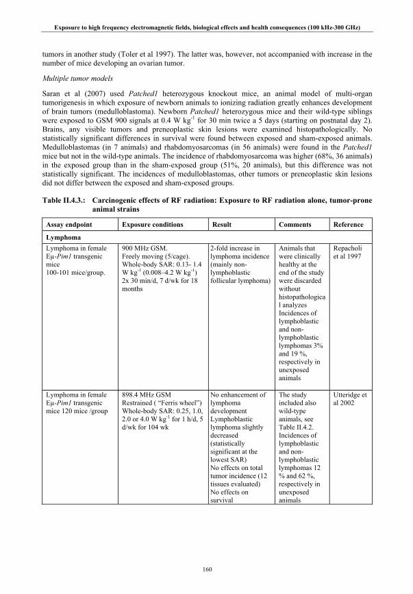
Exposure to high frequency electromagnetic fields, biological effects and health consequences (100 kHz-300 GHz)
160
tumors in another study (Toler et al 1997). The latter was, however, not accompanied with increase in the number of mice developing an ovarian tumor.
Multiple tumor models
Saran et al (2007) used Patched1 heterozygous knockout mice, an animal model of multi-organ tumorigenesis in which exposure of newborn animals to ionizing radiation greatly enhances development of brain tumors (medulloblastoma). Newborn Patched1 heterozygous mice and their wild-type siblings were exposed to GSM 900 signals at 0.4 W kg-1 for 30 min twice a 5 days (starting on postnatal day 2). Brains, any visible tumors and preneoplastic skin lesions were examined histopathologically. No statistically significant differences in survival were found between exposed and sham-exposed animals. Medulloblastomas (in 7 animals) and rhabdomyosarcomas (in 56 animals) were found in the Patched1 mice but not in the wild-type animals. The incidence of rhabdomyosarcoma was higher (68%, 36 animals) in the exposed group than in the sham-exposed group (51%, 20 animals), but this difference was not statistically significant. The incidences of medulloblastomas, other tumors or preneoplastic skin lesions did not differ between the exposed and sham-exposed groups.
Table II.4.3.: Carcinogenic effects of RF radiation: Exposure to RF radiation alone, tumor-prone animal strains
Assay endpoint Exposure conditions Result Comments Reference
Lymphoma Lymphoma in female Eµ-Pim1 transgenic mice 100-101 mice/group.
900 MHz GSM. Freely moving (5/cage). Whole-body SAR: 0.13- 1.4 W kg-1 (0.008–4.2 W kg-1) 2x 30 min/d, 7 d/wk for 18 months
2-fold increase in lymphoma incidence (mainly non-lymphoblastic follicular lymphoma)
Animals that were clinically healthy at the end of the study were discarded without histopathological analyzes Incidences of lymphoblastic and non-lymphoblastic lymphomas 3% and 19 %, respectively in unexposed animals
Repacholi et al 1997
Lymphoma in female Eµ-Pim1 transgenic mice 120 mice /group
898.4 MHz GSM Restrained ( “Ferris wheel”) Whole-body SAR: 0.25, 1.0, 2.0 or 4.0 W kg-1 for 1 h/d, 5 d/wk for 104 wk
No enhancement of lymphoma development Lymphoblastic lymphoma slightly decreased (statistically significant at the lowest SAR) No effects on total tumor incidence (12 tissues evaluated) No effects on survival
The study included also wild-type animals, see Table II.4.2. Incidences of lymphoblastic and non-lymphoblastic lymphomas 12 % and 62 %, respectively in unexposed animals
Utteridge et al 2002

Exposure to high frequency electromagnetic fields, biological effects and health consequences (100 kHz-300 GHz)
161
Assay endpoint Exposure conditions Result Comments Reference Lymphoma in female AKR/J mice 160 mice/group
900 MHz GSM Freely moving (6-7/cage) Whole-body SAR: 0.4 W kg-
1 24 h/d, 7d/wk, for 10 months
No effects on development of lymphoma, differential count of leucocytes or survival. Exposed animals had higher body weights during late stages of the study
Lymphoma incidence 90 % in unexposed animals
Sommer et al 2004
Lymphoma in female and male Eµ-Pim1 transgenic mice 50 females and 50 males/group
900 MHz GSM Restrained (“Ferris wheel”) Whole-body SAR: 0.4, 1.4 or 4.0 W kg-1, 1 h/d, 7 d/wk for 18 months
No effects on the incidence of lymphoma. Harderian gland adenoma increased in male mice, but not in females. Survival was decreased in the exposed animals (significant in males, but no dose-related trend)
Sham-exposed group and cage controls were combined for statistical analysis Incidences of lymphoblastic and non-lymphoblastic lymphomas 4% and 40%, respectively in sham-exposed females and 0% and 18% in sham-exposed males
Oberto et al 2007
Lymphoma in female AKR/J mice, 160 mice/group
1.966 GHz UMTS Freely moving (6-7/cage) Whole-body SAR: 0.4 W kg-
1 24 h/d, 7 d/wk for 35 weeks
No effects on incidence or severity of lymphoma. No effects on survival
Lymphoma incidence 96.7 % in unexposed animals
Sommer et al 2007
Mammary Tumors Mammary gland tumors in female C3H/HeA mice 40 mice/group
2.45 GHz CW Freely moving (10 /cage) 50 W/m2 (SAR: 2–3 W kg-1) or 150 W/m2 (6–8 W kg-1); 2 h/d, 6 d/wk for 10.5 months
Accelerated tumor development Decreased survival due to mammary tumorigenesis
Large uncertainty in estimated SAR Incidence of mammary tumors ≈35% in unexposed animals
Szmigielski et al 1982
Mammary tumors in female C3H/HeJ mice 200 mice/group
435 MHz pulsed (1 µs pulses, 1000 pps) Freely moving (1 /cage) SAR: 0.32 W kg-1 22h/d, 7 d/wk, for 21 months
No effect on mammary gland tumorigenesis Increased number of animals with a bilateral stromal tumors in ovaries (but no effect on incidence of animals with a stromal tumor in ovaries) No effects on other tumors (most organs analyzed). No effects on survival
Incidence of adenocarcinoma ≈ 40 % in unexposed animals
Toler et al 1997

Exposure to high frequency electromagnetic fields, biological effects and health consequences (100 kHz-300 GHz)
162
Assay endpoint Exposure conditions Result Comments Reference Mammary tumors in female C3H/HeJ mice 100 animals /group
2.45 GHz CW Freely moving (1/cage) SAR: 0.3 W kg-1 20 h/d, 7 d/wk for 78 wk
No effects on mammary gland tumorigenesis Decreased incidence of alveolar-bronchiolar adenomas in lungs No effects on tumors in other organs (most organs evaluated) No effects on survival
Mammary gland tumor incidence 55 % in unexposed animals)
Frei et al 1998a
Mammary tumors in female C3H/HeJ mice 100 animals/group
2.45 GHz CW Freely moving (1/cage) SAR: 1.0 W kg-1 20 h/d, 7 d/wk for 78 wk
No effects on mammary gland tumorigenesis No effects on tumors in other organs (most organs evaluated) No effects on survival
Mammary gland tumor incidence 30% in unexposed animals
Frei et al 1998b
Mammary tumors in female C3H/HeJ mice 100 animals/group
UWB pulsed (1.9 ns pulses, 1000 pps) SAR: 0.01 W kg-1 2 min/d, 1d/wk for 12 wk
No effects on mammary gland tumorigenesis No effects on tumors in other organs (all main tissues evaluated) No effects on survival
Mammary gland tumor incidence 52% in unexposed animals
Jauchem et al 2001
Multiple tumors Multiple tumors in newborn Patched1 heterozygous knock-out mice 50-63 animals (22-36 females and 23-29 males)/group
900 MHz GSM Restrained SAR: 0.4 W kg-1 1 h/d, 5d/wk for 1 wk
No significant effect on medulloblastoma, rhabdomyosarcoma, other visible tumors or preneoplastic skin lesions No effects on survival
Many samples for histopathology were lost because of tissue autolysis (too late detection of death) Incidences in unexposed animals: medulloblastoma 8%, rhabdomyosarcoma 51%
Saran et al 2007
II.4.2.2. Combined RF and known genotoxic/carcinogenic agents
Both theoretical considerations (low photon energy) and experimental evidence (reviewed in Chapter II.2.) indicates that direct DNA-damaging effects of weak RF electromagnetic radiation are not likely. Therefore, there has been considerable interest in testing RF radiation as a non-genotoxic carcinogen or a co-carcinogen that enhances the effects of known carcinogenic agents. Methods for detecting non-genotoxic carcinogens and co-carcinogens are less well developed than those for detecting genotoxic carcinogens. It can be argued that classical animal carcinogenicity bioassays should identify carcinogens independently of the mechanisms. However, because of the very low number of tumors induced, such studies (involving exposure to the agent alone, without co-exposures) may suffer from low statistical

Exposure to high frequency electromagnetic fields, biological effects and health consequences (100 kHz-300 GHz)
163
power to detect co-carcinogens. Animal studies on co-carcinogenic effects have usually been designed based on the concepts of "initiation" and "promotion". Such studies involve a short-term exposure to an "initiator" (known DNA-damaging agent), followed by long-term exposure to the putative "cancer promoter". However, it has been questioned whether the initiation-promotion approach is sufficient for describing the complex interaction of genotoxic and non-genotoxic agents (Juutilainen et al 2000). Although most of the studies on co-carcinogenicity of RF radiation have tested RF radiation as a possible “promoter” after a single dose or short-term treatment with a known “initiator”, a few studies have used different approaches such as long-term simultaneous exposure to RF radiation and the known carcinogen, or RF field exposure before treatment with the known carcinogen.
Details of studies evaluating combined exposure to RF radiation with known genotoxic/carcinogenic agents are shown in Table II.4.4. As in the case of genetically predisposed models (see Chapter II.4.2.1.2.), the incidence of tumors in the control group (exposed only to the known carcinogen) should be at an appropriate level to allow detection of a possible further increase related to RF field exposure. Therefore, information of tumor incidence in the control (known carcinogen only) group is included in Table II.4.4.
Brain tumors
Several animal studies have evaluated the effects of low-level RF radiation on tumorigenesis initiated by transplacental administration of a known genotoxic agent, n-ethylnitrosourea (ENU) in Fischer 344 rats (Adey et al 1999; Adey et al 2000; Shirai et al 2005) and in Sprague-Dawley rats (Zook and Simmens 2001). Using a carousel exposure set-up to ensure well defined dosimetry of the head, these studies have provided no evidence that RF radiation can promote the development of CNS tumors in this model.
RF exposure (836.55 MHz; pulsed or continuous) did not increase the incidence of brain tumors induced by transplacental administration of ENU in Fischer 344 rats (Adey et al 1999; Adey et al 2000). North American Digital Cellular (NADC)-modulated RF field exposure appeared to decrease the incidence of ENU-induced glial CNS tumors (similar tendency was seen also in spontaneous tumors), but the difference was not statistically significant. The difference was more evident (statistically significant) if the animals surviving to the end of the experiment were excluded from the analyzes. RF exposure did not statistically significantly affect the mortality of ENU-treated animals, although survival was slightly increased in the RF exposed group.
Similarly, a more recent study reported no statistically significant effects of RF exposure on brain tumorigenesis in ENU-treated Fischer 344 rats, although the incidence of brain tumors in females was slightly lower in both RF-exposed animals compared to the sham-exposed group (Shirai et al 2005). The ENU dose was identical to that used earlier by Adey et al (1999; 2000). Considering other tissues, the incidences of pituitary tumors showed a tendency for increase in both sexes treated with ENU compared to the cage-control animals, the effect being more consistent for females. Compared to the sham-RF-exposed group, incidence of pituitary tumors was decreased in males of both RF-exposed groups. At the higher RF-exposure level (2.0 W kg-1) the decrease was statistically significant, and the incidence was slightly decreased also in females. The authors questioned the biological meaning of high pituitary tumor incidence in their study, and stated that the incidences may still be within the wide range of background data of this strain. An earlier study did not report any effect of RF on tumorigenesis in pituitary glands of ENU treated Sprague-Dawley rats (Zook and Simmens 2001), but the proportion of pituitary glands tissues available for histology was only about 80% in this study. Interestingly, the development of brain tumors in ENU-treated female rats was slightly decreased in both RF-exposed groups, like in the earlier study by Adey et al (1999).
A later study from the same group (Shirai et al 2007) had otherwise similar protocol, but a different mobile phone signal was used (1.95 GHz W-CDMA versus 1.439 GHz TDMA used in the first study). In contrast to the previous study, brain tumor incidences of both females and males tended to be higher in the RF exposed groups than in the sham-exposed group, but no statistically significant effects were reported. However, the statistical method used (two-group comparisons with Fisher’s exact test) is not sensitive for detecting trends with increasing exposure level. Using combined female and male data from the paper, chi-squared test for trend showed a p-value of 0.0395 for an increasing trend from the sham-
Exposure to high frequency electromagnetic fields, biological effects and health consequences (100 kHz-300 GHz)
164
exposed group to the highest exposure group. No differences in pituitary tumors were observed in this study. Given the inconsistent findings and opposite trends observed in these two studies (Shirai 2005; 2007), the differences observed are most likely incidental.
Continuous or pulsed 860 MHz Motorola integrated Radio Services (MiRS) head-mainly RF field exposure at 1 W kg-1 did not significantly affect incidence, volume, multiplicity, malignancy or fatality on ENU-induced brain tumors or development of tumors in eight other organs in Sprague-Dawley rats (Zook and Simmens 2001). There was a slight statistically non-significant tendency toward higher incidence of fatal brain tumors in the group treated with higher level of ENU and exposed to the pulsed RF field.
In a follow-up study, Zook and Simmens (2006) investigated further potential promoting effect of the pulsed RF signal. Latency and other characteristics of neurogenic tumors were investigated in the progeny of pregnant Sprague-Dawley rats treated with 6.25 or 10 mg/kg of ENU. The 1080 offspring were randomized equally by number, sex and ENU dose into pulsed RF, sham and cage control groups. The rats were exposed to the RF field (MiRS signal, 860 MHz, 11.1 pulses per second) 6 h/day 5 days/week at a SAR of 1.0 W kg-1 averaged over the brain (0.27-0.42 W kg-1 averaged over the whole body). The animals were restrained during the exposures. An equal number of rats from each group were killed every 30 days between the ages of 171 and 325 days; 32 rats died and 225 rats were killed when they were moribund. All rats were necropsied and the brain and spinal cord were examined histopathologically. The examinations revealed 38 spinal cord tumors, 191 spinal nerve tumors, 232 cranial nerve tumors, and 823 brain tumors. No evidence was found of RF effects on the incidence, malignancy, volume, multiplicity, latency or fatality associated with any kind of neurogenic tumor. Body weight was higher in the cage control animals than in the other groups, which is most likely related to restraint of the exposed and sham-exposed animals.
Multiple tumors
Heikkinen et al (2006) evaluated possible effects of RF radiation on tumorigenesis induced by the mutagen and multisite carcinogen 3-chloro-4-(dichloromethyl)-5-hydroxy-2(5H)-furanone (MX) given in drinking water continuously during the experiment. Female Wistar rats were exposed to GSM 900 RF at 0.3 or 0.9 W kg-1. The tumor profile in the MX-exposed animals resembled that reported earlier in MX-exposed female Wistar rats. RF radiation did not statistically significantly affect mortality or organ-specific incidence of any tumor type. The only statistically significant difference was an increase in the combined frequency of vascular tumors of the mesenteric lymph nodes in the high-RF group compared to the sham-RF group. However, comparison to cage-control animals suggested that this difference was due to an unusually low frequency of this type of tumors in the sham-RF group, rather than high frequency in the high-RF group.
Lymphoma
Exposure to continuous (frequency modulated) or pulsed (GSM modulation) 902 MHz RF radiation at 1.5 W kg-1 or 0.35 W kg-1 for 1.5 h/d on did not affect development of lymphomas, enhance development of other tumors or affect survival in female CBA/S mice irradiated with X-rays (Heikkinen et al 2001). The X-rays were delivered during the first three weeks of the study in three subdoses, and the exposures to RF radiation continued for 1.5 years. The only statistically significant differences in tumor incidences were decreased incidence of glandular polyps in the continuous wave group, and decreased incidence of a benign pheochromocytomas of adrenal glands in both RF field groups.
Mammary tumors
Several studies have investigated effects of RF field effects on mammary gland tumorigenesis induced by 7,12-dimethylbenz(a)anthracene (DMBA) in rodents. Although some indication of enhanced or decreased tumorigenesis was found in some experiments, these findings were not repeatable in other experiments by the same group, or in studies with similar design by different groups.
The study of Bartsch and co-workers (Bartsch et al 2002) differs from most other RF field studies published this far, in that the daily RF exposure time was long (nearly all time exposure). The study

Exposure to high frequency electromagnetic fields, biological effects and health consequences (100 kHz-300 GHz)
165
involved exposures of freely moving animals (12 per cage) at low SARs levels (0.1 W kg-1 or below). The study consisted of three experiments each started exactly at the same time of the year on three consecutive years. The animals were exposed until “practically all animals had developed a macroscopic mammary tumor” and the last experiment was conducted in blinded fashion. In one of the three experiments median latency for the development of the first malignant tumor was statistically significantly extended in the RF field-exposed group. This finding was not supported by the two other experiments. The overall conclusion was that long-term exposure to RF radiation had no significant effect on the development of DMBA-induced mammary tumors in Spraque-Dawley rats.
The study of Anane et al (2003a) consisted of two separate experiments, one performed in spring-summer and another in autumn. Female Spraque-Dawley rats were administered DMBA ten days before the beginning of the RF field exposures. The group sizes were small (14-16 rats/group in each experiment). Both experiments included one common exposure level, 1.4 W kg-1. In the first experiment, exposure at this SAR led to accelerated development of malignant mammary tumors, whereas an opposite finding was reported in the second experiment. The authors concluded that the study was no overall effect of exposure.
The study by Yu et al, (2006) did not provide evidence for RF field effects on the development of DMBA-initiated development of mammary tumors in rats. Exposure levels up to 4 W kg-1 were covered and 100 animals per group were used. The incidence of mammary gland adenocarcinomas was slightly lower in the group exposed to the lowest SAR, but the tumors were slightly larger compared to the animals exposed to DMBA only. A slightly enhanced development of adenocarcinomas was found at the highest SAR level. However, none of these differences were statistically significant. Significant differences were observed between the cage controls and the other experimental groups, with increased body weight and higher number and more rapid development of mammary tumors in the cage control group. These differences are most likely related to restraint of the sham-exposed and RF-exposed animals. The statistical analysis of tumor appearance was apparently done without making a distinction between tumors observed during the study by palpation and tumors detected in histopathological evaluation. While this could in principle mask differences between the groups (also small non-palpable tumors are detected in histopathology), the data shown in the paper suggest that a different statistical analysis would not essentially change the conclusion that RF radiation did not promote mammary tumor development.
Hruby et al (2008) used a study design similar to that used by Yu et al (2006). There were several statistically significant differences between RF field-exposed groups and the sham-exposed group. All RF-exposed groups had significantly more palpable mammary gland tissue masses than the sham-exposed group, but there were no differences between the three RF-exposed groups. The incidence of malignant mammary tissue tumors was lowest in the sham-exposed group, and significantly increased in the high exposure group. However, the incidence of benign tumors was significantly lower in the three RF exposed groups than in the sham-exposed group. The number of animals with benign or malign neoplasms was similar in the sham-exposed group and in the three RF-exposed groups. The cage control group had the highest incidence and malignancy of neoplasms among all groups. Given that the DMBA mammary tumor model is known to be prone to high variations in the results, the authors’ interpretation was that the differences between the groups were co-incidental. Comparison to the results of the almost identical study of Yu et al (2006) supports this conclusion: both studies reported similar development of mammary tumors in three groups, but lower rate of development (seen in the appearance of palpable tumors and/or reduced malignancy) in one group. Hruby et al found the lowest rate of development in the sham-exposed group, while Yu et al found it in the 0.44 W kg-1 group. Both studies consistently reported highest incidence of tumors in the cage control group, which is most likely related to the different handling of the cage control animals (different stress level, differences in food intake).
Skin tumors
Szudzinski and co-workers reported significant acceleration of the development of benzo(a)pyrene (B(a)P)-induced tumors in mice irradiated with 2.45 GHz (CW) RF at about 6-8 W kg-1 (Szudzinski et al 1982). Exposure to both Ba(a)P and RF radiation were long-term (6 months). Enhanced development of

Exposure to high frequency electromagnetic fields, biological effects and health consequences (100 kHz-300 GHz)
166
skin tumors was reported also if the RF exposure was for 1-3 months prior to the beginning of B(a)P exposures. Some of these results appear to have been reported in another publication (Szmigielski et al 1982) in the same year. There are some inconsistencies between these two reports (e.g. the group size and the exact handling of the sham-exposed animals) which complicate the interpretation of the results. The authors detected no increase of rectal temperature, but admitted that at the highest exposure level formation of significant “hot-spots” was possible due to non-uniform absorption of RF energy.
Low-level RF field exposures (only a few animals were exposed at 0.27 W kg-1, the others at 0.075 W kg-
1) had no effects on tumor appearance or survival in B(a)P-treated female Sprague-Dawley rats (Chagnaud et al 1999). Similarly, the RF field exposures had no effects on the levels of anti-phosphatidylinositol auto-antibodies, a suggested marker of malignant transformation. The animals were exposed to RF radiation for two weeks beginning on day 20, 40 or 75 after B(a)P injection.
RF field exposures have not been observed to induce tumors in skin of DMBA-treated CD-1 mice (Imaida et al 2001) or ICR mice (Huang TQ et al 2005). In both studies mice were subjected to topical application of DMBA on dorsal skin a week before the beginning of RF field exposures. No skin tumors were observed either in sham-RF or RF-field-exposed animals during the 19-week-studies, or not even after a one-year follow-up (Huang TQ et al 2006), whereas a clear tumor response was observed in the positive control animals exposed to repeated topical treatment with the classical tumor promoter phorbol-12-myristate-13-acetate (PMA). RF field exposures did not affect either the epidermal thickness (Imaida et al 2001; Huang TQ et al 2005) or expression of proliferating cell nuclear antigen (Huang TQ et al 2005). No difference was observable in the incidence of lymphoma (Imaida et al 2001; only liver, kidney, adrenal glands and spleen evaluated for lymphomas), and RF field exposures did not affect serum hormonal levels (melatonin, adrenonocorticotrophic hormone (ACTH) or corticosterone) in samples collected at necropsy (Imaida et al 2001).
Heikkinen et al (2003) reported that daily exposure to pulsed 849 or 902 MHz RF with two modulations characteristics (GSM or DAMPS) did not significantly affect the development of skin tumors induced by UV radiation in female ODC-transgenic mice (K2) and in their non-transgenic littermates. Skin tumors were induced by exposure to solar-simulating UV radiation three times a week during the whole two-year study. The development of skin tumors was faster in RF field-exposed animals than in the control group exposed to UV radiation only. This was consistently seen with both RF signals and in both transgenic and non-transgenic animals, but did not reach statistical significance even in a combined analysis.
Colon tumors
Exposure to 2.45 GHz RF, even at relatively high SAR of 10 - 12 W kg-1, did not affect the development of dimethylhydrazine (DMH)-induced colon tumors in Balb/c mice (Wu et al 1994). Although tumor incidence was not increased in animals treated with PMA as a positive control for tumor promotion, this treatment resulted in higher number of tumors per animal and larger tumors.
Medium-term hepatocarcinogenesis models
Exposure to 1.49 GHz (Imaida et al 1998a) or 929.2 MHz (Imaida et al 1998b) pulsed RF at 0.4-0.8 W kg-1 (maximum local values in liver 0.9-2.0 W kg-1) did not promote hepatocarconigenesis in a rat medium term bioassay, in which rats were exposed to diethylnitrosamine (DEN), partially hepatectomised a few weeks later, and exposed to RF radiation for six weeks. Interestingly, the development of gluthathione S-transferase (GST-p) positive liver foci, a preneoplastic rat liver lesion used as an end-point marker in this assay, was slightly decreased in the RF field exposed animals in both studies, the difference being statistically significant at 1.49 GHz. Compared to unrestrained DEN-exposed control animals, the level was about the same in RF field exposed animals, but higher in sham RF field exposed animals. Serum levels of ACTH, corticosterone and melatonin were increased in RF-exposed animals at the end of the study.
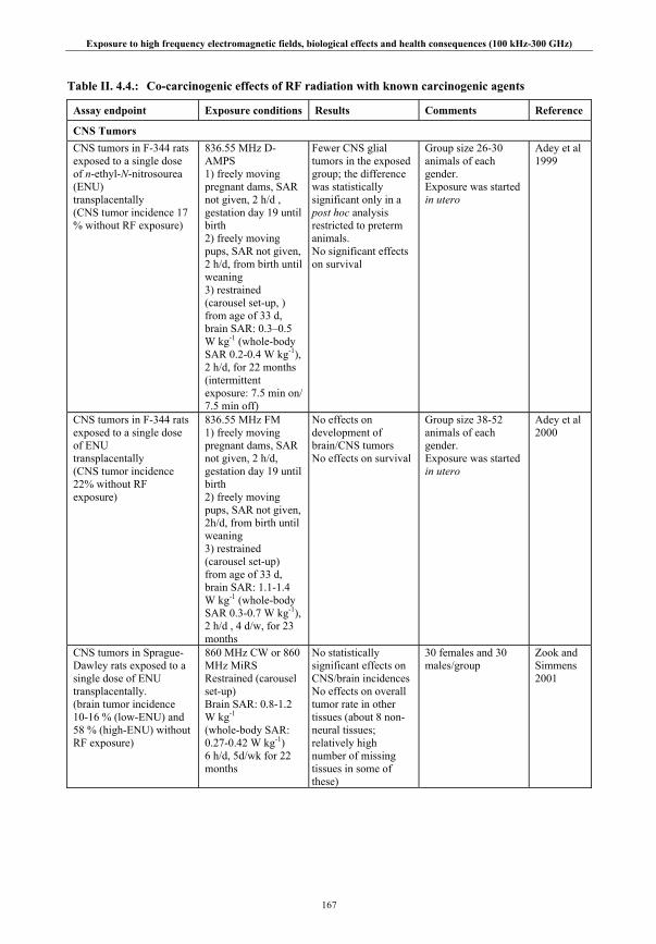
Exposure to high frequency electromagnetic fields, biological effects and health consequences (100 kHz-300 GHz)
167
Table II. 4.4.: Co-carcinogenic effects of RF radiation with known carcinogenic agents
Assay endpoint Exposure conditions Results Comments Reference
CNS Tumors CNS tumors in F-344 rats exposed to a single dose of n-ethyl-N-nitrosourea (ENU) transplacentally (CNS tumor incidence 17 % without RF exposure)
836.55 MHz D-AMPS 1) freely moving pregnant dams, SAR not given, 2 h/d , gestation day 19 until birth 2) freely moving pups, SAR not given, 2 h/d, from birth until weaning 3) restrained (carousel set-up, ) from age of 33 d, brain SAR: 0.3–0.5 W kg-1 (whole-body SAR 0.2-0.4 W kg-1), 2 h/d, for 22 months (intermittent exposure: 7.5 min on/ 7.5 min off)
Fewer CNS glial tumors in the exposed group; the difference was statistically significant only in a post hoc analysis restricted to preterm animals. No significant effects on survival
Group size 26-30 animals of each gender. Exposure was started in utero
Adey et al 1999
CNS tumors in F-344 rats exposed to a single dose of ENU transplacentally (CNS tumor incidence 22% without RF exposure)
836.55 MHz FM 1) freely moving pregnant dams, SAR not given, 2 h/d, gestation day 19 until birth 2) freely moving pups, SAR not given, 2h/d, from birth until weaning 3) restrained (carousel set-up) from age of 33 d, brain SAR: 1.1-1.4 W kg-1 (whole-body SAR 0.3-0.7 W kg-1), 2 h/d , 4 d/w, for 23 months
No effects on development of brain/CNS tumors No effects on survival
Group size 38-52 animals of each gender. Exposure was started in utero
Adey et al 2000
CNS tumors in Sprague-Dawley rats exposed to a single dose of ENU transplacentally. (brain tumor incidence 10-16 % (low-ENU) and 58 % (high-ENU) without RF exposure)
860 MHz CW or 860 MHz MiRS Restrained (carousel set-up) Brain SAR: 0.8-1.2 W kg-1 (whole-body SAR: 0.27-0.42 W kg-1) 6 h/d, 5d/wk for 22 months
No statistically significant effects on CNS/brain incidences No effects on overall tumor rate in other tissues (about 8 non-neural tissues; relatively high number of missing tissues in some of these)
30 females and 30 males/group
Zook and Simmens 2001

Exposure to high frequency electromagnetic fields, biological effects and health consequences (100 kHz-300 GHz)
168
Assay endpoint Exposure conditions Results Comments Reference CNS tumors in F-344 rats exposed to a single dose of ENU transplacentally. (brain tumor incidences 24 % in males and 30% in females without RF exposure)
1.439 GHz PDC Restrained (carousel set-up) Brain SAR: 0.67 W kg-1 or 2.0 W kg-1 (whole-body SAR <0.4 W kg-1) 1.5 h/d, 5d/wk for 104 weeks
No statistically significant effects on CNS/brain tumor incidences Incidence of pituitary tumors decreased in males exposed at 2 W kg-1, no other significant effects (“all organs” evaluated, tissues not listed). No effects on survival
50 females and 50 males/group
Shirai et al 2005
CNS tumors in Sprague-Dawley rats exposed to a single dose of ENU transplacentally. (brain tumor incidence 50% without RF exposure)
860 MHz MiRS Restrained (carousel set-up) Brain SAR: 0.8-1.2 W kg-1 (whole-body SAR: 0.27-0.42 W kg-1) 6 h/d, 5d/wk for 39 weeks
No effects on incidence, malignancy, volume, multiplicity, latency or fatality of any kind of neurogenic tumor
540 females and 540 males/group Two ENU doses An equal number of rats were killed and examined at 30-d intervals
Zook and Simmens 2006
CNS tumors in F-344 rats exposed to a single dose of ENU transplacentally. (brain tumor incidences10 % in males and 8% in females without RF exposure)
1.95 GHz W-CDMA Restrained (carousel set-up) Brain SAR: 0.67 W kg-1 or 2.0 W kg-1 (whole-body SAR <0.5 W kg-1) 1.5 h/d, 5d/wk for 104 weeks
No statistically significant effects on CNS/brain tumor incidence, although there was a tendency towards increased incidence in RF exposed groups compared to sham-exposed group
50 males and 50 females/group. The statistical methods used were not sensitive for detecting a trend with exposure level
Shirai et al 2007
Lymphomas Lymphoma in female CBA/S mice exposed to an initiating dose (consisting of three subdoses)of X-rays (Incidence of lymphoma 24% without RF exposure)
902.5 MHz NMT (analog) or 902.4 MHz GSM Restrained SAR: 1.5 W kg-1 (NMT) or 0.35 W kg-
1 (GSM) 1.5 h/d, 5 d/wk for 78 weeks
No increase in lymphoma No increase in any primary neoplastic change
50 mice/group Heikkinen et al 2001
Mammary tumors Mammary gland tumors in female Sprague-Dawley rats exposed to a single dose of DMBA (practically all animals developed tumors)
900 MHz GSM Freely moving (12/cage) SAR 0.03-0.13 W kg-
1 (young) 0.015-0.06 (adult) 24 h/d, 7 d/wk for 9-11 months
Overall no effects on mammary gland tumorigenesis In one experiment, latency for developing first malignant mammary gland tumor was increased in RF- exposed rats, but not in two other experiments
Three experiments; 60 animals/group in each experiment. Response to DMBA varied between the experiments
Bartsch et al 2002
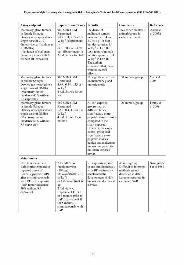
Exposure to high frequency electromagnetic fields, biological effects and health consequences (100 kHz-300 GHz)
169
Assay endpoint Exposure conditions Results Comments Reference Mammary gland tumors in female Sprague-Dawley rats exposed to a single dose of 7,12-dimethylbenz[a]anthracene (DMBA) (Incidence of malignant mammary tumors 60 % without RF exposure)
900 MHz GSM Restrained SAR: 1.4, 2.2 or 3.5 W kg-1 (Experiment I) or 0.1, 0.7 or 1.4 W kg-1 (Experiment II) 2 h/d, 5d/wk for 9wk
Incidence of malignant tumors increased at 1.4 and 2.2 W kg-1 in Exp I, but decreased at 1.4 W kg-1 in Exp II. Less tumors/animals in rats exposed at 1.4 W kg-1 in Exp II. The authors concluded that, there were no overall effects.
Two experiments;16 animals/group in each experiment
Anane et al 2003a
Mammary gland tumors in female Sprague-Dawley rats exposed to a single dose of DMBA (Mammary tumor incidence 45% without RF exposure)
900 MHz GSM Restrained SAR: 0.44, 1.33 or 4 W kg-1 4 h/d, 5 d/wk for 26 wk
No significant effects on mammary gland tumorigenesis
100 animals/group
Yu et al 2006
Mammary gland tumors in female Sprague-Dawley rats exposed to a single dose of DMBA (Mammary tumor incidence 60% without RF exposure)
902 MHz GSM Restrained SAR: 0.4, 1.3 or 0.4 W kg-1 4 h/d, 5 d/wk for 6 months
All RF-exposed groups had, at different times, significantly more palpable tissue masses compared to the sham-exposed. However, the cage-control group had significantly more palpable masses, benign and malignant tumors compared to the sham-exposed group.
100 animals/group
Hruby et al 2008
Skin tumors Skin tumors in male Balb/c mice exposed to repeated doses of Benzo(a)pyrene (BaP) after or simultaneously with RF field exposure (Skin tumor incidence 50% without RF exposure)
2.45 GHz CW Freely moving (10/cage) 50 W/m2 (SAR: 2–3 W kg-1) or 150 W/m2 (6–8 W kg-1) 2 h/d, 6d/wk, Experiment I: for 1 or 3 months prior to BaP; Experiment II: for 5 months simultaneously with BaP
RF exposures (prior to and simultaneously with BP treatments) accelerated the development of skin tumors and decreased survival
40 mice/group Difficult to interpret: methods are not described in detail. Large uncertainty in estimated SAR
Szmigielski et al 1982
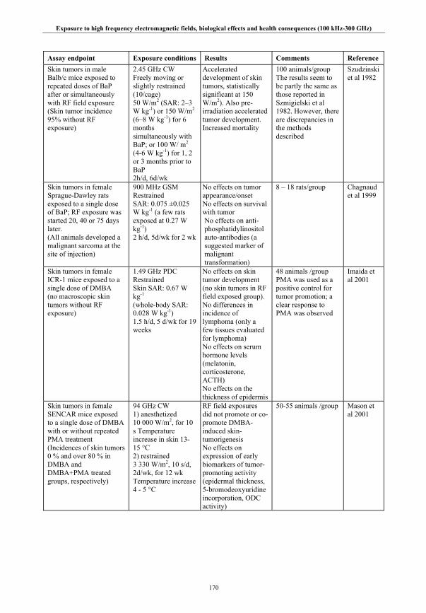
Exposure to high frequency electromagnetic fields, biological effects and health consequences (100 kHz-300 GHz)
170
Assay endpoint Exposure conditions Results Comments Reference Skin tumors in male Balb/c mice exposed to repeated doses of BaP after or simultaneously with RF field exposure (Skin tumor incidence 95% without RF exposure)
2.45 GHz CW Freely moving or slightly restrained (10/cage) 50 W/m2 (SAR: 2–3 W kg-1) or 150 W/m2
(6–8 W kg-1) for 6 months simultaneously with BaP; or 100 W/ m2 (4-6 W kg-1) for 1, 2 or 3 months prior to BaP 2h/d, 6d/wk
Accelerated development of skin tumors, statistically significant at 150 W/m2). Also pre-irradiation accelerated tumor development. Increased mortality
100 animals/group The results seem to be partly the same as those reported in Szmigielski et al 1982. However, there are discrepancies in the methods described
Szudzinski et al 1982
Skin tumors in female Sprague-Dawley rats exposed to a single dose of BaP; RF exposure was started 20, 40 or 75 days later. (All animals developed a malignant sarcoma at the site of injection)
900 MHz GSM Restrained SAR: 0.075 ±0.025 W kg-1 (a few rats exposed at 0.27 W kg-1) 2 h/d, 5d/wk for 2 wk
No effects on tumor appearance/onset No effects on survival with tumor No effects on anti-phosphatidylinositol auto-antibodies (a suggested marker of malignant transformation)
8 – 18 rats/group
Chagnaud et al 1999
Skin tumors in female ICR-1 mice exposed to a single dose of DMBA (no macroscopic skin tumors without RF exposure)
1.49 GHz PDC Restrained Skin SAR: 0.67 W kg-1 (whole-body SAR: 0.028 W kg-1) 1.5 h/d, 5 d/wk for 19 weeks
No effects on skin tumor development (no skin tumors in RF field exposed group). No differences in incidence of lymphoma (only a few tissues evaluated for lymphoma) No effects on serum hormone levels (melatonin, corticosterone, ACTH) No effects on the thickness of epidermis
48 animals /group PMA was used as a positive control for tumor promotion; a clear response to PMA was observed
Imaida et al 2001
Skin tumors in female SENCAR mice exposed to a single dose of DMBA with or without repeated PMA treatment (Incidences of skin tumors 0 % and over 80 % in DMBA and DMBA+PMA treated groups, respectively)
94 GHz CW 1) anesthetized 10 000 W/m2, for 10 s Temperature increase in skin 13-15 °C 2) restrained 3 330 W/m2, 10 s/d, 2d/wk, for 12 wk Temperature increase 4 - 5 °C
RF field exposures did not promote or co-promote DMBA-induced skin-tumorigenesis No effects on expression of early biomarkers of tumor-promoting activity (epidermal thickness, 5-bromodeoxyuridine incorporation, ODC activity)
50-55 animals /group
Mason et al 2001

Exposure to high frequency electromagnetic fields, biological effects and health consequences (100 kHz-300 GHz)
171
Assay endpoint Exposure conditions Results Comments Reference Skin tumors in female transgenic K2 mice and their non-transgenic littermates exposed to repeated doses of UV radiation. (Incidence of skin tumors 22% without RF exposure)
902 MHz GSM or 824 MHz DAMPS Restrained SAR: 0.5 W kg-1 1.5 h/d, 5 d/wk for 52 weeks
No statistically significant increase in skin tumors (although tumor development was faster in both RF field exposed groups compared to the UV only group)
45-49 animals/group K2 mice are more prone to develop skin tumors than their normal counterparts
Heikkinen et al 2003
Skin tumors in male ICR mice exposed to single dose of DMBA (no skin tumors without RF exposure)
849 MHz CDMA or 1.763 GHz CDMA Freely moving SAR: 0.4 W kg-1 2 x 45 min/d, 5 d/wk for 19 wk
No effects on tumor development (no skin tumors in RF-exposed groups). This was confirmed in a replication study with longer follow-up (1 year) No effects on epidermal thickness Staining with anti-proliferating cell nuclear antigen (PCNA) was observed only in the positive control group Cyclin D1 and c-fos proteins were detected only in the skin of the positive controls.
20 animals/group PMA was used as a positive control for tumor promotion; a clear response to PMA was observed
Huang TQ et al 2005
Colon Tumors Colon tumors in BALB/c mice exposed to repeated injections of Dimethylhydrazine (DMH) (colon tumor incidence 46 % without RF exposure)
2.45 GHz CW Restrained SAR: 10–12 W kg-1 3 h/d, 6d/wk, for 5 months
No effects on colon tumorigenesis
26-32 animals /group Positive control (PMA) did not increase tumor incidence, but it accelerated tumor development
Wu et al 1994
Multiple Tumors Several tumor types in female Wistar rats exposed to 3-chloro-4-(dichloromethyl)-5-hydroxy-2(5H)-furanone (MX) continuously in drinking water (Proportion of animals with malignant tumors 51% without RF exposure)
RF 900 MHz GSM Freely moving (1/cage) SAR: 0.3 or 0.9 W kg-1 2 h/d, 5 d/wk for 104 weeks
No increase in the incidence of any primary neoplasm
72 animals/group Heikkinen et al 2006

Exposure to high frequency electromagnetic fields, biological effects and health consequences (100 kHz-300 GHz)
172
Assay endpoint Exposure conditions Results Comments Reference
Liver promotion model Liver, medium-term promotion model: male F-344 rats exposed to a single dose of DEN and partial hepatectomy 3 wk later
1.439 GHz PDC Restrained Liver SAR 0.9-1.9 W kg-1 (peak values), whole-body SAR: 0.45-0.68 W kg-1 1.5 h/d, 5 d/wk for 6 wk
Number of GST-P positive foci decreased Spleen and testis weights decreased; adrenal weight increased Serum levels of ACTH, corticosterone and melatonin increased
45-47 rats/group
Imaida et al 1998a
Liver, medium-term promotion model: male F-344 rats exposed to a single dose of Diethylnitrosamine (DEN) and partial hepatectomy 3 wk later
929.2 MHz PDC Restrained Liver SAR: 1.7-2.0 W kg-1 (local peak values), whole-body SAR: 0.6–0.8 W kg-1, 1.5 h/d, 5 d/wk for 6 wk
No significant effects on the development of GST-P positive foci Serum levels of ACTH, corticosterone and melatonin were increased in RF-field-exposed group
47-48 rats/group
Imaida et al 1998b
II.4.2.3. Effects of RF radiation on tumor transplantation
Szmigielski et al, (1982) exposed BALB/c mice injected with sarcoma cells to 2.45 GHz RF for 1, 2 or 3 months at 50 or 150 W m-2. Significantly elevated numbers of neoplastic lung foci nodules after both 1 and 3 months were reported at both RF exposure levels. The interpretation of this study is complicated by the fact that methods were not described in detail and by uncertainties in dosimetry. Thermal effects are possible at least at the higher exposure level.
Three studies using inoculated/implanted rat glioma cell lines (Salford et al 1993; Salford et al 1997) and gliosarcoma cells (Higashikubo et al 1999) in Fischer 344 rats did not find effects on brain tumor growth from exposure to 835 – 915 MHz RF using several different modulations and SAR levels from 0.008 to 1.67 W kg-1.
Santini and co-workers (Santini et al 1988) exposed C57/6J mice to continuous and pulsed 2.45 GHz RF field at 1.2 W kg-1. After 15 days of exposure animals were subcutaneously implanted with B16 melanoma cells. The results did not indicate significant effects of RF either on tumor development or on survival times.
Four daily 20-min RF field exposures of pregnant dams at thermal exposure levels resulted in retarded development of inoculated sarcomas in offspring of CFW mice, but did not affect the final incidences (Preskorn et al 1978). Mice exposed in utero lived slightly longer. Postnatal exposures treatments did not affect tumor development in this study. Retarded tumor development was also reported in studies involving short-term exposure of the nasal area of mice to 42-61 GHz RF at very high intensities causing temperature elevation (Radzievsky et al 2004; Logani et al 2004; 2006).
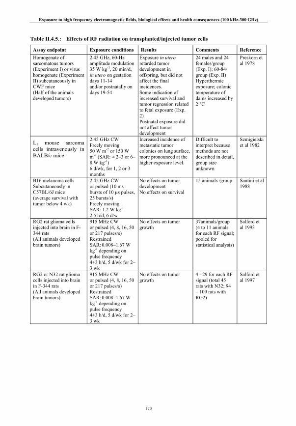
Exposure to high frequency electromagnetic fields, biological effects and health consequences (100 kHz-300 GHz)
173
Table II.4.5.: Effects of RF radiation on transplanted/injected tumor cells
Assay endpoint Exposure conditions Results Comments Reference Homogenate of sarcomatous tumors (Experiment I) or virus homogenate (Experiment II) subcutaneously in CWF mice (Half of the animals developed tumors)
2.45 GHz, 60-Hz amplitude modulation 35 W kg-1, 20 min/d, in utero on gestation days 11-14 and/or postnatally on days 19-54
Exposure in utero retarded tumor development in offspring, but did not affect the final incidences. Some indication of increased survival and tumor regression related to fetal exposure (Exp. 2) Postnatal exposure did not affect tumor development
24 males and 24 females/group (Exp. I); 60-84/ group (Exp. II) Hyperthermic exposure; colonic temperature of dams increased by 2 °C
Preskorn et al 1978
L1 mouse sarcoma cells intravenously in BALB/c mice
2.45 GHz CW Freely moving 50 W m-2 or 150 W m-2 (SAR: ≈ 2–3 or 6–8 W kg-1) 6 d/wk, for 1, 2 or 3 months
Increased incidence of metastatic tumor colonies on lung surface, more pronounced at the higher exposure level.
Difficult to interpret because methods are not described in detail, group size unknown
Szmigielski et al 1982
B16 melanoma cells Subcutaneously in C57BL/6J mice (average survival with tumor below 4 wk)
2.45 GHz CW or pulsed (10 ms bursts of 10 µs pulses, 25 bursts/s) Freely moving SAR: 1.2 W kg-1 2.5 h/d, 6 d/w
No effects on tumor development No effects on survival
15 animals /group
Santini et al 1988
RG2 rat glioma cells injected into brain in F-344 rats (All animals developed brain tumors)
915 MHz CW or pulsed (4, 8, 16, 50 or 217 pulses/s) Restrained SAR: 0.008–1.67 W kg-1 depending on pulse frequency 4+3 h/d, 5 d/wk for 2–3 wk
No effects on tumor growth
37animals/group (4 to 11 animals for each RF signal; pooled for statistical analysis)
Salford et al 1993
RG2 or N32 rat glioma cells injected into brain in F-344 rats (All animals developed brain tumors)
915 MHz CW or pulsed (4, 8, 16, 50 or 217 pulses/s) Restrained SAR: 0.008–1.67 W kg-1 depending on pulse frequency 4+3 h/d, 5 d/wk for 2–3 wk
No effects on tumor growth
4 - 29 for each RF signal (total 45 rats with N32; 94 – 109 rats with RG2)
Salford et al 1997

Exposure to high frequency electromagnetic fields, biological effects and health consequences (100 kHz-300 GHz)
174
Assay endpoint Exposure conditions Results Comments Reference 9L gliosarcoma cells injected into brain in male F-344 rats
835.62 MHz FM or 847.74 MHz CDMA Restrained (carousel set-up) Brain SAR: 0.5-1.0 W kg-1 4 h/d, 5 d/wk, for 4 +21 wk
No effects on tumor growth or survival. Brain weight increased in rats that were implanted with the highest number of viable cells and exposed to the CDMA signal. The authors concluded that this was more likely related to irregularities in sham-exposed group than to RF field exposure
96-101 animals/group (3 subgroups according to the number of viable cells injected: 10-67 rats/subgroup)
Higashikubo et al 1999
B16F10 melanoma cells subcutaneously in male Swiss Webster mice
61.22 GHz, Restrained 130 W/m2, on the nasal area (max. temperature increase at the tip of the nose ≈ 1 º C) 15 min/d for 5 d
Reduced tumor growth, if RF exposures started on day 5. No significant effects, if RF exposures started on day 1 or on day 10 (slightly enhanced tumor growth) Pre-treatment with Naloxane (a non-specific blocker of opioid receptors) blocked the effect
>10 mice/group
Radzievsky et al 2004
B16F10 melanoma cells subcutaneously in female SKH1 hairless mice CPA treatments on days 4-8 (Experiments I-III) or on days 4 and 11 (Experiment IV)
42.2 GHz (60-Hz amplitude modulation) Restrained 185 W m-2 on the nasal area (peak skin SAR: 730 W kg-1, temperature rise 1.5 ºC) 30min/d, for 3-6 d
RF field exposures either concurrently with, prior to or following CPA treatments did not affect tumor development
10 mice/group
Logani et al 2004
B16F10 melanoma cells Intravenously in female C57BL/6 mice, Cyclophospamide (CPA) before RF exposure
42.2 GHz (60-Hz amplitude modulation) Anesthetized 185 W m-2 on the nasal area (peak skin SAR: 730 W kg-1, temperature rise 1.5 ºC) One 30 min exposure.
Decreased number of metastatic lung colonies in animals exposed to RF field alone and in combined exposure with CPA Increased activity of natural killer cells in RF+CPA combined exposure
10 mice/group
Logani et al 2006
II.4.2.4. Summary on cancer
The possible carcinogenicity of RF field exposure has been investigated in a number of experimental models including classical rodent bioassays, studies using genetically predisposed animals, co-carcinogenicity studies involving combined exposure to RF and known carcinogens, and studies evaluating effects on the development of tumors after transplantation of tumor cells. With only a few exceptions, these studies have provided no evidence of carcinogenic effects. Positive findings were reported in some early studies, but these studies are of limited value because of shortcomings in methodology and reporting. A notable positive finding was a two-fold increase in lymphoma incidence in

Exposure to high frequency electromagnetic fields, biological effects and health consequences (100 kHz-300 GHz)
175
a strain of lymphoma-prone transgenic mice following exposure at 900 MHz with a signal similar to that used in GSM mobile phones. However, this finding has not been supported by two subsequent confirmation and extension studies. Recent studies have generally been of high quality and have consistently reported lack of carcinogenic effects in a variety of animal models. This includes several studies involving in utero and postnatal exposures. Overall, the results of these studies are rather consistent and indicate that carcinogenic effects on rodents are not likely at SAR levels up to 4 W kg-1.
II.4.3. Reproduction and development
Reproductive and developmental effects of RF radiation were reviewed by WHO (1993) and more recently by eg, Verschaeve and Maes (1998), O’Connor (1999), IEGMP (2000), Heynick and Merrit (2003), AGNIR (2003), Juutilainen (2005) and Marino et al (2006). The conclusions of these reviews are rather similar, indicating that there is a consensus in the scientific community regarding the interpretation of experimental results on reproductive and developmental effects. Extensive research on a wide range of species has consistently shown effects at exposure levels causing significant temperature increase in tissues, but no effects have been established at non-thermal exposure levels. The present review focuses on studies published after 1992, but some earlier key studies are also included. The review covers classical teratological endpoints such as malformations and fetal loss, postnatal effects of prenatal exposure, and effects on reproduction.
The IEGMP report (IEGMP 2000) focused particularly on possible effects of low level RF radiation on children, particularly in connection with possible effects on the developing nervous system of RF radiation resulting from the use of mobile phone technologies, and recommended further research on this subject. Development after birth largely entails the maturation of existing organ systems (Kheifets et al 2005). With some particular exceptions, most adult neurons are already produced by birth. Two important neurological events that occur postnatally include changes in the number of synapses and increased neuronal myelination, which facilitates the transmission of information within the brain. One recent animal study of the effects of juvenile exposure on subsequent brain histology and the performance of a number of behavioral tests is reviewed.
Studies on avian and other non-mammalian species are also reviewed, although there are fundamental problems in extrapolating such data to mammals, and their relevance to assessment of human health risks is limited. Non-mammalian models are useful for investigating basic mechanisms and as screening tests to detect potential risks that should be studied in mammals or humans.
II.4.3.1. Reproduction
Male fertility has long been recognized as susceptible to heat (AGNIR 2003). Testicular temperature in mammals is normally clearly below that of the rest of the body, and the development of male germ cells can be adversely affected by increased temperatures. Exposure of anaesthetized rodents to RF radiation that elevate testicular or body temperatures can cause depletion of the spermatogenic epithelium and decreased fertility (Gunn et al 1961; Muraca et al 1976; Saunders and Kowalczuk 1981; Kowalczuk et al 1983; Lebovitz et al 1987). In contrast, exposure of conscious animals has resulted in little or no significant effects, except after long exposures at thermally stressful levels (Lebovitz and Johnson 1983, 1987; Johnson et al 1984; Cairnie and Harding 1981; Saunders et al 1988; Berman et al 1980). This difference is most likely explained by the fact that anesthesics impair the regulation of body temperature.
In a small study, Magras and Xenos (1997) reported that exposure to extremely-low-level RF near an antenna park (almost 100 TV and radio antennas), situated at an altitude of ~ 750 m in Northern Greece, produced an apparently dramatic drop in fertility in mice. Twelve male and female mice were caged in various outdoor and indoor locations close to the antenna park. Exposures ranged between 1.7 and 10 mW m-2. After five matings between May and December, the litter size was very small compared to animals living in a microwave-free environment in a laboratory. Unfortunately there was no matched control group, so the result may be due to environmental differences that are unrelated to RF levels.

Exposure to high frequency electromagnetic fields, biological effects and health consequences (100 kHz-300 GHz)
176
Akdağ et al (1999) reported that the epididymal sperm count decreased and the percentage of abnormal sperm increased in rats chronically exposed to 9.45 GHz CW RF at a whole-body SAR of about 2 W kg-1. Testis SAR was not given; because of the high frequency, absorption of power is superficial, so temperature increase of the testis may have occurred because of high local SAR. The same group reported that only the seminiferous tubule diameter in rat testes was decreased after one month of 3-min daily exposures at 890-915 MHz at about 0.14 W kg-1 from a GSM phone (Daşdağ et al 1999b). However, in a subsequent study carried out to explore these results more thoroughly, longer (20 min) daily exposures to pulsed 800-915 MHz GSM-type signals at 0.5 W kg-1 or less had no effect on testicular structure or function (Daşdağ et al 2003). The effects of a 4-week exposure (30 min per day) to 900 MHz CW radiation on testicular morphology were investigated in a small study by Ozguner et al (2005). Significant reductions were observed in the diameter of the seminiferous tubules, in the mean height of the germinal epithelium and in serum testosterone levels. Unfortunately, the dosimetry of the animals was not adequately characterized; SAR values were not given.
II.4.3.2. Development
Effects on non-mammalian species
Several studies have been performed to investigate the effects of 2.45 GHz RF exposure on Japanese quail embryos. The estimated SAR ranged from 3.2 to 25 W kg-1. The results were consistent: no significant effects on hatchability, hatchling weights, viability, or incidence of abnormalities were seen unless the exposure levels were high enough to raise the egg temperatures by a few degrees (see WHO 1993; Heynick and Merritt 2003). The only study that has reported effects on bird embryo development at non-thermal exposure levels was published by Saito et al (1991), who exposed chicken eggs continuously at 428 MHz at low SAR levels (3.1 to 47 mW kg-1 in the exposure area). The findings included decreased hatching, increased mortality and functional abnormality in hatched chickens of the exposed group. The interpretation of these results is difficult due to uncertainties in dosimetry and the fact that the exposed and control eggs were not incubated simultaneously.
Weisbrot et al (2003) exposed fruit flies (Drosophila melanogaster) to GSM multiband (900 and 1900 MHz) mobile phones for 2 h per day during the 10-day developmental period from egg laying to pupation. An increased number of offspring was reported together with increases in stress protein hsp70 level, serum response element (SRE) DNA binding and phosphorylation of the nuclear transcription factor ELK-1. The results are difficult to interpret because of a lack of RF dosimetry.
Effects on mammals
Effects on prenatal development
Numerous studies have shown that RF radiation can cause increased embryo and fetal losses, increased incidence of fetal malformations and anomalies and reduced fetal weight at term, if the SAR level is high enough to raise the maternal body temperature considerably (for detailed review, see WHO 1993; Heynick and Merritt 2003; Juutilainen 2005). The threshold temperature rise for teratogenic effects varies with timing and duration of exposure. The lowest observed thresholds in maternal temperature increase (in experiments with long-term exposure) have been around 1-2°C, which is consistent with the lowest thresholds for effects from hyperthermia induced by other forms of heating (Edwards et al 2003).
In general, no effects have been found at non-thermal exposure levels, even with exposures that lasted for the whole gestation or continued during the postnatal period. The only exception is the study by Tofani et al (1986) who reported increased post-implantation losses in pregnant rats exposed at 27.12 MHz at a very low exposure level. The interpretation of this study is difficult because the increase of post-implantation loss is completely explained by the high percentage (50%) of total resorptions among the exposed dams. Among dams with viable fetuses, no effects were seen on pre- or post-implantation losses, number of viable fetuses or fetal weight. As there is no obvious reason why RF exposure would increase embryonal death only in some of the dams, it remains possible that the increased total resorptions are explained by environmental conditions other than RF field exposure. The exposed and sham-exposed

Exposure to high frequency electromagnetic fields, biological effects and health consequences (100 kHz-300 GHz)
177
animals were not kept in the same room. Other significant findings reported were reduced body weight of dams exposed on days 0-20 of gestation and incomplete ossification of cranial bones of the exposed fetuses.
Exposure to high level RF radiation has also been reported to enhance the effects of chemical teratogens (Marcickiewicz et al 1986; Nelson et al 1991) or ionizing radiation (Roux et al 1986). These effects are most likely due to the rise in fetal temperature. It has been shown that hyperthermia combined with chemical agents such as arsenic, vitamin A, lead and ethanol is more effective in causing developmental effects than when administered alone (Edwards et al 2003).
Klug et al (1997) exposed rat embryos in vitro for up to 36 h to 150 MHz RF modulated at 16, 60 or 120 Hz. The electric field strengths were 60 and 600 V m-1, and the magnetic flux densities were 0.2 and 2 µT. Experiments were also carried out using 900 MHz RF modulated at 217 Hz, at SAR levels of 0.2, 1 and 5 W kg-1. No significant effects were observed on the growth and differentiation of the embryos.
Behavioral teratology
Prenatal exposure of animals and subsequent assessment of postnatal neural or behavioral effects can be considered as one of the most sensitive systems for investigating possible toxic effects.
Some of the early studies reviewed by WHO (1993) and Heynick and Merritt (2003) and Juutilainen (2005) included also assessment of postnatal behavioral effects. The results of these studies (Kaplan et al 1982; Jensh et al 1983b, 1984b) indicate that RF exposure does not cause any consistent effects on behavioral endpoints in the absence of hyperthermia.
One early study using primates merits discussion. Kaplan et al (1982) exposed 33 squirrel monkeys during the second trimester of pregnancy for 3 h/day at 2.45 GHz at whole body SAR of 0.034, 0.34, or 3.4 W kg-1. Some of the offspring were additionally exposed for 18 months postnatally. No significant differences were seen in EEG or the behavioral endpoints tested (righting, orienting, climbing down, climbing up, directed locomotion). However, because of the small group sizes, these results have very limited statistical power.
A series of post-natal studies following prenatal exposure carried out by Jensh et al (1982b, 1983b) and Jensh (1984b, 1997), were more equivocal. These reported a number of minor behavioral changes, such as reduced water T-maze performance by females but not males, in the offspring of rats exposed throughout pregnancy to 6.0 GHz at a whole-body SAR estimated to be about 7 W kg-1. No effects were seen in the offspring of rats similarly exposed to 2.45 GHz or 915 MHz at whole body SARs estimated to be about 2-4 W kg-1 (Jensh 1997). Although an SAR of 7 W kg-1 is usually thermogenic in rats under normal laboratory conditions, the author stated that maternal body temperature was not elevated by exposure to RF radiation in any of these studies.
Bornhausen and Scheingraber (2000) exposed rats to GSM 900 signals continuously during pregnancy. The power density was 1 W m-2, corresponding to a typical level of GSM base station near the antenna. The estimated SAR was 17.5-75 mW kg-1. The offspring were tested at 3 months of age using nine tests of operant behavior performance using tasks with differing levels of complexity. No performance deficits were observed in the exposed animals.
Cobb et al (2000) exposed pregnant rats to ultra-wideband (UWB) pulses (55 kV m-1 peak, 1.8 ns pulse width, 300 ps risetime, 1000 pulses s-1, 0.1-1 GHz, SAR 0.45 mW kg-1). The exposure was 2 min per day during gestation days 3-18, and was continued during 10 postnatal days for part of the animals. Lead acetate was used as a positive control. No changes were found in 39 of the 42 endpoints. The authors concluded that there was no unifying physiological or behavioral relationship among the differences observed (more stress vocalization, longer medial-to-lateral length of the hippocampus, less frequent mating in exposed males but no difference in fertility). The positive control, in contrast, caused significant effects in numerous endpoints.
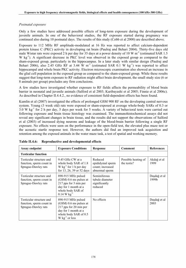
Exposure to high frequency electromagnetic fields, biological effects and health consequences (100 kHz-300 GHz)
178
Postnatal exposure
Only a few studies have addressed possible effects of long-term exposure during the development of juvenile animals. In one of the behavioral studies, the RF exposure started during pregnancy was continued also during 10 postnatal days. The results of this study (Cobb et al 2000) are described above.
Exposure to 112 MHz RF amplitude-modulated at 16 Hz was reported to affect calcium-dependent protein kinase C (PKC) activity in developing rat brain (Paulraj and Behari 2004). Thirty-five days old male Wistar rats were exposed 2 h per day for 35 days at a power density of 10 W m-2 (estimated SAR 1.5 W kg-1). A significant decrease in PKC level was observed in the exposed group as compared to the sham-exposed group, particularly in the hippocampus. In a later study with similar design (Paulraj and Behari 2006), also 2.45 GHz RF at 3.44 W m-2 (estimated SAR 0.1 W kg-1) was reported to affect hippocampal and whole brain PKC activity. Electron microscopic examination also showed an increase in the glial cell population in the exposed group as compared to the sham-exposed group. While these results suggest that long-term exposure to RF radiation might affect brain development, the small study size (6 or 8 animals per group) precludes any firm conclusions.
A few studies have investigated whether exposure to RF fields affects the permeability of blood brain barrier in neonatal and juvenile animals (Salford et al 2003; Kuribayashi et al 2005; Finnie et al 2006c). As described in Chapter II.4.4.2., no evidence of consistent field-dependent effects has been found.
Kumlin et al (2007) investigated the effects of prolonged GSM 900 RF on the developing central nervous system. Young (3 week old) rats were exposed or sham-exposed at average whole-body SARs of 0.3 or 3.0 W kg-1 for 2 h per day, 5 days per week for 5 weeks. A variety of behavioral tests were carried out following exposure and brain tissue histology was examined. The immunohistochemical assays did not reveal any significant changes in brain tissue, and the results did not support the observations of Salford et al (2003) of increased dying neurons and leakage of the blood-brain barrier following a single RF exposure. No effects were seen on the performance in the open-field test, the elevated plus maze test or the acoustic startle response test. However, the authors did find an improved task acquisition and retention among the exposed animals in the water maze task, a test of spatial and working memory.
Table II.4.6: Reproductive and developmental effects
Assay endpoint Exposure Conditions Response Comment References
Testicular function
Testicular structure and function, sperm count in Sprague-Dawley rats
9.45 GHz CW at a whole body SAR of 1.8 W kg-1 for 1 h per day for 13, 26, 39 or 52 days
Reduced epididymal sperm count; increased abnormal sperm
Possible heating of the testis?
Akdağ et al 1999
Testicular structure and function, sperm count in Sprague-Dawley rats
890-915 MHz pulsed (GSM) 0.6 ms pulses at 217 pps for 3 min per day for 1 month at a whole body SAR of 0.14 W kg-1
Seminiferous tubule diameter significantly reduced
Daşdağ et al 1999b
Testicular structure and function, sperm count in Sprague-Dawley rats
890-915 MHz pulsed (GSM) 0.6 ms pulses at 217 pps for 20 min per day for 1 month at a whole body SAR of 0.5 W kg-1 or less
No effects Daşdağ et al 2003

Exposure to high frequency electromagnetic fields, biological effects and health consequences (100 kHz-300 GHz)
179
Assay endpoint Exposure Conditions Response Comment References
Reproductive outcome
Litter number and size in BALB/c/f mice
RF radiation from an ‘antenna park’ over a five month period at various outdoor and indoor locations
Reduced number and size of litters compared to laboratory controls
Lack of matched controls
Magras and Xenos 1997
Behavioral teratology following prenatal exposure
Pregnant rats exposed to RF
915 MHz CW at a whole body SAR estimated to be 3-4 W kg-1 of for 6 h per day throughout gestation
No effect on post-natal and adult behavior
Jensh et al 1982b; Jensh 1997
Pregnant rats exposed to RF
2.45 GHz CW at a whole body SAR estimated to be about 2-4 W kg-1 of for 6 h per day throughout gestation
No effect on most tests of behavior; exposed offspring more active in an open field test.
Jensh et al 1983b; Jensh 1997
Pregnant rats exposed to RF
6.0 GHz CW at a whole body SAR estimated to be about 7 W kg-1 of for 6 h per day throughout gestation
Exposed female offspring showed decreased learning in water T-maze test and decreased activity levels, whereas males showed increased activity levels.
Jensh 1984b, 1997
Exposure of pregnant rats throughout gestation
900 MHz pulsed (GSM) 0.577 pulses at 217 pps; at whole body SAR of between 0.0175 and 0.075 W kg-1 continuously from day 1 to day 20 gestation
No effect on operant task performance.
Bornhausen and Scheingraber (2000)
Exposure of pregnant rats and postnatal exposure of the offspring
UWB (dominant frequency range 0.1-1 GHz) pulses of 1.8 ns pulse width at 1000 pps; Two min per day at average whole body SAR of 0.045 W kg-1 during days 3-18, or during this period and to postnatal day 10.
No statistically significant effects except more stress vocalization, longer hippocampus and lower mating frequency in exposed offspring.
Cobb et al 2000
Postnatal development
Exposure of young (~ 5 week old) rats for a further 5 weeks
112 MHz RF amplitude modulated at 16 Hz; whole body SAR estimated as1.5 W kg-1; 2 h per day for 35 days
A significant decrease in PKC level was observed in the exposed group as compared to the sham exposed group, particularly in the hippocampus.
Paulraj and Behari 2004
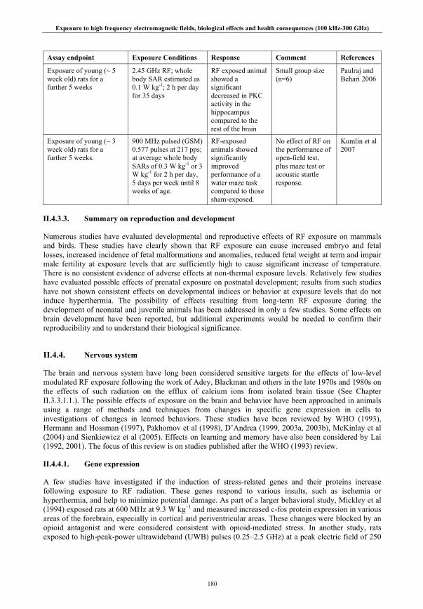
Exposure to high frequency electromagnetic fields, biological effects and health consequences (100 kHz-300 GHz)
180
Assay endpoint Exposure Conditions Response Comment References
Exposure of young (~ 5 week old) rats for a further 5 weeks
2.45 GHz RF; whole body SAR estimated as 0.1 W kg-1; 2 h per day for 35 days
RF exposed animal showed a significant decreased in PKC activity in the hippocampus compared to the rest of the brain
Small group size (n=6)
Paulraj and Behari 2006
Exposure of young (~ 3 week old) rats for a further 5 weeks.
900 MHz pulsed (GSM) 0.577 pulses at 217 pps; at average whole body SARs of 0.3 W kg-1 or 3 W kg-1 for 2 h per day, 5 days per week until 8 weeks of age.
RF-exposed animals showed significantly improved performance of a water maze task compared to those sham-exposed.
No effect of RF on the performance of open-field test, plus maze test or acoustic startle response.
Kumlin et al 2007
II.4.3.3. Summary on reproduction and development
Numerous studies have evaluated developmental and reproductive effects of RF exposure on mammals and birds. These studies have clearly shown that RF exposure can cause increased embryo and fetal losses, increased incidence of fetal malformations and anomalies, reduced fetal weight at term and impair male fertility at exposure levels that are sufficiently high to cause significant increase of temperature. There is no consistent evidence of adverse effects at non-thermal exposure levels. Relatively few studies have evaluated possible effects of prenatal exposure on postnatal development; results from such studies have not shown consistent effects on developmental indices or behavior at exposure levels that do not induce hyperthermia. The possibility of effects resulting from long-term RF exposure during the development of neonatal and juvenile animals has been addressed in only a few studies. Some effects on brain development have been reported, but additional experiments would be needed to confirm their reproducibility and to understand their biological significance.
II.4.4. Nervous system
The brain and nervous system have long been considered sensitive targets for the effects of low-level modulated RF exposure following the work of Adey, Blackman and others in the late 1970s and 1980s on the effects of such radiation on the efflux of calcium ions from isolated brain tissue (See Chapter II.3.3.1.1.). The possible effects of exposure on the brain and behavior have been approached in animals using a range of methods and techniques from changes in specific gene expression in cells to investigations of changes in learned behaviors. These studies have been reviewed by WHO (1993), Hermann and Hossman (1997), Pakhomov et al (1998), D’Andrea (1999, 2003a, 2003b), McKinlay et al (2004) and Sienkiewicz et al (2005). Effects on learning and memory have also been considered by Lai (1992, 2001). The focus of this review is on studies published after the WHO (1993) review.
II.4.4.1. Gene expression
A few studies have investigated if the induction of stress-related genes and their proteins increase following exposure to RF radiation. These genes respond to various insults, such as ischemia or hyperthermia, and help to minimize potential damage. As part of a larger behavioral study, Mickley et al (1994) exposed rats at 600 MHz at 9.3 W kg−1 and measured increased c-fos protein expression in various areas of the forebrain, especially in cortical and periventricular areas. These changes were blocked by an opioid antagonist and were considered consistent with opioid-mediated stress. In another study, rats exposed to high-peak-power ultrawideband (UWB) pulses (0.25–2.5 GHz) at a peak electric field of 250
Exposure to high frequency electromagnetic fields, biological effects and health consequences (100 kHz-300 GHz)
181
kV m−1 for 2 minutes did not show any changes in the expression of c-fos protein levels (Walters et al 1995). Body temperatures of the animals in this study rose by less than 0.5°C.
Fritze et al (1997a) exposed the heads of rats to simulated GSM signals (890–915 MHz pulsed at 217 Hz) at 7.5 W kg−1 and measured changes in the messenger RNAs of hsp70, c-fos, c-jun, and gfap using in situ hybridization histochemistry. Only changes consistent with brain hyperthermia or immobilization stress were found either immediately or 24 hours after exposure. Seven days after exposure, no changes were observed in the levels of the relevant proteins. Similarly, Morrissey et al (1999) reported that local exposure of the heads of mice to a 1.6 GHz Iridium satellite phone signal (pulse modulated at 11 Hz with a duty cycle of 4:1 and a pulse duration of 9.2 ms) for 1 hour only significantly increased c-fos mRNA expression in the forebrain when the average SAR in the brain exceeded 4.3 W kg−1. The pattern of c-fos change was consistent with a thermal stress, thermoregulatory activity and the effects of restraint. There were no differences between continuous and pulsed exposures. Stagg et al (2001) exposed rats for 2 hours to 1.6 GHz Iridium signals using a head-only exposure system that produced local SARs in the brain of up to 5 W kg−1. No significant increases in body temperature were recorded and no field-dependent increases in c-fos and c-jun mRNA were observed. Finnie (2005) found that no increase in c-fos expression was seen in restrained mice given a single far-field exposure to GSM 900 RF at a whole-body SAR of 4 W kg-1 for 1 hr; however, both c-fos expression in both exposed and sham-exposed groups was elevated compared to free-running control mice, suggesting that the experimental restraint significantly elevated c-fos expression. In a subsequent study, Finnie et al (2006a) investigated the expression of c-fos expression in the brain of embryonic and fetal mice exposed or sham-exposed during the whole of gestation (day 1 to day 19) to 900 MHz GSM RF radiation. No effects of GSM RF radiation exposure were seen on c-fos expression in brain tissue compared to the tissue of those sham-exposed, although expression varied between different brain regions.
Belyaev et al (2006) analyzed gene expression profile in RF exposed animals (see also Chapter II.3.3.2.1.2.). Rats were exposed or sham exposed to GSM 900 at whole-body averaged SAR of 0.4 W kg-
1 for 2 h and total RNA was extracted from the cerebellum. In this study, triplicate arrays were applied for three exposed samples or three sham exposed samples. Gene expression profiles were obtained by Affymetrix U34 GeneChips representing 8800 rat genes and analyzed with the Affymetrix Microarray Suite (MAS) 5.0 software. The results showed that 11 genes were up-regulated by about 1.34 - 2.74 fold and one gene was down-regulated 0.48 fold. The induced genes encode proteins with diverse functions including neurotransmitter regulation, blood-brain barrier, and melatonin production. However, these changes in gene expression were not confirmed by other methods.
In general, the only consistent changes seen in gene expression were associated with hyperthermia or restraint stress.
Table II.4.7: Nervous system effects: Gene expression
Assay endpoint Exposure Conditions Response Comment References
Gene expression
Immunocytochemical staining for c-fos protein in rat brain 2 hr after treatment
600 MHz (CW) for 20 min at 9.3 W kg-1 only.
Increase in c-fos protein levels following 1oC rise in brain and body temperature.
A small part of a larger behavioral study.
Mickley et al 1994
Immunocytochemical staining for c-fos protein in rat brain 2 hr after treatment
0.25-2.5 GHz; high peak power UWB radiation; 7-8 ns pulses; 60 pps for 2 min; peak E-field of 250 kV m-1.
No effect on c-fos protein levels; body temperatures rose by less than 0.5oC.
High peak power UWB radiation
Walters et al 1995

Exposure to high frequency electromagnetic fields, biological effects and health consequences (100 kHz-300 GHz)
182
Assay endpoint Exposure Conditions Response Comment References
Genomic response in rat brain tissue immediately following in vivo exposure viz: hsp70, fos and jun mRNA and their protein products assayed 24 hr after exposure
900 MHz CW or pulsed (GSM); 0. 6 ms pulses at 217 Hz pps for 4 h at brain SARs of 0.3 or 1.5 W kg-1 (pulsed) or 7.5 W kg-1 (CW).
Slight increase in hsp 70 expression at the highest SAR (7.5 W kg-1) but no effects on hps70 protein levels or any other exposure-related effects
Fritze et al 1997a
In situ hybridization for fos mRNA in the mouse brain immediately following exposure.
1.6 GHz CW or pulsed (Iridium signal); 9.2 ms pulses at 11 pps for 1 h at average brain SARs of ~ 0.3 - 11 W kg-1
Increased fos expression in stress responsive and thermoregulatory parts of the brain at average brain SARs ≥ ~ 4 W kg-1
Satellite communication system
Morrissey et al 1999
Body temperature, fos, jun and odc mRNA levels in brain tissue and stress-related plasma hormone levels in Fischer rats
1.6 GHz pulse modulated (Iridium signal); 9.2 ms pulses at 11 pps for 2 h at brain SARs of 0.16, 1.6 and 5 W kg-1
No effect on body temperature, gene expression or plasma hormone levels compared to sham values.
Satellite communication system
Stagg et al 2001
Immunocytochemical staining of c-fos levels in mouse brain
900 MHz pulsed (GSM); 0.6 ms pulses at 217 pps for 1 h at whole-body SAR of 4 W kg-1.
No effect of RF exposure on c-fos expression
Elevated c-fos levels in exposed and sham exposed, but not in free moving controls
Finnie 2005
Immunocytochemical staining of c-fos levels in fetal mouse brain
900 MHz pulsed (GSM); 0.6 ms pulses at 217 pps for 1 h from day 1 gestation to day 19 gestation at whole-body SAR of 4 W kg-1.
No effect of RF exposure on c-fos expression in fetal brain tissue
Elevated c-fos levels in some brain regions in exposed and sham-exposed mice
Finnie et al 2006a
II.4.4.2. Blood-brain barrier
The blood-brain barrier is a dynamic interface that regulates the composition of cerebrospinal and interstitial fluid bathing central nervous system tissue. Physically, the ‘barrier’ comprises endothelial cells lining the blood capillaries of the brain and spinal cord and epithelial cells lining the choroid plexuses of the ventricles. ‘Tight’ junctions between these cells restrict the otherwise normal exchange of molecules through extracellular pathways, enabling the endothelial and epithelial cells of blood-brain barrier to regulate the exchange of molecules between the fluid compartments. However, the blood-brain barrier is relatively permeable in some regions of the brain, for example around the ventricles.
About 20 years ago several studies reported that low-level RF exposure may alter the permeability of the blood-brain barrier and cause leakage of molecules from the blood into the cerebrospinal fluid. Such responses could produce severe and lasting consequences. However better conducted studies failed to corroborate these findings and the original observations were ascribed to various confounding factors (see Blackwell and Saunders 1986). Consistent changes in permeability were only found using SARs of about 7 W kg-1 or more, which produced significant heating (WHO 1993, IEGMP 2000; Zmirou 2001; Krewski et al 2001a, Lin 2005). Immobilization stress is also associated with changes in the blood-brain barrier; habituation to experimental conditions is therefore essential when animals are restrained during exposure.
However, some recent studies have again suggested that low level RF exposure may affect the blood-brain barrier. Neubauer et al (1990) reported that significant changes occurred with exposures above 2 W

Exposure to high frequency electromagnetic fields, biological effects and health consequences (100 kHz-300 GHz)
183
kg−1 for 30 minutes or more. Persson et al (1997) and Salford et al (1997) reported that exposure of rats to 915 MHz radiation increased the permeability of the blood-brain barrier to endogenous albumin. Using a TEM cell, animals were exposed in groups of 4 to either a CW field or pulse modulated radiation at pulse repetition rates between 4 and 217 Hz, and exposures lasted from 2 to 960 min at SARs ranging from 0.4-8 mW kg-1 to 1.7-8.3 W kg-1. The number of animals showing increased permeability was reported to depend on both SAR and pulse modulation frequency but generally most exposures increased the leakage of albumin. Furthermore, the largest effects were reported using the weakest radiation, and exposure to CW radiation was reported to produce a greater effect than pulsed radiation. Weaknesses of this study include insufficient description of the experimental and exposure protocols used, and the findings are difficult to assess.
A more recent paper from the same laboratory (Salford et al 2003) reported that single, brief exposure of rats to pulsed 915 MHz radiation for 2 h at SARs of between 2 and 200 mW kg-1 caused increased blood-brain barrier permeability to albumin and neuronal damage throughout the brain (indicated by darkly staining neurons), especially in the cortex, hippocampus, and basal ganglia. However there are a number of caveats with this study. These include not only the modest size of the study (8 animals per group), a rather wide age range (12-26 weeks) of the rats used but also serious uncertainties about the metrology and dosimetry. The quantification of damaged neurons was also highly subjective, and too few data are presented to justify any conclusions. Overall replication using improved methods and with tighter control of experimental variables is necessary before any extrapolation can be made regarding potential human health effects.
Other studies using rats or mice have failed to corroborate these results, and acute or prolonged exposure to the radiation associated with mobile communication has not produced anything more than negligible effects on albumin permeability. Using a head-only exposure system, Fritze et al (1997b) exposed rats at 900 MHz pulsed at 217 Hz for 4 h at local SARs in the brain of 0.3, 1.5 or 7.5 W kg-1. The leakage of albumin across the blood-brain barrier was examined using immuno-histochemical staining either at the end of exposure or 7 days later. Small increases in permeability were observed in all treatment groups examined immediately after exposure, but these numbers only reached significance in the animals exposed at the highest SAR which represented a thermal challenge. No sustained increases in permeability were reported. Using a similar design of exposure system, Tsurita et al (2000) exposed the heads of rats to a pulsed 1439 MHz TDMA field for 1 h a day, 5 days a week for 2 or 4 weeks. The peak SAR in the brain was 2 W kg-1. Permeability was assessed using immuno-histochemical staining and the Evans blue dye injection method. Neither exposure period caused any discernible effect on the permeability of the blood-brain barrier. In addition, exposure had no apparent effect on body weight or on the Purkinje cells and granular cells in the cerebellum. As positive controls, both local cold injury of the skull or 2 h irradiation at 20 W kg-1 produced detectable increases in blood-brain barrier permeability.
Finnie et al (2001) exposed mice to 898 MHz pulsed at 217 Hz for 60 min at 0.4 W kg-1 using a well-characterized whole-body exposure system. This system consisted of a cylindrical parallel plate with the animals restrained in clear acrylic tubes arranged radially around a dipole antenna. Exposure had no significant effect on blood-brain barrier permeability as assessed using immunohistochemical staining for albumin. Where leakage had occurred, it was mainly confined to the leptomeningeal blood vessels which have no recognized blood-brain barrier. The same pattern of responses was reported by Finnie et al (2002) using long-term, repeated exposure. In this study, mice were exposed to 900 MHz pulsed at 217 Hz for 60 min a day, 5 days a week for 104 weeks at whole body SARs of 0.25, 1, 2 and 4 W kg-1. Comparable small numbers of extravasations were observed in the brains of exposed, sham-exposed and freely moving control animals, but statistical analysis was not performed.
More recently, Kuribayashi et al (2005) investigated the effects of exposure to pulsed 1.439 GHz TDMA signals on the blood-brain barrier function in immature (4 week old) and young (10 week old) rats. The authors assessed permeability to dextran and the expression of genes involved in the regulation of barrier function, namely those encoding p-glycoprotein, aquaporin-4 and claudin-5, which regulate transmembrane drug transport, water homeostasis and tight junction integrity respectively. Repeated exposure of the head at 2 or 6 W kg-1 over a one or two week period had no effect on blood-brain barrier permeability or on the expression of related genes. In addition, no histopathalogical lesions such as gliosis or degenerative lesions were seen.

Exposure to high frequency electromagnetic fields, biological effects and health consequences (100 kHz-300 GHz)
184
Cosquer et al (2005a) used the radial arm maze test to investigate whether exposure to RF would increase blood-brain barrier permeability to a drug known to affect radial arm maze performance. The muscarinic antagonist scopolamine hydrobromide readily crosses the blood-brain barrier to alter radial arm maze performance. The authors used a quaternary-ammonium derivative, scopolamine methylbromide, which does not readily cross the blood-brain barrier, to investigate barrier integrity in rats exposed to pulsed 2.45 GHz signals at a whole-body SAR of 2 W kg-1 (3 W kg-1 in the brain) for 45 min. No effect was seen on behavior nor, in separate groups of rats, on leakage of the dye Evans blue, which binds to albumin. A cold-injury positive control induced blood-brain barrier permeability to Evans blue/albumin.
The in utero exposure of embryonic and fetal mice from day 1 to day 19 of gestation for 1 h per day to GSM 900 RF was reported not to increase blood-brain barrier permeability (Finnie et al 2006b), using endogenous albumin as a vascular tracer identified by monoclonal antibody staining. The areas of the brain examined included the cerebral cortex, thalamus, basal ganglia, hippocampus, cerebellum, midbrain and medulla. Positive effects were reported in control animals injected with cadmium chloride. A second experiment (Finnie et al 2006c) examined the effect of a similar exposure for the first seven days following birth, during which time neurogenesis continues. As reported in the previous study, no effects were seen on blood-brain barrier permeability. It is worth noting here that Kumlin et al (2007) did not find any effect on the blood-brain barrier of juvenile (3 week old) rats following a 5 week exposure to 900 MHz GSM mobile phone radiation at average whole-body SARs of 0.3 or 3.0 W kg-1 (see above, Chapter II.4.3.2.2.3. and Table II.4.6.).
Overall, earlier reports of increased blood-brain barrier permeability have not been corroborated by later, better conducted studies.
Table II.4.8.: Nervous system effects: blood brain barrier
Assay endpoint Exposure Conditions Response Comment References
Blood-brain barrier
Fluorescence assay of a tracer (rhodamine-ferritin) of pinocytic uptake in capillary endothelial cells.
2.45 GHz pulse-modulated, 10 μsec pulses at 100 pps, for 15 -120 min; whole-body SAR: ~ 1 or 2 W kg-1
Increased uptake following exposure at 2 W kg-1 for more than 30 min.
Uptake blocked by colchine, which inhibits microtubular formation.
Neubauer et al 1990
Endogenous albumin and fibrinogen immuno-histochemical staining in rat brain tissue
915 MHz CW or pulse-modulated, either 0.57 ms pulses at 4, 8, 16 or 217 pps, or 6.6 ms pulses at 50 pps, for 2-960 min at whole-body SARs: between 0.4-8 mW kg-1 to 1.7-8.3 W kg-1
Increase in albumin permeability at different combinations of SAR and modulation; results for fibrinogen not presented.
Weaknesses include insufficient description of experimental and exposure protocols
Persson et al 1997; Salford et al 1997
Albumin immuno-histochemical staining in rat brain tissue immediately or 7 days after in vivo exposure
900 MHz CW or pulsed (GSM); 0.6 ms pulses at 217 Hz pps for 4 h at brain SARs of 0.3 or 1.5 kg-1 (pulsed) or 7.5 W kg-1 (CW).
Increased extravasation of albumin immediately after exposure at 7.5 W kg-1 but not 7 days later.
Small but detectable increases in extravasation in rats immobilized for 4 h
Fritze et al 1997b
Exposure to high frequency electromagnetic fields, biological effects and health consequences (100 kHz-300 GHz)
185
Assay endpoint Exposure Conditions Response Comment References
Evans blue injection or immunostaining of albumin, and cerebellar Purkinje cell numbers in rat brain tissue exposed in vivo
1439 MHz pulsed (PDC); 6.7 ms pulses at 50 pps for 1 h per day for 10 or 20 days at a brain SAR of 0.2 W kg-1
No effect on blood-brain barrier integrity or Purkinje cell number
Cold injury positive control
Tsurita et al 2000
Albumin immuno-histochemical staining in mouse brain tissue exposed in vivo
898.4 MHz pulsed (GSM); 0.6 ms pulses at 217 pps for 1 h at a whole-body SAR of 4 W kg-1
No effect on blood-brain barrier integrity
Finnie et al 2001
Albumin immuno-histochemical staining in mouse brain tissue exposed in vivo
900 MHz CW or pulsed (GSM); 0.6 ms pulses at 217 pps for 1 h per day, 5 days per week for 104 weeks at a whole-body SAR of 0.25, 1.0, 2.0, or 4.0 W kg-1
The authors report that results suggest negligible effect on blood-brain barrier integrity
Some increased extravasation in exposed animals, but mainly in areas without effective blood-brain barrier.
Finnie et al 2002
Cresyl violet or albumin immunohistochemical staining in rat brain tissue ‘about’ 50 days after in vivo exposure.
898.4 MHz pulsed (GSM) 0.6 ms pulses at 217 pps for 2 h at a whole body SAR of 2, 20, or 200 mW kg-1
Increased presence of albumin and darkly staining neurons in brain tissue of exposed animals.
Modest study size, wide age range, uncertainties with metrology and dosimetry.
Salford et al 2003
Immunocytochemical staining for vascular permeability to dextran and Evans Blue, and RT-PCR for blood-brain barrier-related gene expression, in immature (4 week) and young (10 week) rats
1439 MHz pulsed (PDC); 6.7 ms pulses at 50 pps at head SARs of 2 or 6 W kg-1 for 90 min day-1 for 6 days per week for 1 or 2 weeks.
No effect on vascular permeability, neuropathology or blood-brain barrier -related gene expression.
The genes, involved in blood-brain barrier function, showed only weak responses to chemically-induced barrier disruption
Kuribayashi et al 2005
Behavioral (radial arm maze) performance in response to a drug that crosses the blood-brain barrier poorly and Evans blue extravasation
2.45 GHz pulsed; 2 μS pulses at 500 pps at whole body SAR of 2.0 W kg-1; brain SAR of 3 W kg-1, for 45 min
No effect of exposure on blood-brain barrier permeability as revealed by Evans blue extravasation, or by drug-induced behavioral effects
The study assumed that significant changes in blood-brain barrier permeability would permit drug-induced behavioral changes
Cosquer et al 2005a
Monoclonal antibody staining of endogenous albumin in brain tissue of mice exposed in utero during gestation.
900 MHz pulsed (GSM) 0.6 ms pulses at 217 Hz pps for 1 hr per day, from day 1 gestation to day 19 gestation at a whole-body SAR of 4 W kg-1
No effect on blood-brain barrier permeability
Blood-brain barrier permeability increased in positive control group.
Finnie et al 2006b
Exposure to high frequency electromagnetic fields, biological effects and health consequences (100 kHz-300 GHz)
186
Assay endpoint Exposure Conditions Response Comment References
Monoclonal antibody staining of endogenous albumin in brain tissue of neonatal mice.
900 MHz pulsed (GSM) 0.6 ms pulses at 217 Hz pps for 1 hr per day, from post-natal day 1 - 7 at a whole-body SAR of 4 W kg-1
No effect on blood-brain barrier permeability
Blood-brain barrier permeability increased in positive control group.
Finnie et al 2006c
II.4.4.3. Electrical activity in brain tissues
Neurons and neuronal networks are believed to be potential targets of RF exposure since they are excitable components that are potentially able to interact with induced electric fields. However, few experiments have been done on neuronal systems.
The hippocampal slice preparation has been much used in neurophysiology to study mechanisms associated with memory. Using a novel parallel-plate exposure system, Tattersall et al (2001) exposed slices of rat hippocampus at 700 MHz (CW) at SARs of between 0.6 and 4.4 mW kg−1. Changes were found in the electrically-evoked field potentials, notably the post-synaptic discharge (population spike) in CA1 that depended on the magnitude of the SAR – low field intensities produced an increase in the amplitude of the population spike by up to 20%, but higher intensity radiation produced either increases of up to 120% or decreases of up to 80%. In addition, it was reported that exposure at about 1.1 mW kg−1 reduced or abolished drug-induced epileptiform activity in 36% of slices tested. Any field-induced rises in temperature were too small to be detected even using sensitive measuring equipment. Imposed temperature changes of up to 1°C failed to mimic the effects of RF exposure. However, it was later reported at an international symposium in Dublin (Green et al 2005) that significant heating occurs at the tip of the metallic stimulating electrode at much higher SARs (7-10°C at 400 mW kg-1) which may have influenced these results. Pakhomov et al (2003), using a similar hippocampal slice preparation, found a transient reduction in the electrically-evoked population spike amplitude during exposure to brief, extremely high power (peak SAR of up to 500 MW kg-1) microwave pulses (0.5 – 2.0 μs) at 9.3 GHz that was temperature-dependent; the reported temperature rises ranged up to 6.0°C at time averaged SARs of 3.6 kW kg-1.
The electroencephalogram (EEG) is a description of the spontaneous electrical activity of the brain and can be used to indicate subtle changes in brain function. The interpretation of such studies is often complicated by the possible effects of anesthesia and restraint, and by artifacts resulting from electrical ‘pick-up’ via the recording electrodes. Exposure to very low levels of amplitude-modulated radiation has been reported to alter the EEG of the brain in cats and rabbits (WHO 1993). Complex changes in electrical activity recorded from the surface of the brain itself from implanted electrodes, particularly in the spectral power of various bands of the EEG, have been reported in recent studies using rats, mice and rabbits. Thuróczy et al (1994) reported that the whole-body exposure of anaesthetized rats to continuous wave 2.45 GHz at thermally significant cortical SARs increased the amplitude of the summed power spectrum of the EEG whereas head-only exposure to amplitude-modulated 4 GHz at similar high cortical SARs increased the amplitude of the beta frequency (14.5-30 Hz) band. More recently, Vorobyov et al (1997, 2004) reported that the intermittent application of amplitude-modulated 915 or 945 MHz RF enhanced the amplitude of certain EEG frequency bands recorded during exposure from conscious rats. However SARs were not reported.
Other studies are difficult to evaluate because little experimental detail has been published. Chizhenkova and Safroshkina (1996) reported slow high-amplitude waves accompanied by an increase in the number of spindle-shaped firings in the rabbit brain EEG in response to 3 GHz RF. Pu et al (1997) found an 800 MHz RF-induced inhibition of total EEG energy recorded from the mouse brain. In both studies, the experimental protocol was very briefly described.
Another difficulty with the interpretation of the EEG in individuals at rest is that the intra-individual variability is very high. Overall, because of these problems, it is not possible to draw any general conclusions regarding mobile phone effects on animal EEGs, although some of the changes appear to

Exposure to high frequency electromagnetic fields, biological effects and health consequences (100 kHz-300 GHz)
187
reflect thermal responses. The variability of evoked and event related potentials is much lover, resulting in better reproducibility. Aran et al (2004) found no effect on the electrical activity in the auditory brain stem neural pathways evoked by acoustic stimulation following the chronic exposure of Guinea pigs to GSM 900 RF at local SARs of 4 W kg-1 at the cochlea.
In summary, effects seen in hippocampal brain slice activity appear to be temperature dependant. Otherwise, the reports of effects on EEG are rather variable and may be confounded by various uncontrolled experimental factors including ‘pick-up’ artifacts.
Table II.4.9.: Nervous system effects: brain electrical activity
Assay endpoint Exposure Conditions Response Comment References EEG, DC brain impedance and ECG recorded in anaesthetized rats during RF exposure.
2.45 GHz CW whole body exposure at whole body SARs of 0.2, 2 or 7 W kg-1 for 10 min; 4 GHz, modulated at 16 Hz, head exposure at local SARs of 8 and 17 W kg-1 for 30 min; or 4 GHz CW localized at 42 W kg-1 for 30 min.
Increased EEG activity seen following thermally significant whole body and head exposures
Metal electrodes implanted below the skull may have caused localized RF field distortion. No RF ‘pick-up’ detected.
Thuróczy et al 1994
Electrical activity recorded in awake restrained rabbit brain, following prior electrode implantation, before, during and after RF exposure.
800 MHz at 400 W m-2 for 1 min; precise exposure conditions not given
Slow high amplitude ‘waves’ accompanied by increased in spindle-shaped firing
Experimental details unclear. Metal electrodes may have caused localized RF field distortion. No SARs given
Chizhenkova and Safroshinka 1996
Electrical activity recorded in mouse brain during exposure on 7th day
3 GHz at 50 W m-2 for 1 h day-1 for 7 days; precise exposure details not given
Exposure reported to produce a decrease in electroencephalic energy (expressed in dB).
Experimental details unclear – were the mice anesthetized on 7th day? Metal electrodes may have caused localized RF field distortion.
Pu et al 1997
EEG frequency spectra in rats with chronically implanted electrodes during RF exposure
945 MHz, amplitude modulated at 4 Hz, at 1 – 2 W m-2 applied intermittently (1 min on, 1 min off) for 10 min.
Small but statistically significant differences seen in certain EEG frequency bands during exposure
Implanted carbon electrodes. SARs not given.
Vorobyov et al 1997
Electrically-evoked field potentials recorded in vitro from the CA1 or CA3 region of rat hippocampal slices during RF exposure.
700 MHz CW at SARs (to the tissue slice) estimated between 0.6 an 4.4 mW kg-1 for between 5 and 15 min.
SAR-dependent changes in population spike amplitude
In vitro study; Localized temperature increase possible at tips of electrodes
Tattersall et al 2001; Green et al 2005
Electrically-evoked field potentials recorded in vitro from the CA1 region of rat hippocampal slices before, during and after RF exposure.
9.3 GHz pulsed; 0.5 -2 μs pulses at 0.5-2.0 pps at up to time-averaged SARs of 3.6 kW kg-1; peak SARs of up to 500 MW kg-1.
Time-averaged SAR-dependent reduction in population spike amplitude during exposure.
In vitro study; peak SARs very high
Pakhomov et al 2003
Exposure to high frequency electromagnetic fields, biological effects and health consequences (100 kHz-300 GHz)
188
Assay endpoint Exposure Conditions Response Comment References EEG frequency spectra in rats with chronically implanted electrodes during RF exposure
915 MHz, 20 μs pulses at 4 Hz; 3 W m-2 applied intermittently for 30 min day-1 for 3 days, followed by treatment with a muscarinic cholinergic antagonist and repeated RF exposure.
RF exposure enhanced EEG amplitudes in the 20-26 Hz frequency band and altered EEG responses to the cholinergic antagonist.
Implanted carbon electrodes. SARs not given.
Vorobyov et al 2004
Auditory brain stem response in Guinea pigs following RF exposure.
900 MHz pulsed (GSM); 0.6 ms pulses at 217 Hz pps at localized SARs of 1, 2 or 4 W kg-1. Exposure for 1 h per day, 5 days per week for 2 months
No effects on auditory brain stem evoked response
No evidence for microwave damage to auditory pathways
Aran et al 2004
II.4.4.4. Neurotransmitters
Changes in various neurotransmitter systems have sometimes been reported in a few studies from different laboratories. Many of these data were reviewed by Hermann and Hossman (1997) who ascribed many of the reported changes to spurious temperature effects. The possibility that confinement or other stresses associated with exposure may produce changes in neurotransmitters levels, particularly in the cholinergic systems, should also be considered.
An extensive series of experiments from one laboratory suggests that exposure to low-level RF radiation may affect cholinergic function in a time-dependent fashion (reviewed by Lai 1992). Both pulsed and continuous 2.45 GHz exposure could elicit decreases in cholinergic activity (Lai et al 1987, 1988, 1989a,b). The threshold with pulsed RF (0.45 W kg−1, specific energy absorption per pulse of 0.9 mJ kg−1) was approximately equal to the rat’s auditory perception threshold. It was reported that similar changes in cholinergic function could be induced by stressors such as noise and acute restraint, suggesting that exposure may be associated with mild stress. In addition, exposure was found to increase the concentration of benzodiazepine receptors in the cortex following acute but not repeated exposures (Lai et al 1992a) again suggesting anxiety or stress response. Similar studies provided evidence of the involvement of endogenous opioids (Lai et al 1992b) in the medial septal nucleus (Lai et al 1996).
Testylier et al (2002) reported that RF exposure caused sustained decreases in acetylcholine release from the rat hippocampus. Animals were exposed during the day for 1 h to 2.45 GHz CW or exposed at night to 800 MHz RF modulated at 32 Hz. Acetylcholine release was continuously measured by microdialysis using an implanted membrane in the CA1 region of the hippocampus. No effects were seen using 2.45 GHz at a whole-body SAR of 3.26 W kg-1 but exposure at 6.52 W kg-1 significantly decreased acetylcholine release for several hours after exposure.
Using semi-quantitative immunochemistry and image analysis to assess neurotransmitter content, Mausset et al (2001) reported that exposure to GSM 900RF at an SAR in the head of 4 W kg-1 reduced the cellular GABA neurotransmitter content in the Purkinje cells layer in the rat cerebellum. Similar but more extensive effects were observed following exposure to CW radiation at 32 W kg-1, which suggested that thermal effects may have contributed towards this response. In an extension of this study, Mausset-Bonnefont et al (2004) exposed only the heads of rats to GSM RF for 15 min at a brain-averaged SAR of 6 W kg-1. Using sensitive cellular and molecular techniques, the authors reported significant changes in binding properties of dopamine transporters, GABA receptors, and NMDA receptors in the cortex and striatum, and/or the hippocampus. Exposure was also associated with significant decreases in the expression of NMDA receptor subunits at the postsynaptic membrane, particularly in the striatum. In addition, the amount of glial fibrilliary acidic protein (GFAP), which is considered to be indicative of astrocyte activation, was increased in the cortex, hippocampus and striatum following exposure. The striatum is involved in the control of locomotor activity but a test of this, using an open-field paradigm, did not reveal any change, either immediately or 24 h after exposure. Following a similar exposure protocol, a further study (Brillaud et al 2007), set out to confirm and further evaluate these results on GFAP expression over a 10 day post-exposure period: the authors reported that a significant but transient
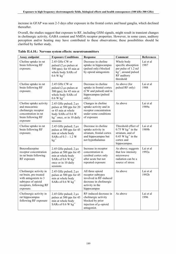
Exposure to high frequency electromagnetic fields, biological effects and health consequences (100 kHz-300 GHz)
189
increase in GFAP was seen 2-3 days after exposure in the frontal cortex and basal ganglia, which declined thereafter.
Overall, the studies suggest that exposure to RF, including GSM signals, might result in transient changes in cholinergic activity, GABA content and NMDA receptor properties. However, in some cases, auditory perception and/or heating may have contributed to these observations; these possibilities should be clarified by further study.
Table II.4.10.: Nervous system effects: neurotransmitters
Assay endpoint Exposure Conditions Response Comment References Choline uptake in rat brain following RF exposure.
2.45 GHz CW or pulsed (2 μs pulses at 500 pps); for 45 min at whole body SARs of 0.6 W kg-1
Decrease in choline uptake in hippocampus (pulsed only) blocked by opioid antagonists
Whole body specific absorption per pulse of 1.2 mJ kg-1, around pulsed RF auditory threshold
Lai et al 1987
Choline uptake in rat brain following RF exposure.
2.45 GHz CW or pulsed (2 μs pulses at 500 pps); for 45 min at whole body SARs of 0.6 W kg-1
Decrease in choline uptake in frontal cortex (CW and pulsed) and in hippocampus (pulsed only).
As above (for pulsed RF only)
Lai et al 1988
Choline uptake activity and muscarinic cholinergic receptor concentration in rat brain following RF exposure
2.45 GHz pulsed; 2 μs pulses at 500 pps for 20 or 45 min at whole body SARs of 0.6 W kg-1 once, or in 10 daily sessions
Changes in choline uptake activity and in receptor concentration under some conditions of exposure
As above Lai et al 1989a
Choline uptake in rat brain following RF exposure.
2.45 GHz pulsed; 2 μs pulses at 500 pps for 45 min at whole body SARs of 0.3 – 1.2 W kg-1
Decrease in choline uptake activity in striatum, frontal cortex and hippocampus but not hypothalamus
Threshold effect of 0.75 W kg-1 in the striatum, and of 0.45 W kg-1 in the cortex and hippocampus.
Lai et al 1989b
Benzodiazepine receptor concentration in rat brain following RF exposure
2.45 GHz pulsed; 2 μs pulses at 500 pps for 45 min at whole body SARs of 0.6 W kg-1 once or in 10 daily sessions
Increase in receptor concentration in cerebral cortex only after acute but not repeated exposure
As above; suggests that low intensity microwave radiation can be a source of stress
Lai et al 1992a
Cholinergic activity in rat brain, pre-treated with antagonists to 3 subtypes of opioid receptors, following RF exposure.
2.45 GHz pulsed; 2 μs pulses at 500 pps for 45 min at whole body SARs of 0.6 W kg-1
All three opioid receptor subtypes involved in RF-induced decrease in cholinergic activity in the hippocampus
As above Lai et al 1992b
Cholinergic activity in rat hippocampus following RF exposure
2.45 GHz pulsed; 2 μs pulses at 500 pps for 45 min at whole body SARs of 0.6 W kg-1
RF-induced decrease in cholinergic activity blocked by prior injection of μ-opioid antagonist.
As above Lai et al 1996

Exposure to high frequency electromagnetic fields, biological effects and health consequences (100 kHz-300 GHz)
190
Assay endpoint Exposure Conditions Response Comment References GABA content of rat cerebellar tissue following in vivo exposure
900 MHz pulsed (GSM) 576 μs pulses at 217 pps for 2 h at brain SARs of 4 W kg-1 (pulsed) or 32 W kg-1 (CW).
Decreased stained area in one cell layer following pulsed RF exposure; reduced optical density in three cell layers following CW exposure.
Mausset et al 2001
ACh release in rat hippocampal tissue during and after exposure in vivo
2.45 GHz CW for 1 h during the day at a whole body SAR of ~ 3 or 6.5 W kg-1; or 800 MHz amplitude modulated at 32 Hz for 1 or 14 h overnight at a whole body SAR of 0.3 W kg-1
Exposure to 2.45 GHz at 6.5 W kg-1 for 1 h, or to 800 MHz for 14 h at 0.3 W kg-1 significantly reduced ACh release.
Testylier et al 2002
Binding properties of neurotransmitter transporters and receptors; number of NMDA receptor subunits and GFAP expression in rat brains
900 MHz pulsed (GSM) 576 μs pulses at 217 pps for 15 min at brain SARs of 6 W kg-
1.
Significant changes were seen in receptor and transporter binding properties and in NMD receptor subunit amount. A strong glial reaction in the striatum
No change in striatum-related locomotor activity.
Mausset-Bonnefont et al 2004
GFAP expression in rat brains 2-10 days after GSM-type exposure.
900 MHz pulsed (GSM) 576 μs pulses at 217 pps for 2 h at brain SARs of 4 W kg-1.
Transient increase in GFAP expression in frontal cortex and basal ganglia 2-3 days after exposure.
Brillaud et al 2007
II.4.4.5. Behavior
Exposure to thermally significant levels of RF induces a heat load that elicits the various physiological and behavioral mechanisms animals use to regulate body temperature. These responses have been studied extensively and were reviewed by WHO (1993) and later by Adair and Black (2003). Most of the relevant animal studies were carried out before 1993. Briefly, in cool environments, animals compensate for RF-induced body heating by lowering their rate of metabolic heating and decreasing their food intake (e.g. Adair and Adams 1982). Other thermoregulatory behaviors exhibited by animals include the selection of cooler environments (e.g. Gordon 1983) and a reduction of spontaneous locomotor activity (eg Mitchell et al 1988). Later studies have focused on volunteer responses (Chapter II.5.3.2.).
In addition, a large number of important studies were carried out mostly in the 1970s and 1980s of aversive responses to RF exposure and of RF effects on food-motivated operant (learned) behaviors (reviewed by D’Andrea 1999; D’Andrea et al 2003a, 2007). The authors observed that the performance of operant tasks in which laboratory rodents and primates were trained to press one or more levers on a prescribed schedule in order to receive the food-reward could be disrupted or completely stopped (the ‘work stoppage’ effect) in a very consistent and repeatable manner by RF exposure sufficient to induce mild, whole-body hyperthermia. In general, behavior was not reliably affected until colonic temperature increased by 1oC or more, corresponding to a whole-body SAR of approximately 4 W kg-1 (3.2 - 8.4 W kg-1), depending on various factors including the frequency of the applied field, the animal size and species and the ambient temperature and relative humidity. The reduction in task performance seen in these studies has been attributed by Stern (1980) to the effects of competing thermoregulatory behaviors such as cooling off or escape, which the author notes may not necessarily be considered adverse.
More recent studies have focused mostly on effects of RF exposure associated with mobile phone use on learned behaviors. In addition, high-peak-power pulsed RF effects on the startle reflex, and in evoking body movement, have also been studied.

Exposure to high frequency electromagnetic fields, biological effects and health consequences (100 kHz-300 GHz)
191
Learned behaviors
This heating effect on learning is illustrated by the results of a study using rats exposed at 600 MHz (Mickley et al 1994). Significant deficits in the performance of a working memory (object recognition) task were observed when exposures caused rises in rectal and brain temperatures of at least 1°C. These changes were correlated with an increase in expression of the c-fos gene in the cortex.
However, results of a few studies using pulsed radar-like signals appear to challenge this conclusion. Lai et al (1989a &b, 1994) reported that the behavior of rats performing a test of spatial memory function in a radial arm maze was disrupted by daily exposure for 20 or 45 minutes to pulsed 2.45 GHz RF at 0.6 W kg−1. It should be noted that the pulse sequence used in these studies (2 μs at 500 pps) results in peak SARs of 600 W kg-1 and absorbed specific energies (SA) of 1.2 mJ kg-1. Exposure did not cause a measurable rise in colonic temperature but acquisition was retarded and exposed animals consistently made more errors than controls, although Cassel et al (2004) noted that differences in performance between these groups existed at the onset of the task, indicating possible differences in anxiety or motivation. Additional results from the Lai group suggested that exposure had activated the endogenous opioid systems and so caused a decrease in cholinergic activity within the hippocampus. Quock et al (1994) reported that brief (5 min) exposure to 4.7 GHz at relatively high whole-body SARs of 36 W kg-1 reversed the sedative and anxiolytic effects of the benzodiazepine drug chlordiazepoxide.
Wang and Lai (2000) placed rats in a Morris water maze immediately after exposure to pulsed 2.45 GHz RF at 1.2 W kg−1 for 1 hour. The animals had to learn to escape from the water by locating a submerged (non-visible) platform. Exposed animals took longer to find the platform than control animals throughout the training sessions, and, in contrast to the control animals, spent much time trying to climb the side walls of the maze. In a probe trial without the platform being present, the exposed animals were reported to have spent less time swimming in the quadrant of the maze that should have contained the platform. Therefore, it was concluded that exposure had disrupted spatial reference memory functions and that the exposed animals had to use other, less efficient, learning strategies to locate the platform. However, statistical analysis of the probe trial data by one-way analysis of variance revealed no significant treatment effect, and only post-hoc analysis suggested a statistical difference between the exposed and control animals (see IEGMP 2000).
In contrast, tests of RF effects on spatial memory carried out at mobile phone frequencies found no effect. Sienkiewicz et al (2000) found that that exposure of mice for 45 minutes to pulsed 900 MHz RF at 0.05 W kg−1 had no significant effects on performance in a radial arm maze. Animals were tested immediately after exposure or following delays of 15 or 30 min. The animals tested without delay took longer to complete the task, possibly due to some mild stress associated with exposure. Similarly, Dubreuil et al (2002) exposed rats to pulsed 900 MHz radiation for 45 min using a head-only system before daily trials either in a radial arm maze or on a food location task in an open field arena (equivalent to a dry-land version of the Morris water maze). No significant effects on the performance of either task were seen using average SARs in the brain of either 1 or 3.5 W kg−1. In an extension of this study, Dubreuil et al (2003) found no effect of a similar exposure on the performance of more complex radial arm maze tasks, or on the performance of a non-spatial object recognition task. A lack of effect on spatial reversal learning in a T-maze was also reported by Yamaguchi et al (2003) following exposure of rats to pulsed 1439 MHz (PDC) at non-thermal levels for either 4 days or 4 weeks. However, performance was significantly impaired by exposure that increased intraperitoneal temperature by up to 2oC.
Two groups have attempted a direct replication and extension of the radial arm maze study by Lai et al (1994) described above, using the same pulsed 2.45 GHz RF exposure at a whole body average SAR of 0.6 W kg-1 for 45 min Cobb et al (2004), using similar experimental procedures to those of Lai et al, including restricted access to distal spatial cues normally used to perform the task, found no effects of exposure on task performance. Similarly, Cassel et al (2004), also using a similar protocol but with distal spatial cues accessible, found that such exposure had no effect on performance. The same group (Cosquer et al 2005b) tested the effect of such exposure, reported by Lai et al (1989a & b, 1992a) to increase the number of benzodiazepine receptors in the cortex, on anxiety responses in rats using the elevated-plus maze test. This maze, which is in the form of a cross, elevated above the floor, has one pair of opposing arms enclosed by high sides, with the adjacent opposing pair were devoid of sides; anxiety increases the
Exposure to high frequency electromagnetic fields, biological effects and health consequences (100 kHz-300 GHz)
192
number of entries into the closed arms. Cosquer et al (2005b) found that exposure had no effect on anxiety levels, either in a low-baseline anxiety test (carried out at low levels of illumination) or in a high baseline anxiety test (high levels of illumination). These findings provide no support for the hypothesis that low level RF radiation exposure increases behavioral measures of anxiety.
In summary, the early studies support the conclusion of WHO (1993) that the performance of learned behaviors is reduced following thermally significant RF exposure. Following a study reporting the reduced performance of a spatial memory task after exposure to pulsed RF, several groups have been unable to replicate or extend the initial observations. In addition, one group reported a lack of effect of pulsed RF on anxiety levels. However, the types of behavioral tasks that have been used are by no means exhaustive.
High peak power pulse effects
The auditory perception of pulsed RF radiation by animals is well established (WHO 1993; Lin and Wang 2007). Following RF absorption, a sound wave is generated in the head by the small and rapid thermoelectric expansion of brain tissue which generates a sound wave that stimulates the cochlea. For short pulses (< 30 μs), thresholds are dependent on the energy per pulse and correspond to a specific absorption per pulse of 0.9-1.8 mJ kg-1 in rats and 10-16 mJ kg-1 in cats (e.g. Guy et al 1975a; Chou et al 1985).
High peak power RF pulses with peak power densities of the order of 10’s – 100’s MW m-2 but of relatively short pulse widths (ns–μs) have been developed for military and other use but their relative biological effectiveness is not well established. Four studies have examined the effects of such pulses on food-reinforced operant behavior.
Using rhesus monkeys D’Andrea et al (1989) examined the effects of such exposure on the performance of a behavioral task that comprised a sequence of three operant schedules: a differential reinforcement of low rate schedule, a time discrimination schedule and a fixed interval schedule. During the performance of these tasks the animals were exposed for 1 h to pulsed 1.3 GHz RF with a pulse width of 3 μs at peak power densities of 1.32 MW m-2 and a specific absorption of 280 mJ kg-1 per pulse. Whole-body time-averaged SARs were varied by adjusting the pulse repetition rate and ranged between 0.05 W kg-1 (at 2 pps) to 0.8 W kg-1 (at 32 pps). The authors found no effects of exposure compared to sham-exposed animals.
D’Andrea et al (1994) exposed rhesus monkeys for 20 min to pulsed 5.62 GHz RF at whole body SARs of 2, 4 or 6 W kg-1 whilst they carried out a variable-interval, color-discrimination task. The monkeys were exposed to RF pulses with a pulse width of 2.8 μs at 100 pulses per second from a military radar either with or without an additional high peak power pulse with a pulse width of ~50 ns superimposed on the radar signal. Peak power densities were 2.77 MW m-2 (radar) and 25.2 MW m-2 (radar plus high peak power pulse). Compared to sham-exposed animals, response rates, reaction time and food pellet rewards significantly declined at whole-body SARs of 4 and 6 W kg-1 suggesting a heating effect; there was no specific effect of the additional high peak power pulse regime.
Akyel et al (1991) examined the operant performance of rats immediately after exposure to high peak power pulsed 1.25 GHz for 10 min. The rats were exposed to 10 μs pulses each of which produced a whole-body specific absorption of 2.1 J kg-1. By adjusting the pulse repetition frequency, whole-body SARs varied from 0.84 W kg-1 to 23 W kg-1. Following exposure, the rats were tested on three successive operant schedules: a fixed-ratio schedule, a variable interval schedule, and a differential reinforcement of low rate schedule. The authors found that the 10 min exposure at 23 W kg-1, which induced a colonic temperature rise of 2.5oC, resulted in the subsequent termination of all operant behavior for about 13 minutes. Afterwards, the animals began to respond, but performance of two of the operant tasks never reached base-line levels, and the performance of the third task was variable. No effects were seen following exposure at the other SARs.
Raslear et al (1993) investigated the effect of exposure of rats to high peak power pulsed RF on their subsequent performance of a time perception and discrimination operant task. In this study, the rats were trained to discriminate between a visual stimulus applied for 0.5 or 5 s for a food reward and were then

Exposure to high frequency electromagnetic fields, biological effects and health consequences (100 kHz-300 GHz)
193
also tested at intermediate durations with no reward following exposure for about 27 min to pulsed 3 GHz RF (80 ns pulse width) at a specific absorption of up to 580 mJ kg-1 per pulse. Whole-body SARs were small (< 0.1 W kg-1). The authors found that the time taken to complete 300 trials and the number of null responses increased with increasing levels of exposure, suggesting a non-thermal effect on cognitive processes.
Other studies have focused on effects on the startle reflex. Seaman and Beblo (1992) studied the effect of exposure to a single high peak power RF ~1 μs pulse (head specific absorption of 22-43 kJ kg-1, or 59-107 kJ kg-1) on a subsequent 100 dB SPL acoustic noise-induced startle response in rats. They found that the low-energy pulse significantly reduced the amplitude of a subsequent startle response, as did prior exposure to an acoustic noise of 60 dB SPL (sound pressure level), whereas the high energy pulse increased the amplitude, but the variability precluded statistical significance. A later study (Seaman et al 1994) reported that a ~1 μs pulse of 66-142 mJ kg-1 and an 8 μs pulse of 525-1056 mJ kg-1 would inhibit and increase that latency of a startle response if given >~10 ms before an acoustic or tactile startle stimulus. An acoustic click given in place of the RF pulse had a similar effect. Both studies suggest that this effect of high peak power pulsed RF was mediated through the field-induced thermoelastic expansion of brain tissue.
Finally, Brown et al (1994) investigated the ability of high peak power RF pulses to evoke body movement in restrained mice. These authors exposed mice either to pulsed 1.25 GHz RF at 80 pps for 2 s, or to ‘gated’ CW 1.25 GHz for a duration of 50-3200 ms and measured induced movement with the aid of piezoelectric sensors. The brain specific energy per pulse varied up to 152 J kg-1, and it was reported that a single pulse could induce body movement. Overall, however, the authors reported that the incidence of evoked body movement increased with the average energy input, and that there was no difference between pulsed RF and the gated CW RF, suggesting a possible heating effect.
In summary, most of the data suggests that high peak power RF pulses have no effect on operant behavior, except at thermogenic levels, when an expected decline in performance ensues. There is however, good evidence that individual high-peak-power pulses reduce and delay the ‘startle’ response to an acoustic noise and may evoke body movement.
Table II.4.11.: Nervous system effects: Behavior
Assay endpoint Exposure Conditions Response Comment References Learned behaviors Radial arm maze (12 arm) performance in rats following RF exposure
2.45 GHz pulsed; 2 μs pulses at 500 pps for 20 or 45 min each day for 10 days at whole body SARs of 0.6 W kg-1
Significantly reduced maze performance following exposure for 45 min, but not 20 min
Whole body specific absorption per pulse of 1.2 mJ kg-1, around pulsed RF auditory threshold for short (< 30 μs) pulses
Lai et al 1989a
Radial arm maze (12 arm) performance in rats following RF exposure
2.45 GHz pulsed; 2 μs pulses at 500 pps for 45 min each day for 10 days at whole body SARs of 0.6 W kg-1
Reduced maze performance reversed by pre-treatment with cholinergic agonist or opioid antagonist
As above Lai et al 1994
Locomotor and rearing in mice, after pre-treatment with chlordiazepoxide, following RF exposure
1.8 or 4.7 GHz CW for 5 min at whole body SARs of 4, 12 or 36 W kg-1
No effect of RF exposure on anxiolytic or sedative effect of drug treatment except at 4.7 GHz, 36 W kg-1
Thermal effect Quock et al 1994
Object recognition - working memory task in rats following RF exposure.
600 MHz (CW) for 20 min; whole-body SAR of 0.1-10 W kg-1.
Impaired performance at > 9.3 W kg-1; 1oC rise in body and brain temperature.
Thermal effect Mickley et al 1994
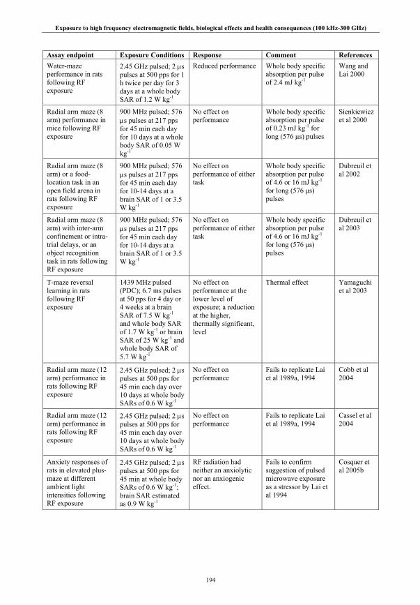
Exposure to high frequency electromagnetic fields, biological effects and health consequences (100 kHz-300 GHz)
194
Assay endpoint Exposure Conditions Response Comment References Water-maze performance in rats following RF exposure
2.45 GHz pulsed; 2 μs pulses at 500 pps for 1 h twice per day for 3 days at a whole body SAR of 1.2 W kg-1
Reduced performance Whole body specific absorption per pulse of 2.4 mJ kg-1
Wang and Lai 2000
Radial arm maze (8 arm) performance in mice following RF exposure
900 MHz pulsed; 576 μs pulses at 217 pps for 45 min each day for 10 days at a whole body SAR of 0.05 W kg-1
No effect on performance
Whole body specific absorption per pulse of 0.23 mJ kg-1 for long (576 μs) pulses
Sienkiewicz et al 2000
Radial arm maze (8 arm) or a food-location task in an open field arena in rats following RF exposure
900 MHz pulsed; 576 μs pulses at 217 pps for 45 min each day for 10-14 days at a brain SAR of 1 or 3.5 W kg-1
No effect on performance of either task
Whole body specific absorption per pulse of 4.6 or 16 mJ kg-1 for long (576 μs) pulses
Dubreuil et al 2002
Radial arm maze (8 arm) with inter-arm confinement or intra-trial delays, or an object recognition task in rats following RF exposure
900 MHz pulsed; 576 μs pulses at 217 pps for 45 min each day for 10-14 days at a brain SAR of 1 or 3.5 W kg-1
No effect on performance of either task
Whole body specific absorption per pulse of 4.6 or 16 mJ kg-1 for long (576 μs) pulses
Dubreuil et al 2003
T-maze reversal learning in rats following RF exposure
1439 MHz pulsed (PDC); 6.7 ms pulses at 50 pps for 4 day or 4 weeks at a brain SAR of 7.5 W kg-1 and whole body SAR of 1.7 W kg-1 or brain SAR of 25 W kg-1 and whole body SAR of 5.7 W kg-1
No effect on performance at the lower level of exposure; a reduction at the higher, thermally significant, level
Thermal effect Yamaguchi et al 2003
Radial arm maze (12 arm) performance in rats following RF exposure
2.45 GHz pulsed; 2 μs pulses at 500 pps for 45 min each day over 10 days at whole body SARs of 0.6 W kg-1
No effect on performance
Fails to replicate Lai et al 1989a, 1994
Cobb et al 2004
Radial arm maze (12 arm) performance in rats following RF exposure
2.45 GHz pulsed; 2 μs pulses at 500 pps for 45 min each day over 10 days at whole body SARs of 0.6 W kg-1
No effect on performance
Fails to replicate Lai et al 1989a, 1994
Cassel et al 2004
Anxiety responses of rats in elevated plus-maze at different ambient light intensities following RF exposure
2.45 GHz pulsed; 2 μs pulses at 500 pps for 45 min at whole body SARs of 0.6 W kg-1; brain SAR estimated as 0.9 W kg-1
RF radiation had neither an anxiolytic nor an anxiogenic effect.
Fails to confirm suggestion of pulsed microwave exposure as a stressor by Lai et al 1994
Cosquer et al 2005b

Exposure to high frequency electromagnetic fields, biological effects and health consequences (100 kHz-300 GHz)
195
Assay endpoint Exposure Conditions Response Comment References High Peak Power Pulse Effects
Multiple schedule operant task performance by rhesus monkeys during exposure.
1.3 GHz pulses, pulse width 3 μs, at 2-32 pps at a head specific absorption of 280 mJ kg-1 per pulse; Peak whole body SARs 8.3 W kg-1; average whole body SARs 0.05-0.8 W kg-1 for 60 min.
No effect on operant task performance.
Head specific absorption per pulse above the auditory stimulus threshold.
D’Andrea et al 1989
Multiple schedule operant task performance by rats after exposure.
1.2 GHz pulses, pulse width 10 μs; average whole body SARs of 0.84, 2.5, 7.6 and 23 W kg-1 for 10 min. Whole body specific absorption of 2.1 J kg-
1 per pulse.
Initial (for 13 min) failure to perform tasks following exposure at 23 W kg-1 followed by reduced task performance.
Colonic temperatures increased in the high exposure group by 2.5oC.
Akyel et al 1991
Startle reflex in rats in response to 100 dB SPL acoustic noise
Single 1.25 GHz pulses, pulse width 0.8-1.0 μs; SA to the head of 22-43 mJ kg-1 or 59-107 mJ kg-1 per pulse. Time-averaged whole-body SARs not given.
The acoustic startle reflex had a lower amplitude following a prior RF pulse at 22-43 kJ kg-1, and a higher but variable amplitude following an RF pulse at 59-107 kJ kg-1.
A prior 60 dB SPL noise increased latency and decreased amplitude of the acoustic startle response.
Seaman and Beblo 1992
Time-discrimination operant behavior in rats after exposure.
3 GHz pulses, pulse width of 80 ns at 0.125 pps for 200 pulses (~27 min) at SA of up to 580 mJ kg-1 per pulse. Time-averaged whole-body SAR of up to 0.072 W kg-1.
Dose-response effects observed for session time and null responses; possible effect on time discrimination.
Co-varying sound (~57-89 dBA per pulse) and x-ray exposure did not correlate with effects.
Raslear et al 1993
Color discrimination operant task performance by rhesus monkeys during exposure.
5.62 GHz pulses, radar pulse width 2.8 μs, with or without additional high peak power pulse width 2 ns, at 100 pps for 20 min at average whole body SARs of 2, 4 or 6 W kg-1.
Responses declined significantly at whole body SARs of 4 and 6 W kg-1, as did reaction time and food pellet consumption.
No effect of pulse regime.
D’Andrea et al 1994.
Startle reflex in rats in response to (1) acoustic noise or (2) tactile (air puff) stimulus
Single 1.25 GHz pulses, (1) pulse width ~1 μs, SA of 16-44 mJ kg-1 or 66-142 kJ kg-1; (2) pulse width ~ 8μs, SA of 525-1056 kJ kg-
1 per pulse
A high intensity 1 μs pulse (1) or an 8 μs pulse (2) affected the amplitude and latency of a subsequent startle reflex
The 8 μs pulse (2) had a similar effect on the tactile startle reflex to that of a 94 dB SPL acoustic noise (click).
Seaman et al 1994

Exposure to high frequency electromagnetic fields, biological effects and health consequences (100 kHz-300 GHz)
196
Assay endpoint Exposure Conditions Response Comment References Evoked body movement in restrained mice.
1.25 GHz pulsed, pulse width 10 μs at 80 pps for up to 2 s; or single gated 1.25 GHz CW, duration 50-3200 ms. Brain SA per pulse up to ~152 J kg-1
Body movement could be induced by a single pulse; generally, incidence increasing with averaged energy input.
No difference between responses to pulsed RF and gated CW suggested a possible skin heating effect.
Brown et al 1994
II.4.4.6. Summary on behavior
Several recent studies support the experimental observations summarized by WHO (1993) that operant behavior in laboratory rodents and primates can be disrupted by thermogenic RF exposure sufficient to raise body temperature by about 1oC. Two studies report the reduced performance of operant tasks during exposure to high peak power RF pulses but attribute these effects to heating. In another study, significant deficits on the performance of a T-maze task were seen only when exposure increased body temperature by 2°C.
Otherwise, studies have continued to investigate the possible effects of RF radiation, often that characteristic of mobile phone use, on the brain and nervous system in animals. Despite sporadic reports of positive effects, most studies have not reported any field-dependent responses either in gene expression or in increased permeability of the blood-brain barrier. One study in particular found a lack of effect on blood-brain barrier permeability following in GSM exposure in utero throughout gestation. The evidence from several laboratories indicates that changes may be induced in cholinergic activity in the brain following relatively intense exposure. Such changes might predict effects on spatial learning and memory, but on balance the evidence does not support this view: two studies from one laboratory have reported deficits in performance of spatial memory tasks using pulsed 2.45 GHz microwaves, but were not confirmed in two independent replications of these studies, nor in three other studies using GSM signals.
Studies of the behavioral effects of high peak power RF pulses such as those used in military applications have been rather sporadic and diverse; pulse widths have varied by two orders of magnitude (80 ns – 10 μs) and the specific absorption per pulse by four orders of magnitude (22 mJ kg-1 – 152 J kg-1). Two rather elegant studies showed the equivalence of pulsed RF to an acoustic ‘click’ in affecting the startle reflex.
II.4.5. Auditory system
It has been known for a long time from extensive electrophysiological and behavioral data indicate that animals can perceive pulsed RF radiation (see WHO 1993; Lin and Wang 2007). As described above, the generally accepted explanation is that a sound wave is generated in the head by the short but rapid thermoelastic expansion of the brain resulting from the absorption of the RF pulse. For short pulses (< 30 μs), thresholds are dependent on the energy per pulse and correspond to a specific absorption per pulse of 0.9-1.8 mJ kg-1 in rats and 10-16 mJ kg-1 in cats. Such effects might be important in the interpretation of behavioral responses to pulsed RF radiation.
Recent studies have focused on possible RF effects on cochlea function per se measuring otoacoustic emission. This is an indicator of the normal mechanical contractility of the outer hair cells of the cochlea and is considered to be a reliable method of assessing cochlea functionality in vivo. The outer hair cells, which are notoriously susceptible to various endogenous and exogenous stressors, generate an acoustic signal in response to auditory stimuli (measured for example as the distortion product otoacoustic emission or DPOE) which can be monitored in the external ear canal (or auditory meatus).
A lack of effect on otoacoustic emissions was reported in four new-born and two groups of seven adult rats exposed or sham exposed to GSM 900 RF for 1 h per day for 30 days (Kizilay et al 2003); unfortunately field measurements and dosimetric assessments of SAR were not given. DPOEs were recorded in anesthetized adult animals before the first exposure and after 30 days exposure; for the new-born rats, the recordings made after 30 days exposure were compared to the results from adults prior to

Exposure to high frequency electromagnetic fields, biological effects and health consequences (100 kHz-300 GHz)
197
exposure. No effects on outer hair cell function were detected. Similarly, Aran et al (2004) found no effect on outer hair cell function in rats following chronic exposure over 2 months to GSM 900 at local cochlea SARs of 1, 2 or 4 W kg-1.
A lack of effect on outer hair cell function by mobile phone RF, as assessed by DPOEs, has also been reported in two papers. Galloni et al (2005a) describe a set of three experiments in which the RF frequency, source, modulation characteristics and period of exposure were varied. In the first experiment, rats were exposed or sham exposed to 936 MHz CW whole-body or to 923 MHz CW head-only for 3 h per day for 5 days; the local SAR to the head was about 1 W kg-1. In the second study, rats were exposed or sham exposed to 960 MHz GSM RF for 3 h per day for 5 days, with a head SAR of 1 W kg-1. In the third study, exposure was to 900 MHz GSM RF for 2 h per day, 5 days per week for 4 weeks, with a head SAR of 2 W kg-1. The authors found no effect of RF exposure on DPOEs in any of these studies. A further study (Galloni et al 2005b) reported that the exposure of rats to GSM signals at 900 MHz or 1800 MHz over a 4 week period at a local SAR of 2 W kg-1 had no effect.
In summary, the evidence is rather consistent and suggests that mobile phone type RF exposure has no effect on auditory function in rodents. It is also clear that animals can hear the pulsed RF characteristic of radar above given thresholds, through a thermoelastic expansion mechanism.
Table II.4.12.: Nervous system effects: auditory functions
Assay endpoint Exposure Conditions Response Comment References DPOEs in newborn and adult rats before and after RF exposure.
900 MHz pulsed (GSM); 0.6 ms pulses at 217 Hz pps. SARs not given. Exposure for 1 h per day for 30 days
No effects on distortion product otoacoustic emissions
Small numbers of animals and absence of dosimetry.
Kizilay et al 2003
DPOEs in Guinea pigs following RF exposure.
900 MHz pulsed (GSM); 0.6 ms pulses at 217 Hz pps at localized SARs of 1, 2 or 4 W kg-1. Exposure for 1 h per day, 5 days per week for 2 months
No effects on distortion product otoacoustic emissions
No evidence for microwave damage to outer hair cells of the cochlea
Aran et al 2004
DPOEs in rats before and after RF exposure.
936 MHz CW whole body or 923 MHz CW head only, 3 h per day for 5 days; head SAR 1 W kg-1; 960 MHz GSM RF, 3 h per day for 5 days, head SAR 1 W kg-1; 900 MHz GSM RF, 2 h per day, 5 days per week for 4 weeks, head SAR 2 W kg-
1.
No effects on distortion product otoacoustic emissions
As above Galloni et al 2005a
DPOEsin rats before, during and after RF exposure.
900 or 1800 MHz pulsed (GSM); 0.6 ms pulses at 217 Hz pps at localized SAR in the ear of 2 W kg-
1 for 2 h per day, 5 days per week for 4 weeks.
No effects on distortion product otoacoustic emissions
As above Galloni et al 2005b
II.4.6. Endocrine system
Most early studies, reviewed for example by WHO (1993) and later by Black and Heynick (2003) described thermally-mediated responses of the endocrine system to RF exposure. Briefly, endocrine responses to acute RF (often CW 2.45 GHz) exposure are generally consistent with the acute responses to

Exposure to high frequency electromagnetic fields, biological effects and health consequences (100 kHz-300 GHz)
198
non-specific stressors such as heat. Several papers report that plasma corticosterone or cortisol levels are significantly enhanced in rodents (Lotz and Michaelson 1978; Lu et al 1980, 1981) and primates (Lotz and Podgorski 1982) by exposures resulting in about a 1oC rise in body temperature; corresponding whole-body SARs were of the order of 4 W kg-1. The response seems to be mediated by the release of adrenocorticotrophic hormone by the hypothalamus via the anterior pituitary gland, and is modulated in amplitude by the circadian rhythm of cortisol or corticosterone levels. The hypothalamus also controls the secretion of growth hormone and thyroxin; stressful stimuli such as significantly elevated body temperatures are known to depress circulating plasma levels of both hormones in rodents (Michaelson et al 1975). However, no effects on growth hormone and thyroxin have been seen in primates (Lotz and Podgorski 1982). In addition, no effects on the circulating levels of a number of hormones have been seen in rats chronically exposed for most of their lives at whole-body SARs of up to 0.4 W kg-1 (Chou et al 1992), a SAR insufficient to significantly affect body temperature.
II.4.6.1. Pineal-melatonin studies
There have been fewer studies of endocrine effects since 1993; those that have been carried out mostly focused on radiation associated with the use of mobile telephony. Several studies have examined possible effects on circulating melatonin, a hormone produced by the pineal gland in a distinct daily or circadian rhythm which is governed by day length, the disturbance of which has been implicated in breast and other cancers (e.g. Stevens 1987).
Vollrath et al (1997) studied the serum melatonin levels and other markers of melatonin synthesis in two strains of rat and in Djungarian hamsters exposed to GSM 900 or CW RF for up to 6 h. Whole body SARs were estimated as ranging from 0.06 – 0.36 W kg-1 in the rats and 0.04 W kg-1 in the hamsters. No effects were seen on any of the endpoints examined. However, interpretation is limited by a number of difficulties; the study comprised 26 experiments which were described and assessed individually; the first 12 experiments were dismissed by the authors because the results were affected by differences in sampling times in exposed and sham-exposed animals due to the sequential nature of the sham and exposure treatments. In addition, the sample numbers in all the individual experiments were small (between 4-6 on average), limiting the statistical power to detect differences.
Bakos et al (2003) examined the daily urinary excretion of 6-sulfatoxymelatonin, a waste product of melatonin metabolism, in male rats exposed or sham exposed to either GSM 900 or 1800 RF for a 2 h period between 8.00 am and noon for 14 days. The exposure levels were chosen to correspond to the occupational (1 W kg-1) and public (100 mW m-2) RF exposure levels that apply in Hungary. The authors found no effect of exposure on daily 6-sulfatoxymelatonin excretion.
Hata et al (2005) measured serum and pineal melatonin levels in rats that were on a reversed day/night schedule and were exposed or sham exposed to mobile phone RF radiation from a Japanese Personal Digital Cellular (PDC) system operating at 1.439 GHz. Treatment (exposure or sham exposure) was for 4 h on one day, beginning at the onset of the 12 h dark period; serum and pineal melatonin were assessed 3 and 6 h after the cessation of exposure. No effects of RF exposure on melatonin levels were observed.
Koyu et al (2005a) looked at nocturnal serum melatonin levels in rats exposed or sham-exposed either to GSM 900 or 1800 RF over a 4 week period. Peak SARs in the head were 2 W kg-1. There was no statistically significant effect on melatonin levels recorded in response to 900 MHz or to 1800 MHz GSM RF radiation.
II.4.6.2. Pituitary-thyroid studies
Even fewer studies have examined hormones controlled by the hypothalamus-pituitary axis. Koyu et al (2005b) investigated the effects in rats of exposure to 900 MHz CW RF on circulating levels of thyroid stimulating hormone (TSH), which is released from the hypothalamus via the anterior pituitary gland and regulates thyroid activity, and serum tri-iodothronine (T3) and thyroxin (T4) levels. The authors found that exposure for 30 min per day for 5 days a week for 4 weeks at a peak SAR in the head of 2 W kg-1 significantly reduced TSH, T3 and T4 levels compared to sham exposed animals. Unfortunately, it is not
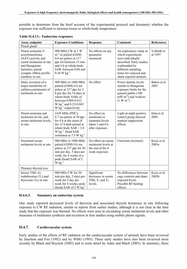
Exposure to high frequency electromagnetic fields, biological effects and health consequences (100 kHz-300 GHz)
199
possible to determine from the brief account of the experimental protocol and dosimetry whether the exposure was sufficient to increase tissue or whole-body temperature.
Table II.4.13.: Endocrine responses
Assay endpoint Exposure Conditions Response Comment References Pineal gland Pineal seratonin N-acetyltransferase (NAT) activity and serum melatonin in rats and Djungarian hamsters; pineal synaptic ribbon profile numbers in rats
900 MHz CW at 1 W m-2 or pulsed (GSM) 0.6 ms pulses at 217 pps for between 15 min and 6 h at whole-body SAR: between 0.04 and 0.36 W kg-1.
No effects on any parameter measured
An exploratory study in which experiments were individually described. Early studies confounded by different sampling times for exposed and sham exposed animals.
Vollrath et al 1997
Daily excretion of a major metabolite (6-sulfatoxymelatonin) of melatonin in rats
900 MHz or 1800 MHz pulsed (GSM) 0.6 ms pulses at 217 pps for 2 h per day for 14 days at whole-body SARs of between 0.009-0.012 W kg-1 and 0.22-0.045 W kg-1 respectively
No effect Power density levels similar to Hungarian exposure limits for the general public (100 mW m-2) and workers (1 W m-2)
Bakos et al 2003
Pineal seratonin and melatonin levels, and serum melatonin levels, in rats.
1439 MHz (PDC); 6.7 ms pulses at 50 pps for 4 h at the onset of the 12 h dark period at whole body SAR ~ 2.0 W kg-1. Head SAR estimated as 7.5 W kg-1
No effect on melatonin or seratonin levels taken 3 and 6 h after exposure.
Light-at-night positive control group showed marked suppression effects.
Hata et al 2005
Nocturnal serum melatonin levels in rats
900 MHz or 1800 MHz pulsed (GSM) 0.6 ms pulses at 217 pps for 30 min per day, 5 days per week, for 4 weeks at a peak (head) SAR of 2 W kg-1
No effect on serum melatonin levels at the end of the 4 week exposure.
Uncertain dosimetry Koyu et al 2005a
Pituitary-thyroid axis Serum TSH, tri-iodothronine (T3) and thyroxine (T4) in rats
900 MHz CW for 30 min per day, 5 days per week for 5 day per week for 4 weeks; peak (head) SAR of 2 W kg-1
Significant decreases in serum TSH, T3 and T4 levels.
No differences between cage controls and sham exposed levels. Possible RF heating effects.
Koyu et al 2005b
II.4.6.3. Summary on endocrine system
One study reported decreased levels of thyroxin and associated thyroid hormones in rats following exposure to CW RF radiation, similar to reports from earlier studies, although it is not clear in the later study that the exposure was thermal. No effects were seen in circulating serum melatonin levels and other measures of melatonin synthesis and excretion in four studies using mobile phone signals.
II.4.7. Cardiovascular system
Early studies of the effects of RF radiation on the cardiovascular system of animals have been reviewed by Jauchem and Frei (1992) and by WHO (1993). These early studies have also been reviewed more recently by Black and Heynick (2003) and in some detail by Adair and Black (2003). In summary, these

Exposure to high frequency electromagnetic fields, biological effects and health consequences (100 kHz-300 GHz)
200
reviews concluded that cardiovascular system responses to RF exposure, such as changes in heart rate and arterial blood pressure, are consistent with those associated with thermoregulatory responses to conventional heating. In general, an increase in body temperature elicits several cardiovascular changes, including increased blood flow to the skin, increasing skin thermal conductance, and increased cardiac output, primarily due to an increase in heart rate, in order to maintain arterial pressure within the normal range. For example, vasodilation of the superficial blood vessels of the skin in primates occurs above a threshold whole-body SAR of about 1 W kg-1 when the RF heating is largely superficial (Adair and Adams 1980). Similar responses occur during exposure of primates to ‘resonant’ frequencies which result in more uniform, less superficial heating (Lotz and Saxton 1987, 1988) but are associated with larger rises in rectal temperature because the less effective stimulation of skin temperature receptors results in reduced thermoregulatory performance. Heart rate was increased in rabbits exposed to 2.45 GHz at whole-body SARs sufficient to raise body temperatures by 0.5oC (Chou et al 1980).
Following the reviews of Jauchem and Frei (1992) and WHO (1993) in the early 90’s, most subsequent studies were of thermoregulatory responses of volunteers to RF exposure (see Chapter II.5.) rather than animals. However, one group carried out a study of the effects of RF exposure during fever, which is generally assumed to increase susceptibility to exogenous sources of heat such as RF radiation. In addition, a series of studies was carried out by another group on health effects primarily associated with military applications of RF, including the responses of anesthetized rats to severe RF heating, and the responses of conscious rats to high peak power RF pulses, or to pulsed ultra-wideband RF radiation.
II.4.7.1. Thermoregulatory changes
Adair et al (1997) investigated the effect of exposure to 450 MHz or 2.45 GHz radiation on thermoregulatory responses during experimentally-induced fever in the conscious squirrel monkey, a non-human primate. The authors found that during RF exposure, the magnitude of the fever remained the same but the absorption of RF energy had proportionately reduced the fever-generated increase in endogenous heat production. However, during exposure at 450 MHz, a resonant frequency in the squirrel monkey, energy is deposited deep within the body and the fever was augmented. In addition, the fever was exacerbated when exposure occurs during the period that the fever abates and body temperature begins to fall.
Jauchem and colleagues investigated the cardiovascular and respiratory responses of rats anesthetized with Ketamine to intense RF radiation. Ketamine is reported to have minimal effects on temperature regulation or on the cardiovascular and respiratory system, but the animals would have been unable to thermoregulate behaviorally. Unusually, the experiments were often continued until the animals died. Jauchem and Frei (1997) investigated the effects of exposure to a sub-resonant RF radiation (350 MHz) at a whole-body SAR of about 13 W kg-1 on the cardiovascular and respiratory responses, namely heart rate, mean arterial blood pressure, respiratory rate and colonic, tympanic and sub-cutaneous temperatures of anesthetized rats. The authors observed that heart rate increased with rising body temperature; mean arterial pressure and respiratory rate were largely unaffected until body temperatures rose above around 42oC, whereupon they declined.
Jauchem et al (2000) investigated the effects of exposure to 1 GHz, 10 GHz, or combined 1 and 10 GHz RF at whole-body SARs of 12 W kg-1 on heart rate, mean arterial blood pressure, respiratory rate and colonic, tympanic and sub-cutaneous temperatures. Colonic temperature was highest in the 1 GHz exposure group, indicating a more uniform heating, whereas subcutaneous temperature on the side facing the antenna was highest in the 10 GHz exposure group, reflecting more superficial heat deposition and greater temperature gradients resulting from exposure to a higher frequency. With regard to the physiological parameters measured, the authors found that the overall pattern of responses was generally similar in all three groups. Heart rate and temperature increased linearly with exposure duration, mean arterial blood pressure increased slightly and then declined, respiration rate initially increased or remained relatively constant and then declined.
These authors also conducted a series of studies of the cardiovascular and respiratory responses of anesthetized rats exposed to 35 GHz RF heating until the death of the animal. Ryan et al (1997a) examined the effect of age and food restriction on the ability of the animal to withstand severe thermal

Exposure to high frequency electromagnetic fields, biological effects and health consequences (100 kHz-300 GHz)
201
challenge. In addition, the effect of various pharmacological manipulations, notably nitric oxide administration (Ryan et al 1997b) and histamine receptor blockade (Jauchem et al 2004), on the ability of the animal to withstand the severe thermal challenge induced by 35 GHz heating was studied. These studies of intense heating effects are reviewed by Jauchem (2006) but are of little direct relevance to occupational or public exposures.
II.4.7.2. High peak power pulses
High peak power RF pulses of relatively short pulse widths are a relatively recent technological development, initially intended for military use but now finding a wider range of application, e.g., in radar. Peak power densities may be of the order of 10’s – 100’s MW m-2, but the short pulse widths (ns–μs) and the low pulse repetition rates result in low average SARs. However, this type of exposure raises a question about the relative biological effectiveness of high peak power SARs compared to low overall average values.
Jauchem and Frei (1995) exposed or sham exposed rats to ten pulses of high peak power density 1.2-1.7 GHz RF. The pulse widths were between 40-70 ns, given at a rate of 1 pulse per second, and the peak power density in each pulse ranged up to a maximum of 520 MW m-2; SARs, however, were not given. The authors reported an initial but transient increase in mean arterial blood pressure and a transient but non-significant decrease in heart rate. These responses disappeared when the acoustic noise associated with the production of each RF pulse was attenuated by Eccosorb® sound attenuator.
II.4.7.3. Ultra-wideband pulses
Ultra-wideband (UWB) RF radiation is a new modality in radar technology that has also been developed initially for military use but which now finds a wide range of application in imaging, sensing and communication systems (ICNIRP 2008). It comprises a RF signal with an ultrashort pulse width (1–10 ns) and a very fast rise-time (10’s–100’s ps). The spectral power of each pulse is very broad, ranging, for example, from 10’s kHz to 10’s GHz. The peak electric field can be in excess of the breakdown voltage of air without arcing, and results in a very high energy absorption per pulse, but with a very low average SAR because of the very low pulse repetition rate. The ratio of peak to average SAR is therefore very much higher than has been addressed hitherto and, as with high peak power pulses, raises a question about biological effectiveness.
Jauchem et al (1998) reported a lack of effect on heart rate and mean arterial blood pressure in anesthetized rats exposed to pulsed UWB RF for 2 min. The rats were exposed to 50, 500 or 1000 pulses per second; the average pulse width was ~ 1 ns, the rise-time was 174-218 ps, and the peak E-field was 87-104 kV m-1. Jauchem et al (1999) reported a lack of effect on heart rate and mean arterial blood pressure in anesthetized rats exposed to pulsed UWB RF radiation for up to 5 min. The pulses had a pulse width of 6 ns and an average rise time of ~ 330 ps, a pulse repetition frequency of 2 kHz and a peak electric field of 19-21 kV m-1; power density and SAR were not given.
Lu et al (1999) exposed or sham exposed conscious rats in a GTEM cell for 6 min to pulsed UWB RF radiation at a whole-body SAR of 0.07 W kg-1, or to pulsed UWB RF radiation at a whole body SAR of 0.121 W kg-1 and reported that systolic and mean arterial blood pressure, and by implication diastolic blood pressure, were significantly decreased during the monitoring period, from 45 min to up to 4 weeks after treatment. In contrast, no effect was seen on heart rate. The exposure was below thermal levels and the specific energy (SA) per pulse was ~0.12 mJ kg-1, about an order of magnitude below the threshold for the ‘microwave hearing’ effect. The authors were unable to account for the UWB radiation-induced hypotension but noted that it was a robust and persistent effect.
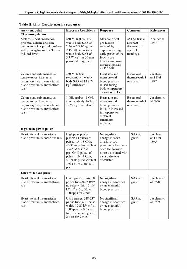
Exposure to high frequency electromagnetic fields, biological effects and health consequences (100 kHz-300 GHz)
202
Table II.4.14.: Cardiovascular responses
Assay endpoint Exposure Conditions Response Comment References Thermoregulation Metabolic heat production, preoptic, colonic and skin temperature in squirrel monkeys with prostaglandin E1 (PGE1)-induced fever
450 MHz (CW) at a whole-body SAR of 2.06 or 3.3 W kg-1 or 2.45 GHz (CW) at a whole-body SAR of 3.3 W kg-1 for 30 min periods during fever
Metabolic heat production reduced by exposure during early period of the fever; core temperature rose during exposure to 450 MHz.
450 MHz is a resonant frequency in squirrel monkeys.
Adair et al 1997
Colonic and sub-cutaneous temperatures, heart rate, respiratory rate, mean arterial blood pressure in anesthetized rats
350 MHz (sub-resonant) at a whole-body SAR of 13.2 W kg-1 until death
Heart rate and mean arterial blood pressure raised during body temperature elevation by 1oC.
Behavioral thermoregulation absent.
Jauchem and Frei 1997
Colonic and sub-cutaneous temperatures, heart rate, respiratory rate, mean arterial blood pressure in anesthetized rats
1 GHz and/or 10 GHz at whole-body SARs of 12 W kg-1 until death.
Heart rate and mean arterial blood pressure initially increased in response to different irradiation regimes.
Behavioral thermoregulation absent.
Jauchem et al 2000
High peak power pulses
Heart rate and mean arterial blood pressure in conscious rats
High peak power pulses: 10 pulses of pulsed 1.7-1.8 GHz; 40-85 ns pulse width at 33-65 MW m-2 at 1 pps. Or 10 pulses of pulsed 1.2-1.4 GHz; 40-70 ns pulse width at 146-561 MW m-2 at 1 pps.
No significant change in mean arterial blood pressure or heart rate once the acoustic noise associated with each pulse was attenuated.
SAR not given
Jauchem and Frei 1995
Ultra-wideband pulses
Heart rate and mean arterial blood pressure in anesthetized rats
UWB pulses: 174-218 ps rise time, 0.97-0.99 ns pulse width, 87-104 kV m-1 at 50, 500 or 1000 pps for 2 min.
No significant change in heart rate or mean arterial blood pressure.
SAR not given
Jauchem et al 1998
Heart rate and mean arterial blood pressure in anesthetized rats
UWB pulses: 318-337 ps rise time, 6 ns pulse width, 19-21 kV m-1 at 1000 pps for 0.5 s or for 2 s alternating with 2 s off for 2 min.
No significant change in heart rate or mean arterial blood pressure.
SAR not given
Jauchem et al 1999

Exposure to high frequency electromagnetic fields, biological effects and health consequences (100 kHz-300 GHz)
203
Assay endpoint Exposure Conditions Response Comment References Heart rate, systolic and diastolic blood pressure, mean arterial blood pressure in conscious rats at 45 min, 24 h, 72 h, and 1, 2, 3 and 4 weeks after exposure.
UWB pulses: 180 ps rise time, 1.00 ns pulse width, 93 kV m-1, at 500 pps; whole-body SAR of 70 mW kg-1. Or 200 ps rise time, 1.03 ns pulse width, 85 kV m-1, at 1000 pps; whole-body SAR of 121 mW kg-1; for 6 min
No effect on heart rate, but significant delayed decrease in diastolic, systolic and mean arterial blood pressure
Lu et al 1999
II.4.7.4. Summary on cardiovascular system
Cardiovascular system responses to RF radiation, such as changes in heart rate and arterial blood pressure, are consistent with those associated with thermoregulatory responses to conventional heating. In general, an RF induced increase in body temperature elicits several cardiovascular changes, including increased blood flow to the skin, increasing skin thermal conductance, and increased cardiac output, primarily due to an increase in heart rate, in order to maintain arterial pressure within the normal range. Exposure to RF radiation during a fever may increase body temperature above that due to the fever itself if the RF energy is deposited deep within the body, or if the exposure takes places as the fever abates and body temperature begins to fall.
Acute exposure to high peak power pulsed RF or to UWB RF radiation in which the energy per pulse is below the threshold for RF auditory effects does not appear to elicit any changes in the cardiovascular system of anesthetized rats, but one study reported persistent delayed hypotension in conscious rats following brief UWB exposure.
II.4.8. Immunology and hematology
Immune responses serve to protect individuals from infectious disease caused by invading micro-organisms such as viruses, bacteria, and various single-celled or multicellular organisms. They can be grouped into acquired or antigen-specific responses and natural or innate responses, which tend to be less specific. The cells that mediate the acquired responses include B-lymphocytes, which secrete antibodies (humoral immunity) which circulate in body fluids, and the T-lymphocytes, which can function as cytotoxic cells (cell-mediated immunity) or as helper T-cells which assist in B- or T-cell activation. The acquired immune responses also involve the recruitment and amplification of the responses of other, less specific parts of the immune system. These include natural killer cells (large granular lymphocytes), mononuclear phagocytes (monocytes and macrophages), granulocytes (neutrophils, eosinophils, and basophils) and the protein complement system, the latter mediating many of the cytolytic and inflammatory effects of humoral immunity. It is generally accepted that the immune system has considerable redundancy in its various components and regulatory mechanisms such that transient and subtle changes in a few components are unlikely to be of much health significance.
Hematology describes the growth and behavior of the cell populations of the blood. Thus, it encompasses the growth and development of the cell populations of the immune system in addition to the erythrocyte populations. The interpretation of changes to cell population estimates can however be complicated by the migration of some cell groups to different body compartments, such as the lymph system, in response to some physiological changes to the body.
A number of studies of RF effects on immune system responsiveness and on the hematological system were carried out mainly in the 1970s and 1980s. They have been reviewed by WHO (1993) and later by Black and Heynick (2003) and are briefly summarized here along with few more recent studies.
Exposure to high frequency electromagnetic fields, biological effects and health consequences (100 kHz-300 GHz)
204
II.4.8.1. Immune system
Studies of immune responses, summarized by WHO (1993), were mostly conducted using 2.45 GHz continuous wave RF. In general, the changes that have been reported with any consistency were usually transient and resulted from acute, thermally-significant exposures. For example, changes in natural killer cell and macrophage activity were reported by several studies after the acute exposure to 2.45 GHz of hamsters at SARs of about 13 W kg-1 or of mice at whole-body SARs of around 21 W kg-1 (Smialowicz et al 1983; Rama Rao et al 1983; Yang et al 1983). An increase in the primary antibody response of B-lymphocytes has been associated with the exposure of mice to 3.0 GHz at whole-body SARs above 4-5 W kg-1 and hamsters to 2.45 GHz at SARs of 8 W kg-1 and above (Shao and Chiang 1989; Rama Rao et al 1985). In primates, an enhanced mitogen response was reported in lymphocytes from rhesus monkeys following exposure to 10.5, 19.27 or 26.6 MHz RF radiation between 0.4 and 2 W kg-1 (Prince et al 1972). The effects in these studies were associated with transiently increased rectal temperatures (WHO 1993).
With regard to effects on the developing immune system, two studies conducted prior to 1993 by the same group of the pre-natal and postnatal exposure of rats to 2.45 GHz at whole-body SARs of 1-5 W kg-
1 (Smialowicz et al 1979) or to 425 MHz at 3-7 W kg-1 (Smialowicz et al 1982) also reported an increased lymphocyte responsiveness to mitogen stimulation at thermogenic levels. In contrast, a lifetime exposure study in which rats were exposed to pulsed 2.45 GHz at whole-body SARs of up to 0.4 W kg-1 between 2 and 27 months of age did not reveal any effects on immunological parameters except for a transient change in the responsiveness of B- and T-lymphocytes to specific mitogens after 13 months exposure (Chou et al 1992).
With regard to studies published after 1993, one group have examined the effect of low-level (whole-body SAR estimated as 2-5 mW kg-1) exposure to 8-18 GHz swept frequency RF on the production of the cytokine tumor necrosis factor (TNF) in the peritoneal macrophages and splenic T-lymphocytes of mice. Fesenko et al (1999) exposed male mice over periods ranging from 0.5 h to 7 days and reported that TNF production was significantly enhanced in both cell types in mice exposed for between 5 h to 3 days compared to sham-exposed mice, and that this persisted over 3 days post-exposure. Following a similar experimental protocol, Novoselova et al (1999) confirmed the increased in TNF production in macrophages and T-lymphocytes following a 5-hour exposure, being maximum about 24 h after exposure in macrophages. T-cell proliferation was also enhanced during this period.
Chagnaud and Veyret (1999) reported a lack of effect on the spleen lymphocyte sub-populations of rats exposed to GSM-modulated RF radiation for 2 h per day for 10 consecutive days at whole-body SARs of 75 mW kg-1 or 270 mW kg-1. No effect was seen on the numbers of cells expressing the surface markers CD4+ (helper T-cells), CD8+ (cytotoxic T-cells) or immunoglobulin A (B-cells). In addition, the mitogenic responses of splenic lymphocytes to the mitogen concanavalin-A were unchanged. These authors subsequently investigated the effects of 900 MHz RF exposure at a head SAR of 1.5 or 6 W kg-1 over a 21-day period on the onset, duration and termination of experimental allergic encephalomyelitis in rats (Anane et al 2003b). This is a demyelinating auto-immune disease that is often used as model for multiple sclerosis. No statistically significant effects of RF exposure were found compared to the responses of sham-exposed animals.
More recently, two studies by one group evaluated the effects of RF radiation on mouse peripheral lymphocytes and on B cell peripheral differentiation and antibody response in mice (Gatta et al 2003; Nasta et al 2006). Mice were exposed or sham-exposed to GSM 900 RF at whole body SARs of 1 or 2 W kg-1 for up to 4 weeks. The first study investigated the effects on T and B lymphocyte frequencies, expression of activation markers (CD28; CD69), cytokine (IL2 and IFNγ) production and T and B cell proliferation (Gatta et al 2003). The second study investigated B cell peripheral differentiation in spleen, and antibody (IgM and IgG) production in response to polyclonal or antigen-specific stimuli (Nasta et al 2006). The authors concluded that T and B lymphocytes were not substantially affected by exposure to RF.

Exposure to high frequency electromagnetic fields, biological effects and health consequences (100 kHz-300 GHz)
205
Table II.4.15.: Immune system responses
Assay endpoint Exposure Conditions Response Comment References TNF production in macrophages and T-lymphocytes in mice
Swept 8-18 GHz RF SAR ~ 2-5 mW kg-1 for 0.5 h to 7 days
Increased TNF production following 5 h to 3 day exposure
Blinded procedures Fesenko et al 1999
TNF production in macrophages and T-lymphocytes in mice
Swept 8-18 GHz RF SAR ~ 2-5 mW kg-1 for 5 h.
Increased TNF production and T-lymphocyte proliferation 24 h after exposure
Blinded procedures Novoselova et al 1999
Cell surface markers (CD8+, CD4+ and IaAG+) and mitogenic activity of lymphocytes in rats
900 MHz GSM RF at a whole-body SAR of 75 or 270 mW kg-1 for 2 h per day for 10 days
No effects on cell surface markers or mitogenic activity
Low-level RF Chagnaud and Veyret 1999
Experimental allergic encephalomyelitis (EAE) in rats; eight rats per group
900 MHz GSM RF at a brain SAR of 1.5 or 6 W kg-1 for 2 h per day for 21 days
No statistically significant effect on onset, duration and termination of EAE crisis.
Cage controls exhibited greatest impairment
Anane et al 2003b
Lymphocyte proliferation, cytokine production and expression of activation markers in mice
900 MHz GSM RF at a whole-body SAR of 1 or 2 W kg-1 for 2 h per day for 1, 2 or 4 weeks
No effects on T or B lymphocyte function, except for transient increase seen in IFNγ after 1 week RF exposure.
Blinded procedures Gatta et al 2003
B-cell peripheral lymphocyte differentiation and antibody production in mice
900 MHz GSM RF at a whole-body SAR of 2 W kg-1 for 2 h per day for 4 weeks
No effects on B cell differentiation or on serum antibody levels
Nasta et al 2006
II.4.8.2. Hematology
A large number of studies of effects of RF exposure on hemopoietic tissues and immune function were also carried out prior to 1993 but the results are not always clear; many reports have yielded conflicting data. In addition, some of the older studies suffered from inadequate dosimetry and poor experimental design. As with the immune system, changes that have been reported were usually transient and resulted from acute, thermally-significant exposures (WHO 1993). One response observed by several authors has been the decrease in peripheral lymphocyte count and an increase in the neutrophil count in mice exposed at 26 MHz at whole-body SARs of 5–13 W kg-1 (Liburdy 1979) and rats exposed to pulsed 24 GHz RF at whole-body SARs of 1.5–3 W kg-1 (Deichmann et al 1959, 1964); sufficient under these particular experimental conditions to raise rectal temperatures by about 1°C. In primates, no field-dependent changes in any of 21 hematological parameters were seen following prolonged (11-day) exposure to 28 MHz at a whole-body SAR of 0.06 W kg-1 (Wright et al 1984).
No consistent effect of RF exposure has been seen on peripheral blood cell populations in developing rats (WHO 1993). No consistent changes in erythrocyte, leucocyte or differential leucocyte cell count were found in rats exposed prenatally and postnatally (for up to 41 days) to 2.45 GHz RF at 1–5 W kg-1, at 100 MHz at 2–3 W kg-1 or to 425 MHz RF at 3–7 W kg-1 (Smialowicz et al 1979, 1981, 1982). In addition, a lifetime exposure study in which rats were exposed to pulsed 2.45 GHz RF at whole-body SARs of up to 0.4 W kg-1 between 2 and 27 months of age did not reveal any effects on hematological parameters except for a transient change in the responsiveness of B- and T-lymphocytes to specific mitogens after 13 months exposure (Chou et al 1992).
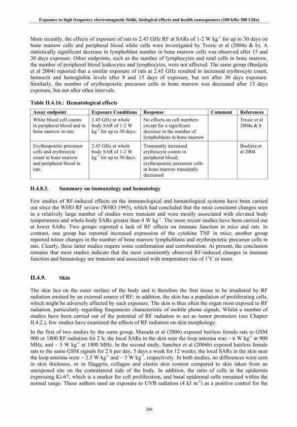
Exposure to high frequency electromagnetic fields, biological effects and health consequences (100 kHz-300 GHz)
206
More recently, the effects of exposure of rats to 2.45 GHz RF at SARs of 1-2 W kg-1 for up to 30 days on bone marrow cells and peripheral blood white cells were investigated by Trosic et al (2004a & b). A statistically significant decrease in lymphoblast number in bone marrow cells was observed after 15 and 30 days exposure. Other endpoints, such as the number of lymphocytes and total cells in bone marrow, the number of peripheral blood leukocytes and lymphocytes, were not affected. The same group (Busljeta et al 2004) reported that a similar exposure of rats at 2.45 GHz resulted in increased erythrocyte count, hemocrit and hemoglobin levels after 8 and 15 days of exposure, but not after 30 days exposure. Similarly, the number of erythropoietic precursor cells in bone marrow was decreased after 15 days exposure, but not after other intervals.
Table II.4.16.: Hematological effects
Assay endpoint Exposure Conditions Response Comment References White blood cell counts in peripheral blood and in bone marrow in rats.
2.45 GHz at whole body SAR of 1-2 W kg-1 for up to 30 days
No effects on cell numbers except for a significant decrease in the number of lymphoblasts in bone marrow
Trosic et al 2004a & b
Erythropoietic precursor cells and erythrocyte count in bone marrow and peripheral blood in rats
2.45 GHz at whole body SAR of 1-2 W kg-1 for up to 30 days
Transiently increased erythrocyte counts in peripheral blood; erythropoietic precursor cells in bone marrow transiently decreased
Busljeta et al 2004
II.4.8.3. Summary on immunology and hematology
Few studies of RF-induced effects on the immunological and hematological systems have been carried out since the WHO RF review (WHO 1993), which had concluded that the most consistent changes seen in a relatively large number of studies were transient and were mostly associated with elevated body temperatures and whole-body SARs greater than 4 W kg-1. The more recent studies have been carried out at lower SARs. Two groups reported a lack of RF effects on immune function in mice and rats. In contrast, one group has reported increased expression of the cytokine TNF in mice; another group reported minor changes in the number of bone marrow lymphoblasts and erythropoietic precursor cells in rats. Clearly, these latter studies require some confirmation and corroboration. At present, the conclusion remains that most studies indicate that the most consistently observed RF-induced changes in immune function and hematology are transient and associated with temperature rise of 1oC or more.
II.4.9. Skin
The skin lies on the outer surface of the body and is therefore the first tissue to be irradiated by RF radiation emitted by an external source of RF; in addition, the skin has a population of proliferating cells, which might be adversely affected by such exposure. The skin is thus often the organ most exposed to RF radiation, particularly regarding frequencies characteristic of mobile phone signals. Whilst a number of studies have been carried out of the potential of RF radiation to act as tumor promoters (see Chapter II.4.2.), few studies have examined the effects of RF radiation on skin morphology.
In the first of two studies by the same group, Masuda et al (2006) exposed hairless female rats to GSM 900 or 1800 RF radiation for 2 h; the local SARs in the skin near the loop antenna was ~ 6 W kg-1 at 900 MHz, and ~ 5 W kg-1 at 1800 MHz. In the second study, Sanchez et al (2006b) exposed hairless female rats to the same GSM signals for 2 h per day, 5 days a week for 12 weeks; the local SARs in the skin near the loop antenna were ~ 2.5 W kg-1 and ~ 5 W kg-1, respectively. In both studies, no differences were seen in skin thickness, or in filaggrin, collagen and elastin skin content compared to skin taken from an unexposed site on the contralateral side of the body. In addition, the ratio of cells in the epidermis expressing Ki-67, which is a marker for cell proliferation, and basal epidermal cells remained within the normal range. These authors used an exposure to UVB radiation (4 kJ m-2) as a positive control for the
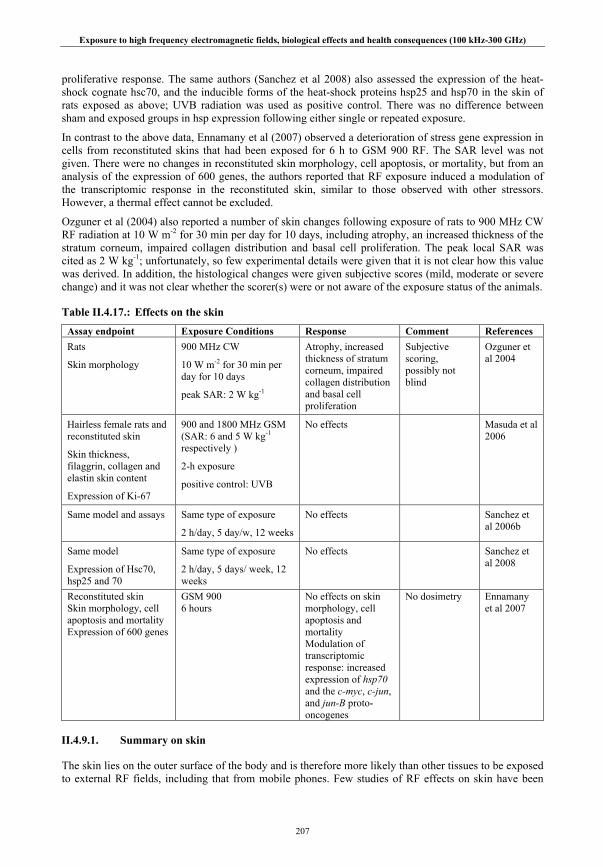
Exposure to high frequency electromagnetic fields, biological effects and health consequences (100 kHz-300 GHz)
207
proliferative response. The same authors (Sanchez et al 2008) also assessed the expression of the heat-shock cognate hsc70, and the inducible forms of the heat-shock proteins hsp25 and hsp70 in the skin of rats exposed as above; UVB radiation was used as positive control. There was no difference between sham and exposed groups in hsp expression following either single or repeated exposure.
In contrast to the above data, Ennamany et al (2007) observed a deterioration of stress gene expression in cells from reconstituted skins that had been exposed for 6 h to GSM 900 RF. The SAR level was not given. There were no changes in reconstituted skin morphology, cell apoptosis, or mortality, but from an analysis of the expression of 600 genes, the authors reported that RF exposure induced a modulation of the transcriptomic response in the reconstituted skin, similar to those observed with other stressors. However, a thermal effect cannot be excluded.
Ozguner et al (2004) also reported a number of skin changes following exposure of rats to 900 MHz CW RF radiation at 10 W m-2 for 30 min per day for 10 days, including atrophy, an increased thickness of the stratum corneum, impaired collagen distribution and basal cell proliferation. The peak local SAR was cited as 2 W kg-1; unfortunately, so few experimental details were given that it is not clear how this value was derived. In addition, the histological changes were given subjective scores (mild, moderate or severe change) and it was not clear whether the scorer(s) were or not aware of the exposure status of the animals.
Table II.4.17.: Effects on the skin
Assay endpoint Exposure Conditions Response Comment References Rats
Skin morphology
900 MHz CW
10 W m-2 for 30 min per day for 10 days
peak SAR: 2 W kg-1
Atrophy, increased thickness of stratum corneum, impaired collagen distribution and basal cell proliferation
Subjective scoring, possibly not blind
Ozguner et al 2004
Hairless female rats and reconstituted skin
Skin thickness, filaggrin, collagen and elastin skin content
Expression of Ki-67
900 and 1800 MHz GSM (SAR: 6 and 5 W kg-1 respectively )
2-h exposure
positive control: UVB
No effects Masuda et al 2006
Same model and assays Same type of exposure
2 h/day, 5 day/w, 12 weeks
No effects Sanchez et al 2006b
Same model
Expression of Hsc70, hsp25 and 70
Same type of exposure
2 h/day, 5 days/ week, 12 weeks
No effects Sanchez et al 2008
Reconstituted skin Skin morphology, cell apoptosis and mortality Expression of 600 genes
GSM 900 6 hours
No effects on skin morphology, cell apoptosis and mortality Modulation of transcriptomic response: increased expression of hsp70 and the c-myc, c-jun, and jun-B proto-oncogenes
No dosimetry Ennamany et al 2007
II.4.9.1. Summary on skin
The skin lies on the outer surface of the body and is therefore more likely than other tissues to be exposed to external RF fields, including that from mobile phones. Few studies of RF effects on skin have been

Exposure to high frequency electromagnetic fields, biological effects and health consequences (100 kHz-300 GHz)
208
carried out. One group has reported an absence of effects of GSM-type mobile phone radiation on a number of different parameters such as skin thickness, elastin and collagen content, proliferative response and the induction of heat-shock proteins. In contrast, two groups reported positive effects, although in both cases there were a number of methodological shortcomings.
II.4.10. Eye
Studies have been carried out on the effects of exposure to RF radiation on the lens of the eye and other tissues including the retina. Many of the early studies carried out in the 1960s and 1970s used rabbits, while later studies tended to use primates because of the greater similarity of their facial and ocular structures to those of humans. These studies have been reviewed by WHO (1993) and more recently by Elder (2003a) and are briefly summarized below, along with a discussion of the evidence from more recent papers.
II.4.10.1. Cataracts
The lens is considered potentially sensitive to RF exposure because it lacks a blood supply and so has a limited ability to dissipate heat. In addition, the fibers which make up the bulk of the lens have only a limited capacity for repair and tend to accumulate the effects of minor insults. Cellular debris resulting from any cytotoxic insult to the lens tends either to be carried to the posterior sub capsular region due to the mechanical forces of epithelial cell proliferation and fiber formation or is trapped in situ in the lens matrix.
Briefly, as noted by WHO (1993), cataract is a well-established thermal effect of RF exposure in anesthetized rabbits (e.g., Kramar et al 1975; Guy et al 1975b; Hagan and Carpenter 1976; Kramar et al 1978; Carpenter 1979). High lens temperatures induced by exposure of the head to RF have been shown to induce cataracts in the lenses of anesthetized rabbits (Guy et al 1975b; Kramar et al 1978); threshold temperatures for prolonged (100-200 min) exposure lie between 41-43oC; corresponding local SARs are in the range 100-140 W kg-1. These high local SARs and temperatures resulted from protracted (>140 min) localized exposure of the eye at 1- 10 GHz at power densities greater than 1.5 kW m-2; whole-body exposure at such levels however is limited by thermal stress (Elder 2003a). The few experiments which have investigated the effect of chronic whole-body exposure of conscious rabbits at lower power densities (up to 100 W m-2) reported a lack of effect on the lens. Cataracts were not observed in rabbits after 2.45 GHz RF radiation at 100 W m-2 (whole-body SAR of 1.5 W kg-1) for up to 17 weeks (Ferri and Hagan 1976). Nor was any change found by in the eyes of rabbits exposed for ~ 6 months at 2.45 GHz where the maximal SAR in the head was 17 W kg-1 (Guy et al 1980). Chou et al (1982, 1983) also reported that low-level pulsed or CW 2.45 GHz RF exposures for 3 months at SARs of 0.55 and 5.5 W kg-1 in the head did not cause cataracts.
These early studies also found primates to be less susceptible to cataract induction than rabbits (WHO 1993). Opacities were induced in the eyes of anesthetized rhesus monkeys after acute localized exposures of up to 5 kW m-2, well above threshold levels for anesthetized rabbits (McAfee et al 1979; 1983). In addition, McAfee and colleagues exposed conscious monkeys to 2.45 GHz CW for up to 12 h over a 4 month period or to pulsed 9.3 GHz RF radiation (pulsed or CW) for up to 15 h over a 34 month period at SARs in the head of up to 40 W kg-1. Eye examinations carried out 1-4 years after exposure revealed no effects on the lens, cornea or retina. The lower susceptibility of primates to cataract induction is thought to result from structural differences in the eyes and skull of the two species resulting in lower power absorption and heating of the thinner primate lens.
More recently, Saito et al (1998) exposed the eyes of conscious rabbits for ~ 2.5 – 4 h at 2.45 GHz at an SAR to the head/eye of 26.5 W kg-1, with the contralateral eye serving as a control, and reported transient conjunctival and corneal edema, contraction of the pupil and pupilliary congestion, and fibrinogenesis in the anterior chamber of the lens of the exposed eyes. In contrast to studies with anesthetized rabbits, using higher local SARs, the authors did not observe cataracts. Studies with both conscious and anesthetized rabbits have been carried out. Kojima et al (2004) assessed the effects of localized exposure of rabbit eyes for 20-60 min to 2.45 GHz RF at a local SAR to the eye of 108 W kg-1; the RF-induced changes, which

Exposure to high frequency electromagnetic fields, biological effects and health consequences (100 kHz-300 GHz)
209
disappeared within a week, included corneal edema, inflammation of the iris and increased light-scattering from the anterior cortex of the lens. These effects were much more marked in the anaesthetized rabbits than in those not anesthetized; reflecting the greater temperatures (of up to 9oC) measured in the posterior (vitreous) chamber and to a lesser extent in the anterior (aqueous) chamber of the eyes of the anesthetized rabbits. Increased heating of the posterior region of the lens, particularly in anesthetized rabbits due to reductions in blood flow, was confirmed in dosimetric and thermal modeling studies by Hirata et al (2006).
Balci et al (2007) placed 900 MHz GSM phones over cages each housing 10 rats. The phones, on standby, were called intermittently (4 times a day for 10 min) over a 4 week period. There was no RF dosimetry. The authors reported a number of effects. Unfortunately, the absence of proper dosimetry and the poor description of the experimental protocol render the results uninterpretable.
Table II.4.18.: Effects on the eye
Assay endpoint Exposure Conditions Response Comment References Cataract formation and other ocular effects in anesthetized rabbits.
2450 MHz (CW) at 150 mW/cm2 for 100 min at the maximum SAR 138 W kg-1 in the eye.
Induce cataract and transient effects (papillary constriction and anterior chamber turbidity).
Guy et al 1975b
Cataract formation in anesthetized rabbits.
2450 MHz CW at 1.8 kW m-2 for 140 min at maximum SAR in the eye 100 W kg-1.
Cataract. This study was performed to determine the cataractogenic threshold.
Kramar et al 1978
Ocular effects in conscious monkeys.
9310 MHz (PW) at 1.5 kW m-2 for 30-40 days with 294-665 min totally.
No ocular effects such as cataract after one year observation.
McAfee et al 1979
Ocular effects such as cataract, visual capability loss in conscious monkeys.
9310 MHz (PW) at 1.5 kW m-2 for 408-946 min over 34 months; 9310 MHz (PW) at 3 kW m-2
for 275-594 min over 34 months; 2450 MHz (PW) at 1.5 kW m-2 for 549-750 min over 4 months.
No cataracts; no effects on cornea, aqueous and vitreous humors or retina; and no loss of visual capability 4 years after 9310 MHz exposure and 1 year after 2450 MHz exposure.
These results support that clinically significant ocular effects have not been confirmed in human populations exposed for long period of time to low level RF radiation.
McAfee et al 1983
The effect of CW irradiation on conscious rabbit eye including cornea, anterior chamber, lens, and other ocular tissues.
2450 MHz (CW) on the eyes at SAR 26.5 W kg-1 for 160 to 240 min.
No cataracts. Other effects (miosis, keratoleucoma and corneal edema, endothelial cell detachment and floating in aqua oculi, and so on)
The miosis and papillary congestion in all irradiated eyes was first to be detected.
Saito et al 1998
Effects of acute RF exposure on ocular tissue in conscious and anesthetized rabbits
2.45 GHz at localized SAR of 108 W kg-1 for 20-20 min.
Corneal edema, inflammation of the iris and increased light-scattering from anterior lens cortex.
Changes more marked in anesthetized animals.
Kojima et al 2004
Oxidative stress in lens tissues in rats.
900 MHz GSM mobile phone signal for 4 weeks; the phone was placed above the cage.
Malondialdehyde level significantly increased in lens and corneal tissue in the mobile phone group compared to controls.
The absence of any RF measurements of any kind renders the experiment uninterpretable.
Balci et al 2007
Exposure to high frequency electromagnetic fields, biological effects and health consequences (100 kHz-300 GHz)
210
II.4.10.2. Other ocular tissues
Degenerative changes have been reported in various eye tissues of primates after exposure to pulsed microwaves. A series of studies (summarized by Kues & Monahan 1992) have indicated that localized exposure of the eyes of anaesthetized monkeys to pulsed 2.45 GHz RF (10 μs pulses at 100 pps) at an SAR in the eye of 2.6 W kg-1 or more for four hours resulted in transient lesions in the corneal endothelium (Kues et al 1985). These were maximal 16-24 h post-exposed and persisted for several days. Such lesions in the cornea were also induced by exposure to CW 2.45 GHz, but less effectively compared to pulsed radiation. Topical pretreatment with the ophthalmic drug timolol maleate (used in the treatment of glaucoma) appeared to reduce the threshold to a localized SAR of 0.26 W kg-1 (Kues et al 1992). In addition, the authors reported a transient increase in the vascular permeability of the iris (blood-aqueous barrier) following similar treatment. In studies by the same group but using conscious monkeys, transient reductions in electroretinogram activity in response to light stimulation have been reported following repeated exposures to pulsed (0.5 μs pulses, 16 pps) 1.25 GHz microwave radiation at a localized SAR of 4.0 W kg-1 (Kues & Monahan 1992). Histopathalogical investigation three weeks after exposure revealed photoreceptor degeneration, which, the authors argue, is consistent with the observed decrements in electroretinogramme activity.
In contrast to these studies, Kamimura et al (1994) reported that they were unable to induce corneal, lenticular or retinal lesions in the eyes of conscious monkeys exposed to CW (but not pulsed) 2.45 GHz radiation at levels exceeding the threshold for CW-induced corneal damage described by Kues et al (1985). The technique used for the identification of corneal lesions (specular microscopy) was the same as that used by Kues et al (1985); although the latter authors used histological techniques to confirm damage to both the cornea and retina, in contrast to Kamimura et al (1994). However, Kamimura and colleagues note that the use of anesthesia by Kues et al may have compromised heat dissipation in the eye (see above) increasing susceptibility to RF heating.
Further studies using unanesthetized monkeys by Lu et al (2000) were unable to confirm these earlier observations of Kues and colleagues. Lu et al (2000) exposed or sham-exposed monkeys to pulsed 1.25 GHz over a 3 week period at localized SARs averaged over the retina of 4.3, 8.4 or 20.2 W kg-1. RF-induced changes in the retina were examined using various measures of retinal integrity including fundus photography, fluorescein and indocyanine green angiography, and electroretinography both before and after exposure, and complete retinal histopathology following termination of the experiment. No significant changes were seen in the exposed eyes compared to those pre- or sham-exposed either in the appearance of the fundus or in the angiography examinations. The electroretinogramme response of cone photoreceptors to light flash was enhanced in monkeys exposed at retinal SARs of 8.4 or 20.2 W kg-1, but not in those exposed at 4.3 W kg-1. The authors suggest that this effect is likely to represent a transient physiological change. Histopathologic examination did not reveal any pathological changes. However, an increase in glycogen storage was seen in the photoreceptors in eyes exposed at 8.4 and 20.2 W kg-1 and, confusingly, also in sham-exposed animals but not those exposed at 4.3 W kg-1.
Table II.4.19.: Effects on other ocular tissues
Assay endpoint Exposure Conditions Response Comment References Corneal endothelium of anesthetized monkeys
CW 2.45 GHz at localized SAR of 5.3-7.8 W kg-`1 (200-300 W m-2) or pulsed 2.45 GHz (10 µs at 100 pps) at localized SARs of 2.6 W kg-1 for 4 h per day, once or repeated over 4 days.
RF-induced transient lesions in the corneal endothelium. Pulsed RF more effective than CW.
Animals used for repeat experiments, separated by 1 week or more.
Kues et al 1985
Exposure to high frequency electromagnetic fields, biological effects and health consequences (100 kHz-300 GHz)
211
Assay endpoint Exposure Conditions Response Comment References Corneal endothelium and vasculature of the iris in anesthetized monkeys with or without timolol maleate application
Pulsed 2.45 GHz (10 µs at 100 pps) at localized SARs of up to 4 W kg-
`1 for 4 h per day for 3 consecutive days
Timolol pretreatment reduced the threshold for coneal lesions to 0.26 W kg-1 and increased vascular leakage from the iris
Kues et al 1992
Scotopic test (rod photoreceptor response) and 30-Hz flicker test (cone-receptor response) of electroretinogram (ERG) in conscious monkey
Pulsed 1.25 GHz (0.5 µs at 16 pps) at a localized SAR of 4 W kg-1 for 4 h.
60% reduction in scotopic ERG amplitude and 90% reduction in flicker test ERG correlated with photoreceptor degeneration
Kues and Monohan 1992
Corneal endothelium, lens, vitreous humor and retina in conscious monkeys
CW 2.45 GHz at up to 430 W m-2 for 4 h.
No abnormalities of the corneal epithelium or lens; or of vitreous humor or retina
Attempted corroboration of Kues et al 1985 with CW 2.45 GHz using supra-threshold exposures
Kamimura et al 1994
Ocular tissues of anesthetized rabbits and monkeys
60 GHz at 100 W m-2 for 8 h, or for 4 h on 5 consecutive days. SAR not given
No histopathalogical effects seen in cornea, iris, or lens
Kues et al 1999
Ocular tissues of conscious monkeys exposed to high peak power RF pulses
1.25 GHz pulsed (5.6 µs at up to 2.8 pps); retinal average SARs of up to 20 W kg-1 (peak of 130 MW kg-1 per pulse), for 4 h per day for 9 days over a 3 week period.
No histopathological effects seen; transient functional changes in electroretinograms and increased photo-receptor glycogen storage seen above 4.3 W kg-1.
Increased glycogen storage also seen in photoreceptors of sham-exposed group.
Lu et al 2000
II.4.10.3. Summary on eye
The lens of the eye is potentially sensitive to RF because it lacks a blood supply and so has a limited ability to dissipate heat. RF-induced cataract is a well-established thermal effect of RF exposure in anesthetized rabbits; thresholds for prolonged (100-200 min) exposure lie between about 41-43oC, corresponding to localized SARs in the range 100-140 W kg-1. However, recent studies have confirmed that the anesthesia restricted lenticular cooling through a reduction in local blood flow, thereby exacerbating the effects observed. Primates appear less susceptible to cataract induction than rabbits, and opacities have not been observed following either acute or prolonged exposures.
Studies from one laboratory suggesting that the exposure of the eyes of anesthetized primates to pulsed RF could result in corneal lesions and vascular leakage from the iris were not corroborated by later studies by other groups using conscious primates. Transient changes were seen in the electroretinogramme responses following exposure at high localized SARs, but the functional significance of this, if any, was not clear.
II.4.11. Summary on animal studies
Overall, studies published after 1993 provide a further support for the conclusions of WHO (1993) that the most consistent and reproducible responses of animal to acute RF exposure result from RF-induced heating. These studies established that, in general, an increase in body temperature elicits several

Exposure to high frequency electromagnetic fields, biological effects and health consequences (100 kHz-300 GHz)
212
cardiovascular changes including increased blood flow to the skin, increasing skin thermal conductance, and increased cardiac output, primarily due to an increase in heart rate, in order to maintain arterial pressure within the normal range. Deficits in learned behaviors, particularly the disruption of ongoing operant behaviors, occur mainly when core temperatures are increased by about 1°C or more. Similar rises in body temperature also result in significantly enhanced plasma corticosterone or cortisol levels in rodents and primates and transient changes in immune function and hematology, generally consistent with the acute responses to non-specific stressors.
In addition, RF radiation can cause increased embryo and fetal losses, increased incidence of fetal malformations and anomalies, reduced fetal weight at term and impair male fertility at exposure levels that are sufficiently high to cause a significant increase in temperature. To date, there is no consistent evidence of effects at non-thermal exposure levels. Relatively few studies have evaluated possible effects of prenatal exposure on postnatal development; results from such studies have not shown consistent effects on developmental indices or behavior at exposure levels that do not induce significant temperature elevation. The few studies that have addressed neonatal exposure or exposure of juvenile animals to low level RF have generally reported a lack of effect on such diverse endpoints such as behavior, blood-brain barrier permeability and tumor induction. However, to date, there remains insufficient evidence to form a firm conclusion regarding neonatal or juvenile sensitivity to RF compared to adults.
RF-induced cataract also remains a well-established thermal effect of RF exposure in anesthetized rabbits. However, recent studies have confirmed that the anesthesia-restricted lenticular cooling through a reduction in local blood flow, thereby exacerbating the effects observed. Primates appear less susceptible to cataract induction than rabbits, and opacities have not been observed in primates following either acute or prolonged exposures. Studies from one laboratory suggesting that the exposure of the eyes of anesthetized primates to pulsed RF could result in corneal lesions and vascular leakage from the iris were not corroborated by later studies by other groups using conscious primates.
Overall, the results of recent carcinogenicity studies are rather consistent and indicate that carcinogenic effects on rodents are not likely at SAR levels up to 4 W kg-1 even for long-term exposure. Genotoxicity studies also generally indicate a lack of effect. A notable positive finding was of a two-fold increase in lymphoma incidence in a strain of lymphoma-prone transgenic mice following exposure at 900 MHz with a signal similar to that used in GSM mobile phones. However, this finding was not confirmed in two subsequent replication and extension studies. In addition, studies report an absence of effects of RF radiation characteristic of mobile phone use on melatonin levels.
Studies of the behavioral effects of high peak power RF pulses used in some military applications have been rather sporadic and diverse; pulse widths have varied by two orders of magnitude and the specific absorption per pulse by four orders of magnitude. Two studies have shown the equivalence of pulsed RF to an acoustic ‘click’ in affecting the startle reflex. Otherwise, the effects seen may be attributed either to heating or auditory perception.
II.5. HUMAN STUDIES
Prior to 1993, laboratory studies using volunteer were confined primarily to studies of cutaneous and auditory perception and effects resulting from localized and whole body heating (WHO 1993). Guy et al (1975c), for example, determined a threshold for the auditory perception of pulsed RF as used in radar as 16 mJ kg-1 energy absorption per pulse in the head. With regard to the effects of RF absorption by the whole body, this was addressed largely in the context of thermoregulation. It was known that healthy individuals can sustain an increase in body temperature up to an upper safe limit of 39oC, at which level the heart rate is considerably elevated and the sweat rate is about 1 liter per hour (WHO 1993). In addition, early studies on the exposure of patients and volunteers to RF fields in magnetic resonance imaging systems reported that whole-body SARs of up to 4 W kg-1 for 20-30 minutes resulted in body temperature increases in the range 0.1-0.5oC (eg Kido et al 1987; Shellock and Crues 1987; Shellock et al 1989).
In subsequent years, the rapid increase in wireless telecommunications, particularly those used in mobile telephony, initiated a number of research programs which included volunteer studies of the possible

Exposure to high frequency electromagnetic fields, biological effects and health consequences (100 kHz-300 GHz)
213
physiological effects of the complex but generally low level RF emitted by such devices. In particular, the proximity of mobile phones to the head raised public concern about a potentially toxic effect of electromagnetic radiation on the central nervous system which has prompted a large number of studies to be carried out. These include investigations of mobile-phone type RF radiation on the electrical activity of the brain and regional cerebral blood flow (rCBF), a marker of neural activity in a local brain region, and on various cognitive functions such as memory, attention and concentration. A number of these studies have been reviewed by Cook et al (2006) and Valentini et al (2007), as well as by the major reviews cited in the Introduction. In addition, a variety of subjective symptoms such as headaches, fatigue, etc., have been reported by some users of mobile phones. Finally, some studies have examined possible effects on the endocrine system, particularly in relation to melatonin, and on the cardiovascular system. With regard to the latter, as indicated above, thermoregulatory responses to heat stress and to RF radiation are well understood and are briefly summarized here.
Experiments using volunteers exposed to RF are restricted for ethical reasons to the investigation of transient physiological phenomena which, in the controlled conditions of a laboratory, are at relatively low exposure levels. It is possible, however, that effects judged to be harmless when experienced transiently in the laboratory, may have adverse health consequences if experienced for long periods in an occupational or public context. The advantage of such experiments is that they indicate the likely response of other people exposed under similar conditions, but the disadvantages include the often short duration of investigation and the small number of subjects usually examined. To some extent, shortcoming such heterogeneity in the study population can be addressed through experimental design, in this example by using a crossover experimental design (see below), or retesting of participants to account for possible differences in response. However, due to practical considerations, subjects have tended to be relatively homogeneous and are therefore unlikely to reflect the range of variability encountered within a population. Nevertheless, within this limited context, volunteer studies can give valuable insight into the physiological effects of exposure in normal, healthy people.
Important factors to consider in the evaluation of these studies include the use of double-blind procedures and crossover and counter-balanced protocols. Double-blind procedures apply when both the experimenters and subjects are unaware of the exposure status of the subjects, and so are less likely to be influenced by any expectation of a particular outcome; single-blind procedures, often used in early studies, are where only the subjects are unaware of their exposure status. A crossover design is where subjects are both exposed and sham exposed in different parts of the experiment, so that they act as their own controls (also known as a within-subjects or repeated measures design). This procedure minimizes the effects of intrinsic differences between subject groups, such as might occur between a sham group and an exposed group, which could affect the experimental outcome. A counter-balanced protocol is where all possible orders of exposures are used, with equal numbers of subjects experiencing each order. This counteracts any effect of time-dependency on the subjects’ responses, resulting for example from improving in task performance or from loss of attention during the course of a study.
II.5.1. Nervous system
II.5.1.1. Electrical activity of the brain
The electroencephalogram (EEG) is a reflection of synchronous activity in relatively large populations of cortical neurons. The 'spontaneous' EEG of awake subjects is conventionally divided into a number of frequency bands, the relative amounts of activity in which depends upon the psychological state of the subject and the nature of the cognitive function in which he or she is engaged. The designation of the frequency bands is not always applied very strictly, which results in specific frequencies sometimes being assigned to different bands in different studies. Generally, the following division is used: delta (δ) < 4 Hz; theta (θ) 4-8 Hz; alpha (α) 8-12 Hz; beta (β) 12-30 Hz; gamma (γ) > 30 Hz. Slightly different band designations are used by some authors which are also cited in this report. The functional significance of these different components of the normal, waking EEG is poorly understood. Thus, while a demonstration that mobile phone signals influenced these components would be indicative of a biological effect of such
Exposure to high frequency electromagnetic fields, biological effects and health consequences (100 kHz-300 GHz)
214
signals, interpretation of the effect in terms of health would be uncertain. In addition, intra-individual variability is very high. However, EEG patterns associated with sleep are well characterized and routinely used as indices of the different sleep stages that a typical healthy individual will move between during the night. There would also be little uncertainty in the interpretation of a change from a normal to a frankly pathological pattern of EEG activity, such as might be observed in epilepsy.
A measure of brain function closely related to the EEG is the 'evoked' or 'event-related' potential (ERP). ERPs are obtained by sampling the EEG time-locked to a reference event such as the presentation of a stimulus or the onset of a motor response, and averaging the samples together so as to obtain an electrical waveform that represents brain activity associated specifically with that class of event. ERPs are commonly used to study the timing and functional integrity of neural systems supporting sensory, cognitive and motor processing. Nevertheless, interpretation is still problematic, since changes in arousal and attention of volunteers can substantially affect the outcome of these studies.
Spontaneous EEG
Laboratory studies investigating the effects of mobile phone signals on the spontaneous EEG have produced somewhat mixed results, although more recent stronger studies point to the existence of effects of exposure primarily to the alpha bands of the EEG.
Reiser et al (1995) reported from a single-blind, sham-controlled crossover experiment that a 15-min exposure to a signal from a GSM mobile phone was associated with an increase some 15 minutes later in the power of EEG frequencies in the 18-35 Hz (defined here as the upper beta) band. The effect is only marginal and the statistical analysis of the data is questionable. Röschke and Mann (1997) were unable to detect any differences in EEG spectra related to exposure to GSM signals. These authors exposed 34 male volunteers in a single blind design to the signal of a GSM mobile phone positioned at 40 cm from the vertex. The power density at the location of the head was 0.5 W m-2. Exposure or sham exposure was for 3.5 min midway during a 10-min EEG recording session. No effect on the EEG was detected and no distinction could be made between sensitive and non-sensitive groups. Hietanen et al (2000) recorded resting EEG from 19 volunteers during sham exposure, and exposure to signals from five different mobile handsets (analogue and GSM at 900 and 1800 MHz) operating at full power and positioned over the left side of the head. Conditions were single blind. Statistical analysis of spectral parameters of the EEG revealed an effect in only absolute but not relative power in one frequency band in one of four brain regions investigated, for one of the analog phones. The authors attributed this to chance.
Lebedeva et al (2000) recorded EEG from 24 subjects during sham exposure and exposure to a 900 MHz signal directed at the back of the head. An index representing the ‘dimensional complexity’ of the EEG signals was reported to vary significantly as a function of exposure condition, with a more pronounced difference between exposure and sham under ‘eyes-closed’ conditions than under ‘eyes open’. The authors concluded that their chosen measure of EEG was more sensitive to the effects of RF signals than conventional indices. A definition of their index and a comparison with conventional indices was not provided, however. There is also almost no information about how the data were analyzed statistically, and no information about levels of statistical significance.
Borbély et al (1999) reported that exposure to a 900 MHz ‘pseudo GSM signal’ immediately prior to sleep increased resting EEG power in the 11-11.5 Hz range only. Exposure and sham were double-blind, randomized and given at 1-week interval, and only 14 of the total sample of 24 participants had sufficient data for the analysis. An extended analysis was provided in Huber et al (2003). In a follow-up study, Huber et al (2000) exposed 16 healthy volunteers for 30 min to a GSM 900 signal immediately before sleep. Resting EEG prior to sleep was reported to be reduced in the 10.5-11 Hz range. An extended analysis of these data was also provided in Huber et al (2003). Huber et al (2002) investigated the effects of GSM 900 signals resembling that of a handset, and of a CW 900 MHz signal on waking EEG. The left side of each subject’s head was exposed to each of these signals for 30 minutes on 3 separate evenings at weekly intervals, before they went to sleep. Power in the alpha band was found to increase for pulse-modulated but not CW exposure.
Exposure to high frequency electromagnetic fields, biological effects and health consequences (100 kHz-300 GHz)
215
Freude et al (2000) performed a single-blind study, where subjects were exposed for the duration of a series of cognitive tests to a signal from a GSM 900 phone. Analysis of the EEGs revealed a decrease of EEG power in all regions except frontal during a visual monitoring task. These effects were stronger in the exposed hemisphere. Croft et al (2002) exposed 24 volunteers to GSM mobile phone type RF radiation and recorded the spontaneous EEG and subsequently phase-locked responses from the EEG during the performance of an auditory discrimination task (results briefly summarized below). The study was single-blind and counterbalanced with a crossover design. Spectral analysis of the spontaneous EEG revealed decreases in the theta (4-8 Hz) EEG frequency band and increases in the alpha (8-12 Hz) frequency band. However, the strength of the dosimetry in that study has been subsequently questioned by the investigators (Croft et al 2008).
D’Costa et al (2003) made EEG recordings from 10 subjects during exposure to a GSM phone positioned behind the head, the antenna pointing towards the head. Two experimental trials were conducted. In the first trial, the GSM phone had its speaker disabled and was configured to transmit at full power. In the second trial, the mobile phone was in active standby mode. For each trial, subjects were exposed under single-blind conditions in 5-min intervals to a randomized, interrupted sequence of 5 active and 5 sham exposures. The average EEG band power in active exposure recordings was compared to the corresponding sham recordings. The EEG alpha (8-12 Hz) and beta (13-30 Hz) bands showed significant differences when the full power mode was on. However, it is difficult to directly compare these results to others as a unique recording method was employed which assessed the 8-12 Hz fluctuations of the ‘difference between EEG activity in left and right hemispheres’, rather than fluctuations of the activity itself, and no control for type I error was employed although numerous statistical tests were conducted.
Kramarenko and Tan (2003) recorded EEG changes during the exposure of adults and children (12 years old) to a GSM phone on standby. They claim to have suppressed the interference caused by emission from the phone by transmission of the EEG signal by telemetry. They observed changes in EEG patterns: after 20-40 s, a slow-wave delta (2.4-6.0 Hz) appeared in areas on the side of the phone, in periods lasting several seconds. After turning off the mobile phone, slow wave activity disappeared. They observed similar changes in children, but the slow-waves with lower amplitude (1-2.5 Hz) appeared earlier in children. According to the authors, these results suggest that cellular phones may induce abnormal slow waves in the EEG of conscious subjects. However, the dosimetry was not well described and the transmission of the signal by telemetry raises doubts about the interpretation of this study. Also the study appears not to have been performed blinded to the subjects, no sham exposure was performed, and no details of appropriate statistics are provided.
Hinrikus et al (2004) exposed 20 healthy volunteers in a single-blind setup to 450-MHz microwaves with 7-Hz on-off modulation. RF stimulation caused changes in the EEG in the frontal region which varied strongly from subject to subject but overall were not statistically significant.
In a study comparing effects in males and females, Papageorgiou et al (2004) exposed healthy volunteers to a GSM-like signal and measured the EEG during the initial anticipatory phase of a memory test. They observed that the baseline EEG power was greater in males than in females and that exposure decreased the power in males and increased it in females. They found no effect of exposure on performance in the memory test. This study suffered from a lack of adequate details of the experimental setup; the exact type of signal and the level and duration of exposure are not given. It is also not clear whether any blinding was observed. Moreover, no actual EEG data are presented, only the differences in overall EEG energies, and these EEG changes are not comparable to those in the other studies, as it was recorded while participants performed a cognitive task.
Curcio et al (2005) used a GSM 900 phone to expose 20 volunteers for 45 min under double-blind conditions. In half of the subjects they measured the EEG after completion of the exposure, in the other half during the last 7 minutes of exposure. They observed a small increase in some frequencies in the alpha band, which was stronger when measured during exposure than after. Maby et al (2006a) exposed healthy volunteers and epileptic patients to a signal from an undefined GSM mobile phone (single blind). In the healthy volunteers they observed a decrease in EEG power in the theta, alpha and beta bands and a decrease in the variations in the delta band. In contrast, in the epileptic patients an increase in power in all EEG bands was observed. Although the authors provide an elaborate description of the methods of

Exposure to high frequency electromagnetic fields, biological effects and health consequences (100 kHz-300 GHz)
216
analysis of the EEG signals, they fail to give sufficient details on the experimental design. Regel et al (2007a) assessed the EEG in awake volunteers in a double-blind counterbalanced exposure setup. They used 900 MHz signals, either GSM-type, or CW, applied for 30 min from a planar antenna. The EEG was recorded immediately and 30 and 60 min after exposure during both eyes-closed and eyes-open conditions. An increase in alpha band activity was observed 30 min after exposure to the pulsed signal with the eyes-closed condition. No effects were seen at other times, neither with eyes open nor after the continuous signal.
A replication of the study by Huber et al (2002) described earlier was published by Perentos et al (2007). They performed an effectively single blind study on healthy volunteers of changes in four specified EEG bands resulting from exposure to either a signal similar to that generated by a 900 MHz GSM mobile phone or a 900 MHz continuous wave. No effect of either type of signal on any EEG band was observed. The authors suggest that the failure to replicate the Huber et al (2002) study might be associated with the very small sample size (n=10) or the differences in exposure pattern. Whereas in the current study a modified phone was used, Huber et al (2002) used a patch antenna, effectively exposing a larger area of the brain.
Croft et al (2008) exposed 120 adult volunteers in a double-blind counterbalanced crossover design to an 875 MHz GSM phone and assessed the EEG in the first and last 10 min of a 30-min exposure. The phones were positioned on either the left or the right side of the head. Comparisons were made between ipsi- and contra-lateral effects and anterior and posterior scalp regions. An increased power in the alpha band was found which was larger on the ipsilateral compared to the contralateral side in posterior regions. This is a well-performed study, with a large number of participants, appropriate control of a number of variables and adequate statistics.
Table II.5.1.: Electrical activity of the brain: spontaneous EEG
Assay endpoint Exposure Conditions Response Comment References EEG in awake healthy volunteers (n=36)
Mega-wave 150/1 therapy instrument (9.6 Hz pulsed 150 kHz) or 904 MHz mobile phone, 40 cm behind head; output power 8 W
EEG for 1 h, exposure during 2nd quarter
Mega-wave: increase in power in alpha (9.75-12.5 Hz) and beta bands (12.75-35 Hz); mobile telephone: increase in beta2 power (18.75-35 Hz) after 15 min delay.
Measures taken to protect against interference.
Unclear statistics.
Reiser et al 1995
EEG in awake healthy volunteers 21-35 y (n=34)
GSM at 40 cm from vertex; power density 0.5 W m-2
EEG 2 x 10 min; exposure or sham for 3.5 min midway; awake, eyes closed
No effect of exposure; no sensitive subgroup detected.
Roschke and Mann 1997
EEG immediately prior to sleep in healthy volunteers 20-25 y (n=14)
GSM signal, base station-like, 900 MHz, 2, 8, 217, 1736 Hz modulation, 3 antennas 30 cm from the head; max SAR: 1 W kg-1
EEG continuous; awake, eyes closed
Increase in power for 11-11.5 Hz band.
Measures taken to protect against interference.
Borbély et al 1999
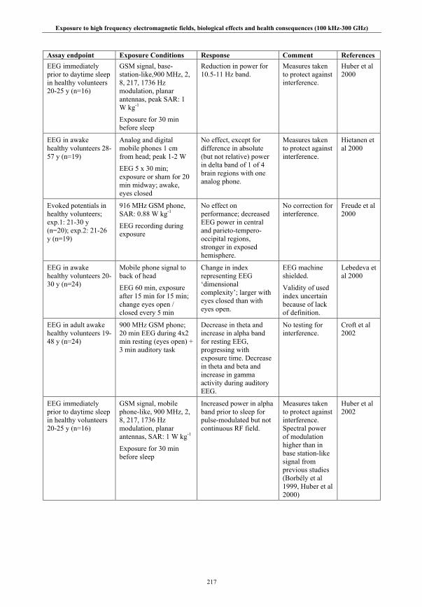
Exposure to high frequency electromagnetic fields, biological effects and health consequences (100 kHz-300 GHz)
217
Assay endpoint Exposure Conditions Response Comment References EEG immediately prior to daytime sleep in healthy volunteers 20-25 y (n=16)
GSM signal, base-station-like,900 MHz, 2, 8, 217, 1736 Hz modulation, planar antennas, peak SAR: 1 W kg-1
Exposure for 30 min before sleep
Reduction in power for 10.5-11 Hz band.
Measures taken to protect against interference.
Huber et al 2000
EEG in awake healthy volunteers 28-57 y (n=19)
Analog and digital mobile phones 1 cm from head; peak 1-2 W
EEG 5 x 30 min; exposure or sham for 20 min midway; awake, eyes closed
No effect, except for difference in absolute (but not relative) power in delta band of 1 of 4 brain regions with one analog phone.
Measures taken to protect against interference.
Hietanen et al 2000
Evoked potentials in healthy volunteers; exp.1: 21-30 y (n=20); exp.2: 21-26 y (n=19)
916 MHz GSM phone, SAR: 0.88 W kg-1
EEG recording during exposure
No effect on performance; decreased EEG power in central and parieto-tempero-occipital regions, stronger in exposed hemisphere.
No correction for interference.
Freude et al 2000
EEG in awake healthy volunteers 20-30 y (n=24)
Mobile phone signal to back of head
EEG 60 min, exposure after 15 min for 15 min; change eyes open / closed every 5 min
Change in index representing EEG ‘dimensional complexity’; larger with eyes closed than with eyes open.
EEG machine shielded.
Validity of used index uncertain because of lack of definition.
Lebedeva et al 2000
EEG in adult awake healthy volunteers 19-48 y (n=24)
900 MHz GSM phone; 20 min EEG during 4x2 min resting (eyes open) + 3 min auditory task
Decrease in theta and increase in alpha band for resting EEG, progressing with exposure time. Decrease in theta and beta and increase in gamma activity during auditory EEG.
No testing for interference.
Croft et al 2002
EEG immediately prior to daytime sleep in healthy volunteers 20-25 y (n=16)
GSM signal, mobile phone-like, 900 MHz, 2, 8, 217, 1736 Hz modulation, planar antennas, SAR: 1 W kg-1
Exposure for 30 min before sleep
Increased power in alpha band prior to sleep for pulse-modulated but not continuous RF field.
Measures taken to protect against interference. Spectral power of modulation higher than in base station-like signal from previous studies (Borbély et al 1999, Huber et al 2000)
Huber et al 2002

Exposure to high frequency electromagnetic fields, biological effects and health consequences (100 kHz-300 GHz)
218
Assay endpoint Exposure Conditions Response Comment References EEG in awake healthy volunteers 18-30 y (n=10)
900 MHz GSM, antenna pointed at the back of the head; full power (250 mW) or standby
EEG 10 x 5 min, random exposure / sham, at 10-15 min intervals
Significant decrease in alpha (8-12 Hz) and beta (13-30 Hz) bands power with full power mode only.
No interference found upon testing.
D’Costa et al 2003
Awake EEG in awake healthy adults (n=10) and children, 12 y (n=10)
900 MHz GSM, 100 MHz radio
Awake, eyes open
Wireless EEG recording system
Appearance of seconds-long periods of slow waves (2.5-6.0 Hz in adults; 1.0-2.5 Hz in children) 20-40 sec after turning on phone.
Incomplete experimental details; no statistics. No exposure-sham comparison.
Kramarem-ko and Tan 2003
EEG in awake healthy volunteers 19-23 y, (n=20)
450 MHz,7 Hz modulated; SAR: 0.0095 Wkg-1
EEG during 10 60-sec periods, at 60-sec intervals; awake, eyes closed
Changes in alpha waves, high inter-individual variability, overall not significant. Less changes in theta waves. Effects increase in subsequent exposure periods.
No mention of measures taken against interference.
Hinrikus et al 2004
EEG during memory task in healthy volunteers 23.3±2.2 y; males (n=9) and females (n=10)
900 MHz GSM signal, mean power 64 mW
EEG recording during anticipatory period of memory test
Baseline EEG power greater in males than in females; exposure decreased power in males and increased power in females. No effect on memory.
Incomplete description of experimental design.
Papageorgiou et al 2004
EEG in awake healthy volunteers 22-31 y (n=20)
900 MHz GSM at max power; SAR: 0.5 Wkg-1; 45 min
EEG after or during last 7 min of exposure; during EEG eyes closed
Increased power in alpha band (9-10 Hz).
No mention of measures taken against interference.
Curcio et al 2005
EEG in healthy volunteers (n=9) and epileptic patients (n=6)
GMS mobile phone signal
EEG before and during exposure or sham
Healthy subjects: decrease in variation in delta band; decrease in power in theta, alpha and beta bands. Epileptics: power increase in all bands.
Incomplete description of experimental design.
Maby et al 2006a
EEG in awake healthy volunteers19-25 y (n=24)
900 MHz, GSM pulsed or continuous, 30 min, SAR:1 W kg-1; planar antenna
EEG immediately, 30 min, 60 min after exposure, during 3 min eyes closed and 3 min eyes open
Increase 10.5-11 Hz power 30 min after GSM with eyes closed only; no effects continuous signal
Regel et al 2007a
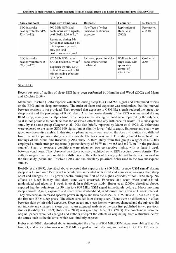
Exposure to high frequency electromagnetic fields, biological effects and health consequences (100 kHz-300 GHz)
219
Assay endpoint Exposure Conditions Response Comment References EEG in awake healthy volunteers19-32 y (n=12)
900 MHz GSM and continuous wave signals, peak SAR: 1.56 W kg-1
Recording during 2-h period that included 3 15 min exposure periods; only pre- and postexposure analyzed
No effects of either pulsed or continuous exposure.
Replication of Huber et al (2002)
Perentos et al 2004
EEG in awake healthy volunteers 18-69 y (n=120)
875 MHz GSM, max SAR in brain: 0.11 W kg-1
Exposure 30 min, EEG in first 10 min and in 10 min following exposure; eyes open
Increased power in alpha band; greater effect ipsilateral.
Well performed large study with appropriate control of interference.
Croft et al 2008
Sleep EEG
Recent reviews of studies of sleep EEG have been performed by Hamblin and Wood (2002) and Mann and Röschke (2004).
Mann and Roschke (1996) exposed volunteers during sleep to a GSM 900 signal and determined effects on the EEG and on sleep architecture. The order of sham and exposure was randomized, but the interval between sessions is not provided. They reported that exposure to GSM-like signals reduced the latency to sleep onset and the percentage of REM sleep. Also the power density of the EEG was increased during REM sleep, mainly in the alpha band. No changes in well-being or mood were reported by the subjects, so it is not possible to conclude that the observed effects had any influence on health. In a subsequent study by the same group (Wagner et al 1998; also briefly reported by Mann et al 1998) 22 volunteers were exposed to the same GSM 900 signal, but at slightly lower field strength. Exposure and sham were given on consecutive nights. In this study a planar antenna was used, so the dose distribution also differed from that in the previous study where a mobile telephone was used. This study failed to replicate the findings of the Mann and Röschke (1996) study. A third study from this group (Wagner et al 2000) employed a much stronger exposure (a power density of 50 W m-2, vs 0.5 and 0.2 W m-2 in the previous studies). Sham or exposure conditions were given on two consecutive nights, with at least 1 week between conditions. They observed no effects on sleep architecture or EEG spectral power density. The authors suggest that there might be a difference in the effects of linearly polarized fields, such as used in the first study (Mann and Röschke 1996), and the circularly polarized fields used in the two subsequent studies.
Borbély et al (1999), described above, reported that exposure to a 900 MHz ‘pseudo GSM signal’ during sleep in a 15 min on / 15 min off schedule was associated with a reduced number of wakings after sleep onset and changes in EEG power spectra during the first of the night’s episodes of non-REM sleep. No effects on sleep latency and sleep state were observed. Exposure and sham were double-blind, randomized and given at 1 week interval. In a follow-up study, Huber et al (2000), described above, exposed healthy volunteers for 30 min to a 900 MHz GSM signal immediately before a 3-hour morning sleep episode. Again, exposure and sham were double-blind, randomized and given at 1 week interval. They observed an increased spectral power in alpha and beta bands (9.75-11.25 Hz and 12.5-13.25 Hz) in the first non-REM sleep phase. The effect subsided later during sleep. There were no differences in effect between right or left-sided exposure. Sleep stages and sleep latency were not changed and the subjects did not indicate any changes in sleep quality. An extended analysis of the data first published in two previous studies (Borbély et al 1999; Huber et al 2000) was given by Huber et al (2003). The conclusions from the original papers were not changed and authors interpret the effects as originating from a structure below the cortex such as the thalamus which was similarly exposed.
Huber et al (2002), described above, investigated the effects of 900 MHz GSM signal resembling that of a handset, and of a continuous wave 900 MHz signal on both sleeping and waking EEG. The left side of
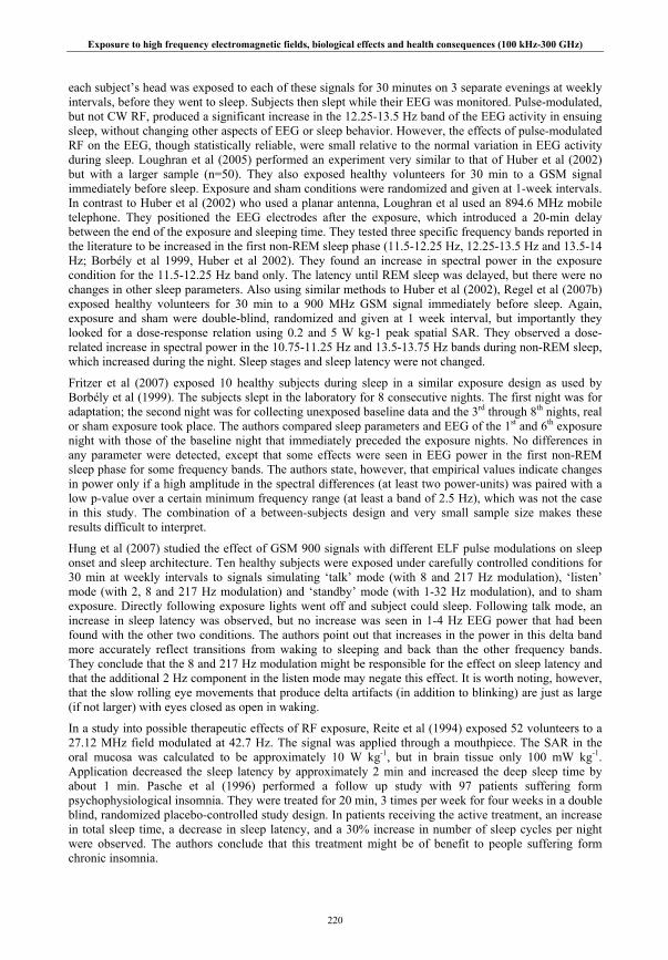
Exposure to high frequency electromagnetic fields, biological effects and health consequences (100 kHz-300 GHz)
220
each subject’s head was exposed to each of these signals for 30 minutes on 3 separate evenings at weekly intervals, before they went to sleep. Subjects then slept while their EEG was monitored. Pulse-modulated, but not CW RF, produced a significant increase in the 12.25-13.5 Hz band of the EEG activity in ensuing sleep, without changing other aspects of EEG or sleep behavior. However, the effects of pulse-modulated RF on the EEG, though statistically reliable, were small relative to the normal variation in EEG activity during sleep. Loughran et al (2005) performed an experiment very similar to that of Huber et al (2002) but with a larger sample (n=50). They also exposed healthy volunteers for 30 min to a GSM signal immediately before sleep. Exposure and sham conditions were randomized and given at 1-week intervals. In contrast to Huber et al (2002) who used a planar antenna, Loughran et al used an 894.6 MHz mobile telephone. They positioned the EEG electrodes after the exposure, which introduced a 20-min delay between the end of the exposure and sleeping time. They tested three specific frequency bands reported in the literature to be increased in the first non-REM sleep phase (11.5-12.25 Hz, 12.25-13.5 Hz and 13.5-14 Hz; Borbély et al 1999, Huber et al 2002). They found an increase in spectral power in the exposure condition for the 11.5-12.25 Hz band only. The latency until REM sleep was delayed, but there were no changes in other sleep parameters. Also using similar methods to Huber et al (2002), Regel et al (2007b) exposed healthy volunteers for 30 min to a 900 MHz GSM signal immediately before sleep. Again, exposure and sham were double-blind, randomized and given at 1 week interval, but importantly they looked for a dose-response relation using 0.2 and 5 W kg-1 peak spatial SAR. They observed a dose-related increase in spectral power in the 10.75-11.25 Hz and 13.5-13.75 Hz bands during non-REM sleep, which increased during the night. Sleep stages and sleep latency were not changed.
Fritzer et al (2007) exposed 10 healthy subjects during sleep in a similar exposure design as used by Borbély et al (1999). The subjects slept in the laboratory for 8 consecutive nights. The first night was for adaptation; the second night was for collecting unexposed baseline data and the 3rd through 8th nights, real or sham exposure took place. The authors compared sleep parameters and EEG of the 1st and 6th exposure night with those of the baseline night that immediately preceded the exposure nights. No differences in any parameter were detected, except that some effects were seen in EEG power in the first non-REM sleep phase for some frequency bands. The authors state, however, that empirical values indicate changes in power only if a high amplitude in the spectral differences (at least two power-units) was paired with a low p-value over a certain minimum frequency range (at least a band of 2.5 Hz), which was not the case in this study. The combination of a between-subjects design and very small sample size makes these results difficult to interpret.
Hung et al (2007) studied the effect of GSM 900 signals with different ELF pulse modulations on sleep onset and sleep architecture. Ten healthy subjects were exposed under carefully controlled conditions for 30 min at weekly intervals to signals simulating ‘talk’ mode (with 8 and 217 Hz modulation), ‘listen’ mode (with 2, 8 and 217 Hz modulation) and ‘standby’ mode (with 1-32 Hz modulation), and to sham exposure. Directly following exposure lights went off and subject could sleep. Following talk mode, an increase in sleep latency was observed, but no increase was seen in 1-4 Hz EEG power that had been found with the other two conditions. The authors point out that increases in the power in this delta band more accurately reflect transitions from waking to sleeping and back than the other frequency bands. They conclude that the 8 and 217 Hz modulation might be responsible for the effect on sleep latency and that the additional 2 Hz component in the listen mode may negate this effect. It is worth noting, however, that the slow rolling eye movements that produce delta artifacts (in addition to blinking) are just as large (if not larger) with eyes closed as open in waking.
In a study into possible therapeutic effects of RF exposure, Reite et al (1994) exposed 52 volunteers to a 27.12 MHz field modulated at 42.7 Hz. The signal was applied through a mouthpiece. The SAR in the oral mucosa was calculated to be approximately 10 W kg-1, but in brain tissue only 100 mW kg-1. Application decreased the sleep latency by approximately 2 min and increased the deep sleep time by about 1 min. Pasche et al (1996) performed a follow up study with 97 patients suffering form psychophysiological insomnia. They were treated for 20 min, 3 times per week for four weeks in a double blind, randomized placebo-controlled study design. In patients receiving the active treatment, an increase in total sleep time, a decrease in sleep latency, and a 30% increase in number of sleep cycles per night were observed. The authors conclude that this treatment might be of benefit to people suffering form chronic insomnia.

Exposure to high frequency electromagnetic fields, biological effects and health consequences (100 kHz-300 GHz)
221
Table II.5.2.: Electrical activity of the brain: sleep EEG
Assay endpoint Exposure Conditions Response Comment References EEG and sleep parameters in healthy volunteers 18-53 y (n=52)
Low energy emission therapy device (27.12 MHz modulated at 42.7 Hz); intrabuccal applicator; max brain SAR:100 mW kg-1
EEG 35 min, exposure 15 min, starting at 5 min after EEG
Decrease in sleep latency and increase in sleep duration.
Reite et al 1994
EEG and sleep parameters in healthy volunteers 21-34 y (n=12)
GSM mobile phone, 900 MHz, 217 Hz modulation, at 40 cm from vertex, 0.5 W m-2
EEG and exposure continuous for 8 h
No effect on sleep efficiency; sleep latency onset and % REM sleep reduced.
Increased spectral power density during REM sleep, mainly in alpha band.
Control for interference.
Mann and Röschke 1996
Sleep parameters in volunteers with psychophysiological insomnia 21-55 y (n=97)
Low energy emission therapy device (27.12 MHz modulated at 42.7 Hz); intrabuccal applicator; max brain SAR: 100 mW kg-
20 min, 3x per week for 4 weeks
Increase in total sleep time, decrease in sleep latency, 30% increase in number of sleep cycles per night.
Pasche et al 1996
EEG and sleep parameters in healthy volunteers 18-37 y (n=22)
GSM signal from planar antenna, 900 MHz, 217 Hz modulation, 40 cm below pillow, 0.2 W m-
2, max SAR: 0.6 W kg-1
EEG and exposure continuous for 8 h
No effects on sleep architecture and EEG.
Control for interference.
Wagner et al 1998
EEG and sleep parameters in healthy volunteers 20-25 y (n=24)
GSM signal, base station-like, 900 MHz, 2, 8, 217, 1736 Hz modulation, 3 antennas 30 cm from the head; max SAR: 1 W kg-1
EEG continuous and exposure 15 min on / 15 min off for 8 h
Reduced waking during sleep only with sham before exposure. No effect on sleep latency and sleep state.
Increase in power for 10-11 Hz and 13.5-14 Hz band during non-REM sleep.
Control for interference.
Borbély et al 1999
EEG and sleep parameters in healthy volunteers 19-36 y (n=20)
GSM signal from horn antenna, 900 MHz, 217 Hz modulation, 40 cm below pillow, 50 W m-2, max SAR: 1.8 W kg-1
EEG and exposure continuous for 8 h on 2 consecutive nights
No effects on sleep architecture and EEG.
Control for interference.
Wagner et al 2000

Exposure to high frequency electromagnetic fields, biological effects and health consequences (100 kHz-300 GHz)
222
Assay endpoint Exposure Conditions Response Comment References EEG and sleep parameters in healthy volunteers 20-25 y (n=16)
GSM signal, base-station-like,900 MHz, 2, 8, 217, 1736 Hz modulation, planar antennas, peak SAR: 1 W kg-1Exposure for 30 min before sleep
Spectral power in 9.75-11.25 Hz and 12.5-13.25 Hz band increased in first non-REM phase. No difference between right or left exposure.
No effect on sleep stage or sleep latency.
Huber et al 2000
EEG in sleeping volunteers (n=20)
Mobile phone
EEG during 8-h sleep; continuous exposure
Increase in alpha band power and changes in sleep pattern during exposure
No information on experimental setup and methods of data analysis
Lebedeva et al 2001
EEG and sleep parameters in healthy males, 20-25 y (n=13).
GSM signal, mobile phone-like, 900 MHz, 2, 8, 217, 1736 Hz modulation, planar antennas, SAR: 1 W kg-1
Exposure for 30 min before sleep
Increased power in alpha band prior to sleep and in the 12.25-13.5 Hz band during non-REM sleep; no fading during sleep. No effects of non pulse-modulated field.
Spectral power of modulation higher than in base station-like signal from previous studies (Borbély et al 1999, Huber et al 2000)
Huber et al 2002
EEG and sleep parameters in healthy volunteers, 18-60 y (n=50).
894.6 MHz GSM mobile phone, peak SAR: 0.29 W kg-1, for 30 min
Decrease in REM sleep latency; increase in power in 11.5-12.25 Hz band during first 30 min of 1st non-REM sleep period.
EEG electrodes positioned after exposure.
Loughran et al 2005
EEG and sleep parameters in healthy males, 22-36 y (n=10).
GSM signal, 900 MHz, 2, 8, 217, 1733 Hz modulation, 3 antennas 30 cm from the head; max SAR: 1 W kg-1
EEG continuous and exposure during sleep for 6 nights
Comparison between unexposed baseline night and 1st and 6th exposure night. No effect on sleep parameters or on EEG power in either night.
Possibly too strict criteria for EEG power changes to become statistically significant.
Fritzer et al 2007
EEG and sleep parameters in healthy volunteers, 18-28 y (n=10).
GSM signal, 900 MHz, pulse modulated with 8, 217 Hz (talk), 2, 8, 217 Hz (listen), or 1-32 Hz (standby); mean SAR resp.: 0.133, 0.015, <0.001 W kg-1, for 30 min
EEG continuous and during exposure and sleep
Delayed sleep latency after talk mode; no similar increase in 1-4 Hz (delta) power as in other conditions.
Hung et al 2007
EEG and sleep parameters in healthy volunteers 20-26 y (n=15)
GSM signal, base-station-like, 900 MHz, 2, 8, 217, 1736 Hz modulation, planar antennas, peak SAR: 0.2 and 5 W kg1
Exposure for 30 min before sleep
Dose-response increase in 10.75-11.25Hz and 13.5-13.75 Hz bands in non-REM, Stage 2 and slow-wave sleep.
No effect on sleep stage or sleep latency.
Regel et al 2007b
Exposure to high frequency electromagnetic fields, biological effects and health consequences (100 kHz-300 GHz)
223
Event-related (evoked) potentials
These studies have examined RF (mostly GSM signals) effects on auditory, visual and somatosensory event-related potentials (ERPs). In addition, one group focused in ERPs related to the performance of cognitive tasks and another examined cortical excitability using transcranial magnetic stimulation applied to the motor cortex before and after RF exposure in order to generate muscle contraction. Most studies were carried out on adults; one study was carried out using children.
Studies with adult subjects
A number of studies have reported that acute exposure to GSM RF radiation can affect auditory ERPs. However, the interpretation of the conflicting results from many of these studies is often weakened by various methodological limitations such as questionable dosimetry, small sample size and single-blinding techniques.
Eulitz et al (1998) observed an increase of high frequency (approximately 18-30 Hz) spectral power in auditory ERP waveforms elicited by infrequent auditory 'oddball' stimuli interspersed among a more frequent class of auditory stimulus. The effect was observed only in the left hemisphere, the side of the exposure. This study in 13 volunteers was single blind and no control of possible interference of the GSM signal with the electrodes or leads was reported. Croft et al (2002; described above) exposed 24 volunteers to GSM mobile phone type RF radiation and recorded phase-locked responses from the EEG during the performance of an auditory discrimination task. The study was single-blind and counterbalanced with a crossover design. A decrease in power of the theta (1-4 Hz) and beta (12-30 Hz) bands and an increase in power of the gamma band (30-45 Hz) in the phase-locked EEG were reported. The strength of the dosimetry in that study was subsequently questioned by the investigators (Croft et al 2008).
Arai et al (2003) studied the neuronal pathways mediating auditory stimulus, from ear to midbrain, by recording the auditory brainstem-evoked responses (ABR). They exposed healthy volunteers for 30 min to a mobile phone operating at full power. ABRs were recorded before and after exposure. They performed three experiments at 1 week intervals to study three different parameters. No effects of the exposure were observed. Bak et al (2003) exposed 45 volunteers to signals of three types of mobile phones operating at different frequencies. Exposure was intermittent at four times per minute at 1-s intervals, for 20 min on both the right and left side of the head. Dosimetry was performed, but exposure levels are unclear. ABRs were not influenced by RF exposure.
A pilot study by Hamblin et al (2004) in 12 subjects reported a decrease in the amplitude and latency of a sensory component of an auditory ERP, and an increase in the latency of a later more cognitive component. This study had a single-blind, counterbalanced crossover design. Sievert et al (2005) reported a lack of effect of exposure to a CW or GSM signal on ABRs before, during and after exposure in a study comprising 12 volunteers. It is not clear whether the study was blinded in any way.
Maby et al (2004) studied auditory ERPs in two groups of 14 subjects: healthy volunteers and epileptic patients. They reported a reduction in the amplitude and latency of the early sensory component of the auditory ERP in the healthy subjects, and an increase in latency and decrease in amplitude for the epileptic patients. In the healthy subjects only, they also observed an increase in the amplitude of a later component. Unfortunately the experimental design was not clearly described. Therefore it is difficult to draw any conclusions on the effect of exposure. Maby et al (2005, 2006b) examined the effects of GSM RF radiation on auditory ERPs evoked by two different sound stimuli in both normal and epileptic subjects. In both studies, nine healthy volunteers and six patients suffering from temporal lobe epilepsy were exposed or sham-exposed to GSM-type RF whilst auditory ERPs were recorded. The authors calculated in each individual the temporal and frequency correlation variations for the auditory ERP responses to the different pairs of stimuli recorded from 14 (out of 32) selected electrodes, with or without RF exposure. Each subject acted as its own control and the study was a single-blind design. Variable exposure-related differences in the correlation coefficients were observed in both healthy and epileptic subjects, but it was not possible to determine the significance of this observation for health.

Exposure to high frequency electromagnetic fields, biological effects and health consequences (100 kHz-300 GHz)
224
Oysu et al (2005) measured ABRs in 18 healthy volunteers. It is not clear whether the subjects were blinded to the exposure conditions. ABRs were determined before and after a 15-min exposure to the signal from a 900 MHz mobile phone. No differences were observed between the before and after measurements.
In an attempt to avoid the methodological weaknesses of previous studies, Hamblin et al (2006) further investigated their earlier study (Hamblin et al 2004) which reported a decrease in the amplitude and latency of a sensory component of an auditory ERP, and an increase in the latency of a later, more cognitive component, using larger number of subjects (120), better dosimetry and a double-blind, counterbalanced, crossover design. Two experimental sessions were held, 1 week apart; in each session subjects were initially sham exposed, and then either exposed or sham-exposed to GSM 895 RF. The authors measured the reaction times for cognitive responses to an auditory and a visual cognitive (oddball) task and recorded the early and late components of ERPs resulting from the auditory and visual stimuli. There were no statistically significant effects on the early or late components of the ERPs, and no effect on reaction times. The authors concluded that there is currently no clear evidence in support of a mobile phone related EMF effect on ERPs or reaction times.
Several studies have examined RF effects on visual ERPs. Freude et al (1998) measured slow response-related brain potentials in a visual monitoring task in a single-blind study in 16 volunteers. They observed a small reduction in the amplitude of potentials in the central and temporo-parieto-occipital regions. In contrast to the study of Eulitz et al (1998), the effect was strongest in the right (contralateral) hemisphere. No effects were found in the potentials preceding spontaneous movements, and neither were there any exposure effects on task performance.
Freude et al (2000) performed a follow-up of this study. They measured visual ERPs in two groups of 20 and 19 volunteers, respectively. In the second group, other evoked potentials were measured. In a single blind design, the subjects were exposed for the duration of the tests to a signal from a GSM 900 phone. Exposure had no effect on this performance, nor on a simple finger tapping task or a more complex task involving two visual stimuli. Urban et al (1998) found no effects in a pilot study of GSM mobile phone radiation effects on the visual ERP evoked by reversal of a checkerboard pattern in 20 volunteers. However, visual ERPs were only measured after exposure and the subjects were aware of whether the phone was on or off.
Jech et al (2001) studied visual ERPs in 22 patients with narcolepsy. In 17 of these patients ERPs were studied during a visual ‘oddball’ task. In this task, rare horizontally-striped ‘targets’ (the oddballs) were interposed among presentations of more frequent non-targets (vertical stripes). Both classes of stimulus could occur either in full-field, or restricted to one or other side of the visual field. Exposure was double-blind, with sham and exposure conditions occurring on separate days (ordering of conditions was counterbalanced). Recordings were obtained during exposure to a GSM 900 signal. Exposure was found to enhance the amplitude of two components of the brain’s response to the oddball stimuli, but only when the stimuli were presented to the right half of the visual field. This effect was most marked in waveforms from right hemisphere electrodes. In addition, exposure was found to shorten reaction time to both stimulus classes by approximately 20 ms. It should be noted however that the majority of the narcoleptic patients were medicated, possibly restricting the generality of these findings. In addition, since stimuli presented to the right visual hemifield project to the left hemisphere, it is not clear that the effect reported over the right hemisphere related to the experimental task.
Yuasa et al (2006) studied the effects of mobile phone RF radiation on somatosensory ERPs in 12 subjects. The experiment was single-blinded. Exposure or sham exposure was to 800 MHz RF radiation from a digital mobile phone held by hand for 30 min within 4 cm of the head. The authors recorded the ERP in the sensory region of the right cortex evoked by median nerve stimulation of the left arm before during and after exposure. They reported that the RF exposure did not affect the somatosensory ERP or its recovery function, suggesting that neither the neural pathways mediating somatosensory stimuli nor the large neurons of the sensory cortex are affected by mobile phone radiation.
Ferreri et al (2006) investigated the effects of GSM mobile phone RF radiation on cortical excitability in fifteen right-handed young male volunteers. Transcranial magnetic stimulation was applied to the motor cortex before and after RF exposure in order to generate motor-evoked potentials in a target muscle in the

Exposure to high frequency electromagnetic fields, biological effects and health consequences (100 kHz-300 GHz)
225
hand. This approach differs from the other ERP studies where a sensory stimulus evoked electrical potentials in the sensory cortex of the exposed or sham-exposed brain. All subjects underwent two trials, separated by one week, in a double-blind cross-over experimental design. The left side of the subject’s head was exposed or sham-exposed to RF radiation for 45 min; the right side served as a control. The effect of main interest was the triple interaction of time, exposure condition and hemisphere. This had a probability of 0.07, which is not statistically significant but is sufficiently borderline to be of interest. It indicates a transient decrease in intracortical inhibition and a transient increase in intracortical facilitation in the RF-exposed hemisphere. However, the analysis and interpretation is complex and depends, for example, on the stability of the base-line response to a single pulse, as indicated by the authors.
Krause et al (2000a; 2000b) investigated the effects of a GSM 900 signal on event-related desynchronization/synchronization (ERD/ERS) of four narrow EEG frequency bands: 4-6 Hz, 6-8 Hz, 8-10 Hz and 10-12 Hz, during performance of a cognitive task. Although not referred to as evoked potentials per se, different EEG frequencies are associated with different mental processes and synchronization or desynchronization of these reflects event-related increases or decreases respectively in the relative EEG power of the different frequency bands. Krause et al (2000a) examined ERD/ERS in 16 subjects during the performance of an auditory memory task during exposure. A counterbalanced, single-blind procedure was used. RF exposure significantly increased the ERD/ERS responses in the 8-10 Hz frequency band only, which is associated with attention and memory functions. They concluded that RF exposure can influence neural oscillatory systems associated with memory retrieval. The second study (Krause et al 2000b) examined ERD/ERS elicited by the visual presentation of letters during an ‘n-back’ working memory task. Twenty-four subjects were employed, using an on/off single-blind procedure with a 900 MHz GSM signal. Exposure effects were observed in two specific bands of the EEG spectrum, at 6-8 and 8-10 Hz. The authors concluded that, as in their previous study, the findings suggested that RF effects on EEG are most prominent during active cognitive processing.
A replication of the Krause et al 2000a study using an auditory memory task was published by Krause et al (2004). In this double-blind study, all 24 subjects performed the memory task both with and without exposure in a counter-balanced order. Although the authors found some effects of RF on the ERD/ERS responses in the 4-8 Hz EEG frequency range, they were not able to replicate the findings from their earlier study. In contrast to the previous study, they did observe an increase in the number of incorrect answers in the memory task during exposure. They concluded that GSM 900 effects on EEG and on the performance of memory tasks are variable and not easily replicable for unknown reasons.
Further puzzling observations were made by the Krause group in a subsequent study (Krause et al 2007). Here they used a double blind crossover experimental design to expose two groups of 36 volunteers to both GSM 900 and CW signals. One group performed a visual memory task during exposure, the other one an auditory memory task. No effects of exposure to either type of signal on performance were observed. In both groups, some small, but inconsistent differences were found in EEG power in the alpha band (8-10 Hz) between CW and GSM conditions, but not with sham. The authors conclude that EMF effects on the EEG are either non-existent or so susceptible to many other factors in standard EEG experiments (such as normal variations in the EEG, attentional effects, random variance, etc.), that they are difficult to capture systematically.
Hinrichs & Heinze (2004) exposed healthy volunteers for 30 minutes to a GSM 1800 signal during the learning (encoding) phase of a memory test. They subsequently measured the total EEG power during memory retrieval, where subjects had to indicate whether the words they were presented were part of the list they had seen during the encoding phase. The reaction time and percentages of correct answers were not influenced by the exposure. In some parts of the left hemisphere (the exposed side) differences in EEG power were detected at some specific time windows after the start of the stimulus. It is not clear, however, whether these differences were increases or decreases in total EEG power. It is also not clear whether any blinding of the subjects to the exposure condition was performed.
Studies with children
Krause et al (2006) used the same experimental design as in previous studies (Krause et al 2000a, 2001, 2004) to examine ERD/ERS responses in 15 subjects aged 10-14 years performing an auditory memory
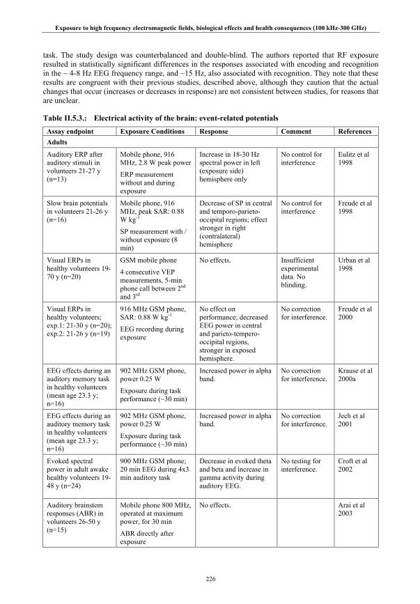
Exposure to high frequency electromagnetic fields, biological effects and health consequences (100 kHz-300 GHz)
226
task. The study design was counterbalanced and double-blind. The authors reported that RF exposure resulted in statistically significant differences in the responses associated with encoding and recognition in the ~ 4-8 Hz EEG frequency range, and ~15 Hz, also associated with recognition. They note that these results are congruent with their previous studies, described above, although they caution that the actual changes that occur (increases or decreases in response) are not consistent between studies, for reasons that are unclear.
Table II.5.3.: Electrical activity of the brain: event-related potentials
Assay endpoint Exposure Conditions Response Comment References Adults
Auditory ERP after auditory stimuli in volunteers 21-27 y (n=13)
Mobile phone, 916 MHz, 2.8 W peak power
ERP measurement without and during exposure
Increase in 18-30 Hz spectral power in left (exposure side) hemisphere only
No control for interference
Eulitz et al 1998
Slow brain potentials in volunteers 21-26 y (n=16)
Mobile phone, 916 MHz, peak SAR: 0.88 W kg-1
SP measurement with / without exposure (8 min)
Decrease of SP in central and temporo-parieto-occipital regions; effect stronger in right (contralateral) hemisphere
No control for interference
Freude et al 1998
Visual ERPs in healthy volunteers 19-70 y (n=20)
GSM mobile phone
4 consecutive VEP measurements, 5-min phone call between 2nd and 3rd
No effects. Insufficient experimental data. No blinding.
Urban et al 1998
Visual ERPs in healthy volunteers; exp.1: 21-30 y (n=20); exp.2: 21-26 y (n=19)
916 MHz GSM phone, SAR: 0.88 W kg-1
EEG recording during exposure
No effect on performance; decreased EEG power in central and parieto-tempero-occipital regions, stronger in exposed hemisphere.
No correction for interference.
Freude et al 2000
EEG effects during an auditory memory task in healthy volunteers (mean age 23.3 y; n=16)
902 MHz GSM phone, power 0.25 W
Exposure during task performance (~30 min)
Increased power in alpha band.
No correction for interference.
Krause et al 2000a
EEG effects during an auditory memory task in healthy volunteers (mean age 23.3 y; n=16)
902 MHz GSM phone, power 0.25 W
Exposure during task performance (~30 min)
Increased power in alpha band.
No correction for interference.
Jech et al 2001
Evoked spectral power in adult awake healthy volunteers 19-48 y (n=24)
900 MHz GSM phone; 20 min EEG during 4x3 min auditory task
Decrease in evoked theta and beta and increase in gamma activity during auditory EEG.
No testing for interference.
Croft et al 2002
Auditory brainstem responses (ABR) in volunteers 26-50 y (n=15)
Mobile phone 800 MHz, operated at maximum power, for 30 min
ABR directly after exposure
No effects.
Arai et al 2003

Exposure to high frequency electromagnetic fields, biological effects and health consequences (100 kHz-300 GHz)
227
Assay endpoint Exposure Conditions Response Comment References ABRs in volunteers 21-28 y (n=45)
Mobile phones, 450, 935, 1800 MHz
4x14 sec min-1, for 20 min, both left and right
No effects. Controlled for interference.
Unclear dosimetry.
Bak et al 2003
Auditory ERP after auditory stimuli in volunteers 19-44 y (n=12)
Mobile phone, 895 MHz, 0.25 W mean power
Exposure 1 h, ERP recording after 30 min
Decrease in amplitude and latency of stimulus-bound ERP components and increase in latency of cognitive components.
Controlled for interference.
Hamblin et al 2004
Auditory ERPs in healthy volunteers (n=14) and epileptic patients (n=14)
GSM signal, maximum SAR: 1.4 W kg-1
Four recording phases: no RF, minimal power, maximal power, minimal power; 3 sessions: first two for right / left ear, 3rd for missing data or placebo
Healthy subjects: decrease in latency and amplitude of early ERP component, increase in latency of later component.
Epileptics: increase in latency and decrease in amplitude of early component.
Inadequate description of experimental design.
Maby et al 2004
EEG effects during an auditory memory task in healthy volunteers 24.3±8.1 y (n=24)
902 MHz GSM phone, SAR: 0.65 W kg-1
Exposure during task performance (~30 min)
No effects on EEG, but increase in incorrect answers.
Replication of Krause et al (2000a)
Krause et al 2004
EEG during memory task in healthy volunteers 18-20 y (n=12)
1870 MHz GSM signal, SAR: 0.61 W kg-1
30 min exposure during memory encoding; EEG measured during retrieval; average power over 0-50 Hz
No effect on reaction time and error rate; differences in total EEG power in left (exposed) hemisphere during retrieval.
Blinding not clear.
Hinrichs and Heinze 2004
ABRs in healthy volunteers 19-57 y (n=12)
GSM phone, 890 MHz, continuous or with 217 Hz modulation, SAR: 1.9 W kg-1
No effects. Sievert et al 2005
ABRs in healthy volunteers 20-28 y (n=18)
900 MHz GSM phone, SAR: 0.82 W kg-1
Measurement before / after 15 min exposure
No effects. Unclear whether blinding occurred.
Oysu et al 2005
Auditory ERPs in healthy volunteers 21-32 y (n=9) and epileptic patients 25-39 y (n=6)
900 MHz GSM mobile phone, max SAR: 1.4 W kg-1
Auditory ERP recording during control exposure, minimal and maximal power
Difference in correlation coefficients between control and experimental sessions.
Complex parameters used in the analysis; low number of subjects.
Maby et al 2005
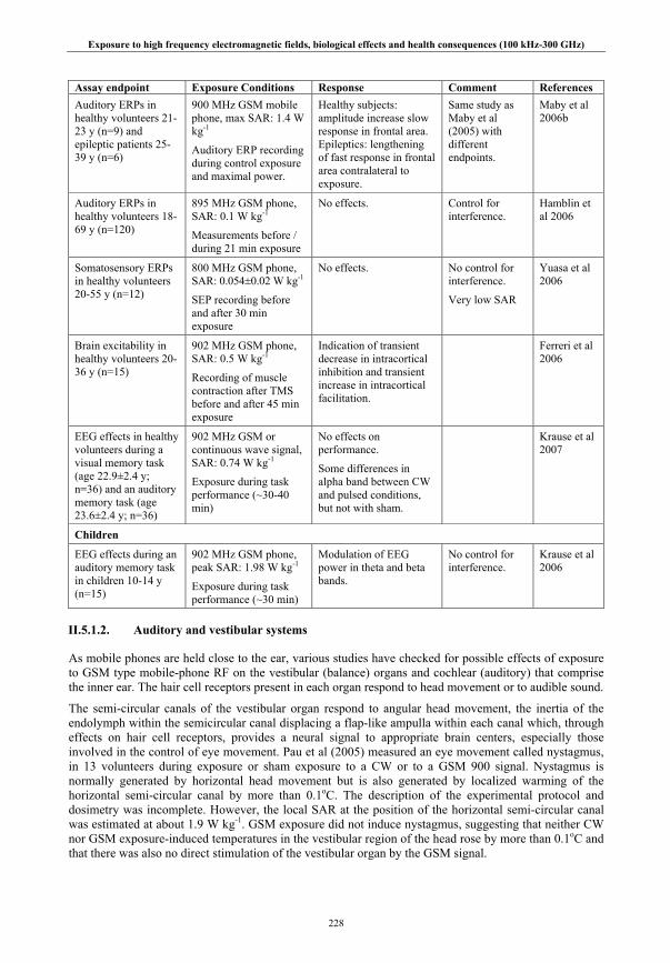
Exposure to high frequency electromagnetic fields, biological effects and health consequences (100 kHz-300 GHz)
228
Assay endpoint Exposure Conditions Response Comment References Auditory ERPs in healthy volunteers 21-23 y (n=9) and epileptic patients 25-39 y (n=6)
900 MHz GSM mobile phone, max SAR: 1.4 W kg-1
Auditory ERP recording during control exposure and maximal power.
Healthy subjects: amplitude increase slow response in frontal area. Epileptics: lengthening of fast response in frontal area contralateral to exposure.
Same study as Maby et al (2005) with different endpoints.
Maby et al 2006b
Auditory ERPs in healthy volunteers 18-69 y (n=120)
895 MHz GSM phone, SAR: 0.1 W kg-1
Measurements before / during 21 min exposure
No effects. Control for interference.
Hamblin et al 2006
Somatosensory ERPs in healthy volunteers 20-55 y (n=12)
800 MHz GSM phone, SAR: 0.054±0.02 W kg-1
SEP recording before and after 30 min exposure
No effects. No control for interference.
Very low SAR
Yuasa et al 2006
Brain excitability in healthy volunteers 20-36 y (n=15)
902 MHz GSM phone, SAR: 0.5 W kg-1
Recording of muscle contraction after TMS before and after 45 min exposure
Indication of transient decrease in intracortical inhibition and transient increase in intracortical facilitation.
Ferreri et al 2006
EEG effects in healthy volunteers during a visual memory task (age 22.9±2.4 y; n=36) and an auditory memory task (age 23.6±2.4 y; n=36)
902 MHz GSM or continuous wave signal, SAR: 0.74 W kg-1
Exposure during task performance (~30-40 min)
No effects on performance.
Some differences in alpha band between CW and pulsed conditions, but not with sham.
Krause et al 2007
Children
EEG effects during an auditory memory task in children 10-14 y (n=15)
902 MHz GSM phone, peak SAR: 1.98 W kg-1
Exposure during task performance (~30 min)
Modulation of EEG power in theta and beta bands.
No control for interference.
Krause et al 2006
II.5.1.2. Auditory and vestibular systems
As mobile phones are held close to the ear, various studies have checked for possible effects of exposure to GSM type mobile-phone RF on the vestibular (balance) organs and cochlear (auditory) that comprise the inner ear. The hair cell receptors present in each organ respond to head movement or to audible sound.
The semi-circular canals of the vestibular organ respond to angular head movement, the inertia of the endolymph within the semicircular canal displacing a flap-like ampulla within each canal which, through effects on hair cell receptors, provides a neural signal to appropriate brain centers, especially those involved in the control of eye movement. Pau et al (2005) measured an eye movement called nystagmus, in 13 volunteers during exposure or sham exposure to a CW or to a GSM 900 signal. Nystagmus is normally generated by horizontal head movement but is also generated by localized warming of the horizontal semi-circular canal by more than 0.1oC. The description of the experimental protocol and dosimetry was incomplete. However, the local SAR at the position of the horizontal semi-circular canal was estimated at about 1.9 W kg-1. GSM exposure did not induce nystagmus, suggesting that neither CW nor GSM exposure-induced temperatures in the vestibular region of the head rose by more than 0.1oC and that there was also no direct stimulation of the vestibular organ by the GSM signal.

Exposure to high frequency electromagnetic fields, biological effects and health consequences (100 kHz-300 GHz)
229
Several authors have examined the effect of mobile phone RF radiation on auditory function in volunteers which can be studied by measuring otoacoustic emission (OAE). This is sound recorded in the outer ear canal thought to result mostly from outer hair cell activity in the cochlea, either spontaneous or following auditory stimulation. Ozturan et al (2002) determined transiently evoked (TE) and distortion product (DP) OAEs in 30 healthy adult volunteers. The authors did not find any effect of a 10 min exposure to the signal from a 900 MHz GSM phone. The level of exposure was not clear, however, because it was not indicated at which output level the mobile phone operated.
Monnery et al (2004) reported that OAEs were unaffected in 12 volunteers during transmission from a mobile phone placed in close proximity to the ear. They did not provide, however, any information on the type of mobile phone used, and on the level and duration of exposure. Kerekhanjanarong et al (2005) studied auditory function in 98 subjects that underwent clinical hearing evaluations and correlated those with reported intensity of mobile phone use. No differences were found in audiometry between the dominant and non-dominant ear. In the eight subjects with the highest telephone use, uncorrelated differences in OAE and ABR were observed between the two ears. These could not be analyzed statistically.
Using a double blind study protocol, Uloziene et al (2005) measured baseline audiological parameters and transient evoked otoacoustic emissions (TEOAE). They exposed healthy volunteers for 10 minutes to GSM 900 or 1800 mobile phones operating at maximum power. Measurements performed directly before and after exposure were not significantly different, nor was there any difference between responses after real and sham exposure.
Two studies have examined possible effects on distortion product OAEs (DPOAEs) which result from the intermodulation products generated by the responses to two tones applied simultaneously. Janssen et al (2005) measured DPOAEs in 28 subjects between consecutive GSM signal pulses from a monopole antenna held 5 cm away from the subject’s ear. No statistically significant changes in DPOAEs were seen in this single blind experiment. However, after correction for variation resulting from effects other than EMFs, DPOAEs were observed to be increased in a few subjects by a physiologically trivial amount (< 1.0 dB compared to a normal dynamic range of 120 dB) but these data were not subject to statistical analysis. Parazzini et al (2005) used mathematical techniques in order to examine two separate components of the DPOAE signal, increasing the sensitivity of the investigation. Fifteen subjects participated; DPOAEs were measured immediately before and after exposure or sham exposure to a GSM signal at 900 MHz or at 1800 MHz; SARs to the cochlea were estimated as 0.41 and 0.19 W kg-1 respectively. The study was double-blind with a within-subject design. No statistically significant effects of mobile phone RF radiation were seen. In a follow-up study by the same group, Paglialonga et al (2007) studied transient evoked otoacoustic emissions (TEOAE) in a group of 29 healthy volunteers. The design of the experiment was identical to the previous one. Also for the TEOAE, no effects of a 10 min exposure were observed.
Oktay and Daşdağ (2006) performed audiometry in three groups of 20 volunteers: one group that had used a mobile phone for more than 2 h daily for 4 years (heavy users); one group that used it daily for 10-20 min for 4 years (moderate users) and a group of non-users. Some control was made for confounding by excluding subjects that had been subjected to loud noises from acoustic devices. No effects were observed on brainstem evoked response audiometry, an objective measure of auditory function. On a more subjective measure, pure tone audiometry, a decrease in hearing was observed in the heavy users group, at 400 Hz for the right ear and 500 and 400 Hz for the left ear. The authors state that no correction could be made for laterality, because 13 of the 20 subjects indicated that they used the phone on both ears. An explanation for the difference between left and right ear is not provided. However, the sound level of the phones was not measured; therefore it is possible that long-term exposure to loud noises from the phones might be the cause of the observed hearing loss.
In a double-blind crossover study with a group of subjects with complaints attributed to using a mobile phone (cases) and a group of control subject without such complaints, Bamiou et al (2008) studied the effect of a 30 min exposure from a modified handset capable of producing GSM 882 and CW signals. Both auditory and vestibular functions were measured, in separate experiments 2-4 weeks apart. No
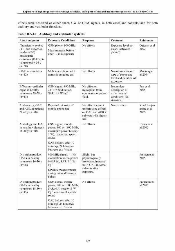
Exposure to high frequency electromagnetic fields, biological effects and health consequences (100 kHz-300 GHz)
230
effects were observed of either sham, CW or GSM signals, in both cases and controls, and for both auditory and vestibular functions.
Table II.5.4.: Auditory and vestibular systems
Assay endpoint Exposure Conditions Response Comment References
Transiently evoked (TE) and distortion product (DP) otoacoustic emissions (OAEs) in volunteers19-36 y (n=30)
GSM phone, 900 MHz
Measurements before / after 10 min exposure
No effects. Exposure level not clear (‘activated phone’).
Ozturan et al 2002
OAE in volunteers (n=12)
Mobile telephone set to transmit outgoing call
No effects. No information on type of phone and level and duration of exposure.
Monnery et al 2004
Effect on vestibular organ in healthy volunteers 29-58 y (n=13)
GSM signal, 890 MHz, 217 Hz modulation, SAR: 1.9 W kg-1
No effect on nystagmus from continuous or pulsed field.
Incomplete description of experimental conditions. No statistics.
Pau et al 2005
Audiometry, OAE and ABR in patients 20-67 y (n=98)
Reported intensity of mobile phone use
No effects, except uncorrelated effects on OAE and ABR in subjects with highest use.
No statistics. Kerekhanjanarong et al 2005
Audiology and OAE in healthy volunteers 18-30 y (n=30)
GSM signal, mobile phone, 900 or 1800 MHz, maximum power (2 resp. 1 W), concurrent speech sound
OAE before / after 10 min exp; 24 h interval between exp / sham
No effects. Uloziene et al 2005
Distortion product OAEs in healthy volunteers 16-30 y (n=28)
900 MHz signal, 41 Hz modulation, mean power 0.465 W , SAR: 0.1 W kg-1
DPOEA measurements during interval between pulses
Slight, but physiologically irrelevant, increase in DPOAE in some subjects after exposure.
Janssen et al 2005
Distortion product OAEs in healthy volunteers 18-30 y (n=15)
GSM signal, mobile phone, 900 or 1800 MHz, SAR: 0.41 resp 0.19 W kg-1, concurrent speech sound
OAE before / after 10 min exp; 24 h interval between exp / sham
No effects. Parazzini et al 2005

Exposure to high frequency electromagnetic fields, biological effects and health consequences (100 kHz-300 GHz)
231
Assay endpoint Exposure Conditions Response Comment References
Transient evoked OAEs in healthy volunteers 23-30 y (n=29)
GSM signal, mobile phone, 900 or 1800 MHz, SAR: 0.41 resp 0.19 W kg-1, concurrent speech sound
TEOAE before / after 10 min exp; 24 h interval between exp / sham
No effects. Paglialonga et al 2007
Audiology in healthy volunteers 22-53 y (n=60)
3 groups of 20: heavy users (>2 h d-1 for 4 y); moderate users (10-20 min d-1 for 4 y); non users
No effect on brainstem evoked response audiometry (BERA); hearing loss in heavy users at 4000 Hz (right ear) and 500 and 4000 Hz (left ear).
Some control for confounding (loud noise from acoustic devices). No correction for laterality possible.
Oktay and Daşdağ 2006
Transient evoked OAEs and nystagmus (video-oculography) in volunteers 20-55 y (subjects with complaints: n=9; controls: n=21)
GSM or continuous signal, 882 MHz, SAR: 1.3 W kg-1,
Measurements before / after 30 min exposure; auditory and vestibular exps 2-4 wk apart
No effects on auditory and vestibular function in either group.
Bamiou et al 2008
II.5.1.3. Regional cerebral blood flow
It is generally assumed that changes in regional cerebral blood flow reflect localized changes in neural activity. Huber et al (2002) studied the effects of EMF signals similar to a GSM phone on regional cerebral blood flow (rCBF) measured by positron emission tomography (PET). A 900 MHz signal simulating that of a GSM mobile phone was delivered by a planar antenna to the left side of the head. The peak SAR was estimated at 1 W kg-1. Thirteen subjects were tested in exposed and sham exposed conditions in counterbalanced order, using a within-subjects double-blind design. At least 1 week elapsed between the two tests. In each test, rCBF was measured over three 1 minute periods, starting 10, 20 and 30 minutes after completion of a 30 minute exposure to pulse-modulated EMF or sham exposure. Subjects were asked to count silently during the scans, to balance cognitive function across scans. The results showed a significant increase in rCBF in the dorsolateral prefrontal cortex of the left (exposed) brain hemisphere. In a follow-up study (Huber et al 2005), the effects of 900 MHz mobile-telephony signals on rCBF were investigated in 12 healthy male volunteers, again using the PET technique. Two types of exposure were used: base-station-like and mobile-phone-like, with a similar exposure setup as in the previous study. The exposure of one side of the head lasted 30 min and resulted in a peak SAR of 1 W kg-1 for both exposure conditions. Following exposure, an increase in rCBF was observed in the dorsolateral prefrontal cortex on the side of exposure only following the mobile phone-like exposure. The authors interpreted this finding, supporting their previous observations, that only pulse-modulated RF (as in the mobile phone-like signal, in contrast with the CW base-station-like signal) is necessary to induce changes in brain physiology.
Haarala et al (2003a) examined the effects of exposure and sham exposure to a GSM 900 signal on rCBF in 14 volunteers, also using PET, under double blind conditions using a counterbalanced order of exposure/sham exposure. In contrast to the experiments of Huber et al (2002, 2005) exposure took place during PET scanning, while the subjects performed a visual working memory task. The main effect of mobile phone exposure was a bilateral decrease in rCBF in the auditory cortex, which the authors attributed to a high frequency auditory signal emitted by the battery of an active phone, since preliminary results from a follow-up study, reported here, indicated that there was no effect with a phone using a

Exposure to high frequency electromagnetic fields, biological effects and health consequences (100 kHz-300 GHz)
232
silent, external power source. There was no effect on the performance of the visual working memory task, including the reaction times and accuracy of the responses. In a follow-up study Aalto et al (2006) improved upon the design of Haarala et al (2003a) by ascertaining that no auditory clues at all were present. They observed a reduced rCBF close to the antenna, and an increase at various other locations deeper in the brain. There was no effect on reaction time. It is not clear from either study whether the changes observed in rCBF were immediately present following the onset of exposure, or that it took some time for them to develop. The papers give no clear indication of the sequence of events.
Table II.5.5.: Regional cerebral blood flow
Assay endpoint Exposure Conditions Response Comment References
Cerebral blood flow in healthy males, 20-25 y (n=13). PET scan 10 min after exposure.
902 MHz, simulating mobile phone, SAR: 1 W kg-1, for 30 min, 1 wk between exposure and sham.
Increased relative rCBF in dorsolateral prefrontal cortex on the side of the exposure.
Exposure less localized than when using mobile telephone.
Huber et al 2002
Cerebral blood flow in healthy males, 20-25 y (n=12). PET scan 10 min after exposure.
902 MHz, simulating mobile phone or base station, SAR: 1 W kg-1, for 30 min, 1 wk between conditions.
Increased relative rCBF in dorsolateral prefrontal cortex on the side of the exposure with mobile phone signal, not with base station signal. No relationship with SAR distribution.
Exposure less localized than when using mobile telephone.
Huber et al 2005
Cerebral blood flow (rCBF) during memory task in healthy volunteers, 21-35 y (n=14). PET scan during exposure.
902 MHz GSM phone, operating at 0.25 W (SAR~1.2 W kg-1), for 45 min; no time between exposure and sham.
Relative decrease in rCBF in auditory cortex, not in area of maximum RF exposure.
Possibly auditory signal. Sequence of events not clear.
Haarala et al 2003a
rCBF by PET scan during a cognitive task and mobile phone exposure in healthy males, 25±2 y (n=12)
902 MHz from a mobile phone operated at 0.25 W, for 30 min.
Reduced rCBF close to antenna, increase at various other locations.
No effect on reaction time.
Sequence of events not clear.
Aalto et al 2006
II.5.1.4. Cognitive performance
Cognitive studies have been carried out in healthy adult volunteers, in adults who report experiencing a variety of symptoms such as headaches in the vicinity of RF sources and, following the recommendations of IEGMP (2000), in children, and also in adolescents. Giedd (2004) for example, notes that there are dynamic changes in brain anatomy throughout childhood and adolescence. The amount of white matter (the myelination of nerve axons), which is related to the speed of neuronal processing, increases linearly throughout adolescence. Changes in grey matter content, thought to reflect changes in size and complexity in neurons such as the number of synaptic connections rather than changes in cell number and considered to be related to the maturation of behavior, are more complex but continue into an individual’s early twenties.
Healthy adults
Preece et al (1999) investigated the performance of 36 volunteers on a wide range of tasks, including short- and long-term memory, simple and choice reaction time, and sustained attention, which, together, yielded a total of 15 dependent variables. Using a double-blind, counterbalanced, randomized cross-over
Exposure to high frequency electromagnetic fields, biological effects and health consequences (100 kHz-300 GHz)
233
design, volunteers were exposed or sham exposed to a continuous or a pulsed 915 MHz GSM-type signal for about 30 min. A statistically significant shortening of reaction time during exposure to the continuous signal in the choice reaction time task was reported. The effect was not accompanied by a reduction in the accuracy of responding, suggesting that it did not reflect a speed-accuracy trade-off. Simple reaction times were unaffected and there were no changes in word, number or picture recall, or in spatial memory. There was no significant effect of exposure to the pulsed GSM signal.
Koivisto et al (2000a) studied 48 volunteers, also using a wide range of cognitive tests. Using a single-blind counterbalanced crossover design, volunteers were exposed or sham-exposed to 902 MHz GSM signal. Koivisto et al reported decreased response times in simple reaction time and vigilance tasks. In addition, the time needed to accomplish a mental arithmetic subtraction task was decreased during exposure. However, the effect of exposure on the choice reaction time task analogous to that employed by Preece et al (1999) was far from significant. In addition, no allowance had been made for multiple testing (IEGMP 2000). Nevertheless, in a second study (Koivisto et al 2000b) using a similar experimental design to investigate GSM RF effects on the performance of a task where working memory load was varied, Koivisto and colleagues reported a statistically significant reduction of reaction time when the memory load was particularly demanding. However, an attempt by the same group to confirm and extend the results from both studies was not successful (Haarala et al 2003b, 2004). Using an improved experimental design by increasing the sample size, performing the study in two independent laboratories and implementing a double-blind design, no consistent field-dependent effects were observed on reaction times or error rates during performance of any of the cognitive tasks.
Lass et al (2002) studied the effect of exposure or sham exposure of the head to a 450 MHz RF signal, amplitude modulated at 7 Hz, on the performance in 3 cognitive tasks by 100 students, randomly allocated to either group. Exposed subjects made significantly fewer errors on the memory recognition task than sham-exposed subjects. In contrast, performance of the other tasks by the exposed group, which were more demanding, produced small and non-significant effects in the opposite direction, showing worse performance and greater inter-subject variability. Edelstyn and Oldershaw (2002) employed a single-blind, between-subjects experimental design to assess the effects of GSM 900 signals on the performance of 19 subjects in six widely-used cognitive tasks. Exposure to a mobile phone held to the left ear was for a total of 30 min; there was no dosimetry. Testing was undertaken in a pre-exposure baseline period, and 15 and 30 min after exposure. The authors reported that exposure facilitated cognitive tasks involving attentional capacity and one task which involved processing speed. Smythe and Costall (2003) also examined the effect of RF exposure from a mobile phone held to the left ear on the performance on a verbal memory task by 62 students, randomly assigned to one of three groups. The authors reported a significant improvement in immediate recall in males but not females. There were no effects of exposure on delayed testing.
Maier et al (2004) studied the effect of exposure to a GSM 900 RF on the performance of an auditory discrimination task. This task required participants to determine whether two successive auditory stimuli were temporally separate and on which side the two stimuli were presented. It was carried out before and immediately after the double-blind exposure or sham exposure of 11 volunteers for 50 min to GSM RF from a mobile phone held 4 cm away from the left ear. The authors reported that exposure significantly reduced the subsequent performance of the task. Cinel et al (2007) replicated this study with a larger number of subjects. Two groups of 84 healthy volunteers were exposed for 40 min to either a 888 MHz GSM signal or a continuous signal. They did not find an effect on performance of the auditory task from either signal type. Both studies do not indicate the type of blinding used, however. Curcio et al (2004) also examined the effect of exposure to a GSM 900 signal on subsequent cognitive task performance. Twenty subjects were randomly assigned to two groups, one group exposed for 45 min before, and the other exposed during a 45 min experimental session. Each subject was tested on four performance tasks: an acoustic simple-reaction time task, a visual search task, an arithmetic descending-subtraction task and an acoustic choice-reaction time task. Using a counter-balanced double-blind paradigm, subjects were subjected to three separate trials separated by 48 h: baseline exposure, real exposure and sham exposure. A significant reduction of both simple and choice reaction times was seen in subjects exposed to a GSM signal. In addition, subjects exposed before testing performed faster than those exposed during testing.
Exposure to high frequency electromagnetic fields, biological effects and health consequences (100 kHz-300 GHz)
234
The authors suggested that RF exposure for a minimum time of around 25-30 min was required for these effects to become manifest.
A different experimental protocol was used by Besset et al (2005) which attempted to better emulate real-life exposure. In a double-blind study, 55 subjects were assigned to GSM 900-exposed or sham-exposed groups, matched for age, sex and IQ. Over the 45-day experimental period, there was a baseline period of 3 days, an exposure period of 28 days during which exposures or sham exposures took place, and a recovery period of 14 days, during which both groups were sham exposed. Exposures or sham exposures were carried out for 2 h per day, five days per week. Subjects held the phone adjacent to their preferred ear during this time; the localized SAR was approximately 0.54 W kg-1. A neuropsychological battery of 22 tasks screened information processing, attention, memory, and executive function on 4 days during the 45-day period, 13 hours after the previous exposure or sham exposure. Statistically significant decreases in reaction time were seen for all tasks between the baseline and exposure periods, indicating a learning effect, but there was no significant effect of RF exposure on task performance. The authors note that, in contrast to other studies, there was a 13 h delay between exposure and testing, which included an overnight sleep. This indicates that there are no lasting effects of exposure, but it precludes any meaningful comparison with studies that looked at short-term effects.
Russo et al (2006) investigated the effects on cognitive performance of exposure to 888 MHz CW or GSM RF using a large number (168) of male and female volunteers, compared to the earlier studies, increasing the statistical power of the study. The subjects were exposed or sham-exposed in two sessions, separated by one week. Half of the subjects had the left side of the head exposed, and half the right side, irrespective of their handedness. Unlike most previous studies, the RF exposure was carried out under double-blind procedures. Cognitive performance was assessed using similar tasks to those used previously, i.e., reaction time task, 10-choice serial reaction time task, subtraction task and vigilance task, which were administered in a counterbalanced order. The authors found no significant effects of RF exposure on task performance, irrespective of whether the left or right side of the head was exposed.
Keetley et al (2006) investigated the effect of exposure to GSM RF radiation on the cognitive performance of 120 male and female volunteers using a double-blind crossover design. The subjects were exposed or sham-exposed in two sessions, separated by one week. Cognitive performance was assessed using a battery of eight cognitive tests: Rey’s audio-visual learning test, digital span test, digital symbol substitution test, speed of comprehension test, trail making task, reaction time task, choice reaction time task and inspection time task, which were administered in a counterbalanced order. After adjusting for known covariates (gender, age and education), simple and choice reaction times showed significant impairment, in contrast to earlier studies (Preece et al 1999; Koivisto et al 2000b), whereas performance on the trail-making task, which involves working memory, significantly improved. However, this study involved numerous comparisons with no adjustment for multiple comparisons (type I error). The authors point out that neither of the earlier studies corrected for known covariates, and that the study of Koivisto et al (2000b) used only a single-blind study design.
Eliyahu et al (2006) examined, in 36 young, right-handed male subjects, the effects of GSM RF exposure of the right or left side of the head on four cognitive tasks selected for high cerebral hemisphere specificity. The authors’ intention was to examine the effect of RF exposure of a specific part of the brain on associated cognitive functions. These were a spatial item recognition task (activating the right premotor cortex), a verbal item recognition task (activating the left posterior parietal cortex and supplementary motor and premotor cortex), and two spatial compatibility tasks (a visual stimulus on the left side of the test screen activating the left posterior parietal cortex, and on the right side activating the right posterior parietal cortex). Each task required right- and left-handed responses. The subjects performed 3 series of tests, with exposure to the right or the left hemisphere or with sham exposure, in either of two 1-h sessions, separated by 5 minutes. The study was conducted under single-blinded conditions, and the exposure regime and task sequence were counterbalanced. The authors analyzed the reaction times for correct responses to each task, comparing the exposure condition (left, right or sham) for left hand or for right hand responses. Generally, right-hand responses were faster than left-hand responses (the subjects were right-handed subjects) and strong training effects (reaction times faster in the second session) were present in most sham responses. The authors reported that exposure of the left hemisphere of the brain resulted in slower left-hand responses in the second session compared to the first,

Exposure to high frequency electromagnetic fields, biological effects and health consequences (100 kHz-300 GHz)
235
for two tasks: the spatial item recognition task, thought to activate the right premotor cortex, and one spatial compatibility task, where left-handed responses are thought to activate the left parietal cortex. Thus, no correlation was seen between exposure of the left hemisphere and the hemisphere-dependence of the two affected tasks.
Haarala et al (2007) also compared responses of exposure of the left and right hemisphere. They exposed 36 healthy male volunteers to a continuous or GSM signal operating at 0.25 W. In a double blind, crossover design, subjects were exposed in 90 min sessions at 1 week intervals. Different cognitive functions tasks were performed: simple reaction time, 10-choice reaction time, subtraction, verification, vigilance, and memory (n-back test). No difference on response was observed for any task for both continuous wave and GSM exposure between exposure to either the left of right hemisphere and sham exposure. The absence of a difference in response with a control group tested without the exposure equipment indicated that the presence of the exposure equipment also was of no influence. The authors comment that they used fairly simple and hemisphere-aspecific behavioral tasks and that it cannot be excluded that more complex or hemisphere-specific cognitive tasks could be more sensitive to effects of RF exposure.
Terao et al (2006) exposed 16 healthy volunteers for 30 min to an 800 MHz mobile phone signal in a double-blind crossover study. Immediately before and after actual or sham exposure a precued choice reaction time test was performed. Exposure did not have any effect on reaction time or accuracy. Schmid et al (2005) exposed 58 healthy volunteers to UMTS signals resulting in SAR values of 0.37 or 0.037 W kg-1, thus simulating a UMTS phone in transmitting or receiving mode. A double-blind crossover design was used. Four visual perception tests were applied: the Critical Flicker Fusion Frequency test, a visual pursuit test, the Tachistoscopic Traffic test Mannheim, and a contrast sensitivity threshold test. The duration of exposure is not given, but was most likely the time it took to perform the tests. Since for all subjects the test procedures were applied on one single day, carry-over effects might be present. However, in none of the tests was an effect of either level of exposure observed.
Regel et al (2007a) investigated reaction time and memory in 20 subjects exposed to either a 900 MHz continuous or GSM-type signal for 30 min. In this double-blind, randomized, counterbalanced cross-over study no effects were observed in single or 2-choice reaction time tests. An improvement in accuracy in the 3-back memory test was found after GSM-type (pulsed field) exposure, but not after CW exposure. Regel et al (2007b) exposed healthy volunteers for 30 min to a GSM 900 signal immediately before sleep while performing cognitive tasks. Exposure and sham were double-blind, randomized and given at 1 week interval, and importantly they looked for a dose-response relation using 0.2 and 5 W kg-1 peak spatial SAR. They observed a dose-related reduction of reaction time with increasing field strength for the 1-back task, and similar relations at trend level for the 2-back task and the choice reaction time task, but no effect on the simple reaction time or 3-back task.
Adults with EMF-attributed symptoms
In a study using exposure similar to that from mobile phone base stations, Zwamborn et al, (2003) investigated subjective feeling and cognitive functions in a group of 36 subjects claiming to experience symptoms in connection with living near a GSM base station and a group of 36 healthy subjects. The groups differed in terms of age and gender distribution and therefore no comparisons could be made between the groups, only within groups for periods with and without exposure. The subjects were exposed to a 1 V/m field at 900 and 1800 MHz (GSM signal), and 2100 MHz (UTMS signal). Each subject participated in three sessions, one of which was unexposed, using a double-blind design. Exposure groups therefore consisted of 24 subjects. Each session took 45 min including exposure (during which cognitive functions were tested), questionnaire, and break. Cognitive function tests included reaction time, memory comparison, dual-tasking, selective visual attention and filtering irrelevant information. A corrected analysis of the data was presented in a report of the Health Council of the Netherlands (2004). In this reanalysis, only one statistically significant result was found with the cognitive function tests. In the control group without symptoms UMTS exposure resulted in an increased completion of the memory comparison test. This could be a chance effect. The results with respect to symptoms have been discussed in the chapter on electrosensitive people.
Exposure to high frequency electromagnetic fields, biological effects and health consequences (100 kHz-300 GHz)
236
The follow-up study by Regel et al (2006) investigated the effect only of the 2140 MHz UMTS base-station-like RF signal, identical to that used by Zwamborn et al, on well-being and cognitive performance in 33 RF-sensitive subjects and in 84 non-sensitive subjects. There were three experimental sessions held at one-week intervals; subjects were randomly assigned to one of six possible sequences of three exposure conditions, each lasting 45 min: 0 V/m (sham), 1 V/m (identical to that used by Zwamborn et al), and 10 V/m (in order to assess any possible dose-response relationship). The study was double-blinded with a randomized crossover design. Cognitive performance was assessed using a simple-reaction time task, a 2-choice reaction time task, the N-back task and the visual selective attention task, the latter also used by Zwamborn et al (2003). No effect of either exposure level was observed on cognitive performance. Again, the results with respect to symptoms have been discussed in the chapter on electrosensitive subjects.
Another recent study (Wilén et al 2006) investigated the effects of mobile phone radiation on various physiological parameters such as heart-rate variability, electrodermal activity, and respiration rate, measured before, during and after exposure, in 20 RF-sensitive subjects and in 20 non-sensitive controls. In addition, tests of arousal and vigilance, short-term memory and reaction times were performed before and after exposure. The subjects were exposed or sham exposed to GSM 900 RF for 30 min on two separate days. The study was single-blinded. No significant effects of RF radiation on any physiological or cognitive variable were found. The results with respect to physiological parameters have been discussed in the chapter on electrosensitivity.
Children and adolescents
Haarala et al (2005) and Preece et al (2005) both exposed children to GSM 900 signals in a double-blind cross-over design. In the study by Haarala et al (2005), 32 children (10-14 years old) performed a battery of cognitive tests that were the same as in previous work of the same group on adults (Haarala, et al 2004). There were no significant differences between the exposure conditions in reaction times and accuracy over all tests. In the study by Preece et al (2005), 18 children (10-12 years of age) were tested using the Cognitive Drug Research cognitive assessment system. The two exposure levels were 0.025 or 0.25 W. There were no significant alterations in any of the tests and in particular in reaction times, which had been found to decrease in adults under exposure to more powerful signals (Preece, et al 1999). However, there are some experimental weaknesses in these two studies that limit their interpretation, such as low exposure levels, limited power, and high variability of the tests of cognitive function and their applicability to children (SSI 2006).
Lee et al (2001) compared the performance of schoolchildren, segregated according to mobile phone usage into two groups (37 users vs. 35 non-users), on three ‘paper-and-pencil’ tests of cognitive function: symbol-digit matching, stroop test, and trail making. Mobile phone users were selected according to self-reported usage; the controls were age and sex-matched. The authors reported a mild facilitating effect on attention in the user group. However, AGNIR (2003) note that the effect may reflect the influence of one or more variables confounded with phone use, rather than a direct effect of mobile phone signals on cognitive function. In addition, there was no correction for multiple testing (Haarala et al 2003a). A later single blind study by the same group (Lee et al 2003) further investigated their earlier observation of a facilitating effect with increasing duration of mobile phone exposure. The authors randomly assigned 78 undergraduate students to an exposure or sham exposure group. The same cognitive tests used by Lee et al (2001) and an additional sustained attention task were performed during exposure or sham exposure to 1900 MHz from a mobile phone situated over the right cerebral hemisphere. The authors reported that reaction time in the sustained attention task was decreased in the RF-exposed group, supporting their earlier observation.

Exposure to high frequency electromagnetic fields, biological effects and health consequences (100 kHz-300 GHz)
237
Table II.5.6.: Human studies cognitive performance
Assay endpoint Exposure conditions Response Comment References
Healthy adults
Cognitive function in two groups of healthy volunteers, 21-60 y (n=18), 20-28 y (n=18).
915-MHz simulated mobile phone signal, 1 W for 25-30 min, continuous or pulsed.
Decrease in reaction time, no effect on other functions.
Effect stronger with continuous field.
Preece et al 1999
Reaction time in healthy volunteers, 18-49 y (n=48).
902-MHz field from GSM phone, operating at 0.25 W, for1 h.
Decrease in reaction time in simple reaction and vigilance tasks; decrease in time for mental arithmetic.
Single blind study. No correction for multiple testing.
Koivisto et al 2000a
Working memory in healthy volunteers, 18-34 y (n=48).
902-MHz field from GSM phone, operating at 0.25 W, for 30 min.
Decrease in reaction time in 3-back test, not in 0-, 1-, or 2-back tests. No change in accuracy.
Single blind study.
Koivisto et al 2000b
Reaction time and accuracy in healthy volunteers, 20-42 y (in two labs: Finland: n=32; Sweden: n=32).
902 MHz GSM phone, operating at 0.25 W, for 65 min, with 24 h between sessions.
No effects on reaction time and accuracy.
Replication of Koivisto et al 2000a
Haarala et al 2003b
Memory in healthy volunteers, 20-42 y (in two labs: Finland: n=32; Sweden: n=32).
902 MHz GSM phone, operating at 0.25 W, for 65 min, with 24 h between sessions.
No effect on memory. Replication of Koivisto et al 2000b
Haarala et al 2004
Attention and memory in students (n=100)
450 MHz, 7 Hz, modulated RF, 0.158 mW/cm2, for 10-20 min
In exposed subjects, decrease in memory errors but worse attention
Lass et al 2002
Various cognitive tests in healthy volunteers, 20-22 y (exposed and controls: n=19.) Testing before, at 15 and 30 min of exposure.
900 MHz from GSM SAR: 1.19 W kg-1, for 30 min.
At 15 min improved memory and attention.
Exposure not clear; no fixed position of phone.
Edelstyn and Oldershaw 2002
Memory in students (male: n=33; female: n=29)
Mobile phone. Improved memory in males, but not in females
Smythe and Costall 2003
Auditory discrimination task in healthy volunteers (n=11).
900 MHz GSM phone, for 50 min.
Test before / after exposure.
Decreased performance in 9 / 11.
Low number of subjects.
Maier et al 2004
Auditory discrimination task in healthy volunteers, 18-42 y (n=84/group).
888 MHz GSM or continuous signal operating at 1.4 W kg-1, for 40 min
Test before / after exposure.
No effect of GSM or continuous signal.
Replication of Maier et al 2004.
Blinding not clear.
Cinel et al 2007
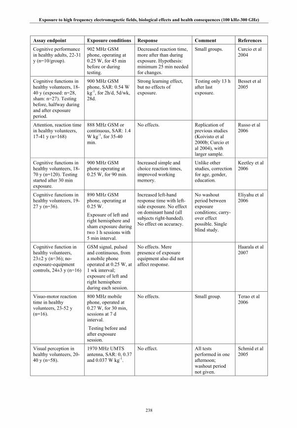
Exposure to high frequency electromagnetic fields, biological effects and health consequences (100 kHz-300 GHz)
238
Assay endpoint Exposure conditions Response Comment References
Cognitive performance in healthy adults, 22-31 y (n=10/group).
902 MHz GSM phone, operating at 0.25 W, for 45 min before or during testing.
Decreased reaction time, more after than during exposure. Hypothesis: minimum 25 min needed for changes.
Small groups. Curcio et al 2004
Cognitive functions in healthy volunteers, 18-40 y (exposed: n=28, sham: n=27). Testing before, halfway during and after exposure period.
900 MHz GSM phone, SAR: 0.54 W kg-1, for 2h/d, 5d/wk, 28d.
Strong learning effect, but no effects of exposure.
Testing only 13 h after last exposure.
Besset et al 2005
Attention, reaction time in healthy volunteers, 17-41 y (n=168)
888 MHz GSM or continuous, SAR: 1.4 W kg-1, for 35-40 min.
No effects. Replication of previous studies (Koivisto et al 2000b; Curcio et al 2004), with larger sample.
Russo et al 2006
Cognitive functions in healthy volunteers, 18-70 y (n=120). Testing started after 30 min exposure.
900 MHz GSM phone operating at 0.25 W, for 90 min.
Increased simple and choice reaction times, improved working memory.
Unlike other studies, correction for age, gender, education.
Keetley et al 2006
Cognitive functions in healthy volunteers, 19-27 y (n=36).
890 MHz GSM phone, operating at 0.25 W.
Exposure of left and right hemisphere and sham exposure during two 1 h sessions with 5 min interval.
Increased left-hand response time with left-side exposure. No effect on dominant hand (all subjects right-handed). No effect on accuracy.
No washout period between exposure conditions; carry-over effect possible. Single blind study.
Eliyahu et al 2006
Cognitive function in healthy volunteers, 23±2 y (n=36); no-exposure-equipment controls, 24±3 y (n=16)
GSM signal, pulsed and continuous, from a mobile phone operated at 0.25 W, at 1 wk interval; exposure of left and right hemisphere during each session.
No effects. Mere presence of exposure equipment also did not affect response.
Haarala et al 2007
Visuo-motor reaction time in healthy volunteers, 23-52 y (n=16).
800 MHz mobile phone, operated at 0.27 W, for 30 min, sessions at 7 d interval.
Testing before and after exposure session.
No effects. Small group. Terao et al 2006
Visual perception in healthy volunteers, 20-40 y (n=58).
1970 MHz UMTS antenna, SAR: 0, 0.37 and 0.037 W kg-1.
No effect. All tests performed in one afternoon; washout period not given.
Schmid et al 2005
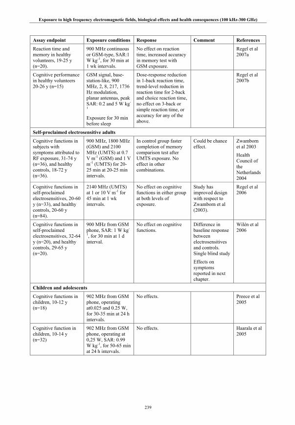
Exposure to high frequency electromagnetic fields, biological effects and health consequences (100 kHz-300 GHz)
239
Assay endpoint Exposure conditions Response Comment References
Reaction time and memory in healthy volunteers, 19-25 y (n=20).
900 MHz continuous or GSM-type, SAR:1 W kg-1, for 30 min at 1 wk intervals.
No effect on reaction time, increased accuracy in memory test with GSM exposure.
Regel et al 2007a
Cognitive performance in healthy volunteers 20-26 y (n=15)
GSM signal, base-station-like, 900 MHz, 2, 8, 217, 1736 Hz modulation, planar antennas, peak SAR: 0.2 and 5 W kg-
1
Exposure for 30 min before sleep
Dose-response reduction in 1-back reaction time, trend-level reduction in reaction time for 2-back and choice reaction time, no effect on 3-back or simple reaction time, or accuracy for any of the above.
Regel et al 2007b
Self-proclaimed electrosensitive adults
Cognitive functions in subjects with symptoms attributed to RF exposure, 31-74 y (n=36), and healthy controls, 18-72 y (n=36).
900 MHz, 1800 MHz (GSM) and 2100 MHz (UMTS) at 0.7 V m-1 (GSM) and 1 V m-1 (UMTS) for 20-25 min at 20-25 min intervals.
In control group faster completion of memory comparison test after UMTS exposure. No effect in other combinations.
Could be chance effect.
Zwamborn et al 2003
Health Council of the Netherlands 2004
Cognitive functions in self-proclaimed electrosensitives, 20-60 y (n=33), and healthy controls, 20-60 y (n=84).
2140 MHz (UMTS) at 1 or 10 V m-1 for 45 min at 1 wk intervals.
No effect on cognitive functions in either group at both levels of exposure.
Study has improved design with respect to Zwamborn et al (2003).
Regel et al 2006
Cognitive functions in self-proclaimed electrosensitives, 32-64 y (n=20), and healthy controls, 29-65 y (n=20).
900 MHz from GSM phone, SAR: 1 W kg-
1, for 30 min at 1 d interval.
No effect on cognitive functions.
Difference in baseline response between electrosensitives and controls. Single blind study
Effects on symptoms reported in next chapter.
Wilén et al 2006
Children and adolescents
Cognitive functions in children, 10-12 y (n=18)
902 MHz from GSM phone, operating at0.025 and 0.25 W, for 30-35 min at 24 h intervals.
No effects. Preece et al 2005
Cognitive function in children, 10-14 y (n=32)
902 MHz from GSM phone, operating at 0,25 W, SAR: 0.99 W kg-1, for 50-65 min at 24 h intervals.
No effects. Haarala et al 2005

Exposure to high frequency electromagnetic fields, biological effects and health consequences (100 kHz-300 GHz)
240
Assay endpoint Exposure conditions Response Comment References
Attention in same-level high school students (mean age 16 y).
Mobile phones users (n=37) vs non-users (n=35)
Better performance in users in 1 of 3 tests.
Large variation in phone use (175 – 27240 min total use time). No correction for multiple testing.
Lee et al 2001
Attention in undergraduate students.
1900 MHz from GSM phone for 25 min, 2 trials 2 min apart.
Decreased reaction time in 2nd trial in users. No effect on accuracy.
Single blind study.
Lee et al 2003
II.5.1.5. Subjective Symptoms
A wide range of subjective symptoms has been attributed to exposure to various sources of RF both at home and at work. Some people report they suffer a variety of subjective complaints, including headaches and migraines, fatigue, skin itches, and sensations of warmth (Frey 1998a & b; Hocking 1998; Chia et al 2000a & b; Hocking and Westerman 2001; Sandström et al 2001; Santini et al 2002, 2003; Rubin et al 2005; Röösli 2008). They attribute these symptoms to exposure from mobile telephones, nearby base stations, DECT cordless phones and, more recently, wireless LAN systems. Less commonly reported symptoms include dizziness, blurred vision, memory loss, confusion and vagueness, toothaches, and nausea. An increasing number of those people considers themselves electrosensitive.
The prevalence of these symptoms, and of the associated self-proclaimed electrical hypersensitivity, has been investigated in several countries. Hillert et al (2002) sent a paper questionnaire to 15000 adult residents of the Stockholm county. With an overall high response rate of 73%, 1.5% of the respondents reported to be sensitive to EMF exposure. The prevalence was higher in women than in men and the highest prevalence was found in the age group 60-69. Eriksson and Stenberg (2006) performed a more general study of symptoms related to the indoor environment, both at home and at work, among 2154 people in Sweden. They found that women reported symptoms more often than men. Symptoms associated with electrical hypersensitivity (EHS) were more frequent among VDU users. Levallois et al (2002) performed telephone interviews with 2072 Californians. They found a self-reported prevalence of 3.2% for being “allergic or very sensitive” to being near electrical devices, which was strongly associated with self-reported environmental illness or multiple chemical sensitivity. EHS only pertained to devices emitting power-frequency fields, however.
Several other studies investigated a possible association between mobile phone use and symptoms through questionnaires. Oftedal et al (2000) sent a questionnaire to 12000 Swedes and 5000 Norwegians. 13% of the respondents in Sweden and 31% in Norway indicated to have at least one of the symptoms questioned. Of those people, 45% indicated that they had taken measures to reduce exposure. The study was not blinded in that it was indicated that it was a study into effects of mobile phone use. This may have influenced the responses. Another problem is that the response rates between the two groups were rather different: 76% in Sweden and 64% in Norway. The main question addressed by this study, however, was whether there would be differences in pattern and types of symptoms between users of analog and digital phones. This was addressed by Sandström et al (2001). It appeared that the prevalence of symptoms was similar in users of either phone type. Overall, longer calls and a higher number of calls were associated with a higher prevalence of warmth behind or around or on the ear and with headaches and fatigue. This observation led to a third analysis that included a subset of 2197 users that made more than 2 calls per day and used one of four different types of GSM phones (Wilén et al 2003). These were selected to result in a range of SAR values. The prevalence of dizziness, discomfort and warmth behind the ear were associated with exposure to SAR values higher than 0.5 W kg-1 and long calling times. According to Sandström et al (2001) confounding factors such as psychosocial workload, occupation, and gender might affect the prevalence of symptoms. In this subset study the authors found that it was not possible, however, to correct for these factors because of the relatively small numbers of cases. The same group also studied heart function and mood in 14 self-proclaimed EHS and an equal-size control group
Exposure to high frequency electromagnetic fields, biological effects and health consequences (100 kHz-300 GHz)
241
(Sandström et al 2003). In the EHS group they observed larger heart rate variability. They also performed ELF measurements in the homes and found that the ELF levels were not associated with mood.
Provocation studies provide the most direct way of studying a possible effect of RF exposure on the occurrence of symptoms. A setback is that such studies only investigate direct, short-term interactions, while symptoms may only occur after some longer exposure time. In a single-blind provocation study, Koivisto et al (2001) presented two groups of 48 individuals without symptoms with either real or sham exposure to a pulsed 902 MHz field. Two experiments were performed. In the first, exposure and sham were in two 1-h sessions separated by 24 h. In the second, 30-min sessions were given without interval. Subjects were asked to rate subjective symptoms and sensations during the sessions. No significant differences were found between exposure conditions, although fatigue and headaches increased toward the end of sessions. In another single-blind provocation study, Hietanen et al (2002) challenged 20 volunteers who reported themselves to be sensitive to RF using analogue or digital phone signals. Blood pressure, heart rate and breathing rate were measured every 5 min and subjects were asked to report any abnormal feelings. Nineteen of the subjects reported symptoms, most of which were sensations in the head of pain or warmth. However, more symptoms were reported during sham exposure than real exposure. The subjects could not indicate whether they were being exposed and by what type of signal. The physiological parameters showed no relevant trends, although they tended to decrease throughout the day.
In a double-blind provocation study, Rubin et al (2006) investigated the effect of exposure to GSM RF on the severity of the symptoms experienced by 60 EHS, compared to 60 ‘non-sensitive’ subjects. Each subject was exposed or sham exposed for 50 min either to a GSM 900 signal or to a non-pulsed signal, both of which induced a localized SAR in the region of the head adjacent to the phone of 1.4 W kg-1. There were three separate experimental sessions over a two-year period within which the order of presentation was randomized and counter-balanced. All subjects were asked to score on visual analogue scales before, during and after exposure, the severity of headaches and various other symptoms such as nausea, fatigue, dizziness. The authors found that the proportion of sensitive participants who believed a signal was present during GSM exposure (60%) was similar to the proportion (63%) who believed one present during sham exposure. In addition, the prevalence of various symptoms experienced during exposure or sham exposure in people who reported themselves as GSM-sensitive was very much higher than in non-sensitive subjects, but this occurred irrespective of the exposure condition. In some cases, for sensitive subjects, the symptoms experienced were so severe that the individual withdrew from the study. Rubin et al (2006) suggested that psychological factors, possibly the conscious expectation of such symptoms (the nocebo effect), might have a key role in the etiology of this condition.
Wilén et al (2006) investigated the effects of mobile phone exposure on various physiological parameters such as heart rate variability, electrodermal activity, and respiration rate, measured before, during and after exposure, in twenty self-proclaimed RF-sensitive subjects and in twenty non-sensitive controls. In addition, cognitive function tests were performed; these have been described in the previous chapter. The subjects were exposed to GSM 900 RF for 30 min or sham exposed on two separate days. The study was single-blinded. No significant effects of RF radiation on any physiological parameter were found. However, people who experienced subjective symptoms showed differences in heart-rate variability compared to controls. Wilén et al (2006) suggested that these results might reflect differences between these two groups in autonomic nervous system function. Using the same exposure setup, Oftedal et al (2007) studied headache, discomfort and various physiological parameters in 17 subjects that attributed their symptoms to mobile phone use, using a double blind, sham-controlled design. Exposure was given in four 30-min sessions separated by 2 days. An increase in headache and discomfort was found after sham, but not after real exposure; subjects could not perceive being exposed. There were no effects on heart rate and blood pressure.
All these studies used mobile telephones as sources of exposure. It is however also of interest to study exposure at levels as experienced continuously in daily life that result from base station antennas. This was the idea behind the study of Zwamborn et al (2003). They explored the effects of exposure to GSM and UMTS signals on and cognitive functions (which have been reported in the previous chapter) and self-reported well-being (that will be reported here). A small, but significant, decrease in well-being after UMTS exposure was seen in both study groups (subjects who had previously reported symptoms
Exposure to high frequency electromagnetic fields, biological effects and health consequences (100 kHz-300 GHz)
242
attributed to GSM radiation and a control group without such symptoms). No effects were seen using GSM signals either at 900 or 1800 MHz. However, the validity of the questionnaire used to score well-being was challenged in the comprehensive analysis of this study by the Health Council of the Netherlands (2004). A follow-up study by Regel et al (2006) using an improved protocol with greater numbers of subjects investigated the effect only of the 2140 MHz UMTS base-station-like RF signal, identical to that used by Zwamborn et al (2003), on cognitive performance (reported in the previous chapter) and well-being in 33 self-proclaimed RF-sensitive subjects and in 84 non-sensitive subjects. Well-being was assessed using three standard questionnaires, one of which was identical to that used in the Zwamborn et al (2003) study. Well-being was not affected by UMTS radiation at either of two exposure levels. Even though RF-sensitive subjects generally reported more health problems, Regel et al (2006) found no difference between the two groups with respect to the applied field conditions. Subjects were also not able to discriminate between exposure levels, but they reported more health complaints when they suspected exposure, suggesting that, as indicated above, psychological factors may be involved in this condition.
This can also be concluded from the study of Lonne-Rahm et al (2000) who did not use exposure to an RF source, but instead used a VDU which only minimally emits RF EMF. Nevertheless the study is worth brief discussion here, because of the similarity of the results with the studies described above. Lonne-Rahm et al studied a group of 24 EHS subjects and 12 controls. They subjected participants to situations with either low or high stress (by exposing them to flashing lights while trying to solve mathematical problems) with or without exposure to EMF from a VDU. The EHS subjects reported increased skin symptoms when they thought that they were perceiving fields, but there was actually no difference between situations with the VDU field present or absent. No effect was detected on inflammatory mediators and skin mast cells.
Table II.5.7.: Subjective symptoms
Study endpoint Exposure conditions Response Comment References
Prevalence
Prevalence of self-reported hypersensitivity to electric or magnetic fields in Stockholm county, (n=15000, 19-80 y) assessed by paper questionnaire.
Response rate 73%.
EHS reported by 1.5% of respondents.
Highest in women and 60-69 y age group.
Hillert et al 2002
Prevalence of symptoms related to indoor environment in Sweden (n=2154, 18-64 y).
Response rate 70%.
High prevalence in VDU users.
Eriksson and Stenberg 2006
Self-reported hypersensitivity to EMF in California, USA (n=2072), assessed by telephone interview.
“Allergic or very sensitive” to being near electrical devices: prevalence = 3.2% (95% CI: 2.8, 3.7).
Alleging environmental illness or multiple chemical sensitivity was the strongest predictor of reporting being hypersensitive to EMFs. Only ELF field exposure questioned.
Levallois et al 2002

Exposure to high frequency electromagnetic fields, biological effects and health consequences (100 kHz-300 GHz)
243
Study endpoint Exposure conditions Response Comment References
Descriptive studies
Symptoms experienced by mobile phone users, assessed by paper questionnaire (Sweden : n=12000, Norway: n=5000).
Telephone use in daily life by people who own a job-related telephone.)
Response rates 76% (Sweden), 64% (Norway).
13% (Sweden) or 31% (Norway) of respondents had at least one symptom.
45% of those had taken measures to reduce exposure.
Study not blinded. Oftedal et al 2000
Comparison of symptoms experienced by users of analogue (n=8113) and digital mobile phones (n=8879).
Telephone use in daily life by people who own a job-related telephone.
No difference in prevalence of symptoms.
Higher prevalence of warmth behind/around or on the ear, headaches and fatigue with longer calling times or # calls.
Same group of subjects as in Oftedal et al (2000.)
Sandström et al 2001
Symptoms experienced by mobile phone users (n=2197) assessed by paper questionnaire.
Telephone use in daily life by people who own a job-related telephone.
Prevalence of some symptoms higher with SAR > 0.5 W kg-1 and long calling times.
Sub group of subjects from Oftedal et al (2000). No correction for possible confounding factors.
Wilén et al 2003
ECG in patients with perceived electrical hypersensitivity (n=14) and controls (n=14). Assessment of mood by paper questionnaire.
Exposure to RF and ELF in daily life; measurement of ELF.
No effect of ELF exposure on mood.
Disturbed pattern of heart rate variability in patients.
Small groups. Sandström et al 2003
Provocation – GSM/UMTS
Subjective symptoms associated with GSM use in subjects without symptoms (experiment 1: 18-49 y, n=48; experiment 2: 18-34 y, n=48).
GSM 900 phone, operating at a mean power of 0.25 W.
Exposures for 60 min at 24 h interval, or for 30 min without interval.
Questionnaire at start, middle, end of session.
No effect. Single blind study. Koivisto et al 2001

Exposure to high frequency electromagnetic fields, biological effects and health consequences (100 kHz-300 GHz)
244
Study endpoint Exposure conditions Response Comment References
Hypersensitivity symptoms associated with mobile phone use in self-reported electrosensitives, 37-67 y (n=20). Measurement of blood pressure, heart rate and breathing frequency every 5 min.
NMT 900 / GSM 900 and 1800 phones, operating at output power of resp. 1, 0.25 and 0.125 W, for 30 min with minimally 60 min interval.
More symptoms reported during sham exposure. No ability to detect exposure.
Single blind study. Hietanen et al 2002
Sensitivity to mobile phone signals in subjects with / without symptoms attributed to GSM exposure (n=60/group).
GSM 900, 900 MHz continuous, SAR:1.4 W kg-1 ± 30%, for 50 min at intervals of at least 24 h.
No difference in reported symptoms between groups and between exposure and sham.
No field perception.
Results suggested as being ‘nocebo’ effect.
Rubin et al 2006
Mobile phone related symptoms in self-proclaimed electrosensitives, 32-64 y (n=20), and healthy controls, 29-65 y (n=20).
900 MHz from GSM phone, SAR:1 W kg-1, for 30 min at 1 d interval.
No effects of exposure. Indication for difference in autonomous nervous system regulation between cases and controls.
Single blind study.
Only subjects with phone-related symptoms, no general electrosensitivity.
Effects on cognitive functions reported in previous chapter.
Wilén et al 2006
Headache associated with mobile phone use in subjects with symptoms attributed to mobile phone use, 20-58 y (n=17).
900 MHz from GSM phone, SAR: 1 W kg-
1, for 30 min at 2 d interval.
Increase in headache and discomfort higher with sham; no effect on heart rate, blood pressure; no perception of exposure.
Only subjects with phone-related symptoms, no general electrosensitivity.
Results explained as ‘nocebo’ effect.
Oftedal et al 2007
Well being in subjects with symptoms attributed to RF exposure, 31-74 y (n=36), and healthy controls, 18-72 y (n=36).
900 MHz, 1800 MHz (GSM) and 2100 MHz (UMTS) at 0.7 V m-1 (GSM) and 1 V m-1 (UMTS) for 20-25 min, at 20-25 min intervals.
Slightly decreased well-being after UMTS exposure.
Hypothesis-generating study; small numbers/group; questionable validity of well-being questionnaire.
Zwamborn et al 2003
Well-being in self-proclaimed electrosensitives, 20-60 y (n=33), and healthy controls, 20-60 y (n=84).
2140 MHz (UMTS) at 1 or 10 V/m for 45 min. at 1 wk intervals.
No effect on well-being in either group at both levels of exposure.
No ability to detect exposure.
Replication of Zwamborn et al 2003 study with improved design.
Regel et al 2006

Exposure to high frequency electromagnetic fields, biological effects and health consequences (100 kHz-300 GHz)
245
Study endpoint Exposure conditions Response Comment References
Provocation – other
Provocation with stress and EMF of patients with "sensitivity to electricity” (n=24) and controls (n=12).
30 min high/low stress with/without VDU exposure, at 1 week interval.
Patients reported increased skin symptoms when perceiving fields.
In effect, no difference between fields on / off.
No effect on inflammatory mediators and skin mast cells.
Small groups. Little RF exposure form VDU.
Lonne-Rahm et al 2000
II.5.1.6. Summary on nervous system
There is some evidence for effects of exposure to a GSM-type signal on the spontaneous EEG. A well-performed large study has confirmed previous smaller studies in finding increased power in the alpha band (8–12 Hz) of brain activity. Effects on other frequency bands of natural brain activity have not been consistently demonstrated. These observations are not corroborated, however, by the results from studies on evoked potentials. Although in some studies some small but inconsistent effects were observed, no effects at all were found when auditory evoked potentials were assessed in the same large study group described above.
A similar conclusion of variable results can be drawn with respect to the effects of exposure to GSM-type signals on sleep, although there is some evidence emerging that suggests there may be an effect on sleep EEG. Some studies, but not all, have indicated effects on EEG power in alpha or beta bands with exposure during sleep. A reported shortening of sleep latency was not subsequently reproduced. Other studies which looked at exposure during 30 min before going to sleep also reported variable results, sometimes reporting increases in alpha and beta band power. In summary, exposure to a GSM-type signal may result in minor effects on brain activity, but it should be stressed that such changes have not been found to relate to any health effects. There are some indications of changes in regional cerebral blood flow during and following RF exposure, but the available data are equivocal. It should be noted that changes in rCBF are not by themselves an indication of health damage. No consistent cognitive performance effects were seen. Studies with larger numbers of subjects generally show no effect. No higher sensitivity was shown in children nor in self-proclaimed electrosensitives compared to healthy adults. If anything, any effect is small and exposure seems to improve performance. It was not possible to derive a dose-response relationship.
The weight of evidence from the studies on auditory and vestibular function indicates that neither hearing nor the sense of balance is influenced by short-term exposure to mobile phone signals.
A wide range of subjective symptoms including headaches and migraine, fatigue, and skin itches have been attributed to various RF sources both at home and at work. However, in provocation studies a causal relation between EMF exposure and symptoms has never been demonstrated. Possibly the conscious expectation of such symptoms may play a role in the etiology of this condition.
II.5.2. Endocrine system
The majority of volunteer studies of the effects of RF radiation on the endocrine system have focused on hormones released into the blood stream by the pineal and pituitary neuroendocrine glands. These are both situated in the head and intimately connected with and controlled by the nervous system. The hormones they release exert a profound influence on body metabolism and physiology, particularly
Exposure to high frequency electromagnetic fields, biological effects and health consequences (100 kHz-300 GHz)
246
during development and reproduction, partly via their influence on the release of hormones from other endocrine glands situated elsewhere in the body.
Most studies have investigated the effects of RF exposure on circulating levels of the pineal hormone melatonin or on the urinary excretion of the major metabolite: 6-sulphatoxymelatonin (aMT6s). Fewer studies have been carried out on circulating levels of pituitary hormones or other hormones released from other endocrine glands such as the adrenal cortex.
II.5.2.1. Melatonin studies
Melatonin in humans is produced in a distinct daily or circadian rhythm, peaking during the night and strongly influences circadian physiology and behavior. Night-time peak values of serum melatonin, however, can vary up to ten-fold between different people.
Mann et al (1998) studied the effect of exposure and sham exposure on two successive nights to circularly polarized GSM 900 RF on the nocturnal profiles of melatonin and three other hormones (see below) in 22 male subjects. The treatments were carried out from 23.00 to 07.00 the following morning in a randomized order of presentation under single-blind conditions. Blood samples were withdrawn from an indwelling catheter every 20 min throughout this period. There was no statistically significant effect on night-time serum melatonin levels.
De Seze et al (1999) evaluated the effect on the serum melatonin levels of 2-h daily exposure, 5 days per week, of 19 male volunteers to GSM 900 and 19 others to GSM 1800 mobile phone radiation over a period of four weeks. Exclusion criteria included night-shift work, endocrine disorders and other factors. However, it is not clear whether confounders such as alcohol, coffee or light-at-night were controlled. Blood samples were taken at regular intervals throughout a 24-h period before exposure, after 2 and 4 weeks exposure, and 2 weeks following exposure. Each subject acted as their own control; sham exposures were carried out on the days of the pre-exposure and post-exposure sampling sessions. There was no effect of exposure to either mobile phone signal on the serum melatonin profiles, suggesting that there was no overall cumulative or persistent effect on melatonin secretion.
Radon et al (2001) investigated the effect of circularly polarized GSM 900 signal on salivary melatonin (and cortisol; see below) in 8 volunteers. The RF signal was transmitted by an antenna positioned 10 cm behind each subjects head. In double-blind trials, each subject underwent a total of 20 randomly allotted 4-h periods of exposure and sham exposure between 12.00 and 16.00 or 22.00 and 02.00 the following morning, over a five month summer period. Each treatment period was separated by 2-3 days. Saliva samples were taken at regular times on the day of treatment and on wakening the following morning. There were no significant differences in salivary melatonin concentrations between the exposed and sham exposed conditions. The data were, however, rather variable, a possible consequence of the small number of subjects.
Bortkiewicz et al (2002) exposed or sham exposed 9 male volunteers to GSM 900 RF between 18.00 and 19.00 h emitted from a mobile phone positioned near the subject’s head. Urine samples were collected at 19.00, 24.00 and 07.00 the following day and were analyzed for aMT6s content, normalized to creatinine content (a standard procedure to account for errors in the estimation of urine volume). Exposure and sham exposure of all 9 subjects took place on separate days more than one week apart. There was no significant change in aMT6s excretion at these three time-points. There was, however, considerable variation in aMT6s excretion between individuals, reducing the power of the study to detect any effect.
Jarupat et al (2003) studied the effect of exposure and sham exposure to 1906 MHz RF from a mobile phone (Japanese signal) on salivary melatonin levels in eight female subjects. The study was a crossover design. The subjects entered a climatically controlled chamber at 10.00 and held the phone to their left ear for 30 min every hour from 19.00 to 01.00 over a two day period. The authors state that the subjects were unaware whether the phone emitting RF radiation or not, but it is not clear whether this sequence was randomized. Salivary samples were taken at the start of the treatment, and one hour after the treatment terminated. Salivary melatonin levels were reported to be significantly depressed following RF exposure compared to levels taken after sham exposure. The volunteers had led ‘a well-regulated life’ for a week
Exposure to high frequency electromagnetic fields, biological effects and health consequences (100 kHz-300 GHz)
247
before the study, but the authors acknowledge there may have been uncontrolled factors and recommended a larger study.
Wood et al (2006) examined the effect of exposure to GSM 900 RF from a mobile handset on the night-time excretion of aMT6s in 55 adult volunteers. The study was a double-blind crossover design; the subjects were both exposed or sham exposed for 30 min in random sequence on 2 successive Sunday nights. Urine collection was taken shortly after exposure, prior to retiring to bed, and on rising next morning. The authors reported that, after normalization to creatinine concentration, the pre-bedtime aMT6s concentrations were significantly reduced (by about 27%) in the exposed group compared to the sham-exposed group. There was no difference between the post-bedtime measures. The authors acknowledge that the significant result may be spurious, but speculate that it may indicate a delay in onset of the night-time rise in circulating melatonin levels, possibly in a sensitive sub-group of 4 individuals. However, given that the pre-bedtime urine sample was taken shortly after the end of the 30 min exposure, it is difficult to see how there would have been sufficient time for any exposure-induced change in the circulating melatonin levels to have significantly influenced the aMT6s urinary content. This suggests that the effect is more likely to be spurious, perhaps a result of possible lifestyle confounders that were uncontrolled.
II.5.2.2. Pituitary and other hormones
The main pituitary hormones investigated in EMF studies include several hormones involved in the control of growth and body physiology, particularly thyroid-stimulating hormone (TSH) that controls the function of the thyroid gland and the release of thyroxin, adrenocorticotrophic hormone (ACTH), which regulates the function of the adrenal cortex and particularly the release of cortisol, and growth hormone (GH). Hormones released by the pituitary which have important sexual and reproductive functions have also been studied, particularly follicle stimulating hormone (FSH), luteinizing hormone (LH) and prolactin (PRL).
Mann et al (1998), as part of a wider study of the effects of circularly polarized GSM 900 RF on the neuroendocrine system, studied effects on the nocturnal profiles of cortisol, GH and LH, as well as melatonin (see above) in 22 male subjects. Exposure and sham exposure were carried out on two successive nights from 23.00 to 07.00 the following morning in a randomized order of presentation under single-blind conditions. Blood samples were withdrawn from an indwelling catheter every 20 min throughout this period. The authors reported no significant effects on GH or LH levels, but there was a slight, transient elevation of cortisol levels immediately after the onset of RF exposure, suggesting a transient activation of the pituitary adrenal axis.
De Seze et al (1998) evaluated the effect on the hormones of the anterior pituitary gland of 2-h daily exposure, 5 days per week, of 18 male volunteers to GSM 900 RF over a period of four weeks. Subjects acted as their own controls. Blood samples were taken during 9 weeks, 3 weekly samples before exposure, 4 during exposure and 2 after exposure. Most sample were taken on a Monday; thus, during the exposure period, blood samples were taken 48 h after GSM exposure on the previous Friday except following the last exposure (week 7), when a blood sample was taken the day after (Saturday). Because some hormone levels respond quickly to the stress of having a blood sample taken a 15-min rest period after skin puncture but before blood withdrawal was instituted. The samples were tested for 6 hormones: ACTH, TSH, GH, PRL, LH and FSH. Statistical analysis was adjusted for multiple comparisons. All mean hormone concentrations remained within the limits of physiological variation; although for some individuals, aberrant levels of ACTH, GH and PRL, which are known to be affected by stress, suggested that puncture stress hadn’t been completely eliminated. Otherwise, there was no significant weekly variation in five of the six hormones studied, but TSH levels showed tendency to decrease from the baseline (i.e. mean pre-exposure value) during exposure, reaching statistical significance (21% decrease) on the 7th sampling, i.e. on the day following the last day of GSM exposure, but recovered to the baseline value during the post-exposure period. The authors concluded that there was no long-lasting or cumulative effect of GSM radiation.
As part of the melatonin study described above, Radon et al (2001) investigated the effect of circularly polarized GSM 900 signal on cortisol levels, directed to the back of the head of 8 volunteers. In double-
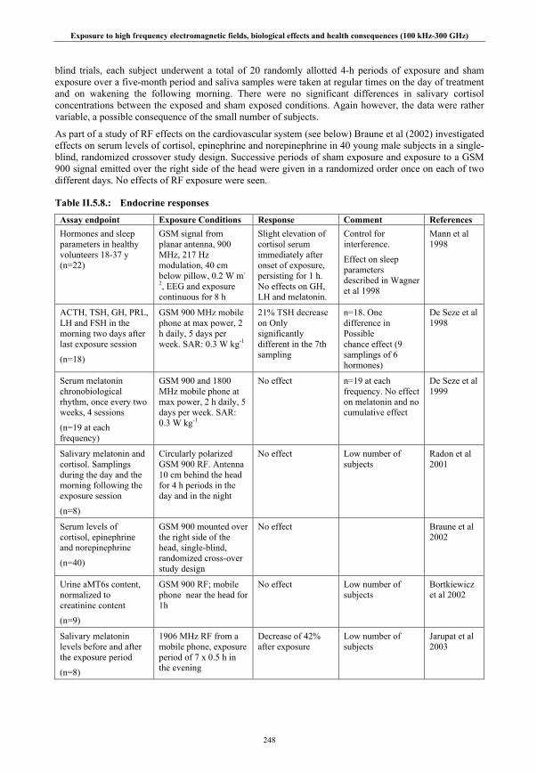
Exposure to high frequency electromagnetic fields, biological effects and health consequences (100 kHz-300 GHz)
248
blind trials, each subject underwent a total of 20 randomly allotted 4-h periods of exposure and sham exposure over a five-month period and saliva samples were taken at regular times on the day of treatment and on wakening the following morning. There were no significant differences in salivary cortisol concentrations between the exposed and sham exposed conditions. Again however, the data were rather variable, a possible consequence of the small number of subjects.
As part of a study of RF effects on the cardiovascular system (see below) Braune et al (2002) investigated effects on serum levels of cortisol, epinephrine and norepinephrine in 40 young male subjects in a single-blind, randomized crossover study design. Successive periods of sham exposure and exposure to a GSM 900 signal emitted over the right side of the head were given in a randomized order once on each of two different days. No effects of RF exposure were seen.
Table II.5.8.: Endocrine responses
Assay endpoint Exposure Conditions Response Comment References Hormones and sleep parameters in healthy volunteers 18-37 y (n=22)
GSM signal from planar antenna, 900 MHz, 217 Hz modulation, 40 cm below pillow, 0.2 W m-
2, EEG and exposure continuous for 8 h
Slight elevation of cortisol serum immediately after onset of exposure, persisting for 1 h. No effects on GH, LH and melatonin.
Control for interference.
Effect on sleep parameters described in Wagner et al 1998
Mann et al 1998
ACTH, TSH, GH, PRL, LH and FSH in the morning two days after last exposure session
(n=18)
GSM 900 MHz mobile phone at max power, 2 h daily, 5 days per week. SAR: 0.3 W kg-1
21% TSH decrease on Only significantly different in the 7th sampling
n=18. One difference in Possible chance effect (9 samplings of 6 hormones)
De Seze et al 1998
Serum melatonin chronobiological rhythm, once every two weeks, 4 sessions
(n=19 at each frequency)
GSM 900 and 1800 MHz mobile phone at max power, 2 h daily, 5 days per week. SAR: 0.3 W kg-1
No effect n=19 at each frequency. No effect on melatonin and no cumulative effect
De Seze et al 1999
Salivary melatonin and cortisol. Samplings during the day and the morning following the exposure session
(n=8)
Circularly polarized GSM 900 RF. Antenna 10 cm behind the head for 4 h periods in the day and in the night
No effect Low number of subjects
Radon et al 2001
Serum levels of cortisol, epinephrine and norepinephrine
(n=40)
GSM 900 mounted over the right side of the head, single-blind, randomized cross-over study design
No effect Braune et al 2002
Urine aMT6s content, normalized to creatinine content
(n=9)
GSM 900 RF; mobile phone near the head for 1h
No effect Low number of subjects
Bortkiewicz et al 2002
Salivary melatonin levels before and after the exposure period
(n=8)
1906 MHz RF from a mobile phone, exposure period of 7 x 0.5 h in the evening
Decrease of 42% after exposure
Low number of subjects
Jarupat et al 2003
Exposure to high frequency electromagnetic fields, biological effects and health consequences (100 kHz-300 GHz)
249
Assay endpoint Exposure Conditions Response Comment References Urine aMT6s content, normalized to creatinine content, on the evening following exposure and on the following morning
(n=55)
GSM 900 RF from a mobile handset for 0.5 h - double-blind crossover design
Decrease of 27% on the evening sampling following exposure.
Not physiologically relevant (no time for urine excretion to occur after exposure)
Wood et al 2006
II.5.2.3. Summary on endocrine system
No cumulative effect seems to occur upon repeated chronic exposure for one month on serum melatonin or pituitary hormones. Most studies did not report effects after acute exposure, but often, statistical power was insufficient because of the low number of volunteers. Only one study with acutely repeated exposure seems worth confirming, showing a melatonin decrease in saliva samplings in the morning after 7 consecutive 0.5-h sessions every hour in the evening.
II.5.3. Cardiovascular function and thermoregulation
Volunteer studies have investigated the effects of mobile phone type RF radiation at levels generally assumed to be too low to induce significant heating. A number of studies have been carried out investigating possible effects on heart rate, heart rate variability and on blood pressure. In addition, there is an established literature on cardiovascular responses to RF heating, such as those involved in thermoregulation, and a number of studies address these endpoints. Indeed, such thermoregulatory responses are mediated primarily through well-understood changes in cardiovascular system dynamics and this topic is therefore included here along with a discussion of heat stress disorders and the effects of localized heating in order to place the possible health consequences of RF heating into a broader occupational and environmental context. In particular, a full evaluation of the possible health effects of an RF heat load should also take into account all sources of heat, such as rate of physical work, and the ease with which heat can be lost from the body, which in turn depends to some extent on climatic conditions, clothing etc.
II.5.3.1. Heart rate and blood pressure changes
Braune et al (1998) have reported acute effects on blood pressure in 10 human volunteers exposed to a conventional GSM digital mobile phone positioned close to the right side of the head. After 35 min of exposure, heart rate, blood pressure and capillary perfusion were measured with the subject either supine or standing for 60 s. They found that the heart rate during these tests was slightly lower after exposure to RF than following non-exposed control sessions, and both systolic and diastolic blood pressure were elevated by 5-10 mm Hg. Since capillary perfusion (blood flow through capillaries of the hand) was decreased, the authors concluded that the effects on blood pressure were due to excessive vasoconstriction. This study has been criticized on the basis of both its design and the statistical analysis (Reid and Gettinby 1998). In particular, the ‘placebo’ (sham exposure) session preceded the test session for all subjects, and therefore the small cardiovascular changes might have been resulted simply from the lengthy period of the experiment.
Braune et al (2002) further investigated these effects on 40 young male subjects in a single-blind, ‘randomized crossover study design. Successive periods of sham exposure and exposure to a GSM 900 source mounted over the right side of the head were given in a randomized order once on each of two different days. As in the previous study, systolic and diastolic pressure showed a slow continuous increase of about 5 mm Hg throughout the 50-min protocol; heart rate remained constant. This change in blood pressure was however independent of RF exposure.
Huber et al (2003) reported on an extended analysis of data first published in two previous studies (Borbély et al 1999; Huber et al 2000), focusing on further analysis of EEG recordings (see Chapter
Exposure to high frequency electromagnetic fields, biological effects and health consequences (100 kHz-300 GHz)
250
II.5.1.1) and on recordings of the ECG, particularly heart rate and heart rate variability. In these previously published studies, volunteers were exposed to GSM 900 signals either during sleep or during the waking period preceding sleep. In the first experiment, subjects were exposed intermittently during an 8-h night-time sleep period and, in the second experiment, on one side of the head for 30 min before a 3-h daytime sleep period. RF exposure prior to sleep reduced the heart rate during waking and stage-1 sleep, but not during RF exposure itself. Heart rate variability was affected during sleep in both experiments, showing both increases and decreases in the spectral power content prior to and during sleep. The authors speculate that this might indicate changes in sympathetic or vagal activity.
Tahvanainen et al (2004) measured heart rate and blood pressure responses in 32 volunteers during and after a randomized double-blind 35 min exposure to 900, 1800 MHz and sham exposure in three separate exposure sessions. Cardiovascular responses were evaluated in terms of blood pressure and heart rate during four different tests of autonomic regulation of these end-points: a spontaneous breathing test, a deep breathing test, a head-up tilt table test and an expiratory breath test. There were no effects of exposure to either RF frequency on diastolic or systolic blood pressure, or on heart rate, either during or after exposure.
Nam et al (2006) investigated the effects of 30-min sham exposure followed, after a break of 30 min, by exposure to CMDA 835 MHz RF, on blood pressure and heart rate, along with respiration rate and skin resistance (measured on two fingers), in a group of 21 teenagers, and in a group of 21 adults. These parameters were measured after an initial 10 min rest, after 15- and 30-min RF exposure or sham exposure, and 10 min after exposure termination. There were no significant changes in heart rate, diastolic or systolic blood pressure, or respiration rate during any part of the study. However, skin resistance reportedly decreased in the teenagers after 15 and 30 min of RF exposure, and in all males, when grouped together for analysis, after 30 min; in both cases returning to baseline levels within 10 min after the cessation of RF exposure. The authors suggest that the result indicates possible teenage and male susceptibility to CDMA RF radiation. However, the study design, in which sham exposure is always followed by RF exposure, mirrors that of the study by Braune et al (1998), criticized for its lack of randomization of the sham/exposure sequence.
Parazzini et al (2007) focused their investigation of possible GSM 900 RF effects on heart rate variability in 26 volunteers. Frequency and time-domain analysis of heart rate variability is thought to provide quantitative information regarding the sympathetic and parasympathetic control of heart rate by the autonomic nervous system. Heart rate variability data were collected during two different sessions, one with a real RF exposure and the other with a sham exposure. These sessions were performed on separate days in a random order following a double blind experimental design. During each 26 min session, the subject underwent a standard rest-to-stand protocol, thought to elicit sympathetic activity. Data analysis revealed that RF exposure did not affect most heart rate variability parameters; however, a few weak but statistically significant changes were seen in some minor indices of heart rate variability such as an increase the low frequency component as subjects moved into the stand position. The authors suggest this might indicate an augmentation of sympathetic activity. However, it is not clear whether the analysis allowed for multiple testing, and so the significance of these minor changes may have been overestimated.
Barker et al (2007) examined the effects of both TETRA and GSM signals on blood pressure and heart rate variability in 120 subjects. In this study, the subjects were seated and blood pressure and heart rate were recorded during a 20-min pre-exposure period, and a 40-min double-blind RF exposure or sham-exposure session. Four different sets of RF signals were applied in addition to the sham exposures: GSM modulated signals, GSM carrier wave, TETRA modulated, and TETRA carrier wave. The authors found no effect of any RF signal on mean arterial blood pressure, or on any measure of heart rate variability, either in the low frequency or high frequency bands. However, mean arterial blood pressure was reduced (by ~ 0.7 mm Hg) for GSM sham exposures and the authors speculate that this might have resulted from a slight increase in the operating temperature of the handset when in this mode.
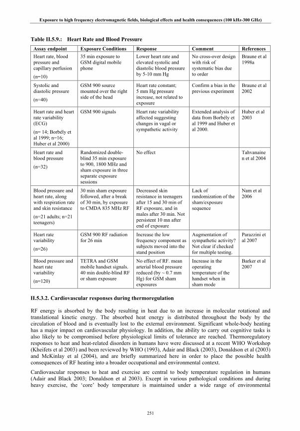
Exposure to high frequency electromagnetic fields, biological effects and health consequences (100 kHz-300 GHz)
251
Table II.5.9.: Heart Rate and Blood Pressure
Assay endpoint Exposure Conditions Response Comment References Heart rate, blood pressure and capillary perfusion
(n=10)
35 min exposure to GSM digital mobile phone
Lower heart rate and elevated systolic and diastolic blood pressure by 5-10 mm Hg
No cross-over design with risk of systematic bias due to order
Braune et al 1998a
Systolic and diastolic pressure
(n=40)
GSM 900 source mounted over the right side of the head
Heart rate constant; 5 mm Hg pressure increase, not related to exposure
Confirm a bias in the previous experiment
Braune et al 2002
Heart rate and heart rate variability (ECG)
(n= 14; Borbély et al 1999; n=16; Huber et al 2000)
GSM 900 signals Heart rate variability affected suggesting changes in vagal or sympathetic activity
Extended analysis of data from Borbély et al 1999 and Huber et al 2000.
Huber et al 2003
Heart rate and blood pressure
(n=32)
Randomized double-blind 35 min exposure to 900, 1800 MHz and sham exposure in three separate exposure sessions
No effect Tahvanainen et al 2004
Blood pressure and heart rate, along with respiration rate and skin resistance
(n=21 adults; n=21 teenagers)
30 min sham exposure followed, after a break of 30 min, by exposure to CMDA 835 MHz RF
Decreased skin resistance in teenagers after 15 and 30 min of RF exposure, and in males after 30 min. Not persistent 10 mn after end of exposure
Lack of randomization of the sham/exposure sequence
Nam et al 2006
Heart rate variability
(n=26)
GSM 900 RF radiation for 26 min
Increase the low frequency component as subjects moved into the stand position
Augmentation of sympathetic activity? Not clear if checked for multiple testing.
Parazzini et al 2007
Blood pressure and heart rate variability
(n=120)
TETRA and GSM mobile handset signals. 40 min double-blind RF or sham exposure
No effect of RF. mean arterial blood pressure reduced (by ~ 0.7 mm Hg) for GSM sham exposures
Increase in the operating temperature of the handset when in sham mode
Barker et al 2007
II.5.3.2. Cardiovascular responses during thermoregulation
RF energy is absorbed by the body resulting in heat due to an increase in molecular rotational and translational kinetic energy. The absorbed heat energy is distributed throughout the body by the circulation of blood and is eventually lost to the external environment. Significant whole-body heating has a major impact on cardiovascular physiology. In addition, the ability to carry out cognitive tasks is also likely to be compromised before physiological limits of tolerance are reached. Thermoregulatory responses to heat and heat-related disorders in humans have were discussed at a recent WHO Workshop (Kheifets et al 2003) and been reviewed by WHO (1993), Adair and Black (2003), Donaldson et al (2003) and McKinlay et al (2004), and are briefly summarized here in order to place the possible health consequences of RF heating into a broader occupational and environmental context.
Cardiovascular responses to heat and exercise are central to body temperature regulation in humans (Adair and Black 2003; Donaldson et al 2003). Except in various pathological conditions and during heavy exercise, the ‘core’ body temperature is maintained under a wide range of environmental

Exposure to high frequency electromagnetic fields, biological effects and health consequences (100 kHz-300 GHz)
252
conditions at a value of about 37°C with a circadian fluctuation of about ±0.5°C. Heat gained at rest, during exercise or exposure to RF, has to be compensated by heat loss and is often accompanied by a small increase in heat storage. The principal heat loss mechanisms in humans are radiant and convective heat loss from the skin through increased skin blood flow and evaporative heat loss from sweat. Heat storage reflects shifts in both peripheral and core temperatures and occurs, for example, during heavy exercise or in hot, humid environments. Prolonged rates of increase in heat storage, such as 0.5–1.0 W kg−1 for 1–2 hours, will lead to unacceptable rises in body temperature (Gordon 1984). In moderate conditions, however, increased skin blood flow will increase heat storage through an increase in the temperature of the peripheral tissues of the body, increasing heat loss without necessarily increasing core temperature.
These relationships can be expressed more formally (Bligh and Johnson 1973) as:
M ± Wo = E ± C ± R± K±S Eqn. 1
where M = rate of metabolic heat production, SAR = specific energy absorption rate of internally absorbed RF radiation, Wo = rate of physical work, E = rate of evaporative heat loss, C = rate of convective heat loss, R = rate of radiant heat loss, K = rate of conductive heat loss, and S = rate of heat storage in the body (All values can be expressed in eg W (ie Watts or Joules s-1), W m-2, or W kg-1).
Generally, values for the rate of whole-body metabolic heat production in humans vary between about 1 W kg-1 and 10 W kg-1; typical average values for many industrial jobs for example vary between about 2.5 W kg-1 for light work and 6 W kg-1 for heavy manual labor (NIOSH 1980). The degree to which humans can increase skin blood flow and sweat confers marked thermoregulatory advantages compared to other mammals, allowing excessive heat loads to be dissipated more effectively (Adair and Black 2003; Adair 2008). In particular, skin blood flow can increase from approximately 0.2-0.5 liters min-1 in thermally neutral conditions, to values exceeding 7-8 liters min-1 during hyperthermia, a dynamic range which is much higher than in other species (Donaldson et al 2003).
The main physical difference between children and adults affecting thermoregulation is the much higher surface-area-to-mass ratio of children (Falk 1998). In a warm environment this allows them to rely more upon increased skin blood flow and heat loss through convection and radiation, and less upon evaporative cooling. The lower sweating rate of children is partly due to a lower sensitivity of the sweating mechanism to thermal stimuli. Nevertheless, during exercise in thermally neutral or warm environments, children thermoregulate as effectively as adults. When ambient temperatures exceed body temperature, however, children are more liable to have a higher rate of heat absorption compared with adults. Also, whilst neither children nor adults sufficiently replace fluid loss during exercise in the heat, dehydration may have a more detrimental effect on children because of their greater reliance upon elevated skin blood flow to dissipate heat.
Whole-body and localized RF heating
RF radiation absorbed by the body provides an additional source of heat that has to be lost through the normal heat loss mechanisms described above. The heat balance equation (1) given above can be modified to account for the absorption of RF radiation (Adair 1996) as follows:
M ± Wo + Arf = E ± C ± R± K±S Eqn. 2
where Arf = the whole body SAR, ie the rate of absorption of RF per unit body mass averaged over the whole body.
The constraints described above apply to the cardiovascular responses and heat loss during exercise in hot environments would also apply to additional heat loads generated by exposure to RF radiation, except that, in the latter case, heating is passive rather than the result of muscular activity, avoiding the potential conflict between the demands for skin and blood flow. However, individuals may of course be exposed to RF radiation whilst engaging in physical activity in hot environments.
Exposure to high frequency electromagnetic fields, biological effects and health consequences (100 kHz-300 GHz)
253
The physiological responses of seated or supine volunteers acutely exposed to RF radiation have been studied mostly by two groups of researchers, namely Shellock et al (1989, 1994), considering mostly the safety of clinical magnetic resonance diagnostic procedures, and by Adair et al (1998, 1999, 2001a, 2001b, 2003, 2005) investigating whole or partial body exposures.
Shellock et al (1989) exposed six volunteers to 64 MHz RF magnetic fields for 30 min; the RF antenna was situated over each subject's abdomen resulting in a partial body exposure. SARs averaged over the whole body mass ranged between 2.7 and 4.0 W kg-1 with a mean value of 3.3 W kg-1. Over the 30 min exposure, body temperature rose by an average of 0.1oC, although this response was variable. Cutaneous blood flow and skin temperature in the abdominal region were significantly increased (and were still rising at the end of exposure). All of the subjects reported that they felt warm during the procedure and each of them had visible signs of perspiration on their forehead, chest and abdomen. Subsequently, Shellock et al (1994) exposed six volunteers to 64 MHz RF magnetic fields for 16 min; exposure was centered over the abdomen as in the previous study. Whole-body SARs were estimated as about 6 W kg-1. The ambient room temperature was 22.3oC, the relative humidity was maintained at 45 and air movement was kept to less than 0.1 m s-1. Tympanic membrane temperature rose significantly by an average of 0.4oC; heart rate rose significantly by 13 bpm. In addition, skin blood flow on the abdomen more than doubled. Each subject had signs of perspiration, especially on the forehead, face, abdomen and chest and there were statistically significant increases in skin temperature on the abdomen, upper arm, hand, chest and thigh.
The physiological responses of healthy volunteers, most of whom engaged in regular exercise programs, given whole or partial body exposure to RF radiation at 450 or 2450 MHz, has been investigated in a series of experiments by Adair et al (1998, 1999, 2001a, 2001b). The RF was directed to the back of each seated volunteer; each exposure lasted 45 min and followed a 30 min equilibration period. In the first study, Adair et al (1998) exposed 7 adult volunteers to 450 MHz at two power densities (180 and 240 W m-2) and at three environmental temperatures (24, 28 and 31oC). Peak surface SARs on the back of 6 and 7.7 W kg-1 were estimated from measurements in phantoms; there was no measure of whole-body heat load. Vigorous increases in sweating rate on the back and chest, directly related to power density, peak SAR and environmental temperature were reported; core body (oesophageal) temperature and metabolic rate were essentially unchanged during exposure.
An experiment by Adair et al (1999) compared the physiological responses induced in the volunteers by exposure at 450 MHz in the previous study (Adair et al 1998) with those induced by a similar exposure at 2450 MHz in the present study, using a different group of 7 subjects (one subject participated in both studies) in a different laboratory. The experimental procedures were similar to those described above except that the power densities at 2450 MHz were adjusted (270 and 350 W m-2) to produce similar peak surface SARs (6.0 and 7.7 W kg-1) to those induced by 450 MHz. There was no difference in metabolic heat production, or in core body temperature at the different RF frequencies. However, skin temperature in the irradiated area was greater at 2450 MHz than at 450 MHz, probably reflecting the better stimulation of thermal skin receptors by the higher frequency, less penetrative, RF fields. Local sweat rates were more variable within and between the different groups, and related to both local SAR and environmental temperature. A later study (Adair et al 2001a) reported that exposure to 2450 MHz at higher power densities (500 or 700 W m-2), where local peak SARs were 11 and 15.4 W kg-1, resulted in increased skin temperatures in the exposed region increased of up to 4.0 oC, and more vigorous local sweating and increased local skin blood flow compared to results from the previous study. There was no statistically significant effect of exposure on core body temperature or metabolic rate; hole body SARs were estimated as approximately 0.7 and 1.0 W kg-1.
Adair et al (2001b) compared the effects of exposure to pulsed and continuous wave 2450 MHz in two separate experiments, carried out one and a half years apart, on the thermoregulatory responses of two different groups of 7 subjects (four subjects participated in both experiments). Similar levels of exposure resulted in peak SARs of 6.0 and 7.7 W kg-1 for both CW and pulsed RF; whole body SARs were estimated as 0.4 and 0.5 W kg-1. The authors reported that there was little change in core body temperature and metabolic heat production in all test conditions with no reliable differences between CW and pulsed RF. The increase in skin temperature in the exposed region of the back was greater following pulsed RF compared to CW RF. Otherwise, there were no reliable differences between pulsed and CW
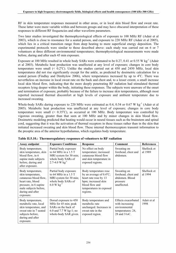
Exposure to high frequency electromagnetic fields, biological effects and health consequences (100 kHz-300 GHz)
254
RF in skin temperature responses measured in other areas, or in local skin blood flow and sweat rate. These latter were more variable within and between groups and may have obscured interpretation of these responses to different RF frequencies and other waveform parameters.
Two later studies investigated the thermophysiological effects of exposure to 100 MHz RF (Adair et al 2003), which is close to resonance for a seated person, and exposure to 220 MHz RF (Adair et al 2005), which lies in a critical transition range from deep heating to more superficial energy deposition. The experimental protocols were similar to those described above: each study was carried out on 6 or 7 volunteers at three different environmental temperatures; thermophysiological measurements were made before, during and after each 45 min exposure.
Exposure at 100 MHz resulted in whole body SARs were estimated to be 0.27, 0.41 or 0.55 W kg-1 (Adair et al 2003). Metabolic heat production was unaffected at any level of exposure; changes in core body temperature were small (< 0.2oC). Unlike the studies carried out at 450 and 2450 MHz, local skin temperatures did not rise significantly except for the ankle, as predicted by dosimetric calculation for a seated person (Findlay and Dimbylow 2006), where temperatures increased by up to 4oC. There was nevertheless an increase in local sweat rate on the back and chest and, to a lesser extent, a small increase local skin blood flow, indicating that the more deeply penetrating RF radiation had stimulated thermal receptors lying deeper within the body, initiating these responses. The subjects were unaware of the onset and termination of exposure, probably because of the failure to increase skin temperatures, although most reported increased thermal discomfort at high levels of exposure and ambient temperature due to increased sweating.
Whole-body SARs during exposure to 220 MHz were estimated as 0.4, 0.54 or 0.67 W kg-1 (Adair et al 2005). Metabolic heat production was unaffected at any level of exposure; changes in core body temperature were small (< 0.35oC), as occurred at 100 MHz. Body temperature was controlled by vigorous sweating, greater than that seen at 100 MHz and by minor changes in skin blood flow. Dosimetric modeling predicted that heating would occur in neural tissues such as the brainstem and spinal cord, suggesting that it was the activation of thermal receptors in these tissues rather than in the skin that initiated increased sweating and skin blood flow. These internal thermoreceptors transmit information to the preoptic area of the anterior hypothalamus, which regulates body temperature.
Table II.5.10.: Thermoregulatory responses of volunteers to RF radiation
Assay endpoint Exposure Conditions Response Comment References Body temperature, skin temperature, skin blood flow, in 6 supine male subjects before, during and after exposure.
Partial body exposure to 64 MHz in a 1.5 T MRI system for 30 min; whole body SARs of 2.7-4.0 W kg-1
No effect on body temperature; increased cutaneous blood flow and skin temperature in exposed regions.
Sweating on forehead, chest and abdomen.
Shellock et al 1989
Body temperature, skin temperature, cutaneous blood flow, heart rate, blood pressure, in 6 supine male subjects before, during and after exposure.
Partial body exposure to 64 MHz in a 1.5 T MRI system for 30 min; whole body SARs of 6.0 W kg-1
Body temperature rose by an average of 0.4oC; heart rate rose by 13 bpm; increased skin blood flow and temperatures in exposed regions.
Sweating on forehead, chest and abdomen. Blood pressure unaffected.
Shellock et al 1994
Body temperature, metabolic rate, local skin temperature, and sweat rate in 7 seated subjects before, during and after exposure.
Dorsal exposure to 450 MHz for 45 min; peak SARs on the back of 6.0 and 7.7 W kg-1; no whole-body SAR given.
Body temperature and metabolic rate unchanged. Increases in sweat rate in the exposed region.
Effects exacerbated with increasing environmental temperatures: 24, 28 and 31oC.
Adair et al 1998

Exposure to high frequency electromagnetic fields, biological effects and health consequences (100 kHz-300 GHz)
255
Assay endpoint Exposure Conditions Response Comment References Body temperature, metabolic rate; skin temperature, and sweat rate in 7 seated subjects before, during and after exposure.
Dorsal exposure to 2450 MHz for 45 min; peak SARs on the back of 6.0 and 7.7 W kg-1; no whole-body SAR given.
Body temperature and metabolic rate unchanged. Increases in skin temperature in exposed areas greater at 2450 MHz.
Increase in local sweat rate slightly lower in exposed region at 2450 MHz, but were variable within and between groups.
Adair et al 1999
Body temperature, metabolic rate; skin temperature, skin blood flow and sweat rate in 7 seated subjects before, during and after exposure.
Dorsal exposure to 2450 MHz for 45 min; peak SARs on the back of 11.0 and 15.4 W kg-1; whole-body SARs of 0.7 and 1.0 W kg-1.
Body temperature and metabolic rate unchanged. Increases in skin temperature, blood flow and sweat rate in exposed region.
Individual skin temperature, blood flow and sweat rate responses variable, particularly at high local SARs and environmental temperatures.
Adair et al 2001a
Body temperature, metabolic rate; skin temperature, blood flow and sweat rate; in two different groups of 6 – 7 seated subjects before, during and after exposure.
Dorsal exposure to CW 2450 MHz or pulsed (65 μs pulses at 104 pps; SA of 0.77 mJ per pulse) 2450 MHz for 45 min; peak SARs on the back of 6.0 and 7.7 W kg-1; whole-body SARs of 0.4 and 0.5 W kg-1.
Body temperature and metabolic rate unchanged in both groups. Skin temperature in exposed region increased more by pulsed RF than by CW.
Increases in skin blood flow and sweat rate variable within and between groups.
Adair et al 2001b
Body temperature, metabolic rate; skin temperature, skin blood flow and sweat rate in 7 seated subjects before, during and after exposure.
Dorsal exposure to 100 MHz for 45 min; whole-body SARs of 0.27, 0.41 and 0.54 W kg-1.
Metabolic rate unchanged. Changes in core temperature small (< 0.2oC). Skin temperature largely unchanged by exposure; but increased in the ankle. Sweat rate increased in exposed region and chest.
Adair et al 2003
Body temperature, metabolic rate; skin temperature, skin blood flow and sweat rate in 6 seated subjects before, during and after exposure.
Dorsal exposure to 100 MHz for 45 min; whole-body SARs of 0.4 and 0.54, or 0.67 W kg-1.
Metabolic rate unchanged. Changes in core temperature small (< 0.3oC). Skin temperature largely unchanged by exposure. Sweat rate increased in exposed region and chest.
Several ‘hot-spots’ identified. Dosimetric modeling suggests heating in brainstem and spinal cord.
Adair et al 2005
Adverse health effects of whole-body and localized heating
There is increasing evidence that cognitive function can be adversely affected by whole-body heat stress, resulting in increased levels of unsafe behavior and reduced task performance (Hancock and Vasmatzidis 2003). For example, Ramsey et al (1983) found a clear correlation between heat stress and unsafe behavior in workers in two industrial plants. A large number of volunteer studies have been carried out over the past 40 years. Most have been in laboratory settings where subjects have performed a variety of cognitive tasks during exposure to a series of thermally stressful conditions. Overall, it appears that simple tasks, such as reaction time and mental calculations, are less vulnerable to heat stress than more complex tasks, such as vigilance, tracking and multiple tasks performed together. [Similar results can be seen in studies with primates (D’Andrea et al 2003a); reduced performance of operant tasks occurs
Exposure to high frequency electromagnetic fields, biological effects and health consequences (100 kHz-300 GHz)
256
reliably at body temperature elevations of 1°C or more.] However, with regard to the volunteer studies, a number of other variables will affect performance of these tasks including the level of skill and acclimatization of the subjects. In addition, core body temperature rises were not measured in the experiments reviewed but extrapolated from other data. The precise relationship between increased body temperature and cognitive performance in humans cannot therefore be defined at present (Goldstein et al 2003c); changes in response from small temperature increments would be particularly difficult to judge.
Heat-related disorders such as heat exhaustion are not uncommon in healthy people unaccustomed to hot environments. Heavy exercise either through work or recreation will further exacerbate any problem, particularly if water and salts lost through sweat are not replenished. In addition, people with a history of heat illness, heat injury or heat intolerance and previous difficulty in acclimatizing to the heat are likely to be at increased risk. A number of drugs and chemicals have direct effects on the control of body temperature, or on metabolism or heat production of the body (NIOSH 1986). Almost any drug that impairs central nervous system activity, cardiovascular reserve or body hydration can reduce heat tolerance (NIOSH 1986). For example, drugs such as barbiturates or phenothiazines depress reflex regulation of body temperature generally, while anticholinergic drugs specifically suppress sweating and vasodilation.
The most important adverse consequence of heat stress is death, and in practice the great majority of excess deaths in hot weather are not due to hyperthermia but to the cardiovascular consequences of heat stress in elderly and vulnerable people (Donaldson et al 2003). The main causes of death are heart failure and stroke. In addition, the elderly appear less effective at maintaining normal body temperature compared to the young, due to declines in sweating and blood flow responses, as well as from decline in the neural control of these responses. Cardiovascular diseases that compromise the circulation, such as peripheral vascular disease, are also highly prevalent in older people (Lakatta 2002). Few of the heat-related deaths are specifically attributed to heat in death certificates and national statistics. Accordingly, they can only be assessed by analysis of mortality statistics at the population level. Donaldson et al (2001) note that such calculations cannot be used directly to quantify mortality, but they imply that a substantial increase in heat load might increase mortality in the elderly in hot weather.
The extent to which RF absorption in tissues or organs of the body results in localized peaks of temperature rise in relation to the average rise in core body temperature depends not only on the local SAR but also on the vascularity and flow of blood through the tissue or organ in question which can vary considerably. Localized heating, for example, usually results in vasodilation and increased blood flow but this response may be compromised by cardiovascular responses to whole-body heating. Whilst cardiac output is maintained in healthy elderly people, total peripheral resistance is increased (Ferrari 2002). Cardiovascular diseases, which will further compromise the circulation, such as peripheral vascular disease, which may be caused, for example, by atherosclerosis or heart failure, are also highly prevalent in the elderly (Corti et al 2001; De Sanctis 2001; Lakatta 2002). In addition, people taking medications such as beta-blockers that affect the peripheral distribution of blood flow may also be compromised in this respect.
There are few studies of localized heating in human subjects. Male germ cells in the testes have been known to be heat sensitive for some time; testicular temperatures in most mammalian species are normally several degrees below body temperature. Repeated heating of the human testis by 3–5°C will result in a decreased sperm count lasting several weeks (Watanabe 1959); similar results have been seen in animal studies. Historically, cataracts have been associated with chronic, occupational exposure to infrared radiation (e.g. Lydahl and Phillipson 1984).
Otherwise, information about the damaging effects of localized increases in tissue temperature can be derived from a number of studies of acute exposure have been carried out both in vitro and in vivo, investigating ‘dose-response’ relationships for tissue damage resulting from localized tissue or whole-body heating in order to determine safe but effective hyperthermia regimes in the treatment of cancer. Temperatures have usually ranged between 40 and 45°C, sometimes up to 50°C or more, for periods lasting from a few minutes to several hours. The results of animal studies and a very small number of human studies (mostly of skin damage) have been summarized by Dewhirst et al (2003). The results from different studies are variable but in many cases lesions occurred when temperatures exceeded 42°C or so
Exposure to high frequency electromagnetic fields, biological effects and health consequences (100 kHz-300 GHz)
257
for periods of more than about 1 hour, occurring with increasing rapidity as temperatures rose further. CNS tissue seems particularly susceptible (Sharma and Hoopes 2003).
II.5.3.3. Summary on cardiovascular function and thermoregulation
The evidence from the few studies examining the effects of low level mobile phone type radiation on blood pressure and heart rate variability was somewhat equivocal: both positive and negative data were reported. In general, most studies report an absence of effects on blood pressure or heart rate and only weak evidence from two studies for effects on some indices of heart rate variability. However, the small number of studies coupled with weaknesses in the experimental design of some of them, preclude definite conclusions being drawn.
The thermoregulatory studies involving whole-body or localized RF-induced heating indicate that adequately hydrated, resting healthy volunteers exposed to RF in laboratory conditions will accommodate whole-body RF heat loads of between approximately 1 W kg-1 for 45 min at environmental temperatures of up to 31oC to 6 W kg-1 for at least 15 min at ambient temperatures with minimal changes in core body temperature. With regard to localized heating of the skin, increased skin blood flow and profuse localized sweating increase in skin temperature by up to 4oC in response to a local peak SAR of about 15 W kg-1 at the irradiated site.
With regard to the possible health consequences of occupational and/or public exposure to RF, a full assessment of the whole-body heat stress can only be properly derived from a consideration of all sources of heat and from the ease with which heat can be dissipated from the body. Heat gain through solar radiation or other sources of radiant heat may also have to be taken into account. The main adverse health effects expected to result from excessive heat loads are heat-related disorders such as heat exhaustion and, in elderly people, an increase in the risk of heat-related mortality. These effects are well documented in people exposed to hot environments and in elderly people during prolonged periods of hot weather, but have not been associated with RF exposure. In addition, adverse effects on cognitive function may be expected to result from increased body temperature with the potential to increase accident rates but this has proved difficult to quantify in volunteer studies.
A number of studies of acute exposure have been carried out on the adverse effects of raised tissue temperature using animals, often in the context of providing guidance on ultrasound use or hyperthermia in clinical practice. Generally, lesions, including those resulting from cell death, occur when temperatures exceed about 42°C for more than about one hour. The CNS and testes appear particularly susceptible to heat induced damage and show significant changes in cell numbers following exposures at 40–41°C and above.
II.5.4. Summary on human studies
The advantage of laboratory studies using human volunteers is that the results indicate the likely response of other people exposed under similar conditions, but the disadvantages include the often short duration of investigation, the small number and larger heterogeneity of volunteers compared to inbred animal strains. One consequence is the often low power to detect any effect. Furthermore, the subjects are usually chosen to be healthy and are therefore unlikely to reflect the range of responses encountered within a population. For example, the very young and the elderly, or people on medication, have rarely been included within experimental study groups. Nevertheless, within this limited context, volunteer studies can give valuable insight into the physiological effects of exposure in normal, healthy people.
The most consistent effects of acute RF exposure on human subjects are the thermoregulatory responses to RF-induced heating. Cardiovascular responses are particularly important in this context, increasing heat loss from the skin through increased skin blood flow and evaporative heat loss from sweat. Overall, volunteer studies indicate that exposed subjects can accommodate whole body RF heat loads of up to several (< 6) watts per kilogram with minimal changes in core temperature. Increased skin blood flow and profuse localized sweating minimize skin temperature rises (< 4oC) in response to high (< 15 W kg-1) local peak SARs.

Exposure to high frequency electromagnetic fields, biological effects and health consequences (100 kHz-300 GHz)
258
Most volunteer studies have investigated the effects of RF exposures characteristic of mobile phone use, usually to the head, on a number of physiological parameters including brain electrical activity and blood flow, cognition, and more generally on the endocrine and cardiovascular systems. The majority of studies have been conducted using healthy human adult subjects. Children and adolescents have become an increasingly important focus of RF studies, given the increasing awareness of the continued maturation of the brain into late adolescence, and a several recent studies using school children have been carried out. In addition, some studies have addressed adults who report themselves to be ‘electrosensitive’.
Some evidence suggests that exposure to a GSM-type signal may affect the spontaneous EEG in volunteers(increased power in the alpha band (8–12 Hz) of brain activity). Effects on other frequency bands of natural brain activity have not been consistently demonstrated. However, these observations are not corroborated by the results from studies on evoked potentials. In addition, there are some indications of changes in regional cerebral blood flow, thought to correlate to changes in neural activity, during and following RF exposure, but the available data are equivocal.
A similar conclusion of variable and inconsistent results can be drawn with respect to the effects of exposure to GSM-type signals on EEGs generated during sleep, although there is some evidence emerging that suggests there may be an increase in sleep alpha and beta band activity, either with exposure during sleep or following exposure before going to sleep. In one study this was observed only after exposure to a modulated but not a continuous signal, while in another study a dose-dependent increase in alpha and beta power was observed. Other studies have reported an increase in time to fall asleep, but no other effects on sleep architecture.
The small changes seen in brain electrical activity and possibly in regional cerebral blood flow may not have any functional significance. Despite there having been a large number of studies of cognitive function, no consistent effects on cognitive performance have been found, although the use of a large variety of techniques to assess cognitive performance increases the difficulty with which the results of different studies may be directly compared. When effects have been found, more often in smaller rather than larger studies, they are of small magnitude and exposure generally seems to improve performance, but it has not been possible to derive any dose-response relationship.
With regard to children and adolescents, several recent studies of brain electrical activity and cognitive performance have been published. The results of the spontaneous EEG studies were somewhat equivocal; no effects were seen in two studies of cognitive performance during mobile phone exposure and two studies comparing cognitive performance in mobile phone users versus non-users report a slight facilitation of performance in the users, although this may of course be due to other uncontrolled variables. Overall, there is no robust evidence of any effect of mobile phone type RF on children or adolescents. With regard to possible thermally significant RF exposures, children have a similar thermoregulatory ability to adults, but may be more vulnerable to dehydration because of their larger surface area to volume ratio.
Otherwise, with regard to more general physiological end-points, the weight of evidence from the studies on auditory and vestibular function indicates that neither hearing nor the sense of balance is influenced by short-term exposure to mobile phone signals. In addition, there is no clear evidence of mobile phone type RF exposure on resting heart rate or blood pressure, nor is there consistent evidence of any effect on serum melatonin, or on pituitary hormone levels. However, small but inconsistent changes in heart rate variability were reported in two studies.
A wide range of subjective symptoms including headaches and migraine, fatigue, and skin itches have been attributed to various RF sources both at home and at work. However, the evidence from double-blind provocation studies suggests that the reported symptoms are not causally related to EMF exposure.

Exposure to high frequency electromagnetic fields, biological effects and health consequences (100 kHz-300 GHz)
259
II.6. SUMMARY AND CONCLUSIONS
II.6.1. Summary
The mechanisms by which RF exposure heats biological tissue are well understood, and the most consistent effects of acute RF exposure on human subjects are the thermoregulatory responses of the cardiovascular system to RF-induced heating, increasing heat loss from the skin through increased skin blood flow and evaporative heat loss from sweat. Children are known to thermoregulate as well as adults in response to exercise and/or hot environments, but may be more vulnerable to dehydration.
Similar cardiovascular responses to RF-induced heating such as increased skin blood flow occur in laboratory animals. However, animals are less effective at dissipating excess heat than humans, being in general less able to increase skin blood flow and sweat although heat loss can also occur via other mechanisms such as panting. The evidence from volunteer studies suggest that cognitive function can be adversely affected by whole-body heat stress, resulting in increased levels of unsafe behavior and reduced task performance, but this has not yet been explored using RF-exposed subjects. However, laboratory animals show a consistent reduction in the performance of learned behaviors when RF exposure increases core body temperatures by about 1°C or more. Similar RF-induced rises in body temperature also result in significantly enhanced plasma corticosterone or cortisol levels in rodents and primates respectively and transient changes in immune function and hematology, generally consistent with the acute responses to non-specific stressors. Again, these thermal effects have not been systematically explored in RF volunteer studies.
Most recent studies of human subjects, including adults, children and adolescents, have focused on the possible effects of essentially non-thermal exposures to mobile phone type RF, often simulating mobile phone use and so only involving localized exposure of part of the head. A number of non-thermal interaction mechanisms have been proposed but to date none have been experimentally verified. Several volunteer studies using adult subjects report that exposure to a GSM-type signal may result in increased power in the alpha band of the spontaneous EEG. Effects on EEGs generated during sleep were more variable and inconsistent although there is some evidence emerging that suggests there may be an effect on alpha and beta band activity. In addition, there are some indications of changes in regional cerebral blood flow, thought to correlate to changes in neural activity, during and following RF exposure, but again the available data are equivocal. Whether these small changes have any functional significance is unclear; no consistent effects on cognitive performance have been found in a large number of volunteer studies. In addition, regarding possible mobile phone type RF effects on EEG and cognitive function in children and adolescents, there is overall no robust evidence of any effect.
In animals, despite there being sporadic reports of positive effects on brain physiology, most studies have not reported any field-dependent responses either in gene expression or in increased permeability of the blood brain barrier. Several studies indicate that changes may be induced by relatively intense RF exposure in cholinergic activity in the brain, but the evidence of any functional consequence for the performance of some behavioral tasks is equivocal.
A wide range of subjective symptoms including headaches and migraine, fatigue, and skin itches have been attributed to various RF sources both at home and at work. However, the evidence from double-blind provocation studies suggests that the reported symptoms are not causally related to EMF exposure.
Otherwise, with regard to more general physiological endpoints, there is no clear evidence of RF exposure on resting heart rate or blood pressure in human subjects, nor is there consistent evidence of any effect on serum melatonin, or on pituitary hormone levels. However, small but inconsistent changes in heart rate variability have been reported. Animal studies report an absence of effects of pulsed RF radiation characteristic of mobile phone use on circulating serum melatonin levels and other measures of body melatonin.
The evidence from the studies on auditory and vestibular function indicates that neither hearing nor the sense of balance is influenced by short-term exposure to mobile phone signals. The evidence from
Exposure to high frequency electromagnetic fields, biological effects and health consequences (100 kHz-300 GHz)
260
laboratory animal studies is rather consistent and suggests that mobile phone type RF exposure has no effect on auditory function. It is also clear that, like humans, animals can hear the pulsed RF characteristic of radar above given thresholds through a thermoelastic expansion mechanism. Studies of the effects of high peak power RF pulses and ultrawide band (UWB) RF has been somewhat diverse and sporadic. Acute exposure to either does not appear to elicit any cardiovascular changes in anesthetized rats.
Overall, the results of recent animal carcinogenicity studies are rather consistent and indicate that such effects on rodents are not likely at SAR levels up to 4 W kg-1. In vivo and in vitro genotoxicity studies also generally indicate a lack of effect. With regard to in vitro studies of non-genotoxic effects such as cell signaling, gene and protein expression, the results are more equivocal. The evidence from studies using measurements of calcium ion concentration, does not support the earlier positive reports of modulated RF effects on calcium ion efflux. There is insufficient research regarding RF effects on nitric oxide signaling, gap junctions and receptor clustering to be conclusive. Recent studies suggest that the RF exposure has no or very little effect on the expression of cancer-related genes (proto-oncogenes and tumor suppressor genes). However, the results of studies of RF exposure on stress protein expression, particularly on hsps, have so far been inconsistent, with both positive and negative outcomes. Heating remains a potential confounder and may account for some of the positive effects reported. More recently, studies using powerful, high-throughput screening techniques, capable of examining changes in the expression very large numbers of genes and proteins, have often shown a limited number of alterations where some genes were up-regulated and others down-regulated, and the expression and phosphorylation of some proteins were changed. However, the magnitude of reported changes was very small and may be of limited functional consequence. In terms of effects on cell behavior, the results of studies on cell proliferation and differentiation, apoptosis and cell transformation are mostly negative.
Thermally significant RF exposure can impair male fertility and cause increased embryo and fetal losses and increase the incidence of fetal malformations and anomalies. Such effects have not been consistently shown at exposure levels that do not induce temperature elevation of 1°C or more. The studies that have addressed postnatal developmental indices or behavior after prenatal exposure to low level RF radiation have generally reported lack of effects. Effects resulting from long-term exposure during the development of juvenile animals have been addressed in only a few studies, and the data are insufficient for conclusions.
Cataract in the eyes of anesthetized rabbits remains a well-established thermal effect of RF exposure. However, primates appear less susceptible to cataract induction than rabbits, and opacities have not been observed in primates following either acute or prolonged exposures.
II.6.2. Conclusions
Overall, it is concluded that:
• The mechanisms by which RF exposure heats biological tissue are well understood and the most marked and consistent effect of RF exposure is that of heating, resulting in a number of heat-related physiological and pathological responses in human subjects and laboratory animals. Heating also remains a potential confounder in in vitro studies and may account for some of the positive effects reported.
• Recent concern has been more with exposure to the lower level RF radiation characteristic of mobile phone use. Whilst it is in principle impossible to disprove the possible existence of non-thermal interactions, the plausibility of various non-thermal mechanisms that have been proposed is very low.
• Concerning cancer-related effects, the recent in vitro and animal genotoxicity and carcinogenicity studies are rather consistent overall and indicate that such effects are unlikely at SAR levels up to 4 W kg-1. With regard to in vitro studies of RF effects on non-genotoxic end-points such as cell signaling and gene/protein expression, the results are more equivocal, but the magnitudes of the reported RF radiation induced changes are very small and of
Exposure to high frequency electromagnetic fields, biological effects and health consequences (100 kHz-300 GHz)
261
limited functional consequence. The results of studies on cell proliferation and differentiation, apoptosis and cell transformation are mostly negative.
• There is some evidence of small changes in brain physiology, notably on spontaneous EEG, and somewhat more variable evidence of changes in sleep EEG and regional cerebral blood flow but these may be of limited functional consequence; no changes were seen in cognitive function. With regard to more general physiological end-points, the evidence suggests that there are no consistent effects of non-thermal RF exposures on cardiovascular physiology, circulating hormone levels or on auditory or vestibular function, except for the auditory perception of pulsed RF such as that characteristic of radar.
• The evidence from double-blind provocation studies suggests that subjective symptoms, such as headaches, that have been identified by some individuals as associated with RF exposure, whilst real enough to the individuals concerned, are not causally related to EMF exposure.
• The experimental data do not suggest so far that children are more susceptible than adults to RF radiation, but few relevant studies have been conducted.
• Studies of the effects of RF modalities such as high peak power pulses have been somewhat diverse and sporadic; no effects have been seen other than those associated with heating and with acoustic perception.
Exposure to high frequency electromagnetic fields, biological effects and health consequences (100 kHz-300 GHz)
262
II.7. REFERENCES
Aalto S, Haarala C, Brück A, Sipilä H, Hämäläinen H, Rinne JO. Mobile phone affects cerebral blood flow in humans. J Cereb Blood Flow Metab 26(7):885-890; 2006.
Adair ER. 1996. Thermoregulation in the presence of microwave fields. In: Polk CK, Postow E (eds). CRC Handbook of biological effects of electromagnetic fields, 2nd edn. Boca Raton: CRC Press:403–434; 1996.
Adair ER. Confessions of a journeyman scientist: studies of thermoregulation in non-human primates and humans. Bioelectromagnetics 29(8):586-597; 2008.
Adair ER, Adams BW. Microwaves induce peripheral vasodilation in squirrel monkey. Science 207:1381-1383; 1980.
Adair ER, Adams BW. Adjustments in metabolic heat production by squirrel monkeys exposed to microwaves. J Appl Physiol: Respirat Environ Exercise Physiol 52:1049-1058; 1982.
Adair ER, Adams BW, Kelleher SA, Street JW. Thermoregulatory responses of febrile monkeys during microwave exposure. Ann N Y Acad Sci 813:497-507; 1997.
Adair ER, Kelleher SA, Mack GW, Morocco TS. Thermophysiological responses of human volunteers during controlled whole-body radiofrequency exposure at 450 MHz. Bioelectromagnetics 19:232-245; 1998.
Adair ER, Cobb BL, Mylacraine KS, Kelleher SA. Human exposure at two radio frequencies (450 and 2450 MHz): similarities and differences in physiological response. Bioelectromagnetics 20:12-20; 1999.
Adair ER, Mylacraine KS, Cobb BL. Human exposure to 2450 MHz CW energy at levels outside the IEEE C95.1 standard does not increase core temperature. Bioelectromagnetics 22:429-439; 2001a.
Adair ER, Mylacraine KS, Cobb BL. Partial-body exposure of human volunteers to 2450 MHz pulsed or CW fields provokes similar thermoregulatory responses. Bioelectromagnetics 22:246-259; 2001b.
Adair ER, Mylacraine KS, Allen SJ. Thermophysiological consequences of whole body resonant RF exposure (100 MHz) in human volunteers. Bioelectromagnetics 24(7):489-501; 2003.
Adair ER, Black DR. Thermoregulatory responses to RF energy absorption. Bioelectromagnetics Suppl 6:17-38; 2003.
Adair ER, Blick DW, Allen SJ, Mylacraine KS, Ziriax JM, Scholl DM. Thermophysiological responses of human volunteers to whole body RF exposure at 220 MHz. Bioelectromagnetics 26(6):448-461; 2005.
Adair RK. Effects of weak high-frequency electromagnetic fields on biological systems. In: Klauenberg BJ, Grandolfo M, Erwin DN (eds). Radiofrequency standards. New York: Plenum Press:207 – 222; 1994.
Adair RK. Vibrational resonances in biological systems at microwave frequencies. Biophys J 82:1147-52; 2002.
Adair RK. Biophysical limits on athermal effects of RF and microwave radiation. Bioelectromagnetics 24:39-48; 2003.
Adey WR, Bawin SM, Lawrence AF. Effects of weak amplitude-modulated microwave fields on calcium efflux from awake cat cerebral cortex. Bioelectromagnetics 3:295-307; 1982.
Adey WR, Byus CV, Cain CD, Higgins RJ, Jones RA, Kean CJ, Kuster N, MacMurray A, Stagg RB, Zimmerman G, Phillips JL, Haggren W. Spontaneous and nitrosourea-induced primary tumors of the central nervous system in Fischer 344 rats chronically exposed to 836 MHz modulated microwaves. Radiat Res 152:293-302; 1999.

Exposure to high frequency electromagnetic fields, biological effects and health consequences (100 kHz-300 GHz)
263
Adey WR, Byus CV, Cain CD, Higgins RJ, Jones RA, Kean CJ, Kuster N, MacMurray A, Stagg RB, Zimmerman G. Spontaneous and nitrosourea-induced primary tumors of the central nervous system in Fischer 344 rats exposed to frequency-modulated microwave fields. Cancer Res 60:1857-1863; 2000.
AFSSE. Report to the French Agency on Environmental Health (AFSSE) on mobile telephony and health; 2003. www.afsse.fr.
AFSSE. Report to the French Agency on Environmental Health (AFSSE) on mobile telephony and health; 2005. www.afsse.fr.
AGNIR. Possible health effects from terrestrial trunked radio (TETRA). Report of an Independent Advisory Group on Non-ionizing Radiation. Docs NRPB 12(2), Chilton, NRPB; 2001.
AGNIR. Health effects from radiofrequency electromagnetic fields. Report of an Independent Advisory Group on Non-ionising Radiation. Docs NRPB 14(2), Chilton, NRPB; 2003.
Aitken RJ, Bennetts LE, Sawyer D, Wiklendt AM, King BV. Impact of radio frequency electromagnetic radiation on DNA integrity in the male germline. Int J Androl 28:171-179; 2005.
Akdağ MZ, Celik MS, Ketani A, Nergiz Y, Deniz M, Daşdağ S. Effect of chronic low-intensity microwave radiation on sperm count, sperm morphology, and testicular and epididymal tissues of rats. Electro- And Magnetobiology 18:133-145; 1999.
Akyel Y, Hunt EL, Gambrill C, Vargas Jr C. Immediate post-exposure effects of high-peak-power microwave pulses on operant behavior of Wistar rats. Bioelectromagnetics 12 183-195; 1991.
Albert EN, Slaby F, Roche J, Loftus J. Effect of amplitude modulated 147 MHz radiofrequency radiation on calcium ion efflux from avian brain tissue. Radiat Res 109:19-27; 1987.
Ames BN, Gold LS. Too many rodent carcinogens: mitogenesis increases mutagenesis. Science 249:970-971; 1990.
Anane R, Dulou PE, Taxile M, Geffard M, Crespeau FL, Veyret B. Effects of GSM-900 microwaves on DMBA-induced mammary gland tumors in female Sprague-Dawley rats. Radiat Res 160:492-497; 2003a.
Anane R, Geffard M, Taxile M, Bodet D, Billaudel B, Dulou PE, Veyret B. Effects of GSM-900 microwaves on the experimental allergic encephalomyelitis (EAE) rat model of multiple sclerosis. Bioelectromagnetics 24(3):211-213; 2003b.
Anderson LE, Sheen DM, Wilson BW, Grumbein SL, Creim JA, Sasser LB. Two-year chronic bioassay study of rats exposed to a 1.6 GHz radiofrequency signal. Radiat Res 162:201-210; 2004.
Anisimov VN, Ukraintseva SV, Yashin AI. Cancer in rodents: does it tell us about cancer in humans? Nat Rev Cancer 5:807-819; 2005.
Antonopoulos A, Eisenbrandt H, Obe G. Effects of high-frequency electromagnetic fields on human lymphocytes in vitro. Mutation Res 395, 209-214; 1997.
Araia N, Enomotoa H, Okabea S, Yuasaa K, Kamimurab Y, Ugawa Y. Thirty minutes mobile phone use has no short-term adverse effects on central auditory pathways. Clinical Neurophysiology 114:1390–1394; 2003.
Aran J-M, Carrere N, Dulou P-E, Larrieu S, Letenneur L, Veyret B, Dulon D. Effects of exposure of the ear to GSM microwaves: in vivo and in vitro experimental studies. Int J Audiol 43:545-554; 2004.
Bak M, Śliwińska-kowalska M, Zmyślony M, Dudarewicz A. No effects of acute exposure to the electromagnetic field emitted by mobile phones on brainstem auditory potentials in young volunteers. Int J Occupat Med Health 36(3):201-208; 2003.
Bakos J, Kubinyi G, Sinay H, Thuróczy G. GSM modulated radiofrequency radiation does not affect 6-sulfatoxymelatonin excretion of rats. Bioelectromagnetics 24:531-534; 2003.

Exposure to high frequency electromagnetic fields, biological effects and health consequences (100 kHz-300 GHz)
264
Balmain A, Harris CC. Carcinogenesis in mouse and human cells: parallels and paradoxes. Carcinogenesis 21(3):371-377; 2000.
Balcer-Kubiczek EK, Harrison GH. Evidence for microwave carcinogenesis in vitro. Carcinogenesis 6(6):859-864; 1985.
Balcer-Kubiczek EK, Harrison GH. Induction of neoplastic transformation in C3H/10T1/2 cells by 2.45-GHz microwaves and phorbol ester. Radiation Research 117(3):531-537; 1989.
Balcer-Kubiczek EK, Harrison GH. Neoplastic transformation of C3H/10T1/2 cells following exposure to 120-Hz modulated 2.45-GHz microwaves and phorbol ester tumor promoter. Radiation Research 126(1):65-72; 1991.
Balci M, Devrim E, Durak I. Effects of mobile phones on oxidant/antioxidant balance in cornea and lens of rats. Curr Eye Res 32(1):21-25; 2007.
Balzano Q. Proposed test for detection of nonlinear responses in biological preparations exposed to RF energy. Bioelectromagnetics 23:278-287; 2002.
Balzano Q. RF nonlinear interactions in living cells – II: Detection methods for specral signatures. Bioelectromagnetics 24:483-488; 2003.
Balzano Q, Sheppard A. RF nonlinear interactions in living cells – I: Non-equilibrium thermodynamic theory. Bioelectromagnetics 24:473-482; 2003.
Balzano Q, Hodzic V, Gammon RW, Davis CC. A Doubly Resonant cavity for detection of RF demodulation by living cells. Bioelectromagnetics 29:81-91; 2008.
Bamiou DE, Ceranic B, Cox R, Watt H, Chadwick P, Luxon LM. Mobile telephone use effects on peripheral audiovestibular function: a case-control study. Bioelectromagnetics 29(2):108-117; 2008.
Bandell M, Macpherson LJ, Patapoutian A. From chills to chilis: mechanisms for thermoreception and chemesthesis via thermoTRPs. Curr Op Neurobiol 17:490-497; 2007.
Barker AT, Jackson PR, Parry H, Coulton LA, Cook GG, Wood SM. The effect of GSM and TETRA mobile handset signals on blood pressure, catechol levels and heart rate variability. Bioelectromagnetics 28(6):433-438; 2007.
Bartsch H, Bartsch C, Seebald E, Deerberg F, Dietz K, Vollrath L, Mecke D. Chronic exposure to a GSM-like signal (mobile phone) does not stimulate the development of DMBA-induced mammary tumors in rats: results of three consecutive studies. Radiat Res 157:183-190; 2002.
Bawin SM, Kaczmarek LK, Adey WR. Effects of modulated VHF fields on the central nervous system. Ann NY Acad Sci 247:74-81; 1975.
Beaubois E, Girard S, Lallechere S, Davies E, Paladian F, Bonnet P, Ledoigt G, Vian A. Intercellular communication in plants: evidence for two rapidly transmitted systemic signals generated in response to electromagnetic field stimulation in tomato. Plant Cell Environ 30:834–844; 2007.
Belloni F, Nassisi V, Alifano P, Monaca C, Tala A, Tredici M, Raino A. A suitable plane transmission line at 900 MHz RF fields for E. coli DNA studies. Rev Scient Instruments 76 (054302); May 2005.
Belyaev IY, Shcheglov VS, Polunin VA. Resonance effect of millimeter waves in the power range from 10(-19) to 3x 10(-3) W/cm2 on Escherichia coli cells at different concentrations. Bioelectromagnetics 17:312-321; 1997.
Belyaev IY, Hillert L, Protopopova M, Tamm C, Malmgren LO, Persson BR, Selivanova G, Harms-Ringdahl M. 915 MHz microwaves and 50 Hz magnetic fields affect chromatin conformation and 53BP1 foci in human lymphocytes from hypersensitive and healthy persons. Bioelectromagnetics 26:173-184; 2005.
Exposure to high frequency electromagnetic fields, biological effects and health consequences (100 kHz-300 GHz)
265
Belyaev IY, Koch CB, Terenius O, Roxström-Lindquist K, Malmgren LOG, Sommer WH, Salford LG, Persson BR. Exposure of rat brain to 915 MHz GSM microwaves induces changes in gene expression but no double stranded DNA breaks or effects on chromatin conformation. Bioelectromagnetics 27:295-306; 2006.
Benham CD, Gunthorpe MJ, Davis JB. TRPV channels as temperature sensors. Cell Calcium 33:479-487; 2003.
Berman E, Kinn JB, Carter HB. Observations of mouse fetuses after irradiation with 2.45 GHz microwaves. Health Phys 35:791–801; 1978.
Berman E, Carter HB, House D. Tests for mutagenesis and reproduction in male rats exposed to 2450 MHz (CW) microwaves. Bioelectromagnetics 1:65-76; 1980.
Berman E, Carter HB, House D. Observations of rat fetuses after irradiation with2450-MHz(CW) microwaves. J Microw Power 16:9–13; 1981.
Berman E, Carter HB, House D. Reduced weight in mice offspring after in utero exposure to 2450-MHz (CW) microwaves. Bioelectromagnetics 3:285–291; 1982.
Berman E, Carter HB, House D. Growth and development of mice offspring after irradiation in utero with 2,450-MHz microwaves. Teratology 30:393–402; 1984a.
Berman E, Carter HB. Decreased body weight in fetal rats after irradiation with 2450-MHz (CW) microwaves. Health Phys 46:537–542; 1984b.
Berridge MJ, Lipp P, Bootman MD. The versatility and universality of calcium signaling. Nature Reviews Molecular Cell Biology 1:11-21; 2000.
Besset A, Espa F, Dauvilliers Y, Billiard M, de Seze R. No effect on cognitive function from daily mobile phone use. Bioelectromagnetics 26:102-108; 2005.
Bhogal N, Grindon C, Combes R, Balls M. Toxicity testing: creating a revolution based on new technologies. Trends in Biotechnol 23:299-307; 2005.
Bisht KS, Moros EG, Straube WL, Baty JD, Roti Roti JL. The effect of 835.62 MHz FDMA or 847.74 MHz CDMA modulated radiofrequency radiation on the induction of micronuclei in C3H 10T(1/2) cells. Radiation Res 157:506– 515; 2002.
Bismuto E, Mancinelli F, d'Ambrosio G, Massa R. Are the conformational dynamics and the ligand binding properties of myoglobin affected by exposure to microwave radiation? Eur Biophys J 32:628-634; 2003.
Black DR, Heynick LN. Radiofrequency (RF) effects on blood, cells, cardiac, endocrine and immunological functions. Bioelectromagnetics 6:187-195; 2003.
Blackman CF, Elder JA, Weil CM, Benane SG, Eichinger DC, House DE. Induction of calcium-ion efflux from brain tissue by radiofrequency radiation: effects of modulation frequency and field strength. Radio Sci 14(S): 93-98; 1979.
Blackman CF, Benane SG, Elder JA, House DE, Lampe JA, Faulk JM. Induction of calcium-ion efflux from brain tissue by radiofrequency radiation: effect of sample number and modulation frequency on the power-density window. Bioelectromagnetics 1:35-43; 1980a.
Blackman CF, Benane SG, Joines WT, Hollis MA, House DE. Calcium-ion efflux from brain tissue: power-density versus internal field-intensity dependencies at 50-MHz RF radiation. Bioelectromagnetics 1:277-283; 1980b.
Blackman CF, Benane S G, Kinney LS, House DE, Joines WT. Effects of ELF (1-120 Hz) and modulated (50 Hz) RF fields on the efflux of calcium ions from brain tissue, in vitro. Bioelectromagnetics 6:1-11; 1985.
Blackwell RP, Saunders RD. The effects of low-level radiofrequency and microwave radiation on brain tissue and animal behaviour. Int J Radiat Biol 50:761-87; 1986.
Exposure to high frequency electromagnetic fields, biological effects and health consequences (100 kHz-300 GHz)
266
Bligh J, Johnson KG. Glossary of terms for thermal physiology. J Appl Physiol 35:941-961; 1973.
Bohr H, Bohr J. Microwave enhanced kinetics observed in Optical Rotational Dispersion studies of a protein. Bioelectromagnetics 21:68-72; 2000a.
Bohr H, Bohr J. Microwave enhanced folding and denaturation of globular proteins. Phys Rev E61:4310–4314; 2000b.
Bonassi S, Abbondandolo A, Camurri L, Dal Prá L, De Ferrari M, Degrassi F, Forni A, Lamberti L, Lando C, Padovani P, Sbrana I, Vecchio D, Puntoni R. Are chromosome aberrations in circulating lymphocytes predictive of future cancer onset in humans? Cancer Genet Cytogenet 79:133-135; 1995.
Bonassi S, Znaor A, Ceppi M, Lando C, Chang WP, Holland N, Kirsch-Volders M, Zeiger E, Ban S, Barale R, Bigatti MP, Bolognesi C, Cebulska-Wasilewska A, Fabianova E, Fucic A, Hagmar L, Joksic G, Martelli A, Migliore L, Mirkova E, Scarfi MR, Zijno A, Norppa H, Fenech M. An increased micronucleus frequency in peripheral blood lymphocytes predicts the risk of cancer in humans. Carcinogenesis 28:625-631; 2007.
Borbély AA, Huber R, Graf T, Fuchs B, Gallmann E, Achermann P. Pulsed high-frequency electromagnetic field affects human sleep and sleep electroencephalogram. Neurosci Lett 275:207-210; 1999.
Bornhausen M, Scheingraber H. Prenatal exposure to 900 MHz, cell-phone electromagnetic fields had no effect on operant-behavior performances of adult rats. Bioelectromagnetics 21:566-574; 2000.
Bortkiewicz A, Pilacik B, Gadzicka E, Szymczak W. The excretion of 6-hydroxymelatonin sulphate in healthy young men exposed to electromagnetic fields emitted by cellular phone – an experimental study. Neuroendocrinol Lett 23(1):88-91; 2002.
Braune S, Wrocklage C, Raczek J, Gailus T, Lücking CH. Resting blood pressure increase during exposure to a radio-frequency electromagnetic field. Lancet 351:1857-1858; 1998.
Braune S, Riedel A, Schulte-Mönting J, Raczek J. Influence of a radiofrequency electromagnetic field on cardiovascular and hormonal parameters of the autonomic nervous system in healthy individuals. Radiation Research 158:352-356; 2002.
Brillaud E, Piotrowski A, de Seze R. Effect of an acute 900 MHz GSM exposure on glia in the rat brain: A time-dependent study. Toxicology 238:23-33; 2007.
Brocklehurst B, McLauchlan KA. Free radical mechanism for the effects of environmental electromagnetic fields on biological systems. Int J Radiat Biol 69(1):3-24; 1996.
Brown DO, Lu S-T, Elson. Characteristics of microwave evoked body movements in mice. Bioelectromagnetics 15:143-161; 1994.
Brown-Woodman PDC, Hadley JA, Waterhouse J, Webster JS. Teratogenic effects of exposure to radiofrequency radiation (27.12 MHz) from a shortwave diathermy unit. Ind Health 26:1–10; 1988.
Brusick D, Albertini R, McRee D, Peterson D, Williams G, Hanawalt P, Preston P. Genotoxicity of radiofrequency radiation. Environ Molec Mutagen 31:1-16; 1998.
Busljeta I, Trosic I, Milkovic-Kraus S. Erythropoietic changes in rats after 2.45 GHz nonthermal irradiation. Int J Hyg Environ Health 207(6):549-554; 2004.
Buttiglione M, Roca L, Montemurno E, Vitiello F, Capozzi V, Cibelli G. Radiofrequency radiation (900 MHz) induces Egr-1 gene expression and affects cell-cycle control in human neuroblastoma cells. J Cell Physiol 213:759-767; 2007.
Cain CD, Thomas DL, Adey WR. Focus formation of C3H/10T½ cells and exposure to a 836.55 MHz modulated radiofrequency field. Bioelectromagnetics 18(3):237-243; 1997.

Exposure to high frequency electromagnetic fields, biological effects and health consequences (100 kHz-300 GHz)
267
Cairnie AB, Harding RK. Cytological studies in mouse testis irradiated with 2.45 GHz continuous wave microwaves. Radiat Res 87:100-108; 1981.
Cao G, Liu LM, Cleary SF. Cell cycle alterations induced by isothermal 27 MHz radio-frequency radiation exposure. Bioelectrochem Bioenerg 37:131-140; 1995.
Capri M, Scarcella E, Fumelli C, Bianchi E, Salvioli S, Mesirca P, Agostini C, Antolini A, Schiavoni A, Castellani G, Bersani F, Franceschi C. In vitro exposure of human lymphocytes to 900 MHz CW and GSM modulated radiofrequency: Studies of proliferation, apoptosis and mitochondrial membrane potential. Radiation Research 162:211–218; 2004a.
Capri M, Scarcella E, Bianchi E, Fumelli C, Mesirca P, Agostini C, Remondini D, Chuderer J, Kuster N, Franscheschi C, Bersani F. 1800 MHz RF does not affect apoptosis and HSP 70 level in peripherical blood mononuclear cells from young and old volunteers. International Journal of Radiation Biology 80:389-397; 2004b.
Caraglia M, Marra M, Mancinelli F, D'Ambrosio G, Massa R, Giordano A, Budillon A, Abbruzzese A, Bismuto E. Electromagnetic fields at mobile phone frequency induce apoptosis and inactivation of the multi-chaperone complex in human epidermoid cancer cells. Journal of Cell Physiology 204:539-548; 2005.
Carpenter RL. Ocular effects of microwave radiation. Bull NYAcad Med 55:1048–1057; 1979.
Cassel J-C, Cosquer B, Galani R, Kuster N. Whole-body exposure to 2.45 GHz electromagnetic fields does not alter radial-arm maze performance in rats. Behav Brain Res 155:37-43; 2004.
Chagnaud J-L, Veyret B. In vivo exposure of rats to GSM-modulated microwaves: flow cytometry analysis of lymphocyte sub-populations and of mitogen stimulation. Int J Radiat Biol 75:111-113; 1999.
Chagnaud JL, Moreau JM, Veyret B. No effect of short-term exposure to GSM-modulated low-power microwaves on benzo(a)pyrene-induced tumors in rat. Int J Radiat Biol 75:1251-1256; 1999.
Chia SE, Chia HP, Tan JS. Health hazards of mobile phones - Prevalence of headache is increased among users in Singapore. British Medical Journal 321:1155-1156; 2000a.
Chia SE, Chia HP, Tan JS. Prevalence of headache among handheld cellular telephone users in Singapore: A community study. Environmental Health Perspectives 108:1059-1062; 2000b.
Challis LJ. Mechanisms for interaction between RF fields and biological tissue. Bioelectromagnetics 7:98-106; 2005.
Chang SK, Choi JS, Gil HW, Yang JO, Jeon YS, Lee ZW, Lee M, Hong MY, Ho Son T, Hong SY. Genotoxicity evaluation of electromagnetic fields generated by 835-MHz mobile phone frequency band. Eur. J Cancer Prev 14:175-179; 2005.
Chauhan V, Mariamplillai A, Bellier PV, Qutob SS, Gajda GB, Lemay E, Thansandote A, McNamee JP. Gene expression analysis of a human lymphoblastoma cell line exposed In Vitro to an intermittent 1.9 GHz pulse-modulated radiofrequency field. Radiat Res 165:424-429; 2006.
Chauhan V, Mariampillai A, Kutzner BC, Wilkins RC, Ferrarotto C, Bellier PV, Marro L, Gajda GB, Lemay E, Thansandote A, McNamee JP. Evaluating the biological effects of intermittent 1.9 GHz pulse-modulated radiofrequency fields in a series of human-derived cell lines. Radiation Research 167:87–93; 2007a.
Chauhan V, Qutob SS, Lui S, Mariampillai A, Bellier PV, Yauk CL, Douglas GR, Williams A, McNamee JP. Analysis of gene expression in two human-derived cell lines exposed in vitro to a 1.9 GHz pulse-modulated radiofrequency field. Proteomics 7(21):3896-905; 2007b.
Chemeris NK, Gapeyev AB, Sirota NP, Gudkova OY, Kornienko NV, Tankanag AV, Konovalov IV, Buzoverya ME, Suvorov VG, Logunov VA. DNA damage in frog erythrocytes after in vitro exposure to a high peak-power pulsed electromagnetic field. Mutation Res 558:27-34; 2004.

Exposure to high frequency electromagnetic fields, biological effects and health consequences (100 kHz-300 GHz)
268
Chemeris NK, Gapeyev AB, Sirota NP, Gudkova OY, Tankanag AV, Konovalov IV, Buzoverya ME, Suvorov VG, Logunov VA. Lack of direct DNA damage in human blood leukocytes and lymphocytes after in vitro exposure to high power microwave pulses. Bioelectromagnetics 27:197-203; 2006.
Chen Q, Zeng QL, Lu DQ, Jiang H. Effects of millimeter wave on gap junctional intercellular communication in human keratinocytes. Zhonghua Yu Fang Yi Xue Za Zhi. 38(1):8-10; 2004a (in Chinese).
Chen Q, Zeng QL, Lu DQ, Chiang H. Millimeter wave exposure reverses TPA suppression of gap junction intercellular communication in HaCaT human keratinocytes. Bioelectromagnetics 25(1):1-4; 2004b.
Chizhenkova RA, Safroshkina AA. Electrical reactions of brain to microwave irradiation. Electro-Magnetobiology 15:253-258; 1996.
Chou C-K, Han LF, Guy AW. Microwave radiation and heart-beat rate of rabbits. J Micro Power 15:87-93; 1980.
Chou C-K, Guy AW, McDougall JA, Han L-F. Effects of continuous and pulsed chronic microwave exposure on rabbits. Radio Sci 17(5S):185–193; 1982.
Chou C-K, Guy AW, Borneman LE, Kunz LL, Kramar P. Chronic exposure of rabbits to 0.5 and 5 mW/cm2 CW microwave radiation. Bioelectromagnetics 4:63–77;1983.
Chou C-K, Yee KC, Guy AW. Auditory response in rats exposed to 2450 MHz electromagnetic fields in a circularly polarized waveguide. Bioelectromagnetics: 323-326; 1985.
Chou C-K, Guy AW, Kunz LL, Johnson RB, Crowley JJ, Krupp JH. Long-term, low level microwave irradiation of rats. Bioelectromagnetics 13:469-496; 1992.
Chowdhury S, Ragaz C, Kreuger E, Narberhaus F. Temperature-controlled structural alterations of an RNA thermometer. J Biol Chem 278(28):47915-47921; 2003.
Ciaravino V, Meltz ML, Erwin DN. Effects of radiofrequency radiation and simultaneous exposure with mitomycin C on the frequency of sister chromatid exchanges in Chinese hamster ovary cells. Environ Mutagen 9:393–399; 1987.
Ciaravino V, Meltz ML, Erwin DN. Absence of synergistic effect between moderate-power radio-frequency electromagnetic radiation and adriamycin on cell-cycle progression and sister chromatid exchange. Bioelectromagnetics 12:289–298; 1991.
Cinel C, Boldini A, Russo R, Fox E. Effects of mobile phone electromagnetic fields on an auditory order threshold task. Bioelectromagnetics 28(6):493-496; 2007.
Cleary SF, Liu LM, Merchant RE. Glioma proliferation modulated in vitro by isothermal radiofrequency radiation exposure. Radiat Res 121(1):38-45; 1990.
Cleary SF, Du Z, Cao G, Liu LM, McCrady C. Effect of isothermal radiofrequency radiation on cytolytic T lymphocytes. FASEB J 10(8):913-919; 1996.
Cleary SF, Cao G, Liu LM, Egle PM, Shelton KR. Stress proteins are not induced in mammalian cells exposed to radiofrequency or microwave radiation. Bioelectromagnetics 18:499-505; 1997.
Cobb BL, Jauchem JR, Adair ER. Radial arm maze performance of rats following repeated low level microwave irradiation exposure. Bioelectromagnetics 25:49-57; 2004.
Cobb BL, Jauchem JR, Mason PA, Dooley MP, Miller SA, Ziriax JM, Murphy MR. Neural and behavioral teratological evaluation of rats exposed to ultra-wideband electromagnetic fields. Bioelectromagnetics 21:524-537; 2000.
Cook CM, Saucier DM, Thomas AW, Prato FS. Exposure to ELF magnetic and ELF-modulated radiofrequency fields: the time course of physiological and cognitive effects observed in recent studies (2001-2005). Bioelectromagnetics 27(8):613-627; 2006.

Exposure to high frequency electromagnetic fields, biological effects and health consequences (100 kHz-300 GHz)
269
Copty AB, Neve-Oz Y, Barak I, Golosovsky M, Davidov D. Evidence for a specific microwave radiation effect on the green fluorescent protein. Biophys J 91:1413-1423; 2006.
Corti R, Fuster V, Badimon JJ, Hutter R, Fayad ZA. New understanding of atherosclerosis (clinically and experimentally) with evolving MRI technology in vivo. Ann New York Acad Sci 947:181-198; 2001.
Cosquer B, de Vasconcelos AP, Frölich J, Cassel J-C. Blood-brain barrier and electromagnetic fields: effects of scopolamine methylbromide on working memory after whole-body exposure to 2.45 GHz in rats. Behavioural Brain Res 161:229-237; 2005a.
Cosquer B, Galani R, Kuster N, Cassel J-C. Whole-body exposure to 2.45 GHz electromagnetic fields does not alter anxiety responses in rats: a plus-maze study including test validation. Behav Brain Res 156:65-74; 2005b.
Cranfield CG, Wood AW, Anderson V, Menezes KG. Effect of mobile phone type signals on calcium levels within human leukaemic T-cells (Jurkat cells). Int J Radiat Biol 77:1207-1217; 2001.
Cranfield CG, Wieser HG, Al Maddam J, Dobson J. Preliminary evaluation of nanoscale biogenic magnetite-based ferromagnetic transduction mechanisms for mobile phone bio-effects. IEEE Trans NanoBioscience 2:40-43; 2003a.
Cranfield CG, Wieser HG, Dobson J. Exposure of magnetic bacteria to simulated mobile phone-type RF radiation has no impact on mortality. IEEE Trans NanoBioscience 2:146-149; 2003b.
Croft RJ, Chandler JS, Burgess AP, Barry RJ, Williams JD, Clarke AR. Acute mobile phone operation affects neural function in humans. Clin Neurophysiol 113:1623-1632; 2002.
Croft RJ, Hamblin DL, Spong J, Wood AW, McKenzie RJ, Stough C. The effect of mobile phone electromagnetic fields on the alpha rhythm of human electroencephalogram. Bioelectromagnetics 29(1):1-10; 2008.
Curcio G, Ferrara M, De Gennaro L, Cristiani R, D’Inzeo G, Bertini M. Time-course of electromagnetic field effects on human performance and tympanic temperature. Neuroreport 15:161-164; 2004.
Curcio, G, Ferrara M, Moroni F, D’Inzeo G, Bertini M, De Gennaro L. Is the brain influenced by a phone call? An EEG study of resting wakefulness. Neurosci Pes 53:265-270; 2005.
Czyz J, Guan K, Zeng Q, Nikolova T, Meister A, Schonborn F, Schuderer J, Kuster N, Wobus AM. High frequency electromagnetic fields (GSM signals) affect gene expression levels in tumor suppressor p53-deficient embryonic stem cells. Bioelectromagnetics 25:296-307; 2004.
D’Ambrosio G, Lioi MB, Scarfi MR, Zeni O. Genotoxic effects of amplitude-modulated microwaves on human lymphocytes exposed in vitro under controlled conditions. Electro-Magnetobiol 14:157-164; 2004.
D’Ambrosio G, Massa R, Scarfi MR, Zeni O. Cytogenetic damage in human lymphocytes following GMSK phase modulated microwave exposure. Bioelectromagnetics 23:7-13; 2002.
D’Andrea JA, Cobb BL, de Lorge JO. Lack of behavioural effects in the rhesus monkey: high peak power microwave pulses at 1.3 GHz. Bioelectromagnetics 10:65-76; 1989.
D’Andrea JA, Thomas A, Hatcher DJ. Rhesus monkey behaviour during exposure to high-peak-power 5.62-GHz microwave pulses. Bioelectromagnetics 15:163-176; 1994.
D’Andrea JA. Behavioral evaluation of microwave irradiation. Bioelectromagnetics 20:64-74; 1999.
D’Andrea JA, Adair ER, de Lorge JO. Behavioural and cognitive effects of microwave exposure. Bioelectromagnetics 6:39-62; 2003a.
D’Andrea JA, Chou C-K, Johnston SA, Adair ER. Microwave effects on the nervous system. Bioelectromagnetics 6:107-147; 2003b.
D’Andrea JA, Ziriax JM, Adair EA. Radio frequency electromagnetic fields: mild hyperthermia and safety standards. Prog Brain Res 162:107-135; 2007.

Exposure to high frequency electromagnetic fields, biological effects and health consequences (100 kHz-300 GHz)
270
D’Costa H, Trueman G, Tang L, Abdel-Rahman U, Abdel-Rahman W, Ong K, Cosic I. Human brain activity during exposure to radiofrequency field emissions from mobile phones. Australas Phys Eng Sci Med 26:162-167; 2003.
Daşdağ S, Celik MS, Uyar F, Akdağ MZ, Sert C, Ensari Y. Effect of nonionizing radiation on plasmid DNA of E. coli puc9. Biochem Arch 15: 317-322; 1999a.
Daşdağ S, Ketani MA, Akdağ Z, Ersay AR, Sari I, Demirtas OC, Celik MS. Whole-body microwave exposure emitted by cellular phones and testicular function of rats. Urol Res 27:219-23; 1999b.
Daşdağ S, Akdağ M, Aksen F, Yilmaz F, Bashan M, Daşdağ M, Celik M. Whole body exposure of rats to microwaves emitted from a cell phone does not affect the testes. Bioelectromagnetics 24:182-188; 2003.
Dawe AS, Smith B, Thomas DW, Greedy S, Vasic N, Gregory A, Loader B, de Pomerai DI. A small temperature rise may contribute towards the apparent induction by microwaves of heat-shock gene expression in the nematode Caenorhabditis Elegans. Bioelectromagnetics 27:88-97; 2006.
Dawe, AS, Nylund, R, Leszczynski D, Kuster N, Reader T, De Pomerai DI. Continuous wave and simulated GSM exposure at 1.8 W/kg and 1.8 GHz do not induce hsp16-1 heat-shock gene expression in Caenorhabditis elegans. Bioelectromagnetics 29(2):92-99; 2007.
Demsia G, Vlastos D, Matthopoulos DP. Effect of 910-MHz electromagnetic field on rat bone marrow. The Scientific World Journal 4(S2):48-54; 2004.
De Pomerai D, Daniells C, David H, Allan J, Duce I, Mutwakil M, Thomas D, Sewell P, Tattersall J, Jones D, Candido P. Non-thermal heat-shock response to microwaves. Nature 405:417-418; 2000.
De Pomerai DI, Smith B, Dawe A, North K, Smith T, Archer DB, Duce IR, Jones D, Candido EP. Microwave radiation can alter protein conformation without bulk heating. FEBS Lett 543:93-97; 2003.
De Sanctis JT. Percutaneous interventions for lower extremity peripheral vascular disease. Am Fam Physician 64(12):1965-1972; 2001.
De Seze R, Fabbro-Peray P, Miro L. GSM radiotelephones do not disturb the secretion of antepituitary hormones in humans. Bioelectromagnetics 19:271-278; 1998.
De Seze R, Ayoub J, Peray P, Mior L, Touitou Y. Evaluation in humans of the effects of radiocellular telephones on the circadian pattern of melatonin sectretion, a chronobiological rhythm marker. J Pineal Res 27:237-242; 1999.
Deichmann WB, Stephens FH, Keplinger M, Lampe KF. Acute effects of microwave radiation on experimental animals (24,000 Megacycles). J Occupat Med 1:369-381; 1959.
Deichmann WB, Miale J, Landeen K. Effect of microwave radiation on the hemopoietic system of the rat. Toxicol Appl Pharmacol 6:71-77; 1964.
Desta AB, Owen RD, Cress LW. Non-thermal exposure to RF energy from digital wireless phones does not affect ODC activity in L929 cells. Radiation Research 160:488-491; 2003.
Dewhirst MW, Lora-Michiels M, Vigilianti BJ, Dewey WC, Repacholi M. Basic principles of thermal dosimetry and thermal thresholds for tissue damage from hyperthermia. Int J Hyperthermia 19(3):267-294; 2003.
Dhalluin S, Elias Z, Cruciani V, Bessi H, Poirot O, Rast C, Gate L, Pages N, Tapiero H, Vasseur P, Nguyen-Ba G. Two-stage exposure of syrian-hamster-embryo cells to environmental carcinogens: superinduction of ornithine decarboxylase correlates with increase of morphological-transformation frequency. Int J Cancer 75:744–749; 1998.
Diem E, Schwartz C, Adlkofer F, Jahn O, Rüdiger H. Non-thermal DNA breakage by mobile-phone radiation (1800 MHz) in human fibroblasts and in transformed GFSH-R17 rat granulosa cells in vitro. Mutation Res 583:178-183; 2005.
Exposure to high frequency electromagnetic fields, biological effects and health consequences (100 kHz-300 GHz)
271
Donaldson GC, Kovats RS, Keatinge WR, McMichael RJ. The potential impact of climate change on heat- and cold-related mortality and morbidity. In: Health effects of climate change in the UK (R Maynard and E Green, Eds). Report to the Department of Health (UK), Chapter 3.1.1; 2001.
Donaldson GC, Keatinge WR, Saunders RD. Cardiovascular responses to heat stress and their adverse consequences in healthy and vulnerable human populations. In: Proceedings of a WHO workshop ‘Adverse Temperature Levels in the Human Body’. Geneva, 21-22 March 2002. Int J Hyperthermia 19(3):225-235; 2003.
Donnellan M, McKenzie DR, French PW. Effects of exposure to electromagnetic radiation at 835 MHz on growth, morphology and secretory characteristics of a mast cell analogue, RBL-2H3. Cell Biol Int 21(7):427-439; 1997.
Dubreuil D, Jay T, Edeline J-M. Does head-only exposure to GSM 900-MHz electromagnetic fields affect the performance of rats in spatial learning tasks? Behav Brain Res 129:203-210; 2002.
Dubreuil D, Jay T, Edeline J-M. Head-only exposure to GSM 900-MHz electromagnetic fields does not alter rat’s memory in spatial and non-spatial tasks. Behav Brain Res 145:51-61; 2003.
Dutta SK, Subramoniam A, Ghosh B, Parshad R. Microwave radiation induced calcium ion efflux from human neuroblastoma cells in culture. Bioelectromagnetics 5:71-76; 1984.
Dutta SK, Ghosh B, Blackman CF. Radiofrequency radiation-induced calcium ion efflux enhancement from human and other neuroblastoma cells in culture. Bioelectromagnetics 10(2):197-202; 1989.
Eberle P, Erdtmann-Vourliotis M, Diener S, Finke H-G, Löffelholz B, Schnor A, Schräder M. Cell proliferation, sister-chromatid exchange, chromosomal aberrations, micronuclei and mutation rate of the HGPRT locus following the exposure of human peripheral lymphocytes to electromagnetic high-frequency fields (440 MHz, 900 MHz, and 1.8 GHz). In: Electromagnetic compatibility of biological systems. Brinkmann K, Friedrich G (eds). VDE-Verlag GMBH, Berlin-Offenbach:135-155; 1997.
Edelstyn N, Oldershaw A. The acute effects of exposure to the electromagnetic field emitted by mobile phones on human attention. Neuroreport 13:119-121; 2002.
Edwards GS, Davis CC, Saffer JD, Swicord ML. Resonant microwave absorption of selected DNA molecules. Phys Rev Lett 53:1284-1287; 1984.
Edwards GS, Davis CC, Saffer JD, Swicord ML. Microwave-field-driven acoustic modes in DNA. Biophysical J 47:799-807; 1985.
Edwards MJ, Saunders RD, Shiota K. Effects of heat on embryos and foetuses. Int J Hyperthermia 19:295-324; 2003.
Elder JA. Ocular effects of radiofrequency energy. Bioelectromagnetics 6:148-161; 2003a.
Elder JA. Survival and cancer in laboratory mammals exposed to radiofrequency energy. Bioelectromagnetics 6:101-106; 2003b.
Eliyahu I, Luria R, Hareuveny R, Margaliot M, Meiran N, Shani G. Effects of radiofrequency radiation emitted by cellular telephones on the cognitive functions of humans. Bioelectromagnetics 27:119-126; 2006.
Ennamany R, Fitoussi R, Vie K, Rambert J, De Benetti L, Mossalayi MD. Exposure to Electromagnetic Radiation Induces Characteristic Stress Response in Human Epidermis J Invest Dermatol 128(3):743-746; 2007.
Eriksson NM, Stenberg BG. Baseline prevalence of symptoms related to indoor environment. Scand J Public Health 34(4):387-396; 2006.
Eulitz C, Ullsperger P, Freude G, Elbert T. Mobile phones modulate response patterns of human brain activity. Neuroreport 9(14):3229-3232; 1998.

Exposure to high frequency electromagnetic fields, biological effects and health consequences (100 kHz-300 GHz)
272
Eveson RW, Timmel CR, Brocklehurst B, Hore PJ, McLauchlan KA. The effects of weak magnetic fields on radical recombination reactions in micelles. Int J Radiat Biol 76(11):1509-1522; 2000.
Falk B. Effects of thermal stress during rest and exercise in the paediatric population. Sports Med 25(4):221-240; 1998.
Fenech M. The in vitro micronucleus technique. Mutation Res 455:81-95; 2000.
Ferrari AU. Modifications of the cardiovascular system with ageing. Am J Geriatric Cardiol 11(1):30-39; 2002.
Ferreira AR, Knakievicz T, de Bittencourt Pasquali MA, Gelain DP, Dal-Pizzol F, Fernández CER, de Almeida de Salles AA, Ferreira HB, Moreira JCF. Ultra high frequency-electromagnetic field irradiation during pregnancy leads to an increase in erythrocytes micronuclei incidence in rat offspring. Life Sci 80:43-50; 2006.
Ferreri F, Curcio G, Pasqualetti P, De Gennaro L, Fini R, Rossini PM. Mobile phone emissions and human brain excitability. Ann Neurol 60(2):188-196; 2006.
Ferri ES, Hagan GJ. Chronic, low-level exposure of rabbits to microwaves. In: Johnson CC, Shore ML, editors. Biological effects of electromagnetic waves. Vol. 1. Rockville, MD: HEW Publication (FDA) 77-8010:129–142; 1976.
Fesenko EE, Makar VR, Novoselova EG, Sadovnikov VB. Microwaves and cellular immunity I. Effect of whole-body microwave irradiation on tumor necrosis factor protection in mouse cells. Bioelectrochemistry and Bioenergetics 49:29-35; 1999.
Findlay RP, Dimbylow PJ. FDTD calculations of specific energy absorption rate in a seated voxel model of the human body from 10 MHz to 3 GHz. Phys Med Biol 51(9):2339-2352; 2006.
Figueiredo ABS, Alves RN, Ramalho AT. Cytogenetic analysis of the effects of 2.5 and 10.5 GHz microwaves on human lymphocytes. Genet Molec Biol 27:460-466; 2004.
Finnie JW, Blumbergs PC, Manavis J, Utteridge TD, Gebski V, Swift JG, Vernon-Roberts B, Kuchel TR. Effect of global system for mobile communication (GSM)-like radiofrequency fields on vascular permeability in mouse brain. Pathology 33:338-340; 2001.
Finnie JW, Blumbergs PC, Manavis J, Utteridge TD, Gebski V, Davies RA, Vernon-Roberts B, Kuchel T R. Effect of long-term mobile communication microwave exposure on vascular permeability in mouse brain. Pathology 34(4):344-347; 2002.
Finnie JW. Expression of the immediate early gene, c-fos, in mouse brain after acute global system for mobile communication microwave exposure. Pathology 37(3):231-233; 2005.
Finnie JW, Cai Z, Blumbergs PC, Manavis J, Kuchel TR. Expression of the immediate early gene, c-fos, in fetal brain after whole of gestation exposure of pregnant mice to global system for mobile communication microwaves. Pathology 38(4):333-335; 2006a.
Finnie JW, Blumbergs PC, Cai Z, Manavis J, Kuchel TR. Effect of mobile telephony on blood-brain barrier permeability in the fetal mouse brain. Pathology 38(1):63-65; 2006b.
Finnie JW, Blumbergs PC, Cai Z, Manavis J, Kuchel TR. Neonatal mouse brain exposure to mobile telephony and effect on blood-brain barrier permeability. Pathology 38(3):262-263; 2006c.
Foster KR, Repacholi MH. Biological effects of radiofrequency fields: does modulation matter? Radiat Res 162:219-225; 2004.
Foster KR, Glaser R. Thermal mechanisms of interaction of radiofrequency energy with biological exposure systems with relevance to exposure guidelines. Health Phys 92(6):609-620; 2007.
Foster KR, Epstein BR, Gealt MA. "Resonances" in the dielectric absorption of DNA? Biophys J 52(3):421-425; 1987.
Foster KR, Baish JW. Viscous damping of vibrations in microtubules. J Biolog Phys 26:255-260; 2000.
Exposure to high frequency electromagnetic fields, biological effects and health consequences (100 kHz-300 GHz)
273
Frei MR, Berger RE, Dusch SJ, Guel V, Jauchem JR, Merritt JH, Stedham MA. Chronic exposure of cancer-prone mice to low-level 2450 MHz radiofrequency radiation. Bioelectromagnetics 19:20-31; 1998a.
Frei MR, Jauchem JR, Dusch SJ, Merritt JH, Berger RE, Stedham MA. Chronic, low-level (1.0 W/kg) exposure of mice prone to mammary cancer to 2450 MHz microwaves. Radiat Res 150:568-576; 1998b.
Freude G, Ullsperger P, Eggert S, Ruppe I. Effects of microwaves emitted by cellular phones on human slow brain potentials. Bioelectromagnetics 19(6):384-387; 1998.
Freude G, Ullsperger P, Eggert S, Ruppe I. Microwaves emitted by cellular phones affect human slow brain potentials. Eur J Appl Physiol 81(1-2):18-27; 2000.
Friedman J, Kraus S, Hauptman Y, Schiff Y, Seger R. Mechanism of short-term ERK activation by electromagnetic fields at mobile phone frequencies. Biochem J 405(3):559-568; 2007.
Fritze K, Wiessner C, Kuster N, Sommer C, Gass P, Hermann DM, Kiessling M, Hossmann K-A. Effect of global system for mobile communication microwave exposure on the genomic response of the rat brain. Neuroscience 81:627-639; 1997a.
Fritze K, Sommer C, Schmitz B, Mies G, Hossmann KA, Kiessling M, Wiessner C. Effect of global system for mobile communication (GSM) microwave exposure on blood-brain barrier permeability in rat. Acta Neuropathol (Berl) 94:465-470; 1997b.
Fritzer G, Göder R, Friege L, Wachter J, Hansen V, Hinze-Selch D, Aldenhoff JB. Effects of short- and long-term pulsed radiofrequency electromagnetic fields on night sleep and cognitive functions in healthy subjects. Bioelectromagnetics 28(4):316-325; 2007. Erratum in: Bioelectromagnetics 28(5):368; 2007.
Frölich H. Long range coherence and energy storage in biological systems. Int J Quantum Chem 2:641-649; 1968.
Gabriel C, Grant EH, Tata R, Brown PR, Gestblom B, Noreland E. Microwave absorption in aqueous solutions of DNA. Nature 328(6126):145-146; 1987.
Galloni P, Lovisolo GA, Mancini S, Parazzini M, Pinto R, Piscitelli M, Ravazzani P, Marino C. Effects of 900 MHz electromagnetic fields on cochlea cells’ functionality in rats: evaluation of distortion product otoacoustic emissions. Bioelectromagnetics 26:536-547; 2005a.
Galloni P, Piscitelli M, Pinto R, Lovisolo GA, Tognola G, Marino C, Ravazzani P. Electromagnetic fields from mobile phones do not affect the inner auditory system of Sprague-Dawley rats. Rad Res 164:798-804; 2005b.
Garaj-Vrhovac V, Horvat D, Koren Z. The effect of microwave radiation on the cell genome. Mutation Res 243:87– 93; 1990.
Garaj-Vrhovac V, Horvat D, Koren Z. The relationship between colony-forming ability, chromosome aberrations and incidence of micronuclei in V79 Chinese hamster cells exposed to microwave radiation. Mutation Res 263:143– 149; 1991.
Garaj-Vrhovac V, Fucic A, Horvat D. The correlation between the frequency of micronuclei and specific chromosome aberrations in human lymphocytes exposed to microwave radiation in vitro. Mutation Res 281:181–186; 1992.
Garaj-Vrhovac V, Vojvodic S, Fucic A, Kubelka D. Effects of 415 MHz frequency on human lymphocyte genome. Proc. IRPA9 Congress, Vienna, Austria, 3:604-606; 1996.
Gatta L, Pinto R, Ubaldi V, Pace L, Galloni P, Lovisolo G A, Marino C, Pioli C. Effects of in vivo exposure to GSM-modulated 900 MHz radiation on mouse peripheral lymphocytes. Radiat Res 60(5):600-605; 2003.
Giedd JN. Structural magnetic resonance imaging of the adolescent brain. Ann N Y Acad Sci 021:7-85; 2004.

Exposure to high frequency electromagnetic fields, biological effects and health consequences (100 kHz-300 GHz)
274
Glaser R. Are thermoreceptors responsible for “non-thermal” effects of RF fields? Edition Wissenschaft FGF 21:2-13; 2005.
Goldstein LS, Kheifets L, van Deventer E, Repacholi M. Comments on "Long-term exposure of Eμ-Pim1 transgenic mice to 898.4 MHz microwaves does not increase lymphoma incidence" by Utteridge et al (Radiat Res 158:357-364; 2002). Radiat Res 159:275-6; 2003a; author reply 276-278.
Goldstein LS, Kheifets L, Van Deventer E, Repacholi M. Further comments on "Long-term exposure of Eμ-Pim1 transgenic mice to 898.4 MHz microwaves does not increase lymphoma incidence" by Utteridge et al (Radiat Res 158:357-364; 2002). Radiat Res 159:835; 2003b; author reply 835-836.
Goldstein LS, Dewhirst MW, Repacholi M, Kheifets L. Summary, conclusions and recommendations: Adverse temperature levels in the human body. In: Proceedings of a WHO workshop ‘Adverse Temperature Levels in the Human Body’. Geneva, 21-22 March 2002. Int J Hyperthermia 19(3), 385-390; 2003c.
Gordon CJ. Behavioral and autonomic thermoregulation in mice exposed to microwave radiation. J Appl Physiol: Respirat Environ Exercise, Physiol 55:1242-1248; 1983.
Gordon CJ. Thermal physiology. In: Biological Effects of Radiofrequency Radiation. Elder JA, Cahill DF (eds). North Carolina, Health Effect Research Laboratory, United States Environmental Protection Agency EPA-600/8-83-026F:4-12; 1984.
Görlitz BD, Muller M, Ebert S, Hecker H, Kuster N, Dasenbrock C. Effects of 1-week and 6-week exposure to GSM/DCS radiofrequency radiation on micronucleus formation in B6C3F1 mice. Radiation Res 164:431-439; 2005.
Gos P, Eicher B, Kohli J, Heyer WD. Extremely high frequency electromagnetic fields at low power density do not affect the division of exponential phase Saccharomyces cerevisiae cells. Bioelectromagnetics 18:142-155; 1997.
Gos P, Eicher B, Kohli J, Heyer WD. No mutagenic or recombinogenic effects of mobile phone fields at 900 MHz detected in the yeast Saccharomyces cerevisiae. Bioelectromagnetics 21:515-523; 2000.
Goswami PC, Albee LD, Parsian AJ, Baty JD, Moros EG, Pickard WF, Roti Roti JL, Hunt CR. Proto-oncogene mRNA levels and activities of multiple transcription factors in C3H 10T½ murine embryonic fibroblasts exposed to 835.62 and 847.74 MHz cellular phone communication frequency radiation. Radiat Res 151:300-309; 1999.
Green AC, Truscott B, Mifsud NCD, Alfadhl Y, Chen X, Tattersall JEH. Thermal imaging and modeling of microelectrodes in a radiofrequency field. Bioelectromagnetics Symposium, Dublin, 2005. P-A-130; 2005.
Gunn SA, Gould TC, Anderson WAD. The effect of microwave radiation on morphology and function of rat testis. Lab Invest 10:301-314; 1961.
Gurisik E, Warton K, Martin DK, Valenzuelaa SM. An in vitro study of the effects of exposure to a GSM signal in two human cell lines: Monocytic U937 and neuroblastoma SK-N-SH Cell Biology International 30:793-799; 2006.
Guy AW, Chou CK, Lin JC, Christensen D. Microwave-induced effects in mammalian auditory systems and physical materials. Ann N Y Acad 247:194-218; 1975a.
Guy AW, Lin JC, Kramar PO, Emery AF. Effect of 2450-MHz radiation on the rabbit eye. IEEE Trans Microwave Theory Tech 23:492–498; 1975b.
Guy AW, Kramar PO, Harris CA, Chou CK. Long-term 2450-MHz CW microwave irradiation of rabbits: Methodology and evaluation of ocular and physiologic effects. J Microw Power 15:37–44; 1980.

Exposure to high frequency electromagnetic fields, biological effects and health consequences (100 kHz-300 GHz)
275
Haarala C, Aalto S, Hautzel H, Julkunen L, Rinne JO, Laine M, Krause B, Hamalainen H. Effects of a 902 MHz mobile phone on cerebral blood flow in humans: a PET study. Neuroreport 14(16):2019-2023; 2003a.
Haarala C, Bjornberg L, Ek M, Laine M, Revonsuo A, Koivisto M, Hamalainen H. Effect of a 902 MHz electromagnetic field emitted by mobile phones on human cognitive function: A replication study. Bioelectromagnetics 24(4):283-288; 2003b.
Haarala C, Ek M, Bjornberg L, Laine M, Revonsuo A, Koivisto M, Hamalainen H. 902 MHz mobile phone does not affect short term memory in humans. Bioelectromagnetics 25(6):452-456; 2004.
Haarala C, Bergman M, Laine M, Revonsuo A, Koivisto M, Hamalainen H. Electromagnetic field emitted by 902 MHz mobile phones shows no effects on children's cognitive function. Bioelectromagnetics 7:144-150; 2005.
Haarala C, Takio F, Rintee T, Laine M, Koivisto M, Revonsuo A, Hamalainen H. Pulsed and continuous wave mobile phone exposure over left versus right hemisphere: effects on human cognitive function. Bioelectromagnetics 28(4):289-295; 2007.
Hagan GJ, Carpenter RL. Relative cataractogenic potencies of two microwave frequencies (2.45 and 10 GHz). In: Johnson CC, Shore ML (eds). Biological effects of electromagnetic waves. Vol 1. Rockville, MD: HEW Publication (FDA) 77-8010:143-155; 1976.
Hagmar L, Brøgger A, Hansteen I-L, Heim S, Högstedt B, Knudsen L, Lambert B, Linnainmaa K, Mitelman F, Nordenson I, Reuterwall C, Salomaa S, Skerfving S, Sorsa M. Cancer risk in humans predicted by increased levels of chromosomal aberrations in lymphocytes: Nordic study group on the health risk of chromosome damage. Cancer Res 54:2919-2922; 1994.
Haider T, Knasmueller S, Kundi M, Haider M. Clastogenic effects of radiofrequency radiations on chromosomes in Tradescantia. Mutation Res 324:65-68; 1994.
Hamblin DL, Wood AW. Effects of mobile phone emissions on human brain activity and sleep variables. Int J Radiat Biol 78(8):659-669; 2002.
Hamblin DL, Wood AW, Croft RJ, Stough C. Examining the effects of electromagnetic fields emitted by GSM mobile phones on human event-related potentials and performance during an auditory task. Clin Neurophysiol 115(1):171-178; 2004.
Hamblin DL, Croft RJ, Wood AW, Stough C, Spong J. The sensitivity of human event-related potentials and reaction time to mobile phone emitted electromagnetic fields. Bioelectromagnetics 27(4):265-273; 2006.
Hancock PA, Vasmatzidis I. Effects of heat stress on cognitive performance: the current state of knowledge. In: Proceedings of a WHO workshop ‘Adverse Temperature Levels in the Human Body’. Geneva, 21-22 March 2002. Int J Hyperthermia 19(3): 355-372; 2003.
Harvey C, French PW. Effects on protein kinase C and gene expression in a human mast cell line, HMC-1, following microwave exposure. Cell Biol Int 23(11):739-748; 1999.
Hata K, Yamaguchi H, Tsurita G, Watanabe S, Wake K, Taki M, Ueno S, Nagawa H. Short term exposure to 1439 MHz pulsed TDMA field does not alter melatonin synthesis in rats. Bioelectromagnetics 26: 49-53; 2005.
Health Council of the Netherlands HCN. 2004-2009. Electromagnetic Fields Annual Update, 2004-2009. www.healthcouncil.nl
Heikkinen P, Kosma VM, Hongisto T, Huuskonen H, Hyysalo P, Komulainen H, Kumlin T, Lahtinen T, Lang S, Puranen L, Juutilainen J. Effects of mobile phone radiation on X-ray-induced tumorigenesis in mice. Radiat Res 156:775-785; 2001.
Heikkinen P, Kosma VM, Alhonen L, Huuskonen H, Komulainen H, Kumlin T, Laitinen JT, Lang S, Puranen L, Juutilainen J. Effects of mobile phone radiation on UV-induced skin tumorigenesis in ornithine decarboxylase transgenic and non-transgenic mice. Int J Radiat Biol 79:221-233; 2003.
Exposure to high frequency electromagnetic fields, biological effects and health consequences (100 kHz-300 GHz)
276
Heikkinen P, Ernst H, Huuskonen H, Komulainen H, Kumlin T, Maki-Paakkanen J, Puranen L, Juutilainen J. No effects of radiofrequency radiation on 3-chloro-4-(dichloromethyl)-5-hydroxy-2(5H)-furanone-induced tumorigenesis in female Wistar rats. Radiat Res 166:397-408; 2006.
Hermann DM, Hossmann K-A. Neurological effects of microwave exposure related to mobile communication. J Neurol Sci 152:1-14; 1997.
Heynick LN, Merritt JH. Radiofrequency fields and teratogenesis. Bioelectromagnetics 6:174-186; 2003.
Hietanen M, Kovala T, Hamalainen AM. Human brain activity during exposure to radiofrequency fields emitted by cellular phones. Scand J Work Environ Health 26:87-92; 2000.
Hietanen M, Hämälainen AM, Husman T. Hypersensitivity symptoms associated with exposure to cellular telephones: No causal link. Bioelectromagnetics 23(4):264–270; 2002.
Higashikubo R, Culbreth VO, Spitz DR, LaRegina MC, Pickard WF, Straube WL, Moros EG, Roti JL. Radiofrequency electromagnetic fields have no effect on the in vivo proliferation of the 9L brain tumor. Radiat Res 152:665-671; 1999.
Higashikubo R, Ragouzis M, Moros EG, Straube WL, Roti Roti JL. Radiofrequency electromagnetic fields do not alter the cell cycle progression of C3H 10T and U87MG cells. Radiation Research 156(6):786-795; 2001.
Hillert L, Berglind, N, Arnetz, BB, Bellander T. Prevalence of self-reported hypersensitivity to electric or magnetic fields in a population-based questionnaire survey. Scand J Environ Health 28(1):33-41; 2002.
Hinrichs H, Heinze HJ. Effects of GSM electromagnetic field on the MEG during an encoding-retrieval task. Neuroreport 15(7):1191-1194; 2004.
Hinrikus H, Parts M, Lass J, Tuulik V. Changes in human EEG caused by low level modulated microwave radiation. Bioelectromagnetics 25:431-440; 2004.
Hirata A, Watanabe S, Kojima M, Hata I, Wake K, Taki M, Sasaki K, Fujiwara O, Shiozawa T. Computational verification of anesthesia effect on temperature variations in rabbit eyes exposed to 2.45 GHz microwave energy. Bioelectromagnetics 27(8):602-612; 2006.
Hirose H, Sakuma N, Kaji N, Suhara T, Sekijima M, Nojima T, Miyakoshi J. Phosphorylation and gene expression of p53 are not affected in human cells exposed to 2.1425 GHz band CW or W-CDMA modulated radiation allocated to mobile radio base stations. Bioelectromagnetics 27(6):494-504; 2006.
Hirose H, Sakuma N, Kaji N, Nakayama K, Inoue K, Sekijima M, Nojima T, Miyakoshi J. Mobile phone base station-emitted radiation does not induce phosphorylation of hsp27. Bioelectromagnetics 28:99-108; 2007.
Hirose H, Suhara T, Kaji N, Sakuma N, Sekijima M, Nojima T, Miyakoshi J. Mobile phone base station radiation does not affect neoplastic transformation in BALB/3T3 cells. Bioelectromagnetics 29(1):55-64; 2008.
Hocking B. Preliminary report: symptoms associated with mobile phone use. Occup Med 48(6):357-360; 1998.
Hocking B, Westerman R. Neurological abnormalities associated with mobile phone use. Occup Med 50(5):366-368; 2001.
Hook GJ, Zhang P, Lagroye I, Li L, Higashikubo R, Moros EG, Straube WL, Pickard WF, Baty JD, Roti Roti JL. Measurement of DNA damage and apoptosis in Molt-4 cells after in vitro exposure to radiofrequency radiation. Radiation Res 161:193-200; 2004a.
Hook GJ, Spitz DR, Sim JE, Higashikubo R, Baty JD, Moros EG, Roti Roti JL. Evaluation of parameters of oxidative stress after in vitro exposure to FMCW- and CDMA-modulated radiofrequency radiation fields. Radiat Res 162(5):497-504; 2004b.
Exposure to high frequency electromagnetic fields, biological effects and health consequences (100 kHz-300 GHz)
277
Höytö A, Sihvonen AP, Alhonen L, Juutilainen J, Naarala J. Modest increase in temperature affects ODC activity in L929 cells: low-level radiofrequency radiation does not. Radiation and Environmental Biophysics 45:231–235; 2006.
Höytö A, Juutilainen J, Naarala J. Ornithine Decarboxylase activity of L929 cells after exposure to continuous wave or 50 Hz- modulated radiofrequency radiation: a replication study. Bioelectromagnetics 28(7):501-508; 2007a.
Höytö A, Juutilainen J, Naarala J. Ornithine decarboxylase activity is affected in primary astrocytes but not in secondary cell lines exposed to 872 MHz RF radiation. Int J Radiat Biol 83(6):367-74; 2007b.
Hruby R, Neubauer G, Kuster N, Frauscher M. Study on potential effects of "902-MHz GSM-type Wireless Communication Signals" on DMBA-induced mammary tumors in Sprague-Dawley rats. Mutat Res 649:34-44; 2008.
Huang TQ, Lee JS, Kim TH, Pack JK, Jang JJ, Seo JS. Effect of radiofrequency radiation exposure on mouse skin tumorigenesis initiated by 7,12-dimethybenz[alpha]anthracene. Int J Radiat Biol 81:861-867; 2005.
Huang T, Lee MS, Bae Y, Park H, Park W, Seo J. Prediction of Exposure to 1763 MHz Radiofrequency Radiation Using Support Vector Machine Algorithm in Jurkat Cell Model System. Genomics and Informatics 4(2):71-76; 2006.
Huang X, Halicka HD, Traganos F, Tanaka T, Kurose A, Darzynkiewicz Z. Cytometric assessment of DNA damage in relation to cell cycle phase and apoptosis. Cell Prolif 38(4):223-243; 2005.
Huber R, Graf T, Cote KA, Wittmann L, Gallman E, Matter D, Schuderer J, Kuster N, Borbely AA, Achermann P. Exposure to pulsed high frequency electromagnetic fields during waking affects human sleep. Neuroreport 11:3321-3325; 2000.
Huber R, Treyer V, Borbely AA, Schuderer J, Gottselig JM, Landolt HP, Werth E, Berthold T, Kuster N, Buck A, Achermann P. Electromagnetic fields, such as those from mobile phones, alter regional cerebral blood flow and sleep and waking EEG. J Sleep Res 11:289-295; 2002.
Huber R, Schuderer J, Graf T, Jutz K, Borbely A, Kuster N, Acherman P. Radiofrequency electromagnetic field exposure in humans: estimation of SAR distribution in the brain, effects on sleep and heart rate. Bioelectromagnetics 24:262-276; 2003.
Huber R, Treyer V, Schuderer J, Berthold T, Buck A, Kuster N, Landolt HP, Achermann P. Exposure to pulse-modulated radio frequency electromagnetic fields affects regional blood flow. Eur J Neurosci 21:1000-1006; 2005.
Hung C-S, Anderson C, Horne JA, McEvoy P. Mobile phone ‘talk-mode’ signal delays EEG-determined sleep onset. Neuroscience Letters 421:82–86; 2007.
IARC. IARC monographs on the evaluation of carcinogenic risks to humans. Preamble as amended on January 2006. Lyon: International Agency for Research on Cancer; 2006.
ICNIRP. Non-thermal Effects of RF Electromagnetic Fields. Proc Int Seminar on biological effects of non-thermal pulsed or amplitude-modulated RF electromagnetic fields and related health risks. Bernhardt JH, Matthes R, Repacholi MH (eds). ICNIRP 3/97. Märkel-Druck, München; 1997.
ICNIRP. Guidelines for limiting exposure to time-varying electric, magnetic, and electromagnetic fields (up to 300 GHz). Health Phys 74:494-522; 1998.
ICNIRP. Biological Effects, Health Consequences and Standards for Pulsed Radiofrequency Fields. Proc. Int. Seminar on biological effects, health consequences and standards for pulsed radiofrequency fields. Bernhardt JH, Matthes R, Repacholi MH (eds). ICNIRP 11/2001. Märkel-Druck, München; 2001.
ICNIRP. Exposure to Static and Low Frequency Electromagnetic Fields, Biological Effects and Health Consequences (0-100 kHz) - Review of the Scientific Evidence and Health Consequences. Eds.
Exposure to high frequency electromagnetic fields, biological effects and health consequences (100 kHz-300 GHz)
278
Bernhardt J H, Matthes R, McKinlay A, Vecchia P, Veyret B. ICNIRP 13/2003. Märkel-Druck, München; 2003.
ICNIRP. ICNIRP statement on EMF-emitting new technologies. Health Phys 94(4):376-392; 2008.
IEEE. Standard for safety levels with respect to human exposure to radio frequency electromagnetic fields, 3 kHz to 300 GHz. Institute of Electrical and Electronics Engineers, Piscataway, NJ; 1999.
IEGMP. Mobile Phones and Health. Report of an Independent Expert Group on Mobile Phones. ISBN 0-85951-450-1; 2000. http://www.iegmp.org.uk.
Imaida K, Taki M, Watanabe S, Kamimura Y, Ito T, Yamaguchi T, Ito N, Shirai T. The 1.5 GHz electromagnetic near-field used for cellular phones does not promote rat liver carcinogenesis in a medium-term liver bioassay. Jpn J Cancer Res 89:995-1002; 1998a.
Imaida K, Taki M, Yamaguchi T, Ito T, Watanabe S, Wake K, Aimoto A, Kamimura Y, Ito N, Shirai T. Lack of promoting effects of the electromagnetic near-field used for cellular phones (929.2 MHz) on rat liver carcinogenesis in a medium-term liver bioassay. Carcinogenesis 19:311-314; 1998b.
Imaida K, Kuzutani K, Wang J, Fujiwara O, Ogiso T, Kato K, Shirai T. Lack of promotion of 7,12-dimethylbenz[a]anthracene-initiated mouse skin carcinogenesis by 1.5 GHz electromagnetic near fields. Carcinogenesis 22:1837-1841; 2001.
Ismail IH, Wadhra TI, Hammarsten O. An optimized method for detecting gamma-H2AX in blood cells reveals a significant interindividual variation in the gamma-H2AX response among humans. Nucleic Acid Res 35(5):e36; 2007. doi:10.1093/nar/gkl1169.
Ivaschuk OI, Jones RA, Ishida-Jones T, Haggren W, Ross Adey W, Phillips JL. Exposure of nerve growth factor treated PC12 rat pheochromocytoma cells to a modulated radiofrequency field at 836.55 MHz: Effects on c-jun and c-fos expression. Bioelectromagnetics 18:223-229; 1997.
Janssen T, Boege P, von Miklusch-Buchberg J. Investigation of potential effects of cellular phones on human auditory function by means of distortion product otoacoustic emissions. J Acoust Soc Am 117(3):1241-1247; 2005.
Jarupat S, Kawabata A, Tokura H, Borkiewicz A. Effects of 1900 MHz electromagnetic field emitted from cellular phone on nocturnal melatonin sectretion. J Physiol Anthropol 22(1):61-63; 2003.
Jauchem JR, Frei MR. Heart rate and blood pressure changes during radiofrequency irradiation and environmental heating. Comp Biochem Physiol 101A(1):1-9; 1992.
Jauchem JR, Frei MR. High-peak-power microwave pulses: effects on heart rate and blood pressure in unanesthetized rats. Aviat. Space Env Med 66(10):992-997; 1995.
Jauchem JR, Frei MR. Body heating induced by sub-resonant (350 MHz) microwave irradiation: cardiovascular and respiratory responses in anesthetized rats. Bioelectromagnetics 18:335-338; 1997.
Jauchem JR, Seaman RL, Lehnert HM, Mathur SP, Ryan KL, Frei MR, Hurt WD. Ultra-wideband electromagnetic pulses: lack of effects on heart rate and blood pressure during two-minute exposures of rats. Bioelectromagnetics 19:330-333; 1998.
Jauchem JR, Frei MR, Ryan KL, Merritt JH, Murphy MR. Lack of effects on heart rate and blood pressure in ketamine-anaesthetized rats exposed briefly to ultra-wideband electromagnetic pulses. IEEE Trans. Biomed Eng 46(1):117-120; 1999.
Jauchem JR, Ryan KL, Frei MR. Cardiovascular and thermal effects of microwave irradiation at 1 and/or 10 GHz in anaesthetized rats. Bioelectromagnetics 21:159-166; 2000.
Jauchem JR, Ryan KL, Frei MR, Dusch SJ, Lehnert HM, Kovatch RM. Repeated exposure of C3H/HeJ mice to ultra-wideband electromagnetic pulses: lack of effects on mammary tumors. Radiat Res 155:369-377; 2001.

Exposure to high frequency electromagnetic fields, biological effects and health consequences (100 kHz-300 GHz)
279
Jauchem JR, Ryan KL, Tehrany MR. Effects of histamine receptor blockade on cardiovascular changes induced by 35 GHz radio frequency radiation heating. Autonomic and Autocoid Pharmacology 24:17-28; 2004.
Jauchem JR. The role of autocoids and the autonomic nervous system in cardiovascular responses to radio-frequency energy heating. Autonomic and Autocoid Pharmacology 26:121-140; 2006.
Jech R, Sonka K, Ruzicka E, Nebuzelsky A, Bohm J, Juklickova M, Nevsimalova S. Electromagnetic field of mobile phones affects visual event related potential in patients with narcolepsy. Bioelectromagnetics 22:519-528; 2001.
Jensh RP. Behavioral teratologic studies using microwave radiation: is there an increased risk from exposure to cellular phones and microwave ovens? Reprod Toxicol 11:601-611; 1997.
Jensh RP, Weinberg I, Brent RL. Teratologic studies of prenatal exposure of rats to 915-MHz microwave radiation. Rad Res 92:160–171; 1982a.
Jensh RP, Vogel WH, Brent RL. Postnatal functional analysis of prenatal exposure of rats to 915 MHz microwave radiation. J Am Coll Toxicol 1:73-90; 1982b.
Jensh RP, Weinberg I, Brent RL. An evaluation of the teratogenic potential of protracted exposure of pregnant rats to 2450-MHz microwave radiation: I. Morphologic analysis at term. J Toxicol Environ Health 11:23–35; 1983a.
Jensh RP, Vogel WH, Brent RL. An evaluation of the teratogenic potential of protracted exposure of pregnant rats to 2450-MHz microwave radiation. II. Postnatal psychophysiologic analysis. J Toxicol Environ Health 11:37-59; 1983b.
Jensh RP. Studies of the teratogenic potential of exposure of rats to 6000-MHz microwave radiation I. Morphologic analysis at term. Rad Res 97:272–281; 1984a.
Jensh RP. Studies of the teratogenic potential of exposure of rats to 6000-MHz microwave radiation. Postnatal psychophysiologic evaluations. Radiat Res 97:282-301; 1984b.
Johnson L, Lebovitz RM, Samson WK. Germ cell degeneration in normal and microwave irradiated rats: Potential sperm production rates at different developmental steps in spermatogenesis. Anat Rec 209:501-507; 1984.
Joubert V, Lévêque P, Rametti A, Collin A, Bourthoumieu S, Yardin C. Microwave exposure of neuronal cells in vitro: Study of apoptosis. International Journal of Radiation Biology 82:267-275; 2006.
Joubert V, Lévêque P, Cueille M, Bourthoumieu S, Yardin C. No apoptosis is induced in rat cortical neurons exposed to GSM phone fields. Bioelectromagnetics 28(2):115-121; 2007.
Joubert V, Bourthoumieu S, Lévêque P, Yardin C. Apoptosis is induced by radiofrequency fields through the caspase-independent mitochondrial pathway in cortical neurons. Radiat Res 169:38-45; 2008.
Juutilainen J. Developmental effects of electromagnetic fields. Bioelectromagnetics 7:107-1015; 2004.
Juutilainen J, Lang S, Rytomaa T. Possible cocarcinogenic effects of ELF electromagnetic fields may require repeated long-term interaction with known carcinogenic factors. Bioelectromagnetics 21:122-128; 2000.
Juutilainen J, Heikkinen P, Soikkeli H, Maki-Paakkanen J. Micronucleus frequency in erythrocytes of mice after long-term exposure to radiofrequency radiation. Int J Radiation Biol 83:213-220; 2007.
Kamimura Y, Saito K-I, Saiga T, Amenmiya Y. Effect of 2.45 GHz microwave irradiation on monkey eyes. IEICE Trans Commun E77-B:762-765; 1994.
Kaplan J, Polson P, Rebert C, Lunan K, Gage M. Biological and behavioral-effects of prenatal and postnatal exposure to 2450-Mhz electromagnetic radiation in the squirrel-monkey. Radio Science 17:135-144; 1982.
Exposure to high frequency electromagnetic fields, biological effects and health consequences (100 kHz-300 GHz)
280
Keenan KP, Laroque P, Ballam GC, Soper KA, Dixit R, Mattson BA, Adams SP, Coleman JB. The effects of diet ad libitum overfeeding and moderate dietary restriction on the rodent bioassay: The uncontrolled variable in safety assessment. Toxicol Pathol 24:757-768; 1996.
Keetley V, Wood AW, Spong J, Stough C. Neuropsychological sequelae of digital mobile phone exposure in humans. Neuropsychologia 44(10):1843-1848; 1996.
Kerbacher JJ, Meltz ML, Erwin DN. Influence of radiofrequency radiation on chromosome aberrations in CHO cells and its interaction with DNA damaging agents. Radiation Res 123:311-319; 1990.
Kerekhanjanarong V, Supiyaphun P, Naratricoon J, Laungpitackchumpon P. The effect of mobile phone to audiologic system. J Med Assoc Thai 88(4):231-234; 2005.
Kheifets L, Repacholi MH, Saunders R. Thermal stress and radiation protection principles. In: Proceedings of a WHO workshop ‘Adverse Temperature Levels in the Human Body’. Geneva, 21-22 March 2002. Int J Hyperthermia 19(3):215-224; 2003.
Kheifets L, Repacholi MH, Saunders RD, van Deventer TE. Sensitivity of Children to EMF. Overview: Proc WHO Workshop on Sensitivity of Children to EMF Exposure, Istanbul, 9-10 June, 2004. Pediatrics 116:303-313; 2005.
Kirschvink JL. Microwave absorption by magnetite: A possible mechanism. Bioelectromagnetics 17:187-194; 1996.
Klurfeld DM, Lloyd LM, Welch CB, Davis MJ, Tulp OL, Kritchevsky D. Reduction of enhanced mammary carcinogenesis in LA/N-cp (corpulent) rats by energy restriction. Proc Soc Exp Biol Med 196(4):381-384; 1991.
Kido DK, Morris JW, Ericson JL, Plewes DB, Simon JH. Physiologic changes during high field strength MR imaging. Am J Neuroradiol 8:263-266; 1987.
Kizilay A, Ozturan O, Erdam T, Kalcioglu T, Miman MC. Effects of chronic exposure of electromagnetic fields from mobile phones on hearing in rats. Auris Nasus Larynx 30:239-245; 2003.
Klokov D, MacPhail SM, Banáth JP, Byrne JP, Olive PL. Phosphorylated histone H2AX in relation to cell survival in tumor cells and xenografts exposed to single and fractionated doses of X-rays. Radiother Oncol 80:223-229; 2006.
Klug S, Hetscher M, Giles S, Kohlsmann S, Kramer K. The lack of effects of nonthermal RF electromagnetic fields on the development of rat embryos grown in culture. Life Sci 61:1789-802; 1997.
Koivisto M, Krause CM, Revonsuo A. The effects of electromagnetic field emitted by GSM phones on working memory. Neuroreport 11(8):1641-1643; 2000a.
Koivisto M, Revonsuo A, Krause CEA. Effects of 902 MHz electromagnetic field emitted by cellular telephones on response times in humans. Neuroreport 11(2):413-415; 2000b.
Koivisto M, Haarala C, Krause CM, Revonsuo A, Laine M, Hamalainen H. GSM phone signal does not produce subjective symptoms. Bioelectromagnetics 22(3):212-215; 2001.
Kojima M, Hata I, Wake K, Watanabe S, Yamanaka Y, Kamimura Y, Taki M, Sasaki K. Influence of anesthesia on ocular effects and temperature in rabbit eyes exposed to microwaves. Bioelectromagnetics 25(3):228-233; 2004.
Komatsubara Y, Hirose H, Sakurai T, Koyama S, Suzuki Y, Taki M, Miyakoshi J. Effects of high-frequency electromagnetic fields with a wide range of SARs on chromosomal aberrations in murine m5S cells. Mutation Res 587:114-119; 2005.
Kowalczuk CI, Saunders RD, Stapleton HR. Sperm count and sperm abnormality in male mice after exposure to 2.45 GHz microwave radiation. Mutat Res 122:155-161; 1983.

Exposure to high frequency electromagnetic fields, biological effects and health consequences (100 kHz-300 GHz)
281
Koyama S, Nakahara T, Wake K, Taki M, Isozumi Y, Miyakoshi J. Effects of high frequency electromagnetic fields on micronucleus formation in CHO-K1 cells. Mutation Res 541:81-89; 2003.
Koyama S, Isozumi Y, Suzuki Y, Taki M, Miyakoshi J. Effects of 2.45-GHz electromagnetic fields with a wide range of SARs on micronucleus formation in CHO-K1 cells. The Scientific World Journal 4(2):29-40; 2004.
Koyu A, Ozguner F, Cesur G, Gokalp O, Mollaoglu H, Caliskan S, Delibas N. No effects of 900 MHz and 1800 MHz electromagnetic fields emitted from cellular phone on nocturnal serum melatonin levels in rats. Toxicol. Ind Health 21:27-31; 2005a.
Koyu A, Cesur G, Ozguner F, Akdogan M, Mollaoglu H, Ozen S. Effects of 900 MHz electromagnetic field on TSH and thyroid hormones in rats. Toxicol Lett 157:257-262; 2005b.
Kramar PO, Emery AF, Guy AW, Lin JC. The ocular effects of microwaves on hypothermic rabbits: a study of microwave cataractogenic mechanisms. Ann N.Y. Acad Sci 247:155-165; 1975.
Kramar P, Harris C, Emery AF, Guy AW. Acute microwave irradiation and cataract formation in rabbits and monkeys. J Microw Power 13:239–249; 1978.
Kramarenko A, Tan U. Effects of high-frequency electromagnetic fields on human EEG: a brain mapping study. Int J Neurosci 113:1007-1019; 2003.
Krause CM, Sillanmäki L, Koivisto M, Häggqvist A, Saarela C, Revonsuo A, Laine M, Hämäläinen H. Effects of electromagnetic field emitted by cellular phones on the EEG during a memory task. Neuroreport 11:761-764; 2000a.
Krause CM, Sillanmäki L, Koivisto M, Häggqvist A, Saarela C, Revonsuo A, Laine M, Hämäläinen H. Effects of electromagnetic fields emitted by cellular phones on the electroencephalogram during a visual working memory task. Int J Radiat Biol 76:1659-1667; 2000b.
Krause CM, Salminen PA, Sillanmäki L, Holopainen IE. Event-related desynchronization and synchronization during a memory task in children. Clin Neurophys 112:2233-2240; 2001.
Krause C, Haarala C, Sillanmäki L, Koivisto M, Alanko K, Revonsuo A, Laine M, Hämäläinen H. Effects of electromagnetic field emitted by cellular phones on the EEG during an auditory memory task: a double blind replication study. Bioelectromagnetics 25:33-40; 2004.
Krause C, Björnberg CH, Personen M, Hulten A, Liesivori T, Koivisto M, Revonsuo A, Laine M, Hämäläinen H. Mobile phone effects on children’s event-related oscillatory EEG during an auditory memory task. Int J Radiat Biol 82(6):443-450; 2006.
Krause CM, Pesonen M, Haarala Björnberg C, Hämäläinen H. Effects of pulsed and continuous wave 902 MHz mobile phone exposure on brain oscillatory activity during cognitive processing. Bioelectromagnetics 28(4):296-308; 2007.
Krewski D, Byus CV, Glickman BW, Lotz WG, Mandeville R, McBride ML, Prato FS, Weaver DF. Potential health risks of radiofrequency fields from wireless telecommunication devices. J Toxicol Environ Health B Crit Rev 4:1-143; 2001a.
Krewski D, Byus CV, Glickman BW, Lotz WG, Mandeville R, McBride ML, Prato FS, Weaver DF. Recent advances in research on radiofrequency fields and health. J Toxicol Environ Health B Crit Rev 4(1):145-59; 2001b.
Krewski D, Glickman BW, Habash RW, Habbick B, Lotz WG, Mandeville R, Prato FS, Salem T, Weaver DF. Recent advances in research on radiofrequency fields and health: 2001-2003. J Toxicol Environ Health B Crit Rev 10(4):287-318; 2007.
Kubota S, Kiyosawa H, Nomura Y, Yamada T, Seyama Y. Ornithine decarboxylase overexpression in mouse 10T1/2 fibroblasts: cellular transformation and invasion. J Nat Canc Inst 89:567-71; 1997.
Exposure to high frequency electromagnetic fields, biological effects and health consequences (100 kHz-300 GHz)
282
Kues HA, Monohan JC. Microwave-induced changes in the primate eye. Johns Hopkins APL Technical Digest 13:244-254; 1992.
Kues HA, Hirst LW, Lutty GA, D'Anna, SA, Dunkelberger GR. Effects of 2.45-GHz microwaves on primate corneal endothelium. Bioelectromagnetics 6:177-188; 1985.
Kues HA, Monohan JC, D'Anna SA, McLeod DS, Lutty GA, Koslov S. Increased sensitivity of the non-human primate eye to microwave radiation following ophthalmic drug pretreatment. Bioelectromagnetics 13:379-393; 1992.
Kumlin T, Iivonen H, Miettinen P, Juvonen A, van Groen T, Puranen L, Pitkaaho R, Juutilainen J, Tanila H. Mobile phone radiation and the developing brain: behavioral and morphological effects in juvenile rats. Radiat Res 168:471-479; 2007.
Kundi M. Comments on "Long-term exposure of Eμ-Pim1 transgenic mice to 898.4 MHz microwaves does not increase lymphoma incidence" by Utteridge et al (Radiat Res 158:357-364; 2002). Radiat Res 159:274; author reply 276-278; 2003a.
Kundi M. Comments on the responses of Utteridge et al (Radiat Res 159:277-278; 2003) to letters about their paper (Radiat Res 158:357-364; 2002). Radiat Res 160:613-4; 2003b.
Kuribayashi M, Wang J, Fujiwara O, Doi Y, Nabae K, Tamano S, Ogiso T, Asamoto M, Shirai T. Lack of effects of 1439 MHz electromagnetic near field exposure on the blood-brain barrier in immature and young rats. Bioelecromagnetics 26:578-588; 2005.
Kwee S, Raskmark P. Changes in cell proliferation due to environmental non-ionising radiation. 2. Microwave radiation. Bioelectrochemistry and Bioenergetics 44: 251 – 255; 1998.
Kwee S, Reskmark P, Veltzerov S. Changes in cellular proteins due to environmental non-ionizing radiation. Electro- and Magnetobiology 20:141-152; 2001.
La Cara F, Scarfi MR, D'Auria S, Massa R, d'Ambrosio G, Franceschetti G, Rossi M, De Rosa M. Different effects of microwave energy and conventional heat on the activity of a thermophilic beta-galactosidase from Bacillus acidocaldarius. Bioelectromagnetics 20:172-176; 1999.
La Regina M, Moros EG, Pickard WF, Straube WL, Baty J, Roti Roti JL. The effect of chronic exposure to 835.62 MHz FDMA or 847.74 MHz CDMA radiofrequency radiation on the incidence of spontaneous tumors in rats. Radiat Res 160:143-151; 2003.
Lagroye I, Hook GJ, Wettring BA, Baty JD, Moros EG, Straube WL, Roti Roti JL. Measurements of alkali-labile DNA damage and protein-DNA crosslinks after 2450 MHz microwave and low-dose gamma irradiation in vitro. Radiation Res 161:201-214; 2004a.
Lagroye I, Anane R, Wettring BA, Moros EG, Straube WL, La Regina M, Niehoff M, Pickard WF, Baty J, Roti Roti JL. Measurements of DNA damage after acute exposure to pulsed-wave 2450 MHz microwaves in rat brain cells by two alkaline comet assay methods. Int J Radiation Biol 80:11-20; 2004b.
Lai H, Carino MA, Horita A, Guy AW. Single vs. repeated microwave exposure: Effects on benzodiazepine receptors in the brain of the rat. Bioelectromagnetics 13:57-66; 1992a.
Lai H, Carino MA, Horita A, Guy AW. Opioid receptor subtypes that mediate a microwave-induced decrease in central cholinergic activity in the rat. Bioelectromagnetics 13:237-246; 1992b.
Lai H, Carino MA, Horita A, Guy AW. Intraseptal microinjection of beta-funaltrexamine blocked a microwave-induced decrease of hippocampal cholinergic activity in the rat. Pharmacol Biochem Behav 53:613-616; 1996.
Lai H, Carino MA, Horita A, Guy AW. Low-level microwave irradiation and central cholinergic systems. Pharmacol Biochem Behav 33:131-138; 1989a.
Lai H, Carino MA, Horita A, Guy AW. Low-level microwave irradiation and central cholinergic activity: a dose-response study. Bioelectromagnetics 10:203-208; 1989b.

Exposure to high frequency electromagnetic fields, biological effects and health consequences (100 kHz-300 GHz)
283
Lai H, Horita A, Guy AW. Microwave irradiation affects radial-arm maze performance in the rat. Bioelectromagnetics 15:95-104; 1994.
Lai H, Horita A, Guy AW. Acute low-level microwave exposure and central cholinergic activity: studies on irradiation parameters. Bioelectromagnetics 9:355-362; 1988.
Lai H, Horita A, Chou C, Guy AW. Low-level microwave irradiations affect central cholinergic activity in the rat. J Neurochem 48:40-45; 1987.
Lai H. Research on the neurological effects of non-ionizing radiation at the University of Washington. Bioelectromagnetics 13:513-526; 1992.
Lai H. Memory and behavior. In: Biological effects, health consequences and standards for pulsed radiofrequency fields (R Matthes, JH Bernhardt and MH Repacholi, Eds). ICNIRP 11/2001:193-209; 2001.
Lai H, Singh NP. Acute low-intensity microwave exposure increases DNA single-strand breaks in rat brain cells. Bioelectromagnetics 16:207– 210; 1995.
Lai H, Singh NP. Single- and double-strand DNA breaks in rat brain cells after acute exposure to low-level radiofrequency electromagnetic radiation. Int J Radiation Biol 69:513-521; 1996a.
Lai H, Singh NP. Reply to “Comment on ‘acute low-intensity microwave exposure increases DNA single-strand breaks in rat brain cells’”. Bioelectromagnetics 17.166, 1996b.
Lai H, Singh NP. Acute exposure to a 60 Hz magnetic field increases DNA strand breaks in rat brain cells. Bioelectromagnetics 18(2):156-65; 1997.
Lai H, Carino M, Singh NP. Melatonin and a spin-trap compound block radiofrequency electromagnetic radiation-induced DNA strand breaks in rat brain cells. Bioelectromagnetics 18:446–454; 1997.
Lai H, Singh NP. Interaction of microwaves and a temporally incoherent magnetic field on single and double DNA strand breaks in rat brain cells. Electromagnetic Biol Med 24:23-29; 2005.
Lakatta EG. Age-associated cardiovascular changes in health: impact on cardiovascular disease in older persons. Heart Failure Rev 7(1):29-49; 2002.
Lantow M, Schuderer J, Hartwig C, Simko M. Free radical release and HSP70 expression in two human immune-relevant cell lines after exposure to 1800 MHz radiofrequency radiation. Radiat Res 165:88-94; 2006a.
Lantow M, Lupke M, Frahm MJ, Mattsson M, Kuster N, Simko M. ROS Release and HSP70 expression after exposure to 1,800 MHz Radiofrequency electromagnetic fields monocytes and lymphocytes. Radiat Environ Biophys 45:55-62; 2006b.
Lantow M, Viergutz T, Weiss DG, Simko M. Comparative study of cell cycle kinetics and induction of apoptosis or necrosis after exposure of human Mono Mac 6 cells to radiofrequency radiation. Radiation Research 166:539–543; 2006c.
Lary JM, Conover DL, Foley ED, Hanser PL. Teratogenic effects of 27.12 MHz radiofrequency radiation in rats. Teratology 26:299–309; 1982.
Lary JM, Conover DL, Johnson PH. Absence of embryotoxic effects from low-level (nonthermal) exposure of rats to 100 MHz radiofrequency radiation. Scand J Work Environ Health 9:120–127; 1983.
Lary JM, Conover DL, Johnson PH, Hornung RW. Dose–response relationship between body temperature and birth defects in radiofrequency-irradiated rats. Bioelectromagnetics 7:141–149; 1986.
Lass J, Tuulik V, Ferenets R, Riisalo R, Hinrikus H. Effects of 7 Hz-modulated 450 MHz electromagnetic radiation on human performance in visual memory tasks. Int J Radiat Biol 78(10):937-944; 2002.

Exposure to high frequency electromagnetic fields, biological effects and health consequences (100 kHz-300 GHz)
284
Laurence JA, French PW, Lindner RA, McKenzie DR. Biological effects of electromagnetic fields -- mechanisms for the effects of pulsed microwave radiation on protein conformation. J Theor Biol 206(2):291-298; 2000.
Laurence JA, McKenzie DR, Foster KR. Application of the heat equation to the calculation of temperature rises from pulsed microwave exposure. J Theor Biol 222(3):403-405; 2003.
Lebedeva NN, Sulimov AV, Sulimova OP, Kotrovskaya TI, Gailus T. Cellular phone electromagnetic field effects on bioelectric activity of human brain. Crit Rev Biomed Eng 28:323-337; 2000.
Lebedeva NN, Sulimov AV, Sulimova OP, Kotrovskaya TI, Gailus T. Investigation of brain potentials in sleeping humans exposed to the electromagnetic field of mobile phones. Crit Rev Biomed Eng 29:125-133; 2001.
Lebovitz RM, Johnson L. Testicular function of rats following exposure to microwave radiation. Bioelectromagnetics 4:107-114; 1983.
Lebovitz RM, Johnson L. Acute, whole body microwave exposure and testicular function of rats. Bioelectromagnetics 8:37-43; 1987.
Lebovitz RM, Johnson L, Samson WK. Effects of pulse modulated microwave radiation and conventional heating on potential of microwaves. J Appl Physiol 62(1):245-252; 1987.
Lee TM, Ho SM, Tsang LY, Yang SH, Li LS, Chan CC, Yang SY. Effect on human attention of exposure to the electromagnetic field emitted by mobile phones. Neuroreport 12(4):729-31; 2001. Erratum in: Neuroreport 12(7): inside back cover; 2001.
Lee TM, Lam PK, Yee LTS, Chan CHC. The effect of the duration of exposure to the electromagnetic field emitted by mobile phones on human attention. Neuroreport 14:1361-1364; 2003.
Lee S, Johnson D, Dunbar K, Dong H, Ge X, Kim YC, Wing C, Jayathilaka N, Emmanuel N, Zhou CQ, Gerber HL, Tseng CC, Wang SM. 2.45 GHz radiofrequency fields alter gene expression in cultured human cells. FEBS Lett 579(21):4829-4836; 2005.
Léonard A, Berteaud AJ, Bruyère A. An evaluation of the mutagenic, carcinogenic and teratogenic potential of microwaves. Mutation Res 123:31–46; 1983.
Lerchl A. Comments on the recent publication on microwave effects on Emicro-Pim1 transgenic mice (Utteridge et al, Radiat Res 158:357-364; 2002). Radiat Res 159:276; author reply 276-8; 2003.
Lerchl A. Comments on “Radiofrequency electromagnetic fields (UMTS, 1,950 MHz) induce genotoxic effects in vitro in human fibroblasys but not in lymphocytes” by Schwarz et al (Int Arch Occup Environ Health 2008: doi:10.1007/s00420-008-0305-5). Int Arch Occup Environ Health; 2008. DOI 10.1007/s00420-008-0323-3.
Leszczynski D, Joenvaara S, Reivinen J, Kuokka R. Non-thermal activation of the hsp27/p38MAPK stress pathway by mobile phone radiation in human endothelial cells: molecular mechanism for cancer- and blood-brain barrier-related effects. Differentiation 70:120-129; 2002.
Leszczynski D, Nylund R, Joenvaara S, Reivinen J. Applicability of discovery science approach to determine biological effects of mobile phone radiation. Proteomics 4(2):426-431; 2004.
Leszczynski D, Meltz ML. Questions and answers concerning applicability of proteomics and transcriptomics in EMF research. Proteomics 6(17):4674-4677; 2006.
Leszczynski D. Mobile phone radiation and gene expression. Radiat Res 167(1):121 author reply 121-123; 2007.
Levallois P, Neutra R, Lee G, Hristova L. Study of self-reported hypersensitivity to electromagnetic fields in California. Environ Health Perspect 10(4):619-623; 2002.
Li L, Bisht KS, Lagroye I, Zhang P, Straube WL, Moro EG, Roti Roti JL. Measurement of DNA damage in mammalian cells exposed in vitro to radiofrequency fields at SARs of 3 – 5 W/kg. Radiation Res 156:328–332; 2001.
Exposure to high frequency electromagnetic fields, biological effects and health consequences (100 kHz-300 GHz)
285
Liburdy RP. Radiofrequency radiation alters the immune system: Modification of T and B lymphocyte levels and cell mediated immunocompetence by hyperthermic radiation. Radiat Res 77:34-46; 1979.
Liburdy RP, Magin RL. Microwave-stimulated drug release from liposomes. Radiat Res 103:266-275; 1985.
Lim HB, Cook GG, Barker AT, Coulton LA. Effect of 900 MHz electromagnetic fields on nonthermal induction of heat-shock proteins in human leukocytes. Radiat Res 163:45-52; 2005.
Lin JC. Interaction of wireless communication fields with the blood-brain barrier of laboratory animals. Radio Science Bulletin 315 (December):33-38; 2005.
Lin JC, Wang Z. Hearing of microwave pulses by humans and animals: effects, mechanism, and thresholds. Health Phys 92(6):621-628; 2007.
Lin-Liu S, Adey WR. Low frequency amplitude modulated microwave fields change calcium efflux rates from synaptosomes. Bioelectromagnetics 3:309-322; 1982.
Linz KW, von Westphalen C, Streckert J, Hansen V, Meyer R. Membrane potential and currents of isolated heart muscle cells exposed to pulsed radio frequency fields. Bioelectromagnetics 20:497-511; 1999.
Litovitz TA, Krause D, Penafiel M, Elson EC, Mullins JM. The role of coherence time in the effect of microwaves on ornithine decarboxylase activity. Bioelectromagnetics 14(5):395-403; 1993.
Litovitz TA, Penafiel LM, Farrel JM, Krause D, Meister R, Mullins JM. Bioeffects induced by exposure to microwaves are mitigated by superposition of ELF noise. Bioelectromagnetics 18(6):422-430; 1997.
Logani MK, Bhanushali A, Anga A, Majmundar A, Szabo I, Ziskin MC. Combined millimeter wave and cyclophosphamide therapy of an experimental murine melanoma. Bioelectromagnetics 25:516-523; 2004.
Logani MK, Szabo I, Makar V, Bhanushali A, Alekseev S, Ziskin MC. Effect of millimeter wave irradiation on tumor metastasis. Bioelectromagnetics 27:258-264; 2006.
Lonne-Rahm S, Andersson B, Melin L, Schultzberg M, Arnetz B, Berg M. Provocation with stress and electricity of patients with "sensitivity to electricity". J Occup Environ Med 42(5):512-516; 2000.
Lotz WG, Michaelson SM. Temperature and corticosterone relationships in microwave-exposed rats. J Appl Physiol: Respirat Environ Exercise Physiol 44:438-445; 1978.
Lotz WG, Podgorski RP. Temperature and adrenocortical responses in Rhesus monkeys exposed to microwaves. J Appl Physiol: Respirat Environ Exercise Physiol 53:1565-1571; 1982.
Lotz WG, Saxton JL. Metabolic and vasomotor responses of Rhesus monkeys exposed to 225-MHz radiofrequency energy. Bioelectromagnetics 8:73-89; 1987.
Lotz WG, Saxton JL. Thermoregulatory responses in the rhesus monkey during exposure at a frequency (225 MHz) near whole-body resonance. IN: Electromagnetic fields and neurobehavioral function. O'Connor ME, Lovely RH (eds). New York, Alan R Liss:203-218; 1988.
Loughran, SP, Wood AW, Barton JM, Croft RJ, Thompson B, Stough C. The effect of electromagnetic fields emitted by mobile phones on human sleep. Neuroreport 16(17):1973-1976; 2005.
Lu S-T, Mathur SP, Akyel Y, Lee JC. Erratum: Ultrawide-band electromagnetic pulses induced hypotension in rats. Physiol and Behav 65(4/5):753-761; 1999.
Lu S-T, Lotz WG, Michaelson SM. Delineating acute neuroendocrine responses in microwave-exposed rats. J Appl Physiol: Respirat Environ Exercise Physiol 48:927-932; 1980.
Lu S-T, Lebda N, Pettit S, Michaelson SM. Microwave-induced temperature, corticosterone, and thyrotropin interrelationships. J Appl Physiol: Respirat Environ Exercise Physiol 50:399-405; 1981.
Exposure to high frequency electromagnetic fields, biological effects and health consequences (100 kHz-300 GHz)
286
Lu S-T, Mathur SP, Stuck B, Zwick H, D'Andrea JA, Ziriax JM, Merritt JH, Lutty G, McLeod DS, Johnson M. Effects of high peak power microwaves on the retina of the rhesus monkey. Bioelectromagnetics 21(6):439-454; 2000.
Lydahl E, Phillipson B. Infrared radiation and cataract: II – Epidemiologic investigation of glassworkers. Acta Ophthalmol 62: 976-992; 1984.
Maby E, Le Bouquin Jeannes R, Liégeois-Chauvel C, Gourevitch B, Faucon G. Analysis of auditory evoked potential parameters in the presence of radiofrequency fields using a support vector machines method. Med Biol Eng Comput 42(4):562-568; 2004.
Maby E, Le Bouquin Jeannes R, Faucon G, Liégeois-Chauvel C, De Sèze R. Effects of GSM signals on auditory evoked responses. Bioelectromagnetics 26:341-350; 2005.
Maby E, Le Bouquin Jeannes R, Faucon G. Short-term effects of GSM mobiles phones on spectral components of the human electroencephalogram. Conf Proc IEEE Eng Med Biol Soc 1:3751-3754; 2006a.
Maby E, Le Bouquin Jeanes R, Faucon G. Scalp localization of human auditory cortical activity modified by GSM electromagnetic fields. Int J Radiat Biol 82(7):465-472; 2006b.
Maes A, Verschaeve L, Arroyo A, De Wagter C, Vercruyssen L. In vitro cytogenetic effects of 2450 MHz waves on human peripheral blood lymphocytes. Bioelectromagnetics 14:495–501; 1993.
Maes A, Collier M, Slaets D, Verschaeve L. Cytogenetic effects of microwaves from mobile communication frequencies (954 MHz). Electro-Magnetobiol 14:91–98; 1995.
Maes A, Collier M, Slaets D, Verschaeve L. 954 MHz microwaves enhance the mutagenic properties of mitomycin C. Environ. Molec. Mutagen 28:26– 30; 1996.
Maes A, Collier M, Van Gorp U, Vandoninck S, Verschaeve L. Cytogenetic effects of 935.2-MHz GSM microwaves alone and in combination with mitomycin C. Mutation Res 393:151– 156; 1997.
Maes A, Collier M, Verschaeve L. Cytogenetic investigations on microwaves emitted by a 455.7 MHz car phone. Folia Biol 46:175-180; 2000.
Maes A, Collier M, Verschaeve L. Cytogenetic effects of 900 MHz (GSM) microwaves on human lymphocytes. Bioelectromagnetics 22:91-96; 2001.
Magras IN, Xenos TD. RF radiation-induced changes in the prenatal development of mice. Bioelectromagnetics 18:455-461; 1997.
Maier R, Greter SE, Maier N. Effects of pulsed EMF on cognitive processes: pilot study on pulsed field interference with cognitive regeneration. Acta Neurol Scand 110:46-52; 2004.
Malyapa RS, Ahern EW, Straube WL, Moros EG, Pickard WF, Roti Roti JL. Measurement of DNA damage after exposure to 2450 MHz electromagnetic radiation. Radiation Res 148:608– 617; 1997a.
Malyapa RS, Ahern EW, Straube WL, Moros EG, Pickard WF, Roti Roti JL. Measurement of DNA damage after exposure to electromagnetic radiation in the cellular phone communication frequency band (835.62 and 847.74 MHz). Radiation Res 148:618– 627; 1997b.
Malyapa RS, Ahern EW, Bi C, Straube WL, LaRegina M, Pickard WF, Roti Roti JL. DNA damage in rat brain cells after in vivo exposure to 2450 MHz electromagnetic radiation and various methods of euthanasia. Radiation Res 149:637– 645; 1998.
Mancinelli F, Caraglia M, Abbruzzese A, d'Ambrosio G, Massa R, Bismuto E. Non-thermal effects of electromagnetic fields at mobile phone frequency on the refolding of an intracellular protein: myoglobin. J Cell Biochem 93:188-196; 2004.
Mann K, Röschke J. Effects of pulsed high-frequency electromagnetic fields on human sleep. Neuropsychobiology 33:41-47; 1996.

Exposure to high frequency electromagnetic fields, biological effects and health consequences (100 kHz-300 GHz)
287
Mann K, Röschke J. Sleep under exposure to high-frequency electromagnetic fields. Sleep Med Rev 8:95-107; 2004.
Mann K, Wagner P, Brunn G, Hassan F, Hiemke C, Röschke J. Effects of pulsed high-frequency electromagnetic fields on the neuroendocrine system. Clin Neuroendocrinol 67:139-144; 1998.
Marchionni I, Paffi A, Pellegrino M, Liberti M, Apollonio F, Abeti R, Fontana F, D'Inzeo G, Mazzanti M. Comparison between low-level 50 Hz and 900 MHz electromagnetic stimulation on single channel ionic currents and on firing frequency in dorsal root ganglion isolated neurons. Biochim Biophys Acta 1758:597-605; 2006.
Marcickiewicz J, Chazan B, Niemiec T, Sokolska G, Troszynski M, Luczak M, Szmigielski S. Microwave radiation enhances teratogenic effect of cytosine arabinoside in mice. Biol Neonate 50:75-82; 1986.
Marinelli F, La Sala D, Cicciotti G, Cattini L, Trimarchi C, Putti S, Zamparelli A, Giuliani L, Tomassetti G, Cinti C. Exposure to 900 MHz electromagnetic field induces an unbalance between pro-apoptotic and pro-survival signals in T-lymphoblastoid leukemia CCFR-CEM cells. J Cell Physiol 198:324-332; 2004a.
Marinelli F, La Sala D, Ciccioti G, Cattinin L, Trimarchi C, Putti S, Zamparelli A, Giuliani L, Tomassetti G, Cinti C. Exposure to 900 MHz EMF induce an unbalance between pro apoptotic and pro survival signals in T-Lymphoblastoid leukemia CCRF-CEM cells. Journal of Cell Physiology 198:324-332. ERRATUM: (2004) Journal of Cellular Physiology 198:479-480; 2004b.
Marino C, Apollonio F, Buschmann J, Friedrich G, Heikkinen P, Lagroye I, Liberti M, Parazzini M, Sanström M, Sienkiewicz Z, Thuroczy G. Effects on reproduction and development. Report D4bis of EMF-NET (Effects of the exposure to electromagnetic fields: from science to public health and safer workplace). EU Sixth Framework Program; 2006.
Markkanen A, Penttinen P, Naarala J, Pelkonen J, Sihvonen AP, Juutilainen J. Apoptosis induced by ultraviolet radiation is enhanced by amplitude modulated radiofrequency radiation in mutant yeast cells. Bioelectromagnetics 25:127-133; 2004.
Markova E, Hillert L, Malmgren L, Person BRR, Belyaev IY. Microwaves from GSM mobile telephones affect 53BP1 and gamma-H2AX foci in human lymphocytes from hypersensitive and healthy persons. Environ. Health Perspect 113:1172-1177; 2005.
Mashevich M, Folkman D, Kesar A, Barbul A, Korenstein R, Jerby E, Avivi L. Exposure of human peripheral blood lymphocytes to electromagnetic fields associated with cellular phones leads to chromosomal instability. Bioelectromagnetics 24:82-90; 2003.
Mason PA, Walters TJ, DiGiovanni J, Beason CW, Jauchem JR, Dick EJ, Mahajan K, Dusch SJ, Shields BA, Merritt JH, Murphy MR, Ryan KL. Lack of effect of 94 GHz radio frequency radiation exposure in an animal model of skin carcinogenesis. Carcinogenesis 22(10):1701-1708; 2001.
Masuda H, Sanchez S, Dulou PE, Haro E, Anane R, Billaudel B, Lévêque P, Veyret B. Effect of GSM-900 and -1800 signals on the skin of hairless rats. I: 2-hour acute exposures. Int J Radiat Biol 82(9):669-674; 2006.
Mateuca R, Lombaerts N, Aka PV, Decordier I, Kirsch-Volders M. Chromosomal changes: induction, detection methods and applicability in human biomonitoring. Biochimie 88:1515-1532; 2006.
Mausset A L, de Seze R, Montpeyroux F, Privat A. Effects of radiofrequency exposure on the GABAergic system in the rat cerebellum: clues from semi-quantitative immunohistochemistry. Brain Res 912:33-46; 2001.
Mausset-Bonnefont AL, Hirbec H, Bonnefont X, Privat A, Vignon J, de Sèze R. Acute exposure to GSM 900-MHz electromagnetic fields induces glial reactivity and biochemical modifications in the rat brain. Neurobiol Dis 17(3):445-454; 2004.
Mayo MS, Gajewski BJ, Morris JS. Some statistical issues in microarray gene expression data. Radiat Res 165(6):745-748; 2006.

Exposure to high frequency electromagnetic fields, biological effects and health consequences (100 kHz-300 GHz)
288
Mazor R, Korenstein-Ilan A, Barbul A, Eshet Y, Shahadi A, Jerby E, Korenstein R. Increased levels of numerical chromosome aberrations after in vitro exposure of human peripheral blood lymphocytes to radiofrequency electromagnetic fields for 72 hours. Radiat Res 169:28-37; 2008.
McAfee RD, Longacre A Jr, Bishop RR, Elder ST, May JG, Holland MG, Gordon R. Absence of ocular pathology after repeated exposure of unanesthetized monkeys to 9.3-GHz microwaves. J Micro Power 14:41–44; 1979.
McAfee RD, Ortiz-Lugo R, Bishop R, Gordon R. Absence of deleterious effects of chronic microwave radiation on the eyes of Rhesus monkeys. Ophthalmology 90:1243–1245; 1983.
McKinlay AF, Allen SG, Cox R, Dimbylow PJ, Mann SM, Muirhead CR, Saunders RD, Sienkiewicz ZJ, Stather JW, Wainwright PJ. Review of the scientific evidence for limiting exposure to electromagnetic fields (0-300 GHz). Docs NRPB, 15(3); 2004.
McNamee JP, Bellier PV, Gajda GB, Miller SM, Lemay EP, Lavallee BF, Marro L, Thansandote A. DNA damage and micronucleus induction in human leukocytes after acute in vitro exposure to a 1.9 GHz continuous-wave radiofrequency field. Radiation Res 158:523-533; 2002a.
McNamee JP, Bellier PV, Gajda GB, Lavallee BF, Lemay EP, Marro L, Thansandote A. DNA damage in human leukocytes after acute in vitro exposure to a 1.9 GHz pulse-modulated radiofrequency field. Radiation Res 158:534–537; 2002b.
McNamee JP, Bellier PV, Gajda GB, Lavallee BF, Marro L, Lemay E, Thansandote A. No evidence for genotoxic effects from 24 h exposure of human leukocytes to 1.9 GHz radiofrequency fields. Radiation Res 159:693–697; 2003.
McNamee JP, Bellier PV. Cytogenetic and carcinogenic effects of exposure to radiofrequency radiation. In: Obe G and Vijayalaxmi (eds). Chromosomal alterations, methods, results and importance in human health, Springer-Verlag, Berlin-Heidelberg-New York. ISBN: 978-3-540-71413-2; 2007.
Meltz ML, Walker KA. Radiofrequency microwave radiation exposure on mammalian cells during UV-induced DNA repair synthesis. Radiation Res 110:255–266; 1987.
Meltz ML, Eagan P, Erwin DN. Absence of mutagenic interaction between microwaves and mitomycin C in mammalian cells. Environ Molec Mutagen 13:294–303; 1989.
Meltz ML, Eagan P, Erwin DN. Proflavin and microwave radiation: absence of a mutagenic interaction, Bioelectromagnetics 11:149–157; 1990.
Meltz ML. Radiofrequency exposure and mammalian cell toxicity, genotoxicity, and transformation. Bioelectromagnetics 6:196-213; 2003.
Merola P, Marino C, Lovisolo GA, Pinto R, Laconi C, Negroni A. Proliferation and apoptosis in a neuroblastoma cell line exposed to 900 MHz modulated radiofrequency field. Bioelectromagnetics 27:164-171; 2006.
Michaelson SM, Houk WM, Lebda NJA, Lu S-T, Magin RL. Biochemical and neuroendocrine aspects of exposure to microwaves. Ann N. Y. Acad Sci 247:21-45; 1975.
Mickley GA, Cobb BL, Mason PA, Farrell S. Disruption of a putative working memory task and selective expression of brain c-fos following microwave-induced hyperthermia. Physiol Behav 55:1029-38; 1994.
Mineta M, Katada R, Yamada T, Nagasawa K, Takahashi K, Aburano T, Yoshida I. Bacterial mutation in high magnetic fields and radiofrequency radiation. Nippon Igaku Hoshasen Gakkai Zasshi 59:467-469; 1999.
Mitchell CL, McRee DI, Peterson J, Tilson HA. Some behavioural effects of short-term exposure of rats to 2.45 GHz microwave radiation. Bioelectromagnetics 9:259-268; 1988.
Miura M, Okada J. Non-thermal vasodilatation by radio frequency burst-type electromagnetic field radiation in the frog. J Physiol 435:257-273; 1991.

Exposure to high frequency electromagnetic fields, biological effects and health consequences (100 kHz-300 GHz)
289
Miura M, Takayama K, Okada J. Increase in nitric oxide and cyclic GMP of rat cerebellum by radio frequency burst-type electromagnetic field radiation. J Physiol 461:513-524; 1993.
Miyakoshi J, Yoshida M, Tarusawa Y, Nojima T, Wake K, Taki M. Effects of high-frequency electromagnetic fields on DNA strand breaks using comet assay method. Electrical Engineering in Japan 141:9-15; 2002.
Miyakoshi J, Takemasa K, Takashima Y, Ding GR, Hirose H, Koyama S. Effects of exposure to a 1950 MHz radio frequency field on expression of Hsp70 and Hsp27 in human glioma cells. Bioelectromagnetics 26:251-257; 2005.
Monnery PM, Srouji EI, Bartlett J. Is cochlear outer hair cell function affected by mobile telephone radiation? Clin Otolaryngol 29:747-749; 2004.
Moran MM, Xu H, Clapham DE. TRP ion channels in the nervous system. Curr Op Neurobiol 14:362-369; 2004.
Morimoto S, Takahashi T, Shimizu K, Kanda T, Okaishi K, Okuro M, Murai H, Nishimura Y, Nomura K, Tsuchiya H, Ohashi I, Matsumoto M. Electromagnetic fields inhibit endothelin-1 production stimulated by thrombin in endothelial cells. J Int Med Res 33:545-554; 2005.
Morrissey JJ, Raney S, Heasley E, Rathina Velu P, Dauphinee M, Fallon JH. Iridium exposure increase c-fos expression in the mouse brain only at the levels which likely result in tissue heating. Neuroscience 92:1539-1546; 1999.
Muraca GJ, Ferri ES, Buchta FL. A study of the effects of microwave irradiation on the rat testes. In: Biological effects of electromagnetic waves. Selected papers of the USNC/URSI Annual Meeting, Boulder, Colorado, October, 1975, Volume 1 (Johnson CC, Shore ML, eds). Rockville, Maryland, US Department of Health, Education, and Welfare. HEW Publication (FDA) 77 8010:484-494; 1976.
Nam KC, Kim SW, Kim SC, Kim DW. Effects of RF exposure of teenagers and adults by CDMA cellular phones. Bioelectromagnetics 27:509-514; 2006.
Nasta F, Prisco MG, Pinto R, Lovisolo GA, Marino C, Pioli C. Effects of GSM-modulated radiofrequency electromagnetic fields on B-cell peripheral differentiation and antibody production. Radiat Res 165(6):664-670; 2006.
Natarajan M, Nayak BK, Galindo C, Mathur SP, Roldan FN, Meltz ML. Nuclear translocation and DNA-binding activity of NFKB (NF-κB) after exposure of human monocytes to pulsed ultra-wideband electromagnetic fields (1 kV/cm) fails to transactivate κB-dependent gene expression. Radiat Res 165:645-654; 2006.
Nazarov IB, Smirnova AN, Krutilina RI, Svetlova MP, Solovjeva LV, Nikoforov AA, Oei SL, Zalenskaya IA, Yau PM, Bradbury EM, Tomilin NV. Dephosphorylation of histone gamma-H2AX during repair of DNA double-strand breaks in mammalian cells and its inhibition by calyculin A. Radiation Res 160:309-317; 2003.
NCRP. Biological Effects of Modulated Radiofrequency Fields. NCRP Commentary No 18. Bethesda, Maryland. National Council on Radiation Protection and Measurements; 2003.
Nelson BK, Conover DL, Brightwell WS, Shaw PB, Werren D, Edwards RM, Lary JM. Marked increase in the teratogenicity of the combined administration of the industrial solvent 2-methoxyethanol and radiofrequency radiation in rats. Teratology 43:621-634; 1991.
Neubauer C, Phelan, AM, Kues H, Lange DG. Microwave irradiation of rats at 2.45 GHz activates pinocytotic-like uptake of tracer by capillary endothelial cells of cerebral cortex. Bioelectromagnetics 11:261-268; 1990.
Nikolova T, Czyz J, Rolletschek A, Blyszczuk P, Fuchs J, Jovtchev G, Schuderer J, Kuster N, Wobus AM. Electromagnetic fields affect transcript levels of apoptosis-related genes in embryonic stem cell-derived neural progenitor cells. FASEB Journal 19:1686-1688; 2005.
Exposure to high frequency electromagnetic fields, biological effects and health consequences (100 kHz-300 GHz)
290
NIOSH. Proceedings of a NIOSH workshop on recommended heat stress standards. (Dukes-Dubos FN, Henschel A; eds) US Department of Health and Human Studies. DHSS(NIOSH) 81; 1980.
NIOSH. Criteria for a recommended standard. Occupational exposure to hot environments. Revised Criteria 1986. US Department of Health and Human Services, DHHS (NIOSH) 86-113; 1986.
Novoselova EG, Fesenko EE, Makar VR, Sadovnikov VB. Microwaves and cellular immunity II. Immunostimulating effects of microwaves and naturally occurring antioxidant nutrients. Biolelectricity and Bioenergetics 49:37-41; 1999.
Nylund R, Leszczynski D. Proteomics analysis of human endothelial cell line EA.hy926 after exposure to GSM 900 radiation. Proteomics 4(5):1359-1365; 2004.
Nylund R, Leszczynski D. Mobile phone radiation causes changes in gene and protein expression in human endothelial cell lines and the response seems to be genome- and proteome-dependent. Proteomics 6(17):4769-4780; 2006.
Oberto G, Rolfo K, Yu P, Carbonatto M, Peano S, Kuster N, Ebert S, Tofani S. Carcinogenicity study of 217 Hz pulsed 900 MHz electromagnetic fields in Pim1 transgenic mice. Radiat Res 168:316-326; 2007.
O’Connor ME. Intrauterine effects in animals exposed to radiofrequency and microwave fields. Teratology 59:287-291; 1999.
Oftedal G, Wilén J, Sandström M, Hansson Mild K. Symptoms experienced in connection with mobile phone use. Occup Med-Oxf 50(4):237–245; 2000.
Oftedal G, Straume A, Johnsson A, Stovner L. Mobile phone headache: a double blind, sham-controlled provocation study. Cephalalgia 27(5):447-55; 2007.
Oktay MF, Daşdağ S. Effects of intensive and moderate cellular phone use on hearing function. Electromagn Biol Med 25(1):13-21; 2006.
Olive PL, Banáth JP. The comet assay: a method to measure DNA damage in individual cells. Nature Protocols 1:23-29; 2006.
Ono T, Saito Y, Komura J, Ikehata H, Tarusawa Y, Nojima T, Goukon K, Ohba Y, Wang JQ, Fujiwara O, Sato R. Absence of mutgenic effects of 2.45 GHz radiofrequency exposure in spleen, liver, brain and testis of LacZ-transgenic mouse exposed in utero. Tohoku J Exp Med 202:93-103; 2004.
Oysu C, Topak M, Celik O, Yilmaz HB, Sahin AA. Effects of the acute exposure to the electromagnetic field of mobile phones on human auditory brainstem responses. Eur Arch Otorhinolaryngol 262(10):839-843; 2005.
Ozguner F, Aydin G, Mollaoglu H, Gökalp O, Koyu A, Cesur G. Prevention of mobile phone induced skin tissue changes by melatonin in rat: an experimental study. Toxicol Ind Health 20(6-10):133-139; 2004.
Ozguner M, Koyu A, Cesur G, Ural M, Ozguner F, Gokcimen A, Delibas N. Biological and morphological effects on the reproductive organ of rats after exposure to electromagnetic field. Saudi Med J 26:405-10; 2005.
Ozturan O, Erdem T, Miman MC, Kalcioglu MT, Oncel S. Effects of the electromagnetic field of mobile telephones on hearing. Acta Otolaryngol 122(3):289-293; 2002.
Pacini S, Ruggiero M, Sardi I, Aterini S, Gulisano F, Gulisano M. Exposure to global system for mobile communication (GSM) cellular phone radiofrequency alters gene expression, proliferation, and morphology of human skin fibroblasts. Oncol Res 13(1):19-24; 2002.
Paglialonga A, Tognola G, Parazzini M, Lutman ME, Bell SL, Thuroczy G, Ravazzani P. Effects of mobile phone exposure on time frequency fine structure of transiently evoked otoacoustic emissions. J Acoust Soc Am 122(4):2174-2182; 2007.
Exposure to high frequency electromagnetic fields, biological effects and health consequences (100 kHz-300 GHz)
291
Pakhomov AG, Akyel Y, Pakhomova ON, Stuck BE, Murphy MR. Current state and implications of research on biological effects of millimeter waves: A review of the literature. Bioelectromagnetics 19:393-413; 1998.
Pakhomov AG, Gajsek P, Allen L, Stuck BE, Murphy MR. Comparison of dose dependences for bioeffects of continuous-wave and high-peak power microwave emissions using gel-suspended cell cultures. Bioelectromagnetics 23:158-167; 2002.
Pakhomov AG, Doyle J, Stuck BE, Murphy MR. Effects of high power microwave pulses on synaptic transmission and long term potentiation in hippocampus. Bioelectromagnetics 24(3):174-181; 2003.
Papageorgiou CC, Nanou ED, Tsiafakis VG, Capsalis CN, Rabavilas AD. Gender related differences on the EEG during a simulated mobile phone signal. Neuroreport 15(16):2557-2560; 2004.
Parazzini M, Bell S, Thuroczy G, Molnar F, Tognola G, Lutman ME, Ravazzani P. Influence on the mechanisms of generation of distortion product otoacoustic emissions of mobile phone exposure. Hear Res 208:68-78; 2005.
Parazzini M, Ravazzini P, Tognola G, Thuróczy G, Molnar F B, Sacchettini A, Ardesi G, Mainardi LC. Electromagnetioc fields produced by cellular phones and heart rate variability. Bioelectromagnetics 28:122-129; 2007.
Pasche B, Erman M, Hayduk R, Mitler MM, Reite M, Higgs L, Kuster N, Rossel C, Dafni U, Amato D, Barbault A, Lebet JP. Effects of low energy emission therapy in chronic psychophysiological insomnia. Sleep 19(4):327-36; 1996.
Patapoutian A, Peier AM, Story GM, Viswanath V. ThermoTRP channels and beyond: mechanisms of temperature sensation. Nature Rev Neurosci 4:529-539; 2003.
Pau H W, Sievert U, Eggert S, Wild W. Can electromagnetic fields emitted by mobile phones stimulate the vestibular organ? Otolaryngol Head Neck Surg 132:43-49; 2005.
Paulraj R, Behari J. Radio frequency radiation effects on protein kinase C activity in rats' brain. Mutat Res 545:127-130; 2004.
Paulraj R, Behari J. Protein kinase C activity in developing rat brain cells exposed to 2.45 GHz radiation. Electromagn Biol Med 25(1):61-70; 2006.
Pavel A, Ungureanu CE, Băra II, Gassner P, Creangă DE. Cytogenetic changes induced by low-intensity microwaves in the species Triticum aestivum. Rev Med Chir Soc Med Nat Iasi 102:89-92; 1998 (in Romanian).
Peinnequin A, Piriou A, Mathieu J, Dabouis V, Sebbah C, Malabiau R, Debouzy JC. Non-thermal effects of continuous 2.45 GHz microwaves on Fas-induced apoptosis in human Jurkat T-cell line. Bioelectrochemistry 51:157-161; 2000.
Penafiel LM, Litovitz T, Krause D, Desta A, Mullins JM. Role of modulation on the effect of microwaves on ornithine decarboxylase activity in L929 cells. Bioelectromagnetics 18(2):132-141; 1997.
Perentos N, Croft RJ, McKenzie RJ, Cvetkovic D, Cosic I. Comparison of the effects of continuous and pulsed mobile phone like RF exposure on the human EEG. Australas Phys Eng Sci Med 30(4); 274-280; 2007.
Persson BRR, Salford LG, Brun A. Blood-brain barrier permeability in rats exposed to electromagnetic fields used in wireless communication. Wireless Networks 3:455-461; 1997.
Phillips JL, Ivaschuk O, Ishida-Jones T, Jones RA, Campbell-Beachler M, Haggren W. DNA damage in Molt-4 T lymphoblastoid cells exposed to cellular telephone radiofrequency fields in vitro. Bioelectrochem Bioenerg 45:103-110; 1998.
Pickard WF, Barsoum YH. Radio-frequency bioeffects at the membrane level: separation of thermal and athermal contributions in the Characeae. J Membrane Biol 61:39-54; 1981.

Exposure to high frequency electromagnetic fields, biological effects and health consequences (100 kHz-300 GHz)
292
Pickard WF, Moros EG. Energy deposition processes in biological tissue: nonthermal biohazards seem unlikely in the ultra-high frequency range. Bioelectromagnetics 22:97-105; 2001.
Platano D, Mesirca P, Paffi A, Pellegrino M, Liberti M, Apollonio F, Bersani F, Aicardi G. Acute exposure to low-level CW and GSM-modulated 900 MHz radiofrequency does not affect Ba 2+ currents through voltage-gated calcium channels in rat cortical neurons. Bioelectromagnetics 28(8):599-607; 2007.
Prausnitz S, Susskind C. Effects of chronic microwave irradiation on mice. IRE Trans Biomed Electron BME-9:104-108; 1962.
Preece AW, Iwi G, Davies SA. Effect of a 915-MHz simulated mobile phone signal on cognitive function in man. Int J Radiat Biol 75(4):447-456; 1999.
Preece AW, Goodfellow S, Wright MG, Butler SR, Dunn EJ, Johnson Y, Manktelow TC, Wesnes K. Effect of a 902 MHz mobile phone transmission on cognitive function in children. Bioelectromagnetics 26 (7):138-143; 2005.
Preskorn SH, Edwards WD, Justesen DR. Retarded tumor growth and greater longevity in mice after fetal irradiation by 2450-MHz microwaves. J Surg Oncol 10:483-492; 1978.
Prince JE, Mori LH, Frazier JW, Mitchell JC. Cytologic aspect of RF radiation in the monkey. Aerosp Med 43:759-761; 1972.
Prohofsky EW. RF absorption involving biological macromolecules. Bioelectromagnetics 25:441-51; 2004.
Pu JS, Chen J, Yang YH, Bai YQ. The effects of 3000 MHz microwave irradiation on electroencephalic energy and energy metabolism in mouse brain. Electro-Magnetobiology 16:243-247; 1997.
Quock RM, Klauenberg J, Hurt WD, Merritt JH. Influence of microwave exposure on chlordiazepoxide effects in the mouse staircase test. Pharmacol Biochem Behav 47(4):845-849; 1994.
Qutob SS, Chauhan V, Bellier PV, Yauk CL, Douglas GR, Berndt L, Williams A, Gajda GB, Lemay E, Thansandote A, McNamee JP. Microarray gene expression profiling of a human glioblastoma cell line exposed in vitro to a 1.9 GHz pulse-modulated radiofrequency field. Radiat Res 165(6):636-644; 2006.
Radon K, Parera D, Rose D-M, Jung D, Vollrath L. No effects of electromagnetic fields on melatonin, cortisol and selected markers of the immune system. Bioelectromagnetics 22:280-287; 2001.
Radzievsky AA, Gordiienko OV, Szabo I, Alekseev SI, Ziskin MC. Millimeter wave-induced suppression of B16 F10 melanoma growth in mice: involvement of endogenous opioids. Bioelectromagnetics 25:466-473; 2004.
Rama Rao G, Cain CA, Lockwood J, Tompkins, WAF. Effects of microwave exposure on the hamster immune system. II. Peritoneal macrophage function. Bioelectromagnetics 4:141-155; 1983.
Rama Rao G, Cain CA, Tompkins WAF. Effects of microwave exposure on the hamster immune system. IV. Spleen cell IgM hemolytic plaque formation. Bioelectromagnetics 6:41-52; 1985.
Ramsey JD, Burford CL, Beshir MY, Jensen RC. Effects of workplace thermal conditions on safe work behaviour. J Safety Res 14:105-114; 1983.
Ramundo-Orlando A, Arcovito C, Palombo A, Seraino A, Mossa G. Enzymatic kinetic change of ascorbate oxidase loaded into liposomes induced by microwave fields exposure. J Liposome Res 3:717-724; 1993.
Ramundo-Orlando A, Gallerano GP, Stano P, Doria A, Giovenale E, Messina G, Cappelli M, D'Arienzo M, Spassovsky I. Permeability changes induced by 130 GHz pulsed radiation on cationic liposomes loaded with carbonic anhydrase. Bioelectromagnetics 28(8):587-98; 2007.
Exposure to high frequency electromagnetic fields, biological effects and health consequences (100 kHz-300 GHz)
293
Ramundo-Orlando A, Liberti M, Mossa G, D'Inzeo G. Effects of 2.45 GHz microwave fields on liposomes entrapping glycoenzyme ascorbate oxidase: evidence for oligosaccharide side chain involvement. Bioelectromagnetics 25:338-345; 2004.
Rao VS, Titushkin IA, Moros EG, Pickard WF, Thatte HS, Cho MR. Nonthermal effects of radiofrequency-field exposure on calcium dynamics in stem cell-derived neuronal cells: elucidation of calcium pathways. Radiat Res 169:319-329; 2008.
Raslear T G, Akyel Y, Bates F, Belt M, Lu S-T. Temporal bisection in rats: The effect of high-peak-power pulsed microwave radiation. Bioelectromagnetics 14:459-478; 1993.
REFLEX. Risk evaluation of potential environmental hazards from low energy electromagnetic field exposure using sensitive in vitro methods; 2004. Final report, http://www.verum-foundation.de.
Regel SJ, Negovetic S, Röösli M, Berdiñas V, Schuderer J, Huss A, Lott U, Kuster N, Achermann P. UMTS base station-like exposure, well-being, and cognitive performance. Environ Health Perspect 114(8):1270-1275; 2006.
Regel SJ, Gottselig JM, Schuderer J, Tinguely G, Rétey JV, Kuster N, Landolt HP, Achermann P. Pulsed radio frequency radiation affects cognitive performance and the waking electroencephalogram.Neuroreport 28 18(8):803-807; 2007a.
Regel SJ, Tinguely G, Schuderer J, Adam M, Kuster N, Landolt HP, Achermann P. Pulsed radio-frequency electromagnetic fields: dose-dependent effects on sleep, the sleep EEG and cognitive performance. J Sleep Res 16(3):253-258; 2007b.
Reid SW, Gettinby G. Radio-frequency electromagnetic field from mobile phones. Lancet 15 352(9127):576-567; 1998.
Reiser H, Dimpfel W, Schober F. The influence of electromagnetic fields on brain activity. Eur J Med Res 1:27-32; 1995.
Reite M, Higgs L, Lebet JP, Barbault A, Rossel C, Kuster N, Dafni U, Amato D, Pasche B. Sleep inducing effect of low energy emission therapy. Bioelectromagnetics 15(1):67-75; 1994.
Remondini D, Nylund R, Reivinen J, Poulletier de Gannes F, Veyret B, Lagroye I, Haro E, Trillo MA, Capri M, Franceschi C, Schlatterer K, Gminski R, Fitzner R, Tauber R, Schuderer J, Kuster N, Leszczynski D, Bersani F, Maercker C. Gene expression changes in human cells after exposure to mobile phone microwaves. Proteomics 6(17):4745-4754; 2006.
Repacholi MH. Radiofrequency field exposure and cancer: what do the laboratory studies suggest? Environ Health Perspect 105(6):1565-1568; 1997.
Repacholi MH, Cardis E. Criteria for Health Risk Assessment. Radiat Prot Dosim 72(3-4); 1997.
Repacholi MH, Basten A, Gebski V, Noonan D, Finnie J, Harris AW. Lymphomas in E mu-Pim1 transgenic mice exposed to pulsed 900 MHZ electromagnetic fields. Radiat Res 147:631-640; 1997.
Ritz T, Thalau P, Phillips JB, Wiltschko R, Wiltschko W. Resonance effects indicate a radical-pair mechanism for avian magnetic compass. Nature 429:177-180; 2004.
Roberts NJ Jr, Michaelson SM. Microwaves and neoplasia in mice: analysis of a reported risk. Health Phys 44:430-433; 1983.
Rogakou EP, Pilch DR, Orr AH, Ivanova VS, Bonner WP. DNA-double strand breaks induce H2AX phophorylation on serine. J Biol Chem 273:5858-5868; 1998.
Röösli M. Radiofrequency electromagnetic field exposure and non-specific symptoms of ill health: a systematic review. Environ Res 107(2):277-87; 2008.
Röschke J, Mann K. No short-term effects of digital mobile radio telephone on the awake human electroencephalogram. Bioelectromagnetics 18:172-176; 1997.

Exposure to high frequency electromagnetic fields, biological effects and health consequences (100 kHz-300 GHz)
294
Roti Roti JL, Malyapa RS, Bisht KS, Ahern EW, Moros EG, Pickard WF, Straube WL. Neoplastic transformation in C3H 10T 1⁄ 2 cells after exposure to 835.62 MHz FDMA and 847.74 MHz CDMA radiations. Radiation Research 155:239–247; 2001.
Roux C, Elefant E, Gaboriaud G, Jaullery C, Gardette J, Dupuis R, Lambert D. Association of microwaves and ionizing radiation: potentiation of teratogenic effects in the rat. Radiat Res 108:317-326; 1986.
Roux D, Vian A, Girard S, Bonnet P, Paladian F, Davies E, Ledoigt G. Electromagnetic fields (900 MHz) evoke consistent molecular responses in tomato plants. Physiol Plant 128:283-288; 2006.
Roux D, Vian A, Girard S, Bonnet P, Paladian F, Davies E, Ledoigt G. High frequency (900 MHz) low amplitude (5 V m-1) electromagnetic field: a genuine environmental stimulus that affects transcription, translation, calcium and energy charge in tomato. Planta 227(4):883-891; 2008.
Rubin GJ, Das Munshi J, Wessely S. Electromagnetic hypersensitivity: a systematic review of provocation studies. Psychosom Med 67(2):224-32; 2005.
Rubin GJ, Hahn G, Everitt BS, Cleare AJ, Wessely S. Are some people sensitive to mobile phone signals? Within participants double blind randomised provocation study. BMJ 332(7546):886-891; 2006.
Rüdiger HW. Answer to comments by A. Lerchl on "Radiofrequency electromagnetic fields (UMTS, 1,950 MHz) induce genotoxic effects in vitro in human fibroblasts but not in lymphocytes" published by Schwarz C et al. Int Arch Occup Environ Health; 2008. (DOI 10.1007/s00420-008-0323-3.) In: Int Arch Occup Environ Health. (DOI 10.1007/s00420-008-0330-4).
Russo R, Fox, E, Cinel C, Boldini A, Defeyer M A, Mirshekar-Syahkal D, Mehta A. Does acute exposure to mobile phones affect human attention? Bioelectromagnetics 27(3):215-220; 2006.
Ryan KL, Walters TJ, Tehrany MR, Lovelace JD, Jauchem JR. Age does not affect thermal and cardiorespiratory responses to microwave heating in calorically restricted rats. Shock, 8(1):55-60; 1997a.
Ryan KL, Lovelace JD, Frei MR, Jauchem JR. Administration of a nitric oxide donor does not affect hypotension induced by 35-GHz microwave heating. Meth Find Exp Clin Pharmacol 19(7):455-464; 1997b.
Sagripanti J-L, Swicord M. DNA structural changes caused by microwave radiation. Int J Radiation Biol 50:47-50; 1986.
Sagripanti J-L, Swicord M, Davis CC. Microwave effects on plasmid DNA. Radiation Res 110:219-231; 1987.
Saito K, Saiga T, Suzuki K. Reversible irritative effect of acute 2.45GHz microwave exposure on rabbit eyes--a preliminary evaluation. J Toxicol Sci 23:197-203; 1998.
Saito K, Suzuki K, Motoyoshi S. Lethal and teratogenic effects of long-term low-intensity radio frequency radiation at 428 MHz on developing chick embryo. Teratology 43:609-614; 1991.
Sakuma N, Komatsubara Y, Takeda H, Hirose H, Sekijima M, Nojima T, Miyakoshi J. DNA strand breaks are not induced in human cells exposed to 2.1325 GHz band CW and W-CDMA modulated radiofrequency fields allocated to mobile radio base stations. Bioelectromagnetics 27, 51-57; 2006.
Salford LG, Brun A, Persson BRR, Eberhardt J. Experimental studies of brain-tumor development during exposure to continuous and pulsed 915 MHz radiofrequency radiation. Bioelectrochemistry and Bioenergetics 30:313-318; 1993.
Salford LG, Brun A, Persson B. Brain tumor development in rats exposed to electromagnetic fields used in wireless communication. Wireless Networks 3:463–469; 1997.

Exposure to high frequency electromagnetic fields, biological effects and health consequences (100 kHz-300 GHz)
295
Salford LG, Brun AE, Eberhardt JL, Malmgren L, Persson BR. Nerve cell damage in mammalian brain after exposure to microwaves from GSM mobile phones. Environ Health Perspect 111:881-883; 2003.
Sanchez S, Milochau A, Ruffie G, Poulletier de Gannes F, Lagroye I, Haro E, Surleve-Bazeille JE, Billaudel B, Lassegues M, Veyret B. Human skin cell stress response to GSM-900 mobile phone signals. In vitro study on isolated primary cells and reconstructed epidermis. FEBS J 273(24):5491-5507; 2006a.
Sanchez S, Haro E, Ruffié G, Veyret B, Lagroye I. In vitro study of the stress response of human skin cells to GSM-1800 mobile phone signals compared to UVB radiation and heat shock. Radiation Research 167:572–580; 2007.
Sanchez S, Masuda H, Billaudel B, Haro E, Anane R, Lévêque P, Ruffie G, Lagroye I, Veyret B. Effect of GSM-900 and -1800 signals on the skin of hairless rats. II: 12-week chronic exposures. Int J Radiat Biol 82(9):675-80; 2006b.
Sanchez S, Masuda H, Ruffié G, De Gannes FP, Billaudel B, Haro E, Lévêque P, Lagroye I, Veyret B. Effect of GSM-900 and -1800 signals on the skin of hairless rats. III: Expression of heat shock proteins. Int J Radiat Biol 84(1):61-68; 2008.
Sandström M, Wilén J, Oftedal G, Hansson Mild K. Mobile phone use and subjective symptoms. Comparison of symptoms experienced by users of analogue and digital mobile phones. Occup Med-Oxf 51(1):25–35; 2001.
Sandström M, Lyskov E, Hörnsten R, Hansson Mild K, Wiklund U, Rask P, Klucharev V, Stenberg B, Bjerle P. Holter ECG monitoring in patients with perceived electrical hypersensitivity. Int J Psychophysiol 49(3):227–235; 2003.
Sannino A, Calabrese ML, d’Ambrosio G, Massa R, Petraglia G, Mita P, Sarti M, Scarfi MR. Evaluation of cytotoxic and genotoxic effects in human peripheral blood leukocytes following exposure to 1950-MHz modulated signal. IEEE Transactions on Plasma Science 34:1441-1448; 2006.
Santini R, Hosni M, Deschaux P, Pacheco H. B16 melanoma development in black mice exposed to low-level microwave radiation. Bioelectromagnetics 9:105-107; 1988.
Santini R, Santini P, Danze JM, Le Ruz P, Seigne M. [Investigation on the health of people living near mobile telephone relay stations: I/Incidence according to distance and sex] Pathol Biol (Paris) 50(6):369-373; 2002 (in French). Erratum in: Pathol Biol (Paris) 50(10):621; 2002.
Santini R, Santini P, Danze JM, Le Ruz P, Seigne M. Symptoms experienced by people in vicinity of base stations: II/ Incidences of age, duration of exposure, location of subjects in relation to the antennas and other electromagnetic factors. Pathol Biol (Paris) 51(7):412-415; 2003 (in French).
Saran A, Pazzaglia S, Mancuso M, Rebessi S, Di Majo V, Tanori M, Lovisolo GA, Pinto R, Marino C. Effects of exposure of newborn patched1 heterozygous mice to GSM, 900 MHz. Radiat Res 168:733-740; 2007.
Sarimov R, Malmgren L, Markova E, Persson B, Belyaev IY. Non-thermal GSM microwaves affect chromatin conformation in human lymphocytes similar to heat shock. IEEE Trans Plasma Sci 32:1600-1608; 2004.
Sarkar S, Ali S, Behari J. Effect of low power microwave on the mouse genome: a direct DNA analysis. Mutat Res 320(1-2):141-147; 1994.
Saunders RD, Kowalczuk CI. Effects of 2.45 GHz microwave radiation and heat on mouse spermatogenic epithelium. Int J Radiat Biol 40:623-632; 1981.
Saunders RD, Kowalczuk CI, Beechey CV, Dunford R. Studies of the induction of dominant lethals and translocations in male mice after chronic exposure to microwave radiation. Int J Radiat Biol 53:983-992; 1988.

Exposure to high frequency electromagnetic fields, biological effects and health consequences (100 kHz-300 GHz)
296
Scarfi MR, Lioi MB, d’Ambrosio G, Massa R, Zeni O, De Pietro R, De Bernardino D. Genotoxic effects of mytomicin-C and microwave radiation on bovine lymphocytes. Electro-Magnetobiol 15:99-104; 1996.
Scarfi MR, Fresegna AM, Villani P, Pinto R, Marino C, Sarti M, Altavista P, Sannino A, Lovisolo GA. Exposure to radiofrequency radiation (900 MHz, GSM signal) does not affect micronucleus frequency and cell proliferation in huan peripheral blood lymphocytes: an interlaboratory study. Radiation Res 165:655-663; 2006.
Schmid G, Sauter C, Stepansky R, Lobentanz IS, Zeitlhofer J. No influence on selected parameters of human visual perception of 1970 MHz UMTS-like exposure. Bioelectromagnetics 26(4):243-250; 2005.
Schönborn F, Pokovic K, Kuster N. Dosimetric analysis of the carousel setup for the exposure of rats at 1.62 GHz. Bioelectromagnetics 25:16-26; 2004.
Schwarz C, Kratochvil E, Pilger A, Kuster N, Adlkofer F, Rüdiger HW. Radiofrequency electromagnetic fields (UMTS, 1,950 MHz) induce genotoxic effects in vitro in human fibroblasys but not in lymphocytes. Int Arch Occup Environ Health 81:755-767; 2008.
Seaman RL, Beblo DA. Modification of acoustic startle by microwave pulses in the rat: A preliminary report. Bioelectromagnetics 13:323-328; 1992.
Seaman RL, Beblo DA, Raslear TG. Modification of acoustic and tactile startle stimulus by single microwave pulses. Physiol Behav 55(3):587-595; 1994.
Serganov A, Patel DJ. Ribozymes, riboswitches and beyond: regulation of gene expression without proteins. Nature Rev Genet 8(10):776-790; 2007.
Shamovsky I, Ivannikov M, Kandel ES, Gershon D, Nudler E. RNA-mediated response to heat shock in mammalian cells. Nature 440(7083):556-60; 2006.
Shao B-J, Chiang H. The effects of microwaves on the immune system in mice. J. Bioelectricity, 8, 1-10; 1989.
Sharma HS, Hoopes PJ. Hyperthermia-induced pathophysiology of the central nervous system. IN: Proceedings of a WHO workshop ‘Adverse temperature levels in the human body’. Geneva, 21st, 22nd March 2002. Int J Hyperthermia 19(3):325-355; 2003.
Shellock FG, Crues JV. Temperature, heart rate and blood pressure changes associated with clinical MR imaging at 1.5 T. Radiology 163:259-262; 1987.
Shellock FG, Schaefer DJ, Crues JV. Alterations in body and skin temperatures caused by magnetic resonance imaging: Is the recommended exposure for radiofrequency radiation too conservative? Br J Radiol; 62:904-909; 1989.
Shellock FG, Schaefer DJ, Kanal E. Physiologic responses to an MR Imaging procedure performed at a specific energy absorption rate of 6.0 W/kg. Radiology 192:865-868; 1994.
Shelton WW, Merritt JH. In vitro study of microwave effects on calcium efflux in rat brain. Bioelectromagnetics 2:161-167; 1981.
Sheppard AR, Bawin SM, Adey WR. Models of long-range order in cerebral macromolecules. Effects of sub-ELF and modulated VHF and UHF fields. Radio Sci 14(6S):141-145; 1979.
Shirai T, Kawabe M, Ichihara T, Fujiwara O, Taki M, Watanabe S, Wake K, Yamanaka Y, Imaida K, Asamoto M, Tamano S. Chronic exposure to a 1.439 GHz electromagnetic field used for cellular phones does not promote N-ethylnitrosourea induced central nervous system tumors in F344 rats. Bioelectromagnetics 26:59-68; 2005.
Shirai T, Ichihara T, Wake K, Watanabe S, Yamanaka Y, Kawabe M, Taki M, Fujiwara O, Wang J, Takahashi S, Tamano S. Lack of promoting effects of chronic exposure to 1.95-GHz W-CDMA signals for IMT-2000 cellular system on development of N-ethylnitrosourea-induced central nervous system tumors in F344 rats. Bioelectromagnetics 28:562-572; 2007.
Exposure to high frequency electromagnetic fields, biological effects and health consequences (100 kHz-300 GHz)
297
Sienkiewicz ZJ, Blackwell RP, Haylock RGE, Saunders RD, and Cobb BL. Low-level exposure to pulsed 900 MHz microwave radiation does not cause deficits in the performance of a spatial memory task Bioelectromagnetics 21:159-158; 2000.
Sienkiewicz Z, Jones N, Bottomley A. Neurobehavioural effects of electromagnetic fields. Bioelectromagnetics S7:116-126; 2005.
Sievert U, Eggert S, Pau HW. Can mobile phone emissions affect auditory functions of cochlear or brainstem? Otolaryngol Head Neck Surg 132:451-455; 2005.
Singh NP, McCoy MT, Tice RR, Schneider EL. A simple technique for quantitation of low levels of DNA damage in individual cells. Exp Cell Res 175:184-191; 1988.
Sinha DK, Gebhard RL, Pazik JE. Inhibition of mammary carcinogenesis in rats by dietary restriction. Cancer Lett 40(2):133-141; 1988.
Smialowicz RJ, Kinn JB, Elder JA. Perinatal exposure of rats to 2450 MHz CW microwave radiation: Effects on lymphocytes. Radio Sci 14(S): 147-153; 1979.
Smialowicz RJ, Ali JS, Berman E, Bursian SJ, Kinn JB, Liddle CG, Reiter LW, Weil CM. Chronic exposure of rats to 100 MHz (CW) radiofrequency radiation: Assessment of biological effects. Radiat Res 86:488-505; 1981.
Smialowicz RJ, Weil CM, Kinn JB, Elder JA. Exposure of rats to 425 MHz (CW) radiofrequency radiation: Effects on lymphocytes. J Microwave Power 17:211-221; 1982.
Smialowicz RJ, Rogers RR, Garner RJ, Riddle MM, Luebke RW, Rowe DG. Microwaves (2,450 MHz) suppress murine natural killer cell activity. Bioelectromagnetics 4:371-381; 1983.
Smith P, Kuster N, Ebert S, Chevalier HJ. GSM and DCS wireless communication signals: combined chronic toxicity/carcinogenicity study in the Wistar rat. Radiat Res 168:480-492; 2007.
Smythe JW, Costall B. Mobile phone use facilitates memory in male, but not female subjects. Neuroreport 14:243-246; 2003.
Sommer AM, Streckert J, Bitz AK, Hansen VW, Lerchl A. No effects of GSM-modulated 900 MHz electromagnetic fields on survival rate and spontaneous development of lymphoma in female AKR/J mice. BMC Cancer 4:77; 2004.
Sommer AM, Bitz AK, Streckert J, Hansen VW, Lerchl A. Lymphoma development in mice chronically exposed to UMTS-modulated radiofrequency electromagnetic fields. Radiat Res 168:72-80; 2007.
Speit G, Schütz P, Hoffmann H. Genotoxic effects of exposure to radiofrequency electromagnetic fields (RF-EMF) in cultured mammalian cells are not independently reproducible.Mutation Res 626:42-47; 2007.
Stagg RB, Thomas WJ, Jones RA, Adey WR. DNA synthesis and cell proliferation in C6 glioma and primary glial cells exposed to a 836.55 MHz modulated radiofrequency field. Bioelectromagnetics 18(3):230-236; 1997.
Stagg RB, Hawel LH III, Pastorian K, Cain C, Adey WR, Byus CV. Effect of immobilization and concurrent exposure to a pulse-modulated microwave field on core body temperature, plasma ACTH and corticosteroid, and brain ornithine decarboxylase, Fos and Jun mRNA. Radiat Res 155:584-592; 2001.
Stern SL. Behavioural effects of microwaves. Neurobehav Toxicol 2:49-58; 1980.
Stevens RG. Electric power use and breast cancer. Am J Epidemiol 125:556-561; 1987.
Stronati L, Testa A, Moquet J, Edwards A, Cordelli E, Villani P, Marino C, Fresegna AM, Appoloni M, Lloyd D. 935 MHz cellular phone radiation. An in vitro study of genotoxicity in human lymphocytes. Int J Radiation Biol 82:339-346; 2006.
Exposure to high frequency electromagnetic fields, biological effects and health consequences (100 kHz-300 GHz)
298
Sun LX, Yao K, Wang KJ, Lu DQ, Hu HJ, Gao XW, Wang BH, Zheng W, Luo JL, Wu W. Effects of 1.8 GHz radiofrequency field on DNA damage and expression of heat shock protein 70 in human lens epithelial cells. Mutat Res 602(1-2):135-42; 2006. [Sometimes incorrectly referenced as Lixia et al, 2006.]
Swedish Radiation Protection Authority SSI. 2004-2008. Recent research on EMF and health risks. Annual reports from SSI’s International Independent Expert Group on Electromagnetic Fields. 2004-2008. www.ssi.se.
Swicord ML, Davis CC. Microwave absorption of DNA between 8 and 12 GHz. Biopolymers 21:2453-2460; 1982.
Sykes PJ, McCallum BD, Bangay MJ, Hooker AM, Morley AA. Effect of exposure to 900 MHz radiofrequency radiation on intrachromosomal recombination in pKZ1 mice. Radiat Res 156(5 Pt 1):495-502; 2001.
Szmigielski S, Szudzinski A, Pietraszek A, Bielec M, Janiak M, Wrembel JK. Accelerated development of spontaneous and benzopyrene-induced skin cancer in mice exposed to 2450-MHz microwave radiation. Bioelectromagnetics 3:179-191; 1982.
Szudzinski A, Pietraszek A, Janiak M, Wrembel J, Kalczak M, Szmigielski S. Acceleration of the development of benzopyrene-induced skin cancer in mice by microwave radiation. Arch Dermatol Res 274:303-312; 1982.
Tabib A, Bachrach U. Role of polyamines in mediating malignant transformation and oncogene expression. Int J Biochem Cell Biol 31:1289-1295; 1999.
Tahvanainen K, Niño J, Halonen P, Kuusela T, Laitinen T, Länsimies E, Hartikain J, Hietenan M, Lindholm H. Cellular phone use does not acutely affect blood pressure or heart rate of humans. Bioelectromagnetics 25:73-83; 2004.
Takahashi S, Inaguma S, Cho Y-M, Imaida K, Wang J, Fujiwara O, Shirai T. Lack of Mutation Induction with Exposure to 1.5 GHz Electromagnetic Near Fields Used for Cellular Phones in Brains of Big Blue Mice. Cancer Res 62:1956-1960; 2002.
Takashima Y, Hirose H, Koyama S, Suzuki Y, Taki M, Miyakoshi J. Effects of Continuous and Intermittent Exposure to RF Fields With a Wide Range of SARs on Cell Growth, Survival, and Cell Cycle Distribution. Bioelectromagnetics 27(5):392-400; 2006.
Tattersall JE, Scott IR, Wood SJ, Nettell JJ, Bevir MK, Wang Z, Somasiri NP, Chen X. Effects of low intensity radiofrequency electromagnetic fields on electrical activity in rat hippocampal slices. Brain Res 904(1):43-53; 2001.
Terao Y, Okano T, Furubayashi T, Ugawa Y. Effects of thirty-minute mobile phone use on visuo-motor reaction time. Clin Neurophysiol 117(11):2504-2511; 2006.
Testylier G, Tonduli L, Malabiau R, Debouzy JC. Effects of exposure to low level radiofrequency fields on acetylcholine release in hippocampus of freely moving rats. Bioelectromagnetics 23:249-255; 2002.
Thalau P, Ritz T, Stapput K, Wiltschko R, Wiltschko W. Magnetic compass orientation of migratory birds in the presence of a 1.315 MHz oscillating field. Naturwissenschaften 92(2):86-90; 2005.
Thuróczy G, Kubinyi G, Bodó M, Bakos J, Szabó LD. Simultaneous response of brain electrical activity (EEG) and cerebral circulation (REG) to microwave exposure in rats. Rev Environ Health 10:135-148; 1994.
Tian F, Nakahara T, Wake K, Taki M, Miyakoshi J. Exposure to 2.45 GHz electromagnetic fields induces hsp70 at a high SAR of more than 20 W/kg but not at 5W/kg in human glioma MO54 cells. Int J Radiat Biol 78:433-440; 2002.
Exposure to high frequency electromagnetic fields, biological effects and health consequences (100 kHz-300 GHz)
299
Tice RR, Hook GG, Donner M, McRee DI, Guy AW. Genotoxicity of radiofrequency signals: I. Investigation of DNA damage and micronuclei induction in cultured human blood cells. Bioelectromagnetics 23:113–126; 2002.
Tillmann T, Ernst H, Ebert S, Kuster N, Behnke W, Rittinghausen S, Dasenbrock C. Carcinogenicity study of GSM and DCS wireless communication signals in B6C3F1 mice. Bioelectromagnetics 28:173-187; 2007.
Timmel CR, Henbest KB. A study of spin chemistry in weak magnetic fields. Philos Transact A Math Phys Eng Sci 362(1825):2573-2589; 2004.
Tofani S, Agnesod G, Ossola P, Ferrini S, Bussi R. Effects of continuous low-level exposure to radiofrequency radiation on intrauterine development in rats. Health Phys 51:489-499; 1986.
Toler JC, Shelton WW, Frei MR, Merritt JH, Stedham MA. Long-term, low-level exposure of mice prone to mammary tumors to 435 MHz radiofrequency radiation. Radiat Res 148:227-234; 1997.
Trosic I, Busljeta I, Kasuba V, Rozgaj R. Micronucleus induction after whole-body microwave irradiation of rats. Mutation Res 521:73–79; 2002.
Trosic I, Busljeta I, Modlic B. Investigation of the genotoxic effect of microwave irradiation in rat bone marrow cells: in vivo exposure. Mutagenesis 19: 361-364; 2004a.
Trosic I, Busljeta I, Pavicic I. Blood-forming system in rats after whole-body microwave exposure; reference to the lymphocytes.Toxicol Lett 154(1-2):125-132; 2004b.
Trosko JE, Upham BL. The emperor wears no clothes in the field of carcinogen risk assessment: ignored concepts in cancer risk assessment. Mutagenesis 20:81-92; 2005.
Tsurita G, Nagawa H, Ueno S, Watanabe S, Taki M. Biological and morphological effects on the brain after exposure of rats to a 1439 MHz TDMA field. Bioelectromagnetics 21:364-371; 2000.
Uloziene I, Uloza V, Gradauskiene E, Saferis V. Assessment of potential effects of the electromagnetic fields of mobile phones on hearing. BMC Public Health 5 39:1-9; 2005.
Urban P, Lukas E, Roth Z. Does acute exposure to the electromagnetic field emitted by a mobile phone influence visual evoked potentials? A pilot study. Cent Eur J Public Health 6(4):288-290; 1998.
Utteridge TD, Gebski V, Finnie JW, Vernon-Roberts B, Kuchel TR. Long-term exposure of Eμ-Pim1 transgenic mice to 898.4 MHz microwaves does not increase lymphoma incidence. Radiat Res 158:357-364; 2002.
Valentini E, Curcio G, Moroni F, Ferrara M, De Gennaro L, Bertini M. Neurophysiological effects of mobile phone electromagnetic fields on humans: a comprehensive review. Bioelectromagnetics 28(6):415-432; 2007.
Vanderstraeten J, Vander Vorst A. Theoretical evaluation of dielectric absorption of microwave energy at the scale of nucleic acids. Bioelectromagnetics 25:380-389; 2004.
Velizarov S, Raskmark P, Kwee S. The effects of radiofrequency fields on cell proliferation are non-thermal. Bioelectrochem Bioenerg 48(1):177-180; 1999.
Verschaeve L. Can non ionizing radiation induce cancer? Cancer J 8:237-249; 1995.
Verschaeve L. Genetic effects of radiofrequency radiation (RFR). Toxicol Appl Pharmacol 207:336-341; 2005.
Verschaeve L, Maes A. Genetic, carcinogenic and teratogenic effects of radiofrequency fields. Mutat Res 410:141-65; 1998.
Verschaeve L, Heikkinen P, Verheyen G, Van Gorp U, Boonen F, Vander Plaetse F, Maes A, Kumlin T, Mäki-Paakkanen J, Puranen L, Juutilainen J. Investigation of co-genotoxic effects of radiofrequency electromagnetic fields in vivo. Radiation Res 165:598-607; 2006.

Exposure to high frequency electromagnetic fields, biological effects and health consequences (100 kHz-300 GHz)
300
Vian A, Faure C, Girard S, Davies E, Hallé F, Bonnet P, Ledoigt G, Paladian F. Yes, Plants do respond to GSM-like radiations !!! Plant Sign Behav 2:1-3; 2007.
Vian A, Roux D, Girard S, Bonnet P, Paladian F, Davies E, Ledoigt G. Microwave irradiation affects gene expression in plants. Plant Sign Behav 1:67-69; 2006.
Vijayalaxmi. Cytogenetic studies in human blood lymphocytes exposed in vitro to 2.45 GHz or 8.2 GHz radiofrequency radiation. Radiat Res 166:532-538; 2006.
Vijayalaxmi, Mohan M, Meltz ML, Wittler A. Proliferation and cytogenetic studies in human blood lymphocytes exposed in vitro to 2450 MHz radiofrequency radiation. Int J Radiation Biol 72:751-757; 1997a.
Vijayalaxmi, Frei MR, Dusch SJ, Guel V, Meltz ML, Jauchem JR. Frequency of micronuclei in the peripheral blood and bone marrow of cancer-prone mice chronically exposed to 2450 MHz radiofrequency radiation. Radiation Res 147:495-500; 1997. Erratum in Radiat Res 149:308; 1997b.
Vijayalaxmi, Seaman RL, Belt ML, Doyle JM, Mathur SP, Prihoda TJ. Frequency of micronuclei in the blood and bone marrow cells of mice exposed to ultra-wideband electromagnetic radiation. Int J Radiation Biol 75:115-120; 1999.
Vijayalaxmi, Leal BZ, Szilagyi M, Prihoda TJ, Meltz ML. Primary DNA damage in human blood lymphocytes exposed in vitro to 2450 MHz radiofreqency radiation. Radiation Res 153:479-486; 2000.
Vijayalaxmi, Pickard WF, Bisht KS, Prihoda TJ, Meltz ML, LaRegina MC, Roti Roti JL, Straube WL, Moros EG. Micronuclei in the peripheral blood and bone marrow cells of rats exposed to 2450 MHz radiofrequency radiation. Int J Radiation Biol 77:1109–1115; 2001a.
Vijayalaxmi, Bisht KS, Pickard WF, Meltz MJ, Roti Roti JL, Moros EG. Chromosome damage and micronucleus formation in human blood lymphocytes exposed in vitro to radiofrequency radiation at a cellular telephone frequency (847.74 MHz, CDMA). Radiation Res 156:430-432; 2001b.
Vijayalaxmi, Leal BZ, Meltz ML, Pickard WF, Bisht KS, Roti Roti JL, Straube WL, Moros EG. Cytogenetic studies in human blood lymphocytes exposed in vitro to radiofrequency radiation at a cellular telephone frequency (835.62 MHz, FDMA). Radiation Res 155:113–121; 2001c.
Vijayalaxmi, Logani MK, Bhanushali A, Ziskin MC, Prihoda TJ. Micronuclei in peripheral blood and bone marrow cells of mice exposed to 42 GHz electromagnetic millimeter waves. Radiation Res 161:341-345; 2004.
Vijayalaxmi, Sasser LB, Morris JE, Wilson BW, Anderson LE. Genotoxic potential of 1.6 GHz wireless communication signal: in vivo two-year bioassay. Radiation Res 159:558-564; 2003.
Vijayalaxmi, Obe G. Controversial cytogenetic observations in mammalian somatic cells exposed to radiofrequency radiation. Radiation Res 162:481-496; 2004.
Vijayalaxmi, McNamee JP, Scarfi MR. Letter to the editor. Comments on: “DNA strand breaks” by Diem et al [Mutat Res 583: 178-183; 2005] and Ivancsits et al [Mutat Res 583:184-188; 2005]. Mutation Res 603:104-106; 2006.
Vijayalaxmi, Prihoda TJ. Genetic damage in mammalian somatic cells exposed to radiofrequency radiation: a meta-analysis of data from 63 publications (1990-2005). Radiation Res 169:561-574; 2008.
Vollrath L, Spessert R, Kratzsch T, Keiner M, Hollman H. No short-term effects of high frequency electromagnetic fields on the mammalian pineal gland. Bioelectromagnetics 18:376-387; 1997.
Vorobyov VV, Galchenko AA, Kukushkin NL, Akoev IG. Effects of weak microwave fields amplitude modulated at ELF on EEG of symmetric brain areas in rats. Bioelectromagnetics 18:293-298; 1997.
Exposure to high frequency electromagnetic fields, biological effects and health consequences (100 kHz-300 GHz)
301
Vorobyov V, Pešić V, Janać B, Prolić Z. Repeated exposure to low-level extremely low frequency-modulated microwaves affects baseline and scopolamine-modified electroencephalograms in freely moving rats. Int J Radiat Biol 80:691-698; 2004.
Wagner P, Roschke J, Mann K, Hiller W, Frank C. Human sleep under the influence of pulsed radiofrequency electromagnetic fields: a polysomnographic study using standardised conditions. Bioelectromagnetics 19(3):199-202; 1998.
Wagner P, Roschke J, Mann K, Fell J, Hiller W, Frank C, Grozinger M. Human sleep EEG under the influence of pulsed radio frequency electromagnetic fields. Results from polysomnographies using submaximal high power flux densities. Neuropsychobiology 42(4):207-212; 2000.
Walters TJ, Mason PA, Sherry CJ, Steffen C, Merritt JH. No detectable bioeffects following acute exposure to high peak power ultra-wide band electromagnetic radiation in rats. Aviat Space Environ Med 66:562-567; 1995.
Wang B, Lai H. Acute exposure to pulsed 2450-MHz microwaves affects water-maze performance of rats. Bioelectromagnetics 21:52-56; 2000.
Wang B, He J, Jin L, Lu D, Zheng W, Lou J, Deng H. Studying the synergistic damage effects induced by 1.8 GHz radiofrequency field radiaition (RFR) with four chemical mutagens on human lymphocyte DNA using comet assay in vitro. Mutation Res 578:149-157; 2005. [Sometimes incorrectly referenced as Baohong W et al, 2005.]
Wang B, Jin L, Li L, Lou J, Lu D, Zheng W, He J. Evaluating the combinative effects on human lymphocyte DNA damage induced by ultraviolet ray C plus 1.8 GHz microwaves using comet assay in vitro. Toxicology 232:311-316; 2007. [Sometimes incorrectly referenced as Baohong W et al, 2007.]
Wang J, Sakurai T, Koyama S, Komatubara Y, Suzuki Y, Taki M, Miyakoshi J. Effects of 2450 MHz electromagnetic fields with a wide range of SARs on methylcholanthrene-induced transformation in C3H10T½ cells. J Radiat Res (Tokyo) 46(3):351-61; 2005.
Wang J, Koyama S, Komatsubara Y, Suzuki Y, Taki M, Miyakoshi J. Effects of a 2450 MHz high-frequency electromagnetic field with a wide range of SARs on the induction of heat-shock proteins in A172 cells. Bioelectromagnetics 27:479-86; 2006.
Wang X, Fenech M. A comparison of folic acid and 5-methyltetrahydrofolate for prevention of DNA damage and cell death in human lymphocytes in vitro. Mutagenesis 18:81-86; 2003.
Watanabe A. The effect of heat on the human spermatogenesis. Kyushu J Med Sci 10:101-117; 1959.
Weisbrot D, Lin H, Ye L, Blank M, Goodman R. Effects of mobile phone radiation on reproduction and development in Drosophila melanogaster. J Cell Biochem 89:48-55; 2003.
Whitehead TD, Moros EG, Brownstein BH, Roti Roti JL. Gene expression does not change significantly in C3H 10T(1/2) cells after exposure to 847.74 CDMA or 835.62 FDMA radiofrequency radiation. Radiat Res 165(6):626-635; 2006a.
Whitehead TD, Moros EG, Brownstein BH, Roti Roti JL. The number of genes changing expression after chronic exposure to code division multiple access or frequency DMA radiofrequency radiation does not exceed the false-positive rate. Proteomics 6(17):4739-4744; 2006b.
Whitehead TD, Brownstein B, Roti Roti JL. Response to letter of Dariusz Leszczynski. Rad Res 167:122-123; 2007.
WHO. Environmental Health Criteria 137. Electromagnetic fields (300 Hz-300 GHz). Geneva, World Health Organization; 1993.
WHO. Environmental Health Criteria 238. Extremely low frequency (ELF) fields. Geneva: World Health Organization; 2007.
Wilén J, Sandström M, Hansson Mild K. Subjective symptoms among mobile phone users - a consequence of absorption of radiofrequency fields? Bioelectromagnetics 24(3):152-159; 2003.

Exposure to high frequency electromagnetic fields, biological effects and health consequences (100 kHz-300 GHz)
302
Wilén J, Johansson A, Kalezic N, Lyskov E, Sandström M. Psychophysiological tests and provocation of subjects with mobile phone related symptoms. Bioelectromagnetics 27(3):204-214; 2006.
Williams GM. Comment on ‘acute low-intensity microwave exposure increases DNA single-strand breaks in rat brain cells’ by Henry Lai and Narendra P Singh. Bioelectromagnetics 17:165; 1996.
Wolke S, Neibig U, Elsner R, Gollnick F, Meyer R. Calcium homeostasis of isolated heart muscle cells exposed to pulsed high-frequency electromagnetic fields. Bioelectromagnetics 17(2):144-153; 1996.
Wood A, Loughran S, Stough C. Does evening exposure to mobile phone radiation affect subsequent melatonin production? Int J Radiat Biol 82(2):69-76; 2006.
Wright NA, Borland RG, Cookson JH, Coward RF, Davies JA, Nicholson AN, Christie JL, Flanagan NG, Goodridge VD. Biological studies with continuous-wave radiofrequency (28 MHz) radiation. Radiat Res 97:468-477; 1984.
Wu RY, Chiang H, Shao BJ, Li NG, Fu YD. Effects of 2.45-GHz microwave radiation and phorbol ester 12-O-tetradecanoylphorbol-13-acetate on dimethylhydrazine-induced colon cancer in mice. Bioelectromagnetics 15:531-538; 1994.
Xie L, Chiang H, Sun WJ, Fu YT, Lu DQ. GSM 1800 MHz radiofrequency electromagnetic fields induced clustering of membrane surface receptors and interference by noise magnetic fields. Zhonghua Lao Dong Wei Sheng Zhi Ye Bing Za Zhi 24(8):461-464; 2006.
Xu S, Ning W, Xu Z, Zhou S, Chiang H, Luo J. Chronic exposure to GSM 1800-MHz microwaves reduces excitatory synaptic activity in cultured hippocampal neurons. Neurosci Lett 398:253-257; 2006.
Yamaguchi H, Tsurita G, Ueno S, Watanabe S, Wake K, Taki M, Nagawa H. 1439 MHz pulsed TDMA fields affect performance of rats in a T-maze only when body temperature is elevated. Bioelectromagnetics 24:223-230; 2003.
Yang HK, Cain CA, Lockwood J, Tompkins WAF. Effects of microwave exposure on the hamster immune system. I. Natural killer cell activity. Bioelectromagnetics 4:123-139; 1983.
Yao K, Wang KJ, Sun ZH, Tan J, Xu W, Zhu LJ, Lu DQ. Low power microwave radiation inhibits the proliferation of rabbit lens epithelial cells by upregulating P27Kip1 expression. Mol Vis 10:138-43; 2004.
Ye J, Yao K, Zeng Q, Lu D. Changes in gap junctional intercellular communication in rabbits lens epithelial cells induced by low power density microwave radiation. Chin Med J (Engl) 115(12):1873-6; 2002.
Yu D, Shen Y, Kuster N, Fu Y, Chiang H. Effects of 900 MHz GSM wireless communication signals on DMBA-induced mammary tumors in rats. Radiat Res 165:174-80; 2006.
Yuasa K, Arai N, Okabe S, Tarusawa Y, Nojima T, Hanajima R, Terao Y, Ugawa Y. Effects of thirty minutes mobile phone use on the human sensory cortex. Clin Neurophysiol 117(4):900-905; 2006.
Zeng Q, Chen G, Weng Y, Wang L, Chiang H, Lu D, Xu Z. Effects of global system for mobile communications 1800 MHz radiofrequency electromagnetic fields on gene and protein expression in MCF-7 cells. Proteomics 6(17):4732-4738; 2006.
Zeni O, Chiavoni AS, Sannino A, Antolini A, Forigo D, Bersani F, Scarfi MR. Lack of genotoxic effects (micronucleus formation) in human lymphocytes exposed in vitro to 900 MHz electromagnetic fields. Radiation Res 160: 152-158; 2003.
Zeni O, Romano M, Perrotta A, Lioi MB, Barbieri R, d'Ambrosio G, Massa R, Scarfi MR. Evaluation of genotoxic effects in human peripheral blood leukocytes following an acute in vitro exposure to 900 MHz radiofrequency fields. Bioelectromagnetics 26:258-265; 2005.
Exposure to high frequency electromagnetic fields, biological effects and health consequences (100 kHz-300 GHz)
303
Zeni O, Di Pietro R, d'Ambrosio G, Massa R, Capri M, Naarala J, Juutilainen J, Scarfi MR. Formation of reactive oxygen species in L929 cells after exposure to 900 MHz RF radiation with and without co-exposure to 3-chloro-4-(dichloromethyl)-5-hydroxyl-2(5H)-furanone 167:306-311; 2007.
Zeni O, Schiavoni A, Perrotta A, Forigo D, Deplano M, Scarfi MR. Evaluation of genotoxic effects in human leukocytes after in vitro exposure to 1950 MHz UMTS radiofrequency field. Bioelectromagnetics 29:177-184; 2008.
Zhang DY, Xu ZP, Chiang H, Lu DQ, Zeng QL. Effects of GSM 1800 MHz radiofrequency electromagnetic fields on DNA damage in Chinese hamster lung cells. Zhongua Yu Fang Yi Xue Za Zhi 40:149-152; 2006.
Zhang MB, He JL, Jin LF, Lu DQ. Study of low-intensity 2450-MHz microwave exposure enhancing the genotoxic effects of mitomycin C using micronucleus test and comet assay in vitro. Biomed Environ Sci 15:283-290; 2002.
Zhao R, Zhang S, Xu Z, Ju L, Lu D, Yao G. Studying gene expression profile of rat neuron exposed to 1800MHz radiofrequency electromagnetic fields with cDNA microassay. Toxicology 235(3):167-175; 2007.
Zhao TY, Zou SP, Knapp PE. Exposure to cell phone radiation up-regulates apoptosis genes in primary cultures of neurons and astrocytes. Neurosci Lett 412(1):34-38; 2007.
Zmirou D. Mobile phones, their base stations, and health. Report to the French Health Directorate, Chairman, Denis Zmirou. France, DGS; 2001. http://www.sante.gouv.fr.
Zmyślony M, Politanski P, Rajkowska E, Szymczak W, Jajte J. Acute exposure to 930 MHz CW electromagnetic radiation in vitro affects reactive oxygen species level in rat lymphocytes treated by iron ions. Bioelectromagnetics 25:324-328; 2004.
Zook BC, Simmens SJ. The effects of 860 MHz radiofrequency radiation on the induction or promotion of brain tumors and other neoplasms in rats. Radiat Res 155:572-583; 2001.
Zook BC, Simmens SJ. The effects of pulsed 860 MHz radiofrequency radiation on the promotion of neurogenic tumors in rats. Radiat Res 165:608-615; 2006.
Zotti-Martelli L, Peccatori M, Scarpato R, Migliore L. Induction of micronuclei in human lymphocytes exposed in vitro to microwave radiation. Mutation Res 472:51-58; 2000.
Zotti-Martelli L, Peccatori M, Maggini V, Ballardin M, Barale R. Individual responsiveness to induction of micronuclei in human lymphocytes after exposure in vitro to 1800-MHz microwave radiation. Mutation Res 582:42-52; 2005.
Zwamborn APM, Vossen SHJA, Leersum BV, Ouwens MA, Mäkel WN. Effects of global communication system radio-frequency fields on well being and cognitive functions of human subjects with and without subjective complaints. The Hague: TNO Physics and Electronics Laboratory. FEL-03-C148; 2003.

III. Epidemiology
A. Epidemiology of health effects of radiofrequency exposure
B. Epidemiologic evidence on mobile phone and tumor risk
ICNIRP Standing Committee I – Epidemiology
Ahlbom A, Feychting M, Green A, Kheifets L, Savitz D, and Swerdlow A


Exposure to high frequency electromagnetic fields, biological effects and health consequences (100 kHz-300 GHz)
307
III.A. EPIDEMIOLOGY OF HEALTH EFFECTS OF RADIOFREQUENCY EXPOSURE*
ABSTRACT
We have undertaken a broad review of epidemiological knowledge about the effects of RF on human health in order to summarize the current state of knowledge, to explain the methodological issues that are involved, and to aid in the planning of future studies. We have looked at epidemiological studies on chronic disease causation; for completeness we have also included epidemiological studies on symptoms although such studies are usually better conducted by laboratory volunteer experiments. For the purpose of this review we have divided the literature into studies of RF exposure from occupational sources, from transmitters, and from mobile phones.
Results of epidemiological studies to date give no consistent or convincing evidence of a causal relation between RF exposure and any adverse health effect. On the other hand, these studies have too many deficiencies to rule out an association. A key concern across all studies is the quality of assessment of RF exposure. Despite the rapid growth of new technologies using RF, little is known about population exposure from RF sources and even less about the relative importance of different sources. An important element in improving future studies would be the use of a meter to monitor individual exposure. The need for better exposure assessment is particularly strong in relation to transmitter studies, because the relation between distance and exposure is very weak. Although the likelihood is low fields emanating from base stations would create a health hazard, because of their weakness, this possibility is nevertheless a concern for many people. Another general concern in mobile phone studies is that the lag periods that have been examined to date are necessarily short. The implication is that if a longer period is required for a health effect to occur, the effect could not be detected in these studies. The majority of research has focused on brain and head and neck tumors but studies on other health effects may be equally justified. Another gap in research is children. Children are increasingly heavy users of mobile phones, they may be particularly susceptible to harmful effects, and they are likely to accumulate many years of exposure.
* This review was first published in Environmental Perspectives 112 (17): 1741-1754; Dec. 2004 and is reproduced here with updates drafted in 2009.
Exposure to high frequency electromagnetic fields, biological effects and health consequences (100 kHz-300 GHz)
308
III.A.1. INTRODUCTION
The advent of mobile phones, now used by about 1.6 billion people worldwide, has been accompanied by an upsurge in public and media concern about the possible hazards of this new technology, and specifically of radiofrequency electromagnetic field (RF) exposure. Although some epidemiological research was conducted several decades ago on RF in occupational settings, in general the effects of RF in humans are an emerging area of investigation, and most studies are recent or not yet published. Furthermore, although the results of studies of mobile phone risks have received widespread public attention, their interpretation is not straightforward because of methodological difficulties. In particular, because RF is invisible and imperceptible individuals cannot directly report on their exposure, and therefore the quality of exposure assessment needs particularly careful consideration when interpreting epidemiological studies. In order to summarize the current state of knowledge, to explain the methodological issues that need to be considered when assessing studies, and to aid in planning future studies, we have undertaken a broad review of epidemiological knowledge about the effects of RF on human health. We have divided the literature, for this purpose, into studies of RF exposure from occupational sources, from transmitters, and from mobile phones.
This review covers the possible effects of long-term exposure to RF - defined as 100 KHz to 300 GHz - on the risk of diseases: for instance, cancer, heart disease and adverse outcomes of pregnancy. We have not reviewed the health consequences of communications technology that are indirect or unlikely to be due to radiation. In particular, RF can interfere with implanted medical devices, such as cardiac pacemakers, but the effects on health are a consequence of this interference, rather than a direct effect on the body; phone conversations by drivers of moving vehicles appear to raise the risk of motor vehicle accidents, but this is probably related to distraction rather than RF exposure. While anxieties and psychosomatic illnesses might be caused by knowledge of the presence of phones or phone masts, again this would not be an effect of RF and is not discussed.
As well as epidemiological studies of chronic disease causation some studies have been published that use an epidemiological design to investigate whether mobile phones can affect acute symptoms, such as headaches. For completeness we have included these in this review, although such investigations are usually better conducted by laboratory volunteer experiments rather than by observational epidemiology, given the high degree of susceptibility to biased reporting in response to concerns.
Because this is primarily an epidemiological review we have not detailed the physics and dosimetry of RF from different sources, which are described elsewhere (Hitchcock & Patterson 1995; Mantiply et al 1997; IEGM 2000). However, because understanding of mobile phone-related epidemiology is critically dependent on understanding of mobile phone technology, we have included some information explaining this technology. We have also included, because of its importance to future research advance, some comments on the interface between physics and epidemiology, and the gaps to be bridged between these disciplines if more rigorous investigation of potential RF effects is to be achieved.
As the review was written initially in 2004, we have added addenda for the current book, outlining the subsequent literature and its impact on the conclusions. The references include all years of the literature.
III.A.2. EXPOSURE
III.A.2.1. Sources of exposure
Communications sources have increased greatly in recent years, and there is continuing change in the frequencies used and variety of applications. The first mobile phone systems were analogue and utilized 450 and 900 MHz. Digital systems, operating at somewhat higher frequencies (1800-1900 MHz) and using different modulation techniques, became prevalent in the early 1990s. Currently, the
Exposure to high frequency electromagnetic fields, biological effects and health consequences (100 kHz-300 GHz)
309
third generation systems (3G) using the Universal Mobile Telecommunication System (UMTS) are being introduced, which will operate in the 1900-2200 MHz frequency range. Occupational RF exposures occur to workers engaged in a number of industrial processes, particularly when using dielectric heaters for wood lamination and the sealing of plastics and industrial induction heaters. Relatively high levels of exposure to RF fields can occur to workers in the broadcasting, transport and communications industries, and the military, when they work in close proximity to RF transmitting antennas and radar systems. Medical exposures can come from medical diathermy equipment to treat pain and inflammation, electrosurgical devices for cutting tissues, and diagnostic equipment such as Medical Resonance Imaging (MRI).
III.A.2.2. Distribution of exposure in the population
Despite the rapid growth of new technologies using RF, little is known about population exposure from these and other RF sources and even less about the relative importance of different sources. In a typical house, non-occupational exposure could come from external sources, such as radio, TV, and cellular base stations, as well as internal sources, such as a faulty microwave oven, in-house bases for cordless phones, or use of mobile phones.
Radio and TV transmitters have a large coverage area and therefore operate at relatively high power levels up to about 1MW (Dahme 1999). Although these transmitters could generate fairly high fields at ground level, most are not located in heavily populated areas and thus do not lead to high exposure of the population.
Cellular phone base stations are low-powered radio antennas that communicate with users’ handsets. In early 2000, there were about 20,000 base stations in the United Kingdom and about 82,000 cell sites in the United States. Base stations can transmit power levels of 100 W or more (Schüz & Mann 2000). It is expected that the number of base stations will roughly double to accommodate new technology and a larger percentage of sites will have to be shared between operators, complicating exposure assessment. The power density levels inside a building can be from 1 to 100 times lower than outside, depending on the type of building construction (Schüz & Mann 2000). In addition, exposure can vary substantially within the building. For example, exposure was found to be about twice as high (and more variable) in the upper compared with the lower floors of a building (Anglesio et al 2001). Driven by a typical pattern of use, the exposure from base stations shows a distinct diurnal pattern, characterized by lowest values during the night and by two maxima during the day, the first from 10 a.m. to 1 p.m. and the second from 6 to 10 p.m. (Silvi et al 2001). Compared with spatial variations, however, these variations are normally less than one order of magnitude (COST 2001).
There have been few and limited efforts to characterize population exposures; all of them have been small (usually areas around 10-20 base stations) (Cost 2000; Schüz & Mann 2000; Anglesio et al 2001). The total power density from the base stations was slightly higher than, but comparable with, the background power density from all other RF sources combined. Mobile phones operate at a typical power of 0.25W. Analogue systems operated at higher power levels than the newer digital systems. Similarly older cordless phones operated to the analogue standard, while modern ones operate to the digital with a transmitted power of a base around 0.09W in a home but higher in a business setting. The actual exposure of the user depends on a number of factors such as characteristics of the phone, particularly the type and location of the antenna; on the way the phone is handled; and most importantly, on the adaptive power control (APC), which may reduce the emitted power by orders of magnitude (up to a factor of 1,000). Factors that influence APC include distance from the base station, the frequency of handovers and traffic conditions. Thus the emitted power is higher in rural than in urban areas and when the user is moving (e.g. in a car). In areas where there is a great deal of phone use, phones may operate more than half of the time at the highest power levels. To compensate for the shielding effect of materials, power levels of phones are, on average, higher when a phone is used indoors than outdoors. Handheld phones constitute the highest source of concentrated exposure to the brain. RF absorption is maximal on the side of the head to which the phone is held, greatest close to the antenna, and falls off to less than a tenth on the opposite side of the head (Dimbylov & Mann 1999).

Exposure to high frequency electromagnetic fields, biological effects and health consequences (100 kHz-300 GHz)
310
In an occupational setting, higher exposures occur, albeit infrequently; for example, radar exposed workers in the US Navy had potential for exposures greater than 100 mW/cm2 (Groves et al 2002).
III.A.2.3. Epidemiological considerations in exposure assessment
General: In the absence of information on what biological mechanism is relevant, it is unclear what aspect of exposure needs to be captured in epidemiologic studies. Because thermal heating is the only known effect of RF, most research has assumed that the metric of choice must be a function of the Specific Absorption Rate (SAR). Metrics proven to be useful in epidemiologic studies of other agents, such as cumulative exposure, average exposure over specific time intervals, and peak exposure need to be considered. Given the uncertainty about the relevant interaction mechanism, the dose needs to be assessed not just as external field intensity, but also as cumulative exposure, as well as SAR for specific anatomical sites. Integrating exposure over time is further complicated by the fact that sources vary markedly over very brief time periods relative to the time periods of interest.
Epidemiologic studies thus far have relied on rather crude proxies for exposure, such as job title, proximity to a base station, or use of a mobile telephone. Refinement of exposure assessment is critical to improved epidemiology. This requires a bridge between the rather disparate worlds of epidemiology and physics. While it is of interest to know about sources of variation or uncertainty in general, the critical need in epidemiological studies is to identify those variables that are most important in determining exposure levels and most amenable to capture within populations.
A key element in linking the complexity of the exposure sources and patterns with the needs of epidemiology is a meter that is capable of monitoring individual exposure. Such meters have now been developed (HPA-NRPB 2003).
Ideally, the dose, time pattern, and frequencies (wavelengths) of exposure from all key sources should be estimated for each individual in the study. Dose- and duration-response analyses are important to assessment of etiology, but have often been absent in the existing literature (Swerdlow 1999). In addition, the possible lag period between exposure and disease manifestation needs to be considered. Hand-held mobile phones were not used regularly until the 1990s. Thus, studies published to date have had little power to detect possible effects with long induction periods or latencies, or effects from long-term heavy exposure to mobile phones or base stations.
Methodologically, it would be desirable to conduct studies to clarify the relative contributions of different spheres of life. Such knowledge would allow epidemiologists to design studies that incorporate all important sources of RF exposure, or at least determine how much it matters that the occupational studies to date have taken no account of residential or mobile phone exposures and vice versa.
Occupational exposures: Most occupational epidemiological studies have based their exposure assessments simply on job titles and have included no measurements (see Tables III.A.1, 2, 3, 4). It is possible that some jobs, e.g., radar operator, are adequate indicators of RF exposure. However, many job titles that have been previously considered to indicate exposure may often provide a poor proxy for RF exposure.
In addition to improving exposure assessment in individual studies, there is the potential to develop job-exposure matrices, with the rows corresponding to relatively homogeneous groups with respect to RF exposure, defined by job title, perhaps specific work location, calendar time, and other recordable work history, and the columns corresponding to RF exposure metrics.
Transmitter exposures: All published epidemiological studies of transmitter exposures have based exposure assessment solely on distance from the transmitter. The relation between exposure and distance from the antenna is usually very complex, especially in urban areas. Close to the antenna, the field is very low due to the directional antenna characteristics. As one moves away, the field pattern can be complicated, with peaks and valleys in field intensity with increasing distance from the antenna.

Exposure to high frequency electromagnetic fields, biological effects and health consequences (100 kHz-300 GHz)
311
Estimation of community exposure to RF from transmission towers may, however, be amenable to refinement. Geographic information systems allow for precise assignment of residence, topography, and other likely determinants of exposure. Historical information on power output from the base towers may well be available. This information combined with personal measurements may provide refined measures of exposure that can be applied retrospectively, with empirical validation.
Mobile phones exposures: Studies on mobile phones have used the simple dichotomy of user versus non-user, with some incorporating information on years of use, number of phone calls per day and duration of calls. Some studies have separated analogue and digital phone use. Few have included use of cordless phones, from which exposure pattern is different and exposure generally much lower.
Ongoing studies are attempting to incorporate information on intensity of use, place of use, position of the telephone, type of telephone, and calendar period of use. Each of these extensions need to be evaluated, however, to determine (a) whether they are truly important determinant of exposure and (b) whether they are amenable to accurate historical reconstruction through recall or some type of written record There is little benefit in knowing that the intensity of exposure varies by a parameter that cannot be captured, or gathering relatively precise information about, say, model of mobile phone, if no useful exposure variable can be derived from it.
III.A.3. MECHANISMS
Heating of cells and tissues from RF exposure can have benign or adverse biological effects. These effects, which reflect an imbalance in the amount of heat built up in the body and the effectiveness of mechanisms to remove it, can be due to either elevated temperatures or increased physiological strain from attempts to remove the heat. Of particular concern for whole body heating are effects in the elderly, people taking certain kinds of drugs, and the embryo and fetus? Cardiovascular mortality, birth defects and impaired ability to perform complex tasks are among the outcomes that have been associated with whole body heating. The sensitivity of various tissues and cells to thermal damage to both localized and whole body heating varies. The central nervous system, testis and lens of the eye seem to be particularly sensitive, the last due to a limited capacity to dissipate heat rather than due to a greater sensitivity of its cells to heat-induced damage.
Laboratory studies suggest that adverse biological effects can be caused by temperature rises in tissue that exceed 1°C above their normal temperatures (Goldstein et al 2003). In addition to the absolute increase in temperature, duration of heating and thermoregulatory capacity of the body are important determinants of the harmful levels of tissue heating. High rates of physical activity, and warm and humid environments, will reduce tolerance to the additional heat loads.
There has been concern about possible carcinogenic effects of RF below levels that cause detectably harmful heating. Since RF is not sufficiently energetic to destabilize electron configurations within DNA molecules, there is no direct link between RF exposure and genotoxic effects such as DNA mutations, DNA strand breaks, or other genetic lesions. Experimental evidence from animal and laboratory studies at the cellular level confirm the lack of genotoxic effect of RF (Moulder et al 1999; Krewski et al 2001). Similarly, investigations in rodents do not support the suggestion that growth of tumors induced by other agents may be promoted by RF from mobile phone signals (Imaida et al 2001; Mason et al 2001). However, no data exist that examine the carcinogenic risks of chronic thermal exposures below the threshold for detectable tissue damage, either alone or in combination with known carcinogens.
Repacholi et al (1997), evaluated the effects of radiofrequency fields on tumorigenesis in a moderately lymphoma-prone Eμ-Pim1 oncogene-transgenic mouse line. Exposure was associated with a statistically significant, 2.4-fold increase in the risk of developing lymphoma. Utteridge et al (2002), recently repeated this study with a larger number of mice and with several refinements in the experimental design and failed to demonstrate any difference in the incidence or type of lymphomas that developed between control and treated groups. Questions have been raised about the conduct and reporting of both studies and the inconsistency has not been resolved (Goldstein et al 2002). Additionally, extrapolating the transgenic model to humans remains controversial.
Exposure to high frequency electromagnetic fields, biological effects and health consequences (100 kHz-300 GHz)
312
III.A.4. OUTCOMES
The greatest public concern appears to be that the use of hand-held mobile phones may be linked to the occurrence of malignant disease, especially brain cancer and, to a lesser extent leukemia. Other tumors such as acoustic neuroma that occur in the head and neck region have also been investigated. Each of these conditions is rare. The incidence of malignant tumors of the brain in the general population is around 10 to 15 per 100,000 each year (Behin et al 2003), the annual incidence of benign extra cerebral tumors such as meningiomas is about 3 per 100,000, and benign tumors of the cranial nerves such as acoustic neuromas, are rarer still. Because tumor incidence is so low, investigators have so far relied on case-control studies or, in a few instances, retrospective cohort studies. In addition, different tumor subtypes are likely to have different causes, as evidenced among brain tumors by the different molecular pathways leading to malignant astrocytomas on the one hand and benign meningiomas and acoustic neuromas on the other (Inskip et al 1995). Similarly there are a variety of types of leukemia each probably with differences in causation, making it even more difficult to ascertain sufficient numbers of homogeneous tumors for study. Epidemiological assessments have been further complicated because the environmental risk factors for malignant and benign brain tumors (Inskip et al 1995), and hence potential confounders, are largely unknown beyond high-dose ionizing radiation. For leukemia (Petridou et al 2002) knowledge of potential confounders is greater, but still limited: other risk factors, as well as ionizing radiation, include exposure to chemotherapy, cigarette smoking, benzene, and constitutional chromosomal abnormalities among children in particular.
Available evidence suggests that induction of a proportion of brain tumors occurs over decades following tumorigenic exposures early in life. Latency of tumors varies from months to years depending on how aggressive tumor growth is and the location of the tumor. Epidemiological studies should therefore in principle allow for a lead time between potentially causal exposure and disease, although in the absence of biological or epidemiological evidence it is unclear what length this should be for potential RF effects.
Other chronic diseases such as cardiovascular disease, as well as symptoms, both acute and chronic, have been studied in relation to RF exposure. Headaches and other cranial discomforts including sensations of local warmth or heating, dizziness, visual disturbances, fatigue and sleeplessness are the main symptoms volunteered by users of mobile phones. All of these are common symptoms in humans.
III.A.5. REVIEW OF STUDIES ON OCCUPATIONAL EXPOSURE
III.A.5.1. Cancer
Information on cancer risks in relation to occupational RF exposure comes from three types of epidemiological study: cohort studies, investigating a wide range of cancer (and non-cancer) outcomes in groups with potential RF exposure (Tables III.A.1. and 2.); case-control studies of specific cancer sites, investigating occupational RF as well as other exposures (Table III.A.3.); and analyses of routinely collected datasets on cancer incidence or mortality, in which risks of cancer have been assessed in relation to job title (Table III.A.4.). The most extensive literature addresses brain tumors and leukemia.
Considering study size, design, and likely quality of RF assessment, the most informative studies (Groves et al 2002; Milham 1988; Morgan et al 2000) provide little evidence of an association with either brain tumors or leukemia. The one possible exception was a raised risk of non-lymphocytic leukemia in radar-exposed navy veterans (Milham 1988) restricted to only one of three highly exposed occupations (aviation electronics technicians), but this finding was divergent from that of an earlier study of US naval personnel (Garland et al 1990). Two US case-control studies of brain tumor
Exposure to high frequency electromagnetic fields, biological effects and health consequences (100 kHz-300 GHz)
313
etiology have shown elevated odds ratios of around 1.5 in relation to jobs believed to have RF exposure RF exposure RF exposure. However, the study by Thomas et al (1987) was based on interviews with relatives of dead cases, and hence was unable to identify exposure with much certainty. The other study (Grayson 1996) assessed exposures by a job exposure matrix based on historical reports of incidents of exposure above permissible limits (10 mW/cm2). No clear or consistent trend was found in risk of brain tumor in relation to exposure score. A widely cited study of US Moscow embassy staff and their dependents with possible RF exposure was only published as a précis by a third party (Goldsmith 1995); this leaves the study methods unclear, but few brain tumors or leukemia occurred, and half were in dependents who lived outside the embassy.
A key concern across all these studies is the quality of assessment of RF exposure, including the question of whether it was truly present at all, and if so, for what proportion of the cohort. Although the published studies do not give consistent evidence for a raised leukemia or brain cancer risk, they cannot be counted as substantial evidence against a possible association. Most of the studies suffer from severe imprecision, with the cancers of greatest interest rarely found in cohort studies of modest size and the exposure of interest rarely found in geographically based case-control studies. The cohort studies generally lack data on other relevant exposures, including non-RF frequencies of radiation, as well as on RF exposures outside the workplace (e.g., mobile phones). The studies based on routine data are vulnerable to publication bias given the many datasets worldwide that could be used to address this issue. Several of these studies did not follow workers after they left the job of interest (Garland et al 1990; Grayson 1996; Szmigielski et al 2001), with the potential for bias if individuals left employment because of health problems that subsequently turned out to be due to cancer – this might especially be a problem for some types of brain tumor, which can be present for long periods before diagnosis. In addition, several studies have had substantial methodological inadequacies – for instance one study that found apparently raised risks for many different cancers used more sources of exposure information for cancer cases than for non-cancer subjects, and was analyzed improperly (Tynes et al 1996).
Several studies have investigated the risk of breast cancer in relation to RF exposure. A cohort study of radio and telegraph operators in Norwegian merchant ships by Tynes et al (1996) found a relative risk of breast cancer of 1.5 (1.1 – 2.0), based on 50 cases in women working in this occupation, stronger for women aged 50 and above (2.6 (1.3 – 5.5)). An elevated relative risk found also for endometrial cancer suggests that reproductive and hormonal factors (for which full adjustment could not be made), not RF, may have been responsible for the raised breast cancer risk. A large case-control study based on job titles from death certificates in the US found no trend in risk of breast cancer in relation to probability or to level of occupational RF exposure (Cantor et al 1995). A case-control study in the US of men with breast cancer found an odds ratio of 2.9 (0.8 – 10) in radio and communication workers (Demers et al 1991), based on 7 cases in exposed men, and with a low response rate in controls. A study of US embassy personnel with potential RF exposure found 2 breast cancers with 0.5 expected (Goldsmith 1995). Other studies of male (Groves 2002) and female Morgan et al 2000; Lagorio et al 1997) breast cancers, with few cases, did not report increased risks. The available data are insufficient to reach any conclusion on whether RF exposure is related to breast cancer risk, but the results of Tynes et al (1996) do support continued evaluation of the possibility.
Testicular cancer was considered in a US case-control study (Hayes et al 1990). A significantly raised risk was found for self-reported occupational exposure to microwave and other radio waves (0R = 3.1) but not for self-reported radar exposure nor for radar or other microwave exposure assessed by an occupational hygienist based on job history. A cluster of testicular cancer (observed/expected ratio = 6.9) was reported in 6 police officers in Washington State, US, who routinely used hand-held traffic radar guns (Davis & Mostofi 1993) (exposure levels are usually less than 20 W/cm2 (Lotz et al 1995). In a large US Navy cohort with radar exposure, testicular cancer mortality was lower than expected (SMR 0.6 (0.2 – 1.4), n = 5) in the group with potential for high exposure (Groves et al 2002).
Ocular melanoma was associated with self-reported exposure to microwaves (excluding domestic microwave ovens) or radar (0R 2.1 (1.1 – 4.0)) in a case-control study (Holly et al 1996). Stang et al (2001) found a raised risk of ocular melanoma in subjects with self-reported occupational exposure for at least 6 months and several hours per day to RF (14% of cases, 10% of controls) and for

Exposure to high frequency electromagnetic fields, biological effects and health consequences (100 kHz-300 GHz)
314
occupational exposure several hours per day to radio sets (0R 3.3 (1.2 – 9.2)). There was no relation of risk to duration of this exposure, however, and risk was not raised for radar exposure (0R 0.4 (0.0 – 2.6)). The study was small, and combined subjects from two different study designs.
A nested case-control study of electrical utility workers thought to be exposed to pulsed electromagnetic fields found a significant excess of lung cancer (Armstrong et al 1994) and a dose-response gradient with increasing cumulative exposure. Adjustment for crude indicators of smoking and other factors left the results little changed. In an attempt to address a similar exposure in a cohort of US electric utility workers, limited due to the ill-defined agent addressed in the previous study, no increased risk of lung cancer was found (Savitz et al 1997), and no other studies of RF have reported associations with lung cancer (Groves et al 2002; Milham 1988; Morgan et al 2000; Szmigielski 1996 and 2001; Tynes 1996; Lagorio 1997; Milham 1985; Muhm 1992).
In conclusion, there is no cancer site for which there is consistent evidence, or even an individual study providing strong evidence, that occupational exposure to RF affects risk. The quality of information on exposure has generally been poor, however, and it is not clear that the heterogeneous exposures studied can be regarded as a single etiological entity. This, combined with imprecision and methodological limitations, leave unresolved the possibility of an association between occupational RF and cancer.
Addendum: One further study of brain tumor risk, from German population-based case-control data, showed no significant risk of glioma or meningioma from occupational RF exposure, based on self-reported exposure data, although a non-significant increase in risk was found for 10+ years of high exposure (Berg et al 2006). An Australian population-based interview case-control study found no significant risk or trend in risk for exposure-matrix derived occupational RF exposure, although there was a non-significant raised risk, based on small numbers, in the highest exposure category (Karipidis et al 2007). Our conclusions above still stand.
III.A.5.2. Other outcomes
Adverse Reproductive Outcomes
A wide range of potential reproductive consequences of RF exposure have been investigated (Table III.A.5.), with a focus on exposures of physiotherapists to therapeutic short wave diathermy (typically 27.12 MHz). Depending on the type of equipment used and the location of the operator in relation to the equipment, substantial peak exposures can occur (Larsen et al 1991a). Many of the studies analyzed levels of exposure, on the basis of duration of work and type of equipment used (shortwaves or microwaves).
There are isolated suggestions of an association between RF exposure and delayed conception (Larsen et al 1991b), spontaneous abortion (Ouellet-Hellstrom and Stewart 1993; Taskinen et al 1990), stillbirth (Larsen et al 1991b), pre-term birth with exposure to fathers (Larsen et al 1991b), birth defects in aggregate (Larsen 1991), and increased male to female sex ratio (Larsen et al 1991b). Almost always, however, either the finding was not corroborated in other studies of comparable quality or there are no other studies available. The evidence is strongest for spontaneous abortion (based on two independent studies with some support) and perhaps sex ratio (based on a single study with rather striking findings). Potential confounding by other aspects of work activity (e.g., physical exertion) needs to be considered, however.
Semen parameters have been examined among men with varying forms of military exposure to microwaves and radar (Table III.A.5.). Three of these studies found reductions in sperm density, (Lancranjan et al 1975; Weyandt et al 1996; Hjollund 1997), with variable results for other semen parameters, but one did not report such an association (Schrader et al 1998; Grajewski et al 2000). Several of these reports were based purely on volunteers with no attempt to sample from a defined population (Lancranjan et al 1975; Weyandt et al 1996 Schrader et al 1998), and those that did provide information about response proportions (Hjollund et al 1997; Grajewski et al 2000) had substantial
Exposure to high frequency electromagnetic fields, biological effects and health consequences (100 kHz-300 GHz)
315
non-response. However, given the well-known susceptibility of spermatogenesis to even subtle heating, the possibility of reduced fertility in exposed men is reasonable to evaluate.
Overall, problems of exposure assessment temper any conclusions regarding reproductive outcomes, and no adverse effects of RF have been substantiated.
Addendum: Since 2004 two studies of reproductive health have been conducted in men occupationally exposed to RF fields, both in Norway (Mjoen et al, 2006; Mollerlokken and Moen 2008). There were no differences in the numbers of children born to exposed men and their unexposed counterparts despite some positive associations with reported difficulty in conceiving (Mollerlokken and Moen 2008; Baste et al, 2008). Furthermore there were no associations between paternal occupational exposure to RF and poor obstetric outcomes or overall occurrence of birth defects (Mjoen et al 2006). Similar conclusions were drawn from an investigation carried out in two villages in Cyprus situated near a military air base with visible antennae, which found no associated increase in adverse obstetric outcomes or birth defects (Preece et al 2007).
Cardiovascular Disease
Several methodologically weak studies from the Soviet Union addressed microwave exposure and acute effects on cardiovascular physiology (e.g., hypotension, bradycardia, tachycardia) as part of a set of ill-defined conditions (Jauchem 1997). Additional studies of indirect relevance considered symptoms among a range of potentially exposed groups including radar workers, pilots, radio broadcasting workers, and electronics industry workers. The variability in research methods, exposure characteristics, and outcome measures makes it difficult to draw conclusions: there are sporadic reports of symptoms among some groups of workers, but no obvious pattern is present.
Major clinical outcomes have been examined less frequently. In a mail survey of US physical therapists (Hamburger et al 1989 men more highly exposed to microwave and shortwave radiation, based on indices including length of employment and frequency of treatments, tended to report a significantly greater prevalence of heart disease, with odds ratios of 2-3. Selective response to this survey must be considered among possible explanations for the associations that were observed. In US Navy veterans potentially exposed to radar (Groves et al 2002) and in a cohort of nearly 200,000 Motorola workers (Morgan et al 2000), heart disease SMRs were well below 1.0, and analyses of mortality (Groves et al 2002), hospital admissions and disability compensation (Robinette et al 1980) did not support greater risk with greater potential exposure. Other cohorts reporting cardiovascular mortality have had small numbers (Lagorio et al 1997; Muhm 1992).
Overall, the literature on RF and cardiovascular symptoms and disease provides little suggestion of an association, but is at too rudimentary a level to draw firm conclusions.
Addendum: Putative alterations in some cardiovascular parameters with RF exposure in an epidemiologic setting have not been replicated in exposed volunteers under experimental conditions (Jauchem 2008), and a neurological study in Sweden found no measurable differences in blood-brain barrier integrity among frequent users of wireless telephones (short- or long-term) compared with infrequent users (Soderqvist et al 2008). All-cause mortality among Belgian military personnel who were radar operators for many years showed no increase compared with their counterparts who were never exposed to radars (Degrave et al 2005). Finally two recent and extensive reviews have found no substantive evidence of adverse health outcomes arising as a result of high levels of RF exposure (Valberg et al 2007; Jauchem 2008).
Cataract
Laboratory research indicates that the lens of the eye is highly sensitive to heat, and damage can occur from even a single acute exposure. Hence there is a potential mechanism for RF to lead to increased cataract incidence. Epidemiologic research has been limited, however, especially with regard to exposure assessment.
Exposure to high frequency electromagnetic fields, biological effects and health consequences (100 kHz-300 GHz)
316
Based on hospital records of US military veterans (Cleary et al 1965), men with cataracts were no more likely than men with other medical conditions to have been radar workers (OR 0.67, p>0.10). Age was adjusted using broad groupings, with little change to the result.
In two studies in the US military, ocular examinations were conducted on microwave-exposed and unexposed workers, without knowledge of exposure status by the examiner. In one (Cleary et al 1966) a tendency towards increased minor lens changes was found among exposed workers, characterized as the equivalent of 5 years advanced ageing in the exposed compared with unexposed workers around age 60. In the other (Shacklett et al 1975), prevalence of lens opacities was similar in exposed and unexposed individuals matched on age.
In an Australian study of workers who built and maintained radio and television broadcasting towers, compared with unexposed workers from the same geographic regions (Hollows & Douglas 1984), posterior subcapsular opacities were in excess in exposed workers (borderline significant) but nuclear sclerosis prevalence was similar in exposed and unexposed workers. It was not specified whether evaluators were aware of exposure history. Exposures were estimated to be from 0.08 to 3956 mW/cm2, with brief, intense exposures thought to be quite common.
The study designs above are limited with respect to exposure assessment and selection of unexposed workers. Solar radiation exposure, a known risk factor for cataracts, was not considered and could have differed between RF exposed and unexposed workers. Not all of the opacities were of direct clinical importance, but they would be pertinent to a pathway that could lead to cataract later in life. The plausibility of a causal relation supports more extensive investigation.
III.A.6. REVIEW OF STUDIES ON ENVIRONMENTAL EXPOSURE FROM TRANSMITTERS
The primary concern with transmitters has been with cancer risk among populations who live in proximity to broadcast towers, including those that are used for transmitting radio, television, microwave, and cellular telephone communications. There is a long history of public concern and resistance to the siting of such towers, for reasons involving aesthetics and property value, as well as health concerns. Much of the research has been conducted in response to such concerns, either based solely on the exposure source or on a perceived cancer cluster among persons living in the vicinity.
The studies of which we are aware are listed in Table III.A.6. together with some fundamental characteristics and major findings.
The first study, in San Francisco (Selvin et al 1992) was focused on statistical analysis of spatial data and the results are not reported according to standard epidemiologic practice. Indeed the authors did not even report a relative risk. The source of exposure was a large TV tower, and the three statistical methods considered in the paper all showed that the pattern of cancer incidence was essentially random with respect to the tower. A case-control study based on an apparent cluster of childhood leukemia (Maskarinec et al 1994) was prompted by an observation of an unusually high number of childhood leukemia cases in a region of Hawaii. There were 12 leukemia cases, and the odds ratio for having lived within 2.6 miles of the radio towers before diagnosis was 2.0 (95% c.l.: 0.06 – 8.3). Hocking et al compared cancer incidence in three municipalities immediately surrounding three TV towers in northern Sydney to the cancer incidence in six adjacent municipalities, estimating power densities from information on commencement of service of each tower , power and frequency band Hocking et al 1996. For leukemia incidence in adults they found a relative risk of 1.24 (95% c.l. 1.09-1.40) for the inner three municipalities compared with the surrounding municipalities. Their highest relative risk, 1.67 (1.12-2.49), was for the subcategory other leukemia. For childhood leukemia they observed a relative risk of 1.58 (1.07-2.34). Neither for adults nor for children were there any risk elevations for brain tumor.
Dolk et al reported on an apparent cluster of leukemia and lymphomas near a UK radio and TV transmitter at Sutton Coldfield (Dolk et al 1997a). The study area was defined as a 10 km radius circle

Exposure to high frequency electromagnetic fields, biological effects and health consequences (100 kHz-300 GHz)
317
around the transmitter. Ten bands of increasing distance from the antenna were defined as the basis of testing for declining incidence with increasing distance. The relative risk of adult leukemia within 2 km was 1.83 (95% c.l.: 1.22-2.74) and there was a statistically significant decline in risk with increasing distance from the antenna. In children, under 15 years, there were 2 cases compared with 1.1 expected within the 2 km radius circle. The authors concluded that there was an excess risk of adult leukemia in the vicinity of the transmitter. Field strength measurements in the vicinity of the transmitter showed a maximum total power density at any one point of 0.013 W/m2 for TV and 0.057 W/m2 for FM radio with considerable variability between different measurement points.
A second investigation with a similar design to the first one was extended to include 20 high power TV and FM radio transmitters (Dolk et al 1997b). Inside the 2 km radius circle the O/E ratio for adult leukemia was 0.97 (95% c.l.: 0.78 – 1.21) and for childhood leukemia was 1.12 (95% c.l.0.61-2.06). Thus these results gave no more than very weak support to the original results.
McKenzie et al re-examined the Sydney results discussed above (McKenzie et al 1998). They found that the excess risk reported by Hocking et al (1996) was mainly limited to one local government area within the studied region.
The Sutton Coldfield results have also been followed up by another group (Cooper et al 2001). They used more recent cancer data to reanalyze cancer incidence around the transmitter and found considerably weaker results than the original.
An Italian study occasioned by local concerns investigated leukemia incidence in children and mortality in adults within a 10 Km circle around the Vatican radio station (Michelozzi et al 2002). The station consists of numerous transmitters with different transmission powers ranging from 5 to 600 kW and with different frequency ranges. In adults of both sexes taken together the SMR within 2 km of the station was 1.8 (95% c.l.: 0.3-5.5) based on 2 cases. Stone’s test for trend in rates over successive 2 Km bands around the station gave a p-value of 0.14. The excess risk and the trend were essentially confined to males. In children the SIR for those living within the 2 km radius circle was 6.1 (95% c.l.: 0.4-27.5) based on one case. Elevated rates were observed for all cumulative bands up to 10 km but all had wide confidence intervals and the total number of cases within the 10 km radius circle was 8. The Stone test for trend was reported as p=0.004. No systematic RF measurements have been made in the area and the epidemiologic analyses are based on the simplistic proxy, distance from the source. The numbers of cases were small, especially for children, which precludes firm conclusions. For adults the results are somewhat inconsistent in that the risk elevations were largely confined to males.
Addendum: All studies available at the time of the previous review were ecological studies, with no individual exposure assessment. Since then, two studies on childhood leukemia in relation to environmental RF exposure have been published (Ha et al 2007; Merzenich et al 2008; Schüz et al 2008). The study from South Korea (Ha et al 2007; Schüz et al 2008) included 1,928 childhood leukemia cases diagnosed between 1993 and 1999, and one hospital based control per case. Exposure assessment for each individual child was made through calculations of the RF fields generated by nearby AM radio transmitters. There was no association between childhood leukemia and estimated RF fields; OR=0.83, 95% CI: 0.63-1.08 in the highest exposure quartile. A study from Germany (Merzenich et al 2008) included 1,959 childhood leukemia cases diagnosed between 1984 and 2003 and 5,848 population-based controls. Individual exposure assessment was made through calculations of the RF exposure from AM and FM radio and television broadcast transmitters. An OR of 0.86 (95% CI: 0.67-1.11) was observed for the upper >95% quantiles compared to the <90% quantiles of the exposure distribution. Stratification of the analyses according to time period revealed no difference in the results before and after the introduction of mobile phones. These studies provide evidence against an association between RF exposure from broadcast transmitters and the risk of childhood leukemia.

Exposure to high frequency electromagnetic fields, biological effects and health consequences (100 kHz-300 GHz)
318
Symptoms
A number of cross-sectional studies on the occurrence of subjective symptoms and well-being in relation to RF exposure from mobile phone base-stations or mobile phone use have been published since the 2004 review (Abdel-Rassoul et al 2007; Berg-Beckhoff et al 2009; Blettner et al 2009; Hutter et al 2006; Preece et al 2007; Soderqvist et al 2008; Thomas et al 2008). Methodological limitations inherent in the cross-sectional design make it difficult to draw conclusions about cause and effect based on these studies. Particular difficulties relates to the nature of the studied outcomes, which can only be estimated through self-reports. In addition, exposure to RF fields has rarely been measured, but has often been based on self-reports of mobile phone use or distance to base stations, assessed at the same time as the studied outcomes, or on ecological data, which makes the results prone to bias. Some of the later studies, however, have improved the exposure assessment. An Austrian study was one of the first to perform RF measurements in homes (Hutter et al 2006), but the actual measurements were not used in the analyses of associations with symptoms. Instead the maximum exposure from the base station was computed based on measurements of broadcast channels. Statistically significantly increased 1.3 to 1.6-fold prevalence of three out of 14 subjective symptoms (headaches, cold hands or feet and concentration difficulties) was reported in the group with the highest exposure. No effect was found on sleep quality, although concern for adverse effects of base stations was associated with poorer sleep quality. A German study measured distance to base stations through geo-coding (Blettner et al 2009), and found a slightly higher prevalence of health complaints among people living within 500 meters of a base station. People who were concerned about or attributed adverse health effects to exposure from mobile phone base stations reported a higher prevalence of health complaints. The German study also included a component where RF exposure in the homes of a subset of participants were estimated through individual RF measurements of the background RF-EMFs from mobile phone base stations and other external sources (Berg-Beckhoff et al 2009). People who attributed adverse health effects to mobile phone base stations reported significantly more sleep disturbances and health complaints, but the actual measurements of the RF fields were not associated with health complaints. Another German study used personal measurements of RF fields from mobile communication systems during waking hours to estimate exposure (Thomas et al 2008), which includes also mobile phone use, and found no associations between exposure levels and chronic or acute symptoms.
Generally, studies of symptoms and well-being find a higher prevalence of symptoms and less well-being among persons who are concerned about exposure from base-stations, whereas there is little evidence for an association between measured RF levels and the studied outcomes.
Discussion
The research on community exposures to radiofrequency fields and cancer gives a very weak test of the possibility of a relation. Diverse exposure sources, poorly estimated population exposures, small numbers of cases, and selective investigation in response to cluster concerns have resulted in a literature that is of limited value. Despite apparent positive relations between proximity and leukemia incidence in some analyses (Hocking et al 1996; Michelozzi 2002), the results have not been consistent within or between studies, and do not show relations to RF exposure levels . It seems to us that a prerequisite for a new generation of informative studies to emerge is the use of an RF meter.
Some of the concern about health risks from living near transmitter towers is directed toward symptoms such as fatigue, sleep disturbances, and frequent headaches. It may be tempting to address such issues in a cross-sectional study on people living near transmitters in which subjects are asked to report their symptoms. Indeed, such studies have been done, as discussed above. However, this is a design in which exposure is often poorly characterized and reporting bias with respect to symptoms of profound concern. Experimental designs easily overcome these biases and thus would be preferable, although they have their own limitations such as difficulty in practice in detecting effects present in a small percentage of a population or when the effect is not immediate. In these latter situations, an observational study would be the design of choice, but only if a design was found that avoided reporting bias.

Exposure to high frequency electromagnetic fields, biological effects and health consequences (100 kHz-300 GHz)
319
III.A.7. REVIEW OF STUDIES ON MOBILE PHONE USE
Most studies of association between cancer and mobile phone use have evaluated the risk of brain tumors (Table III.A.7.); though in a few instances the risks of other tumors have been explored. Also studies of symptoms in relation to mobile phone use have been conducted (Table III.A.8.). The first case-control study of brain tumors was conducted in Sweden (Hardell et al 1999; 2000; 2001) and included adult cases diagnosed in two regions in Sweden between 1994 and 1996 and still alive, with two controls per case matched for region of residence. Details of intensity and duration of mobile phone use, preferred side (ear) of use and whether phones were analogue or digital, and handheld or hands-free, were gathered by postal questionnaire followed by telephone interview (Hardell 1999). 209 cases (only about a third of the malignant cases occurring in the study geographical area in the period (Ahlbom et al 1999) took part along with 425 controls (a reported 91% response rate – extraordinarily high for a contemporary population-based study). There was no association of phone use with brain tumors (Hardell et al 1999), though later re-analysis of side of use in relation to tumor site suggested a possible relationship (Hardell et al 2001). A second larger study a few years later by the same authors (2002; 2003) was similar in design to the first. It involved 1303 living cases (51% of all brain tumors diagnosed 1997 – 2000) and their controls. Cumulative phone use for over 85 hours, 10 years before case diagnosis, gave ORs for brain tumors of 1.9 (1.1-3.2) and 3.0 (0.6-14.9) respectively for analogue and cordless phones, but not raised for digital. There was no adjustment for confounding variables. Ipsilateral use of analogue phones was related to temporal tumors, OR=2.5 (1.3-4.9), and general analogue phone use was associated with acoustic neuroma, OR= 3.5 (1.8-6.8) (Hardell et al 2002; 2003).
Muscat et al conducted two hospital-based case-control studies in the USA, one of malignant brain tumors (Muscat et al 2000), the other of acoustic neuroma (Muscat et al 2002) using the same ascertainment and data collection procedures (Table III.A.7.). The first study included 469 cases of brain cancer (70% response rate), and 422 matched controls from the same hospitals (90% response rate) with a variety of malignant and benign conditions. Information about mobile phone use was obtained by standard interview (of proxies for 9% of cases and 1% of controls). No raised risks were seen relating to frequency or duration of use, or for site or histologic subtype of brain cancer. An excess of brain cancer was found on the same side of the head as reported phone use among 41 cases with assessable data (p = 0.06), compared with a deficit on the side of mobile phone use for tumors specifically located in the temporal lobe (p = 0.33). In the acoustic neuroma study, 90 cases were compared with 86 controls, and no associations were seen with level or laterality of phone use.
In another US hospital-based case-control study (Inskip 2001) interview data were obtained from 782 cases with brain tumors (92% response rate; via proxies for 16% and 3% of glioma and acoustic neuroma patients respectively) and 799 matched hospital controls with non-malignant conditions (88% response; 3% by proxy). Results adjusted for potential confounders showed no association between cumulative use of mobile phones (mainly analogue) and brain tumor overall or by histological subtype or anatomical location.
Subscription records of national network providers were used to characterize mobile phone users in a Finnish case-control study (Auvinen et al 2002). All people (398) diagnosed with brain tumors in 1996, ascertained from the National Cancer Registry, were matched with 5 controls per case drawn from the national population register (Table III.A.7.). The OR for brain tumors with ever-subscription to phones was 2.1(1.3 - 3.4) for analogue phones and 1.0 for digital, and the OR for glioma was 1.5 (1.0 – 2.4) (null for other brain tumor histologies) for any phone subscription. The average duration of subscription was 2-3 years for analogue phones and less for digital. Adjusting for potential confounders did not alter results. No information was available about the frequency or duration of calls or about corporate subscriptions.
Of two cohort studies, an early US study (Rothman et al 1996; Dreyer et al 1999) analyzed one year of follow-up of mortality in a cohort of 285,561 non-corporate users of mobile phones with at least 2 billing cycles from two US carriers. No relation was found between mortality from brain cancer (based on 6 cases ascertained from the National Death Index) and the use of handheld versus non-handheld phones. The overall mortality of the cohort was less that in the general population. The

Exposure to high frequency electromagnetic fields, biological effects and health consequences (100 kHz-300 GHz)
320
second cohort study was in Denmark (Johansen et al 2002a) and comprised 420,095 private cellular network subscribers (80% of all subscribers), with average follow-up for analogue and digital subscribers of 3.5 and 1.9 years respectively. Standardized incidence ratios comparing cancer rates in phone users with national rates allowing for sex, age and period, showed no relation to risk of brain and nervous system cancers (SIR= 0.95, 0.81 – 1.21) and reduced risk of smoking related-cancers. Risks did not vary by age at, or time since, first subscription, phone type or tumor location. Again no information was available about the frequency or duration of calls or about corporate subscriptions.
Regarding other head and neck cancers, no association with parotid gland tumors (34 cases) was seen in the Finnish case-control study (Auvinen 2002), or in the Danish cohort study (Johansen et al 2002a). A mixed population and hospital-based case-control study of uveal melanoma (Stang et al 2001) included 118 cases and 475 controls. Occupational exposure to mobile phones for several hours a day for 6 months or more assessed by interview gave a raised OR (4.2, 1.2 – 14.5), reflecting result in the hospital-based participants (OR = 10.1), although there was no raised risk of uveal melanoma in the Danish mobile phone user cohort (Johansen et al 2002b). Finally, leukemia was assessed in both cohort studies, but no relation with phone use was found.
The first report from the multicentre Interphone study has recently been published. This study focused on acoustic neuroma and was negative; however, the number of long term users was small (Christensen et al 2004).
Subjective symptoms, including tinnitus, headache, dizziness, fatigue, sensations of warmth, dysesthesia of the scalp, visual symptoms such as flashes, memory loss and sleep disturbance have been investigated in relation to mobile phone use (Chia et al 2000; Oftedal et al 2000; Sandstrom 2001) - see Table III.A.8. for details. As discussed above in relation to transmitter studies, such research is highly susceptible to recall bias. For completeness we have also added a table with experimental studies on mobile phone use and symptoms.
Discussion
Handheld mobile phones were not used regularly until the 1990s, so published studies at present can only assess relatively short lag periods before cancer manifestation. The relevant lag periods are unknown. Even in the large Danish study (Johansen et al 2002a), long-term (15 years) subscribers to analogue phones comprised only a small proportion of users.
Another issue relates to choice of study population. No study populations to date have included children, yet children are increasingly heavy users of mobile phones and they are potentially highly susceptible to harmful effects (although some of these effects might not manifest until adulthood). So far study populations have been ascertained from population registers in Nordic studies, hospital in-patients in the US case-control studies, and cellular network private subscribers in the two cohort studies and the Finnish study. While the population-based studies should have avoided the selection biases inherent in the hospital based studies, this was not so in population-based case-control studies of prevalent living cases with low participation rates (Hardell et al 1999; 2002) since inter alia those with high grade tumors tend to be excluded. While rapid recruitment of incident brain tumor cases was facilitated in the hospital-based studies, loss due to death was still greater for malignant than benign tumors as reflected in differential proxy response rates by tumor type (Inskip et al 2001), and there is a major weakness in using hospital controls with a variety of conditions of unknown relationship to mobile phone use.
Differential recall of mobile phone use among those with and without a cerebral tumor in case-control studies is a major potential source of bias, exacerbated by differential timing of data collection from cases and controls in the hospital studies. Reporting bias is also likely since presence of a brain tumor may distort both memory and hearing. Bias is also likely introduced by the use of proxies, especially as use of proxies was more common for cases than controls. Relying on private cellular network subscription as a proxy for mobile phone use would also have resulted in substantial misclassification because subscribers bear only a modest relation to users (Funch et al 1996) and because corporate users, likely to be among the earliest and heaviest users of mobile phones, were either excluded or
Exposure to high frequency electromagnetic fields, biological effects and health consequences (100 kHz-300 GHz)
321
included in the unexposed group. Until there is some objective measure of RF exposure, or at least validation of self-reported records, the validity of self-reported indices of phone use e.g. average minutes of use per day (Hardell et al 2002; Inskip et al 2001) or minutes/hours per month as indicators of RF exposure, remains unknown.
Overall, while occasional significant associations between various types of brain tumor and analogue mobile phone use have emerged (often seen after multiple testing), no single association has been consistently reported across population-based studies. The timing of epidemiological studies and the lack of knowledge about actual RF exposure to the brain from mobile phone use to date (Gandhi et al 1999) mitigate strongly against current ability to detect any true association. Thus current evidence is inconclusive regarding cancer risk following heavy RF exposure from mobile phones. Similarly the studies of symptoms to date do not suggest that a single exposure to RF from a mobile phone results in immediately identifiable symptoms, but there are no adequate population-based data available about the symptomatic effects of repeated mobile phone use, especially among those who claim hypersensitivity to RF.
III.A.8. GENERAL CONCLUSIONS AND RECOMMENDATIONS
Results of epidemiological studies to date give no consistent or convincing evidence of a causal relation between RF exposure and any adverse health effect. On the other hand, these studies have too many deficiencies to rule out an association.
A key concern across all studies is the quality of assessment of RF exposure, including the question of whether such exposure was present at all. Communication sources have increased greatly in recent years, and there is continuing change in the frequencies used and the variety of applications. Despite the rapid growth of new technologies using RF, little is known about population exposure from these and other RF sources and even less about the relative importance of different sources. Certain studies that are currently under way have made serious attempts to improve exposure assessment, based on attempts to learn more about determinants of RF exposure levels. A key element in improving future studies would be the use of a meter that monitors individual exposure. In the absence of information on what biological mechanism is relevant, if any, it is unclear what aspect of exposure needs to be captured in epidemiological studies. Ideally, the dose needs to be assessed not just as external field intensity, but also as cumulative exposure, as well as SAR, for specific anatomical sites.
The need for better exposure assessment is particularly strong in relation to transmitter studies, because the relation between distance and exposure is very weak. There is no point in conducting such studies unless it has been established that exposure levels vary substantially within the study area, and measurements of these RF levels are available. In the future, methods need to be developed to infer exposure based on some combination of knowledge regarding the sources of exposure, the levels of exposure, and location of people in relation to those sources, ideally informed by selective measurements.
Although the likelihood is low that fields emanating from base stations would create a health hazard, because of their weakness, this possibility is nevertheless a concern for many people. To date no acceptable study on any outcome has been published on this. On the one hand, results from valid studies would be of value in relation to a social concern; on the other hand, it would be difficult to design and conduct a valid study, and there is no scientific point in conducting an invalid one.
Another general concern in mobile phone studies is that the lag periods that have been examined to date are necessarily short. The implication is that if a longer lag period is required for a health effect to occur, the effect could not be detected in these studies. Only in the few countries where mobile phones were introduced very early has it been possible to look at ten years of usage or more. Much longer lag periods have been examined for occupational RF exposures, however. The published studies include some large occupational cohorts of good design and quality, except that there has been poor assessment of the degree of RF exposure, which render the results difficult to interpret.

Exposure to high frequency electromagnetic fields, biological effects and health consequences (100 kHz-300 GHz)
322
The majority of research has focused on brain tumors and to some extent on leukemia. However, because the RF research questions are not driven by a specific biophysical hypothesis but rather by a general concern that there are unknown or misunderstood effects of RF fields, studies on other health effects may be equally justified. Examples are eye diseases, neurodegenerative diseases, and cognitive function. Given the increase of new mobile phone technologies, it is essential to follow various possible health effects from the very beginning, particularly since such effects may be detected only after a long duration, due to the prolonged latency period of many chronic diseases. Thus, research is needed to address long-term exposure, as well as diseases other than those included in the ongoing case-control studies.
Another gap in the research is children. No study population to date has included children, with the exception of studies of people living near radio and TV antennas. Children are increasingly heavy users of mobile phones, they might be particularly susceptible to harmful effects (although there is no evidence of this), and they are likely to accumulate many years of exposure during their lives..

Exposure to high frequency electromagnetic fields, biological effects and health consequences (100 kHz-300 GHz)
323
Tab
le II
I.A. 1
.: C
ohor
t stu
dies
of r
isk
of c
ance
r in
rel
atio
n to
occ
upat
iona
l or
hobb
y R
F ex
posu
re -
desc
ript
ion
of st
udie
s
Aut
hors
Yea
r
Occ
upat
iona
l gro
up
Sex
No
of su
bjec
ts
Mea
sure
of e
xpos
ure
Out
com
e
Milh
am 1
988
Am
ateu
r rad
io o
pera
tors
M
ale
67
,829
H
obby
title
M
orta
lity
Gar
land
et a
l 199
0 N
avy
pers
onne
l: el
ectro
nics
te
chni
cian
s, av
iatio
n el
ectro
nics
te
chni
cian
s, fir
e co
ntro
l tec
hnic
ians
*
Mal
e - **
Jo
b tit
le
Inci
denc
e
Muh
m e
t al 1
992
Elec
trom
agne
tic p
ulse
test
wor
kers
Mal
e 30
4 Jo
b tit
le
Mor
talit
y
Tyne
s et a
l 199
6 R
adio
& te
legr
aph
oper
ator
s on
mer
chan
t shi
ps
Fem
ale
2,61
9 M
easu
res i
n ra
dio
room
s of
3 sh
ips
Inci
denc
e
Szm
igie
lski
199
6 M
ilita
ry c
aree
r per
sonn
el
Mal
e 12
8,00
0 to
tal‡
3,70
0 ex
pose
d‡
Mili
tary
hea
lth re
cord
s;
repr
esen
tativ
e ex
posu
re
leve
ls g
iven
, bas
ed o
n m
easu
rem
ents
(n
o. n
ot st
ated
)
Inci
denc
e
Szm
igie
lski
et a
l 200
1
Mili
tary
car
eer p
erso
nnel
M
ale
124,
500
tota
l 3,
900
expo
sed
Lago
rio e
t al 1
997
Die
lect
ric R
F he
at se
aler
ope
rato
rs
Fem
ale
481
Unc
lear
– st
ated
that
>1
0W/m
2 ‘fre
quen
tly
exce
eded
’
Mor
talit
y
Mor
gan
et a
l 200
0 M
otor
ola
empl
oyee
s 56
% m
ale
44%
fem
ale
195,
775
tota
l 24
,621
exp
osed
Jo
b tit
le, w
ith e
xper
t as
sess
men
t (no
t mea
sure
d)
of u
sual
exp
osur
es
Mor
talit
y
Gro
ves e
t al 2
002
Nav
y pe
rson
nel w
ith p
oten
tial r
adar
ex
posu
re
Mal
e 40
,581
tota
l 20
,021
hig
h ex
posu
re
Job
title
, plu
s exp
ert
asse
ssm
ent o
n po
tent
ial f
or
high
exp
osur
e, a
nd
info
rmat
ion
on ty
pe a
nd
pow
er o
f rad
ar u
nits
Mor
talit
y
Lilie
nfel
d ci
ted
by G
olds
mith
19
95
US
emba
ssy
pers
onne
l M
ales
&
Fem
ales
N
ot st
ated
M
osco
w e
mba
ssy
serv
ice
M
orta
lity
* We
have
ext
ract
ed fr
om th
e pu
blis
hed
pape
r dat
a on
thos
e jo
bs st
ated
by
Gro
ves e
t al (
2002
) to
have
gre
ates
t RFR
exp
osur
e. *
*Not
stat
ed.
† Not
stri
ctly
a c
ohor
t stu
dy –
ther
e do
es n
ot a
ppea
r to
be a
ny fo
llow
-up.
Des
ign
appe
ars
to b
e ca
lcul
atio
n of
ann
ual r
ates
, bas
ed o
n an
nual
inci
denc
e an
d co
unts
of e
mpl
oyed
pop
ulat
ion,
and
then
ave
ragi
ng o
f the
se
rate
s.
‡ ”Mea
n co
unt e
ach
year
“. P
resu
mab
ly m
any
but n
ot a
ll of
the
pers
onne
l will
hav
e be
en th
e sa
me
indi
vidu
als f
rom
yea
r to
year
of t
he st
udy.
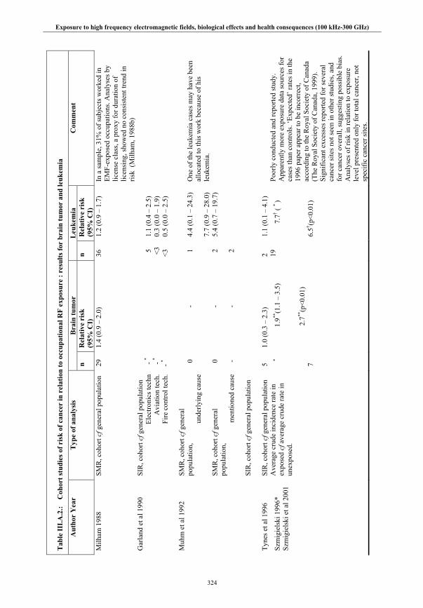
Exposure to high frequency electromagnetic fields, biological effects and health consequences (100 kHz-300 GHz)
324
Tab
le II
I.A.2
.: C
ohor
t stu
dies
of r
isk
of c
ance
r in
rel
atio
n to
occ
upat
iona
l RF
expo
sure
: re
sults
for
brai
n tu
mor
and
leuk
emia
Bra
in tu
mor
L
euke
mia
A
utho
r Y
ear
Typ
e of
ana
lysi
s n
Rel
ativ
e ri
sk
(95%
CI)
n
Rel
ativ
e ri
sk
(95%
CI)
Com
men
t
Milh
am 1
988
SMR
, coh
ort c
f gen
eral
pop
ulat
ion
29
1.4
(0.9
– 2
.0)
36
1.2
(0.9
– 1
.7)
In a
sam
ple,
31%
of s
ubje
cts w
orke
d in
EM
F-ex
pose
d oc
cupa
tions
. Ana
lyse
s by
licen
se c
lass
, a p
roxy
for d
urat
ion
of
licen
sing
, sho
wed
no
cons
iste
nt tr
end
in
risk
(Milh
am, 1
988b
) G
arla
nd e
t al 1
990
SIR
, coh
ort c
f gen
eral
pop
ulat
ion
Elec
troni
cs te
chn
Avi
atio
n te
ch.
Fire
con
trol t
ech.
- * - *
- *
5 <3
<3
1.1
(0.4
– 2
.5)
0.3
(0.0
– 1
.9)
0.5
(0.0
– 2
.5)
Muh
m e
t al 1
992
SMR
, coh
ort c
f gen
eral
po
pula
tion,
u
nder
lyin
g ca
use
SMR
, coh
ort c
f gen
eral
po
pula
tion,
m
entio
ned
caus
e SI
R, c
ohor
t cf g
ener
al p
opul
atio
n
0 0 -
- - -
1 2 2
4.4
(0.1
– 2
4.3)
7.
7 (0
.9 –
28.
0)
5.4
(0.7
– 1
9.7)
One
of t
he le
ukem
ia c
ases
may
hav
e be
en
allo
cate
d to
this
wor
k be
caus
e of
his
le
ukem
ia.
Tyne
s et a
l 199
6 SI
R, c
ohor
t cf g
ener
al p
opul
atio
n 5
1.0
(0.3
– 2
.3)
2 1.
1 (0
.1 –
4.1
)
Szm
igie
lski
199
6*
Szm
igie
lski
et a
l 200
1
Ave
rage
cru
de in
cide
nce
rate
in
expo
sed
cf a
vera
ge c
rude
rate
in
unex
pose
d.
*
7
1.9**
(1.1
– 3
.5)
2.
7**(p
<0.0
1)
19
7.7† (
* )
6.
5† (p<0
.01)
Poor
ly c
ondu
cted
and
repo
rted
stud
y.
App
aren
tly m
ore
expo
sure
dat
a so
urce
s for
ca
ses t
han
cont
rols
. ‘Ex
pect
ed’ r
ates
in th
e 19
96 p
aper
app
ear t
o be
inco
rrec
t, ac
cord
ing
to th
e R
oyal
Soc
iety
of C
anad
a
(The
Roy
al S
ocie
ty o
f Can
ada,
199
9).
Sign
ifica
nt e
xces
ses r
epor
ted
for s
ever
al
canc
er si
tes n
ot se
en in
oth
er st
udie
s, an
d fo
r can
cer o
vera
ll, su
gges
ting
poss
ible
bia
s. A
naly
ses o
f ris
k in
rela
tion
to e
xpos
ure
leve
l pre
sent
ed o
nly
for t
otal
can
cer,
not
spec
ific
canc
er si
tes.

Exposure to high frequency electromagnetic fields, biological effects and health consequences (100 kHz-300 GHz)
325
Tab
le II
I.A.2
.: C
ohor
t stu
dies
of r
isk
of c
ance
r in
rel
atio
n to
occ
upat
iona
l RF
expo
sure
: re
sults
for
brai
n tu
mor
and
leuk
emia
Bra
in tu
mor
L
euke
mia
A
utho
r Y
ear
Typ
e of
ana
lysi
s n
Rel
ativ
e ri
sk (9
5% C
I)
n R
elat
ive
risk
(95%
C
I)
Com
men
t
Lago
rio e
t al 1
997
SMR
, coh
ort c
f gen
eral
po
pula
tion
1 10
1
5 Po
tent
ial c
onfo
undi
ng b
y ch
emic
al
expo
sure
s. Lo
sses
to fo
llow
-up
treat
ed
as a
live
to e
nd o
f stu
dy p
erio
d.
Mor
gan
et a
l 200
0 SM
R, e
xpos
ed w
orke
rs c
f ge
nera
l pop
ulat
ion
Rat
e ra
tio e
xpos
ed c
f une
xpos
ed
in c
ohor
t, cu
mul
ativ
e ex
posu
re
Non
e
<med
ian
≥med
ian
17 34
7
10
0.5
(0.2
– 1
.1)
1.0
1.0
(0.4
– 2
.2)
0.9
(0.4
– 1
.9)
21 66
8
13
0.8
(0.4
– 1
.4)
1.0
0.6
(0.3
– 1
.3)
0.6
(0.3
– 1
.0)
No
dura
tion-
resp
onse
tren
d
Gro
ves e
t al 2
002
SMR
, ove
rall
coho
rt cf
gen
eral
po
pula
tion
SMR
, hig
h ex
posu
re c
ohor
t cf
gene
ral p
opul
atio
n R
elat
ive
risk,
exp
osed
cf
unex
pose
d in
coh
ort
88 37
37/5
1
0.
9 (0
.7 –
1.1
)
0.7
(0.5
– 1
.0)
0.
6 (0
.4 –
1.0
)
11
3 69
69/4
4
1.
0 (0
.8 –
1.2
)
1.1
(0.9
– 1
.4)
1.
5 (1
.0 –
2.2
)
Sign
ifica
nt ra
ised
risk
for
nonl
ymph
ocyt
ic le
ukem
ia in
hig
h ex
posu
re c
ohor
t, bu
t onl
y ra
ised
in o
ne
of 3
hig
h-ex
posu
re o
ccup
atio
ns.
Lilie
nfel
d ci
ted
by
Gol
dsm
ith 1
995#
Obs
erve
d an
d ex
pect
ed, b
ut
sour
ce o
f lat
ter u
ncle
ar
A
dults
: 2/1
.9
Chi
ldre
n: 0
/-
2/2.
0 2/
4.0
Dat
a al
so p
rese
nted
for o
ther
US
emba
ssie
s in
East
ern
Euro
pe, b
ut
uncl
ear w
heth
er th
ey w
ere
expo
sed.
B
oth
brai
n tu
mor
s and
one
leuk
emia
in a
ch
ild w
ere
in d
epen
dent
s who
live
d ou
t of
the
emba
ssy.
* N
o da
ta p
ublis
hed.
For
Szm
igie
lski
it is
impl
ied
that
ther
e w
ere
2-3
brai
n tu
mor
s and
X le
ukem
iae
in th
e ex
pose
d gr
oup,
in w
hich
cas
e w
e im
ply
that
the
CI f
or b
rain
tum
or is
inco
rrec
t. **
Ner
vous
syst
em.
† Cal
cula
ted
from
dat
a in
the
pape
r. N
otes
: Stu
dy n
ot p
ublis
hed
by L
ilien
feld
, and
too
little
info
rmat
ion
give
n in
pré
cis b
y G
olds
mith
for u
nder
stan
ding
or e
valu
atio
n of
the
met
hods
. # Smal
l num
bers
of c
ance
rs, a
nd se
vera
l of t
he c
ance
rs o
ccur
red
in
pers
ons w
ho li
ved
out o
f the
em
bass
y (i.
e. p
resu
mab
ly w
ere
in th
e em
bass
y lit
tle o
f the
tim
e, e
spec
ially
chi
ldre
n). B
reas
t can
cer i
n em
ploy
ees 2
obs
erve
d, 0
.5 e
xp. C
ance
rs o
f fem
ale
geni
talia
4 o
bser
ved,
0.8
ex
pect
ed. E
xpos
ures
est
imat
ed to
rang
e fr
om 5
– 1
8μ W
/cm
2 – b
asis
of e
stim
ate
not s
tate
d.

Exposure to high frequency electromagnetic fields, biological effects and health consequences (100 kHz-300 GHz)
326
Tab
le II
I.A.3
.: C
ase-
cont
rol s
tudi
es o
f ris
k of
bra
in tu
mor
and
leuk
emia
in r
elat
ion
to o
ccup
atio
nal R
F ex
posu
re
R
esul
ts
Aut
hors
Y
ear
Sour
ces o
f cas
es
and
cont
rols
* M
easu
re o
f ex
posu
re
Exp
osur
e da
ta
colle
ctio
n m
etho
d
Mor
talit
y or
in
cide
nce
Nos
. of
case
s/
cont
rols
T
ype
of a
naly
sis
Bra
in tu
mor
O
R
(95%
CI)
Leu
kem
ia
OR
(9
5% C
I)
Thom
as e
t al
1987
C
ases
: de
ath
certi
ficat
es
Con
trols
: dea
th
certi
ficat
es fo
r de
aths
from
oth
er
caus
es, e
xcep
t ep
ileps
y, st
roke
, su
icid
e, h
omic
ide
Job
title
& in
dust
ry
Inte
rvie
w
with
re
lativ
es
Mor
talit
y 43
5/38
6 O
dds r
atio
cf n
ever
occ
. ex
pose
d 1.
6 (1
.0-2
.4)
-
A
mst
rong
et
al 1
994
Elec
trica
l util
ity
wor
kers
(nes
ted
case
-con
trol)
Job
expo
sure
mat
rix
base
d on
1 w
eek
met
er m
easu
rem
ents
at
5-2
0 M
Hz**
for
>100
0 w
orke
rs,
asse
ssin
g ex
posu
re to
pu
lsed
el
ectro
mag
netic
fie
lds
Com
pany
re
cord
s In
cide
nce
84/3
25
95/3
74
Odd
s rat
io fo
r ≥m
edia
n ex
posu
re
Odd
s rat
io fo
r ≥90
th
perc
entil
e O
dds r
atio
for ≥
med
ian
expo
sure
O
dds r
atio
for ≥
90th
pe
rcen
tile
0.8
(0.5
– 1
.5) †
1.
9 (0
.5 –
7.6
) †
- -
- 0.
7 (0
.4 –
1.2
) 0.
8 (0
.2 –
3.4
)
G
rays
on
1996
U
SAF
(nes
ted
case
-con
trol)
Job
title
& w
heth
er
repo
rts o
f inc
iden
ts
of h
igh
expo
sure
for
each
job
title
Mili
tary
re
cord
s In
cide
nce
230/
920
Odd
s rat
io c
f nev
er-e
xpos
ed
1.4
(1.0
-1.9
) -
* All
stud
ies r
estri
cted
to m
en.
**B
ut it
was
subs
eque
ntly
foun
d th
at th
e m
eter
s als
o re
spon
ded
to fi
elds
of 1
50 a
nd 3
00 M
Hz
and
to ra
dio
trans
mis
sion
s. † M
alig
nant
bra
in tu
mor
s.

Exposure to high frequency electromagnetic fields, biological effects and health consequences (100 kHz-300 GHz)
327
Tab
le II
I.A.4
.: A
naly
ses o
f rou
tinel
y co
llect
ed d
ata
on b
rain
tum
or a
nd le
ukem
ia r
isk
in r
elat
ion
to o
ccup
atio
nal R
F ex
posu
re
Bra
in tu
mor
Leu
kem
ia
Aut
hor
Yea
r T
ype
of
anal
ysis
E
xpos
ed g
roup
* C
ompa
riso
n co
hort
/ con
trol
gr
oup
Mor
talit
y or
in
cide
nce
n** R
R (9
5% C
I)
n**
R
R (9
5% C
I)
Wrig
ht e
t al 1
982
Pr
opor
tiona
l in
cide
nce
Rad
io &
TV
re
pairm
en. T
elep
hone
lin
esm
en.
All
othe
r can
cers
In
cide
nce
-† -†
1 2
1.2
(-† )
3.1
(-† )
Cal
le&
Sav
itz 1
985
Prop
ortio
nal
mor
talit
y R
adio
& te
legr
aph
oper
ator
s.
Rad
io &
TV
repa
irmen
All
caus
es o
f dea
th
Mor
talit
y -† -†
6 3
2.3
(-† )
0.
9 (-
† )
Lin
et a
l 198
5 C
ase-
cont
rol
Elec
tric
& te
leph
one
linem
en, s
ervi
cem
en
Non
-can
cer d
eath
s M
orta
lity
27
(-
† )
Milh
am 1
985
Prop
ortio
nal
mor
talit
y R
adio
& te
legr
aph
oper
ator
s.
Rad
io &
TV
repa
irmen
All
caus
es o
f de
aths
M
orta
lity
1 2
0.4
(-† )
0.
6 (-
† )
5 7
1.0
(-† )
1.
8 (-
† )
Pear
ce e
t al 1
989
C
ase-
cont
rol
Rad
io &
TV
repa
irmen
All
othe
r can
cers
In
cide
nce
-†
2
7.9
(2.2
– 2
8.1)
Tyne
s et a
l 199
2 C
ohor
t R
adio
freq
uenc
y ex
pose
d oc
cupa
tions
Econ
omic
ally
ac
tive
mal
es
Inci
denc
e 3
0.6
(0.1
– 1
.8)
9
2.8
(1.3
– 5
.4)
* All
stud
ies a
re o
f mal
es. E
xpos
ure
asse
ssm
ent f
or a
ll is
bas
ed so
lely
on
job
title
, with
no
mea
sure
s of e
xpos
ure.
**
No.
in e
xpos
ed g
roup
. † N
o da
ta p
ublis
hed.

Exposure to high frequency electromagnetic fields, biological effects and health consequences (100 kHz-300 GHz)
328
Tab
le II
I.A.5
.: Su
mm
ary
of li
tera
ture
on
radi
ofre
quen
cy e
xpos
ure
and
repr
oduc
tive
heal
th o
utco
mes
Out
com
e M
easu
re
Ref
eren
ce
Geo
grap
hic
Sett
ing
Popu
latio
n Si
ze &
Sou
rce
Res
ults
: Exp
osur
e &
Out
com
e C
omm
ents
Sem
en p
aram
eter
s
La
ncra
njan
et a
l 197
5 R
oman
ia
Mic
row
ave
expo
sure
(3
1) v
s. co
ntro
ls (3
0)
Sper
m c
ount
: 50
(Exp
), 60
(Ctl)
mill
ion/
ml
Mot
ility
: 36%
(Exp
), 54
% (C
tl)
W
eyan
dt e
t al 1
996
US
Mili
tary
inte
llige
nce
(20)
vs.
cont
rols
(30)
Sp
erm
den
sity
: 13
(Exp
), 35
(Ctl)
Pe
rcen
t nor
mal
: 69
(Exp
), 73
(Ctl)
Pe
rcen
t mot
ile: 3
2 (E
xp),
43 (C
tl)
H
jollu
nd &
Bon
de 1
997
Den
mar
k M
ilita
ry: m
issi
le o
pera
tors
(19)
, ot
her (
489)
Sp
erm
den
sity
: 40
(Exp
), 62
(Ctl)
Im
mot
ile %
: 52
(Exp
), 33
(Ctl)
Pe
rcen
t nor
mal
: 61
(Exp
), 68
(Ctl)
Sc
hrad
er e
t al 1
998
US
(Tex
as)
Mili
tary
: rad
ar o
pera
tors
(33)
, co
ntro
ls (1
03)
Sper
m d
ensi
ty: 2
9 (E
xp),
32 (C
tl)
Perc
ent n
orm
al: 4
6 (E
xp),
42 (C
tl)
Perc
ent m
otile
: 46
(Exp
), 45
(Ctl)
G
raje
wsk
i et a
l 200
0 U
S (M
aryl
and)
R
F he
ater
ope
rato
rs
Sper
m d
ensi
ty: 4
7 (E
xp),
45 (C
tl)
Sper
m c
ount
: 73
(Exp
), 93
(Ctl)
M
otile
(%):
67 (E
xp),
52 (C
tl)
Nor
mal
mor
phol
ogy:
81
(Exp
), 79
(Ctl)
Fert
ility
La
rsen
et a
l 199
1 D
enm
ark
Phys
ioth
erap
ists
49
tim
e to
pre
gnan
cy
over
6 m
os
TWA
Exp
osur
e &
TTP
>6
mon
ths
RR
= 1
.0, 0
.8 (0
.2-2
.2),
1.7
(0.7
-4.1
):
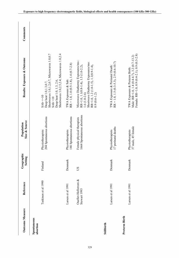
Exposure to high frequency electromagnetic fields, biological effects and health consequences (100 kHz-300 GHz)
329
Out
com
e M
easu
re
Ref
eren
ce
Geo
grap
hic
Sett
ing
Popu
latio
n Si
ze &
Sou
rce
Res
ults
: Exp
osur
e &
Out
com
e C
omm
ents
Spon
tane
ous
abor
tion
Ta
skin
en e
t al 1
990
Finl
and
Phys
ioth
erap
ists
20
4 Sp
onta
neou
s abo
rtion
s SA
b <=
10
Dee
p he
at 1
.0,1
.3,0
.7,
Shor
twav
es 1
.0,1
.2,0
.7, M
icro
wav
es 1
.0,0
.7
SAb
>10
Dee
p he
at 1
.0, 1
.3, 2
.6,
Shor
twav
es 1
.0,2
.5,2
.4; M
icro
wav
es 1
.0,2
.4
La
rsen
et a
l 199
1 D
enm
ark
Phys
ioth
erap
ists
14
6 Sp
onta
neou
s abo
rtion
s
TWA
Exp
osur
e &
Sab
: R
R =
1.0
, 1.0
(0.5
-1.8
), 1.
4 (0
.7-2
.8)
O
uelle
t-Hel
lstro
m &
St
ewar
t 199
3 U
S Fe
mal
e ph
ysic
al th
erap
ists
16
64 S
pont
aneo
us a
borti
ons
Mic
row
ave
Dia
ther
my
Expo
sure
s/m
o.:
RR
=1.0
, 1.1
(0.8
-1.4
), 1.
5 (1
.0-2
.2),
1.6
(1.0
-2.6
) Sh
ortw
ave
Dia
ther
my
Expo
sure
s/m
o:
RR
=1.0
, 1.2
(1.0
-1.5
), 1.
1(0.
9-1.
4),
0.9
(0.6
-1.2
)
Still
birt
h
La
rsen
et a
l 199
1 D
enm
ark
Phys
ioth
erap
ists
17
per
inat
al d
eath
s
TWA
Exp
osur
e &
Per
inat
al D
eath
: R
R =
1.0
, 1.5
(0.3
-5.3
), 2.
9 (0
.6-1
0.7)
Pret
erm
Bir
th
La
rsen
et a
l 199
1 D
enm
ark
Phys
ioth
erap
ists
37
mal
e, 4
5 fe
mal
e TW
A E
xpos
ure
& P
rete
rm B
irth:
M
ale:
RR
=1.0
, 1.4
(0.4
-4.7
), 3.
2 (0
.7-1
3.2)
Fe
mal
e: R
R=1
.0, 0
.9 (0
.4-2
.1),
0.9
(0.3
-2.8
)
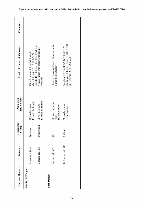
Exposure to high frequency electromagnetic fields, biological effects and health consequences (100 kHz-300 GHz)
330
Out
com
e M
easu
re
Ref
eren
ce
Geo
grap
hic
Sett
ing
Popu
latio
n Si
ze &
Sou
rce
Res
ults
: Exp
osur
e &
Out
com
e C
omm
ents
Low
Bir
th W
eigh
t
La
rsen
et a
l 199
1 D
enm
ark
Phys
ioth
erap
ists
15
mal
e, 2
4 fe
mal
e TW
A E
xpos
ure
& L
ow B
irthw
eigh
t: M
ale:
RR
=1.0
, 0.0
, 5.9
(1.0
-28.
2)
Fem
ale:
RR
=1.0
, 1.2
(0.4
-3.3
), 0.
7 (0
-3.2
)
G
uber
an e
t al 1
994
Switz
erla
nd
Phys
ioth
erap
ists
11
mal
e, 1
4 fe
mal
e N
o as
soci
atio
n w
ith sh
ortw
aves
(RR
not
re
porte
d)
Bir
th D
efec
ts
Lo
gue
et a
l 198
5 U
S Ph
ysic
al th
erap
ists
(m
ale)
19
2 bi
rth d
efec
ts
Obs
erve
d:ex
pect
ed ra
nge
-- "
appe
ars t
o be
hi
gher
than
exp
ecte
d"
Ta
skin
en e
t al 1
990
Finl
and
Phys
ioth
erap
ists
51
birt
h de
fect
s D
eep
heat
1.0
, 2.4
(1.0
-5.3
), 0.
9 (0
.3-2
.7)
Shor
twav
es 1
.0, 2
.7 (1
.2-6
.1),
1.0
(0.3
-3.1
) M
icro
wav
es 1
.0, 0
.5 (0
.1-3
.9)

Exposure to high frequency electromagnetic fields, biological effects and health consequences (100 kHz-300 GHz)
331
Tab
le II
I.A.6
.: Su
mm
ary
of st
udie
s on
tran
smitt
ers a
nd c
ance
r
Ref
eren
ce
Sour
ce o
f exp
osur
e C
ompa
riso
n E
ndpo
ints
N
r. C
ases
R
esul
ts
Sett
ing
Com
men
ts
Selv
in e
t al
1992
M
W to
wer
In
tern
al
Chi
ldho
od le
ukem
ia
123
52
Ran
dom
pat
tern
Sa
n
Fran
cisc
o A
naly
sis o
f spa
tial
data
; no
epi p
aram
.
Mas
karin
ec e
t al
1994
LF
radi
o (2
3.4
kHz)
<2
.6 m
iles
Chi
ldho
od le
ukem
ia12
2.
0: 0
.06-
8.3
Haw
aii;
case
-co
ntro
l SI
R a
naly
sis o
n sa
me
case
s:
2.09
: 1.0
8-3.
65
H
ocki
ng e
t al
1996
TV
tow
ers
Inne
r/ ou
ter
All
age
leuk
emia
C
hild
hood
leuk
emia
1.
24: 1
.09-
1.40
1.
58: 1
.07-
2.34
N
orth
ern
Sydn
ey
8 –0
.2µW
/cm
2
Dol
k et
al 1
997
I TV
and
FM
radi
o
<2 k
m
Adu
lt le
ukem
ia
23
1.83
:1.2
2-2.
74
Sutto
n C
oldf
ield
Dol
k et
al 1
997
II
TV a
nd F
M ra
dio
<2
km
Le
ukem
ia
79
0.97
: 0.7
8-1.
21
All
GB
McK
enzi
e et
al
1998
TV tr
ansm
issi
on
ante
nnas
C
ont.
µW/c
m2
mod
el
Chi
ldho
od le
ukem
ia
Sy
dney
R
eana
lysi
s of
Hoc
king
s; c
oncl
. One
LG
A e
xpla
ins
Coo
per D
et a
l 200
1 TV
and
FM
radi
o <2
km
A
ll ag
e le
ukem
ia
Chi
ldho
od le
ukem
ia20
1
1.32
. 0.8
1-2.
05
1.13
: 0.0
3-6.
27
Sutto
n C
oldf
ield
R
eana
lysi
s, m
ore
timel
y ca
ncer
dat
a
Mic
helo
zzi e
t al
2002
R
adio
stat
ion
<6 k
m
Chi
ldho
od le
ukem
iaA
dult
leuk
emia
8 23
2.
2: 1
.0-4
.1
1.2:
.8-1
.8
Vat
ican

Exposure to high frequency electromagnetic fields, biological effects and health consequences (100 kHz-300 GHz)
332
Tab
le II
I.A.7
.: S
umm
ary
of st
udie
s of m
obile
pho
ne u
se a
nd r
isk
of b
rain
tum
ors
Aut
hors
Yea
r (s
tudy
des
ign)
St
udy
popu
latio
n T
umor
type
(num
bers
ca
ses/
con
trol
s)
Exp
osur
e as
sess
men
t
Phon
e ty
pe;
Dur
atio
n of
use
in
cont
rols
Phon
e ev
er-u
se R
R
(95%
CI)
Har
dell
et a
l 199
9 (c
ase-
cont
rol)
Swed
en. C
ases
: 20-
80 y
r. C
ontro
ls: r
egio
nal p
opul
atio
n re
gist
ers,
Upp
sala
-Ore
bro
1994
-96,
Sto
ckho
lm 1
995-
96
All
tum
ors (
209/
425
) A
cous
tic n
euro
ma
Rec
alle
d m
obile
pho
ne
use
by
ques
tionn
aire
an
d in
terv
iew
Mai
nly
anal
ogue
, 450
or
900
MH
z; 1
6% >
5 yr
.
1.
0 (0
.7 –
1.4
)*
0.8
(0.1
– 4
.2)
Mus
cat e
t al 2
000
(cas
e-co
ntro
l) U
SA: H
ospi
tal i
npat
ient
s,
NY
, Pro
vide
nce,
Bos
ton.
C
ases
:18-
80 y
r ,19
94-9
8.
Con
trols
: Mal
igna
nt a
nd n
on-
mal
igna
nt c
ondi
tions
.
Mal
igna
nt b
rain
tum
or
(469
/ 422
) R
ecal
led
mob
ile p
hone
us
e vi
a in
terv
iew
Mai
nly
anal
ogue
800
–
900
MH
z;
5%>4
yr.
0.
9 (0
.6 –
1.2
)
Insk
ip e
t al 2
001
(cas
e-co
ntro
l) U
SA: H
ospi
tal i
npat
ient
s, B
osto
n; P
hoen
ix; P
ittsb
urgh
. C
ases
:18+
yr,
1994
-98.
C
ontro
ls: n
on-m
alig
nant
co
nditi
ons
All
tum
ors (
782/
799)
G
liom
a (4
89/7
99 )
Men
ingi
oma
(197
/ 799
) A
cous
tic n
euro
ma
(96/
799)
Rec
alle
d m
obile
pho
ne
use
via
inte
rvie
w
Mai
nly
anal
ogue
800
–
900
MH
z;
8% >
3 yr
.
0.
9 (0
.7 –
1.1
) 1.
0 (0
.7 –
1.4
) 0.
8 (0
.5 –
1.2
) 0.
8 (0
.5 –
1.4
)
Mus
cat e
t al 2
002
(cas
e-co
ntro
l) U
SA: H
ospi
tal i
npat
ient
s,
New
Yor
k. C
ases
:18+
yr,
1997
-99.
Con
trols
: Non
-m
alig
nant
con
ditio
ns.
Aco
ustic
neu
rom
a (9
0/86
) R
ecal
led
mob
ile p
hone
us
e vi
a qu
estio
nnai
re
Mai
nly
anal
ogue
800
–
900
MH
z;
7% 3
-6 y
r.
0.
9

Exposure to high frequency electromagnetic fields, biological effects and health consequences (100 kHz-300 GHz)
333
Aut
hors
Yea
r (s
tudy
des
ign)
St
udy
popu
latio
n T
umor
type
(num
bers
ca
ses/
con
trol
s)
Exp
osur
e as
sess
men
t
Phon
e ty
pe;
Dur
atio
n of
use
in
cont
rols
Phon
e ev
er-u
se R
R
(95%
CI)
Auv
inen
et a
l 200
2 (c
ase-
cont
rol)
Finl
and.
Cas
es: 2
0-69
yr,
1996
. Con
trols
: Nat
iona
l po
pula
tion
regi
ster
.
All
tum
ors (
398/
1986
) G
liom
a (1
98/9
89)
Ben
ign
(129
/643
) Sa
livar
y gl
and
(34/
170)
Dur
atio
n of
pr
ivat
e ce
llula
r ne
twor
k su
bscr
iptio
n
Ana
logu
e, a
vera
ge 2
-3
yr su
bscr
iptio
n;
digi
tal,
aver
age
<1 y
r su
bscr
iptio
n.
1.
3 (0
.9 –
1.8
) 1.
5 (1
.0 –
2.4
) 1.
1 (0
.5 –
2.4
) 1.
3 (0
.4 –
4.7
)
Har
dell
et a
l 200
2
(cas
e-co
ntro
l) H
arde
ll et
al 2
003
(cas
e-co
ntro
l)
Swed
en.
Cas
es: 2
0-80
yr.
1997
-200
0.
Con
trols
: 4 re
gion
al
popu
latio
n re
gist
ers.
All
tum
ors (
1303
/130
3)
A
cous
tic n
euro
ma(
159/
42
2)
Rec
alle
d m
obile
pho
ne
use
via
ques
tionn
aire
Ana
logu
e 45
0 or
900
M
Hz,
med
ian
8
yr.
Dig
ital 1
900
MH
z,
med
ian
3 yr
.
Ana
logu
e
Dig
ital
1.3
(1.0
– 1
.6)*
1.0
(0.8
– 1
.2)
3.
5 (1
.8 –
6.8
) 1.
2 (0
.7 –
2.2
)
Dre
yer e
t al 1
999
(coh
ort)
USA
. Sub
scrib
ers o
f 2 la
rge
cellu
lar n
etw
orks
. 199
3.
Cas
es: ≥
20 y
r dea
ths 1
994
Mal
igna
nt b
rain
tum
or
(6).
Dur
atio
n of
su
bscr
iptio
n A
nalo
gue.
1 y
r fo
llow
-up
_ _
Joha
nsen
et a
l 200
2 (c
ohor
t) D
enm
ark.
Priv
ate
cellu
lar
netw
ork
subs
crib
ers.
1982
-95
. C
ases
: ≥18
yea
rs. 1
982-
96.
All
tum
ors (
154
) G
liom
a ( 6
6 )
Men
ingi
oma
(16
)
Dur
atio
n of
su
bscr
iptio
n A
nalo
gue
(450
or 9
00
MH
z) o
r dig
ital.
U
p to
15
yr fo
llow
-up
_ SI
R 1
.0 (0
.8-1
.1)
(0.7
– 1
.2)
0.9
(0.5
– 1
.4)
Chr
iste
nsen
et a
l 20
04
Den
mar
k Po
pula
tion-
base
d ca
se-
cont
rol
Aco
ustic
neu
rom
a (1
06)
Popu
latio
n co
ntro
ls (2
12)
- -
- 0.
90 (0
.51
– 1.
6)

Exposure to high frequency electromagnetic fields, biological effects and health consequences (100 kHz-300 GHz)
334
Tab
le II
I.A.8
.: Su
mm
ary
of st
udie
s of m
obile
pho
ne u
se a
nd sy
mpt
oms
Aut
hors
Yea
r (s
tudy
des
ign)
St
udy
popu
latio
n A
naly
ses
Exp
osur
e as
sess
men
t O
utco
me
asse
ssm
ent
Res
ults
Hoc
king
199
8 (c
ase-
serie
s)
Aus
tralia
ns w
ith
sym
ptom
s on
mob
ile
phon
e us
e w
ho
resp
onde
d to
not
ice
in
med
ical
jour
nal o
r med
ia
publ
icity
N=4
0)
Des
crip
tion
of ty
pe o
f sy
mpt
oms r
epor
tedl
y du
e to
mob
ile p
hone
use
No
form
al a
sses
smen
t of
am
ount
or
freq
uenc
y of
mob
ile
phon
e us
e
Que
stio
nnai
re a
bout
de
tails
of s
ympt
oms
asso
ciat
ed w
ith
mob
ile p
hone
use
Mos
t res
pond
ents
repo
rted
unus
ual s
ensa
tions
af
fect
ing
the
head
, suc
h as
dul
l pai
n,
unpl
easa
nt w
arm
th. *
*
Ofte
dal e
t al
2000
(cro
ss-
sect
iona
l)
Swed
ish
and
Nor
weg
ian
mob
ile p
hone
use
rs,
sele
cted
from
net
wor
k op
erat
or re
gist
ers.
Onl
y in
clud
ed p
eopl
e w
ho
used
pho
ne fo
r job
. N
=106
31
Num
ber o
f res
pond
ents
w
ith a
ny sy
mpt
om
attri
bute
d to
mob
ile
phon
es
Num
ber o
f res
pond
ents
w
ho h
ad ta
ken
step
s to
redu
ce sy
mpt
oms
Not
wel
l des
crib
ed, b
ut
one
tabl
e re
ports
nu
mbe
r of c
alls
and
ca
lling
tim
e pe
r day
, su
gges
ting
repo
rted
in
a qu
estio
nnai
re.
Self-
repo
rted
freq
uenc
y of
sy
mpt
oms.
Patie
nt
cons
ider
ed to
hav
e sy
mpt
om if
oc
curr
ed a
t lea
st
once
per
wee
k.
1.13
% o
f par
ticip
ants
in S
wed
en a
nd 3
1% in
N
orw
ay re
porte
d at
leas
t 1 sy
mpt
om in
co
nnec
tion
with
use
of a
mob
ile p
hone
. Mos
t co
mm
on, w
arm
th a
roun
d ea
r.
22%
of N
orw
egia
ns a
nd 7
% o
f Sw
edes
ex
perie
nced
sym
ptom
oth
er th
an w
arm
th.
2. 4
5% o
f peo
ple
expe
rienc
ing
sym
ptom
s had
ta
ken
step
s to
redu
ce th
em, s
uch
as re
duce
d ca
lling
tim
e, u
se o
f han
ds fr
ee k
it, c
hang
ing
side
pho
ne u
sed.

Exposure to high frequency electromagnetic fields, biological effects and health consequences (100 kHz-300 GHz)
335
Aut
hors
Yea
r (s
tudy
des
ign)
St
udy
popu
latio
n A
naly
ses
Exp
osur
e as
sess
men
t O
utco
me
asse
ssm
ent
Res
ults
Sand
stro
m e
t al
200
1 (c
ross
-se
ctio
nal)
Swed
ish
and
Nor
weg
ian
mob
ile p
hone
use
rs,
sele
cted
from
net
wor
k op
erat
or re
gist
ers.
N=1
6,99
2
Com
paris
on o
f GSM
ve
rsus
NM
T m
obile
ph
one
user
s Tr
ends
with
incr
easi
ng
time
of p
hone
usa
ge
Self-
com
plet
ed
ques
tionn
aire
, va
riabl
es: t
rans
mitt
er
syst
em, c
allin
g tim
e pe
r day
and
num
ber o
f ca
lls p
er d
ay
Self-
repo
rted
freq
uenc
y of
rang
e of
sym
ptom
s. Pa
rtici
pant
co
nsid
ered
to h
ave
sym
ptom
s if
occu
rred
at l
east
on
ce p
er w
eek.
OR
am
ong
GSM
cf N
MT
phon
es:
No
incr
ease
d ris
k fo
r any
sym
ptom
s. G
SM
user
s at l
ower
risk
of w
arm
th b
ehin
d ea
r (O
R:
0.64
, 95%
CI 0
.51-
0.80
) or o
n ea
r (O
R: 0
.68,
95
% C
I 0.5
3-0.
86).
GSM
use
rs in
Sw
eden
at
low
er ri
sk o
f hea
dach
es (O
R: 0
.73,
95%
CI
0.56
-0.9
5) a
nd fa
tigue
(OR
: 0.8
0, 9
5% C
I 0.6
5-0.
99)
With
incr
easi
ng m
inut
es o
f pho
ne u
se th
ere
was
an
incr
ease
d od
ds o
f rep
ortin
g fa
tigue
, he
adac
hes,
war
mth
, bur
ning
and
tigh
tnes
s at
leas
t onc
e pe
r wee
k. *
Chi
a et
al
2000
(cro
ss-
sect
iona
l)
Ran
dom
sam
ple
of 6
35
hous
ehol
ds in
hou
sing
es
tate
in S
inga
pore
. 808
re
spon
dent
s (N
B
resp
onse
rate
less
than
60
%).
Prev
alen
ce ra
tio o
f he
adac
he in
mob
ile p
hone
us
ers v
s non
-use
rs
Ass
ocia
tion
betw
een
min
utes
pho
ne u
se a
nd
head
ache
Inte
rvie
wer
-ad
min
iste
red
ques
tionn
aire
. Pur
pose
of
stud
y m
aske
d.
Cla
ssifi
ed a
s MP
user
if
used
at l
east
on
ce/d
ay.
Que
stio
nnai
re
conc
erni
ng n
atur
e an
d se
verit
y of
‘C
NS
sym
ptom
s.’
(hea
dach
e,
dizz
ines
s, w
arm
th,
tingl
ing,
vis
ual
dist
urba
nces
). N
B
the
freq
uenc
y of
he
adac
hes r
equi
red
befo
re a
resp
onde
nt
was
cla
ssifi
ed a
s a
head
ache
suff
erer
w
as n
ot sp
ecifi
ed.
1. 4
5% m
obile
pho
ne u
sers
3%
exp
erie
nced
CN
S pr
oble
ms
Adj
uste
d pr
eval
ence
ratio
for h
eada
che
amon
g us
ers c
f non
-use
rs 1
.31
(95%
CI 1
.00-
1.70
). N
o si
gnifi
cant
diff
eren
ces f
or a
ny o
ther
sym
ptom
s. 2.
Sig
nific
ant p
ositi
ve tr
end
for i
ncre
asin
g tim
e sp
ent o
n th
e m
obile
pho
ne a
nd p
reva
lenc
e of
he
adac
he (p
=0.0
4).
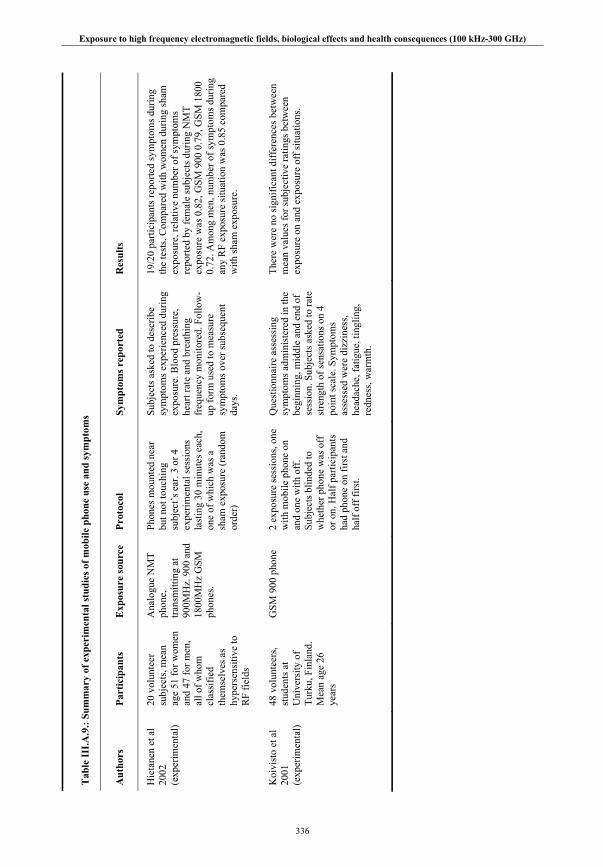
Exposure to high frequency electromagnetic fields, biological effects and health consequences (100 kHz-300 GHz)
336
Tab
le II
I.A.9
.: Su
mm
ary
of e
xper
imen
tal s
tudi
es o
f mob
ile p
hone
use
and
sym
ptom
s
Aut
hors
Pa
rtic
ipan
ts
Exp
osur
e so
urce
Pr
otoc
ol
Sym
ptom
s rep
orte
d R
esul
ts
Hie
tane
n et
al
2002
(e
xper
imen
tal)
20 v
olun
teer
su
bjec
ts, m
ean
age
51 fo
r wom
en
and
47 fo
r men
, al
l of w
hom
cl
assi
fied
them
selv
es a
s hy
pers
ensi
tive
to
RF
field
s
Ana
logu
e N
MT
phon
e,
trans
mitt
ing
at
900M
Hz.
900
and
18
00M
Hz
GSM
ph
ones
.
Phon
es m
ount
ed n
ear
but n
ot to
uchi
ng
subj
ect’s
ear
. 3 o
r 4
expe
rimen
tal s
essi
ons
last
ing
30 m
inut
es e
ach,
on
e of
whi
ch w
as a
sh
am e
xpos
ure
(ran
dom
or
der)
Subj
ects
ask
ed to
des
crib
e sy
mpt
oms e
xper
ienc
ed d
urin
g ex
posu
re. B
lood
pre
ssur
e,
hear
t rat
e an
d br
eath
ing
freq
uenc
y m
onito
red.
Fol
low
-up
form
use
d to
mea
sure
sy
mpt
oms o
ver s
ubse
quen
t da
ys.
19/2
0 pa
rtici
pant
s rep
orte
d sy
mpt
oms d
urin
g th
e te
sts.
Com
pare
d w
ith w
omen
dur
ing
sham
ex
posu
re, r
elat
ive
num
ber o
f sym
ptom
s re
porte
d by
fem
ale
subj
ects
dur
ing
NM
T ex
posu
re w
as 0
.82,
GSM
900
0.7
9, G
SM 1
800
0.72
. Am
ong
men
, num
ber o
f sym
ptom
s dur
ing
any
RF
expo
sure
situ
atio
n w
as 0
.85
com
pare
d w
ith sh
am e
xpos
ure.
Koi
vist
o et
al
2001
(e
xper
imen
tal)
48 v
olun
teer
s, st
uden
ts a
t U
nive
rsity
of
Turk
u, F
inla
nd.
Mea
n ag
e 26
ye
ars
GSM
900
pho
ne
2 ex
posu
re se
ssio
ns, o
ne
with
mob
ile p
hone
on
and
one
with
off
. Su
bjec
ts b
linde
d to
w
heth
er p
hone
was
off
or
on.
Hal
f par
ticip
ants
ha
d ph
one
on fi
rst a
nd
half
off f
irst.
Que
stio
nnai
re a
sses
sing
sy
mpt
oms a
dmin
iste
red
in th
e be
ginn
ing,
mid
dle
and
end
of
sess
ion.
Sub
ject
s ask
ed to
rate
st
reng
th o
f sen
satio
ns o
n 4
poin
t sca
le. S
ympt
oms
asse
ssed
wer
e di
zzin
ess,
head
ache
, fat
igue
, tin
glin
g,
redn
ess,
war
mth
.
Ther
e w
ere
no si
gnifi
cant
diff
eren
ces b
etw
een
mea
n va
lues
for s
ubje
ctiv
e ra
tings
bet
wee
n ex
posu
re o
n an
d ex
posu
re o
ff si
tuat
ions
.
Exposure to high frequency electromagnetic fields, biological effects and health consequences (100 kHz-300 GHz)
337
III.A.9. REFERENCES
Abdel-Rassoul G, El-Fateh OA, Salem MA, Michael A, Farahat F, El-Batanouny M, Salem E. Neurobehavioral effects among inhabitants around mobile phone base stations. Neurotoxicology 28(2):434-40; 2007.
Ahlbom A, Feychting M. Use of cellular phones and the risk of brain tumors: a case-control study. Int J Oncol 15:1045-7; 1999.
Anglesio L, Benedetto A, Bonino A, Colla D, Martire F, Saudino Fusette S, d'Amore G. Population exposure to electromagnetic fields generated by radio base stations: evaluation of the urban background by using provisional model and instrumental measurements. Radiat Prot Dosimetry 97:355-8; 2001.
Armstrong B, Theriault G, Guenel P, Deadman J, Goldberg M, Heroux P. Association between exposure to pulsed electromagnetic fields and cancer in electric utility workers in Quebec, Canada, and France. Am J Epidemiol 140:805-20; 1994.
Auvinen A, Hietanen M, Luukkonen R, Koskela RS. Brain tumors and salivary gland cancers among cellular telephone users. Epidemiology 13:356-9; 2002.
Baste V, Riise T, Moen BE. Radiofrequency electromagnetic fields; male infertility and sex ratio of offspring. Eur J Epidemiol 23:369-77; 2008.
Behin A, Hoang-Xuan K, Carpentier AF, Delattre JY. Primary brain tumors in adults. Lancet 361:323-31; 2003.
Berg G, Spallek J, Schüz J, Schlehofer B, Böhler E, Schlaefer K, Hettinger I, Kunna-Grass K, Wahrendorf J, Blettner M. Interphone Study Group, Germany. Occupational exposure to radio frequency/microwave radiation and risk of brain tumors: Interphone Study Group, Germany. Am J Epidemiol 164:538-548; 2006.
Berg-Beckhoff G, Blettner M, Kowall B, Breckenkamp J, Schlehofer B, Schmiedel S, Bornkessel C, Reis U, Potthoff P, Schüz J. Mobile phone base stations and adverse health effects: phase 2 of a cross-sectional study with measured radio frequency electromagnetic fields. Occup Environ Med 66(2):124-30; 2009.
Blettner M, Schlehofer B, Breckenkamp J, Kowall B, Schmiedel S, Reis U, Potthoff P, Schüz J, Berg-Beckhoff G. Mobile phone base stations and adverse health effects: phase 1 of a population-based, cross-sectional study in Germany. Occup Environ Med 66(2):118-23; 2009.
Cantor KP, Stewart PA, Brinton LA, Dosemeci M. Occupational exposures and female breast cancer mortality in the United States. J Occup Environ Med 37:336-48; 1995.
Chia SE, Chia HP, Tan JS. Prevalence of headache among handheld cellular telephone users in Singapore: a community study. Environ Health Perspect 108:1059-62; 2000.
Christensen HC, Schüz J, Kosteljanetz M, Poulsen HS, Thomsen J, Johansen C. Cellular telephone use and risk of acoustic neuroma. Am J Epidemiol 159:277-83; 2004.
Cleary SF, Pasternack BS, Beebe GW. Cataract incidence in radar workers. Arch Environ Health 11:179-82; 1965.
Cleary SF, Pasternack BS. Lenticular changes in microwave workers. A statistical study. Arch Environ Health 12:23-9; 1966.
Cooper D, Hemmings K, Saunders P. Cancer incidence near radio and television transmitters in Great Britain. I. Sutton Coldfield transmitter; II. All high power transmitters. Am J Epidemiol 153:202-4; 2001.
COST. Report of a short term mission within COST 244 bis; 2001. Dahme M. Residential RF exposures. Radiat Prot Dosimetry 83:113-117; 1999. Davis RL, Mostofi FK. Cluster of testicular cancer in police officers exposed to hand-held radar. Am J
Indust Med 24:231-233; 1993. Degrave E, Autier P, Grivegnée AR, Zizi M. All-cause mortality among Belgian military radar operators:
a 40-year controlled longitudinal study. Eur J Epidemiol 20:677-81; 2005.
Exposure to high frequency electromagnetic fields, biological effects and health consequences (100 kHz-300 GHz)
338
Demers PA, Thomas DB, Rosenblatt KA, al. e. Occupational exposure to electromagnetic fields and breast cancer in men. Am J Epidemiol 134:340-347; 1991.
Dimbylow PJ, Mann SM. Characterization of energy deposition in the head from cellular phones. Radiat Prot Dosimetry 83:113-117; 1999.
Dolk H, Shaddick G, Walls P, Grundy C, Thakrar B, Kleinschmidt I, Elliott P. Cancer incidence near radio and television transmitters in Great Britain. I. Sutton Coldfield transmitter. Am J Epidemiol 145:1-9; 1997a.
Dolk H, Elliott P, Shaddick G, Walls P, Thakrar B. Cancer incidence near radio and television transmitters in Great Britain. II. All high power transmitters. Am J Epidemiol 145:10-7; 1997b.
Dreyer NA, Loughlin JE, Rothman KJ. Cause-specific mortality in cellular telephone users. Jama 282:1814-6; 1999.
Funch DP, Rothman KJ, Loughlin JE, Dreyer NA. Utility of telephone company records for epidemiologic studies of cellular telephones. Epidemiology 7:299-302; 1996.
Gandhi OP, Lazzi G, Tinniswood A, Yu QS. Comparison of numerical and experimental methods for determination of SAR and radiation patterns of handheld wireless telephones. Bioelectromagnetics 20:93-101; 1999.
Garland FC, Shaw E, Gorham ED, Garland CF, White MR, Sinsheimer PJ. Incidence of leukemia in occupations with potential electromagnetic field exposure in United States Navy personnel. Am J Epidemiol 132:293-303; 1990.
Goldsmith JR. Epidemiologic Evidence of Radiofrequency Radiation (Microwave) Effects on Health in Military, Broadcasting, and Occupational Studies. Int J Occup Environ Health 1:47-57; 1995.
Goldstein LS, Kheifets L, van Deventer E, Repacholi M. Comments on "Long-term exposure of Emicro-Pim1 transgenic mice to 898.4 MHz microwaves does not increase lymphoma incidence" by Utteridge et al, Radiat. Res. 158, 357-364 (2002). Radiat Res 159:275-6; author reply 276-8; 2003.
Grajewski B, Cox C, Schrader SM, Murray WE, Edwards RM, Turner TW, Smith JM, Shekar SS, Evenson DP, Simon SD, Conover DL. Semen quality and hormone levels among radiofrequency heater operators. J Occup Environ Med 42:993-1005; 2000.
Grayson JK. Radiation exposure, socioeconomic status, and brain tumor risk in the US Air Force: a nested case-control study. Am J Epidemiol 143:480-6; 1996.
Groves FD, Page WF, Gridley G, Lisimaque L, Stewart PA, Tarone RE, Gail MH, Boice JD, Jr., Beebe GW. Cancer in Korean war navy technicians: mortality survey after 40 years. Am J Epidemiol 155:810-8; 2002.
Ha M, Im H, Lee M, Kim HJ, Kim BC, Gimm YM, Pack JK. Radio-frequency radiation exposure from AM radio transmitters and childhood leukemia and brain cancer. Am J Epidemiol 166(3):270-9; 2007.
Hayes RB, Brown LM, Pottern LM, Gomez M, Kardaun JW, Hoover RN, O'Connell KJ, Sutzman RE, Javadpour N. Occupation and risk for testicular cancer: a case-control study. Int J Epidemiol 19:825-31; 1990.
Hamburger S, Logue JN, Silverman PM. Occupational exposure to non-ionizing radiation and an association with heart disease: an exploratory study. J Chronic Dis 36:791-802; 1983.
Hardell L, Nasman A, Pahlson A, Hallquist A, Hansson Mild K. Use of cellular telephones and the risk for brain tumors: A case-control study. Int J Oncol 15:113-6; 1999.
Hardell L, Nasman A, Pahlson A, Hallquist A. Case-control study on radiology work, medical x-ray investigations, and use of cellular telephones as risk factors for brain tumors. MedGenMed 2:E2; 2000.
Hardell L, Mild KH, Pahlson A, Hallquist A. Ionizing radiation, cellular telephones and the risk for brain tumors. Eur J Cancer Prev 10:523-9; 2001.
Hardell L, Mild KH, Carlberg M. Case-control study on the use of cellular and cordless phones and the risk for malignant brain tumors. Int J Radiat Biol 78:931-6; 2002.

Exposure to high frequency electromagnetic fields, biological effects and health consequences (100 kHz-300 GHz)
339
Hardell L, Mild KH, Carlberg M. Further aspects on cellular and cordless telephones and brain tumors. Int J Oncol 22:399-407; 2003.
Hjollund NH, Bonde JP, Skotte J. Semen analysis of personnel operating military radar equipment (letter). Reprod Toxicol 11:897; 1997.
Hitchcock RT, Patterson RM. Radio-frequency and ELF electromagnetic energies. A handbook for health professionals. New York:van Nostrand Reinhold; 1995.
Hocking B, Gordon IR, Grain HL, Hatfield GE. Cancer incidence and mortality and proximity to TV towers. Med J Aust 165:601-5; 1996.
Hollows FC, Douglas JB. Microwave cataract in radio linemen and controls. Lancet 2:406-7(1984). Holly EA, Aston DA, Ahn DK, Smith AH. Intraocular melanoma linked to occupations and chemical
exposures. Epidemiology 7:55-61; 1996. HPA (NRPB). Report of an independent advisory group on non-ionising radiation. Health effects from
radiofrequency electromagnetic fields. Chilton: Documents of the NRPB. National Radiation Protection Board; 2003.
Hutter HP, Moshammer H, Wallner P, Kundi M. Subjective symptoms, sleeping problems, and cognitive performance in subjects living near mobile phone base stations. Occup Environ Med 63(5):307-13; 2006.
IEGMP. Mobile phones and health. Chilton, Didcot: Independent Expert Group on Mobile Phones (Chairman: Sir William Stewart); 2000.
Imaida K, Kuzutani K, Wang J, Fujiwara O, Ogiso T, Kato K, Shirai T. Lack of promotion of 7,12-dimethylbenz[a]anthracene-initiated mouse skin carcinogenesis by 1.5 GHz electromagnetic near fields. Carcinogenesis 22:1837-41; 2001.
Inskip PD, Linet MS, Heineman EF. Etiology of brain tumors in adults. Epidemiol Rev 17:382-414; 1995.
Inskip PD, Tarone RE, Hatch EE, Wilcosky TC, Shapiro WR, Selker RG, Fine HA, Black PM, Loeffler JS, Linet MS. Cellular-telephone use and brain tumors. N Engl J Med 344:79-86; 2001.
Jauchem JR. Exposure to extremely-low-frequency electromagnetic fields and radiofrequency radiation: cardiovascular effects in humans. Int Arch Occup Environ Health 70:9-21; 1997.
Jauchem JR. Effects of low-level radio-frequency (3kHz to 300GHz) energy on human cardiovascular, reproductive, immune, and other systems: a review of the recent literature. Int J Hyg Environ Health 211:1-29; 2008.
Johansen C, Boice JDJ, McLaughlin JK, Olsen JH. Cellular telephones and cancer - a nationwide cohort study in Denmark. J Natl Cancer Inst 93:203-207; 2002a.
Johansen C, Boice JD Jr, McLaughlin JK, Christensen HC, Olsen JH. Mobile phones and malignant melanoma of the eye. Br J Cancer 86:348-9; 2002b.
Karipidis KK, Benke G, Sim MR, Kauppinen T, Kricker A, Hughes AM, Grulich AE, Vajdic CM, Kaldor J, Armstrong B, Fritschi L. Occupational exposure to ionizing and non-ionizing radiation and risk of non-Hodgkin lymphoma. Int Arch Occup Environ Health 80:663-670; 2007.
Krewski D, Byus CV, Glickman BW, Lotz WG, Mandeville R, McBride ML, Prato FS, Weaver DF. Potential health risks of radiofrequency fields from wireless telecommunication devices. J Toxicol Environ Health B Crit Rev 4:1-143; 2001.
Lancranjan I, Maicanescu M, Rafaila E, Klepsch I, Popescu HI. Gonadic function in workmen with long-term exposure to microwaves. Health Phys 29:381-3; 1975.
Lagorio S, Rossi S, Vecchia P, De Santis M, Bastianini L, Fusilli M, Ferrucci A, Desideri E, Comba P. Mortality of plastic-ware workers exposed to radiofrequencies. Bioelectromagnetics 18:418-21; 1997.
Larsen AI, Skotte J. Can exposure to electromagnetic radiation in diathermy operators be estimated from interview data? A pilot study. Am J Ind Med 19:51-7; 1991a.

Exposure to high frequency electromagnetic fields, biological effects and health consequences (100 kHz-300 GHz)
340
Larsen AI, Olsen J, Svane O. Gender-specific reproductive outcome and exposure to high-frequency electromagnetic radiation among physiotherapists. Scand J Work Environ Health 17:324-9; 1991b.
Larsen AI. Congenital malformations and exposure to high-frequency electromagnetic radiation among Danish physiotherapists. Scand J Work Environ Health 17:318-23; 1991.
Lotz WG, Rinsky RA, Edwards RD. Occupational exposure of police officers to microwave radiation from traffic radar devices. National Institute for Occupational Safety and Health, NIOSH PB95-261350, USA; 1995.
Mantiply ED, Pohl KR, Poppell SW, Murphy JA. Summary of measured radiofrequency electric and magnetic fields (10 kHz to 30 GHz) in the general and work environment. Bioelectromagnetics 18:563-77; 1997.
Maskarinec G, Cooper J, Swygert L. Investigation of increased incidence in childhood leukemia near radio towers in Hawaii: preliminary observations. J Environ Pathol Toxicol Oncol 13:33-7; 1994.
McKenzie DR, Yin Y, Morrell S. Childhood incidence of acute lymphoblastic leukemia and exposure to broadcast radiation in Sydney--a second look. Aust N Z J Public Health 22:360-7; 1998.
Merzenich H, Schmiedel S, Bennack S, Bruggemeyer H, Philipp J, Blettner M, Schüz J. Childhood leukemia in relation to radio frequency electromagnetic fields in the vicinity of TV and radio broadcast transmitters. Am J Epidemiol 168(10):1169-78; 2008.
Michelozzi P, Capon A, Kirchmayer U, Forastiere F, Biggeri A, Barca A, Perucci CA. Adult and childhood leukemia near a high-power radio station in Rome, Italy. Am J Epidemiol 155:1096-103; 2002.
Milham S, Jr. Silent keys: leukemia mortality in amateur radio operators. Lancet 1:812; 1985. Milham S, Jr. Increased mortality in amateur radio operators due to lymphatic and hematopoietic
malignancies. Am J Epidemiol 127:50-4; 1988. Mjøen G, Saetre DO, Lie RT, Tynes T, Blaasaas KG, Hannevik M, Irgens LM. Paternal occupational
exposure to radiofrequency electromagnetic fields and risk of adverse pregnancy outcome. Eur J Epidemiol 21:529-35; 2006.
Møllerløkken OJ, Moen BE. Is fertility reduced among men exposed to radiofrequency fields in the Norwegian Navy? Bioelectromagnetics 29:345-52; 2008.
Morgan RW, Kelsh MA, Zhao K, Exuzides KA, Heringer S, Negrete W. Radiofrequency exposure and mortality from cancer of the brain and lymphatic/hematopoietic systems. Epidemiology 11:118-27; 2000.
Moulder JE, Erdreich LS, Malyapa RS, Merritt J, Pickard WF, Vijayalaxmi. Cell phones and cancer: what is the evidence for a connection? Radiat Res 151:513-31; 1999.
Muhm JM. Mortality investigation of workers in an electromagnetic pulse test program. J Occup Med 34:287-92; 1992.
Muscat JE, Malkin MG, Thompson S, Shore RE, Stellman SD, McRee D, Neugut AI, Wynder EL. Handheld cellular telephone use and risk of brain cancer. Jama 284:3001-7; 2000.
Muscat JE, Malkin MG, Shore RE, Thompson S, Neugut AI, Stellman SD, Bruce J. Handheld cellular telephones and risk of acoustic neuroma. Neurology 58:1304-6; 2002.
Navarro EA, Segura J, Portolés M, al. e. The microwave syndrome: a preliminary study in Spain. Electromagnetic biology and medicine 22:161-169; 2003.
Oftedal G, Wilen J, Sandstrom M, Mild KH. Symptoms experienced in connection with mobile phone use. Occup Med (Lond) 50:237-45; 2000.
Ouellet-Hellstrom R, Stewart WF. Miscarriages among female physical therapists who report using radio- and microwave-frequency electromagnetic radiation. Am J Epidemiol 138:775-86; 1993.
Petridou E, Trichopoulos D. Leukemias. In: Textbook of cancer epidemiology (Hunter D, ed). Oxford. Oxford University Press: 556-72; 2002.
Preece AW, Georgiou AG, Dunn EJ, Farrow SC. Health response of two communities to military antennae in Cyprus. Occup Environ Med 64(6):402-8; 2007.
Exposure to high frequency electromagnetic fields, biological effects and health consequences (100 kHz-300 GHz)
341
Repacholi MH, Basten A, Gebski V. Lymphomas in E muPim1 transgenic mice exposed to pulsed 900 MHZ electromagnetic fields. Radiat Res 147:631-40; 1997.
Robinette CD, Silverman C, Jablon S. Effects upon health of occupational exposure to microwave radiation (radar). Am J Epidemiol 112:39-53; 1980.
Rothman KJ, Loughlin JE, Funch DP, Dreyer NA. Overall mortality of cellular telephone customers. Epidemiology 7:303-5; 1996.
Sandstrom M, Wilen J, Oftedal G, Hansson Mild K. Mobile phone use and subjective symptoms. Comparison of symptoms experienced by users of analogue and digital mobile phones. Occup Med (Lond) 51:25-35; 2001.
Santini R, Santini P, Danze JM, Le Ruz P, Seigne M. Investigation on the health of people living near mobile telephone relay stations: I/ Incidence according to distance and sex. Pathol Biol (Paris) 50:369-73; 2002.
Santini R, Santini P, Danze JM, Le Ruz P, Seigne M. Symptoms experienced by people in vicinity of base stations: II/ Incidences of age, duration of exposure, location of subjects in relation to the antennas and other electromagnetic factors. Pathol Biol (Paris) 51:412-5; 2003.
Savitz DA, Dufort V, Armstrong B, Theriault G. Lung cancer in relation to employment in the electrical utility industry and exposure to magnetic fields. Occup Environ Med 54:396-402; 1997.
Schrader SM, Langford RE, Turner TW, Breitenstein MJ, Clark JC, Jenkins BL, Lundy DO, Simon SD, Weyandt TB. Reproductive function in relation to duty assignments among military personnel. Reprod Toxicol 12:465-8; 1998.
Schüz J, Mann S. A discussion of potential exposure metrics for use in epidemiological studies on human exposure to radiowaves from mobile phone base stations. J Expo Anal Environ Epidemiol 10:600-5; 2000.
Schüz J, Philipp J, Merzenich H, Schmiedel S, Bruggemeyer H. Radio-frequency radiation exposure from AM radio transmitters and childhood leukemia and brain cancer. Am J Epidemiol 167(7):883-4; 2008.
Selvin S, Schulman J, Merrill DW. Distance and risk measures for the analysis of spatial data: a study of childhood cancers. Social Science and Medicine 34:769-77; 1992.
Shacklett DE, Tredici TJ, Epstein DL. Evaluation of possible microwave-induced lens changes in the United States Air Force. Aviat Space Environ Med 46:1403-6; 1975.
Silvi AM, Zari A, Licitra G. Assessment of the temporal trend of the exposure of people to electromagnetic fields produced by base stations for mobile telephones. Radiat Prot Dosimetry 97:387-390; 2001.
Söderqvist F, Carlberg M, Hardell L. Use of wireless telephones and self-reported health symptoms: a population-based study among Swedish adolescents aged 15-19 years. Environ Health 7:18, 2008.
Söderqvist F, Carlberg M, Hardell L. Use of wireless telephones and serum S100B levels: a descriptive cross-sectional study among healthy Swedish adults aged 18-65 years. Sci Total Environ 407:798-805; 2009.
Stang A, Anastassiou G, Ahrens W, Bromen K, Bornfeld N, Jockel KH. The possible role of radiofrequency radiation in the development of uveal melanoma. Epidemiology 12:7-12; 2001.
Swerdlow AJ. Measurement of radiofrequency radiation exposure in epidemiological studies. Radiat Prot Dosimetry 83:149-153; 1999.
Szmigielski S. Cancer morbidity in subjects occupationally exposed to high frequency (radiofrequency and microwave) electromagnetic radiation. Sci Total Environ 180:9-17; 1996.
Szmigielski S, Sobiczewska E, Kubacki R. Carcinogenic potency of microwave radiation: overview of the problem and results of epidemiological studies on Polish military personnel. Eur J Oncol 6:193-199; 2001.
Taskinen H, Kyyronen P, Hemminki K. Effects of ultrasound, shortwaves, and physical exertion on pregnancy outcome in physiotherapists. J Epidemiol Community Health 44:196-201; 1990.

Exposure to high frequency electromagnetic fields, biological effects and health consequences (100 kHz-300 GHz)
342
Thomas TL, Stolley PD, Stemhagen A, Fontham ET, Bleecker ML, Stewart PA, Hoover RN. Brain tumor mortality risk among men with electrical and electronics jobs: a case-control study. J Natl Cancer Inst 79:233-8; 1987.
Thomas S, Kuhnlein A, Heinrich S, Praml G, Nowak D, von Kries R, Radon K. Personal exposure to mobile phone frequencies and well-being in adults: a cross-sectional study based on dosimetry. Bioelectromagnetics 29(6):463-70; 2008.
Tynes T, Hannevik M, Andersen A, Vistnes AI, Haldorsen T. Incidence of breast cancer in Norwegian female radio and telegraph operators. Cancer Causes Control 7:197-204; 1996.
Utteridge TD, Gebski V, Finnie JW, Vernon-Roberts B, Kuchel TR. Long-term exposure of E-mu-Pim1 transgenic mice to 898.4 MHz microwaves does not increase lymphoma incidence. Radiat Res 158:357-64; 2002.
Valberg PA, van Deventer TE, Repacholi MH. Workgroup report: base stations and wireless networks-radiofrequency (RF) exposures and health consequences. Environ Health Perspect 115:416-24; 2007.
Weyandt TB, Schrader SM, Turner TW, Simon SD. Semen analysis of military personnel associated with military duty assignments. Reprod Toxicol 10:521-8; 1996.

Exposure to high frequency electromagnetic fields, biological effects and health consequences (100 kHz-300 GHz)
343
III.B. EPIDEMIOLOGIC EVIDENCE ON MOBILE PHONES AND TUMOR RISK: A REVIEW*
ABSTRACT
This review summarizes and interprets epidemiologic evidence bearing on a possible causal relation between radiofrequency field exposure from mobile phone use and tumor risk. In the last few years epidemiologic evidence on mobile phone use and the risk of brain and other tumors of the head in adults has grown in volume, geographic diversity of study settings, and the amount of data on longer-term users. However, some key methodologic problems remain, particularly with regard to selective non-response and inaccuracy and bias in recall of phone use. Most studies of glioma show small increased or decreased risks among users, although a subset of studies show appreciably elevated risks. We considered methodologic features that might explain the deviant results, but found no clear explanation. Overall the studies published to date do not demonstrate an increased risk within approximately ten years of use for any tumor of the brain or any other head tumor. Despite the methodologic shortcomings and the limited data on long latency and long-term use, the available data do not suggest a causal association between mobile phone use and fast-growing tumors such as malignant glioma in adults (at least for tumors with short induction periods). For slow-growing tumors such as meningioma and acoustic neuroma, as well as for glioma among long-term users, the absence of association reported thus far is less conclusive because the observation period has been too short.
* This review was first published in Epidemiology 20(5):639-652; 2009 and is reproduced here with the kind permission of the Journal.
Exposure to high frequency electromagnetic fields, biological effects and health consequences (100 kHz-300 GHz)
344
Mobile phone use has increased with extraordinary rapidity, and is now nearly universal in some countries, with over two billion subscribers worldwide. The rise in use has generated concerns about safety, particularly potential cancer risk. When we reviewed this subject several years ago, we concluded that the studies at that time gave no consistent or convincing evidence of a causal relation between radiofrequency (RF) exposure and any adverse health effect. However, we could not rule out an association because of deficiencies in the research (Ahlbom et al 2004). Mobile phone studies at that time had been able to address only relatively short induction and latency periods, and included a relatively small number of heavy users. In the last five years, the volume of literature has more than doubled. We have therefore conducted a new review of the cumulated evidence on tumor risk in mobile phone users.
The emphasis of our review, and of the majority of recently published studies, is on tumors of the brain and other sites in the head that have the highest exposure from mobile phones held against the ear. These include the glial and meningeal tissue close to the surface of the head, the vestibular portion of the eighth cranial nerve where acoustic neuromas (vestibular Schwannomas) develop, and the parotid gland. For the rest of the human body the exposure is negligible except for the skin, hand and other potential sites where hands-free devices are placed. We first discuss the key methodologic issues, then review in sequence the study methods, results, and interpretation of findings for each of the cancers for which there is a substantial literature: glioma, meningioma, acoustic neuroma, and salivary glands.
III.B.1. METHODOLOGIC CONSIDERATIONS
III.B.1.1. Exposure Characteristics
The first mobile phone systems were analog and operated at 450 and 900 MHz. Digital systems, operating at higher frequencies (1,800–1,900 MHz) and using different modulation techniques, became prevalent in the early 1990s. Around 2004, third-generation systems using the Universal Mobile Telecommunication System, which operates in the 1,900–2,200 MHz frequency range, were introduced.
The systems differ also in other parameters that can influence radiofrequency exposure, including maximum power output and patterns of handovers (the manner in which the phone’s connection is handed over from one base station to another). Analog systems operated at higher power levels than digital systems and probably resulted in a higher exposure per unit of use. Adaptive power control (a technology to adapt the transmission power to what is required given actual conditions, such as distance between the phone and base station) may reduce the emitted power by as much as a thousand-fold. With adaptive power control, exposure is generally higher at greater distance from the base station (e.g., in rural areas), when the user is moving (e.g., in a car), and in places where there is intensive use with frequent handovers (Hillert et al 2006; Lonn et al 2004a). To compensate for the shielding effect of building materials, power levels of phones are, on average, higher when a phone is used indoors than outdoors (Hillert et al 2006; Lonn et al 2004a). The importance of the various usage circumstances may vary with geographic location and over time (Hillert et al 2006; Lonn et al 2004a). In addition to system characteristics, the radiofrequency exposure also depends on the characteristics of the phone itself, including the type and location of the antenna (e.g., pull-out rod or built-in) and the tilt of the phone relative to the head. The spatial distribution of RF energy in the brain has been studied using measurements made on phantoms (Cardis et al 2008).4 It appears that nearly all of the energy (97-99%) is absorbed in the brain hemisphere on the side where the phone is used, mainly (50-60%) in the temporal lobe. Hands-free devices substantially reduce exposure to the head.
Most studies of mobile phones and cancer have asked the participants (or their proxies) directly about their history of use, including frequency and duration of calls. Some studies have also asked for more detail, including questions about types of phones. A few studies have instead used information on calls recorded by network operators for billing purposes. Each approach has advantages and disadvantages. More detailed data can be collected when information is obtained directly from the participants, but at the price of compromised accuracy and increased potential for recall and reporting bias. Validation

Exposure to high frequency electromagnetic fields, biological effects and health consequences (100 kHz-300 GHz)
345
studies have shown that healthy individuals have a tendency to overestimate the length of their calls and to underestimate the frequency (Vrijheid et al 2009a; Vrijheid et al 2006). This pattern was dependent on the amount of use; heavy users tended to overestimate, whereas light users underestimated their use. A validation study including both brain tumor cases and healthy controls (Vrijheid et al 2009a) found a similar pattern among cases; however, the overestimation by cases increased with increasing time before interview, which was not seen among controls. The potential differential exposure misclassification in studies using self-reported phone use, especially for more distant time periods, may cause positive bias in estimates of disease risk. Network operator information is presumably more accurate and objective, but may be lacking in validity: some networks have information only about outgoing calls, and the information they have refers to subscribers rather than actual users. Neither self-report nor records provide all the relevant or completely accurate data. Thus, all studies based on phone use are affected by exposure misclassification, which (if non-differential) could dilute risk estimates. This is in addition to the errors inherent in inferring radiofrequency radiation exposure even from accurate information on use, for the reasons noted above.
III.B.1.2. Tumor location and laterality of tumor in relation to habitual side of phone use
When a mobile phone is held to the ear, maximum RF energy absorption occurs within the lobes of the brain or other sites near the ear that are within a few centimeters of the phone antenna. Thus, tumors in these locations are more plausibly associated with RF exposure from mobile phones than tumors at other locations.
Some case-control studies have asked about the habitual side of mobile phone use when the phone is hand-held, and have sought to investigate the association with ipsilateral and contralateral brain tumors. However, there is no evidence of consistency over time in a person’s preferred side of use. Retrospective self-report of preferred side of use may be subject to bias. If cases believe that mobile phone use may have caused their tumor, they might overreport mobile phone use on the same side as the tumor. In addition, analysis of data regarding laterality of phone use presents analytic problems. First, a method is needed for handling cases and controls who say they have no preferred side of use. Second, the analysis of control data regarding laterality of mobile phone is problematic because controls have no tumor to determine a reference side. Several techniques have been employed to deal with this issue (Inskip et al 2001; Lonn et al 2004b; Takebayashi et al 2006). One should keep in mind that the one employed by Inskip et al (2001) results in a relative risk that cannot be compared with other relative risks. If a causal effect were operative, one would expect null findings for contralateral use and elevated risk for ipsilateral use, with an overall elevation in risk for all users. On the other hand, if individuals with cancer believed that phone use caused their tumor and overreport use on the affected side, this would result in an apparent excess risk of brain tumor on the side of reported phone use and a deficit in risk on the other side.
III.B.1.3. Induction and latency periods
Because mobile phones are a new technology, there is epidemiologic evidence on cancer risk only for relatively short periods since first exposure; data on exposures more than 10 years before cancer diagnosis are still limited. Most types of cancer occur many years, or even decades, after initial exposure to known carcinogens. A widely expressed view has been that it is therefore too soon to know whether mobile phones have an effect on cancer risk. However, the important issue is not how long it takes for maximum risk to occur, but how long before detectable risk is present. Even for asbestos, a carcinogen that has a notoriously long induction period, detectable elevations in risk occur 10-14 years after first exposure (Walker 1984). Futhermore, it has been argued that RF fields cannot plausibly initiate cancer since they do not damage DNA, and that if RF acts at a later stage in carcinogenesis, the effects on tumor occurrence should be relatively rapid. However, epidemiologic studies are based on diagnosed tumors, whose identification depends not just on the induction period (period between exposure and initiation of disease) but also on their latency (i.e., how long they are present before being detected). Latency is likely to be short for fast-growing malignancies, but could be decades for less-aggressive tumors such as acoustic
Exposure to high frequency electromagnetic fields, biological effects and health consequences (100 kHz-300 GHz)
346
neuromas and benign meningiomas. Hence for glioma (or at least the subset of gliomas that are fast-growing) information on risks 10 or 15 years after first exposure could provide meaningful information for determining whether mobile phone use has an etiologic effect, although this may not be true for slower-growing tumors.
III.B.1.4. Definition of Cases
The constitution of case groups has differed across studies, in some instances in clear and logically defined ways. For example, cases may be restricted to malignant or benign tumors or defined by histologic grade or anatomic location to create the subgroup of interest. Comparison of results across studies is challenging when the diagnostic groups are overlapping but not entirely consistent. Also, the varying ways of handling attrition from the target case group of interest - eg losses due to death, inability to provide exposure or covariate information, and refusal - can be problematic methodologically.
III.B.1.5. Selection of Controls
The goal of identifying controls who are a representative sample from the population that gave rise to the cases is straightforward in principle, but it is not easily achieved in practice. For studies that identify cases comprehensively from a geographically-defined population, the desired composition of the control group is clear, although such controls are not necessarily easy to recruit and interview, as shown in two Nordic studies (Auvinen et al 2002; Lonn et al 2005). For hospital-based case-control studies, the health conditions of controls that resulted in their inclusion in the study need to be scrutinized for potential associations with mobile phone use, as seen for example in two US studies (Inskip et al 2001; Muscat et al 2000).
III.B.1.6. Response rates
Reported participation proportions have varied across studies, with inconsistent methods of calculation distorting comparisons (eTable 1). While attrition from the intended study population is fully reported in some studies, incomplete reporting makes assessment of the potential effect of selection difficult in many studies.
The cohort studies and the registry-based case-control study did not require active subject participation, allowing essentially all of the subjects to be included. Other studies required personal contact and the completion of an interview, with lower participation rates. Participation has been highest in the Scandinavian countries, with reported rates above 70% for both cases and controls in Sweden, and generally worse in other countries.
In several studies, there were indications that non-participation was related to exposure status, with mobile phone users more willing to participate than non-users (Vrijheid et al 2009b). To evaluate the potential magnitude of selection bias, most of the study centers of one study (Interphone; mentioned later) sought a short interview with non-participants (Vrijheid et al 2009b). They were able to elicit responses from 57% of control refusers and 41% of case refusers. In all centers, a lower rate of regular mobile phone use was found in controls who refused the full interview (56% overall) compared with controls who were full participants (69%), regardless of whether the study was presented as a “mobile phone” study or not. The same pattern was found for cases: 50% of case refusers were regular mobile phone users, compared with 66% among full participants. Selection bias introduced by non-participation was estimated to cause a downward bias of around 10% in odds ratios for regular mobile phone use (Vrijheid et al 2009b). It is not known if such a bias would be present differentially among various categories of users (eg between regular versus infrequent users).
Exposure to high frequency electromagnetic fields, biological effects and health consequences (100 kHz-300 GHz)
347
III.B.1.7. Precision of risk estimates
Precision is a concern in research on rare health outcomes, which applies to all the cancers of interest here. Nonetheless, large numbers of cases have been identified for study through population registries. The other determinant of precision is the prevalence of the exposure, i.e., mobile phone use. The dramatic increase in mobile phone use over the past 20 years has implications for the power of epidemiologic studies to detect an association, with the optimal exposure prevalence for maximum power being 50%. For long-term exposure, which requires early usage given the secular trends, the numbers remain small and result in limited precision of effect estimates.
III.B.2. METHODS OF STUDIES
eTable 1 summarizes the methods of studies to date, conducted in ten countries. Aside from a group of early studies conducted in the US (Inskip et al 2001; Muscat et al 2000; Dreyer et al 1999; Muscat et al 2002; Warren et al 2003) the vast majority of publications have come from Scandinavia. One set of studies within Scandinavia was conducted by Hardell and coworkers: three on brain tumors (Hardell et al 2005a; 2006a; 2002a; 1999) and one each on salivary gland tumors (Hardell et al 2004), non-Hodgkin’s lymphoma (Hardell et al 2005b), and testicular cancer (Hardell et al 2007), as well as pooled analyses of two of the brain tumor studies (Hardell et al 2006b;c). In addition, a large number of re-analyses of the brain tumor studies have been published. In this review we have considered the original publications; re-analyses were considered only if they provided relevant information not available in the original publication (Hardell et al 2002b; 2001). A third set of studies was conducted within the Interphone collaboration. Interphone consisted of a series of 16 coordinated case-control studies conducted in 13 countries. While the overall results have not been published, results of several of the national analyses (Lonn et al 2004b; Takebayashi et al 2006; Lonn et al 2005; Christensen et al 2005; Christensen et al 2004; Hepworth et al 2006; Klaeboe et al 2007; Schlehofer et al 2007; Schuz et al 2006a; Takebayashi et al 2008; Sadetzki et al 2008; Lonn et al 2006; Hours et al 2007) and pooled studies from the Nordic countries and UK (Lahkola et al 2007; 2008; Schoemaker et al 2005) have been published and are considered here. A group of independent studies the two Nordic studies (Auvinen 2002; Johansen et al 2001; Schuz et al 2006b) using subscriber data for exposure assessment and one German study (Stang et al 2001) on uveal melanoma-comprise the fourth group.
The tables in this manuscript are organized in the sequence of the preceding paragraph: Early US studies, Hardell studies, Interphone studies, and Subscriber list based studies.
Only two studies have been cohort studies (Dreyer et al 1999; Johansen et al 2001; Schuz et al 2006b) with the rest being case-control studies. All of the studies were limited to adults, although the age ranges varied somewhat. Most of the case-control studies were population-based, except for the US studies, which were hospital-based. Proxies were used to varying degrees for some of the deceased and ill cases (generally less than 10%).
The US Studies and some of the Swedish studies were based on case ascertainment that started as early as 1994, while the Interphone studies ascertained cases from 2000 through 2004. Therefore lifetime exposure prevalence among controls has varied substantially from <10% to 65%. In addition, exposure definitions and methods of categorization (ever/never use of mobile phones; definition of regular, heavy, and long-term use; and the exposure cutpoints) were inconsistent across studies, making direct comparison difficult. Tables III.B.1-5 present all the published original studies, plus published pooled analyses of the two sets of related studies (Hardell, Interphone). Pooled estimates across the overall literature are also presented. There are numerous further papers in the literature that at first sight appear to present different material but are in fact the same data analyzed in different ways or combinations. Figures 1-4 display the key results of the studies graphically. For details about the figures, refer to the footnotes in the corresponding tables.

Exposure to high frequency electromagnetic fields, biological effects and health consequences (100 kHz-300 GHz)
348
Figure III.B.1. : Mobile phone use and risk of glioma. A, short-term use (for pooled estimate, P for homogeneity = 0.138; without Hardell et al (2006a) P = 0.443); B, long-term use (for pooled estimate, P for homogeneity = 0.001; without Hardell et al (2006a), P = 0.251.
Figure III.B.2. : Mobile phone use and risk of meningioma. A, short-term use (for pooled estimate, P for homogeneity = 0.602); B, long-term use (for pooled estimate, P for homogeneity = 0.119). * Upperlimit = 12
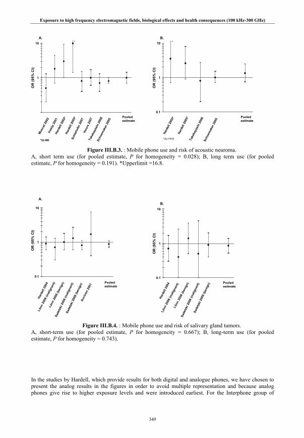
Exposure to high frequency electromagnetic fields, biological effects and health consequences (100 kHz-300 GHz)
349
Figure III.B.3. : Mobile phone use and risk of acoustic neuroma. A, short term use (for pooled estimate, P for homogeneity = 0.028); B, long term use (for pooled estimate, P for homogeneity = 0.191). *Upperlimit =16.8.
Figure III.B.4. : Mobile phone use and risk of salivary gland tumors. A, short-term use (for pooled estimate, P for homogeneity = 0.667); B, long-term use (for pooled estimate, P for homogeneity = 0.743).
In the studies by Hardell, which provide results for both digital and analogue phones, we have chosen to present the analog results in the figures in order to avoid multiple representation and because analog phones give rise to higher exposure levels and were introduced earliest. For the Interphone group of
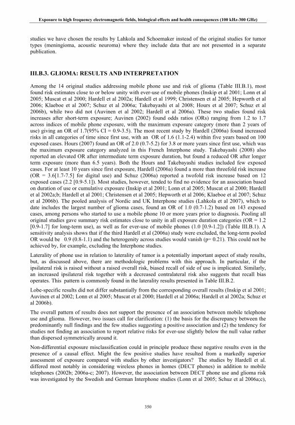
Exposure to high frequency electromagnetic fields, biological effects and health consequences (100 kHz-300 GHz)
350
studies we have chosen the results by Lahkola and Schoemaker instead of the original studies for tumor types (meningioma, acoustic neuroma) where they include data that are not presented in a separate publication.
III.B.3. GLIOMA: RESULTS AND INTERPRETATION
Among the 14 original studies addressing mobile phone use and risk of glioma (Table III.B.1), most found risk estimates close to or below unity with ever-use of mobile phones (Inskip et al 2001; Lonn et al 2005; Muscat et al 2000; Hardell et al 2002a; Hardell et al 1999; Christensen et al 2005; Hepworth et al 2006; Klaeboe et al 2007; Schuz et al 2006a; Takebayashi et al 2008; Hours et al 2007; Schuz et al 2006b), while two did not (Auvinen et al 2002; Hardell et al 2006a). These two studies found risk increases after short-term exposure; Auvinen (2002) found odds ratios (ORs) ranging from 1.2 to 1.7 across indices of mobile phone exposure, with the maximum exposure category (more than 2 years of use) giving an OR of 1.7(95% CI = 0.9-3.5). The most recent study by Hardell (2006a) found increased risks in all categories of time since first use, with an OR of 1.6 (1.1-2.4) within five years based on 100 exposed cases. Hours (2007) found an OR of 2.0 (0.7-5.2) for 3.8 or more years since first use, which was the maximum exposure category analyzed in this French Interphone study. Takebayashi (2008) also reported an elevated OR after intermediate term exposure duration, but found a reduced OR after longer term exposure (more than 6.5 years). Both the Hours and Takebayashi studies included few exposed cases. For at least 10 years since first exposure, Hardell (2006a) found a more than threefold risk increase (OR = 3.6[1.7-7.5] for digital use) and Schuz (2006a) reported a twofold risk increase based on 12 exposed cases (2.2 [0.9-5.1]). Most studies, however, tended to find no evidence for an association based on duration of use or cumulative exposure (Inskip et al 2001; Lonn et al 2005; Muscat et al 2000; Hardell et al 2002a;b; Hardell et al 2001; Christensen et al 2005; Hepworth et al 2006; Klaeboe et al 2007; Schuz et al 2006b). The pooled analysis of Nordic and UK Interphone studies (Lahkola et al 2007), which to date includes the largest number of glioma cases, found an OR of 1.0 (0.7-1.2) based on 143 exposed cases, among persons who started to use a mobile phone 10 or more years prior to diagnosis. Pooling all original studies gave summary risk estimates close to unity in all exposure duration categories (OR = 1.2 [0.9-1.7] for long-term use), as well as for ever-use of mobile phones (1.0 [0.9-1.2]) (Table III.B.1). A sensitivity analysis shows that if the third Hardell et al (2006a) study were excluded, the long-term pooled OR would be 0.9 (0.8-1.1) and the heterogenity across studies would vanish (p= 0.21). This could not be achieved by, for example, excluding the Interphone studies.
Laterality of phone use in relation to laterality of tumor is a potentially important aspect of study results, but, as discussed above, there are methodologic problems with this approach. In particular, if the ipsilateral risk is raised without a raised overall risk, biased recall of side of use is implicated. Similarly, an increased ipsilateral risk together with a decreased contralateral risk also suggests that recall bias operates. This pattern is commonly found in the laterality results presented in Table III.B.2.
Lobe-specific results did not differ substantially from the corresponding overall results (Inskip et al 2001; Auvinen et al 2002; Lonn et al 2005; Muscat et al 2000; Hardell et al 2006a; Hardell et al 2002a; Schuz et al 2006b).
The overall pattern of results does not support the presence of an association between mobile telephone use and glioma. However, two issues call for clarification: (1) the basis for the discrepancy between the predominantly null findings and the few studies suggesting a positive association and (2) the tendency for studies not finding an association to report relative risks for ever-use slightly below the null value rather than dispersed symmetrically around it.
Non-differential exposure misclassification could in principle produce these negative results even in the presence of a causal effect. Might the few positive studies have resulted from a markedly superior assessment of exposure compared with studies by other investigators? The studies by Hardell et al. differed most notably in considering wireless phones in homes (DECT phones) in addition to mobile telephones (2002b; 2006a-c; 2007). However, the association between DECT phone use and glioma risk was investigated by the Swedish and German Interphone studies (Lonn et al 2005; Schuz et al 2006a;c),

Exposure to high frequency electromagnetic fields, biological effects and health consequences (100 kHz-300 GHz)
351
without finding an increased risk of glioma. The exposure assessment methods of Auvinen et al (2002) are similar to the ones used in Schuz et al (2006b), and the methods of Schuz et al (2006a) and Hours et al (2007) are indistinguishable from those of other Interphone studies. Another potential reason for the discrepant results is selection bias through non-response among controls who did not use mobile phones, as discussed above. However, selection bias within the Interphone study was estimated to cause a downward bias in risk estimates of approximately 10% (Vrijheid et al 2009b); if this estimate is correct, this source of selection bias does not appear large enough to explain the differences in results.
If the series of negative studies is correct, it is appropriate to consider the potential reasons, including random error, for spurious positive findings in the studies generating positive results. The positive studies do not appear to have structural features with regard to case and control group constitution that would bias associations in a positive direction. The basic approach to exposure assessment does not appear to differ from that of other studies, with most studies based on self-report of use and various derived indices of exposure. While on the surface, the positive studies, including those by Hardell et al., are very much like the studies that obtained quite different results, subtle aspects of data collection and methods of analysis may be responsible for the apparent discrepancies. Investigators must make decisions regarding the exact constitution of the case groups, such as, whether to restrict by anatomic location, histology, stage, or malignancy. Exposure assignment requires even more complex decisions, including analog or digital phone use; how to define regular use; how to categorize hours of use or cumulative exposure; consideration of laterality of use and tumor location; and selection of reference dates of use for controls in relation to the timing of disease diagnosis. There is potential for differing recruitment methods to affect the magnitude and pattern of non-response, for interviewer training and monitoring to affect reporting tendencies of cases and controls, and even for the wording of questions to have subtle effects on the resulting data. Every team of investigators faces these decisions, and, presuming that there are compensating practices, the series of studies in the literature overall is expected to converge on a valid result. These decisions represent a major reason why replication of results by different research groups is needed before results can be considered as established.
The studies by Hardell and colleagues are particularly problematic because of variation across their publications in the exact constitution of case groups, criteria for exclusion, exposure definitions, and the selection of results for presentation in the multiple overlapping publications. In our view, the series of decisions in methods, analysis, and presentation provide the most plausible explanation for the deviation of the findings of the Hardell studies from those of other investigators. This does not address the other positive reports, but they seem to fit more in the distribution of results expected given random error across studies.
In summary, the complete array of available data does not suggest a causal association of mobile phone use with risk of glioma. However, there remains some uncertainty due to inconsistencies across the studies, as well as the recognized problems of exposure misclassification and potential for bias due to selective participation. As discussed previously, non-participation in the Interphone studies has been estimated to result in a 10% downward bias of the odds ratios, which can not explain all of the observed risk reduction. In addition, the period between exposure to a causal agent and manifestation of glioma may range from 5 to 20 years or more, judging from the intervals observed between ionizing radiation exposure and tumor diagnosis. Symptoms depend on the site and nature of the tumor, with slowest onset for low-grade tumors and rapid onset for highly malignant and swiftly-growing tumors. The data for long-term phone use of more than 10 years are still sparse, and any increased risk of slow-growing tumors may not yet have become manifest.
III.B.4. MENINGIOMA: RESULTS AND INTERPRETATION
Eleven original case-control studies (Inskip et al 2001; Auvinen et al 2002; Lonn et al 2005; Hardell et al 2005a; 2002a; 1999; Christensen et al 2005; Klaeboe et al 2007; Schuz et al 2006a; Takebayashi et al 2008; Hours et al 2007), one cohort study (Johansen et al 2001; Schuz et al 2006b), and two pooled analyses (Hardell et al 2006c; Lahkola et al 2008) have investigated the association between mobile phone use and meningioma. With the exception of the most recent study by Hardell (2005a), all studies

Exposure to high frequency electromagnetic fields, biological effects and health consequences (100 kHz-300 GHz)
352
found risk estimates close to or below unity, regardless of time since first mobile phone use (Table III.B.3). The study by Hardell (2005a) found an increased risk with ever-use of an analog mobile phone (OR = 1.7 [1.0-3.0]), with the highest risk estimate for more than 10 years since first use (2.1 [1.1-4.3]). The largest study so far- the pooled analysis of the Nordic and UK Interphone studies - found an OR of 0.9 (0.7-1.3) for long term use. Pooling all original studies gave risk estimates close to or below unity (Table III.B.3). Thus, there is no consistent evidence of an increased risk of meningioma among mobile phone users.
Many of the methodologic concerns discussed above for glioma apply also to meningioma, since they were typically evaluated within the same epidemiologic studies. A particular consideration in the interpretation of studies of mengioma is the long latency for this disease. Unlike gliomas, meningiomas are typically very slow-growing tumors with probable latencies of up to 30 yrs or more (Choudhary et al 2006). Cases may have no symptoms for a long period before detection of their tumor because meningiomas compress rather than invade the brain. A proportion of patients diagnosed with meningiomas in the 1990s and included in early studies could well have had the tumor present prior to any substantive exposure to mobile phones. Thus, the negative results give weaker evidence regarding an absence of association than the corresponding negative results for glioma.
III.B.5. ACOUSTIC NEUROMA: RESULTS AND INTERPRETATION
The 13 original studies of acoustic neuroma (Inskip et al 2001; Lonn et al 2004b; Takeyabashi et al 2006; Muscat et al 2002; Warren et al 2003; Hardell et al 2005a; 2002a; 1999; Christensen et al 2004; Klaeboe et al 2007; Schlehofer et al 2007; Hours et al 2007; Johansen et al 2001; Schuz et al 2006b) (Table III.B.4) generally included small numbers of cases. The pooled analyses are larger (Hardell et al 2006c; Schoemaker et al 2005), especially the Nordic-UK pooled analysis (Schoemaker et al 2005). Response rates for cases have been relatively high, reflecting the benign nature of this tumor, but control response rates have generally been lower. For ever-use of a mobile phone, all studies found risk estimates close to or below unity, except the two most recent studies by Hardell et al (2005a; 2002a), where up to fourfold risk increases were reported. It is notable that Hardell et al (2005a; 2002a; 2006c) observed considerably increased risks also within a short time period since first use. Acoustic neuroma is a very slow-growing tumor (Thomsen et al 1990) and it seems likely that the majority of cases diagnosed within five years of their first mobile phone use would have had their tumor already present before they started to use the mobile phone. Two of the US studies (Inskip et al 2001; Muscat et al 2002) also reported somewhat elevated ORs relatively soon after first mobile phone use, but these were based on small numbers of exposed cases (Table III.B.4).
For long durations of exposure (10 years or more), the Nordic-UK pooled analysis included the largest number of cases, and reported an OR of 1.0 (0.7-1.5). Most studies found risk estimates below one, sometimes with a considerable risk reduction (eg Christensen (2004), with an OR of 0.2 [0.2-1.1], although the Swedish Interphone study (Lonn et al 2004b) found an OR of 1.9 (0.9-4-1). The two recent Hardell studies (2005a; 2002a) generated results that are discrepant from the other studies, with increased ORs of 3.5 (0.7-16.8) and 2.6 (0.9-8.0) for long-term analog phone use. Pooling all studies gave summary risk estimates of 1.2 (0.8-2.0) for long-term use, and 1.1 (0.8-1.4) for ever-use. Analyses in relation to cumulative hours of use or cumulative number of calls likewise indicated no clear associations except in one of the Hardell studies (2005a).
The risk of acoustic neuroma after reported regular ipsilateral phone use was not increased in the Nordic-UK analysis (OR 0.9 [0.7-1.1]). The same was true in the other datasets (Inskip et al 2001; Lonn et al 2004b; Takebayashi et al 2006; Muscat et al 2002; Klaeboe et al 2007; Hours et al 2007) except one by Hardell (2005a), in which there were ORs of 5.1 (1.9-14) for analog use and 2.9 (1.4-6.1) for digital use. There was, however, a raised risk associated with first ipsilateral phone use at least 10 years prior to diagnosis in the study by Lonn (OR = 3.9 [1.6-9.5]). The corresponding result in the Nordic-UK pooled analysis was 1.3 (0.8-2.0), although a raised risk was associated with at least 10 years of use (OR = 1.8 [1.0-3.3]) (Schoemaker et al 2005). Handedness has not been associated with ipsilateral tumor risk (Schoemaker et al 2005).
Exposure to high frequency electromagnetic fields, biological effects and health consequences (100 kHz-300 GHz)
353
Acoustic neuroma can cause unilateral deafness, which could lead to cessation of phone use (and hence spuriously reduced risks). Alternatively, the deafness could lead to the diagnosis of an otherwise unrecognized tumor and hence lead to spuriously increased risks. Hearing loss associated with acoustic neuromas may influence the side of phone use as the tumor progresses, resulting in preferred contralateral phone use relative to the tumor. This is not predictable, however, since hearing can be preserved in the presence of large vestibular schwannomas and, conversely, hearing loss can frequently occur as the result of radiologically static, small tumors (Rutherford et al 2005). Potential effects on the side of mobile phone use or earlier detection of tumors should, however, affect all available studies similarly; this cannot explain the discrepancies in the results.
Unlike the situation for gliomas and meningiomas, laterality virtually defines the anatomical position of acoustic neuromas, and all ipsilateral acoustic neuromas arise close to the mobile phone handset position. Therefore if reliable unbiased information on side of exposure could be obtained, it would be possible to conduct a powerful unbiased analysis of the effect of mobile phone exposure on acoustic neuroma risk. This analysis, however, is hampered by inconsistency in side of phone use, reporting bias resulting from the tumor diagnosis, and the symptom-based changes in use noted above. The results indicating an increased risk associated with ipsilateral phone use but no overall raised risk again raise questions about the contribution of reporting bias. Thus, the elevated ipsilateral risk beyond 10 years in the large Nordic-UK analysis seems more likely to represent reporting bias than a causal effect, because the latter should lead to a raised risk (although diluted) for users overall beyond 10 years - a finding that was not seen in the overall Nordic-UK data.
As was the case for meningioma, acoustic neuromas are often present for years before diagnosis. Thus, the only data about phone use that are of any potential relevance to acoustic neuroma etiology may be the exposure occurring many years before diagnosis. The available data make it unlikely that there is any substantial raised risk of acoustic neuroma in relation to mobile phone use in the ten years preceding the diagnosis of the tumor. The results leave uncertainty as to whether there are raised risks beyond 10 years from initial use.
III.B.6. SALIVARY GLAND TUMORS: RESULTS AND INTERPRETATION
There is no consistent evidence of an increased risk of salivary gland tumors among mobile phone users (Table III.B.5, Fig. III.B.4) based on four case-control studies (Auvinen et al 2002; Hardell et al 2004; Sadetzki et al 2008; Lonn et al 2006) and one cohort study (Schuz 2006b). One study (Auvinen et al 2002) showed an increase in risk for ever-use compared with never-use and for greater cumulative years of exposure, but the results were based on few cases and had very wide confidence intervals. There was no indication of a raised risk in any of the other studies including that of Hardell. Pooling the results from all studies gave risk estimates slightly below unity in all exposure categories (Table III.B.5). Both publications from the Interphone study reported higher risk estimates associated with ipsilateral phone use at least 10 years prior to diagnosis, with an OR of 2.6 (0.9-7.9) in the Lonn study (2006), and 1.6 (0.7-3.7) in the study by Sadetzki et al (2008). Corresponding ORs for contralateral use were, however, considerably reduced in both studies: 0.3 (0.0-2.3) and 0.6 0.2-2.3), respectively. Thus, reporting bias seems likely to explain these findings.
Single studies of tumors at other sites (pituitary adenoma (Takebayashi et al 2008), non-Hodgkin’s lymphoma (Hardell et al 2005b), testicular cancer (Hardell et al 2007), uveal melanoma (Stang et al 2001) are not discussed here. The main results for these cancer sites are shown in eTable 2.
III.B.7. CONCLUSIONS
In the last few years the epidemiologic evidence on mobile phone use and risk of brain and other tumors of the head has grown considerably. In our opinion, overall the studies published to date do not demonstrate a raised risk within approximately ten years of use for any tumor of the brain or any other
Exposure to high frequency electromagnetic fields, biological effects and health consequences (100 kHz-300 GHz)
354
head tumor. However, some key methodologic problems remain - for example, selective non-response and exposure misclassification. Despite these methodologic shortcomings and the still limited data on long latency and long-term use, the available data do not suggest a causal association between mobile phone use and fast-growing tumors such as malignant glioma in adults, at least those tumors with short induction periods. For slow-growing tumors such as meningioma and acoustic neuroma, as well as for glioma among long-term users, the absence of associations reported thus far is less conclusive because the current observation period is still too short. Currently data are completely lacking on the potential carcinogenic effect of exposures in childhood and adolescence.
Exposure to high frequency electromagnetic fields, biological effects and health consequences (100 kHz-300 GHz)
355
III.B.8. REFERENCES
Ahlbom A, Green A, Kheifets L, Savitz D, Swerdlow A. Epidemiology of health effects of radiofrequency exposure. Environ Health Perspect 112(17):1741-54; 2004.
Auvinen A, Hietanen M, Luukkonen R, Koskela RS. Brain tumors and salivary gland cancers among cellular telephone users. Epidemiology 13(3):356-9; 2002.
Cardis E, Deltour I, Mann S, Moissonnier M, Taki M, Varsier N, Wake K, Wiart J. Distribution of RF energy emitted by mobile phones in anatomical structures of the Brain. Phys Med Biol 53:2771-83; 2008.
Choudhary A, Pradhan S, Huda MF, Mohanty S, Kumar M. Radiation induced meningioma with a short latent period following high dose cranial irradiation - case report and literature review. J Neurooncol 77(1):73-7; 2006.
Christensen HC, Schuz J, Kosteljanetz M, Poulsen HS, Thomsen J, Johansen C. Cellular telephone use and risk of acoustic neuroma. Am J Epidemiol 159(3):277-83; 2004.
Christensen HC, Schuz J, Kosteljanetz M, Poulsen HS, Boice JD, Jr., McLaughlin JK, Johansen C. Cellular telephones and risk for brain tumors: a population-based, incident case-control study. Neurology 64(7):1189-95; 2005.
Dreyer NA, Loughlin JE, Rothman KJ. Cause-specific mortality in cellular telephone users. Jama 282(19):1814-6; 1999.
Hardell L, Nasman A, Pahlson A, Hallquist A, Hansson Mild K. Use of cellular telephones and the risk for brain tumours: A case-control study. Int J Oncol 15(1):113-6; 1999.
Hardell L, Mild KH, Pahlson A, Hallquist A. Ionizing radiation, cellular telephones and the risk for brain tumours. Eur J Cancer Prev 10(6):523-9; 2001.
Hardell L, Hallquist A, Mild KH, Carlberg M, Pahlson A, Lilja A. Cellular and cordless telephones and the risk for brain tumours. Eur J Cancer Prev 11(4):377-86; 2002a.
Hardell L, Mild KH, Carlberg M. Case-control study on the use of cellular and cordless phones and the risk for malignant brain tumours. Int J Radiat Biol 78(10):931-6; 2002b.
Hardell L, Hallquist A, Hansson Mild K, Carlberg M, Gertzen H, Schildt EB, Dahlqvist A. No association between the use of cellular or cordless telephones and salivary gland tumours. Occup Environ Med 61(8):675-9; 2004.
Hardell L, Carlberg M, Hansson Mild K. Case-control study on cellular and cordless telephones and the risk for acoustic neuroma or meningioma in patients diagnosed 2000-2003. Neuroepidemiology 25(3):120-8; 2005a.
Hardell L, Eriksson M, Carlberg M, Sundstrom C, Mild KH. Use of cellular or cordless telephones and the risk for non-Hodgkin's lymphoma. Int Arch Occup Environ Health 78(8):625-32; 2005b.
Hardell L, Carlberg M, Mild KH. Case-control study of the association between the use of cellular and cordless telephones and malignant brain tumors diagnosed during 2000-2003. Environ Res 100(2):232-41; 2006a.
Hardell L, Carlberg M, Hansson Mild K. Pooled analysis of two case-control studies on use of cellular and cordless telephones and the risk for malignant brain tumours diagnosed in 1997-2003. Int Arch Occup Environ Health 79(8):630-9; 2006b.
Hardell L, Carlberg M, Hansson Mild K. Pooled analysis of two case-control studies on the use of cellular and cordless telephones and the risk of benign brain tumours diagnosed during 1997-2003. Int J Oncol 28(2):509-18; 2006c.
Hardell L, Carlberg M, Ohlson CG, Westberg H, Eriksson M, Hansson Mild K. Use of cellular and cordless telephones and risk of testicular cancer. Int J Androl 30(2):115-22; 2007.
Hepworth SJ, Schoemaker MJ, Muir KR, Swerdlow AJ, van Tongeren MJ, McKinney PA. Mobile phone use and risk of glioma in adults: case-control study. Bmj 332(7546):883-7; 2006.
Hillert L, Ahlbom A, Neasham D, Feychting M, Jarup L, Navin R, Elliott P. Call-related factors influencing output power from mobile phones. J Expo Sci Environ Epidemiol 16(6):507-14; 2006.
Exposure to high frequency electromagnetic fields, biological effects and health consequences (100 kHz-300 GHz)
356
Hours M, Bernard M, Montestrucq L, Arslan M, Bergeret A, Deltour I, Cardis E. [Cell Phones and Risk of brain and acoustic nerve tumours: the French INTERPHONE case-control study]. Rev Epidemiol Sante Publique 55(5):321-32; 2007.
Inskip PD, Tarone RE, Hatch EE, Wilcosky TC, Shapiro WR, Selker RG, Fine HA, Black PM, Loeffler JS, Linet MS. Cellular-telephone use and brain tumors. N Engl J Med 344(2):79-86; 2001.
Johansen C, Boice J, Jr., McLaughlin J, Olsen J. Cellular telephones and cancer--a nationwide cohort study in Denmark. J Natl Cancer Inst 93(3):203-7; 2001.
Klaeboe L, Blaasaas KG, Tynes T. Use of mobile phones in Norway and risk of intracranial tumours. Eur J Cancer Prev 16(2):158-64; 2007.
Lahkola A, Auvinen A, Raitanen J, Schoemaker MJ, Christensen HC, Feychting M, Johansen C, Klaeboe L, Lonn S, Swerdlow AJ, Tynes T, Salminen T. Mobile phone use and risk of glioma in 5 North European countries. Int J Cancer 120(8):1769-75; 2007.
Lahkola A, Salminen T, Raitanen J, Heinavaara S, Schoemaker M, Christensen HC, Feychting M, Johansen C, Klaeboe L, Lonn S, Swerdlow A, Tynes T, Auvinen A. Meningioma and mobile phone use--a collaborative case-control study in five North European countries. Int J Epidemiol; 2008.
Lonn S, Forssen U, Vecchia P, Ahlbom A, Feychting M. Output power levels from mobile phones in different geographical areas; implications for exposure assessment. Occup Environ Med 61(9):769-72; 2004a.
Lonn S, Ahlbom A, Hall P, Feychting M. Mobile phone use and the risk of acoustic neuroma. Epidemiology 15(6):653-9; 2004b.
Lonn S, Ahlbom A, Hall P, Feychting M. Long-term mobile phone use and brain tumor risk. Am J Epidemiol 161(6):526-35; 2005.
Lonn S, Ahlbom A, Christensen H, Johansen C, Schuz J, Edstrom S, Henriksson G, Lundgren J, Wennerberg J, Feychting M. Mobile phone use and risk of parotid gland tumor. Am J Epidemiol 164:637-43; 2006.
Muscat JE, Malkin MG, Thompson S, Shore RE, Stellman SD, McRee D, Neugut AI, Wynder EL. Handheld cellular telephone use and risk of brain cancer. Jama 284(23):3001-7; 2000.
Muscat JE, Malkin MG, Shore RE, Thompson S, Neugut AI, Stellman SD, Bruce J. Handheld cellular telephones and risk of acoustic neuroma. Neurology 58(8):1304-6; 2002.
Sadetzki S, Chetrit A, Jarus-Hakak A, Cardis E, Deutch Y, Duvdevani S, Zultan A, Novikov I, Freedman L, Wolf M. Cellular phone use and risk of benign and malignant parotid gland tumors--a nationwide case-control study. Am J Epidemiol 167(4):457-67; 2008.
Schlehofer B, Schlaefer K, Blettner M, Berg G, Bohler E, Hettinger I, Kunna-Grass K, Wahrendorf J, Schuz J. Environmental risk factors for sporadic acoustic neuroma (Interphone Study Group, Germany). Eur J Cancer 43(11):1741-7; 2007.
Schoemaker MJ, Swerdlow AJ, Ahlbom A, Auvinen A, Blaasaas KG, Cardis E, Christensen HC, Feychting M, Hepworth SJ, Johansen C, Klaeboe L, Lonn S, McKinney PA, Muir K, Raitanen J, Salminen T, Thomsen J, Tynes T. Mobile phone use and risk of acoustic neuroma: results of the Interphone case-control study in five North European countries. Br J Cancer 93(7):842-8; 2005.
Schuz J, Bohler E, Berg G, Schlehofer B, Hettinger I, Schlaefer K, Wahrendorf J, Kunna-Grass K, Blettner M. Cellular phones, cordless phones, and the risks of glioma and meningioma (Interphone study group, Germany). Am J Epidemiol 163(6):512-520; 2006a.
Schuz J, Jacobsen R, Olsen JH, Boice JD, Jr., McLaughlin JK, Johansen C. Cellular telephone use and cancer risk: update of a nationwide Danish cohort. J Natl Cancer Inst 98(23):1707-1713; 2006b.
Schuz J, Bohler E, Schlehofer B, Berg G, Schlaefer K, Hettinger I, Kunna-Grass K, Wahrendorf J, Blettner M. Radiofrequency electromagnetic fields emitted from base stations of DECT cordless phones and the risk of glioma and meningioma (Interphone Study Group, Germany). Radiat Res 166(1 Pt 1):116-9; 2006c.
Stang A, Anastassiou G, Ahrens W, Bromen K, Bornfeld N, Jockel KH. The possible role of radiofrequency radiation in the development of uveal melanoma. Epidemiology 12(1):7-12; 2001.
Rutherford SA, King AT. Vestibular schwannoma management: What is the 'best' option? Br J Neurosurg 19(4):309-16; 2005.
Exposure to high frequency electromagnetic fields, biological effects and health consequences (100 kHz-300 GHz)
357
Takebayashi T, Akiba S, Kikuchi Y, Taki M, Wake K, Watanabe S, Yamaguchi N. Mobile phone use and acoustic neuroma risk in Japan. Occup Environ Med 63(12):802-7; 2006.
Takebayashi T, Varsier N, Kikuchi Y, Wake K, Taki M, Watanabe S, Akiba S, Yamaguchi N. Mobile phone use, exposure to radiofrequency electromagnetic field, and brain tumour: a case-control study. Br J Cancer 98(3):652-9; 2008.
Thomsen J, Tos M. Acoustic neuroma: clinical aspects, audiovestibular assessment, diagnostic delay, and growth rate. Am J Otol 11(1):12-9; 1990.
Vrijheid M, Cardis E, Armstrong BK, Auvinen A, Berg G, Blaasaas KG, Brown J, Carroll M, Chetrit A, Christensen HC, Deltour I, Feychting M, Giles GG, Hepworth SJ, Hours M, Iavarone I, Johansen C, Klaeboe L, Kurttio P, Lagorio S, Lonn S, McKinney PA, Montestrucq L, Parslow RC, Richardson L, Sadetzki S, Salminen T, Schuz J, Tynes T, Woodward A. Validation of short term recall of mobile phone use for the Interphone study. Occup Environ Med 63(4):237-43; 2006.
Vrijheid M, Armstrong BK, Bedard D, Brown J, Deltour I, Iavarone I, Krewski D, Lagorio S, Moore S, Richardson L, Giles GG, McBride M, Parent ME, Siemiatycki J, Cardis E. Recall bias in the assessment of exposure to mobile phones. J Expo Sci Environ Epidemiol:369-81; 2009a.
Vrijheid M, Richardson L, Armstrong BK, Auvinen A, Berg G, Carroll M, Chetrit A, Deltour I, Feychting M, Giles GG, Hours M, Iavarone I, Lagorio S, Lonn S, Mcbride M, Parent M-E, Sadetzki S, Salminen T, Sanchez M, Schlehofer B, Schuz J, Siemiatycki J, Tynes T, Woodward A, Yamaguchi N, Cardis E. Quantifying the impact of selection bias caused by nonparticipation in a case–control study of mobile phone use. Ann Epidemiol 19:33-41; 2009b.
Walker AM. Declining relative risks for lung cancer after cessation of asbestos exposure. J Occup Med 26(6):422-6; 1984.
Warren HG, Prevatt AA, Daly KA, Antonelli PJ. Cellular telephone use and risk of intratemporal facial nerve tumor. Laryngoscope 113(4):663-7; 2003.

Exposure to high frequency electromagnetic fields, biological effects and health consequences (100 kHz-300 GHz)
358
Tab
le II
I.B.1
.: R
esul
ts o
f stu
dies
on
mob
ile p
hone
use
and
risk
of g
liom
aa
Tim
e si
nce
first
use
Sh
ort-
term
use
Inte
rmed
iate
-ter
m u
se
L
ong-
term
use
Eve
r/ne
ver
use
Ref
eren
ce
No.
O
R (9
5% C
I)
N
o.
OR
(95%
CI)
No.
O
R (9
5% C
I)
N
o.
OR
(95%
U
S St
udie
s
Mus
cat e
t al 2
000
(Mal
igna
nt b
rain
) 49
0.
9 (0
.6-1
.4)b
17
0.
7 (0
.4-1
.4)
66
0.7
(0.5
-1.1
) In
skip
et a
l 200
1 (G
liom
a)
31
0.9
(0.5
-1.6
)
11
0.5
(0.2
-1.3
)
20
1 1.
0 (0
.7-1
.4)
Har
dell
Stud
ies
H
arde
ll et
al 1
999
(All
brai
n)
78
1.0
(0.7
-1.4
)
34
0.8
(0.5
-1.4
)
16
1.2
(0.6
-2.6
)
78
1.0
(0.7
-1.4
) H
arde
ll et
al 2
002a
,b (A
ll 36
(ana
log)
1.
1 (0
.7-1
.8)
43 (a
nalo
g)
1.2
(0.8
-1.8
)
79 a
nalo
g 1.
1 (0
.8-1
.6)
Har
dell
et a
l 200
6a (A
ll m
alig
nant
) 0
(ana
log)
-
20
(ana
log)
1.
8 (0
.9-3
.5)
48
(ana
log)
3.
5 (2
.0-6
.4)
68
ana
log
2.6
(1.5
-4.3
) H
arde
ll po
oled
ana
lysi
s
H
arde
ll et
al 2
006b
c (All
mal
igna
nt)
39 (a
nalo
g)
1.2
(0.8
-1.8
)
57 (a
nalo
g)
1.1
(0.8
-1.6
)
82 (a
nalo
g)
2.4
(1.6
-3.4
)
178
anal
og
1.5
(1.1
-1.9
) In
terp
hone
Stu
dies
Chr
iste
nsen
et a
l 200
5 (G
liom
a)d
43
0.7
(0.4
-1.0
)
42
0.6
(0.4
-1.0
)
14
0.7
(0.3
-1.6
)
106
0.7
(0.5
-1.0
) Lo
nn e
t al 2
005
(Glio
ma)
11
2 0.
8 (0
.6-1
.1)
75
0.
7 (0
.5-1
.0)
25
0.
9 (0
.5-1
.5)
21
4 0.
8 (0
.6-1
.0)
Schu
z et
al 2
006a
(Glio
ma)
82
0.
9 (0
.6-1
.2)
39
1.
0 (0
.6-1
.5)
12
2.
2 (0
.9-5
.1)
13
8 1.
0 (0
.7-1
.3)
Hep
wor
th e
t al 2
006
(Glio
ma)
2
71
0.9
(0.7
-1.1
)
170
1.0
(0.8
-1.3
)
66
0.9
(0.6
-1.3
)
508
0.9
(0.8
-1.1
) K
laeb
oe e
t al 2
007
(Glio
ma)
27
0.
6 (0
.4-1
.1)
64
0.
5 (0
.3-0
.8)
70
0.
8 (0
.5-1
.2)
16
1 0.
6 (0
.4-0
.9)
Hou
rs e
t al 2
007
(Glio
ma)
38
0.
9 (0
.5-1
.6)e
21
2.
0 (0
.7-5
.2)
59
1.2
(0.7
-2.1
) Ta
keba
yash
i et a
l 200
8 (G
liom
a)
32
1.3
(0.7
-2.3
)f
17
1.9
(0.8
-4.4
)
7 0.
6 (0
.2-1
.8)
56
1.
2 (0
.6-2
.4)
Inte
rpho
ne p
oole
d an
alys
is
Lahk
ola
et a
l 200
7g (Glio
ma)
38
4 0.
8 (0
.7-0
.9)
34
2 0.
8 (0
.6-0
.9)
14
3 1.
0 (0
.7-1
.2)
86
7 0.
8 (0
.7-0
.9)
Subs
crib
er li
st S
tudi
es
Auv
inen
et a
l 200
2 (G
liom
a)
25
1.5
(0.9
-2.4
)h
11
1.7
(0.9
-3.5
)
36
1.
5 (1
.0-2
.4)
Schu
z et
al 2
006b
(Ner
vous
syst
em)
266
1.0
(0.9
-1.2
)
235
1.0
(0.8
-1.1
)
28
0.7
(0.4
-1.0
)
580
1.0
(0.9
-1.0
) Po
olin
g al
l stu
dies
i
P fo
r hom
ogen
eity
1.
0 (0
.9-1
.1)
0.13
8
0.9
(0.8
-1.1
) 0.
010
1.
1 (0
.8-1
.4)
0.00
1
1.0
(0.8
-1.2
) 0.
001
a All
stud
ies a
re c
ase-
cont
rol s
tudi
es e
xcep
t Sch
uz e
t al 2
006b
b P
oole
d re
sult
for 1
yea
r and
2-3
yea
rs
c D
ata
from
Har
dell
2002
b an
d 20
06a
d Po
oled
resu
lts fo
r low
gra
de a
nd h
igh
grad
e gl
iom
a e P
oole
d re
sult
for <
1.3
year
s, 1.
3-2.
25 y
ears
, and
2.2
5-3.
8 ye
ars
f Po
oled
resu
lt fo
r <2.
2 ye
ars a
nd 2
.2-4
.7 y
ears
g D
ata
from
Chr
iste
nsen
200
5, L
onn
2005
, Kla
ebo
2007
, par
t of H
epw
orth
200
6, a
nd d
ata
from
Fin
land
no
t pre
viou
sly
publ
ishe
d
h Po
oled
resu
lt fo
r <1
year
and
1-2
yea
rs
i Po
olin
g al
l stu
dies
exc
ept H
arde
ll 20
06b
and
Lahk
ola
2007
, usi
ng th
e ra
ndom
eff
ects
mod
el. F
rom
H
arde
ll 20
02 a
nd 2
006,
whe
n re
sults
for b
oth
anal
ogue
and
dig
ital p
hone
use
wer
e av
aila
ble,
onl
y th
e re
sults
for a
nalo
gue
phon
e us
e w
ere
incl
uded
to a
void
incl
udin
g du
plic
ate
data
. O
R in
dica
tes o
dds r
atio
; CI,
conf
iden
ce in
terv
al.

Exposure to high frequency electromagnetic fields, biological effects and health consequences (100 kHz-300 GHz)
359
Tab
le II
I.B.2
.: R
esul
ts o
f lat
eral
ity a
naly
ses i
n st
udie
s on
mob
ile p
hone
use
and
risk
of g
liom
a
E
ver/
neve
r us
e
>10
year
s sin
ce fi
rst u
se
Ip
sila
tera
l
Con
tral
ater
al
Ip
sila
tera
l
Con
tral
ater
al
Ref
eren
ce
RR
(95%
CI)
RR
(95%
CI)
RR
(95%
CI)
RR
(95%
CI)
C
omm
ent
Har
dell
et a
l 199
9/20
01
1.1
(0.6
-1.8
)
0.7
(0.4
-1.2
)
Har
dell
et a
l 200
2a,b
1.
9 (1
.2-3
.0)
1.6
(1.1
-2.4
)
0.
6 (0
.4-1
.1)
0.9
(0.5
-1.4
)
1.
8 (1
.0-3
.4)a
2.3
(0.6
-8.9
)a
0.
7 (0
.4-1
.6)a
0.3
(0.0
-2.9
)a
Ana
log
Dig
ital
Har
dell
et a
l 200
6a
3.1
(1.6
-6.2
)
2.6
(1.6
-4.1
)
2.
6 (1
.3-5
.4)
1.3
(0.8
-2.2
)
Ana
log
Dig
ital
Lonn
et a
l 200
5 1.
1 (0
.8-1
.5)
0.
7 (0
.5-1
.0)
1.
6 (0
.8-3
.4)
0.
7 (0
.3-1
.5)
Hep
wor
th e
t al 2
006
1.2
(1.0
-1.5
)
0.8
(0.6
-0.9
)
1.6
(0.9
-2.8
)
0.8
(0.4
-1.4
)
Kla
eboe
et a
l 200
7 1.
0 (0
.7-1
.4)
0.
7 (0
.5-1
.1)
1.
3 (0
.8-2
.1)b
0.
8 (0
.5-1
.4)b
Hou
rs e
t al 2
007
1.2
(0.6
-2.4
)
1.2
(0.5
-2.7
)
Take
baya
shi e
t al 2
008
1.2
(0.7
-2.3
)
1.1
(0.6
-2.0
)
Lahk
ola
et a
l 200
7 1.
1 (1
.0-1
.3)
0.
8 (0
.6-0
.9)
1.
4 (1
.0-1
.9)
1.
0 (0
.7-1
.4)
a >6
yea
rs
b >6
yea
rs
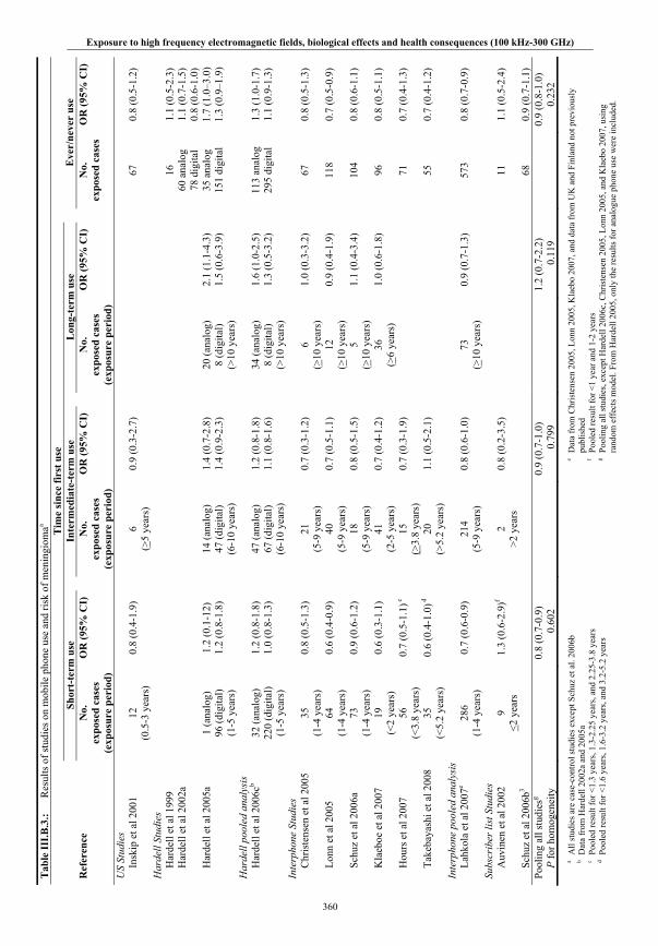
Exposure to high frequency electromagnetic fields, biological effects and health consequences (100 kHz-300 GHz)
360
Tab
le II
I.B.3
.: R
esul
ts o
f stu
dies
on
mob
ile p
hone
use
and
risk
of m
enin
giom
aa
Tim
e si
nce
first
use
Sh
ort-
term
use
Inte
rmed
iate
-ter
m u
se
L
ong-
term
use
E
ver/
neve
r us
e R
efer
ence
N
o.
expo
sed
case
s (e
xpos
ure
peri
od)
OR
(95%
CI)
No.
ex
pose
d ca
ses
(exp
osur
e pe
riod
)
OR
(95%
CI)
No.
ex
pose
d ca
ses
(exp
osur
e pe
riod
)
OR
(95%
CI)
No.
ex
pose
d ca
ses
OR
(95%
CI)
US
Stud
ies
Insk
ip e
t al 2
001
12
(0.5
-3 y
ears
) 0.
8 (0
.4-1
.9)
6
(>5
year
s)
0.9
(0.3
-2.7
)
67
0.
8 (0
.5-1
.2)
Har
dell
Stud
ies
Har
dell
et a
l 199
9
16
1.1
(0.5
-2.3
)
Har
dell
et a
l 200
2a
60
ana
log
78 d
igita
l 1.
1 (0
.7-1
.5)
0.8
(0.6
-1.0
)
Har
dell
et a
l 200
5a
1 (a
nalo
g)
96 (d
igita
l) (1
-5 y
ears
)
1.2
(0.1
-12)
1.
2 (0
.8-1
.8)
14
(ana
log)
47
(dig
ital)
(6-1
0 ye
ars)
1.4
(0.7
-2.8
) 1.
4 (0
.9-2
.3)
20
(ana
log)
8
(dig
ital)
(>10
yea
rs)
2.1
(1.1
-4.3
) 1.
5 (0
.6-3
.9)
35
ana
log
151
digi
tal
1.7
(1.0
–3.0
) 1.
3 (0
.9–1
.9)
Har
dell
pool
ed a
naly
sis
H
arde
ll et
al 2
006c
b 32
(ana
log)
22
0 (d
igita
l) (1
-5 y
ears
)
1.2
(0.8
-1.8
) 1.
0 (0
.8-1
.3)
47
(ana
log)
67
(dig
ital)
(6-1
0 ye
ars)
1.2
(0.8
-1.8
) 1.
1 (0
.8-1
.6)
34
(ana
log)
8
(dig
ital)
(>10
yea
rs)
1.6
(1.0
-2.5
) 1.
3 (0
.5-3
.2)
11
3 an
alog
29
5 di
gita
l 1.
3 (1
.0-1
.7)
1.1
(0.9
-1.3
)
Inte
rpho
ne S
tudi
es
Chr
iste
nsen
et a
l 200
5
35
(1-4
yea
rs)
0.8
(0.5
-1.3
)
21
(5-9
yea
rs)
0.7
(0.3
-1.2
)
6 (>
10 y
ears
) 1.
0 (0
.3-3
.2)
67
0.
8 (0
.5-1
.3)
L
onn
et a
l 200
5
64
(1-4
yea
rs)
0.6
(0.4
-0.9
)
40
(5-9
yea
rs)
0.7
(0.5
-1.1
)
12
(>10
yea
rs)
0.9
(0.4
-1.9
)
118
0.7
(0.5
-0.9
)
S
chuz
et a
l 200
6a
73
(1-4
yea
rs)
0.9
(0.6
-1.2
)
18
(5-9
yea
rs)
0.8
(0.5
-1.5
)
5 (>
10 y
ears
) 1.
1 (0
.4-3
.4)
10
4 0.
8 (0
.6-1
.1)
K
laeb
oe e
t al 2
007
19
(<2
year
s)
0.6
(0.3
-1.1
)
41
(2-5
yea
rs)
0.7
(0.4
-1.2
)
36
(>6
year
s)
1.0
(0.6
-1.8
)
96
0.8
(0.5
-1.1
)
H
ours
et a
l 200
7 56
(<
3.8
year
s)
0.7
(0.5
-1.1
) c
15
(>
3.8
year
s)
0.7
(0.3
-1.9
)
71
0.
7 (0
.4-1
.3)
T
akeb
ayas
hi e
t al 2
008
35
(<5.
2 ye
ars)
0.
6 (0
.4-1
.0) d
20
(>5.
2 ye
ars)
1.
1 (0
.5-2
.1)
55
0.7
(0.4
-1.2
)
Inte
rpho
ne p
oole
d an
alys
is
L
ahko
la e
t al 2
007e
286
(1-4
yea
rs)
0.7
(0.6
-0.9
)
214
(5-9
yea
rs)
0.8
(0.6
-1.0
)
73
(>10
yea
rs)
0.9
(0.7
-1.3
)
573
0.8
(0.7
-0.9
)
Subs
crib
er li
st S
tudi
es
A
uvin
en e
t al 2
002
9 <2
yea
rs
1.3
(0.6
-2.9
)f
2 >2
yea
rs
0.8
(0.2
-3.5
)
11
1.
1 (0
.5-2
.4)
S
chuz
et a
l 200
6b3
68
0.
9 (0
.7-1
.1)
Pool
ing
all s
tudi
esg
P
for h
omog
enei
ty
0.
8 (0
.7-0
.9)
0.60
2
0.9
(0.7
-1.0
) 0.
799
1.
2 (0
.7-2
.2)
0.11
9
0.9
(0.8
-1.0
) 0.
232
a All
stud
ies a
re c
ase-
cont
rol s
tudi
es e
xcep
t Sch
uz e
t al.
2006
b
b Dat
a fr
om H
arde
ll 20
02a
and
2005
a c P
oole
d re
sult
for <
1.3
year
s, 1.
3-2.
25 y
ears
, and
2.2
5-3.
8 ye
ars
d Poo
led
resu
lt fo
r <1.
6 ye
ars,
1.6-
3.2
year
s, an
d 3.
2-5.
2 ye
ars
e Dat
a fr
om C
hris
tens
en 2
005,
Lon
n 20
05, K
laeb
o 20
07, a
nd d
ata
from
UK
and
Fin
land
not
pre
viou
sly
publ
ishe
d
f Po
oled
resu
lt fo
r <1
year
and
1-2
yea
rs
g Poo
ling
all s
tudi
es, e
xcep
t Har
dell
2006
c, C
hris
tens
en 2
005,
Lon
n 20
05, a
nd K
laeb
o 20
07, u
sing
ra
ndom
eff
ects
mod
el. F
rom
Har
dell
2005
, onl
y th
e re
sults
for a
nalo
gue
phon
e us
e w
ere
incl
uded
.
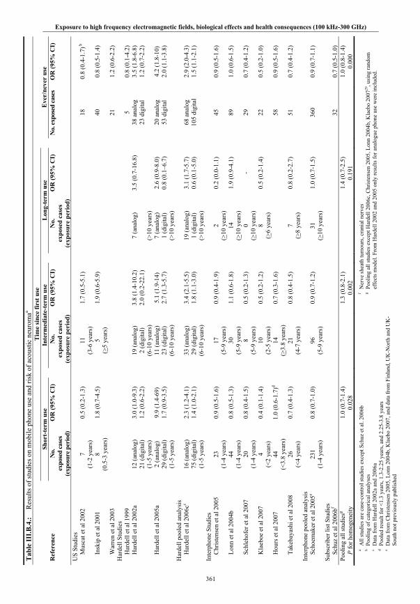
Exposure to high frequency electromagnetic fields, biological effects and health consequences (100 kHz-300 GHz)
361
Tab
le II
I.B.4
.: R
esul
ts o
f stu
dies
on
mob
ile p
hone
use
and
risk
of a
cous
tic n
euro
maa
T
ime
sinc
e fir
st u
se
Shor
t-te
rm u
se
In
term
edia
te-t
erm
use
Lon
g-te
rm u
se
Eve
r/ne
ver
use
Ref
eren
ce
No.
ex
pose
d ca
ses
(exp
osur
e per
iod)
OR
(95%
CI)
No.
ex
pose
d ca
ses
(exp
osur
e pe
riod
)
OR
(95%
CI)
No.
ex
pose
d ca
ses
(exp
osur
e pe
riod
)
OR
(95%
CI)
No.
expo
sed
case
s O
R (9
5% C
I)
US
Stud
ies
Mus
cat e
t al 2
002
7
(1-2
yea
rs)
0.5
(0.2
-1.3
)
11
(3-6
yea
rs)
1.7
(0.5
-5.1
)
18
0.
8 (0
.4-1
.7) b
In
skip
et a
l 200
1 8
(0.5
-3 y
ears
) 1.
8 (0
.7-4
.5)
5
(>5
year
s)
1.9
(0.6
-5.9
)
40
0.
8 (0
.5-1
.4)
W
arre
n et
al 2
003
21
1.2
(0.6
-2.2
) H
arde
ll St
udie
s
H
arde
ll et
al 1
999
5
0.8
(0.1
-4.2
)
Har
dell
et a
l 200
2a
12 (a
nalo
g)
21 (d
igita
l) (1
-5 y
ears
)
3.0
(1.0
-9.3
) 1.
2 (0
.6-2
.2)
19
(ana
log)
2
(dig
ital)
(6-1
0 ye
ars)
3.8
(1.4
-10.
2)
2.0
(0.2
-22.
1)
7
(ana
log)
(>10
yea
rs)
3.5
(0.7
-16.
8)
38 a
nalo
g 23
dig
ital
3.5
(1.8
-6.8
) 1.
2 (0
.7-2
.2)
H
arde
ll et
al 2
005a
2
(ana
log)
29
(dig
ital)
(1-5
yea
rs)
9.9
(1.4
-69)
1.
7 (0
.9-3
.5)
11
(ana
log)
23
(dig
ital)
(6-1
0 ye
ars)
5.1
(1.9
-14)
2.
7 (1
.3-5
.7)
7
(ana
log)
1
(dig
ital)
(>10
yea
rs)
2.6
(0.9
-8.0
) 0.
8 (0
.1–6
.7)
20
ana
log
53 d
igita
l 4.
2 (1
.8-1
0)
2.0
(1.1
-3.8
)
Har
dell
pool
ed a
naly
sis
H
arde
ll et
al 2
006c
c
16 (a
nalo
g)
75 (d
igita
l) (1
-5 y
ears
)
2.3
(1.2
-4.1
) 1.
4 (1
.0-2
.1)
33
(ana
log)
29
(dig
ital)
(6-1
0 ye
ars)
3.4
(2.1
-5.5
) 1.
8 (1
.1-3
.0)
19
(ana
log)
1
(dig
ital)
(>10
yea
rs)
3.1
(1.7
-5.7
) 0.
6 (0
.1-5
.0)
68
ana
log
105
digi
tal
2.9
(2.0
-4.3
) 1.
5 (1
.1-2
.1)
Inte
rpho
ne S
tudi
es
Chr
iste
nsen
et a
l 200
5 23
(1
-4 y
ears
) 0.
9 (0
.5-1
.6)
17
(5
-9 y
ears
) 0.
9 (0
.4-1
.9)
2
(>10
yea
rs)
0.2
(0.0
-1.1
)
45
0.9
(0.5
-1.6
)
L
onn
et a
l 200
4b
44
(1
-4 y
ears
) 0.
8 (0
.5-1
.3)
30
(5
-9 y
ears
) 1.
1 (0
.6-1
.8)
14
(>
10 y
ears
) 1.
9 (0
.9-4
.1)
89
1.
0 (0
.6-1
.5)
S
chle
hofe
r et a
l 200
7 20
(1
-4 y
ears
) 0.
8 (0
.4-1
.5)
8
(5-9
yea
rs)
0.5
(0.2
-1.3
)
0 (>
10 y
ears
) -
29
0.
7 (0
.4-1
.2)
K
laeb
oe e
t al 2
007
4 (<
2 ye
ars)
0.
4 (0
.1-1
.4)
10
(2
-5 y
ears
) 0.
5 (0
.2-1
.2)
8
(>6
year
s)
0.5
(0.2
-1.4
)
22
0.5
(0.2
-1.0
)
H
ours
et a
l 200
7 44
(<
3.8
year
s)
1.0
(0.6
-1.7
)d
14
(>3.
8 ye
ars)
0.
7 (0
.3-1
.6)
58
0.9
(0.5
-1.6
)
T
akeb
ayas
hi e
t al 2
008
26
(<4
year
s)
0.7
(0.4
-1.3
)
21
(4-7
yea
rs)
0.8
(0.4
-1.5
)
7 (>
8 ye
ars)
0.
8 (0
.2-2
.7)
51
0.
7 (0
.4-1
.2)
Inte
rpho
ne p
oole
d an
alys
is
S
choe
mak
er e
t al 2
005e
231
(1-4
yea
rs)
0.8
(0.7
-1.0
)
96
(5-9
yea
rs)
0.9
(0.7
-1.2
)
31
(>10
yea
rs)
1.0
(0.7
-1.5
)
360
0.9
(0.7
-1.1
)
Subs
crib
er li
st S
tudi
es
S
chuz
et a
l 200
6bf
32
0.
7 (0
.5-1
.0)
Pool
ing
all s
tudi
esg
P
for h
omog
enei
ty
1.
0 (0
.7-1
.4)
0.02
8
1.
3 (0
.8-2
.1)
0.00
2
1.
4 (0
.7-2
.5)
0.19
1
1.
0 (0
.8-1
.4)
0.00
0 a A
ll st
udie
s are
cas
e-co
ntro
l stu
dies
exc
ept S
chuz
et a
l. 20
06b
b P
oolin
g of
cat
egor
ical
ana
lyse
s
c Dat
a fr
om H
arde
ll 20
02a
and
2006
a d P
oole
d re
sult
for <
1.3
year
s, 1.
3-2.
25 y
ears
, and
2.2
5-3.
8 ye
ars
e Dat
a fr
om C
hris
tens
en 2
005,
Lon
n 20
04b,
Kla
ebo
2007
, and
dat
a fr
om F
inla
nd, U
K-N
orth
and
UK
-So
uth
not p
revi
ousl
y pu
blis
hed
f N
erve
shea
th tu
mou
rs, c
rani
al n
erve
s g P
oolin
g al
l stu
dies
exc
ept H
arde
ll 20
06c,
Chr
iste
nsen
200
5, L
onn
2004
b, K
laeb
o 20
0729
, usi
ng ra
ndom
ef
fect
s mod
el. F
rom
Har
dell
2002
and
200
5 on
ly re
sults
for a
nalo
gue
phon
e us
e w
ere
incl
uded
.
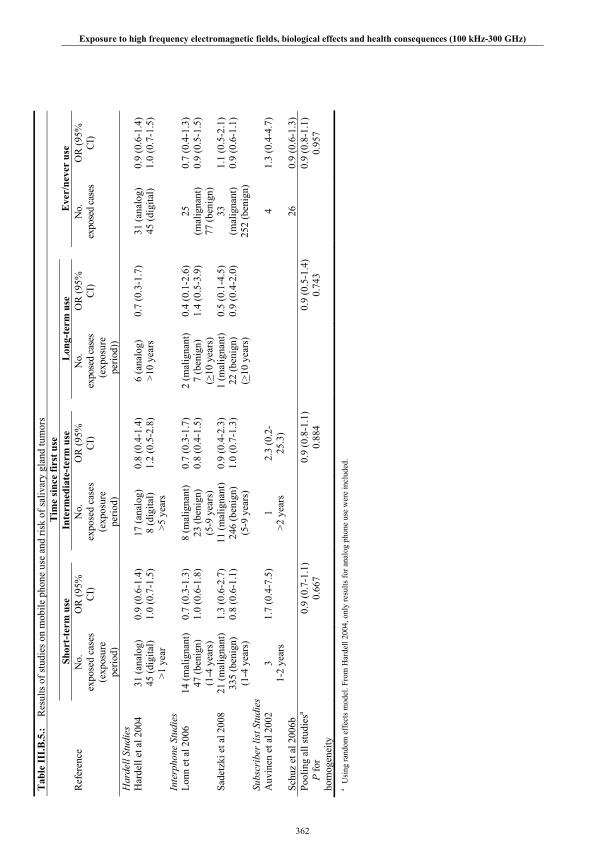
Exposure to high frequency electromagnetic fields, biological effects and health consequences (100 kHz-300 GHz)
362
Tab
le II
I.B.5
.: R
esul
ts o
f stu
dies
on
mob
ile p
hone
use
and
risk
of s
aliv
ary
glan
d tu
mor
s
Tim
e si
nce
first
use
Sh
ort-
term
use
Inte
rmed
iate
-ter
m u
se
L
ong-
term
use
E
ver/
neve
r us
e R
efer
ence
N
o.
expo
sed
case
s (e
xpos
ure
perio
d)
OR
(95%
C
I)
N
o.
expo
sed
case
s (e
xpos
ure
perio
d)
OR
(95%
C
I)
N
o.
expo
sed
case
s (e
xpos
ure
perio
d))
OR
(95%
C
I)
N
o.
expo
sed
case
s O
R (9
5%
CI)
Har
dell
Stud
ies
H
arde
ll et
al 2
004
31 (a
nalo
g)
45 (d
igita
l) >1
yea
r
0.9
(0.6
-1.4
) 1.
0 (0
.7-1
.5)
17
(ana
log)
8
(dig
ital)
>5 y
ears
0.8
(0.4
-1.4
) 1.
2 (0
.5-2
.8)
6
(ana
log)
>1
0 ye
ars
0.7
(0.3
-1.7
)
31 (a
nalo
g)
45 (d
igita
l) 0.
9 (0
.6-1
.4)
1.0
(0.7
-1.5
)
Inte
rpho
ne S
tudi
es
Lo
nn e
t al 2
006
14
(mal
igna
nt)
47 (b
enig
n)
(1-4
yea
rs)
0.7
(0.3
-1.3
) 1.
0 (0
.6-1
.8)
8
(mal
igna
nt)
23 (b
enig
n)
(5-9
yea
rs)
0.7
(0.3
-1.7
) 0.
8 (0
.4-1
.5)
2
(mal
igna
nt)
7 (b
enig
n)
(>10
yea
rs)
0.4
(0.1
-2.6
) 1.
4 (0
.5-3
.9)
25
(m
alig
nant
) 77
(ben
ign)
0.7
(0.4
-1.3
) 0.
9 (0
.5-1
.5)
Sade
tzki
et a
l 200
8 21
(mal
igna
nt)
335
(ben
ign)
(1
-4 y
ears
)
1.3
(0.6
-2.7
) 0.
8 (0
.6-1
.1)
11
(mal
igna
nt)
246
(ben
ign)
(5
-9 y
ears
)
0.9
(0.4
-2.3
) 1.
0 (0
.7-1
.3)
1
(mal
igna
nt)
22 (b
enig
n)
(>10
yea
rs)
0.5
(0.1
-4.5
) 0.
9 (0
.4-2
.0)
33
(m
alig
nant
) 25
2 (b
enig
n)
1.1
(0.5
-2.1
) 0.
9 (0
.6-1
.1)
Su
bscr
iber
list
Stu
dies
A
uvin
en e
t al 2
002
3 1-
2 ye
ars
1.7
(0.4
-7.5
)
1 >2
yea
rs
2.3
(0.2
-25
.3)
4 1.
3 (0
.4-4
.7)
Schu
z et
al 2
006b
26
0.9
(0.6
-1.3
) Po
olin
g al
l stu
dies
a
P
for
hom
ogen
eity
0.
9 (0
.7-1
.1)
0.66
7
0.9
(0.8
-1.1
) 0.
884
0.
9 (0
.5-1
.4)
0.74
3
0.9
(0.8
-1.1
) 0.
957
a Usi
ng ra
ndom
eff
ects
mod
el. F
rom
Har
dell
2004
, onl
y re
sults
for a
nalo
g ph
one
use
wer
e in
clud
ed.

Exposure to high frequency electromagnetic fi elds, biological effects and health consequences (100 kHz-300 GHz)
363
eTab
le 1
. Met
hods
of
stud
ies
on m
obile
pho
ne u
se a
nd t
umor
ris
k.
Rep
orte
d re
spon
se r
ate
Ref
eren
ce
Geo
g.L
ocat
ion
Per
iod
of
case
asce
rtai
n.
Age
Ran
ge
Hos
pita
l or po
pula
tio
n ba
sed
Tum
ors
cons
ider
ed &
w
heth
er h
isto
l;
grad
e; lo
be
cove
red
Cas
es
Con
trol
s %
pro
xy
inte
rvie
ws
% e
xpos
ed
amon
g co
ntro
lsC
omm
ents
US
Stud
ies
Dre
yer e
t al.
1999
USA
19
94
20+
Popu
latio
n To
tal m
orta
lity
Coh
ort b
ased
on
oper
ator
dat
a. F
ollo
w
up to
Rot
hman
199
6.
Subj
ects
usi
ng
hand
held
pho
nes
com
pare
d to
subj
ects
us
ing
bag
phon
es.
M
usca
t et a
l. 20
00M
usca
t et a
l. 20
02U
SA
1994
-199
8 18
-80
Hos
pita
l M
alig
nant
bra
in
tum
ours
(IC
D9-
CM
cod
es 1
91.0
-19
1.9)
Lo
be
G
rade
A
cous
tic n
euro
ma
75%
90%
9% b
rain
tu
mor
ca
ses
1.1%
AN
ca
ses
1.4%
co
ntro
ls
18%
bra
in
tum
or c
ontro
ls
27%
AN
co
ntro
ls
(eve
r had
a
mob
ile p
hone
su
bscr
iptio
n)
Dat
a co
llect
ion
thro
ugh
pers
onal
inte
rvie
ws.
Insk
ip e
t al.
2001
USA
19
94-1
998
18+
Hos
pita
l G
liom
a,
Men
ingi
oma,
A
cous
tic n
euro
ma
Gra
de
Lobe
(IC
D &
mor
ph-
olog
y co
des i
n pa
per)
92%
86
%
16
%
glio
ma
8%
men
ingi
oma 3%
AN
3%
co
ntro
ls
22%
(r
egul
ar u
se, i
.e.
at le
ast t
wic
e pe
r wee
k)
45%
(e
ver u
se)
Dat
a co
llect
ion
thro
ugh
pers
onal
inte
rvie
ws.
War
ren
et a
l. 20
03U
SA
1995
-200
0 N
ot
stat
ed
Hos
pita
l Fa
cial
ner
ve
Aco
ustic
neu
rom
a N
ot
stat
ed
Not
st
ated
0%
38%
(e
ver u
se)
Dat
a co
llect
ion
thro
ugh
tele
phon
e in
terv
iew
s.

Exposure to high frequency electromagnetic fi elds, biological effects and health consequences (100 kHz-300 GHz)
364
Har
dell
Stud
ies
Har
dell
et a
l. 19
99Sw
eden
1994
-199
6
20-8
0 Po
pula
tion
All
brai
n tu
mor
s M
alig
n+B
enig
n A
cous
tic n
euro
ma
Gra
de
Lobe
90%
91%
D
eath
s ex
clud
ed
38%
(a
t lea
st 8
hou
rs
of u
se)
Res
pons
e pr
opor
tions
ex
clud
e de
aths
, ph
ysic
ian
refu
sals
from
de
nom
inat
or in
all
Har
dell
stud
ies.
Dat
a co
llect
ion
thro
ugh
post
al q
uest
ionn
aire
s.
Har
dell
et a
l. 20
02a,
bSw
eden
19
97-2
000
20-8
0 Po
pula
tion
All
brai
n tu
mor
s M
alig
n+B
enig
n A
cous
tic n
euro
ma
Gra
de
Lobe
88%
91%
Dea
ths
excl
uded
A
nalo
gue
15%
D
igita
l 30%
C
ordl
ess 2
7%
(eve
r use
)
Rep
eate
d in
terv
iew
s of
sele
cted
subj
ects
; D
ata
colle
ctio
n th
roug
h po
stal
que
stio
nnai
res.
H
arde
ll et
al.
2005
a
Har
dell
et a
l. 20
06a
Swed
en
20
00-2
003
20-8
0 Po
pula
tion
Ben
ign
brai
n tu
mor
s A
cous
tic n
euro
ma
Men
ingi
oma
Lobe
s M
alig
nant
bra
in
tum
ors
Hig
h gr
ade
astro
cyto
mas
89%
be
nign
88
%
mal
ign
84%
D
eath
s ex
clud
ed
Ana
logu
e 11
%
Dig
ital 5
0%
Cor
dles
s 44%
A
ny ty
pe 6
6%
(eve
r use
)
Dat
a co
llect
ion
thro
ugh
post
al q
uest
ionn
aire
s.
Har
dell
et a
l. 20
04
Swed
en
19
94-2
000
21-8
0 Po
pula
tion
Saliv
ary
glan
d A
lso
by
loca
lizat
ion
and
hist
opat
holo
gy
91%
90
%
Dea
ths
excl
uded
A
nalo
gue
13%
D
igita
l 16%
C
ordl
ess 1
9%
Any
type
33%
(e
ver u
se)
Maj
ority
of c
ontro
ls
from
a 2
002
brai
n tu
mor
stud
y D
ata
colle
ctio
n th
roug
h po
stal
que
stio
nnai
res.
H
arde
ll et
al.
2005
b
Swed
en
19
99-2
002
18-7
4 Po
pula
tion
Non
Hod
gkin
Ly
mph
oma
B-c
ell
T-ce
ll (f
urth
er
subd
ivid
ed)
Oth
er
91%
92%
D
eath
s ex
clud
ed
Ana
logu
e 18
%
Dig
ital 5
5%
Cor
dles
s 41%
A
ny ty
pe 6
8%
(eve
r use
)
Con
trols
recr
uite
d on
“s
ever
al o
ccas
ions
”, 3
0 ca
ses e
xclu
ded
afte
r as
certa
inm
ent o
f ex
posu
re a
s NLH
not
co
nfirm
ed
Dat
a co
llect
ion
thro
ugh
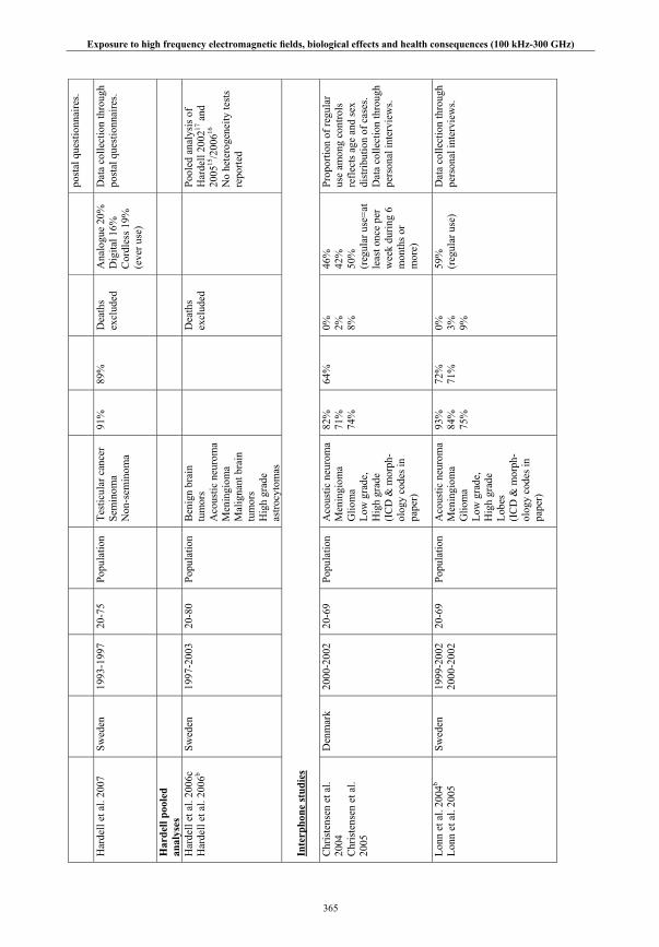
Exposure to high frequency electromagnetic fi elds, biological effects and health consequences (100 kHz-300 GHz)
365
post
al q
uest
ionn
aire
s.
Har
dell
et a
l. 20
07Sw
eden
19
93-1
997
20-7
5 Po
pula
tion
Test
icul
ar c
ance
r Se
min
oma
Non
-sem
inom
a
91%
89
%
Dea
ths
excl
uded
A
nalo
gue
20%
D
igita
l 16%
C
ordl
ess 1
9%
(eve
r use
)
Dat
a co
llect
ion
thro
ugh
post
al q
uest
ionn
aire
s.
Har
dell
pool
ed
anal
yses
Har
dell
et a
l. 20
06c
Har
dell
et a
l. 20
06b
Swed
en
1997
-200
3 20
-80
Popu
latio
n B
enig
n br
ain
tum
ors
Aco
ustic
neu
rom
a M
enin
giom
a M
alig
nant
bra
in
tum
ors
Hig
h gr
ade
astro
cyto
mas
Dea
ths
excl
uded
Pool
ed a
naly
sis o
f H
arde
ll 20
0217
and
20
0515
/200
616
No
hete
roge
neity
test
s re
porte
d
Inte
rpho
ne s
tudi
es C
hris
tens
en e
t al.
2004
Chr
iste
nsen
et a
l. 20
05
Den
mar
k 20
00-2
002
20-6
9 Po
pula
tion
Aco
ustic
neu
rom
a M
enin
giom
a G
liom
a Lo
w g
rade
, H
igh
grad
e (I
CD
& m
orph
-ol
ogy
code
s in
pape
r)
82%
71
%
74%
64%
0%
2%
8%
46%
42
%
50%
(r
egul
ar u
se=a
t le
ast o
nce
per
wee
k du
ring
6 m
onth
s or
mor
e)
Prop
ortio
n of
regu
lar
use
amon
g co
ntro
ls
refle
cts a
ge a
nd se
x di
strib
utio
n of
cas
es.
Dat
a co
llect
ion
thro
ugh
pers
onal
inte
rvie
ws.
Lonn
et a
l. 20
04b
Lonn
et a
l. 20
05
Swed
en
1999
-200
2 20
00-2
002
20-6
9 Po
pula
tion
Aco
ustic
neu
rom
a M
enin
giom
a G
liom
a
Low
gra
de,
Hig
h gr
ade
Lobe
s (I
CD
& m
orph
-ol
ogy
code
s in
pape
r)
93%
84
%
75%
72%
71
%
0%
3%
9%
59%
(r
egul
ar u
se)
Dat
a co
llect
ion
thro
ugh
pers
onal
inte
rvie
ws.

Exposure to high frequency electromagnetic fi elds, biological effects and health consequences (100 kHz-300 GHz)
366
Schl
ehof
er e
t al.
2007
Schu
z et
al.
2006
a
Ger
man
y 20
01-2
003
30-6
9 Po
pula
tion
A
cous
tic n
euro
ma
Men
ingi
oma
Glio
ma
Low
gra
de,
Hig
h gr
ade
89%
88
%
80%
55%
in
AN
stud
y 63
% in
br
ain
tum
or
stud
y
0%
1%
11%
38%
37
%
39%
(r
egul
ar u
se)
Dat
a co
llect
ion
thro
ugh
pers
onal
inte
rvie
ws.
Kla
eboe
et a
l. 20
07
N
orw
ay
2001
-200
2 19
-69
Popu
latio
n A
cous
tic n
euro
ma
Men
ingi
oma
Glio
ma
68%
71
%
77%
69%
0%
0%
36
%
63%
(r
egul
ar u
se)
Dat
a co
llect
ion
thro
ugh
pers
onal
inte
rvie
ws.
Larg
e pr
opor
tion
of
inte
rvie
ws w
as m
ade
over
the
phon
e.
Ta
keba
yash
i et a
l. 20
06Ta
keba
yash
i et a
l. 20
08
Japa
n 20
00-2
004
30-6
9 H
ospi
tal
case
s Po
pula
tion
cont
rols
Aco
ustic
neu
rom
a M
enin
giom
a G
liom
a Pi
tuita
ry a
deno
ma
(IC
D &
mor
ph-
olog
y co
des i
n pa
per)
84%
78
%
59%
76
%
52%
52
%
53%
49
%
0%
58%
52
%
65%
65
%
(reg
ular
use
)
Som
e ho
spita
ls d
id n
ot
parti
cipa
te.
Con
trols
sele
cted
th
roug
h ra
ndom
dig
it di
alin
g.
Dat
a co
llect
ion
thro
ugh
pers
onal
inte
rvie
ws.
Hep
wor
th e
t al.
2006
UK
20
00-2
004
18-6
9 Po
pula
tion
Glio
ma
Low
gra
de,
Hig
h gr
ade
(IC
D &
mor
ph-
olog
y co
des i
n pa
per)
51%
45
%
7% c
ases
52
%
(reg
ular
use
) St
udy
incl
udes
dat
a fr
om tw
o ce
nter
s in
the
UK
. D
ata
colle
ctio
n th
roug
h pe
rson
al in
terv
iew
s.
Hou
rs e
t al.
2007
Fran
ce
2001
-200
3 30
-59
Hos
pita
l ca
ses
Popu
latio
n co
ntro
ls
Men
ingi
oma
Glio
ma
AN
60%
78
%
81%
75%
4%
cas
es
56%
(r
egul
ar u
se)
Som
e ho
spita
ls d
id n
ot
parti
cipa
te.
Dat
a co
llect
ion
thro
ugh
pers
onal
inte
rvie
ws.
Lonn
et a
l. 20
06D
enm
ark
and
Swed
en
2000
-200
2
20-6
9 Po
pula
tion
(mal
igna
nt
case
s and
al
l co
ntro
ls)
Hos
pita
l
Mal
igna
nt p
arot
id
glan
d B
enig
n pl
eom
orph
ic
aden
oma
85%
(M
alig
n)
88%
(B
enig
n)
60%
(D
enm
ark)
72
%
(Sw
eden
)
1 M
alig
n ca
se in
Sw
eden
60%
(r
egul
ar u
se)
Mat
ched
con
trols
in
Den
mar
k R
esul
ts p
rese
nted
for
two
coun
tries
co
mbi
ned
Dat
a co
llect
ion
thro
ugh
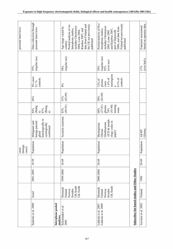
Exposure to high frequency electromagnetic fi elds, biological effects and health consequences (100 kHz-300 GHz)
367
case
s (b
enig
n ca
ses)
pers
onal
inte
rvie
ws.
Sade
tzki
et a
l. 20
08Is
rael
20
01-2
003
18-5
9 Po
pula
tion
Mal
igna
nt a
nd
beni
gn p
arot
id
glan
d hi
stol
ogic
ally
or
cyto
logi
cally
co
nfirm
ed
84%
(M
alig
n)
87%
(B
enig
n)
66%
4%
cas
es
0.1
%
cont
rols
55%
(r
egul
ar u
se)
Dat
a co
llect
ion
thro
ugh
pers
onal
inte
rvie
ws.
Inte
rpho
ne p
oole
d an
alys
es
Scho
emak
er e
t al.
2005
Den
mar
k Fi
nlan
d N
orw
ay
Swed
en
UK
-Sou
th
UK
-Nor
th
1999
-200
4 18
-69
Popu
latio
n A
cous
tic n
euro
ma
83%
(6
9-91
) 51
%
(42-
69)
0%
54%
(r
egul
ar u
se)
Age
rang
e va
ried
by
coun
try.
Pool
ed a
naly
sis o
f six
In
terp
hone
stud
ies,
incl
udes
Chr
iste
nsen
20
04, L
onn
2004
, K
laeb
oe 2
007,
and
da
ta fr
om F
inla
nd a
nd
the
UK
not
pre
viou
sly
publ
ishe
d
Lahk
ola
et a
l. 20
07La
hkol
a et
al.
2008
Den
mar
k Fi
nlan
d N
orw
ay
Swed
en
UK
-Sou
th
2000
-200
4 18
-69
Popu
latio
n M
enin
giom
a G
liom
a G
liobl
asto
ma
(IC
D &
mor
ph-
olog
y co
des i
n pa
per)
60%
(3
7-81
) gl
iom
a 74
%
(55-
90)
men
ing
iom
a
50%
(4
2-69
)
12%
of
glio
ma
ca
ses
1.6%
of
men
ingi
oma 0.
1% o
f co
ntro
ls
59%
(r
egul
ar u
se)
92%
(e
ver u
se)
Pool
ed a
naly
sis o
f fiv
e In
terp
hone
stud
ies,
incl
udes
Chr
iste
nsen
20
05, L
onn
2005
an
d K
laeb
oe 2
007,
pa
rt of
Hep
wor
th
2006
, and
dat
a fr
om
Finl
and
not p
revi
ousl
y pu
blis
hed
Subs
crib
er li
st b
ased
stu
dies
and
Oth
er S
tudi
es A
uvin
en e
t al.
2002
Finl
and
1996
20
-69
Popu
latio
n A
ll B
T G
liom
a,
11
%
(eve
r had
a
Expo
sure
ass
essm
ent
base
d on
ope
rato
r dat
a
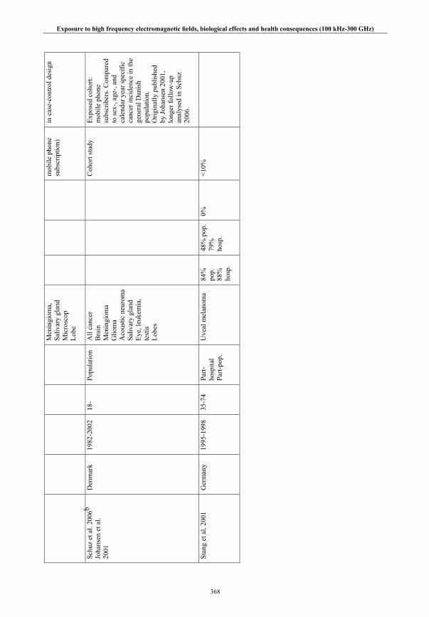
Exposure to high frequency electromagnetic fi elds, biological effects and health consequences (100 kHz-300 GHz)
368
Men
ingi
oma,
Sa
livar
y gl
and
Mic
rosc
op
Lobe
mob
ile p
hone
su
bscr
iptio
n)
in c
ase-
cont
rol d
esig
n
Schu
z et
al.
2006
b
Joha
nsen
et a
l. 20
01
Den
mar
k 19
82-2
002
18-
Popu
latio
n A
ll ca
ncer
B
rain
M
enin
giom
a G
liom
a
Aco
ustic
neu
rom
a Sa
livar
y gl
and
Eye,
leuk
emia
, te
stis
Lo
bes
C
ohor
t stu
dy
Expo
sed
coho
rt:
mob
ile p
hone
su
bscr
iber
s. C
ompa
red
to se
x-, a
ge-,
and
cale
ndar
yea
r spe
cific
ca
ncer
inci
denc
e in
the
gene
ral D
anis
h po
pula
tion.
O
rigin
ally
pub
lishe
d by
Joha
nsen
200
1,
long
er fo
llow
-up
anal
ysed
in S
chuz
20
06.
St
ang
et a
l. 20
01G
erm
any
1995
-199
8 35
-74
Part-
hosp
ital
Part-
pop.
Uve
al m
elan
oma
84%
po
p.
88%
ho
sp.
48%
pop
. 79
%
hosp
.
0%
<10%

Exposure to high frequency electromagnetic fi elds, biological effects and health consequences (100 kHz-300 GHz)
369
eTab
le 2
. R
esul
ts o
f st
udie
s on
mob
ile p
hone
use
and
ris
k of
glio
ma
Ref
eren
ce
Dia
gnos
tic
grou
p
No.
cas
es
ever
/nev
er
user
No.
con
trol
s ev
er/n
ever
us
er
OR
eve
r*
cf n
ever
(9
5% C
I) u
ser
OR
(95
% C
I )
for
max
yrs
exp
. (c
ut p
oint
)
OR
(95
% C
I)
for
max
cu
mul
ativ
eex
posu
re
OR
for
eve
r-an
alog
ue u
se
OR
(95
% C
I)
Lat
eral
ity
(eve
r/ne
ver)
ip
si/c
ontr
a
US
Stud
ies
Dre
yer e
t al.
1999
Bra
in
2/4
No
exce
ss
Too
smal
l nu
mbe
rs
for a
naly
sis
Mus
cat e
t al.
2000
Non
-men
ingi
oma
brai
n (m
ainl
y m
alig
nant
) A
stro
cytic
66/4
03
41/3
13
76/3
46
0.7
(0.5
-1.1
) 0.
8 (0
.5-1
.2)
0.7
(0.4
-1.4
) (>
4)
0.7
(0.3
-1.4
) (>
480
hrs)
Insk
ip e
t al.
2001
Glio
ma
201/
285
35
8/44
0 1.
0 (0
.7-1
.4)
0.6
(0.3
-1.4
) (>
5)
0.5
(0.2
-1.3
) (>
500h
)
In
skip
met
hod:
R
R=0
.9, p
=0.7
7
Har
dell
Stud
ies
Har
dell
et a
l. 19
99/2
001
All
brai
n A
stro
cyt/g
liobl
ast
78/1
31
36/5
8
161/
264
1.
0 (0
.7-1
.4)
1.1
(0.6
-1.8
)
1.2
(0.6
-2.6
) (>
10)
1.1
(0.3
-3.4
) (>
968h
) 0.
9 (0
.6-1
.4)
1.1
(0.6
-1.8
)/
0.7
(0.4
-1.2
)
Har
dell
et a
l. 20
02a,
bA
ll m
alig
nant
A
stro
cyto
ma
low
gr
ade
Ast
rocy
tom
a hi
gh
grad
e
79 a
nalo
gue
112
digi
tal
12 a
nalo
gue
16 d
igita
l
46 a
nalo
gue
64 d
igita
l ?
unex
pose
d
70 a
nalo
gue
99 d
igita
l 8
anal
ogue
19
dig
ital
37 a
nalo
gue
52 d
igita
l ?
unex
pose
d
1.1
(0.8
-1.6
) 1.
1 (0
.9-1
.5)
1.2
(0.8
-1.9
) 1.
2 (0
.8-1
.8)
1.5
(0.6
-3.7
) 0.
8 (0
.4-1
.6)
1.2
(0.8
-1.8
) 1.
7 (0
.7-4
.3)
(>6)
1.1
(0.8
-1.6
) 1.
9 (1
.2-3
.0)/
0.6
(0.4
-1.1
) (A
nalo
gue)
1.
6 (1
.1-2
.4)/
0.9
(0.5
-1.4
) (D
igita
l)
Har
dell
et a
l. 20
06a
All
mal
igna
nt
68 a
nalo
gue
79 a
nalo
gue
2.6
(1.5
-4.3
) 3.
5 (2
.0-6
.4)
4.0
(2.2
-7.3
) 2.
6 (1
.5-4
.3)
3.1
(1.6
-6.2
)/
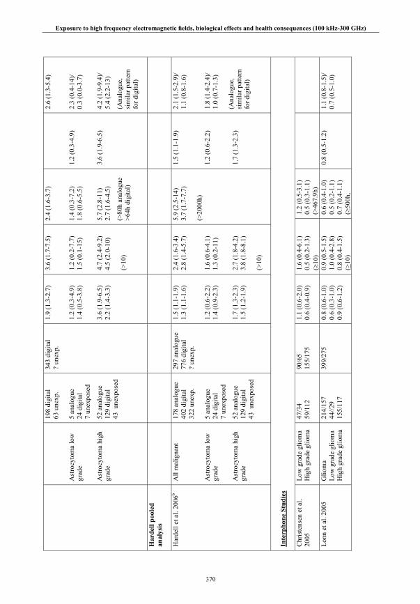
Exposure to high frequency electromagnetic fi elds, biological effects and health consequences (100 kHz-300 GHz)
370
Ast
rocy
tom
a lo
w
grad
e
Ast
rocy
tom
a hi
gh
grad
e
198
digi
tal
63 u
nexp
. 5
anal
ogue
24
dig
ital
7 un
expo
sed
52 a
nalo
gue
129
digi
tal
43 u
nexp
osed
343
digi
tal
? un
exp.
1.9
(1.3
-2.7
) 1.
2 (0
.3-4
.9)
1.4
(0.5
-3.8
) 3.
6 (1
.9-6
.5)
2.2
(1.4
-3.3
)
3.6
(1.7
-7.5
) 1.
2 (0
.2-7
.7)
1.5
(0.1
-15)
4.
7 (2
.4-9
.2)
4.5
(2.0
-10)
(>
10)
2.4
(1.6
-3.7
) 1.
4 (0
.3-7
.2)
1.8
(0.6
-5.5
) 5.
7 (2
.8-1
1)
2.7
(1.6
-4.5
)
(>80
h an
alog
ue
>64h
dig
ital)
1.2
(0.3
-4.9
) 3.
6 (1
.9-6
.5)
2.6
(1.3
-5.4
) 2.
3 (0
.4-1
4)/
0.3
(0.0
-3.7
) 4.
2 (1
.9-9
.4)/
5.4
(2.2
-13)
(Ana
logu
e,
sim
ilar p
atte
rn
for d
igita
l)
Har
dell
pool
ed
anal
ysis
Har
dell
et a
l. 20
06b
All
mal
igna
nt
Ast
rocy
tom
a lo
w
grad
e
Ast
rocy
tom
a hi
gh
grad
e
178
anal
ogue
40
2 di
gita
l 32
2 un
exp.
5
anal
ogue
24
dig
ital
7 un
expo
sed
52 a
nalo
gue
129
digi
tal
43 u
nexp
osed
297
anal
ogue
77
6 di
gita
l ?
unex
p.
1.5
(1.1
-1.9
) 1.
3 (1
.1-1
.6)
1.2
(0.6
-2.2
) 1.
4 (0
.9-2
.3)
1.7
(1.3
-2.3
) 1.
5 (1
.2-1
.9)
2.4
(1.6
-3.4
) 2.
8 (1
.4-5
.7)
1.6
(0.6
-4.1
) 1.
3 (0
.2-1
1)
2.7
(1.8
-4.2
) 3.
8 (1
.8-8
.1)
(>10
)
5.9
(2.5
-14)
3.
7 (1
.7-7
.7)
(>20
00h)
1.5
(1.1
-1.9
)
1.2
(0.6
-2.2
)
1.7
(1.3
-2.3
)
2.1
(1.5
-2.9
)/ 1.
1 (0
.8-1
.6)
1.8
(1.4
-2.4
)/ 1.
0 (0
.7-1
.3)
(Ana
logu
e,
sim
ilar p
atte
rn
for d
igita
l)
Inte
rpho
ne S
tudi
es C
hris
tens
en e
t al.
2005
Low
gra
de g
liom
a H
igh
grad
e gl
iom
a 47
/34
59/1
12
90/6
5 15
5/17
5 1.
1 (0
.6-2
.0)
0.6
(0.4
-0.9
) 1.
6 (0
.4-6
.1)
0.5
(0.2
-1.3
) (
10)
1.2
(0.5
-3.1
) 0.
5 (0
.3-1
.1)
(>46
7.9h
)
Lonn
et a
l. 20
05
Glio
ma
Low
gra
de g
liom
a H
igh
grad
e gl
iom
a
214/
157
44//2
9 15
5/11
7
399/
275
0.8
(0.6
-1.0
) 0.
6 (0
.3-1
.0)
0.9
(0.6
-1.2
)
0.9
(0.5
-1.5
) 1.
0 (0
.4-2
.8)
0.8
(0.4
-1.5
) (>
10)
0.6
(0.4
-1.0
) 0.
5 (0
.2-1
.1)
0.7
(0.4
-1.1
) (≥
500h
,
0.8
(0.5
-1.2
) 1.
1 (0
.8-1
.5)/
0.7
(0.5
-1.0
)

Exposure to high frequency electromagnetic fi elds, biological effects and health consequences (100 kHz-300 GHz)
371
hand
sfre
e ad
just
ed)
Sc
huz
et a
l. 20
06a
Glio
ma
138/
228
28
3/44
9 1.
0 (0
.7-1
.3)
2.
2 ( 0
.9-5
.1)
(≥10
) 1.
0 (0
.6-1
.6)
(>19
5h)
Hep
wor
th e
t al.
2006
Glio
ma
508/
456
898/
818
0.9
(0.8
-1.1
)
0.9
(0.6
-1.3
) (>
10)
0.9
(0.7
-1.2
) ( >
544h
)0.
9 (0
.7-1
.2)
1.2
(1.0
-1.5
)/ 0.
8 (0
.6-0
.9)
Kla
eboe
et a
l. 20
07G
liom
a 16
1/12
8
227/
131
0.
6 (0
.4-0
.9)
0.8
(0.5
-1.2
) (
6)
0.7
(0.4
-1.3
) (
425h
, ha
ndsf
ree
adju
sted
)
0.7
(0.4
-1.1
)
1.0
(0.7
-1.4
)/
0.7
(0.5
-1.1
)
Hou
rs e
t al.
2007
Glio
ma
59/3
7
54/4
2
1.2
(0.7
-2.1
) 2.
0 (0
.7-5
.2)
(≥3.
8)
1.8
(0.7
-4.3
) (≥
260h
)
1.2
(0.6
-2.4
)/
1.2
(0.5
-2.7
)
Take
baya
shi e
t al.
2008
Glio
ma
56/2
7 10
6/57
1.
2 (0
.6-2
.4)
0.6
(0.2
-1.8
) (>
6.5)
1.
7 (0
.7-4
.3)
(>62
0h)
0.8
(0.2
-3.0
) 1.
2 (0
.7-2
.3)/
1.1
(0.6
-2.0
)
Inte
rpho
ne p
oole
d an
alys
is
Lahk
ola
et a
l. 20
07G
liom
a G
liobl
asto
ma
867/
629
368/
330
1853
/128
1 0.
8 (0
.7-0
.9)
0.8
(0.6
-0.9
) 0.
95 (0
.7-1
.2)
0.9
(0.6
-1.2
) (>
10)
0.9
(0.7
-1.1
) 0.
9 (0
.6-1
.1)
(>50
3h,
hand
sfre
e ad
just
ed)
0.9
(0.7
-1.1
) 1.
1 (1
.0-1
.3)/
0.8
(0.6
-0.9
)
Subs
crib
er li
st b
ased
Stu
dies
Auv
inen
et a
l. 20
02G
liom
a 36
/360
119/
1859
1.
5 (1
.0-2
.4)
1.
7 (0
.9-3
.5)
(>2)
2.
1 (1
.3-3
.4)
Schu
z et
al.
2006
b
N
ervo
us sy
stem
G
liom
a 58
0 25
7 1.
0 (0
.9-1
.0)
1.0
(0.9
–1.1
) 0.
7 (0
.4-1
.0)
(>
10)

Exposure to high frequency electromagnetic fi elds, biological effects and health consequences (100 kHz-300 GHz)
372
eTab
le 3
. Res
ults
of
stud
ies
on m
obile
pho
ne u
se a
nd r
isk
of m
enin
giom
a
Ref
eren
ce
Dia
gnos
tic
grou
p
No.
cas
es
ever
/nev
er
user
No.
con
trol
s ev
er/n
ever
us
er
OR
eve
r*
cf n
ever
(9
5% C
I) u
ser
OR
(95
% C
I )
for
max
yrs
exp
. (c
ut p
oint
)
OR
(95
% C
I)
for
max
cu
mul
ativ
eex
posu
re
OR
for
eve
r-an
alog
ue u
se
OR
(95
% C
I)
Lat
eral
ity
(eve
r/ne
ver)
ip
si/c
ontr
a
US
Stud
ies
Insk
ip e
t al.
2001
Men
ingi
oma
67/1
30
35
8/44
0 0.
8 (0
.5-1
.2)
0.9
(0.3
-2.7
) (>
5)
0.7
(0.2
-2.4
) (>
500h
)
In
skip
met
hod:
R
R=0
.9, p
=1.0
Har
dell
Stud
ies
Har
dell
et a
l. 19
99M
enin
giom
a
16/3
0
161/
264
1.
1 (0
.5-2
.3)
Har
dell
et a
l. 20
02a
Men
ingi
oma
60
ana
logu
e 78
dig
ital
? un
expo
sed
56 a
nalo
gue
102
digi
tal
? un
expo
sed
1.1
(0.7
-1.5
) 0.
8 (0
.6-1
.0)
1.
1 (0
.7-1
.5)
Har
dell
et a
l. 20
05a
Men
ingi
oma
35 a
nalo
gue
151
digi
tal
103
unex
pose
d
79 a
nalo
gue
343
digi
tal
? un
expo
sed
1.7
(1.0
–3.0
) 1.
3 (0
.9–1
.9)
2.1
(1.1
–4.3
) 1.
5 (0
.6–3
.9)
(>10
)
2.9
(1.1
–8.1
) 1.
5 (0
.6–3
.9)
(>80
h an
alog
ue >
64h
digi
tal)
1.7
(1.0
–3.0
) 1.
6 (0
.7–3
.9)/
2.6
(1.1
–6.0
) (a
nalo
gue)
1.
5 (0
.9–2
.5)/
1.5
0(.9
–2.3
) (d
igita
l)
Har
dell
pool
ed
anal
ysis
Har
dell
et a
l. 20
06c
Men
ingi
oma
11
3 an
alog
ue
295
digi
tal
455
unex
pose
d
297
anal
ogue
77
6 di
gita
l ?
unex
pose
d.
1.3
(1.0
-1.7
) 1.
1 (0
.9-1
.3)
1.6
(1.0
-2.5
) 1.
3 (0
.5-3
.2)
(>10
)
1.4
(0.5
-3.8
) 0.
7 (0
.3-1
.4)
(>10
00h)
1.3
(1.0
-1.7
) 1.
3 (0
.9-2
.0)/
1.2
(0.7
-1.8
) (a
nalo
gue)
1.
4 (1
.0-1
.8)/
1.1
(0.8
-1.5
) (d
igita
l)
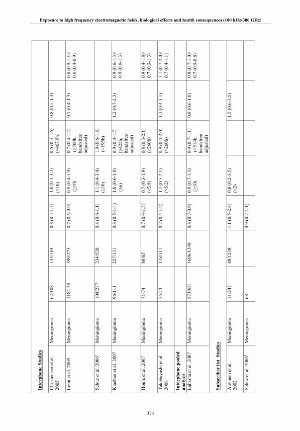
Exposure to high frequency electromagnetic fi elds, biological effects and health consequences (100 kHz-300 GHz)
373
Inte
rpho
ne S
tudi
es C
hris
tens
en e
t al.
2005
Men
ingi
oma
67/1
08
13
3/18
3 0.
8 (0
.5-1
.3)
1.0
(0.3
-3.2
) (
10)
0.6
(0.3
-1.6
) (>
467.
9h)
0.8
(0.5
-1.3
)
Lonn
et a
l. 20
05
Men
ingi
oma
11
8/15
5 39
9/27
5 0.
7 (0
.5-0
.9)
0.9
(0.4
-1.9
) (>
10)
0.7
(0.4
-1.2
) (≥
500h
, ha
ndsf
ree
adju
sted
)
0.7
(0.4
-1.3
) 0.
8 (0
.5-1
.1)/
0.6
(0.4
-0.9
)
Schu
z et
al.
2006
aM
enin
giom
a 10
4/27
7 23
4/52
8 0.
8 (0
.6-1
.1)
1.
1 (0
.4-3
.4)
(≥10
) 1.
0 (0
.6-1
.8)
(>19
5h)
Kla
eboe
et a
l. 20
07M
enin
giom
a 96
/111
227/
131
0.
8 (0
.5-1
-1)
1.0
(0.6
-1.8
) (
6)
0.9
(0.4
-1.7
) (
425h
, ha
ndsf
ree
adju
sted
)
1.2
(0.7
-2.3
)
0.9
(0.6
-1.3
)/
0.9
(0.6
-1.3
)
Hou
rs e
t al.
2007
Men
ingi
oma
71/7
4 80
/65
0.
7 (0
.4-1
.3)
0.7
(0.3
-1.9
) (≥
3.8)
0.8
(0.3
-2.1
) (≥
260h
)
0.9
(0.4
-1.8
)/
0.7
(0.3
-1.3
)
Take
baya
shi e
t al.
2008
Men
ingi
oma
55/7
3 11
8/11
1 0.
7 (0
.4-1
.2)
1.1
(0.5
-2.1
) (>
5.2)
0.
9 (0
.4-2
.0)
(>26
0h)
1.1
(0.4
-3.1
) 1.
1 (0
.7-2
.0)/
0.7
(0.4
-1.1
)
Inte
rpho
ne p
oole
d an
alys
is
Lahk
ola
et a
l. 20
07M
enin
giom
a
573/
631
1696
/124
9 0.
8 (0
.7-0
.9)
0.9
(0.7
-1.3
) (>
10)
0.9
(0.7
-1.1
) (>
514h
, ha
ndsf
ree
adju
sted
)
0.8
(0.6
-1.0
) 0.
8 (0
.7-1
.0)/
0.7
(0.5
-0.8
)
Subs
crib
er li
st S
tudi
es A
uvin
en e
t al.
2002
Men
ingi
oma
11/2
47
48
/123
8 1.
1 (0
.5-2
.4)
0.
8 (0
.2-3
.5)
(>2)
1.
5 (0
.6-3
.5)
Schu
z et
al.
2006
b
M
enin
giom
a 68
0.
9 (0
.7-1
.1)
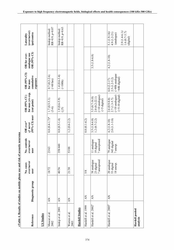
Exposure to high frequency electromagnetic fi elds, biological effects and health consequences (100 kHz-300 GHz)
374
eTab
le 4
. Res
ults
of
stud
ies
on m
obile
pho
ne u
se a
nd r
isk
of a
cous
tic
neur
oma
Ref
eren
ce
Dia
gnos
tic
grou
p
No.
cas
es
ever
/nev
er
user
No.
con
trol
s ev
er/n
ever
us
er
OR
eve
r*
cf n
ever
(9
5% C
I) u
ser
OR
(95
% C
I )
for
max
yrs
exp
. (c
ut p
oint
)
OR
(95
% C
I)
for
max
cu
mul
ativ
eex
posu
re
OR
for
eve
r-an
alog
ue u
se
OR
(95
% C
I)
Lat
eral
ity
(eve
r/ne
ver)
ip
si/c
ontr
a
US
Stud
ies
Mus
cat e
t al.
2002
AN
18
/72
23/6
3 0.
8 (0
.4-1
.7)*
1.
7 (0
.5-5
.1)
(3-6
) 0.
7 (0
.2-2
.6)
(>60
hrs
)
In
skip
met
hod:
R
R=0
.9, p
=0.0
7
Insk
ip e
t al.
2001
AN
40
/56
358/
440
0.8
(0.5
-1.4
)
1.9
(0.6
-5.9
) (>
5)
1.4
(0.6
-3.4
) (>
100h
)
In
skip
met
hod:
R
R=0
.9, p
=0.6
3
War
ren
et a
l. 20
03A
N
21
/30
53/8
8 1.
2 (0
.6-2
.2)
Har
dell
Stud
ies
Har
dell
et a
l. 19
99A
N
5/
8
0.
8 (0
.1-4
.2)
Har
dell
et a
l. 20
02a
AN
38 a
nalo
gue
23 d
igita
l ?
unex
pose
d
11 a
nalo
gue
19 d
igita
l ?
unex
pose
d
3.5
(1.8
-6.8
) 1.
2 (0
.7-2
.2)
3.5
(0.7
-16.
8)
2.0
(0.2
-22.
1)
(>10
ana
logu
e)
(>5
digi
tal)
3.5
(1.8
-6.8
)
Har
dell
et a
l. 20
05a
AN
20
ana
logu
e 53
dig
ital
18 u
nexp
.
79 a
nalo
gue
343
digi
tal
? un
exp.
4.2
(1.8
-10)
2.
0 (1
.1-3
.8)
2.6
(0.9
-8.0
) 2.
7 (1
.3-5
.7)
(>10
ana
logu
e)
(>5-
10 d
igita
l)
6.0
(2.2
-17)
2.
5 (1
.2-5
.2)
(>80
h an
alog
ue
>64h
dig
ital)
4.2
(1.8
-10)
5.1
(1.9
-14)
/ 4.
9 (1
.2-2
1)
(ana
logu
e)
2.9
(1.4
-6.1
)/ 1.
6 (0
.7-3
.7)
(dig
ital)
H
arde
ll po
oled
an
alys
is

Exposure to high frequency electromagnetic fi elds, biological effects and health consequences (100 kHz-300 GHz)
375
Har
dell
et a
l. 20
06c
AN
68 a
nalo
gue
105
digi
tal
88 u
nexp
osed
297
anal
ogue
77
6 di
gita
l ?
unex
p.
2.9
(2.0
-4.3
) 1.
5 (1
.1-2
.1)
3.1
(1.7
-5.7
) 1.
8 (1
.1-3
.0)
(>10
ana
logu
e)
(>5-
10 d
igita
l)
5.1
(1.9
-14)
3.
1 (1
.5-6
.4)
(>10
00h)
2.9
(2.0
-4.3
) 3.
0 (1
.9-5
.0)/
2.4
(1.4
-4.2
) (a
nalo
gue)
1.
7 (1
.1-2
.6)/
1.3
(0.8
-2.0
) (d
igita
l)
Inte
rpho
ne S
tudi
es C
hris
tens
en e
t al.
2005
AN
45
/61
97
/115
0.
9 (0
.5-1
.6)
0.2
(0.0
-1.1
) (
10)
0.7
(0.3
-1.7
) (>
467.
9h)
0.3
(0.1
-0.8
)
Lonn
et a
l. 20
05
AN
89
/59
356/
248
1.0
(0.6
-1.5
) 1.
9 (0
.9-4
.1)
(>10
)1.
1 (0
.6-2
.1)
(≥45
0h,
hand
sfre
e ad
just
ed)
1.6
(0.9
-2.8
) 1.
1 (0
.7-1
.6)/
0.9
(0.6
-1.4
)
Schl
ehof
er e
t al.
2006
AN
29
/68
74/1
20
0.7
(0.4
-1.2
)
0.5
(0.2
-1.3
) (5
-9)
0.4
(0.1
-1.0
) (>
195h
)
Kla
eboe
et a
l. 20
07A
N
22/2
3
227/
131
0.
5 (0
.2-1
.0)
0.
5 (0
.2-1
.4)
(6)
0.
5 (0
.2-1
.6)
(42
5h,
hand
sfre
e ad
just
ed)
0.8
(0.3
-2.2
)
0.7
(0.3
-1.4
)/
0.9
(0.5
-1.9
)
Hou
rs e
t al.
2007
AN
58
/51
12
3/91
0.9
(0.5
-1.6
) 0.
7 (0
.3–1
.6)
(≥3.
8)
0.9
(0.4
-2.1
) (≥
260h
)
0.6
(0.3
-1.2
)/
1.2
(0.6
-2.4
)
Take
baya
shi e
t al.
2006
AN
51
/46
192/
138
0.7
(0.4
-1.2
) 0.
8 (0
.2-2
.7)
(>8)
0.
7 (0
.3-1
.8)
(>90
0h)
1.2
(0.4
-3.8
) 0.
9 (0
.5-1
.6)/
0.
9 (0
.6-1
.6)
In
terp
hone
poo
led
anal
ysis
Scho
emak
er e
t al.
2005
AN
360/
316
1934
/161
2 0.
9 (0
.7-1
.1)
1.
0 (0
.7-1
.5)
(>10
) 0.
9 (0
.7-1
.2)
(>53
4h)
0.9
(0.7
-1.2
) 0.
9 (0
.7-1
.1)/
1.1
(0.9
-1.4
)

Exposure to high frequency electromagnetic fi elds, biological effects and health consequences (100 kHz-300 GHz)
376
Subs
crib
er li
st b
ased
Stu
dies
Schu
z et
al.
2006
b
N
erve
shea
th
tum
ours
, cra
nial
ne
rves
32
0.7
(0.5
-1.0
)
* Poo
ling
of c
ateg
oric
al a
naly
ses
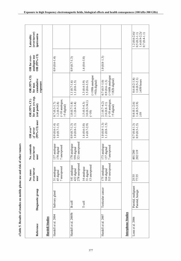
Exposure to high frequency electromagnetic fi elds, biological effects and health consequences (100 kHz-300 GHz)
377
eTab
le 5
. Res
ults
of
stud
ies
on m
obile
pho
ne u
se a
nd r
isk
of o
ther
tum
ors
Ref
eren
ce
Dia
gnos
tic
grou
p
No.
cas
es
ever
/nev
er
user
No.
con
trol
s ev
er/n
ever
us
er
OR
eve
r*
cf n
ever
(9
5% C
I) u
ser
OR
(95
% C
I )
for
max
yrs
exp
. (c
ut p
oint
)
OR
(95
% C
I)
for
max
cu
mul
ativ
eex
posu
re
OR
for
eve
r-an
alog
ue u
se
OR
(95
% C
I)
Lat
eral
ity
(eve
r/ne
ver)
ip
si/c
ontr
a
Har
dell
Stud
ies
Har
dell
et a
l. 20
04Sa
livar
y gl
and
31
ana
logu
e 45
dig
ital
? un
expo
sed
137
anal
ogue
17
0 di
gita
l ?
unex
pose
d
0.9
(0.6
-1.4
) 1.
0 (0
.7-1
.5)
0.7
(0.3
-1.7
) 1.
2 (0
.5-2
.8)
(>10
ana
logu
e,
>5 d
igita
l)
0.
9 (0
.6-1
.4)
Har
dell
et a
l. 20
05b
B-c
ell
T-ce
ll
141
anal
ogue
42
2 di
gita
l 27
8 un
expo
sed
14 a
nalo
gue
31 d
igita
l 13
une
xpos
ed
178
anal
ogue
55
9 di
gita
l 32
1 un
expo
sed
0.9
(0.7
-1.3
) 1.
0 (0
.8-1
.3)
1.6
(0.6
-3.8
) 1.
4 (0
.7-2
.9)
1.0
(0.7
-1.4
) 1.
1 (0
.4-3
.4)
1.5
(0.5
-4.3
) 3.
0 (0
.3-3
4.1)
(>
10)
1.1
(0.7
-1.6
) 1.
1 (0
.8-1
.5)
1.3
(0.4
-3.9
) 1.
5 (0
.6-3
.5)
(>19
8h a
nalo
gue
>91h
dig
ital)
0.9
(0.7
-1.3
)
1.6
(0.6
-3.8
)
Har
dell
et a
l. 20
07Te
stic
ular
can
cer
175
anal
ogue
16
4 di
gita
l 51
5 un
expo
sed
173
anal
ogue
13
7 di
gita
l ?
unex
pose
d
1.0
(0.8
–1.3
) 1.
1 (0
.8–1
.5)
2.1
(0.7
–6.2
) 2.
8 (0
.8–1
1)
(>10
ana
logu
e,
>5 d
igita
l)
0.7
(0.5
–1.0
) 0.
9 (0
.6–1
.3)
(>16
0h a
nalo
gue
>182
h di
gita
l)
1.0
(0.8
–1.3
)
Inte
rpho
ne S
tudi
es Lo
nn e
t al.
2006
Pa
rotid
, mal
igna
nt
Paro
tid, b
enig
n
25/3
5 77
/35
401/
280
20
2/11
9
0.7
(0.4
-1.3
) 0.
9 (0
.5-1
.5)
0.4
(0.1
-2.6
) 1.
4 (0
.5-3
.9)
≥10
0.6
(0.2
-1.8
) 1.
0 (0
.5-2
.1)
≥4
50 h
ours
1.
2 (0
.6-2
.6)/
0.5
(0.2
-1.1
) 1.
4 (0
.9-2
.2)/
0.7
(0.4
-1.1
)
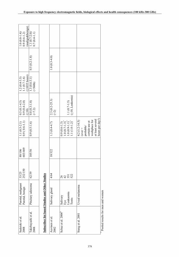
Exposure to high frequency electromagnetic fi elds, biological effects and health consequences (100 kHz-300 GHz)
378
Sade
tzki
et a
l. 20
08Pa
rotid
, mal
igna
nt
Paro
tid, b
enig
n
33/2
5 25
2/15
0 88
/106
60
3/46
9
1.1
(0.5
-2.1
) 0.
9 ( 0
.6-1
.1)
0.5
(0.1
-4.5
) 0.
9 (0
.4-2
.0)
(≥10
)
1.2
(0.4
-3.5
) 1.
1 (0
.7-1
.6)
(≥10
35 h
ours
)
1.
0 (0
.8-1
.4)/
0.9
(0.6
-1.2
) (m
alig
n&be
nign
) Ta
keba
yash
i et a
l. 20
08Pi
tuita
ry a
deno
ma
62/3
9 10
5/56
0.
9 (0
.5-1
.6)
0.8
(0.3
-1.8
) (>
7.2)
1.
3 (0
.6-3
.1)
(>56
0h)
0.5
(0.2
-1.8
) 1.
1 (0
.7-2
.0)/
0.7
(0.4
-1.1
)
Subs
crib
er li
st b
ased
Stu
dies
and
Oth
er S
tudi
es A
uvin
en e
t al.
2002
Saliv
ary
glan
d 4/
64
18/3
22
1.3
(0.4
-4.7
) 2.
3 (0
.2-2
5.3)
(>
2)
1.
0 (0
.3-4
.0)
Schu
z et
al.
2006
b
Sa
livar
y
Eye
Leuk
aem
ia
Test
is
26
42
351
522
0.9
(0.6
-1.3
) 1.
0 (0
.7-1
.3)*
1.0
(0.9
-1.1
)*
1.1
(1.0
-1.2
)*
1.1
(0.7
-1.5
) (>
10, L
euke
mia
)
Stan
g et
al.
2001
Uve
al m
elan
oma
4.2
(1.2
-14.
5)
(eve
r =
prob
able
/ ce
rtain
use
at
wor
kpla
ce fo
r at
leas
t sev
eral
ho
urs p
er d
ay!)
* Poo
led
resu
lts fo
r men
and
wom
en

