expression of matrix metalloproteinase-9 (gelatinase b) in benign
TRANSCRIPT

Summary. The matrix metalloproteinases (MMPs) are afamily of proteolytic zinc-containing enzymes, whichare responsible for the breakdown of the extracellularmatrix components in pathological and physiologicalconditions. They are involved in basement membranedisruption, stroma and blood vessel penetration,metastasis and more recently there is evidence that theyparticipate in tumor growth and angiogenic events.Matrix metalloproteinase 2 and 9 (MMP 2 and 9) belongto the gelatinases, a subgroup of MMPs, and have thecapacity to degrade the triple helix type IV collagen ofbasal lamina of the basement membrane. With thepresent study, we tried to demonstrate the expression ofMMP-9 immunohistochemically, comparatively inbenign, premalignant and malignant lesions of thelarynx. We studied 154 laryngeal lesions including 55squamous cell carcinomas, 8 in situ carcinomas, 54 casesof dysplasia (of low and intermediate grade), 13papillomas and 24 cases of keratosis. Overexpression ofMMP 9 was observed in 74.4% and 50% in invasive andin situ squamous cell carcinomas respectively. Indysplastic cases, in papillomas and in keratoses thepercentage of overexpression was 62.9%, 61.53% and54.16% respectively and the expression of MMP-9 wassignificantly higher in invasive squamous cellcarcinomas compared to dysplasias (p=0.000004). Alsosignificantly higher was the expression of MMP-9 indysplastic cases compared to papillomas (p=0.023). TheMMP-9 expression was related neither to survival nor tothe other available clinicopathological parameters(tumor size, grade, clinical stage, lymph node status andpatient age). In conclusion, our study indicates that theexpression of MMP-9 is up-regulated in a stepwisefashion, with two main steps, the first one, when adysplastic lesion evolves and the next one, when thedysplasia progresses to invasive carcinoma.
Key words: Matrix metalloproteinase 9, Laryngealcancer, Laryngeal lesions
Introduction
Local growth, spread and metastasis are features thatare shared among all malignant tumors, and require thepresence of the appropriate microenvironment of thehost. The matrix metalloproteinases (MMPs) are afamily of proteolytic zinc-containing enzymes, whichare responsible for the breakdown of the extracellularmatrix components in pathological and physiologicalconditions (Jones et al., 1999). Matrix metallo-proteinases (MMPs) are involved in basement membranedisruption, stroma and blood vessel penetration,metastasis and more recently there is evidence that theyparticipate in tumor growth and angiogenic events(Nelson et al., 2000). Additionally, they participate innormal extracellular matrix remodeling (Brenner et al.,1989), as well as in wound healing and bone resorption(Delaisse and Vaes, 1992; Wolf et al., 1992). Thebasement membrane is composed of type IV collagen,laminin, entactin, proteoglycans, andglycosaminoglycans (Nelson et al., 2000), but among allthese components, type IV collagen predominates(Leblond and Inoue, 1989) and it is thought to act as abarrier to the penetration of carcinoma cells into thematrix (Krecicki et al., 2001). The MMPs comprise alarge family of proteolytic enzymes, which is dividedaccording to their substrate specificity in four groups:the collagenases (MMP 1, 8 and 13), the gelatinases(MMP 2 and 9), the stromelysins (MMP 3, 10, 11 and19) and the membrane-bound MMPs (MT-MMP 1, 4)(Murphy et al., 1991). The gelatinases degrade the triplehelix type IV collagen of basal lamina of the basementmembrane (Nethery and O’Grady, 1989). Additionally,they have the capacity to degrade collagen types V, VII,IX and X, fibronectin and elastin (Senior et al., 1991).The tissue inhibitors of matrix metalloproteinases,
Expression of matrix metalloproteinase-9 (gelatinase B)in benign, premalignant and malignant laryngeal lesionsD. Peschos1, C. Damala2, D. Stefanou2, E. Tsanou2, D. Assimakopoulos3, T. Vougiouklakis1, K. Charalabopoulos4 and N.J. Agnantis51Department of Forensic Medicine, 2Pathology-Cytology, 3Otolaryngology and 4Physiology, Medical School, University of Ioannina, Ioannina, Greece
Histol Histopathol (2006) 21: 603-608
Offprint requests to: Professor N.J. Agnantis MD. PhD. FRCPATH,Department of Pathology, Medical School, University of Ioannina, 45110Ioannina, Greece. e-mail: [email protected]
DOI: 10.14670/HH-21.603
http://www.hh.um.es
Histology andHistopathologyCellular and Molecular Biology

known as TIMPs inhibit the catalytic activity of MMPsby binding to the active MMP and also they inhibit theconversion of the inactive proenzyme to the activeenzyme (Magary et al., 2000). Increased expression ofMMP-9 has been showed in various tumors (Coussensand Werb, 1996; Westermarck and Kahari, 1999; McCawley and Matrisian, 2000). With the present study, wetry to demonstrate the expression of MMP-9immunohistochemically, comparatively in benign,premalignant and malignant lesions of the larynx and tohighlight any possible relation to survival in invasivesquamous cell carcinomas. Also emphasis is placed onthe regulatory mechanisms that control the productionand activation of MMP-9. Materials and methods
Patients and study design
Formalin-fixed and paraffin-embedded tissues from154 laryngeal lesions including 55 squamous cellcarcinomas, 8 in situ carcinomas, 54 cases of dysplasia(20 of low, 25 of intermediate and 9 of severe grade), 13papillomas and 24 cases of keratosis were retrieved fromthe archives of the Pathology Department of UniversityHospital of Ioannina. Detailed clinical and laboratorydata, along with follow-up data were available for 46patients with invasive squamous cell carcinoma. Allrelevant data such as age, clinical stage and number ofextranodal disease sites were recorded. The mean agewas 58.5 and they were followed up for a period of 4years (range 1-7). Thirteen patients died during thefollow-up period (six died as a result of therapy), whiletwenty four presented complete remission of the diseaseand four presented partial remission. To assess theadvancement of the disease, stage classificationaccording to the American Joint Committee on Cancerwas used. Main clinical and pathological characteristicsare analyzed in Table 1. All tumor samples wereobtained by biopsy or surgery before any particulartherapy and were fixed in 10% buffered formalin andembedded in paraffin for immunohistochemical analysis.Immunohistochemistry
Immunohistochemistry was performed afterselecting one representative paraffin block, for eachcase, on 4 µm thick tissue sections placed on poly-L-lysine-coated glass slides. Tissue sections weredeparaffinized in xylene and rehydrated to distilledwater. A step of immersion in sodium citrate buffer(0.01M, pH 6.0) in plastic coplin jars and subjection tomicrowave irradiation twice for 15 minutes was used.Sections were then placed in 1.5% hydrogenperoxide/methanol for 10 minutes in order to block theendogenous peroxidase activity. Monoclonal antibodydirected against matrix metalloproteinase 9 (clone 2C3,Novocastra, dilution 1:50), was applied for 60 minutes attemperature 25°C. Subsequently the tissues were
incubated with the secondary antibody for 30 minutes.We used the method involving the avidin-biotin-peroxidase complex and developed the chromogen withimmersion of the slides in a diaminobenzidine-H2O2substrate for 5 min. The slides were counterstained inHarris’ haematoxylin, dehydrated and mounted. Toassess the specificity of the reaction, positive, as well asnegative control slides were used in all cases. Immunohistochemical evaluation
At least 10 high power fields (x 400) were countedindependently by two observers (pathologists) and thepercentage of positive staining of neoplastic, dysplasticor hyperplastic squamous epithelium was recorded.Whenever there was a disagreement between the twoobservers with a difference in the percentage levels>5%, the sections were reassessed simultaneously by thetwo pathologists. For statisistical analysis of the survivaldata we established, as described by other authors(Franchi et al., 2002) a three-scaled system on the basisof the percentage of positive cells: +, low expression(<10% positive cells); ++, moderate expression (10-50%positive cells) and +++, diffuse expression (>50%positive cells). For practical reasons we defined asoverexpression the presence of more than 50% positivecells for MMP-9. Non-specific immunostainings wereomitted from the study. Statistical analysis
The program SPSS for Windows Release 10.0 wasused for statistical analysis. Pearson’s and Spearman’scorrelation coefficients were used for the assessment ofcorrelation between continuous variables. The results
604Expression of MMP-9 in laryngeal lesions
Table 1. Patients’ characteristics.
CHARACTeRISTIC VAlUe
AgeMean 58.5
SexMale 55Female 2
Tumor stageT1 9T2 10T3 16T4 6
Tumor differentiationWell 12Moderately 9Poorly 2
lymph node metastasisAbsent (pN0) 26Present (pN+) 10
Distant metastasisM1 2

were considered as statistically significant when p<0.05.Results
Details on the expression levels of MMP-9 for all lesionsare shown in Table 2
Invasive carcinomasOverexpression of MMP-9 (>50% positive cells)
was observed in 47/55 cases (75.5%) of invasivesquamous cell carcinoma. The immunoreactivity ofMMP 9 was significantly higher in invasive carcinomasthan in dysplasias (p=0.000004). The MMP-9 expressionwas related neither to survival nor to the otherclinicopathological parameters that were available(tumor size, grade, clinical stage, lymph node status,patient age). MMP-9 was predominantly localized toepithelial cells, although it was also evident in thestroma, to varying degrees. Stromal cells surroundingthe tumors stained in general more intensely. Leukocytesand especially macrophages showed stain reactivity aswell; the same was the case for endothelial cells aroundthe tumors. Staining results are shown at Figure 1. Nosignificant correlation was seen between the threesubgroups of MMP-9 protein expression and prognosis.(Table 3).In situ carcinomas
As expected, a lower percentage of in situ
carcinomas 5/8 (62.5%), demonstrated strongimmunoreactivity for MMP 9, compared to invasivecarcinomas, although this difference was not statisticallysignificant (p=0.13).Dysplasias
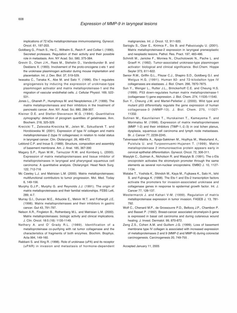
37/54 dysplastic cases (68.5%) presented high levelsof MMP-9 expression. Staining results are shown inFigure 2. The expression of MMP-9 was significantlyhigher in dysplasias, when compared to papillomas(p=0,023) and similarly the dysplastic cases exhibitedsignificantly higher levels of MMP-9 in comparison withthe keratotic lesions (p=0.000036). In addition, nodifferences in MMP-9 expression was observed betweengroups of low, moderate and severe dysplasia, using thechi square test (p=0.219).
605Expression of MMP-9 in laryngeal lesions
Fig. 1. High expression of MMP-9 ininvasive squamous cell carcinoma,moderately differentiated (ABC, x 400).
Table 2. expression levels of MMP-9 in different lesions.
MMP-9 % N Mean Std. Deviation Minimum Maximum
Cancer 55 74,1091 20,02191 10,00 96,00Dysplasia 54 62,9630 23,32135 5,00 95,00In situ carcinoma 8 52,5000 18,51640 30,00 85,00Papilloma 13 59,2308 22,25292 15,00 90,00Keratosis 24 55,8333 24,30185 5,00 90,00
Papillomas
Concerning papillomas, 9/13 (63.2%) showed highMMP-9 expression. Keratoses
For keratotic lesions 12/24 cases (50%) presentedstrong immunoreactivity for MMP-9.Discussion
Matrix metalloproteinases (MMPs) are believed toparticipate in complex processes during the developmentof malignant tumors, acting at several points. Variousstudies have been carried out in order to elucidate therole of MMPs in malignant tumors and to highlight theirpossible prognostic significance. Our study failed todocument any significant correlation between theexpression of MMP-9 in invasive squamous cellcarcinomas and clinicopathological parameters, such astumor stage, lymph node status, age and grade. Nor theexpression of MMP-9 was related to survival. This is inagreement with other studies. Bogusiewicz et al. foundno correlation between the activity of MMP-9 andclinical stage or tumor size of laryngeal carcinomas(Bogusiewicz et al., 2003). Similarly, the study ofFranchi et al. revealed no significant correlation betweenMMP-9 expression and clinical parameters (Franchi etal., 2002). Also in gastric cancers, Murray et al.
demonstrated no association between the expression ofMMP-9 and tumor stage or lymph node status, nor tosurvival (Murray et al., 1998). Although for members ofthe MMP family there is evidence of a stepwise increaseof their expression levels, our results failed to revealsignificant differences in the expression of MMP-9between invasive and in situ squamous cell carcinomas.(Campo et al., 1992; Garzetti et al., 1996; Sutinen et al.,
606Expression of MMP-9 in laryngeal lesions
Fig. 2. High expression of MMP-9 insquamous dysplasia (ABC, x 400).
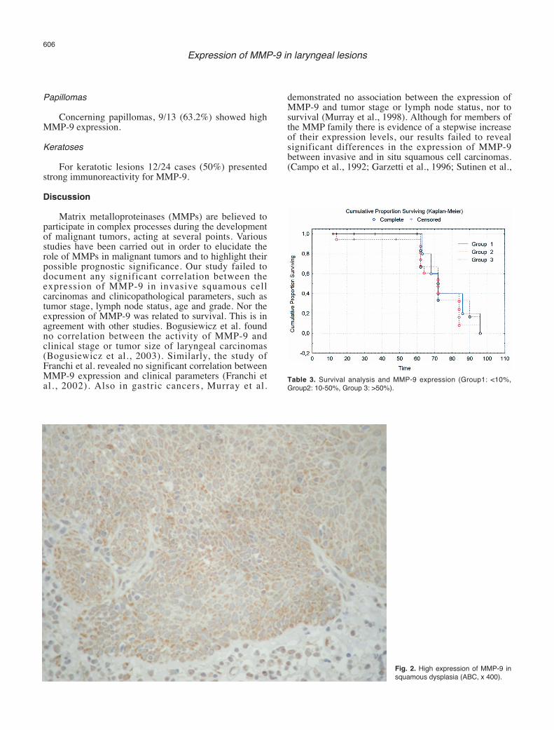
Table 3. Survival analysis and MMP-9 expression (Group1: <10%,Group2: 10-50%, Group 3: >50%).

1998; Talvensaari-Mattila et al., 1999; Zeng et al.,1999). Sarioglu et al. demonstrated such a sequential risein the MMP-2 expression, with carcinomas exhibitingthe highest expression; atypical hyperplasias lowerexpression and in situ carcinomas showing intermediatescores between the two former entities (Sarioglu et al.,2001). During the process of malignant transformation,where low grade dysplasia and invasive carcinomarepresent the two ends of the same spectrum, wedemonstrated only one step of increase in the values ofMMP-9, namely in the transition of dysplasia to invasivecarcinoma. We suggest that this is the crucial point atwhich there is a rise of MMP-9 and that for the transitionfrom in situ to invasive carcinoma, only a slight increaseof MMP-9 takes place. Significant differences occurredin the expression of MMP-9 between dysplasias and thefrankly benign lesions (papillomas, keratoses), a fact thatindicates that already from the evolution of apremalignant lesion, MMP-9 is up regulated. It is wellestablished that MMP activity is regulated at severallevels, including the induction by different cytokines andgrowth factors, such as EGF, bFGF and TNFα(Goldberg et al., 1990; Schmitt et al., 1992; Rabbani andXing, 1998), as well as by various oncogenes (Jones etal., 1999). The gene encoding for MMP-9 is one of thetarget genes of Ets-1 transcriptional factor, which isencoded by the ets 1 protooncogene (Wasylyk et al.,1991; Grevin et al., 1993; Iwasaka et al., 1996; Watabeet al., 1998). Behrens et al demonstrated in his study asignificant upregulation of Ets-1 transcripts and protein,as well as of MMP-9 protein in the stroma of invasivesporadic colorectal carcinomas, pointing out at theimportant role of Ets-1 in the induction of MMP-9(Behrens et al., 2003). Moreover, the status of tumorsuppressor gene p-53 has been linked to the expressionof various MMPs (Sun et al., 1999, 2000). Mutant p-53activates the transcription of MMP genes (Sun et al.,1999, 2000). Overexpression of MMP-9 is correlatedstrongly with the presence of p53 mutations in head andneck squamous cell carcinomas and this indicates thatMMP-9 up regulation is a result of p-53 inactivation(Franchi et al., 2002). Also, activated molecules ofMMPs can activate MMP proenzymes by positivefeedback mechanisms (Jones et al., 1999). MMP-9 isactivated by other MMPs, namely MMP-3 and MMP-2(Friedmann et al., 1995).
It seems that for further progression of a high gradeintraepithelial lesion to invasive carcinoma, otherregulatory mechanisms exist. Posttranscriptionally,MMP activity is controlled by proteolytic activation oflatent proenzymes and by specific TIMPs (Jones et al.,1999). We believe that in order to precede aprecancerous lesion to invasiveness, the ratio ofactive/inactive form of MMP-9 turns out to be moreimportant rather than the absolute expression levels ofMMP-9 themselves. Since immunohistochemicalevaluation cannot discriminate latent from the activatedenzyme, the enzymatic activity can be measured bysubstrate gel electrophoresis, named zymography
(Kleiner and Stetler-Stevenson, 1994). Laryngeal cancerdisplays significantly higher MMP-9 activity byzymography, compared with normal mucosa(Bogusiewicz et al., 2003). In addition TIMPs, which arenegative regulators of MMPs also play an important rolein keeping the balance of MMP levels. Extracellularmatrix degradation with concomitant tumor progressionhas been linked to altered TIMP levels (Di Nezza et al.,2002).
In conclusion, the present study indicates that theexpression of MMP-9 is up regulated early, when adysplastic lesion evolves and the crucial step is when thedysplasia progresses to invasive carcinoma. Theobservation that the levels of MMP-9 did not differsignificantly between in situ and invasive carcinomas,reflects the existence of post-transcriptional mechanismswhich act synergistically to the increase of theproduction of MMP-9. References
Behrens P., Mathiak M., Mangold e., Kirdorf S., Wellmann A., Fogt F.,Rothe M., Florin A. and Wernert N. (2003). Stromal expression ofinvasion-promoting, matrix-degrading proteases MMP-1 and –9 andthe ets 1 transcription factor in HNPCC carcinomas and sporadiccolorectal cancers. Int. J. Cancer 107, 183-188.
Bogusiewicz M., Stryjecka-Zimmer M., Szymanski M., Rechberger T.and Golabek W. (2003). Activity of matrix metalloproteinases-2 and–9 in advanced laryngeal cancer. Otolaryngol. Head Neck Surg.128, 132-136,
Brenner C.A., Adler R.R., Rappolee D.A., Pedersen R.A. and Werb Z.(1989). Genes for extracellular matrix degrading MMP and theirinhibitor TIMP are expressed during early mammalian development.Genes Dev. 3 848-859.
Campo e., Tavassoli F.A., Charonis A.S., Stetler-Stevenson W.G.,liotta l.A. and Merino M.J. (1992). evaluation of basementmembrane components and the 72 Kda type IV collagenase inserous tumors of the ovary. Am. J. Pathol. 6, 500-507
Coussens l.M. and Werb Z. (1996). Matrix metalloproteinases and thedevelopment of cancer. Chem. Biol. 3, 895-904.
Delaisse J.M. and Vaes G. (1992). Mechanism of mineral solublizationand matrix degradation in osteoclastic bone resorption. In: Biologyand physiology of the osteoclast. Rifkin B.R. and Gay C.V. (eds).CRC Press Inc. Philadelphia. pp 397-441.
Di Nezza l., Misajon A., Zhang J., Jobling T., Quinn M.A., Ostor A.G.Nie G., lopata A. and Salamonsen l.A. (2002). Presence of activegelatinases in endometrial carcinoma and correlation of matrixmetalloproteinase expression with increasing tumor grade andinvasion. Cancer 94, 1466-1475.
Franchi A., Santucci M., Masini e., Sardi I.,Paglierani M. and Gallo O.(2002). expression of matrix metalloproteinase 1, matrixmetalloproteinase 2, and matrix metalloproteinase 9 in carcinoma ofthe head and neck. Cancer 95, 1902-1910.
Friedmann R., Toth M., Pena D. and Mobashery S. (1995). Activation ofprogelatinase B (MMP-9) by gelatinase A (MMP-2). Cancer Res. 55,2548-2555.
Garzetti G.G., Ciavattini A., lucarini G., Goteri G., Romani C. andBiagini G. (1996). Microinvasive cervical carcinoma and cervicalintraepithelial neoplasia: Biologic significance and clinical
607Expression of MMP-9 in laryngeal lesions
implications of 72 kDa metalloproteinase immunnostaining. Gynecol.Oncol. 61, 197-203.
Goldberg G., Frisch S., He C., Wilhelm S., Reich F. and Collier I. (1990).Secreted proteases. Regulation of their activity and their possiblerole in metastasis. Ann. NY Acad. Sci. 580, 375-384.
Grevin D., Chen J.H., Raes M., Stehelin D., Vandenbunder B. andDesbiens X. (1993). Involvement of the proto-oncogene c-ets 1 andthe urokinase plasminogen activator during mouse implantation andplacentation. Int. J. Dev. Biol. 37, 519-529.
Iwasaka C., Tanaka K., Abe M. and Sato Y. (1996). ets-1 regulatesangiogenesis by inducing the expression of urokinase-typeplasminogen activator and matrix metalloproteinase-1 and themigration of vascular endothelial cells. J. Cellular Physiol. 169, 522-531.
Jones l., Ghaneh P., Humphreys M. and Neoptolemos J.P. (1999). Thematrix metalloproteinases and their inhibitors in the treatment ofpancreatic cancer. Ann. NY. Acad. Sci. 880, 288-307.
Kleiner D.e. and Stetler-Stevenson W.G. (1994). Quantitativezymography: detection of picogram quantities of gelatinases. Ann.Biochem. 218, 325-329.
Krecicki T., Zalesska-Krecicka M., Jelen M., Szkudlarek T. andHorobiowska M. (2001). expression of type IV collagen and matrixmetalloproteinase-2 (type IV collagenase) in relation to nodal statusin laryngeal cancer. Clin. Otolaryngol. 26, 469-472.
leblond C.P. and Inoue S. (1989). Structure, composition and assemblyof basement membrane. Am. J. Anat. 185, 367-390
Magary S.P., Ryan M.W., Tarnuzzer R.W. and Kornberg l. (2000).expression of matrix metalloproteinases and tissue inhibitor ofmetalloproteinases in laryngeal and pharyngeal squamous cellcarcinoma: A quantitative analysis. Otolaryngol. Head Neck Surg.122, 712-716
Mc Cawley l.J. and Matrisian l.M. (2000). Matrix metalloproteinases:multifunctional contributors to tumor progression. Mol. Med. Today6, 149-156.
Murphy G.J.P., Murphy G. and Reynolds J.J. (1991). The origin ofmatrix metalloproteinases and their familial relationships. FeBS lett,289, 4-7.
Murray G.I., Duncan M.e., Arbuckle e., Melvin W.T. and Fothergill J.e.(1998). Matrix metalloproteinases and their inhibitors in gastriccancer. Gut 43, 791-797.
Nelson A.R., Fingleton B., Rothenberg M.l. and Matrisian l.M. (2000).Matrix metalloproteinases: biologic activity and clinical implications.J. Clin. Oncol. 18:5 (18): 1135-1149.
Nethery A. and O’ Grady R.l. (1989). Identif ication of ametalloproteinase co-purifying with rat tumor collagenase and thecharacteristics of fragments of both enzymes. Biochim. Biophys.Acta 994, 149-160.
Rabbani S. and Xing R. (1998). Role of urokinase (uPA) and its receptor(uPAR) in invasion and metastasis of hormone-dependent
malignancies. Int. J. Oncol. 12, 911-920. Sarioglu S., Ozer e., Kirimca F., Sis B. and Pabuccuoglu U. (2001).
Matrix metalloproteinase-2 expression in laryngeal preneoplasticand neoplastic lesions. Pathol. Res. Pract. 197, 483-486.
Schmitt M., Janicke F., Moniwa N., Chucholowski N., Pache l. andGraeff H. (1992). Tumor-associated urokinase-type plasminogenactivator: biological and clinical significance. Biol.Chem. HoppeSeyler 373, 611-622.
Senior R.M., Griffin G.l., Fliszar C.J., Shapiro S.D., Goldberg G.I. andWelgus H.G. (1991). Human 92- and 72-kilodalton type IVcollagenases are elastases. J. Biol. Chem. 266, 7870-7875.
Sun Y., Wenger l., Rutter J.l., Brinckerhoff C.e. and Cheung H.S.(1999). P53 down-regulates human matrix metalloproteinase-1(collagenase-1) gene expression. J. Biol. Chem. 274, 11535-11540.
Sun Y., Cheung J.M. and Martel-Pelletier J. (2000). Wild type andmutant p53 differentially regulate the gene expression of humancollagenase-3 (hMMP-13). J. Biol. Chem. 275, 11327-11332.
Sutinen M., Kaunilainen T., Hurskainen T., Kameyema T. andMorimatsu M. (1998). expression of matrix metalloproteinases(MMP 1-2) and their inhibitors (TIMP-1,-2,-3) in oral lichen planus,dysplasia, squamous cell carcinoma and lymph node metastases.Br. J. Cancer 77, 2239-2245.
Talvensaari-Mattila A., Apaja-Sakkinen M., Hoythya M., Westurland A.,Puistula U. and Turpeennuemi-Hujanen T. (1999). Matrixmetalloproteinase 2 immunoreactive protein appears early incervical epithelial differentiation. Gynecol. Oncol. 72, 306-311.
Wasylyk C., Gutman A., Nicholson R. and Wasylyk B. (1991). The c-etsoncoprotein activates the stromelysin promoter through the sameelements as several non-nuclear oncoproteins. eMBO J. 10, 1127-1134.
Watabe T., Yoshida K., Shindoh M., Kaya M., Fujikawa K., Sato H., IshiiS. and Fujinaga K. (1998). The ets-1 and ets-2 transcription factorsactivate the promoters for invasion-associated urokinase andcollagenase genes in response to epidermal growth factor. Int. J.Cancer 77, 128-137.
Westermarck J. and Kahari V-M. (1999). Regulation of matrixmetalloproteinase expression in tumor invasion. FASeB J. 13, 781-792.
Wolf C., Chenard M.P., de Grossouvre P.D., Bellocq J.P., Chambon P.and Basset P. (1992). Breast-cancer associated stromelysin-3 geneis expressed in basal cell carcinoma and during cutaneous woundhealing. J. Invest. Dermatol. 99, 870-872.
Zeng Z.S., Cohen A.M. and Guillem J.G. (1999). loss of basementmembrane type IV collagen is associated with increased expressionof metalloproteinases 2 and 9 (MMP-2 and MMP-9) during colorectalcarcinogenesis. Carcinogenesis 20, 749-755 .
Accepted January 11, 2005
608Expression of MMP-9 in laryngeal lesions