fabrication of gold arrays for electrochemical detection...
TRANSCRIPT

Fabrication of Gold Arrays for Electrochemical Detection of Cancer Biomarkers
Chi Tang and James F. Rusling
University of Connecticut
Department of Chemistry
February 24, 2010

Introduction
Cancer statistics
Cancer is the 2nd leading cause of death
Approx. 596,000 patients per year
Early Detection of cancer
Improve prognosis for future patients
Cancer biomarker
Any measurable or observable factors in human body that indicates
cancer or related diseases
Proteins, mutated DNAs, cell deaths, and physical symptoms
Interleukin 6 (IL-6)
Enzyme Linked Immunosorbent Assay (ELISA/Immunoassay)
Use to detect and quantify proteins based on antibody-antigen
interaction and specificity
98/384 wells
1. Rusling, Analyst 2010 (135) 2496-2511
2. U.S. Center of Disease Control and Prevention

Goal
Inexpensive and easy fabrication method for electrochemical
arrays
Integration with Microfluidics
Point of Care device
Compact Disc Arrays

Gold CD-Rs
650 MB Compact Disc Recordable
Relatively Cheap – $1.5 per disk
Easy to prepare
Can be cut into desired shape for different applications
50 to 100 nm single layer of sputtered gold (99.9% pure)
Protective layer Gold
Organic dye (Phthalocyanine)
Polycarbonate

LaserJet Printing
Reproducibility of printed patterns
LaserJet Printer
HP LaserJet 1020 (600 dpi)
Styrene acrylic copolymer (melt at 125ºC)
Iron Oxide
Cost: <$200

Printing process
Charging the photoconductor drum: Photoconductor surface
is charged
Exposure to light: The charged surface is exposed to a laser
Development: Negatively charged toner particle is brought to
the photoconductor drum
Image transfer: Toner is transferred from the photoconductor
drum onto paper
Fusing of Toner

Gold Arrays form CD-R
3. Deniel, et. al. Electrochem Comm 2003
1 min 1 min 10 min 1 min

Electrochemical arrays
Electrode area reproducibility: ± 10 % (n=8)
Cost: $0.18 per array
Time: Approx. 1hr for ~8 arrays
Resistance: 21. 4 ± 0.5 Ω

Micro-wells
Micro-well

Micro-wells (cont.)

Electrochemical methods
Electrochemical cell
3 electrodes system
Cyclic voltammetry
Amperometry
Applied Resultant

Surface Area
CV at 50 mV s-1 of gold CD-R array in 5 mM Ru(NH3)6Cl6
and 0.1 M Sodium triflouroacetate (NaTFA)
-3 10-6
-2 10-6
-1 10-6
0
1 10-6
2 10-6
3 10-6
4 10-6
5 10-6
-0.6-0.4-0.200.2
E1
E2
E3
E4
E5
E6I,
A
Potential, V
ip = (2.687x10⁵) n3/2 v1/2 D1/2 A C
ip – peak current
n – number of electrons
v – scan rate
D – diffusion coefficient
A – electro-active surface area
C – concentration of probe
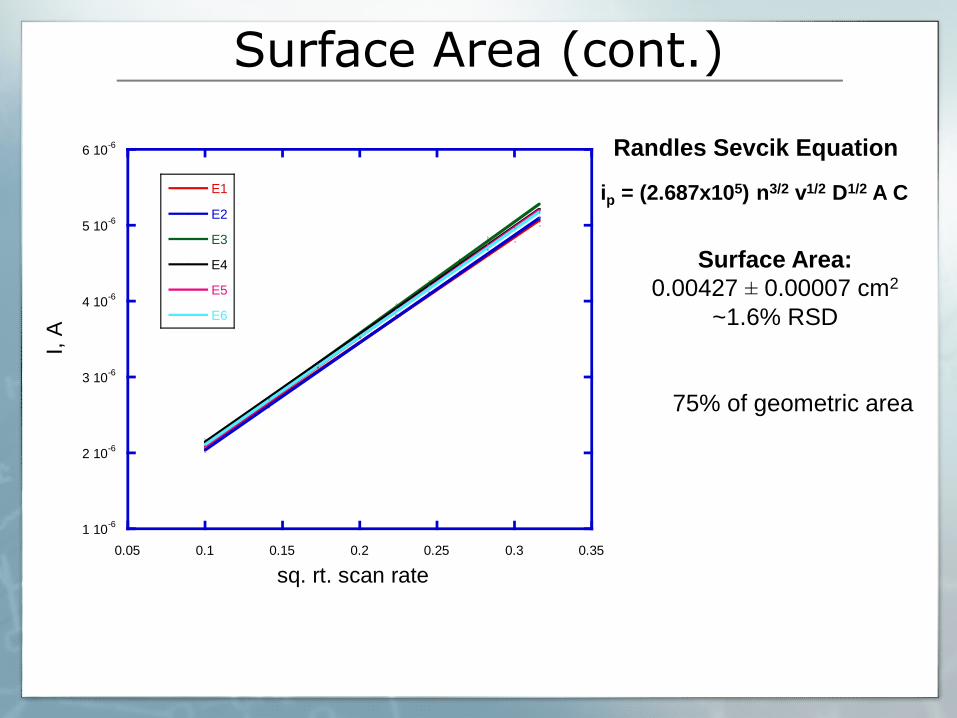
Surface Area (cont.)
1 10-6
2 10-6
3 10-6
4 10-6
5 10-6
6 10-6
0.05 0.1 0.15 0.2 0.25 0.3 0.35
E1
E2
E3
E4
E5
E6
I, A
sq. rt. scan rate
ip = (2.687x10⁵) n3/2 v1/2 D1/2 A C
Randles Sevcik Equation
Surface Area:
0.00427 ± 0.00007 cm2
~1.6% RSD
75% of geometric area

Summary
Old New
Old design New design
Surface Area (cm2) 0.036 0.042
Reproducibility ~10% ~2%
Hold < 1 µL drop of reagents No Yes

Sandwich Immunoassay Interleukin-6 (IL-6)
Normal patient range: < 6 pg/mL
Cancer patient range: > 20 pg/mL

Integration with Microfluidic Device
PDMS channel
Array

Instrument Setup
Pump Injector Valve Microfluidic
device Waste
Potentiostat

Calibration Plot for Interleukin-6
0
5
10
15
20
0 200 400 600 800 1000
y = 5.2222 + 0.011823x R= 0.98187
I, n
A[IL-6] fg/mL
Amperometric response of Au/MPA/Ab1/Ag/Ab2/Strep-HRP with 1 mM
HQ and injection of 100 uM H2O2 at -0.3 V vs. SCE in microfluidic device
Limit of Detection:
100 fg/mL
0
5
10
15
20
0 2000 4000 6000 8000 10000 12000
I, n
A
Time
Control
100
250
500
1000

Conclusion
Fabricate gold electrode array at low cost (~$0.20)
Reproducible electrode areas (~2% RSD)
Successful development of immunosensor for IL-6
Low detection limit: 100 fg/mL
Integration with microfluidics
Multiplexing
Point of Care device

Acknowledgment
Dr. James Rusling
Dr. Vaze and Dr. Liu
Group members
NIH

Thank You!
Questions?