facteurs de pronostic visuel des endophtalmies aiguës
TRANSCRIPT
HAL Id: dumas-00704867https://dumas.ccsd.cnrs.fr/dumas-00704867
Submitted on 6 Jun 2012
HAL is a multi-disciplinary open accessarchive for the deposit and dissemination of sci-entific research documents, whether they are pub-lished or not. The documents may come fromteaching and research institutions in France orabroad, or from public or private research centers.
L’archive ouverte pluridisciplinaire HAL, estdestinée au dépôt et à la diffusion de documentsscientifiques de niveau recherche, publiés ou non,émanant des établissements d’enseignement et derecherche français ou étrangers, des laboratoirespublics ou privés.
Facteurs de pronostic visuel des endophtalmies aiguësbactériennes post-chirurgie de cataracte
Aurélie Combey-De-Lambert
To cite this version:Aurélie Combey-De-Lambert. Facteurs de pronostic visuel des endophtalmies aiguës bactériennespost-chirurgie de cataracte. Human health and pathology. 2012. �dumas-00704867�
UNIVERSITE JOSEPH FOURIER
FACULTE DE MEDECINE DE GRENOBLE
Année : 2012 N°
FACTEURS DE PRONOSTIC VISUEL DES
ENDOPHTALMIES AIGUES BACTERIENNES POST-
CHIRURGIE DE CATARACTE
THESE PRESENTEE POUR L’OBTENTION DU DOCTORAT EN MEDECINE
DIPLOME D’ETAT
Aurélie COMBEY-de LAMBERT
Née le 09.02.1982 à Chambéry
THESE SOUTENUE PUBLIQUEMENT A LA FACULTE DE MEDECINE DE
GRENOBLE
Le 24 mai 2012
DEVANT LE JURY COMPOSE DE
Président du jury : Monsieur le Professeur Jean-Paul ROMANET
Membres :
Monsieur le Professeur Christophe CHIQUET, directeur de thèse
Monsieur le Professeur Max MAURIN
Monsieur le Professeur Gilles THURET

1
A Alb�,
Mon �me sÏur.
A Ninon et Gaspard,
Mes merveilles. Vous �tes ma plus belle r�ussite.

2
Aux membres du jury
A Monsieur le Pr Jean-Paul Romanet,
Votre d�vouement pour vos patients, votre investissement pour la p�dagogie sont
exceptionnels. Mais combien dÕheures comptent donc vos journ�es ? Soyez assurez de ma
plus grande reconnaissance.
A Monsieur le Professeur Christophe Chiquet,
Votre passion pour la recherche est exceptionnelle. Merci de nous pousser � donner le
meilleur de nous m�me.
A Monsieur le Professeur Max Maurin
Votre aide pour mes premiers pas en bact�riologie a �t� tr�s pr�cieuse. Merci de juger ce
travail.
A Monsieur le Professeur Gilles Thuret
Merci pour votre accueil chaleureux ; je garderai un excellent souvenir de ma parenth�se
st�phanoise.
A mes ma�tres
A Olivier
Mon mod�le professionnel. Je suis fi�re de notre amiti�.
CÕest un immense privil�ge de te rejoindre � Chamb�ry.
A Karine,
En souvenir de toutes nos discussions au bloc tr�s Ç f�minines ÈÉEt vive Barry White !
A Rux,
Pour ta disponibilit� et tous tes avis en r�tine medicalÉ
A Adel,
Merci pour tous les tr�s bons conseils chirurgicaux et ton excellent accueil.
A Viviane,
Notre meilleure fournisseuse de chocolat !
A Sabine,
Pour nos 6 mois tr�s sympas sur Chamb�.
A Florent
Bonne route � Grenoble.
A Monsieur le Professeur Gain,
Un tr�s grand merci pour ces six mois � Saint Etienne. Et f�licitations pour votre �quipe
parfaite !

3
A Nelly,
Ta gentillesse et ta g�n�rosit� nÕont dÕ�gales que tes comp�tences professionnelles. Je suis
tr�s heureuse dÕavoir fait ta connaissance et te souhaite tout le bonheur que tu m�rites.
A Damien
Ton sens de la p�dagogie est exceptionnel. Les internes ont beaucoup de chance de tÕavoir.
A Pierre
La star de lÕophtalmo-ped !
A Patricia
La championne du glaucome !
A Catherine
La reine du diab�te !
A Manu Bozonnet
En souvenir des ap�ros et ton accompagnement en chirurgie plastique.
Aux Dr Xavier Piquot et Patrice Moyenin
Pour mes premiers pas en ophtalmo
A mes co-internes Qui pour beaucoup maintenant sont devenus chefs !
A Elisabeth
La plus grenobloise dÕentre nous! Le meilleur est � venirÉ
A Matthieu
Nous nÕaurons malheureusement que tr�s peu travaill� ensemble. Un jour peut-�tre ?
A Cindy
La joie de vivre. Dommage quÕon ne se voit pas plus souvent.
A Hafide
Le speedy-gonzales de la cataracte!
A Pierre-Marie
LÕexpert du barbec ! Pour les grillades � venir avec tous nos loulousÉ
A Youssef
Mon youyou � moi ! Je suis heureuse que tu tÕ�panouisses dans Ton service. Bonne route
chef !
A Tiffany
Quel bonheur de partager mon assistanat avec toi.

4
A Diane
Miss 100 000 Volts. Comment fais-tu ?
A Ahmed
Pour ton courage exemplaire.
A Mag
Merci pour ces 6 derniers mois vraiment supers sympas � Grenoble. Tu es une fille en or !
Une petite pens�e pour nos r�visions acharn�es de lÕEBOÉ et bon mariage !
A Eva
Aussi belle que brillante. Bonne continuation � Bourgoin.
A Ralitsa
Un caract�re bien tremp�!! JÕadorerais nÕavoir que 10% de ton franc parlerÉ Mais je suis
surtout ravie dÕavoir d�couvert ta grande g�n�rosit�. Christian est un homme chanceuxÉ
A Benj
Le meilleur dÕentre nous. Je te souhaite tout le bonheur que tu m�rites avec ton Aur�.
A Thierry
La gentillesse � lÕ�tat pur.
A Jos�phine
Bon s�jour � Marseille. Reviens-nous quand m�me !
A Antoine
La zenitudeÉ
A Mathilde
Je tÕaurai bien envoy�e passer lÕEBO � ma place mais il para�t que ce nÕest pas possibleÉ A
d�faut je tÕai emmen�e avec moi pour mes 6 derniers mois � Chamb� ! Tu es une fille extra :
ne change rien.
A Nischal, Caro, Fred, Georges, Marc-Antoine, Perrine, C�cile, Adriane, Olivier, Julie et
Rachel : courage pour la suite !
A Aur�lie, Mathieu, Marie-Caroline, Mansour, Marine, Maher, R�mi et Claire : mes supers
co-internes st�phanois.
A ma famille
A ma Maman,
Tu as fais ce que je suis. Il mÕest impossible dÕexprimer toute ma gratitude. Merci pour tout
lÕamour que tu donnes maintenant � mes enfants.

5
A mon Papa,
Qui mÕa toujours regard�e avec tant de fiert� : tu as fait ma force.
A C�cilia,
Ma petite sÏur ador�e. Je donnerais beaucoup pour tÕavoir plus pr�s de nous. Un jour peut-
�tre ?
A Pierre,
Beau-p�re mod�le.
A Laurence,
Une super mamie suppl�mentaire pour Ninon et Gaspard.
A Yannick,
Mon super beau-fr�re. Entre deux randos, continue de rendre ma petite sÏur heureuseÉ
A Monique et Thierry,
Des beaux-parents en or. Je suis heureuse dÕ�tre rentr�e dans votre famille.
A Elo, Gaetan, Boris, Marie, Coline et Andr�a,
Je compte sur vous pour la faire grandir cette famille !
A Julia
Une adorable premi�re cousine pour Ninon et Gaspard.
A mamie Pounette et mon Grand-P�re,
Pour toutes les heures que vous mÕavez consacr�es. Et parce que la vocation est certainement
h�r�ditaireÉ
A mamie Boisserette et Papi,
Les vendanges, la r�colte des pommes de terre et des framboises � vos c�t�s font partis de
mes tr�s beaux souvenirs de petite fille.
A Jean-christophe, Jean-Louis, Claudine, Marie, Georges, Jean-Paul, Claudine, Charles,
Adrien, Hugo, Gaston, Florence : une jolie tribu !
A Lise
Qui me fait lÕhonneur dÕ�tre pr�sente ce jour, comme � tous les moments importants de ma
vie.
A mes amis
A Marie
Ma meilleure amie. Merci pour toutes ces belles ann�es. On aura quand m�me r�ussi � les
finir ces �tudes !
6
A Hugo, Julie, Colin et Maud
Des amis tr�s chers. Votre famille est pour moi un exemple. Je regrette encore votre d�part
mais cÕest toujours une immense joie de vous revoir (avec une petite pens�e sp�ciale pour les
moules marini�resÉ)
A Myl�ne,
Que je ne vois malheureusement pas assez souvent.
A Aur�, Rom et L�a
Mes Ç exil�s È lyonnais pr�f�r�s. Grenoble se languit de vousÉ
A ma tio
Reine de la bonne humeur mais aussi de la d�co. Quel we je r�serve pour tes chambres
dÕh�te ?
A Fio
Devenue une vraie Parisienne ! Mais je sais que tu ne nous oublies pas.
A Delphine
Une fille en or.
A Guillaume et Muriel,
Des amis tr�s chers. Merci dÕavoir toujours �t� l� pour nousÉ
A Antoine, Romain, Lolo, J�r�me et Julien,
Des sacr�s carabins.
A Sylvain,
Bravo pour ton parcours et plein de bonheur avec Alice.
A Aur�, Patrick, Manon et Thibault,
Nos supers voisins.
A Fanny, Julien, Mathis et Ilian,
Bis. Fanny, je ne pourrais jamais assez te remercier de veiller aussi bien sur mes enfants ;
vivement lÕ�t� prochain que lÕon puisse vous honorer � votre juste valeurÉ
A Arnaud, Domi et Anouk,
On se suit depuis le lyc�e, je suis ravie de vous retrouver sur Chamb�.
A Bernadette,
Ma petite maman de lÕophtalmologie ! Ton d�vouement au service est remarquable ; ta
g�n�rosit� extraordinaire. Quelle chance de travailler � tes c�t�s et de tÕavoir comme amie.

7
Aux �quipes
Du service, du bloc et de la consultation de Grenoble,
Merci pour votre gentillesse et votre comp�tence durant toutes ces ann�esÉUne pens�e bien
s�r particuli�re pour notre ch�re Catherine.
Du service, du bloc et de la consultation de Chamb�ry,
Vous faites de lÕh�pital mon petit paradisÉ Merci pour votre si bel accueil � mon retour.
Du service, du bloc et de la consultation de Saint Etienne
De bact�riologie du CHU de Grenoble,
Pour mon ann�e de master.
A G�raldine, Sylvain, Marie, Carole, Fabrice, Yann, Monique, Delphine : pour votre soutien
tout au long de lÕinternat.
8
Baseline predictive factors of visual prognosis in acute
bacterial postcataract endophthalmitis
Aur�lie Combey1,2
, Nelly Campolmi3, Florent Aptel
1,2, Max
Maurin1,3
, Pierre-Loic Cornut5, Gilles Thuret
3, Alain Bron
6,
Philippe Denis5, Jean-Paul Romanet
1,2, Catherine Creuzot-
Garcher6, Christophe Chiquet
1,2
On behalf of the FRIENDS (French Institutional Endophthalmitis Study) group
1 - UJF-Grenoble 1, Grenoble, F-38041, France
2 - Department of Ophthalmology, University Hospital, Grenoble, F-38043,
France
3 - Department of Ophthalmology, University Hospital, Saint-Etienne, France
4 -Department of Microbiology, University Hospital, Grenoble, F-38043,
France
5 - Department of Ophthalmology, University Hospital Edouard Herriot, Civil
Hospices of Lyon, University Lyon I, Lyon, France
6 - Department of Ophthalmology, University Hospital, Dijon, France
Keywords: endophthalmitis, cataract surgery, prognosis, microbiology
9
Abstract
Objective: To identify baseline clinical factors of visual prognosis in patients with
acute endophthalmitis following cataract surgery.
Design: prospective cohort study
Participants: 99 patients hospitalized for acute postcataract endophthalmitis in four
academic hospitals (Dijon, Grenoble, Lyon, Saint-Etienne; FRIENDS group).
Methods: Factors related to the cataract surgery (complications), the initial clinical
presentation, and the microbiological diagnosis were analyzed based on the final
visual outcome, defined as poor (<20/100) or good (!20/40) using univariate and
multivariate analysis (backward-stepping procedure).
Main outcome measures: visual acuity (LogMAR), microbiological identification
using culture and panbacterial PCR.
Results: The significant baseline factors for good visual outcome (45% of the series)
were the winter season, absence of complications during cataract surgery, initial
visual acuity (VA), microbiological investigations revealing no microorganism or a
coagulase-negative Staphylococcus sp. (CNS), and fundus visibility. Quantitative
factors associated with a good clinical prognosis were shorter duration of cataract
surgery, younger age, and a hypopyon equal to or less than 1.5 mm. Significant
factors associated with poor visual outcome were infection of the right eye, initial VA,
corneal edema, hypopyon larger than 1.5 mm, detection of bacterial species other
than a CNS, and the absence of fundus visibility. Multiple logistic regression analysis
showed that high bacterial virulence was the only independent factor (odds ratio
[OR]= 14, 95% confidence interval [CI]: 2.7Ð71, p=0.001) for poor visual outcome. On
the other hand, low bacterial virulence (OR=0.1 95%CI: 0.03Ð0.6, p=0.01) and the
10
absence of complications (OR=0.07, 95%CI: 0.01Ð0.4, p=0.003) during cataract
surgery were independent factors for good VA.
Conclusion: Visual outcome factors in acute postcataract endophthalmitis identified
in this prospective study (2004Ð2007) were similar to those reported by the EVS 10
years ago. The bacterial virulence level was the main predictive factor of final visual
prognosis. This emphasizes the need for rapid bacterial identification and
characterization using new biomolecular techniques.
11
INTRODUCTION
Postcataract endophthalmitis is a rare (approximately 0.08% of cataract
surgeries)1, 2 but serious complication, which can lead to anatomical and/or functional
loss of the eye. Significant advances have been achieved in the field of antibiotic
prophylaxis (cefuroxime intracameral injection),3-5 and microbiological diagnosis.6-9
The identification of baseline prognostic factors at the time of modern clear corneal
cataract surgery remains an important step to identify patients who need aggressive
therapy as early as possible. Polymerase chain reaction (PCR)-based techniques
now provide accurate and rapid identification of the microorganisms directly involved
in intraocular samples. This will make it possible to rapidly adapt the therapeutic
strategy to both clinical and microbiological factors, and to give more objective
information to the patient.
The anatomical and functional prognoses have been reported to be associated
with baseline factors such as initial vision,10-12 absence of a red reflex, history of
diabetes or glaucoma, and the bacterial species involved.10-17 These data were
mainly obtained 15 years ago on a selected population to be randomized for pars
plana vitrectomy (PPV) in the EVS (Endophthalmitis Vitrectomy Study), with inclusion
of patients with clear enough cornea to allow vitrectomy and exclusion of patients
who did not consent to therapeutic randomization, VA equal to or better than 20/100).
10 Given the changes in cataract surgery techniques and different epidemiological
characteristics of the population studied, new prospective data are needed for the
identification of prognostic factors.
The FRench Institutional ENDophthalmitis Study (FRIENDS) group included
100 consecutive patients with acute postcataract endophthalmitis, which allowed the
prospective study of the largest series after the EVS. Previous microbiological
12
results, using conventional cultures and panbacterial polymerase chain reaction
(PCR) have been previously published.6, 18-21
The aim of this report was to identify baseline clinical and microbiological
factors of good (VA !20/40) and poor (VA <20/100) visual outcome at 6 months in a
large series of acute postcataract endophthalmitis.
PATIENTS AND METHODS
This prospective study included 100 patients (100 eyes) with acute (<6 weeks)
postcataract endophthalmitis, between March 2004 and December 2005 in four
French academic hospitals (Dijon, Grenoble, Lyon, Saint-Etienne). One patient was
excluded from the analysis given his age (2 years) and the inability to obtain reliable
postoperative vision measurement. The study followed the Declaration of Helsinki
guidelines for research involving human subjects and was approved by the local
Institutional Review Board (Comit� de Protection des Personnes, CPP Lyon B). The
inclusion criteria, data sheet, sampling techniques and microbiological procedures,
antibiotics used for intravitreal injections, and follow-up visits were standardized in
the four centers.
Clinical data collection:
Patients were eligible if they had clinical signs of endophthalmitis within 6 weeks after
cataract surgery. The diagnosis of acute endophthalmitis was made on the basis of
clinical features including pain, decreased visual acuity (VA), diffuse bulbar
conjunctival hyperemia, chemosis, inflammation of the anterior segment (anterior
chamber reaction including one of the following criteria: flare, cells, hypopyon height,
fibrin, cyclitic membrane), and posterior segment inflammation (all patients had
13
vitreous infiltration diagnosed with biomicroscopy or ophthalmic ultrasound). A
baseline evaluation form was completed during the initial examination of each patient
and included demographic features, past medical history, ocular history, details of
cataract surgery (retrospectively collected from the cataract surgeon including
capsular rupture and vitreous loss), and data of the initial ocular examination (see
factors in Table 1)..
Performing pars plana vitrectomy (PPV) was left to the discretion of the
treating physician, generally following the EVS guidelines. During the 6-month follow-
up, patients were defined as having a good (final VA "0.3 LogMAR, !20/40) or poor
(final VA > 0.7 LogMAR, <20/100) visual prognosis. Incidence of phthisis was also
recorded at 6 months.
Microbiological data:
On admission, an immediate tap of aqueous humor and/or vitreous was
performed followed by intravitreal injection of vancomycin (1 mg/0.1 mL) and
ceftazidime (2 mg/0.1 mL). Conventional cultures were made by direct inoculation of
intraocular samples in Brain Heart Infusion (BHI) broth (BioM�rieux, Marcy lÕEtoile,
France). These culture media were incubated for 2 weeks at 37¡C, and were
subcultured on chocolate agar supplemented with PolyViteX (BioM�rieux) and
Columbia agar supplemented with 5% sheep blood (Biom�rieux) when bacterial
growth was detected or systematically after 2 weeks of incubation if no growth was
detected. For positive cultures, Gram stains were performed, and bacterial
identifications and antibiograms were obtained using phenotypic methods (Vitek II,
BioM�rieux, or BD Phoenix, Becton Dickinson). Species identification was confirmed
by 16SrDNA amplification and sequencing.
14
Bacterial DNA extraction, 16S rDNA PCR amplification and sequencing were
performed as previously described.6 Panbacterial PCR was able to detect 500 cfu of
Staphylococcus epidermidis. Amplification of the human betaglobulin gene served as
an internal positive extraction and amplification control. The 16S rDNA sequences
obtained were compared with those available in GenBank, EMBL, and DDBJ
databases with the BIBI (Bio Informatic Bacterial Identification; http://pbil.univ-
lyon1.fr/bibi/query.php) program. Identification to the species level was defined as
99% or greater 16S rDNA sequence similarity with that of the GenBank prototype
strain sequence. Identification to the genus level was defined as a 97% or greater
16S rDNA sequence similarity with that of the GenBank prototype strain sequence. A
failure to identify was defined as a 16S rDNA sequence similarity less than 97% with
sequences deposited in GenBank at the time of the analysis.
Patients were then further classified into the following groups according to
microbiological results: detection of no microorganism, CNS, or detection of a more
virulent species, including Streptococcus sp., S. aureus, S. lugdunensis, and Gram-
negative bacteria. 18
Statistical analysis:
Quantitative data were expressed as mean ± standard deviation (SD). Associations
between prognosis and clinical data were primarily studied using ANOVA for
quantitative data and chi-square analysis (incorporating YatesÕs correction when
appropriate) for qualitative data. To determine which of these variables were
combined with clinical factors to predict visual prognosis, logistic regression models
were fitted using a backward-stepping procedure. The odds ratio (OR), which
provides an estimate of the relative risk of good or poor prognosis, was calculated.
15
Multivariate logistic regression analysis was performed using clinical factors that were
found to be statistically significant in univariate analysis (with p < 0.1). The statistical
analysis was performed using the Statistical Package for the Social Science program
(SPSS 17.0 for Windows, Chicago, IL, USA). The tests were two-tailed and statistical
significance was set p<0.05.
RESULTS
Ninety-nine patients, with a mean age of 73 ± 9 years, were included with
acute (<6 weeks) postcataract endophthalmitis. Patients were hospitalized with a
median delay of 6 days (range, 1Ð38 days) after cataract surgery (clear corneal
phacoemulsification, n=95, manual extracapsular extraction, n=4) associated with
posterior (n= 89) or anterior (n=6) lens implantation or with no intraocular lens (n=4).
The median time between surgery and the onset of symptoms was 5 days (range, 0Ð
35 days). No intracameral antibiotics were used at the time of cataract surgery.
Complicated cataract surgery (capsular rupture with or without vitreous loss) was
reported in 20% of the cases, including 12 eyes that had undergone an anterior
vitrectomy during cataract surgery. Other systemic and ocular risk factors of
endophthalmitis were immunosuppression (n=7), diabetes mellitus (n=13), oral
steroids (n=7), atopic dermatitis (n=1), blepharitis (n=1), dry-eye syndrome (n=2), and
contact lens wear (n=1).
Thirty-one percent of the population had a final VA greater than 0.7 logMAR (group
of poor visual outcome) including 10 patients with light perception only, and 45% of
the population had a final VA lower than 0.3 logMAR (good visual outcome group).
Finally, 24% of the patients had a final VA between 20/100 and 20/50.
16
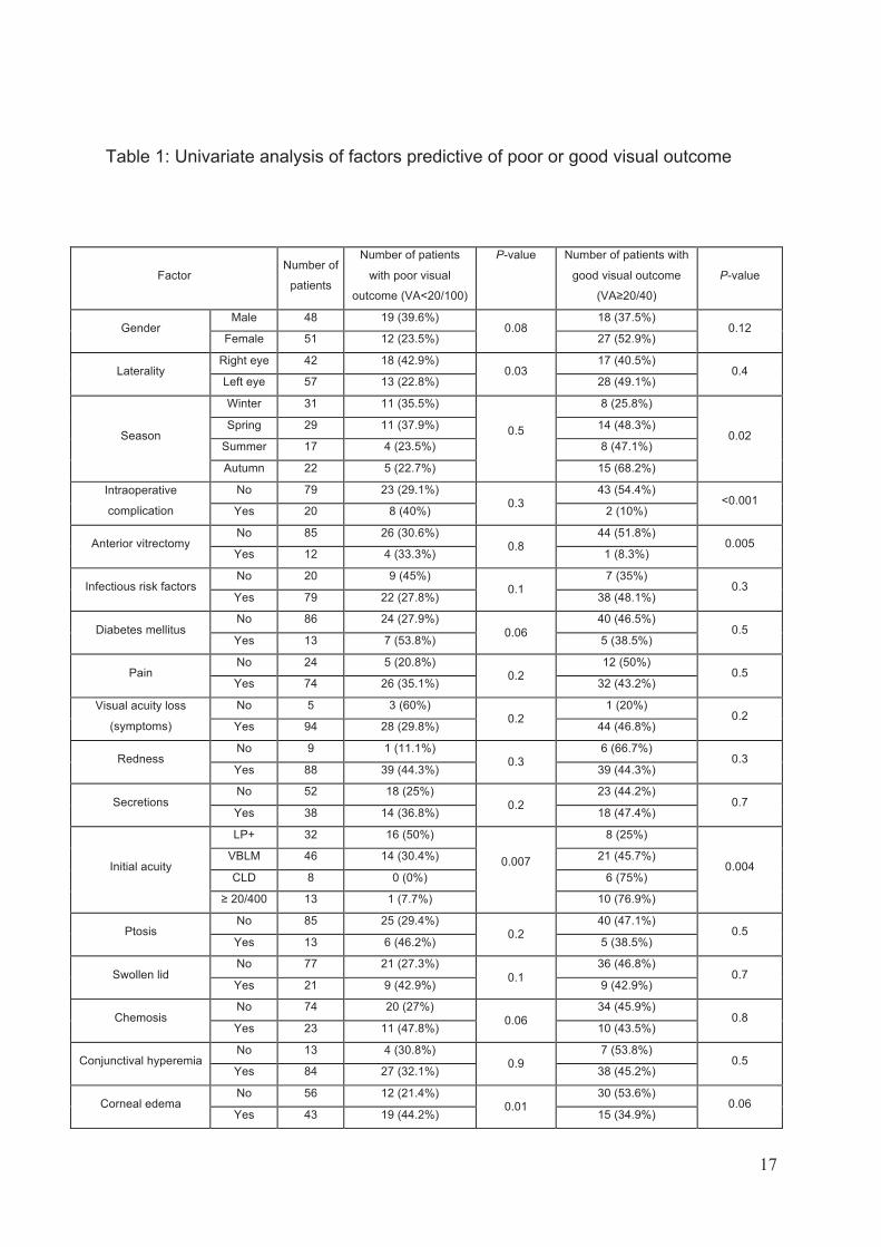
Univariate analysis (Table 1):
The significant initial factors of good visual outcome were the winter season,
absence of complications during cataract surgery, initial VA, detection of CNS or
absence of microorganism detection, and fundus visibility. Quantitative factors
associated with good clinical prognosis were a shorter duration of cataract surgery,
younger age, and a smaller hypopyon.
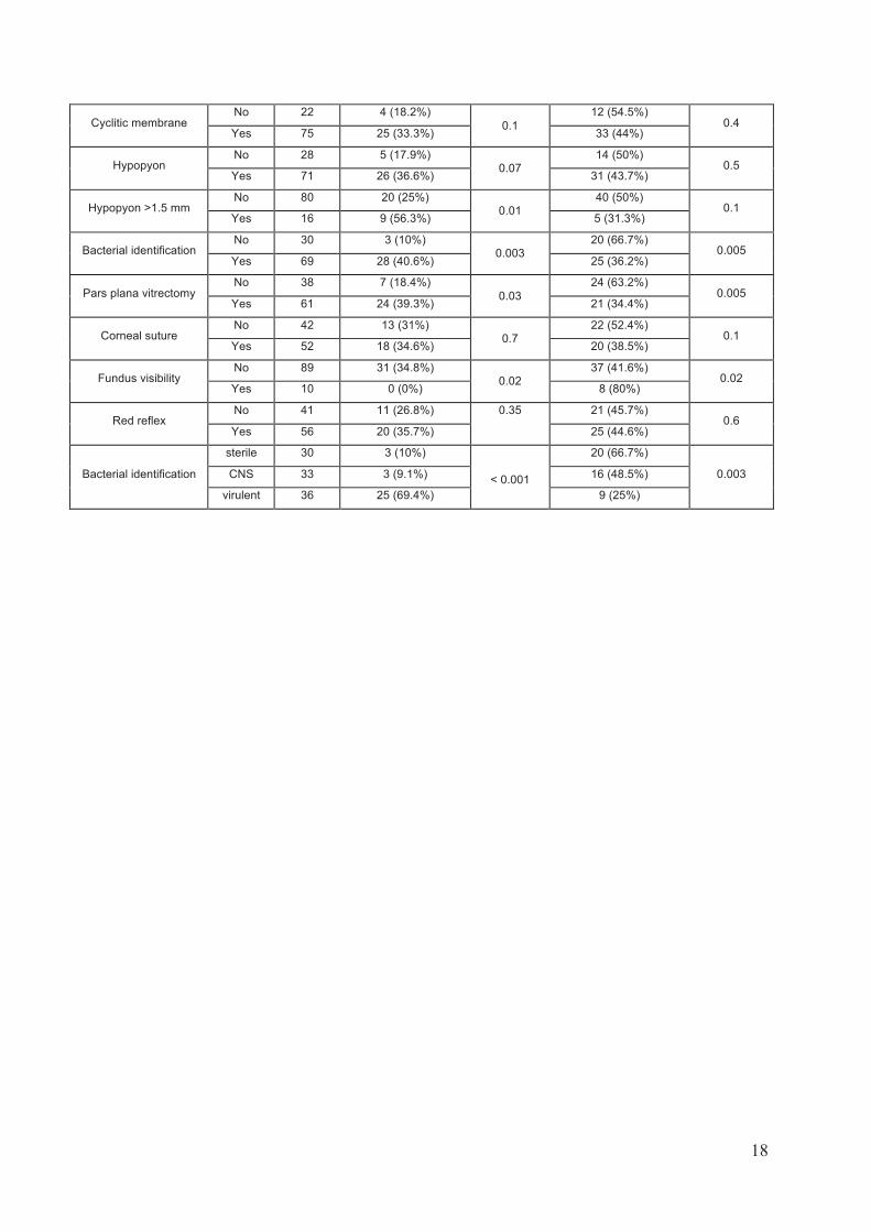
Significant factors associated with poor visual outcome were infection of the
right eye, initial VA, corneal edema, hypopyon larger than 1.5 mm, detection of a
bacterial species other than a CNS, and the absence of fundus visibility.
Considering treatment, the number of intravitreal injections was not
significantly associated with the visual outcome. PPV was more frequently used in
cases of poor visual outcome. PPV was also significantly associated with earlier
presentation (p= 0.03), detection of a virulent species (p=0.008), initial VA (p<0.001),
and loss of red reflex (p=0.01). PPV was performed in 91% of the patients with initial
VA limited to light perception (LP), 56.5% of patients with Òhand motionÓ VA, 37.5% of
patients with Òcounting fingersÓ VA, and 28% of patients with VA !20/400.
17
Table 1: Univariate analysis of factors predictive of poor or good visual outcome
Factor Number of
patients
Number of patients
with poor visual
outcome (VA<20/100)
P-value Number of patients with
good visual outcome
(VA!20/40)
P-value
Gender Male 48 19 (39.6%)
0.08 18 (37.5%)
0.12 Female 51 12 (23.5%) 27 (52.9%)
Laterality Right eye 42 18 (42.9%)
0.03 17 (40.5%)
0.4 Left eye 57 13 (22.8%) 28 (49.1%)
Season
Winter 31 11 (35.5%)
0.5
8 (25.8%)
0.02 Spring 29 11 (37.9%) 14 (48.3%)
Summer 17 4 (23.5%) 8 (47.1%)
Autumn 22 5 (22.7%) 15 (68.2%)
Intraoperative
complication
No 79 23 (29.1%) 0.3
43 (54.4%) <0.001
Yes 20 8 (40%) 2 (10%)
Anterior vitrectomy No 85 26 (30.6%)
0.8 44 (51.8%)
0.005 Yes 12 4 (33.3%) 1 (8.3%)
Infectious risk factors No 20 9 (45%)
0.1 7 (35%)
0.3 Yes 79 22 (27.8%) 38 (48.1%)
Diabetes mellitus No 86 24 (27.9%)
0.06
40 (46.5%) 0.5
Yes 13 7 (53.8%) 5 (38.5%)
Pain No 24 5 (20.8%)
0.2 12 (50%)
0.5 Yes 74 26 (35.1%) 32 (43.2%)
Visual acuity loss
(symptoms)
No 5 3 (60%) 0.2
1 (20%) 0.2
Yes 94 28 (29.8%) 44 (46.8%)
Redness No 9 1 (11.1%)
0.3 6 (66.7%)
0.3 Yes 88 39 (44.3%) 39 (44.3%)
Secretions No 52 18 (25%)
0.2
23 (44.2%) 0.7
Yes 38 14 (36.8%) 18 (47.4%)
Initial acuity
LP+ 32 16 (50%)
0.007
8 (25%)
0.004 VBLM 46 14 (30.4%) 21 (45.7%)
CLD 8 0 (0%) 6 (75%)
! 20/400 13 1 (7.7%) 10 (76.9%)
Ptosis No 85 25 (29.4%)
0.2 40 (47.1%)
0.5 Yes 13 6 (46.2%) 5 (38.5%)
Swollen lid No 77 21 (27.3%)
0.1
36 (46.8%) 0.7
Yes 21 9 (42.9%) 9 (42.9%)
Chemosis No 74 20 (27%)
0.06
34 (45.9%) 0.8
Yes 23 11 (47.8%) 10 (43.5%)
Conjunctival hyperemia No 13 4 (30.8%)
0.9 7 (53.8%)
0.5 Yes 84 27 (32.1%) 38 (45.2%)
Corneal edema No 56 12 (21.4%)
0.01 30 (53.6%)
0.06 Yes 43 19 (44.2%) 15 (34.9%)
18
Cyclitic membrane No 22 4 (18.2%)
0.1
12 (54.5%) 0.4
Yes 75 25 (33.3%) 33 (44%)
Hypopyon No 28 5 (17.9%)
0.07 14 (50%)
0.5 Yes 71 26 (36.6%) 31 (43.7%)
Hypopyon >1.5 mm No 80 20 (25%)
0.01 40 (50%)
0.1 Yes 16 9 (56.3%) 5 (31.3%)
Bacterial identification No 30 3 (10%)
0.003 20 (66.7%)
0.005 Yes 69 28 (40.6%) 25 (36.2%)
Pars plana vitrectomy No 38 7 (18.4%)
0.03
24 (63.2%) 0.005
Yes 61 24 (39.3%) 21 (34.4%)
Corneal suture No 42 13 (31%)
0.7 22 (52.4%)
0.1 Yes 52 18 (34.6%) 20 (38.5%)
Fundus visibility No 89 31 (34.8%)
0.02 37 (41.6%)
0.02 Yes 10 0 (0%) 8 (80%)
Red reflex No 41 11 (26.8%) 0.35 21 (45.7%)
0.6 Yes 56 20 (35.7%) 25 (44.6%)
Bacterial identification
sterile 30 3 (10%)
< 0.001
20 (66.7%)
0.003 CNS 33 3 (9.1%) 16 (48.5%)
virulent 36 25 (69.4%) 9 (25%)
19
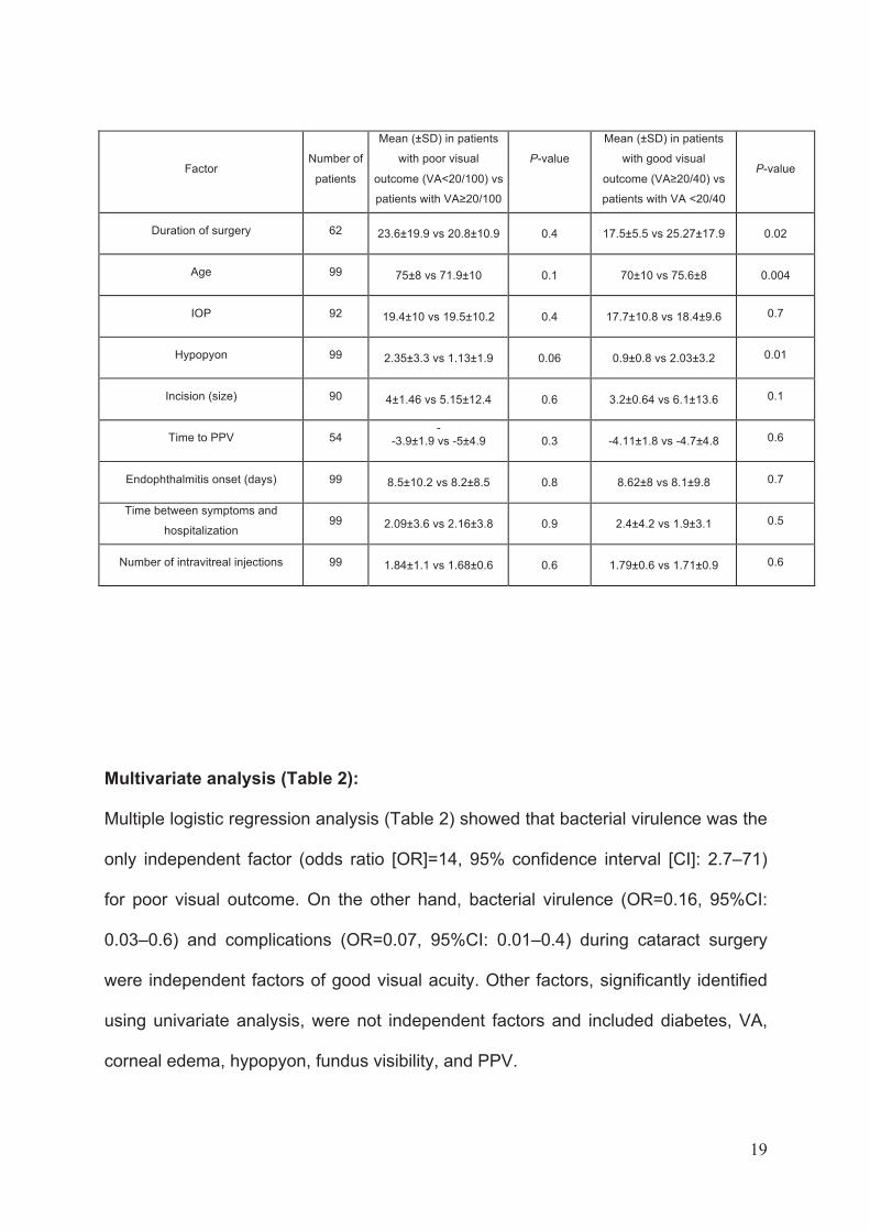
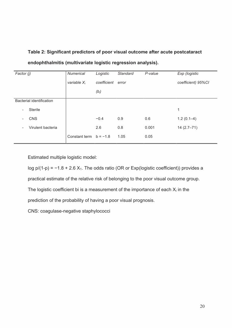
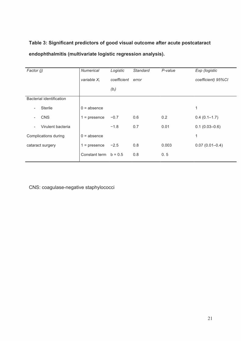
Multivariate analysis (Table 2):
Multiple logistic regression analysis (Table 2) showed that bacterial virulence was the
only independent factor (odds ratio [OR]=14, 95% confidence interval [CI]: 2.7Ð71)
for poor visual outcome. On the other hand, bacterial virulence (OR=0.16, 95%CI:
0.03Ð0.6) and complications (OR=0.07, 95%CI: 0.01Ð0.4) during cataract surgery
were independent factors of good visual acuity. Other factors, significantly identified
using univariate analysis, were not independent factors and included diabetes, VA,
corneal edema, hypopyon, fundus visibility, and PPV.
Factor Number of
patients
Mean (±SD) in patients
with poor visual
outcome (VA<20/100) vs
patients with VA!20/100
P-value
Mean (±SD) in patients
with good visual
outcome (VA!20/40) vs
patients with VA <20/40
P-value
Duration of surgery 62
23.6±19.9 vs 20.8±10.9
0.4
17.5±5.5 vs 25.27±17.9
0.02
Age 99
75±8 vs 71.9±10
0.1
70±10 vs 75.6±8
0.004
IOP 92
19.4±10 vs 19.5±10.2
0.4
17.7±10.8 vs 18.4±9.6 0.7
Hypopyon 99
2.35±3.3 vs 1.13±1.9
0.06
0.9±0.8 vs 2.03±3.2 0.01
Incision (size) 90
4±1.46 vs 5.15±12.4
0.6
3.2±0.64 vs 6.1±13.6 0.1
Time to PPV 54 -
-3.9±1.9 vs -5±4.9
0.3
-4.11±1.8 vs -4.7±4.8 0.6
Endophthalmitis onset (days) 99
8.5±10.2 vs 8.2±8.5
0.8
8.62±8 vs 8.1±9.8 0.7
Time between symptoms and
hospitalization 99
2.09±3.6 vs 2.16±3.8
0.9
2.4±4.2 vs 1.9±3.1 0.5
Number of intravitreal injections 99
1.84±1.1 vs 1.68±0.6
0.6
1.79±0.6 vs 1.71±0.9 0.6
20
Table 2: Significant predictors of poor visual outcome after acute postcataract
endophthalmitis (multivariate logistic regression analysis).
Factor (j) Numerical
variable Xi
Logistic
coefficient
(bi)
Standard
error
P-value Exp (logistic
coefficient) 95%CI
Bacterial identification
- Sterile
- CNS
- Virulent bacteria
#0.4
2.6
0.9
0.8
0.6
0.001
1
1.2 (0.1Ð4)
14 (2.7Ð71)
Constant term b = #1.8 1.05 0.05
Estimated multiple logistic model:
log p/(1-p) = #1.8 + 2.6 X1. The odds ratio (OR or Exp(logistic coefficient)) provides a
practical estimate of the relative risk of belonging to the poor visual outcome group.
The logistic coefficient bi is a measurement of the importance of each Xi in the
prediction of the probability of having a poor visual prognosis.
CNS: coagulase-negative staphylococci
21
Table 3: Significant predictors of good visual outcome after acute postcataract
endophthalmitis (multivariate logistic regression analysis).
CNS: coagulase-negative staphylococci
Factor (j) Numerical
variable Xi
Logistic
coefficient
(bi)
Standard
error
P-value Exp (logistic
coefficient) 95%CI
Bacterial identification
- Sterile
- CNS
- Virulent bacteria
0 = absence
1 = presence
#0.7
#1.8
0.6
0.7
0.2
0.01
1
0.4 (0.1Ð1.7)
0.1 (0.03Ð0.6)
Complications during
cataract surgery
0 = absence
1 = presence
#2.5
0.8
0.003
1
0.07 (0.01Ð0.4)
Constant term b = 0.5 0.8 0. 5
22
Discussion
This prospective study analyzing baseline prognostic clinical and
microbiological factors shows that bacterial virulence is the main independent factor
of good or poor visual outcome after acute postcataract endophthalmitis. One
additional independent factor of good visual outcome is the absence of complications
during cataract surgery.
Most studies considering prognostic factors after acute postoperative
endophthalmitis 10-16 were retrospective. We should emphasize that only prospective
studies are contributive to collecting baseline clinical and microbiological data
because initial examination is difficult in the emergency context and the
microbiological diagnosis should be performed using standardized methods. Our
study was based on previously validated microbiological techniques.6, 18, 19 Our
prospective study differed from the EVS by less restrictive inclusion criteria since it
was not designed to randomize patients for treatment (in the EVS: inclusion of
patients with cornea clear enough to allow PPV, exclusion of patients who did not
consent to therapeutic randomization, VA equal to or better than 20/100). 10 Other
considerations should also be taken into account, such as the number of patients
studied (99 patients in FRIENDS vs 420 in EVS), the number of centers (4 vs 24), the
duration (1.5 years vs 4 years), and the study period (2004Ð2005 vs 1990Ð1994),
and the type of surgery (phacoemulsification rate, scleral or corneal incisions,
secondary implantation). One additional strength of the present study was that the
population was not limited to culture-positive cases 15 and included microbiologically
unproven cases (n=30) and cases diagnosed by PCR only (n=25 in this series).
Complications at the time of cataract surgery were also included.
23
The final visual outcome data reported in this recent series showed that 31%
of patients had a poor visual outcome, while 45% had a good visual outcome (VA
!20/40). These results are similar to those described in the EVS (53% !20/40) and
other reports (see Table 4). Defining prognostic factors of poor visual outcome at
admission may be useful for clinicians since these patients would probably benefit
from more aggressive treatment. The initial VA level has previously shown a good
correlation with the visual prognosis.10, 12, 13, 15, 16, 22 It was identified as one of the
most important prognostic factors in the EVS (OR=2), in addition to diabetes (OR
1.6), corneal infiltrate and/or ring ulcer (OR=1.7), posterior capsular rupture (OR=1.9)
and rubeosis iridis (OR=1.8). Other previously reported significant factors such as
absence of fundus visibility 10 and absence of red reflex 10 were also identified in the
present series. Diabetes is a systemic condition that potentially has an effect on the
severity of endophthalmitis, especially with a higher Gram-positive and CNS infection
identification rate. 10 We previously showed 18 that eyes infected by Streptococcus
species were more frequently noted in diabetic patients. The frequency of diabetes in
the current study was 13%, nearly identical to that in the EVS patients (14%). 10 A
Gram-positive microorganism was always identified in this situation, and in more than
half of the cases (7/13) it belonged to the Streptococcus species. We found a trend
toward a higher frequency of diabetic patients in the group with poor VA. One
limitation of all reported studies is the absence of information concerning severity and
treatment of diabetes.
The season could also play a role in the severity of infection and therefore in
the prognosis. Variations in the bacteria spectrum and virulence may explain the
seasonal variation in severity of postoperative endophthalmitis. We previously
showed that Streptococcus sp., uniformly frequent (30Ð36%) from winter to summer,
24
were less frequent in autumn. These data should be taken into account when noting
the increased rate of S. pneumoniae found in conjunctival cultures in the same
periods (March, November, and December).23 A retrospective Australian study 14
showed a trend toward more isolates in the cooler wetter seasons of autumn and
winter.
After univariate analysis, the multivariate analysis identified the bacterial
virulence as the main independent factor of visual outcome. We classified patients
into three groups: virulent, CNS, and sterile. This classification is based on the
reported virulence of bacteria in cohort or case series, such as Pseudomonas, 12, 24, 25
Enterococcus, 26 Streptococcus, 12, 27, 28 Staphylococcus aureus, 28, 29 and
Staphylococcus lugdunensis 20 in acute postoperative endophthalmitis. Virulence is
strongly associated with bacterial characteristics, such as production of toxins
(cytolysin of Enterococcus faecalis, the quorum-sensing fsr system regulating
gelatinase and serine protease, pneumolysin and autolysin of S. pneumoniae, and S.
aureus alpha-toxin and beta-toxin), production of biofilms (Enterococcus,
Staphylococcus including CNS), and motility in the eye (B. cereus).30
Several reports have emphasized the prognostic role of bacterial virulence in
postoperative endophthalmitis 10-16 but this is only the second prospective study after
EVS that shows the influence of bacterial virulence on final visual prognosis, taking
into account baseline clinical factors. Intraocular lesions during infection have been
extensively studied in animal models of endophthalmitis, and experimental studies
have strongly suggested that ocular lesions during infection are related to bacterial
virulence and secondary intraocular inflammation to the host response to bacterial
toxins and cell wall constituents (cellular infiltration, production of cytokines,
chemokines and adhesion molecules, Fas ligand).31 Culture-negative cases are
25
usually associated with a better final visual outcome (Table 4) 11, 32 The sterile group
of patients with endophthalmitis (i.e., with negative microbiological results) probably
has a low inoculum undetected by conventional cultures and PCR techniques despite
infection. This situation is close to CNS infections, which are traditionally associated
with a less severe disease course.11, 28, 32 However, a subpopulation of patients with
CNS infection has a severe prognosis, 9% in our series. The more severe CNS
infections may be related to specific virulence traits of the involved strains, such as
higher adhesion or biofilm formation. 33 Further development in the field of CNS
virulence may help better define specific phenotypic and genotypic traits associated
with the occurrence of more severe endophthalmitis cases. Also, it has been
previously shown that acquired antibiotic resistance is frequent in S. epidermidis
isolated from eyes with endophthalmitis (e.g., methicillin resistance), 33 The
development of diagnostic tests allowing rapid evaluation of the virulence and
antibiotic resistance traits of CNSs may help better define the population of CNS
endophthalmitis patients with poorer outcome. Previous studies have shown that the
turn-around time of panbacterial PCR is 2Ð3 days, 6 and this time could be reduced
to only a few hours with real-time PCR, as recently described in the literature. 7, 8 , 9
Our analysis found the virulence factor to be a powerful indicator of visual
prognosis, and VA was not an independent prognostic factor, in contrast to the EVS
findings. This discrepancy could be due to different inclusion criteria. Finally, case
series and cohort studies, including this one, strongly suggest that identification of
Gram-positive cocci belonging to Streptococcus sp., S. aureus, or S. lugdunensis,
strongly predict a poor visual outcome.11 For instance, only 29% of patients infected
with virulent Gram-positive cocci had a final VA of 20/40 or better. 11 Another
example concerns infections with S. aureus, streptococci, or enterococci with 50%,
26
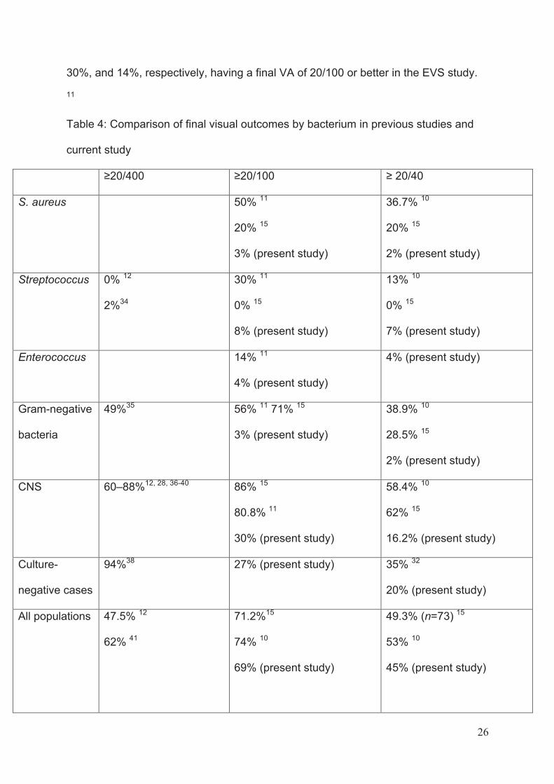
30%, and 14%, respectively, having a final VA of 20/100 or better in the EVS study.
11
Table 4: Comparison of final visual outcomes by bacterium in previous studies and
current study
!20/400 !20/100 ! 20/40
S. aureus 50% 11
20% 15
3% (present study)
36.7% 10
20% 15
2% (present study)
Streptococcus 0% 12
2%34
30% 11
0% 15
8% (present study)
13% 10
0% 15
7% (present study)
Enterococcus 14% 11
4% (present study)
4% (present study)
Gram-negative
bacteria
49%35 56% 11 71% 15
3% (present study)
38.9% 10
28.5% 15
2% (present study)
CNS 60Ð88%12, 28, 36-40 86% 15
80.8% 11
30% (present study)
58.4% 10
62% 15
16.2% (present study)
Culture-
negative cases
94%38 27% (present study) 35% 32
20% (present study)
All populations 47.5% 12
62% 41
71.2%15
74% 10
69% (present study)
49.3% (n=73) 15
53% 10
45% (present study)
27
The need for pars plana vitrectomy was associated with a poor visual outcome
but also with other clinical factors correlated with the severity of the disease (poor
initial VA, loss of red reflex, corneal edema, infection with a virulent species, and
early onset of symptoms). For this reason, after multivariate analysis, the need for
PPV was no longer identified as an independent prognostic factor. This relationship
is easier to understand with the legacy of the EVS, showing the benefit of PPV in
more severe patients, i.e., those having LP at admission. 10 Another nonrandomized
study 15 showed that PPV was associated with a worse prognosis due to the
indication bias of vitrectomy.
In conclusion, this prospective study shows that bacterial virulence is a major
prognostic factor of final visual function in patients with acute endophthalmitis after
cataract surgery. Whereas most patients are currently treated binarily, using PPV or
intravitreal injections, mostly according to EVS guidelines based on initial VA,
perspectives of rapid bacterial identification and characterization (virulence, antibiotic
resistance) are of major interest. This new diagnostic strategy could guide clinicians
in adapting the therapeutic regimen to each patient.
28
THE FRIENDS GROUP
FRench Institutional ENDophthalmitis Study group
Participants:
Study coordinator: Christophe Chiquet Statistics, methodology, microbiological techniques: Fran�ois Vandenesch, Max Maurin Database management: Pierre-Lo�c Cornut Ophthalmology: - University Hospital of Dijon: Pierre-Olivier Lafontaine, Marie Passemard,
Catherine Creuzot-Garcher, Alain Bron - University Hospital of Grenoble: Christophe Chiquet, Aur�lie Combey de Lambert,
Karine Palombi, Jean-Paul Romanet - University Hospital of Lyon (E. Herriot Hospital): Pierre-Lo�c Cornut, Fr�d�ric
Rouberol, Philippe Denis - University Hospital of Saint-Etienne: Gilles Thuret, Philippe Gain Microbiology: - University Hospital of Dijon: Andr� P�chinot, Catherine Neuwirth - University Hospital of Grenoble: Jacques Croiz�, Max Maurin - University Hospital of Lyon: J�r�me Etienne, Yvonne Benito, Sandrine Boisset,
Anne Tristan, Fran�ois Vandenesch - University Hospital of Saint-Etienne: Anne Carricajo, G�rard Aubert Mycology: - University Hospital of Dijon: Fr�d�ric Dalle, Alain Bonin - University Hospital of Grenoble: Bernadette Lebeau, Herv� Pelloux - University Hospital of Lyon: Fr�d�rique de Montbrison, St�phane Picot - University Hospital of Saint-Etienne: H�l�ne Raberin, Roger Tran Manh Sung
29
References 1. Fisch A, Salvanet A, Prazuck T, Forestier F, Gerbaud L, Coscas G, et al.
Epidemiology of infective endophthalmitis in France. The French Collaborative Study Group
on Endophthalmitis. Lancet. 1991; 338(8779): 1373-6.
2. Kattan HM, Flynn HW, Jr., Pflugfelder SC, Robertson C, Forster RK. Nosocomial
endophthalmitis survey. Current incidence of infection after intraocular surgery.
Ophthalmology. 1991; 98(2): 227-38.
3. Yiu G, Young L, Gilmore M, Chodosh J. Prophylaxis against postoperative
endophthalmitis in cataract surgery. Int Ophthalmol Clin. 2011; 51(4): 67-83.
4. Cochereau I, Korobelnik JF, Robert PY, Hajjar J. [Antibioprophylaxis in ocular
surgery: AFSSAPS recommendations]. J Fr Ophtalmol. 2011; 34(6): 428-30.
5. Yu-Wai-Man P, Morgan SJ, Hildreth AJ, Steel DH, Allen D. Efficacy of intracameral
and subconjunctival cefuroxime in preventing endophthalmitis after cataract surgery. J
Cataract Refract Surg. 2008; 34(3): 447-51.
6. Chiquet C, Cornut PL, Benito Y, Thuret G, Maurin M, Lafontaine PO, et al.
Eubacterial PCR for bacterial detection and identification in 100 acute postcataract surgery
endophthalmitis. Invest Ophthalmol Vis Sci. 2008; 49(5): 1971-8.
7. Bispo PJ, de Melo GB, Hofling-Lima AL, Pignatari AC. Detection and gram
discrimination of bacterial pathogens from aqueous and vitreous humor using real-time PCR
assays. Invest Ophthalmol Vis Sci. 2011; 52(2): 873-81.
8. Sugita S, Shimizu N, Watanabe K, Katayama M, Horie S, Ogawa M, et al. Diagnosis
of bacterial endophthalmitis by broad-range quantitative PCR. Br J Ophthalmol. 2011; 95(3):
345-9.
9. Goldschmidt P, Degorge S, Benallaoua D, Basli E, Batellier L, Boutboul S, et al. New
test for the diagnosis of bacterial endophthalmitis. Br J Ophthalmol. 2009; 93(8): 1089-95.
10. Results of the Endophthalmitis Vitrectomy Study. A randomized trial of immediate
vitrectomy and of intravenous antibiotics for the treatment of postoperative bacterial
endophthalmitis. Endophthalmitis Vitrectomy Study Group. Arch Ophthalmol. 1995; 113(12):
1479-96.
11. Microbiologic factors and visual outcome in the endophthalmitis vitrectomy study.
Am J Ophthalmol. 1996; 122(6): 830-46.
12. Bohigian GM, Olk RJ. Factors associated with a poor visual result in endophthalmitis.
Am J Ophthalmol. 1986; 101(3): 332-41.
13. Lafontaine PO, Bron AM, Creuzot-Garcher C. [Postoperative acute endophthalmitis: a
prospective study. Clinical presentation, management and prognostic factors]. J Fr Ophtalmol.
2005; 28(2): 135-48.
14. Ng JQ, Morlet N, Pearman JW, Constable IJ, McAllister IL, Kennedy CJ, et al.
Management and outcomes of postoperative endophthalmitis since the endophthalmitis
vitrectomy study: the Endophthalmitis Population Study of Western Australia (EPSWA)'s
fifth report. Ophthalmology. 2005; 112(7): 1199-206.
15. Lalwani GA, Flynn HW, Jr., Scott IU, Quinn CM, Berrocal AM, Davis JL, et al.
Acute-onset endophthalmitis after clear corneal cataract surgery (1996-2005). Clinical
features, causative organisms, and visual acuity outcomes. Ophthalmology. 2008; 115(3):
473-6.
30
16. Wu PC, Kuo HK, Li M, Lai IC, Fang PC, Lin SA, et al. Nosocomial postoperative
endophthalmitis: a 14-year review. Graefes Arch Clin Exp Ophthalmol. 2006; 244(8): 920-9.
17. Al-Mezaine HS, Al-Assiri A, Al-Rajhi AA. Incidence, clinical features, causative
organisms, and visual outcomes of delayed-onset pseudophakic endophthalmitis. Eur J
Ophthalmol. 2009; 19(5): 804-11.
18. Cornut PL, Thuret G, Creuzot-Garcher C, Maurin M, Pechinot A, Bron A, et al.
Relationship between baseline clinical data and microbiologic spectrum in 100 patients with
acute postcataract endophthalmitis. Retina. 2012; 32(3): 549-57.
19. Chiquet C, Maurin M, Thuret G, Benito Y, Cornut PL, Creuzot-Garcher C, et al.
Analysis of diluted vitreous samples from vitrectomy is useful in eyes with severe acute
postoperative endophthalmitis. Ophthalmology. 2009; 116(12): 2437-41 e1.
20. Chiquet C, Pechinot A, Creuzot-Garcher C, Benito Y, Croize J, Boisset S, et al. Acute
postoperative endophthalmitis caused by Staphylococcus lugdunensis. J Clin Microbiol. 2007;
45(6): 1673-8.
21. Moreau-Gaudry V, Chiquet C, Boisset S, Croize J, Benito Y, Cornut PL, et al. Three
cases of post-cataract surgery endophthalmitis due to Rhizobium (Agrobacterium)
radiobacter. J Clin Microbiol. 2012.
22. Carrim ZI, Richardson J, Wykes WN. Incidence and visual outcome of acute
endophthalmitis after cataract surgery--the experience of an eye department in Scotland. Br J
Ophthalmol. 2009; 93(6): 721-5.
23. Rubio EF. Climatic influence on conjunctival bacteria of patients undergoing cataract
surgery. Eye (Lond). 2004; 18(8): 778-84.
24. Pinna A, Usai D, Sechi LA, Zanetti S, Jesudasan NC, Thomas PA, et al. An outbreak
of post-cataract surgery endophthalmitis caused by Pseudomonas aeruginosa. Ophthalmology.
2009; 116(12): 2321-6 e1-4.
25. Chen KJ, Wu WC, Sun MH, Lai CC, Chen TL. Pseudomonas endophthalmitis.
Ophthalmology. 2010; 117(8): 1657-8; author reply 8-9.
26. Chen KJ, Lai CC, Sun MH, Chen TL, Yang KJ, Kuo YH, et al. Postcataract
endophthalmitis caused by Enterococcus faecalis. Ocul Immunol Inflamm. 2009; 17(5): 364-
9.
27. Miller JJ, Scott IU, Flynn HW, Jr., Smiddy WE, Corey RP, Miller D. Endophthalmitis
caused by Streptococcus pneumoniae. Am J Ophthalmol. 2004; 138(2): 231-6.
28. Verbraeken H, Rysselaere M. Bacteriological study of 92 cases of proven infectious
endophthalmitis treated with pars plana vitrectomy. Ophthalmologica. 1991; 203(1): 17-23.
29. Deramo VA, Lai JC, Winokur J, Luchs J, Udell IJ. Visual outcome and bacterial
sensitivity after methicillin-resistant Staphylococcus aureus-associated acute endophthalmitis.
Am J Ophthalmol. 2008; 145(3): 413-7.
30. Callegan MC, Novosad BD, Ramirez R, Ghelardi E, Senesi S. Role of swarming
migration in the pathogenesis of bacillus endophthalmitis. Invest Ophthalmol Vis Sci. 2006;
47(10): 4461-7.
31. Callegan MC, Gilmore MS, Gregory M, Ramadan RT, Wiskur BJ, Moyer AL, et al.
Bacterial endophthalmitis: therapeutic challenges and host-pathogen interactions. Prog Retin
Eye Res. 2007; 26(2): 189-203.
32. Okhravi N, Towler HM, Hykin P, Matheson M, Lightman S. Assessment of a standard
treatment protocol on visual outcome following presumed bacterial endophthalmitis. Br J
Ophthalmol. 1997; 81(9): 719-25.
33. Duggirala A, Kenchappa P, Sharma S, Peeters JK, Ahmed N, Garg P, et al. High-
resolution genome profiling differentiated Staphylococcus epidermidis isolated from patients
with ocular infections and normal individuals. Invest Ophthalmol Vis Sci. 2007; 48(7): 3239-
45.
31
34. Mao LK, Flynn HW, Jr., Miller D, Pflugfelder SC. Endophthalmitis caused by
streptococcal species. Arch Ophthalmol. 1992; 110(6): 798-801.
35. Irvine WD, Flynn HW, Jr., Miller D, Pflugfelder SC. Endophthalmitis caused by
gram-negative organisms. Arch Ophthalmol. 1992; 110(10): 1450-4.
36. Bode DD, Jr., Gelender H, Forster RK. A retrospective review of endophthalmitis due
to coagulase-negative staphylococci. Br J Ophthalmol. 1985; 69(12): 915-9.
37. O'Day DM, Jones DB, Patrinely J, Elliott JH. Staphylococcus epidermidis
endophthalmitis. Visual outcome following noninvasive therapy. Ophthalmology. 1982;
89(4): 354-60.
38. Driebe WT, Jr., Mandelbaum S, Forster RK, Schwartz LK, Culbertson WW.
Pseudophakic endophthalmitis. Diagnosis and management. Ophthalmology. 1986; 93(4):
442-8.
39. Ficker LA, Meredith TA, Wilson LA, Kaplan HJ. Role of vitrectomy in
Staphylococcus epidermidis endophthalmitis. Br J Ophthalmol. 1988; 72(5): 386-9.
40. Ormerod LD, Ho DD, Becker LE, Cruise RJ, Grohar HI, Paton BG, et al.
Endophthalmitis caused by the coagulase-negative staphylococci. 1. Disease spectrum and
outcome. Ophthalmology. 1993; 100(5): 715-23.
41. Barry P, Gardner S, Seal D, Gettinby G, Lees F, Peterson M, et al. Clinical
observations associated with proven and unproven cases in the ESCRS study of prophylaxis
of postoperative endophthalmitis after cataract surgery. J Cataract Refract Surg. 2009; 35(9):
1523-31, 31 e1.