faculty of mechanical engineering innovative technology
TRANSCRIPT

23
2010Helmholtz-Institute for Biomedical EngineeringRWTH Aachen University
Chair of Medical EngineeringFaculty of Mechanical Engineering
Innovative Technology for Smart TherapyDirectorUniv.-Prof. Dr.-Ing. Klaus Radermacher
Helmholtz-Institute for Biomedical EngineeringPauwelsstr. 20, D-52074 Aachen
Phone: +49 (0) 241 80-23870 (Secretary) +49 (0) 241 80-23873 (Office)Fax: +49 (0) 241 80-22870 Email: [email protected] : http://www.meditec.hia.rwth-aachen.de
StaffAl Hares, Ghaith, Dipl.-Ing. (SY)Bisplinghoff, Stefan, Dipl.-Ing.Belei, Peter, Dipl.-Ing. (Team Leader Biomechanics)Benzenberg, Jan, Dipl.-Ing.Benzko, Julia, Dipl.-Inform.Chuembou Pekam, Fabrice, Dipl.-Ing.Crets, Hans, CAD TechnicianDannenberg, Kirsa, M.A.Dell’Anna, Jasmin, Dipl.-Ing. (FH), M.Sc.Dienst, Svenja, TraineeDrexhage, Christine, Dipl.-Biol. Elfring, Robert, Dipl.-Ing., MBAEschweiler, Jörg, Dipl.-Ing. (FH), M.Sc. Fieten, Lorenz, Dipl.-Ing.Follmann, Axel, Dipl.-Ing. (Team Leader Smart Instruments)Fuente Klein, Matias de la, Dr.-Ing. (Team Leader Navigation and Robotics)Fürtjes, Tobias, Dipl.-Ing. Dipl.-Wirt.Ing. Goffin, Christine, Dipl.-Ing.Hänisch, Christoph, Dipl.-Ing.Heger, Stefan, Dr.-Ing. (Team Leader Ultrasound)Hübner, Moritz, TraineeIbach, Bastian, Dipl.-Ing.Jansen, Arne, Dipl.-Ing. (FH), M.Sc.Janß, Armin, Dipl.-Ing.Korff, Alexander, Dipl.-Ing.Kostrykin, Leonid, TraineeLauer, Wolfgang, Dr.-Ing. (Team Leader Ergonomics and Risk Management)Leucht, Gero, Math.-Tech. AssistantLinke, Sabine, Dipl.-Ing.Long, Yifei, Dipl.-Ing. Maciejasz, Pawel, Dipl.-Ing., M.Sc. (PL)Müller, Meiko, Dipl.-Inform.Niggemeyer, Martin, Dipl.-Ing.Palm, Fabian, TraineeSchmidt, Frauke, Dipl.-Ing. (FH), M.Sc.Stockschläder-Krüger, Sabine, M.A., Team Management AssistantStrake, Melanie, Dipl.-Math. (FH)Vargas da Silveira Cunha-Cruz, V. C., Dipl.-Ing. (BR), M.Sc.

24
2010Helmholtz-Institute for Biomedical EngineeringRWTH Aachen University
Introduction
The Chair of Medical Engineering (mediTEC) of the Faculty of Mechanical Engineering of the RWTH Aachen University is engaged in basic research issues as well as application oriented aspects of computer assisted diagno-sis and model-guided therapy systems engineering. In this context the activities are structured along the following areas: image, signal and information processing as well as biomechanical modeling and simulation as an essential ba-sis for computer assisted model-based therapy planning, surgical navigation and smart mechatronic instruments, devices and robotics, ultrasound and medical shock wave technology as well as ergonomics and safety in medicine. Actual projects in the domain of Orthopedic and Trauma Surgery, Neurosurgery, General Endoscopic Surgery, Cardi-ology, Interventional Radiology, Maxillofacial Surgery, Dental Therapy and Rehabilitation are ranging from feasibility stud-ies (proof of concept) and system development to usability analysis and clinical field tests. The OrthoMIT project (mini-mal invasive orthopedic therapy; 7/2005-6/2011; 24 partners;
14.5 M€ overall funding by the German Feder-al Ministry of Education and Research - BMBF) in cooperation with the Orthopedic Clinic of the University Hospital Aachen continues to be one major framework of our research activ-ities. This also holds for the BMBF pro-ject IDA on the devel-opment of an intraoral ultrasound based mi-cro-scanner (Medical Technology Innovation Award 2008) in cooper-ation with the Depart-ment of Prostho dontics and Dental Materials,
University Hospital Aachen and industrial partners. More-over, our activities towards advanced IT concepts for inte-grated operating room solutions have been enforced by the establishment of the new initiative and project “smartOR” coordinated by our chair (4/2010-3/2013; 8 partners, 2,8 M€ overall funding by the German Federal Ministry of Economics and Technology - BMWi). Additionally, various new research grants related to basic research issues as well as innovative application oriented concepts, patent applications and indus-trial cooperations have been established. Moreover, in 2010 we saw the market launch and continuing successful market application of several products originally developed in our lab. Based on our long-standing research activities on ergo-nomics in medicine, we established a portfolio of tools and methods for usability engineering of medical products. Espe-cially our new method and software tool mAIXuse for hu-man error risk analysis has been applied with a very positive response in cooperation with various industrial partners. We congratulate Dipl.-Ing. A. Janß of our team, who received the renowned Walter Masing Award of the German Association for Quality (DGQ) for his work in this domain.
Image and Model-Based Functio-nal Surgical Planning
Optimized surgical treatment of pathological musculoskel-etal deformities and related functional limitations requires customized computational models. These models provide valuable information on morphology and function along with better understanding of the pathology and enhanced biomechanical surgical planning in individual cases. Models can be customized, based on patient-specific imaging data (e.g. MRI/CT).Within several research projects we focus on the develop-ment of model-based approaches for the segmentation and modeling of musculoskeletal structures from medical im-ages. This includes for example the non-rigid adaptation of functional and morphological reference models (CT/MRI/X-ray/US) to patient-specific anatomic data for humeral head (a) and scapula (b), as well as e.g. level-set deformable model segmentation for knee surgery (c) (Fig. 2). Our stud-ies also address the evaluation of biomechanical models (e.g. hip) and their integration for treatment planning (Fig. 3).
In another project context, model-based image analysis is used for the comparative study of different approaches in (minimal invasive) trauma surgery (Fig. 4).
Fig. 2: Non-rigid functional and morphological modeling.
Fig. 3: Image- and model-based evaluation on the hip.
Medical Engineering
Fig. 1: Dipl.-Ing. A. Janß receives the Walter Masing Award 2010.
Fig. 4: Analysis of a Digital Subtraction Angio graphy for the evaluation of organ blood supply after minimal inva-sive surgery.

25
2010Helmholtz-Institute for Biomedical EngineeringRWTH Aachen University
Robust Registration in Computer Assisted Surgery
Intra-operative rigid registration is used to establish the spa-tial relationship between pre-operative images and the sur-gical site. It relies on reference structures localized in both reference frames. However, the measurement of anatomi-cal landmarks and reference surface structures is prone to anisotropic measurement errors. To increase the robust-ness of registration algorithms, we developed a novel regis-tration approach accounting for a-priori knowledge on error distributions.
Smart Instruments and Mechatronic Systems
In craniofacial surgery (e.g. trepanation) as well as in spine surgery or sternotomy the pres-ervation of underlying soft tissue is of utmost importance while efficiently cutting the bone. The smartCUT approach combines a novel soft tissue preserving efficient saw with an automat-ic depth control based on op-tical and/or ultrasound sensors providing information on the actual penetration depth of the sawblade. In the context of hydrocephalus therapy, a new extra-ventricu-lar drainage system as well as an implantable iShunt system is de-veloped in cooperation with the Chair of Medical Information Technology at our institute. The
system comprises sensors to monitor and actively control
the brain pressure. In this framework our team es-
pecially addresses the conceptual de-sign and the devel-opment of mechanic components.
A Modular Robot System for Knee, Hip and Spine Surgery
In the framework of our OrthoMIT project, the need for miniaturized robotic devices has been identified for differ-ent applications in orthopedic surgery. The removal of the periprosthetic bone cement in revision total hip replace-ment surgery as well as the implantation of a free-form shaped knee implant are two examples of potential applica-tions. Based on the MINARO modular robot architecture application specific robot configurations can be easily as-sembled by combining basic and application-specific hard- and software modules.
Fig. 8: The modular MINARO robot system [Source: BMBF/PT DLR].
Intraoral Ultrasound-Based Micro-Scanning
Computer-integrated manufacturing of dental prostheses such as crowns, bridges and inlays gains more and more im-portance due to its high accuracy and time efficiency. The precise intraoral digitization of the tooth preparation is a crucial step of the CAD/CAM process. Ultrasound (US)-based scanning could be an alternative to optical methods since gingiva, saliva and blood have less influence on the ac-curacy of the digitization process and no powder is need-ed to compensate for unintentional reflections. Moreover, sub-gingival preparations can be scanned without invasive exposure of the preparation boundary. The objective of the interdisciplinary IDA-project is the development of an US-based intraoral micro-scanner, replacing the conventional casting process without the drawbacks of current optical scanners.
Med
ical
Eng
inee
ring
Fig. 6: First prototype of a saw blade with an integrated sensor.
Fig. 7: Functional prototype of an extra-ventricular drainage system.
Fig. 5: Consideration of localization uncertainty in transcu-taneous registration using A-mode ultrasound.
Fig. 9: FEM-Simulation of a micro-positioning device for a miniaturized US-scanner.

26
2010Helmholtz-Institute for Biomedical EngineeringRWTH Aachen University
Simulation and Optimization of Medical Shock Wave Applications
Physical reasons of the destructive character of shock-waves have been studied during the last decades. However, the causes for the therapeutic effects still remain unclear. Depending on the shockwave device used (method of shock-wave generation) and the device settings, differ-ent mechanical stimuli act on the target tissue. Based on profound knowledge about the correlation between the shock wave generation patterns and strategies, on the me-chanical stress/strain caused by the result-ing shock wave and on the biological re-sponse, new target-oriented treatments would become possible. To bet-ter understand the cause-effect relation of shockwaves and biological responses a simulation model of the non-linear shock-wave propagation has been implement-ed as well as two simplified models for the shockwave treat-ment – one for cell culture flasks and one for isolated rat hearts. During simu-lation the normal and shear stress and strain caused by the shockwave can be calculated at each time step. In our on-going experimental evaluation the model will be iteratively validated and optimized by experimental data and correlat-ed to verified biological findings.
Zero-Dose C-Arm Navigation
The Zero-Dose C-Arm Navigation approach developed in our institute, offers a radiation free, model-based preview of the expected X-ray image in real-time during C-arm alignment. Apart from a significant reduction of radiation due to the elimination of initial “pilot shots”, the acquisition of optimal X-ray pro-jections increases the safety and reliabili-ty of fluoroscopically guided interventions. Especially in spine surgery (as well as in trauma surgery on the pelvic ring), the placement and con-trol of e.g. screws should be based on adequate X-ray projections. After a successful dem-onstration of the
Contrast Enhanced Ultrasound-Based Analysis of Compartment Syndrome
In traumatology, the compartment syndrome is one of the most common complications during bone fracture healing with serious consequences for the patient. However, no objective non-invasive diagnostic methods exist. In coop-eration with the Department of Orthopedics and Trauma Surgery of the University Hospital Aachen elastography and contrast enhanced ultrasound measurement of the micro-circulation are used to analyse micro perforations and inner pressure of the muscle. To extract clinically reliable data, a software framework has been developed.
Immediate Cardiac Vitality Diagnosis Using Ultrasound-Based Strain Analysis
An exact differentiation between vital and non-vital myocar-dial tissues of patients with a restricted left ventricular ejec-tion fraction has a great relevance for diagnosis and therapy as only vital myocard benefits from a coronary revascular-ization. 2D strain analysis is an innovative echocardiographic method enabling diagnostic evidence on myocardial vital-ity comparable to MRI. In the framework of the project CardioFusion (funded by the German Research Foundation - DFG) in cooperation with the Cardiology Department of the University Hospital Aachen, we are developing a system for the fusion of 2D strain-US analysis and X-ray based biplanar coronary angiography, providing intraprocedural spatial cor-respondence between the stenosed coronary arteries and
the vitality of related myocardial segments. Based on this infor-mation, a decision on an intervention (PCI) could be made imme-diately without delays due to MR imaging.
Medical Engineering
Fig. 12: Simulation of piezoelectric shockwave generation and non-linear propagation.
Fig. 13: Zero-Dose X-ray preview (right), corresponding X-ray image (left).
Fig. 10: Ultrasound-based monitoring of muscular structures.
Fig. 11: Fusion of 2D-US-strain analysis and fluoroscopic images.

27
2010Helmholtz-Institute for Biomedical EngineeringRWTH Aachen University
principle of the Zero-Dose C-Arm Navigation Module for interventions on the hip and lower extremities, a spine module as well as enhanced modes of visualization have been developed providing a more realistic preview of the relevant structures (anatomy, OR-table components, in-struments visible in the relevant projection). Additionally, the module can be used for educational purposes when rel-evant anatomical structures like nerve roots, facet joints or vertebral discs are highlighted within the spine model. In-vitro studies have proven that the radiation exposure can be significantly reduced by using the system.
Modular Integration of Surgical Worksystems
In the framework of the OrthoMIT project, a modular and flexible integrated surgical work system based on the Service Oriented Architecture (SOA) concept has been developed and realized in a first prototype. The smartOR project uses these concepts as a basis for further devel-opment of standardized protocols and interfaces, security concepts for plug-and-play integration of medical devices, innovative and standardized man-machine-interaction and risk-management of adaptable integrated operating room systems.
Fig. 14: Laboratory setup and evaluation of integrated OR-components [Source: BMBF/PT DLR].
SensoPAL - Sensor Integrated Patient Support In cooperation with SurgiTAIX AG, Aachen, the Chair of Medical Information Technology and several clini-cal partners, we are developing a sensor integrated pa-tient support which could be applied in emergency cots, transportation and positioning systems or OR ta-bles. Capacitive ECG enables the diagnosis of heart ac-tivity without fixing adhesive electrodes, even through the clothing. The build-in tracking equipment allows for easy and distortion-corrected electromagnetic position tracking while the design enables X-ray imaging without interferences. The SensoPAL project is supported by the European Union and the State of North Rhine-Westphalia (Ziel2/EFRE program) and was elected as Project of the Month March 2010 by the Ministry of Economic Affairs of the State of North Rhine-Westphalia.
INKA – An Integrated Workstation for Head Surgery
Today, Neuro- and ENT-Surgery de-pend on multi-modal information as well as multi-ple technical de-vices and systems to be integrated in the operating room. Based on an in-depth anal-ysis of workflows and related nec-essary and suffi-cient information or technical assis-tance respective-ly, a novel concept of an integrat-ed work station is developed in co-operation with a
consortium of four clinical and three industrial partners. Apart from the integration of tracking and navigation de-vices, enhanced concepts of man-machine-interaction as well as modular mechatronic assistance systems are main aspects of our work. Standardized interfaces based on a Service Oriented Architecture (SOA), provide a modular and flexible integration and extension of the workstation.
Human Risk Analysis and Usability Engineering
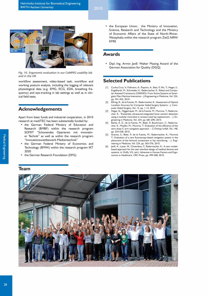
According to international standards such as IEC 62366 and ISO 14971 systematic usability engineering and risk manage-ment processes are required during the complete product development as well as in the marketing phase in order to increase usability and reliability of medical products. In med-ical applications the analysis and evaluation of risks for hu-man error are of increasing importance as more and more complex technical equipment is introduced into clinical as well as home care applications. In this context, the mAIX-use approach for human risk analysis (awarded in 2010 by the German Association for Quality (DGQ), see Fig. 1) has been successfully introduced into industrial application. The method can be used from early developmental stages up to validation of existing prototypes as well as in preclinical and clinical studies. Moreover, comparative evaluation of the us-ability of different products in a given clinical context of use is another scenario for the application of mAiXuse. Workshops and seminars on usability and risk evaluation methods as well as on the conduction of user based stud-ies (e.g. with the mAIXuse method) have been success-fully implemented for medical device manufacturers and hospitals.Our CeMPEG usability laboratory provides facilities for comprehensive user-centered interaction tests and
Med
ical
Eng
inee
ring
Fig. 15: A typical set-up in neuro-surgery.
28
2010Helmholtz-Institute for Biomedical EngineeringRWTH Aachen University
workflow assessment, video-based task, workflow and working posture analysis, including the logging of relevant physiological data (e.g. EMG, ECG, EDA, breathing fre-quency) and eye-tracking in lab settings as well as in clin-ical field tests.
Acknowledgements
Apart from basic funds and industrial cooperation, in 2010 research at mediTEC has been substantially funded by: • the German Federal Ministry of Education and
Research (BMBF) within the research program SOMIT “Schonendes Operieren mit innovativ-er Technik” as well as within the research program “Innovationswettbewerb Medizintechnik”
• the German Federal Ministry of Economics andTechnology (BMWi) within the research program IKT 2020
• theGermanResearchFoundation(DFG)
• the European Union, the Ministry of Innovation,Science, Research and Technology and the Ministry of Economic Affairs of the State of North-Rhine-Westphalia within the research program Ziel2.NRW/EFRE
Awards
• Dipl.-Ing. Armin Janß:WalterMasing Award of theGerman Association for Quality (DGQ)
Selected Publications [1] Cunha-Cruz, V.; Follmann, A.; Popovic, A.; Bast, P.; Wu, T.; Heger, S.;
Engelhardt, M.; Schmieder, K.; Radermacher, K.: Robot and Compu-ter Assisted Craniotomy (CRANIO): From Active Systems to Syner-gistic Man-Machine Interaction – J.Engineering in Medicine, Vol. 224, pp. 441-452, 2010.
[2] Elfring, R.; de la Fuente, M.; Radermacher, K.: Assessment of Optical Localizer Accuracy for Computer Aided Surgery Systems – J. Com-puter Aided Surgery, Vol. 15, pp. 1-12, 2010.
[3] Heger, St.; Niggemeyer, M.; de la Fuente, M.; Mumme, T.; Raderma-cher, K.: Trackerless ultrasound-integrated bone cement detection using a modular minirobot in revision total hip replacement. – J. En-gineering in Medicine, Vol. 224, pp. 681-690, 2010.
[4] Barbe, A. G.; de la Fuente, M.; Belei, P.; Buschmann, C.; Raderma-cher, K.; Mueller, M.; Mumme, T.: Evaluation of the efficiency of the zero-dose-C-arm navigation approach. – Z Orthop Unfall, Vol. 148, pp. 554-558, 2010.
[5] Gravius, S.; Belei, P.; de la Fuente, M.; Radermacher, K.; Mumme T.: Evaluation of a new fluoroscopy-based navigation system in the placement of the femoral component in hip resurfacing. – J. Engi-neering in Medicine, Vol. 224, pp. 565-576, 2010.
[6] Janß, A.; Lauer, W.; Chuembou; F.; Radermacher, K.: A new model-based approach for the user interface design of medical devices and systems. in: Duffy, V.G. (ed.): Advances in Human Factors and Ergo-nomics in Healthcare. CRC-Press, pp. 499-508, 2010.
Medical Engineering
Team
Fig. 16: Ergonomic evaluation in our CeMPEG usability lab and in the OR.