femoropopliteal-crural graft patency is improved by an intensive surveillance program: a prospective...
TRANSCRIPT

Femoropopliteal-crural graft patency is improved by an intensive surveillance program: A prospective randomized study Anders Lundell , MD, PhD, Bengt Lindblad, MD, PhD, David Bergqvist, MD, PhD, and Fleming Hansen, MD, Malmo, Sweden
Purpose: The purpose of this study was to evaluate whether intensive surveillance compared with routine follow-up examinations improves femoropopliteal/crural graft patency. Methods: After operation the patients were randomized to intensive (n = 79) or routine surveillance (n = 77). The groups were matched with regard to sex, diabetes, indication for surgical procedure, surgical procedure, and graft material. Intensive surveillance was clinical examination, ankle/brachial index measurements, and duplex scans 1, 3, 6, 9, 12, 15, 18, 21, 24, and 36 months after operation. Routine surveillance was clinical examination and ankle/brachial index measurements without duplex scanning 1, 12, 24, and 36 months after operation. Grafts with a decrease in ankle/brachial index of more than 0.15 compared with the initial postoperative ankle/brachial index or a duplex scan showing a graft or anastomotic stenosis of more than 50% underwent angiography and if necessary, a revision or repeat procedure. Occluded grafts were reopened with thrombectomy or thrombolysis or were replaced with a new graft. Results: Assisted primary cumulative vein graft patency in the intensive group (n = 56) compared with that in the routine surveillance group (n = 50) after 3 years was 78% versus 53% (chi square analysis, 4.51; one degree of freedom; p < 0.05). Secondary patency was 82% versus 56% (chi square analysis, 5.62; one degree offreedom;p < 0.05). Assisted primary cumulative e-polytetrafluoroethylene and composite graft patency after 1 year in the intensive group (n = 23) compared with that of the routine surveillance group (n = 20) was 57% vs 50% (chi square analysis, 2.17; one degree of freedom; p > 0.1). Secondary patency was 67% vs 54% (chi square analysis, 1.85; one degree of freedom; p > 0.1). Revisions were made on 14 patent and 10 thrombosed grafts in the intensive group and on four patent and 15 thrombosed grafts in the routine surveillance group. All except eight were made during the first postoperative year. Conclusions: Intensive surveillance identified failing vein grafts leading to a significantly higher cumulative assisted primary and secondary patency compared with cumulative assisted primary and secondary patency after routine follow-up examination. The patency of e-polytetrafluoroethylene and composite grafts was not influenced by intensive surveillance. (J VASC SURG 1995;21:26-34.)
The success o f a femorocrural reconstruction depends on the patient selection, the operative
From the Department of Surgery, Department of Clinical Physiology*% Maim6 General Hospital *Department of Sur- gery, Uppsala University Hospital (at time of the study Department of Surgery, Malta6 General Hospital)
Supported by grants from Swedish Medical Research Council 00759, Swedish Heart Lung Foundation, Hulda Almroth Foundation.
Presented at the Forty-eighth Annual Meeting of the Society for Vascular Surgery, Seattle, Wash., June 6-8, 1994.
Reprint requests: Anders Lundell, MD, PhD, Department of Surgery, Maim6 General Hospital, 21401 Malm6, Sweden.
Copyright © 1995 by The Society for Vascular Surgery and International Society for Cardiovascular Surgery, North Ameri- can Chapter.
0741-5214/95/$3.00 + 0 24/6/60826
26
procedure, and possibly postoperative surveillance. It is more difficult to restore patency in a thrombosed graft than to make a revision to maintain patency on a hemodynamically failing but still open graft. 1,2 The importance o f a postoperative surveillance program in detecting failing grafts has been stressed by others. 3,4 However, no prospective randomized study evaluating whether intensive postoperative surveillance actually leads to increased patency has been published.
Our aim with this study was to investigate in a prospective randomized series whether an intensive postoperative surveillance program compared with routine surveillance would assist in improving pa- tency after femorocrural reconstruction with regard

JOURNAL OF VASCULAR SURGERY Volume 21, Number I Lundell et al. 2 7
Table I. Patient material, presence of diabetes, and indication for operation
Intensive Routine
Age (yrs) 74 (66-81) 76 (66-81) Sex 40 Female 45 Female
39 Male 32 Male Diabetes 29/79 25/77 Intermittent claudication 2 2 Rest pain 33 29 Ischemic ulcer 23 31 Gangrene 16 14 Popliteal aneurysm 5 1
both to vein and e-polytetrafluoroethylene (ePTFE) and composite grafts.
MATERIAL AND M E T H O D S
Patients from the city of Maim6 undergoing a primary femoropopliteal or femorodistal reconstruc- tion from January 1,1988, to August 31, 1992, at the Department of Surgery, Malm6 General Hospital, included. Indication for surgical procedure was critical leg ischemia (presence of rest pain with a duration of more than 2 weeks, ischemic ulcer or gangrene of any part of the foot) or popliteal aneurysm. Patients with intermittent claudication underwent operation only if their symptoms were considered to diminish their quality of life. The preoperative examinations included an aortofemoral angiography, a chest x-ray examination, and an electrocardiographic examination. The patients un- derwent operation while they were under epidural anesthesia~ Autologous saphenous vein (reversed or in situ) was used as graft material for reconstructions with the distal anastomosis below knee level. Com- posite grafts (proximal part ePTFE and distal part autologous saphenous vein) were used when the length and quality of the available vein were insuffi- cient. For reconstructions with the distal anastomosis above knee level, ePTFE was used. After the recon- struction was completed and before the wound was closed, the reconstruction was assessed with a table angiogram and a handheld continuous Doppler scanner. A total of 6 gm antibiotic prophylaxis (Cloxacillin, Ekvacinin, As tra AB, Sweden) was given in three doses as intravenous infusion during the surgical procedure and at 12-hour intervals during the first 24 postoperative hours. Dextran (Macro&x, Pharmacia AB, Sweden) was used as a thrombosis prophylaxis; 1000 ml was administered during the surgical procedure and for the first 24 postoperative hours. The patients received a further dose of 500 ml dextran during the first to third postoperative days.
Table II. Type of reconstruction and graft material
Vein Reversed in situ vein Composite ePTFE
Intensive surveil- lance group Femoropopfiteal 1 1 0 15
above knee Femoropopliteal 29 6 2 2
below knee Femorodistal 15 4 3 1
Routine surveil- lance group Femoropopliteal 2 0 0 16
above knee Femoropopfiteal 28 3 1 6
below knee Femorodistal 13 4 4 0
Randomization to an intensive surveillance group or to a routine surveillance group was done after the surgical procedure. The study was approved by the Ethics Committee, University of Lurid, and patient consents were obtained.
Surveillance. Intensive surveillance was done during outpatient visits to a vascular surgeon 1, 3, 6, 9, 12, 15, 18, 24, and 36 months after the operation. These visits included clinical examination of the graft, an ankle/brachiat index (3331) measurement, and a duplex scan. Routine surveillance was conducted during outpatient visits to a vascular surgeon. These visits included clinical examination of the graft and measurement of ABI but did not include duplex scans. They were done 1, 12, 24, and 36 months after the operation. Surveillance data were registered in separate protocols. Data from each examination were sent to the study controller, who decided whether further investigations should be made.
Duplex. The duplex scans were performed by a vascular technician. A Diasonic CV 400 (Diasonics, Inc., Milpitas, Calif.) with a 7.5 MHz B-mode real- time scanner and a 4.5 MHz pulsed Doppler scanner were used, and after January 1990 an Acuson XP 10 (Acuson, Inc., Mountain View, Calif.) with a 5 MHz B-mode real-time scanner and a 3.5 MHz pulsed and color Doppler scanner were used. The whole length of the graft including the proximal and distal anastomosis was examined. A graft stenosis of more than 50% was thought to be present when the peak flow velocity exceeded 200 cm/sec at any part of the graft, s Peak flow velocities less than 45 cm/sec were considered to indicate an inflow or outflow vessel stenosis. 6 Graft stenoses less than 50% were not registered.
Angiography. Patients with signs of a failing
JOURNAL OF VASCULAR SURGERY 28 Lundell et al. lanuary 1995
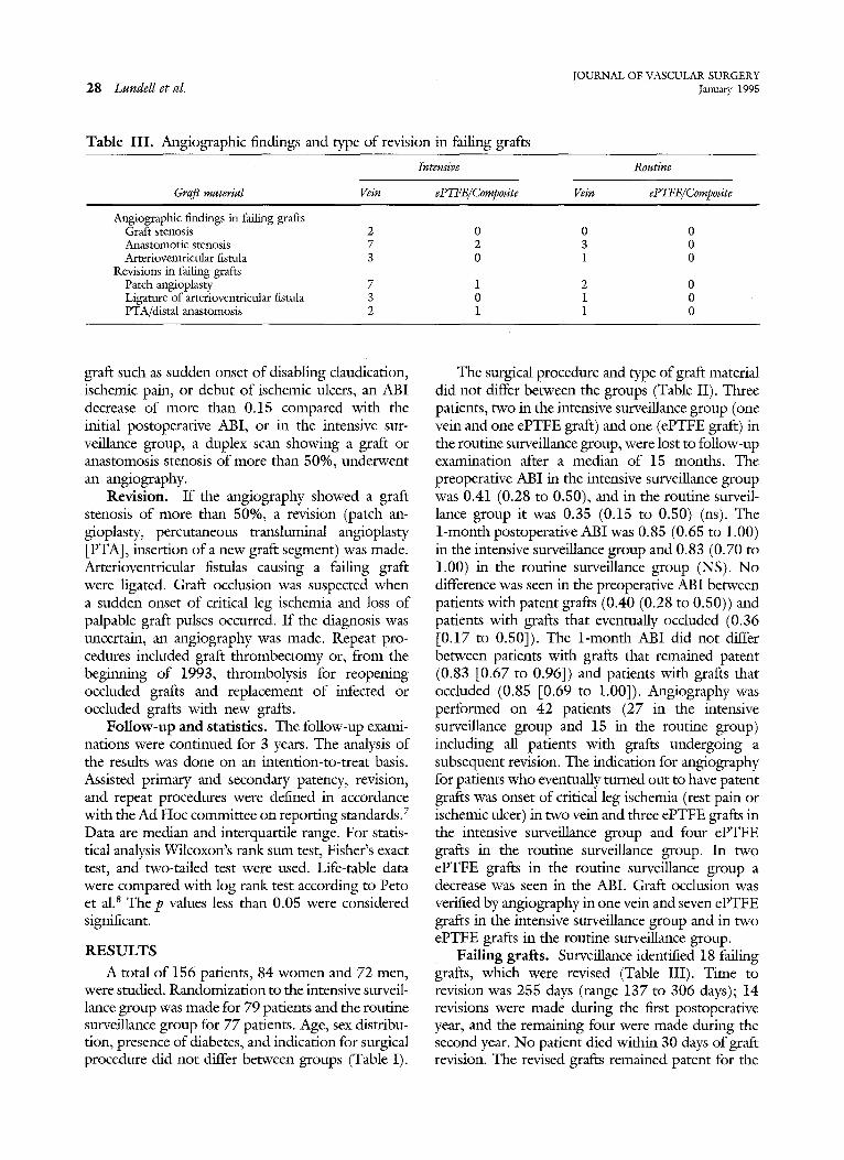
Table III. Angiographic findings and type of revision in Failing grafts
Intensive Routine
Graj~ material V e i n ePTFE/Composite V e i n ePTFE/Composite
Angiographic findings in failing grafts Graft stenosis Anastomofic stenosis Arterioventricular fistula
Revisions in failing grafts Patch angioplasty Ligature of arterioventricular fistula PTA/distal anastomosis
2 0 0 0 7 2 3 0 3 0 1 0
7 1 2 0 3 0 1 0 2 1 1 0
graft such as sudden onset of disabling claudication, ischemic pain, or debut of ischemic ulcers, an ABI decrease of more than 0.15 compared with the initial postoperative ABI, or in the intensive sur- veillance group, a duplex scan showing a graft or anastomosis stenosis of more than 50%, underwent an angiography.
Revision. If the angiography showed a graft stenosis of more than 50%, a revision (patch an- gioplasty, percutaneous transluminal angioplasty [PTA], insertion of a new graft segment) was made. Arterioventricular fistulas causing a failing graft were ligated. Graft occlusion was suspected when a sudden onset of critical leg ischemia and loss of palpable graft pulses occurred. If the diagnosis was uncertain, an angiography was made. Repeat pro- cedures included graft thrombectomy or, from the beginning of 1993, thrombolysis for reopening occluded grafts and replacement of infected or occluded grafts with new grafts.
Follow-up and statistics. The follow-up exami- nations were continued for 3 years. The analysis of the results was done on an intention-to-treat basis. Assisted primary and secondary patency, revision, and repeat procedures were defined in accordance with the Ad Hoc committee on reporting standards.7 Data are median and interquartile range. For statis- tical analysis Wilcoxon's rank sum test, Fisher's exact test, and two-tailed test were used. Life-table data were compared with log rank test according to Peto et al.s The p valucs less than 0.05 were considered significant.
RESULTS
A total of 156 patients, 84 women and 72 men, were studied. Randomization to the intensive surveil- lance group was made for 79 patients and the routine surveillance group for 77 patients. Age, sex distribu- tion, presence of diabetes, and indication for surgical proccdure did not differ between groups (Table I).
The surgical procedure and type of graft material did not differ between the groups (Table II). Three patients, two in the intensive surveillance group (one vein and one ePTFE graft) and one (ePTFE graft) in the routine surveillance group, were lost to follow-up examination after a median of 15 months. The preoperative ABI in the intensive surveillance group was 0.41 (0.28 to 0.50), and in the routine surveil- lance group it was 0.35 (0.15 to 0.50) (ns). The 1-month postoperative ABI was 0.85 (0.65 to 1.00) in the intensive surveillance group and 0.83 (0.70 to 1.00) in the routine surveillance group (NS). No difference was seen in the preoperative ABI between patients with patent grafts (0.40 (0.28 to 0.50)) and patients with grafts that eventually occluded (0.36 [0.17 to 0.50]). The 1-month ABI did not differ between patients with grafts that remained patent (0.83 [0.67 to 0.96]) and patients with grafts that occluded (0.85 [0.69 to 1.00]). Angiography was performed on 42 patients (27 in the intensive surveillance group and 15 in the routine group) including all patients with grafts undergoing a subsequent revision. The indication for angiography for patients who eventually turned out to have patent grafts was onset of critical leg ischemia (rest pain or ischemic ulcer) in two vein and three ePTFE grafts in the intensive surveillance group and four ePTFE grafts in the routine surveillance group. In two ePTFE grafts in the routine surveillance group a decrease was seen in the ABI. Graft occlusion was verified by angiography in one vein and seven ePTFE grafts in the intensive surveillance group and in two ePTFE grafts in the routine surveillance group.
Failing grafts. Surveillance identified 18 falling grafts, which were revised (Table III). Time to revision was 255 days (range 137 to 306 days); 14 revisions were made during the first postoperative year, and the remaining four were made during the second year. No patient died within 30 days of graft revision. The revised grafts remained patent for the

JOURNAL OF VASCULAR SURGERY Volume 21, Number i Lunde l l e t al. 29
Number of occlusions 13
11
10
9
8
7
6
5
4
3
2
1
0 t- n
Intensive Routine Intensive Routine surveilance vein surveillance vein surveillance surveillance
grafts grafts ePTFE+Composite ePTFE+Composite grafts grafts
Fig. 1. Most graft occlusions occurred during first postoperative year. Late failures were pPTFE and composite grafts.
duration of the follow-up period. Thirteen grafts (nine vein and two ePTFE in the intensive surveil- lance group and two vein grafts in the routine surveillance group) were found with a combination of ABI measurements and duplex scanning. Duplex examinations alone identified three failing vein grafts in the intensive surveillance group without any signs of decrease in ABI. Two patients with failing vein grafts in the routine surveillance group had a sudden onset of rest pain. In addition two graft infections occurred: one ePTFE graft in the intensive surveil- lance group and one in situ vein graft in the routine surveillance group. The infected grafts were replaced with new grafts.
Graft occlusion. A total of 65 grafts occluded: 27 in the intensive surveillance group and 38 in the routine surveillance group. Eleven vein graft occlu- sions were in the intensive surveillance group, and 20 were in the routine surveillance group. Most of the occlusions occurred during the first 12 postoperative months (Fig. 1).
Median time to graft occlusion from surgical procedure was 176 days (range 52 to 423 days) for both groups. The median time from the last outpa- tient visit to occlusion was 48 days (range 19 to 60 days) in the intensive surveillance group compared
with 113 days (range 40 to 223 days) in the routine surveillance group (p < 0.01). In three patients (with two vein and one ePTFE graft) in the intensive surveillance group a decrease was seen in ABI and a duplex-verified anastomotic stenosis, but before re- vision was made the grafts occluded. Two patients did not want to participate in the surveillance program after their 6-month visit, and one patient had a myocardial infarction and subsequent graft occlusion. For 21 graft occlusions (eight vein, three composite, and 10 ePTFE grafts) in the intensive surveillance group and for the occlusions in the routine surveillance group no cause for the occlusion was registered.
The occlusion was diagnosed clinically and veri- fied by angiography in 11 patients. Revisions on occluded grafts were made on 10 grafts in the intensive surveillance group and on 15 grafts in the routine surveillance group (Table IV). Most (20) of the revisions on occluded grafts were made during the first postoperative year, three were made during the second year, and two were made during the third postoperative year.
The revisions on occluded grafts were successful in seven (two vein and five ePTFE or composite) grafts in the intensive surveillance group and in three
JOURNAL OF VASCULAR SURGERY 30 Lundell et al. January 1995
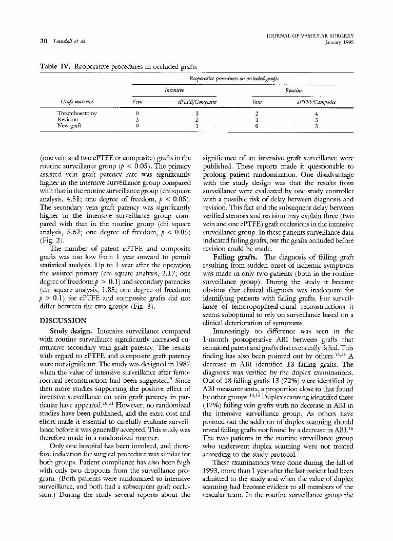
Table IV. Reoperative procedures in occluded grafts
Graft material Vein
Reoperative procedures on occluded grafts
Intensive Routine
ePTFE/Composite Vein ePTFE/Composite
Thromboectomy 0 3 2 4 Revision 2 2 3 3 New graft 0 3 0 3
(one vein and two ePTFE or composite) grafts in the routine surveillance group (p < 0.05). The primary assisted vein graft patency rate was significantly higher in the intensive surveillance group compared with that in the routine surveillance group (chi square analysis, 4.51; one degree of freedom, p < 0.05). The secondary vein graft patency was significantly higher in the intensive surveillance group com- pared with that in the routine group (chi square analysis, 5.62; one degree of freedom, p < 0.05) (Fig. 2).
The number of patent ePTFE and composite grafts was too low from 1 year onward to permit statistical analysis. Up to 1 year after the operation the assisted primary (chi square analysis, 2.17; one degree offreedom;p > 0.1) and secondary patencies (chi square analysis, 1.85; one degree of freedom; p > 0.1) for ePTFE and composite grafts did not differ between the two groups (Fig. 3).
D I S C U S S I O N
Study design. Intensive surveillance compared with routine surveillance significantly increased cu- mulative secondary vein graft patency. The results with regard to ePTFE and composite graft patency were not significant. The study was designed in 1987 when the value of intensive surveillance after femo- rocrural reconstruction had been suggested. 9 Since then more studies supporting the positive effect of intensive surveillance on vein graft patency in par- 6cular have appeared. ~°,11 However, no randomized studies have been published, and the extra cost and effort made it essential to carefully evaluate surveil- lance before it was generally accepted. This study was therefore made in a randomized manner.
Only one hospital has been involved, and there- fore indication for surgical procedure was similar for both groups. Patient compliance has also been high with only two dropouts from the surveillance pro- gram. (Both patients were randomized to intensive surveillance, and both had a subsequent graft occlu- sion.) During the study several reports about the
significance of an intensive graft surveillance were published. These reports made it questionable to prolong patient randomization. One disadvantage with the study design was that the results from surveillance were evaluated by one study controller with a possible risk of delay between diagnosis and revision. This fact and the subsequent delay between verified stenosis and revision may explain three (two vein and one ePTFE) graft occlusions in the intensive surveillance group. In these patients surveillance data indicated failing grafts, but the grafts occluded before revision could be made.
Failing grafts. The diagnosis of failing graft resulting from sudden onset of ischemic symptoms was made in only two patients (both in the routine surveillance group). During the study it became obvious that clinical diagnosis was inadequate for identifying patients with failing grafts. For surveil- lance of femoropopliteal-crural reconstructions it seems suboptimal to rely on surveillance based on a clinical deterioration of symptoms.
Interestingly no difference was seen in the 1-month postoperative ABI between grafts that remained patent and grafts that eventually failed. This finding has also been pointed out by others. 12,13 A decrease in ABI identified 13 failing grafts. The diagnosis was verified by the duplex examinations. Out of 18 failing grafts 13 (72%) were identified by ABI measurements, a proportion close to that found by other groups.14,1s Duplex scanning identified three (17%) failing vein grafts with no decrease in ABI in the intensive surveillance group. As others have pointed out the addition of duplex scanning should reveal failing grafts not found by a decrease in ABI. 16 The two patients in the routine surveillance group who underwent duplex scanning were not treated according to the study protocol.
These examinations were done during the fall of 1993, more than 1 year after the last patient had been admitted to the study and when the value of duplex scanning had become evident to all members of the vascular team. In the routine surveillance group the

JOURNAL OF VASCULAR SURGERY Volume 21, Number 1 Lundell et aL 31
Cumulative vein graft patency
1
0,9
0,8
0,7
0,6
0,5
0,4
0,3
0,2
0,1
q~
- i £ @ A A ,= v v v
Numbers a t r isk : 0 1 3 6 9 12 15 18 21 24 3 6 m o n t h s
[ ] Intensive survei l lance assisted primary patency 56 54 50 44 37 36 31 30 29 28 26
o Intensive survei l lance secondary patency 56 54 51 45 38 37 33 32 31 30 28
r3 Rout ine survei l lance assisted pr imary patency 50 46 41 37 30 28 27 24 24 22 16
o Rout ine survei l lance secondary patency 50 47 42 38 31 29 26 25 25 23 17
I I 1 I I I I I I 1 I 0 3 6 9 12 15 18 21 24 27 30 33
Fig. 2. Cumulative assisted and secondary vein graft patency was significantly higher in intensive surveillance group compared with routine surveillance group (chi square analysis, 4.51; one degree of fteedom;p < 0.05; and chi square analysis, 5.62; one degree of freedom; p < 0.05, respectively).
Months
1 36
long interval between examinations was probably the main reason for the low number of identified failing grafts. Surveillance based on clinical examination and measurement of ABI at short regular intervals will identify close to two thirds of all failing grafts, but the ultimate vein graft surveillance should be based on duplex examinations.
The canse for the occlusion was registered for six of the graft failures in the intensive surveillance group, but for the remaining occlusions in both groups no cause has been registered. Patients with vein grafts in the intensive surveillance group could have had borderline changes in the duplex examina- tions before the occlusion.
Only patients with a stenosis of more than 50%, which was indicated by a peak flow velocity less than 45 cm/sec or in some instances more than 200 cm/sec, underwent angiography and later graft revi- sion. Stenoses less than 50% were not registered. This attitude could be questioned, especially because the total number of graft occlusions in spite o f
intensive surveillance was high. It has been suggested that patency in grafts with stenoses less than 50% is improved by revision. 17 We found that l0 of the 12 vein graft stenoses were located at the distal anasto- mosis, and only two were in the graft itself. This finding is in contrast to the findings of other groups, where most stenoses were located in the graft) 8 One plausible explanation for this discrepancy could be that the duplex examinations during the first 2 years were done without color coding. In spite of intensive surveillance, failing ePTFE or composite grafts were not identified. The lack of influence of intensive surveillance on ePTFE or composite graft patency is reflected in the cumulative patency rates, which did not differ between the groups. Patients undergoing operation with ePTFE or composite grafts should perhaps be submitted to a surveillance program not based on duplex examinations) 9 Digital subtraction angiography performed at regular postoperative intervals has been suggested to be a better alter- native. 2°

JOURNAL OF VASCULAR SURGERY 32 Lundell et al. lanuary 1995
1
0,9
0,8
0,7
0,6
0,5
0,4
0 ,3
0,2
0,1
0
Cumula t ive ePTFE and Compos i t e graft patency
i . . . . ,
IiL_o._.__ I,, I . . . . . . Iq o - -- - ¢ - . . . . . . - ] 8 % _ _ _
Numbers at risk:
o Intensive surveillance assisted primary patency
* Intensive surveillance secondary patency • Routine surveillance assisted primary patency [] Routine surveillance secondary patency
I I I I i 3 6 9 12 15
I I I
LA...~
0 1 3 6 9 12 15 18 21 24 3 6 m o n t h s
23 21 19 16 14 12 9 8 6 6 3
23 22201817 14 11 10 8 8 2 27 23201716 13 11 11 9 8 5
27 24 21 18 17 15 13 13 11 10 7
I i t I 18 2I 24 27
I I i [
[ I I-"
i t_ I
--t.-.~-.----..-..---~
Months
t I I 30 33 36
Fig. 3. No difference in patency was seen between ePTFE and composite grafts in intensive surveillance group compared with routine surveillance group. From 1 year onward number of patent grafts was too small to permit statistical analysis.
Occluded grafts. The significant difference of successful revisions on occluded grafts between the two groups seems to be due to the fact that the graft occlusions in the routine group were diagnosed too late to permit a successful revision. This occurrence is reflected in a significantly longer time from the preceding outpatient visit to occlusion in the routine group compared with that of the intensive surveil- lance group. It seems that intensive surveillance not only identifies failing grafts but also makes a higher successful revascularization rate possible after an occlusion. This finding can be partly explained by the shorter interval between the outpatient visit and graft occlusion in the intensive group compared with that of the routine surveillance group.
Revisions. The preferred procedure for graft revision was patch angioplasty. The results have been encouraging with no postoperative deaths and patent grafts for the duration of the follow-up period. Our experiences with PTA are limited, but the results from this study seem equal to those of patch angioplasty. We do therefore use PTA more often now for correction of vein graft stenoses, particularly
because our radiologists are becoming more familiar with the technique. However, the possibility of a restenosis after PTA has to be considered. 16 For ePTFE graft stenosis caused by intimal hyperplasia in the anastomosic regions, we still consider patch angioplasty the method of choice.
Experiences. Most of the failing grafts and graft occlusions in the study were found during the first year. Based on the results from this study the examinations in our surveillance program are con- centrated to the first postoperative year. Outpatient visits include duplex and ABI measurements 1, 3, 6, 9, 12, and 24 months after the surgical procedure, which is in accordance with other suggested regi- mens. 21 Both vein and ePTFE and composite grafts are examined. The ABI measurements and the clinical examination are done by an outpatient nurse, and the duplex examinations are done by a vascular techni- cian. I f the examinations show signs of a failing graft including a suspect graft stcnosis less than 50%, the patient is examined by a vascular surgeon who decides whether the interval to the next duplex examination should be shortened or an angiography

JOURNAL OF VASCULAR SURGERY Volume 21, Number 1 Lundell et al. 33
should be done. I t has been reported that more failing grafts migh t be identified, if the intervals between visits were only 6 weeks for the first 6 months . 22 However , a risk exists that the advanced age o f the patients and their l imited mobil i ty in spite o f patent grafts and viable fimbs could make such a p rog ram impractical. O n the other hand the consequences o f a graft occlusion for this g roup o f patients are severe, making it impor tan t to have an opt imal surveillance to maintain femorocrural graft patency. 23
Intensive surveillance based on ABI and duplex examinations at short intervals dur ing the first and second postoperat ive year identified failing vein grafts, which led to a significantly higher vein graft patency after revision compared wi th patency after routine fol low-up examinations. The patency o f ePTFE and composi te grafts was no t influenced by intensive surveiUance.
KEFEKENC, ES 1. Bandyk DF, Kaebnick HW, Stewart GW, Towne JB. Dura-
bility of in situ saphenous vein bypass: a comparison of primary and secondary patency. J VASC SURG 1987;5:256-68.
2. Veith F, Gupta SK, Ascer E. Improved strategies for secondary operations on infrainguinal arteries. Ann Vasc Surg 1990;3:85-93.
3. Turnipsed WD, Acher CW. Postoperative surveillance: an effective means of detecting correctable lesions that threaten graft patency. Arch Surg 1985;120:324-8.
4. Berkowitz HD, Hobbs CL, Roberts B, et al. Value of routine vascular laboratory studies to identify vein graft stenosis. Surgery 1981;90:971-9.
5. Cossman DV, Ellison IE, Wagner WH, et al. Comparison of contrast arteriography to arterial mapping with color-flow duplex imaging in the lower extremities. J VASC SURG 1989;10:522-9.
6. Bandyk DF, Seabrook GR, Moldenauer P, et al. Hemody- namics of vein graft stenoses. J VASC SURG 1988;8: 688-95.
7. Rutherford RB, Hanigan DP, Gupta SK, et al. Suggested standards for reports dealing with lower extremity ischemia. J VASC SURG 1986;4:80-94.
8. Peto R, Pike MC, Armitage P, et al. Design and analysis of randomized clinical trials ~equiring prolonged observation of each patient. Br J Cancer 1977;35:1-39.
9. Bandyk DF, Cato RF, Towne JB. A low blood flow velocity
predicts failure of femoropopliteal and femorodistal bypass grafts. Surgery 1985;98:799-809.
10. Grigg MJ, Nicolaides AN, Wolfe JHN. Femorodistal vein graft stenoses. Br J Surg 1988;75:737-40.
11. Green RM, MacNamara J, Oufiel K, de Weese JA. Compari- son of infrainguinal graft surveillance techniques. I VAsc SVRG 1990;1i:207-15.
I2. Barnes RW, Thomson BW, McDonald CM, et al. Serial ' noninvasive studies do not herald postoperative failure of femoropopliteal or femorotibial bypass grafts. Surgery 1989; 106:486-94.
13. Prime HT, Myers KA, Matthews PG, Zeng G. Early postoperative pressure indices help predict late patency rates after femoro-distal vein bypass grafting. J Cardiovasc Surg 199;33:426-31.
14. Berkowitz HI), Greenstein SM. Improved patency in reversed femoro-infrapopriteal autogenous vein grafts by early detec- tion and treatment of the fairing graft. I VAsc SURG 1987;5:755-61.
15. Brennan IA, Walsh AKM, Beard JD, Bolla AA, Bell PRF. The role of simple noninvasive testing in infrainguinal vein graft surveillance. Eur J Vasc Surg 1991 ;5:13-7.
16. Cheshire NJW, Wolfe JHN. Infrainguinal graft surveillance: a biased overview. Semin Vasc Surg 1993;6:143-9.
17. Moody P, Cossart LM, Douglas HM, Harris PL. Asymptom- atic strictures in femoropopllteal vein grafts. Eur J Vasc Surg I989;3:389-92.
18. London NM, Sayers RD, Thomson MM, Naylor A R , Hartshorne T, Ratliff DA, Bell PRF, Boria A. Interventional radiology in the maintenance ofinfrainguinal graft patency. Br J Surg 1993;80:187-93.
I9. von Jessen F, Rordam P, Sillesen HH, Schroeder TV. Postoperative duplex ultrasonic surveillance of infrainguinal arterial reconstructions. Ugeskr Laeger 1992;154:1402-6.
20. GriggMJ, NicolaidesAN, WolfeJHN. Detection and grading of femorodistal vein graft stenoses: duplex velocity measure- ments compared with angiography. I VASC SUV, G 1988;8: 661-6.
21. Bandyk DF, Schmitr DD, Seabrook GR, Adams MB, Towne lB. Monitoring functional patency of in sire saphenous vein bypasses: the impact of a surveillance protocol and elective revision. J VASC SURG 1989;9:286-92.
22. Taylor P, Wolfe J, Tyrell M, et al. Graft stenosis: justification for 1 year surveillance. Br J Surg 1990;77:1125-8.
23. Harris PL. Vein graft surveillance-all part of the service. Br J Surg 1992;79:97-8.
Submitted lune 10, 1994; accepted Sept. 24, 1994.
DISCUSSION Dr. Dennis F. Bandyk (Tampa, Fla.). Previous studies
deafing with infrainguinal graft surveillance were prospec- tive cohort trials but lacked an appropriate control group. The study conducted by Dr. Lnndell and his colleagues is the first randomized clinical trial reported. Both vein and PTFE grafts were studied, and the intensive surveillance protocol included color duplex scanning. Duplex scanning
was compared with clinical assessment and measurements of ankle systolic pressure. The study outcome confirmed duplex surveillance increased secondary and cumulative graft patency compared with clinical evaluation, but the benefit was observed only after vein bypass grafting. Intensive surveillance did not improve the patency of PTFE or PTFE composite grafts. Overall the graft patency rates
JOURNAL OF VASCULAR SURGERY 34 Lundell et aL ~lanuary 1995
of this study were comparable to other recent reports of infrainguinal bypass in patients with critical limb ischemia. Randomization of patients was performed after operation, but graft surveillance was not initiated until a month later. Thus the influence of duplex surveillance on early graft failure was not examined, and indeed the 30-day graft
• failure rates were the same in both study groups. A novel feature of this study was the use of a study
controller who reviewed the data from each study group and then decided what further investigations were neces- sary. This aspect of the study design introduced delay in the evaluation of patients who were identified to have failing grafts. All abnormal grafts were subjected to angiographic confirmation of the duplex-identified abnormality before revision. In the article the authors admit the delay introduced by this process accounted for approximately 20% of the vein graft failure in the intensive surveillance group. Given that the duplex criteria were used to identify a graft stenosis, I was surprised that a greater number of abnormalities were not detected, particularly at the 1- and 3-month scan times. Since 42 patients had postoperative arteriography, I would like to ask the authors whether the accuracy of duplex scanning was calculated. How often were intrinsic graft stenoses or anastomotic abnormalities not detected by duplex scanning?
The number of graft occlusions that developed despite surveillance was also a concern. When changes in graft function were detected, and duplex scanning identified a stenosis, the surveillance protocol was not altered. For lesions that did not meet the criteria for revision, progres- sion to graft thrombosis may have occurred before the next scheduled visit. How did the authors follow lesions with borderline criteria for further evaluation or revision? Of note, 77% of vein graft occlusions and two thirds of PTFE graft occlusions occurred within 9 months of the proce- dure, and this incidence was not decreased by intensive surveillance. The earliest graft revision was undertaken at 4.5 months. This outcome varies from my experience with duplex surveillance of in situ saphenous vein bypass where 10% of revisions were performed within 1 month of operation, and one third occurred within 3 months.
I would like the authors to comment on whether they observed a learning curve in the application of duplex surveillance in this study. I have found that with time our technologists and the surgeons have improved the scanning
technique and interpretation of duplex graft examinations. Do the authors believe the observed incidence of graft occlusion was the result of failed graft revision proce- dures or inadequate graft surveillance, for example, too infrequent testing schedule or improper duplex criteria for intervention? Finally, given the results for postoperative surveillance of PTFE grafts, what are the authors' recom- mendations regarding follow-up of prosthetic bypasses?
Dr. Anders Lundell. The study was designed in 1987 and started in 1988. At that time we had limited experience in graft surveillance, so there is a considerable learning curve in the study. Duplex scanning identified 14 stenoses, and angiography confirmed 12, which I think is a fairly high accuracy for duplex scanning.
When we found duplex abnormalities indicating bor- derline stenosis, we adhered to the protocol, and action was not taken until there was a graft stenosis of more than 50%.
The main cause for graft occlusions must have been inadequate graft surveillance. I did not mention it in the speech, but all our revisions have been successful, with no postoperative deaths after revision, and the grafts have remained patent for the duration of follow-up.
The results with regard to ePTFE and composite grafts have given us great concern. Duplex scanning did not identify failing grafts, and neither did ABI. The patients had a sudden occlusion that came without any preceding symptoms. We are now discussing whether we should change the surveillance, not making duplex scans but perhaps instead digital subtraction angiography, but then you have the cost problem. You must also take into consideration that there was a steady decline of patent ePTFE grafts. The majority of vein graft occlusions were during the first postoperative year, whereas the ePTFE graft occlusions occurred from the first to the third postoperative year. That means that we shall perhaps have a program that follows these grafts for a longer period than 3 years.
Dr. Norman R. Hertzer (Cleveland, Ohio). I might just comment that, as those of you in the United States know, the Health Care Financing Administration has been reluctant to reimburse for ongoing surveillance of synthetic grafts, whereas it will reimburse for ongoing surveillance of autogenous grafts. This work tends to support that conclusion.