fine structure of e. coli rna polymerase-promoter
TRANSCRIPT
Fine structure of E. coli RNApolymerase-promoter interactions:� subunit binding to the UP elementminor grooveWilma Ross,1 Alexander Ernst,2,3 and Richard L. Gourse1,4
1Department of Bacteriology, University of Wisconsin-Madison, Madison, Wisconsin 53706, USA; 2Departmentof Chemistry and Chemical Biology, Harvard University, Cambridge, Massachusetts 02138, USA
The � subunit of E. coli RNAP plays an important role in the recognition of many promoters by binding tothe A+T-rich UP element, a DNA sequence located upstream of the recognition elements for the � subunit,the −35 and −10 hexamers. We examined DNA–RNAP interactions using high resolution interference andprotection footprinting methods and using the minor groove-binding drug distamycin. Our results suggest that� interacts with bases in the DNA minor groove and with the DNA backbone along the minor groove, butthat UP element major groove surfaces do not make a significant contribution to � binding. On the basis ofthese and previous results, we propose a model in which � contacts UP element DNA through amino acidresidues located in a pair of helix–hairpin–helix motifs. Furthermore, our experiments extend existinginformation about recognition of the core promoter by �70 by identifying functional groups in the majorgrooves of the −35 and −10 hexamers in which modifications interfere with RNAP binding. These studiesgreatly improve the resolution of our picture of the promoter–RNAP interaction.
[Key Words: RNA polymerase; promoter; alpha subunit; UP element; minor groove; helix–hairpin–helix]
Received November 29, 2000; revised version accepted January 16, 2001.
Promoters in bacteria contain recognition sequences forRNA polymerase (RNAP, subunit composition �2�����)in up to three distinct regions: the −10 and −35 elements,both recognized by the �70 subunit (for review, see Rec-ord et al. 1996; Gross et al. 1998), and the UP element,recognized by the � subunit (for review, see Gourse et al.2000). The UP element, located just upstream of the −35hexamer, was identified as a binding site for � in therRNA promoter rrnB P1 of Escherichia coli, where itstimulates promoter activity ∼30-fold (Ross et al. 1993).UP elements have been identified in other promoters inE. coli, in other bacterial species, and in promoters tran-scribed by holoenzymes containing alternative � factors(e.g., Moran et al. 1982; Newlands et al. 1992; Ross et al.1993, 1998 Fredrick et al. 1995; Negre et al. 1997). Theoptimal (consensus) UP element sequence was identifiedby in vitro selection and contains alternating A and Ttracts in two distinct subsites (proximal and distal; Es-trem et al. 1998, 1999). Analysis of the E. coli genomesequence suggests that UP elements, consisting of either
one or two subsites with no more than two mismatchesto consensus, occur in more than one-third of stableRNA promoters, but are also found in about 4% ofmRNA promoters (Estrem et al. 1999). Because se-quences with much lesser degrees of match to consensuscan still significantly affect promoter function (Ross etal. 1998), the number of promoters in which UP ele-ments play some role in transcription is likely to bemuch larger. DNA–� interactions also occur adjacent tomany activator protein-binding sites, although, in thesecases, � interactions are often DNA sequence indepen-dent, the binding site(s) do not resemble the UP elementconsensus, and � binding does not occur in the absenceof the bound activator (Hochschild and Dove 1998;Busby and Ebright 1999; Gourse et al. 2000).
The ∼37 kD � subunit contains two independentlyfolded domains (Blatter et al. 1994). The amino-terminaldomain (residues 8–233) is responsible for dimerizationand interactions with � and �� (Hayward et al. 1991), andthe carboxy-terminal domain (�CTD, residues 245–329)contains the DNA-binding determinants (Blatter et al.1994). Genetic analyses have identified amino acid resi-dues in the �CTD critical for DNA binding and UP ele-ment function (Gaal et al. 1996; Murakami et al. 1996).These residues are essential for cell viability (Gaal et al.1996) and are highly conserved in prokaryotic � se-
3Present address: Medicinal Chemistry, Preclinical Drug Research,Schering AG, D-13342 Berlin, Germany.4Corresponding author.E-MAIL [email protected]; FAX (608) 262 9865.Article and publication are at www.genesdev.org/cgi/doi/10.1101/gad.870001.
GENES & DEVELOPMENT 15:491–506 © 2001 by Cold Spring Harbor Laboratory Press ISSN 0890-9369/01 $5.00; www.genesdev.org 491
Cold Spring Harbor Laboratory Press on April 7, 2022 - Published by genesdev.cshlp.orgDownloaded from
quences. Amino acid sequence comparisons in conjunc-tion with the solution structure of the �CTD, deter-mined by nuclear magnetic resonance spectroscopy (Jeonet al. 1995; Gaal et al. 1996), suggest that � is a memberof the helix–hairpin–helix (HhH) domain family of DNA-binding proteins (Shao and Grishin 2000).
Defining the interactions between components of thetranscription initiation apparatus remains a central prob-lem in molecular biology. Recently, there have been dra-matic advances in our understanding of the structure ofbacterial core RNAP at the atomic level (Zhang et al.1999), but the �CTD was not resolved in this structure,and no information at the atomic level exists for com-plexes containing DNA bound to � or RNAP holoen-zyme.
Footprinting and cross-linking studies have providedsome information about the �–UP element interactionand the promoter–RNAP interaction. Footprints ofRNAP on rrnB P1 and other promoters extend about 60bp upstream of the transcription start site, and protec-tion upstream of ∼−40 is attributable to interactions with� (Kolb et al. 1993; Ross et al. 1993). In the rrnB P1 UPelement, DNA backbone regions centered at ∼−42 and∼−52 are protected against hydroxyl radical cleavage by �or RNAP (Newlands et al. 1991; Estrem et al. 1998).However, a full understanding of the mode of � bindinghas been hindered by complicating features of its inter-actions with different promoter upstream regions. Notonly can � bind to DNA in either a sequence-specific ora sequence-nonspecific manner, but �-dependent protec-tion signals have also been observed upstream of −60,and sometimes multiple regions of protection have beenobserved in the same upstream region (Newlands et al.1992; Kolb et al. 1993; Fredrick et al. 1995; Belyaeva et al.1998). Comprehensive cross-linking studies further sug-gest that the two �CTDs may oscillate among differentbinding sites (like fingers on a keyboard; Naryshkin et al.2000), or alternatively may bind to different sites in dif-ferent molecules in the population. Variability in theposition(s) of � binding sites is most likely attributableto the long, flexible tether linking the � amino- and car-boxy-terminal domains (Blatter et al. 1994; Jeon et al.1997), as well as to intrinsic DNA distortions and pro-tein-induced DNA bending in initiation complexes con-taining activators (Gourse et al. 2000; Naryshkin et al.2000).
A binding site for a single � subunit (the consensusproximal subsite; see Fig. 1A) was defined by in vitroselection and by the use of RNAPs heterodimeric for �;[i.e., containing one wild-type � subunit and one � lack-ing its DNA-binding domain (��CTD; Estrem et al.1999)]. Only one �CTD was required for function of anUP element with only a proximal subsite and for protec-tion of this subsite in hydroxyl radical footprints. Theprotected positions (about 4 bp on each DNA strand) areoffset on the two strands by ∼3 bp, and are thus locatedin close proximity across the DNA minor groove (Estremet al. 1999). Likewise, studies with a cross-linking agenttethered by a 10-Å linker indicated that �CTD interac-tions with a promoter upstream region lacking a func-
tional UP element are centered on the DNA minorgroove (Naryshkin et al. 2000). However, the footprint-ing and cross-linking studies do not provide sufficient
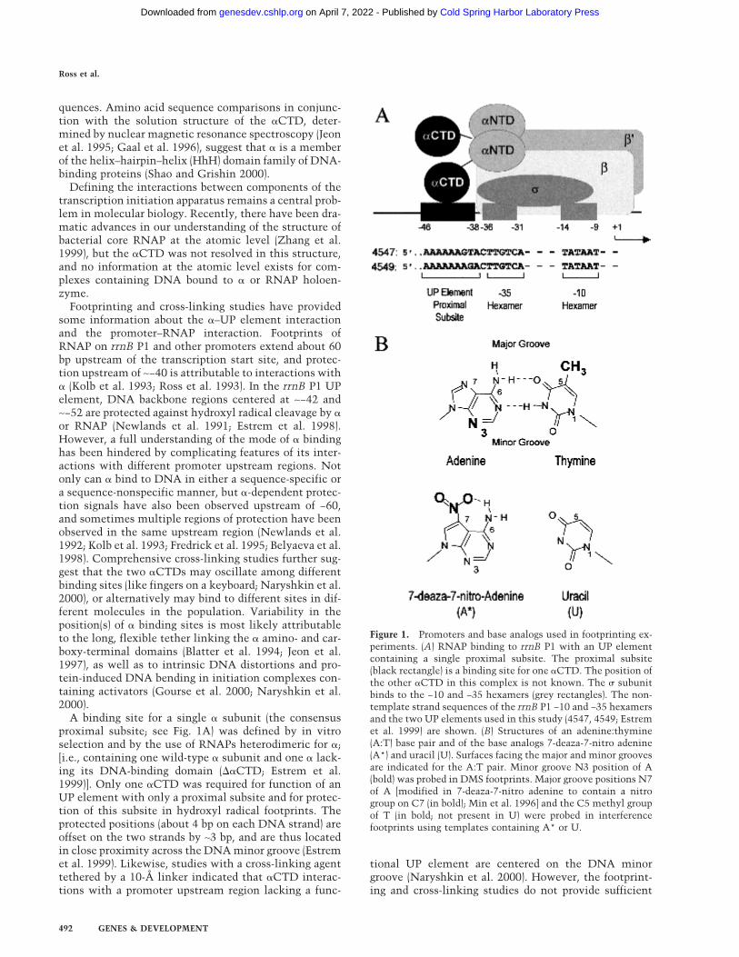
Figure 1. Promoters and base analogs used in footprinting ex-periments. (A) RNAP binding to rrnB P1 with an UP elementcontaining a single proximal subsite. The proximal subsite(black rectangle) is a binding site for one �CTD. The position ofthe other �CTD in this complex is not known. The � subunitbinds to the −10 and −35 hexamers (grey rectangles). The non-template strand sequences of the rrnB P1 −10 and −35 hexamersand the two UP elements used in this study (4547, 4549; Estremet al. 1999) are shown. (B) Structures of an adenine:thymine(A:T) base pair and of the base analogs 7-deaza-7-nitro adenine(A*) and uracil (U). Surfaces facing the major and minor groovesare indicated for the A:T pair. Minor groove N3 position of A(bold) was probed in DMS footprints. Major groove positions N7of A [modified in 7-deaza-7-nitro adenine to contain a nitrogroup on C7 (in bold); Min et al. 1996] and the C5 methyl groupof T (in bold; not present in U) were probed in interferencefootprints using templates containing A* or U.
Ross et al.
492 GENES & DEVELOPMENT
Cold Spring Harbor Laboratory Press on April 7, 2022 - Published by genesdev.cshlp.orgDownloaded from
information to build a structural model of the sequence-specific �CTD–UP element interaction; for example,proteins that bind to two successive major grooves arealso centered on the minor groove. The previous studiesdo not distinguish whether the �CTD makes base con-tacts within a DNA groove, or whether these contacts, ifthey occur, are with the major groove or minor groovesurfaces of the bases.
In this work, we address the basis for the specific in-teraction of � with UP element DNA. An understandingof bacterial promoter recognition and the architecture oftranscription complexes containing activators requiresdetailed information about the �CTD–DNA interaction.Therefore, we carried out RNAP protection footprintingstudies with dimethylsulfate (DMS) and interferencefootprinting studies with promoter fragments containingthe thymine analog uracil or the adenine analog 7-deaza-7-nitro-adenine (A*; Fig. 1B). We also examined the ef-fects of the minor groove-binding drug distamycin on �binding and on UP element-dependent transcription.These results and those from hydroxyl radical footprint-ing studies (Estrem et al. 1999) indicate that � binds tothe DNA backbone across the minor groove and to baseswithin the minor groove. Together with genetic analysesof �CTD (Gaal et al. 1996; Murakami et al. 1996) and therecent proposal that � contains two HhH DNA bindingmotifs [an (HhH)2 domain; Shao and Grishin 2000)], ourdata suggest a model for the interaction of � with UPelement DNA. In addition, our experiments provide de-tailed information about the proximity of the RNAP �subunit to the major groove surface of thymine and ad-enine bases in the −35 and −10 hexamers.
Results
We carried out footprinting studies of RNAP on tem-plates containing the rrnB P1 core promoter fused to UPelements consisting of only a single proximal subsite(UP elements 4547 or 4549; Estrem et al. 1999; see Fig.1A). UP elements containing only a proximal subsite arethe most frequent class of UP elements in the E. coligenome (Estrem et al. 1999). Most important, use of tem-plates with only a proximal subsite simplifies interpre-tation to a single DNA–� interaction.
The UP elements used in these experiments are repre-sentative of the proximal subsite sequences identified byin vitro selection and can stimulate promoter activity>100-fold in vivo. Each contains an A tract extendingfrom −46 to −41 but has a different sequence at positions−40 and −39 (Estrem et al. 1999). The region upstream ofthe proximal subsite in these promoters is not protectedby RNAP and does not affect promoter function (Estremet al. 1999).
Missing bases in the UP element interfere with RNAPbinding
Limited information was available about the relative im-portance of individual positions in the UP elements, and
no information was available about whether bases on thenontemplate strand, template strand, or both make con-tacts with �. As a first approach to understanding therole of specific bases in proximal subsite function, wecarried out missing base interference footprinting(Brunelle and Schleif 1987). This method allows the im-portance of a single base at any promoter position to bedetermined by the selective depletion of templates lack-ing that base from the population of fragments able toform open complexes. Modified promoter fragmentpopulations that contained on average one missing thy-mine per molecule were prepared by low frequency sub-stitution of dUTP for TTP (Fig. 1B) followed by removalof the uracil base by use of uracil DNA glycosylase(UDG; Devchand et al. 1993). Only the base was re-moved, leaving the phosphodiester backbone intact.
Labeled promoter fragments containing missing baseswere then incubated with RNAP. Because the �–UP el-ement interaction increases the rate of association of thepromoter with RNAP (Rao et al. 1994), the RNAP bind-ing reaction was carried out at low RNAP concentrationand in 20-fold molar excess of modified promoter frag-ment relative to RNAP, conditions in which complexformation was strongly dependent on the �–UP elementinteraction. The resulting promoter–RNAP complexeswere separated from unbound DNA by electrophoresison a nondenaturing gel, purified, subjected to strandcleavage at the abasic sites, and then fractionated on ahigh resolution denaturing gel. Missing bases that inter-fered with RNAP binding were under-represented in thestrand cleavage profiles of bound DNA relative to theirrepresentation in the DNA population that was not in-cubated with RNAP.
A typical missing T interference experiment carriedout on a promoter with a consensus proximal subsite isshown in Figure 2. Each of the thymine positions in theproximal subsite (eight of nine positions on the templatestrand) was severely depleted in the RNAP-boundsample, indicating that a missing T at each of eight po-sitions in the UP element interfered with RNAP binding.The strongest interference signals occurred in the centerof the T tract (−41 to −45), whereas missing Ts upstreamof the UP element sequence did not interfere with RNAPbinding (Fig. 2 and data not shown). Identical resultswere obtained with an RNAP preparation containingonly one �CTD (�I/��II heterodimeric RNAP; data notshown), indicating that, as expected from previous work(Estrem et al. 1999), missing Ts interfering with wild-type RNAP binding to the proximal subsite are attribut-able to interactions involving only one � subunit. Nointerference signals in the UP element were observedwith RNAP lacking both �CTDs (data not shown).
Missing Ts at some positions on each strand in thecore promoter region also strongly affected RNAP bind-ing (Fig. 2 and data not shown; summarized in Fig. 6,below). All missing Ts in the −35 hexamer and some inthe spacer region between the −10 and −35 hexamersreduced RNAP binding. In contrast, most missing Ts inthe −10 hexamer strongly stimulated RNAP binding (allbut nontemplate strand position −9), most likely because
RNAP � subunit binding to the DNA minor groove
GENES & DEVELOPMENT 493
Cold Spring Harbor Laboratory Press on April 7, 2022 - Published by genesdev.cshlp.orgDownloaded from

unpaired nucleotides facilitate strand opening (see Fig. 2and Discussion).
Missing purine interference experiments (data notshown) with templates prepared by limited acid depuri-nation (Maxam and Gilbert 1980), were used to examinethe interaction of RNAP with the nontemplate strand.The conclusions from these experiments were similar tothose from the missing T experiments: missing bases atthe eight UP element positions that are purines, and atpurine positions in the spacer, but not at control posi-tions further upstream, resulted in reduced RNAP bind-ing (for summary, see Fig. 6, below). Missing purines atpositions −41 and −42 reduced RNAP binding the most.As reported previously (Li and McClure 1998), and asfound in the missing T experiments, most missing pu-rines in the −10 region (all but nontemplate strand −13)stimulated RNAP binding.
Major groove modifications affect RNAP bindingto the core promoter but not to the UP element
The missing-base interference experiments indicatedthat each base in the UP element proximal subsite was
important for � binding and suggested that bases in thecenter of the subsite (−41 to −45) are most critical. How-ever, these results did not distinguish which base sur-faces (major groove, minor groove, or both) are contactedby �, or alternatively, whether the interference signalsmight derive solely from indirect effects of missing baseson DNA backbone structure.
To determine whether � makes base contacts in themajor groove, interference footprinting was carried outwith promoter fragments containing adenine or thyminebases modified on their major groove surfaces. Modifica-tions were introduced by limited incorporation of thenucleotide analogs dUTP or dA*TP (7-deaza-7-nitrodATP; Fig. 1B). U is identical to T except that it lacks theprominent major groove C5 methyl group. A* (Min et al.1996) disrupts contacts to adenine at position 7 throughsteric exclusion and at position 6-NH through intramo-lecular hydrogen bonding. Both U and A* could poten-tially disrupt protein binding directly through loss offunctional groups that contact amino acids, but, in addi-tion, the adenine modification could potentially disruptprotein binding by steric hindrance. dUTP or dA*TPhave not to our knowledge been used for footprints ofRNAP, although they have been used in footprintingstudies of other proteins (Pu and Struhl 1992; Devchandet al. 1993; Min et al. 1996).
To carry out dUTP interference footprints, RNAP–pro-moter complexes were formed with templates contain-ing, on average, one U substitution per molecule (Fig.3A,B). Complexes were then gel isolated, and the DNAwas purified, subjected to strand cleavage at modifiedbases, and fractionated on a denaturing gel. Uracil sub-stitution at any of the eight thymine positions in the UPelement had very little effect on RNAP binding (tem-plate strand, Fig. 3B), suggesting that � does not makecritical contacts with major groove thymine methylgroups in the UP element. In contrast, U substitutions atany of three core promoter positions (−36, −35, and −14)strongly reduced or eliminated RNAP binding, and Usubstitutions at either of two other core promoter posi-tions (−31 and −9) also reduced RNAP binding (Fig. 3A,B;for summary, see Fig. 6, below).
Similar experiments were carried out with promoterfragments containing A* substitutions (Fig. 3C,D). As inthe U substitution experiments, A* at UP element posi-tions had little or no effect on RNAP binding (nontem-plate strand, Fig. 3C). However, A* substitutions hadlarge effects on RNAP interactions with the core pro-moter. A* substitutions reduced binding at five of the sixA residues in the two strands of the −10 hexamer (Fig.3C,D; for summary, see Fig. 6, below); A* at nontem-plate strand position −10 virtually eliminated binding ofRNAP, and at template strand position −33 (in the −35hexamer) strongly enhanced RNAP binding (see Discus-sion).
In summary, the U and A* interference experimentsindicate that major groove base surfaces in the UP ele-ment play little if any direct role in RNAP binding, al-though they do play a major role in interactions betweenRNAP and the −10 and −35 elements (presumably by
Figure 2. Missing thymine (T) base interference footprint ofRNAP binding to rrnB P1 containing UP element 4549. Thepromoter fragment, with, on average, one missing T per mol-ecule, was labeled in the template strand. RNAP–promotercomplexes were isolated, and after strand cleavage at abasicsites, profiles were examined by gel electrophoresis. Gel lanescontain RNAP-bound populations (+) and control samples with-out RNAP (−). Superimposed profiles from scans of normalizedlanes are aligned with the gel. (Purple solid line) RNAP-boundDNA fragments. (Black dotted line) control DNA fragments. UPelement proximal subsite and −35 and −10 hexamer templatestrand sequences are shown; all Ts are in purple. (Circles) Miss-ing Ts interfering with RNAP binding. (Arrows) missing Tsstimulating RNAP binding.
Ross et al.
494 GENES & DEVELOPMENT
Cold Spring Harbor Laboratory Press on April 7, 2022 - Published by genesdev.cshlp.orgDownloaded from

contacting the � subunit; see Fig. 6, below and Discus-sion).
Distamycin inhibits � binding and UP elementfunction
Hydroxyl radical footprints with RNAP and with puri-fied � previously suggested that � contacts the DNAbackbone along the minor groove in the UP elementproximal subsite (Newlands et al. 1991; Estrem et al.1999). To investigate the role of the minor groove in �binding, we used reagents that bind in the minor groove(distamycin) or that modify the N3 of adenine, which islocated in the minor groove (dimethylsulfate, DMS).
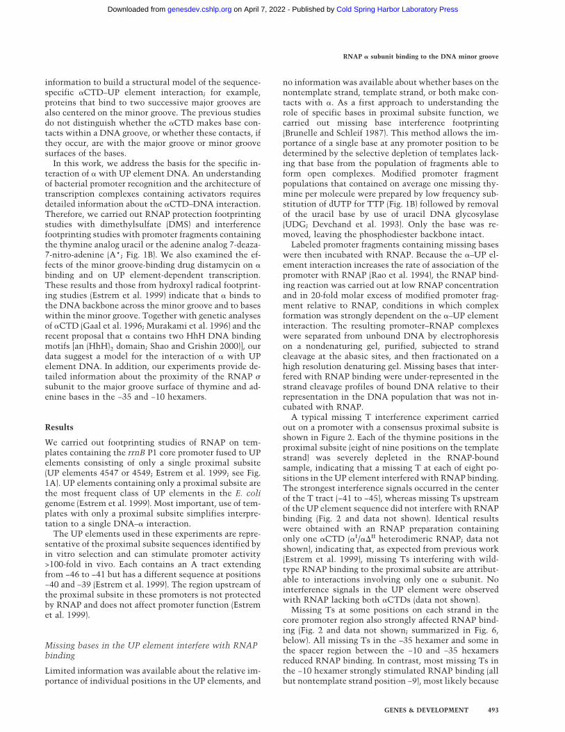
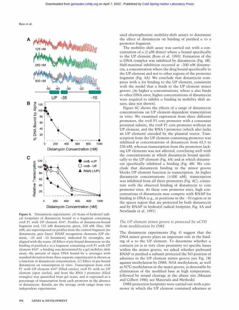
Distamycin, a small, well-characterized oligopeptidethat binds within the minor groove of A+T-rich DNA(Coll et al. 1987; Churchill et al. 1990), was used to de-termine whether insertion of a small molecule in theminor groove of the UP element could compete with andprevent � binding to the promoter. Binding sites for dis-tamycin in the promoter fragment were first character-ized by hydroxyl radical footprinting to determinewhether, as expected, the drug would bind preferentiallyto the UP element (Fig. 4A). Specific, but partial, protec-tion in the UP element (positions −42 to −46) was ob-served at 125 nM distamycin. Protection of this site wasmore complete at 250 nM, but, at 500 nM, protection bydistamycin also occurred at three other sites: in the −35hexamer, in an A+T-rich region of the spacer (−20 to−24), and in the −10 hexamer (−10 to −14). The length ofthe protected regions, 4–5 bp, is consistent with thelength reported previously in hydroxyl radical footprintswith distamycin (Churchill et al. 1990).
Because � and distamycin protect the same UP ele-ment positions against hydroxyl radical cleavage, theidentity of the molecule binding to the UP element can-not be distinguished by footprinting when both the pro-tein and the drug are present. However, purified �–UPelement complexes migrate differently on gels from freeor distamycin-bound DNA fragments. Therefore, we
Figure 3. Interference footprints with the base analogs uracil(U; A,B) or 7-deaza-7-nitro adenine (A*; C,D). Promoter frag-ments (rrnB P1 with UP element 4549), containing, on average,one modified base per molecule, were labeled on the nontem-plate strand (A,C) or the template strand (B,D). RNAP–promotercomplexes were isolated, and samples were examined by gelelectrophoresis after strand cleavage at modified bases. Super-imposed profiles from scans of normalized lanes are alignedwith gels; RNAP-bound samples (+) are indicated by solid bluelines (uracil; A,B) or green lines (A*; C,D), and control sampleswithout RNAP (−) are black dotted lines. UP element, −35 and−10 hexamer sequences are shown, with all T positions in blue(A,B), and all A positions in green (C,D). Modified bases thataffected RNAP binding are indicated: (blue or green circles) in-terference with binding; (blue or green arrows) stimulation ofbinding. Symbol sizes correlate with the approximate degree ofinterference or stimulation observed.
RNAP � subunit binding to the DNA minor groove
GENES & DEVELOPMENT 495
Cold Spring Harbor Laboratory Press on April 7, 2022 - Published by genesdev.cshlp.orgDownloaded from
used electrophoretic mobility-shift assays to determinethe effect of distamycin on binding of purified � to apromoter fragment.
The mobility shift assay was carried out with a con-centration of � (2 µM dimer) where � bound specificallyto the UP element (Ross et al. 1993). Formation of the�–DNA complex was inhibited by distamycin (Fig. 4B).Half-maximal inhibition occurred at ∼200 nM distamy-cin, a concentration where the drug bound specifically tothe UP element and not to other regions of the promoterfragment (Fig. 4A). We conclude that distamycin com-petes with � for binding to the UP element, consistentwith the model that � binds to the UP element minorgroove. (At higher � concentrations, where � also bindsto other DNA sites, higher concentrations of distamycinwere required to inhibit � binding in mobility shift as-says; data not shown).
Figure 4C shows the effects of a range of distamycinconcentrations on UP element-dependent transcriptionin vitro. We examined expression from three differentpromoters, the rrnB P1 core promoter with a consensusproximal subsite, the rrnB P1 core promoter without anUP element, and the RNA I promoter (which also lacksan UP element) encoded by the plasmid vector. Tran-scription from the UP element-containing promoter wasinhibited at concentrations of distamycin from 62.5 to250 nM, whereas transcription from the promoters lack-ing UP elements was not affected, correlating well withthe concentrations at which distamycin bound specifi-cally to the UP element (Fig. 4A) and at which distamy-cin specifically inhibited � binding (Fig. 4B). We con-clude that distamycin binding in the minor grooveblocks UP element function in transcription. At higherdistamycin concentrations (�500 nM), transcriptionwas inhibited from all three promoters (Fig. 4C), consis-tent with the observed binding of distamycin to corepromoter sites. At these core promoter sites, high con-centrations of distamycin may compete with RNAP forbinding to DNA (e.g., at positions in the −10 region or inthe spacer region that are protected by both distamycinand by RNAP in hydroxyl radical footprint; Fig. 4A andNewlands et al. 1991).
The UP element minor groove is protected by �CTDfrom modification by DMS
The distamycin experiments (Fig. 4) suggest that theDNA minor groove plays an important role in the bind-ing of � to the UP element. To determine whether �contacts (or is in very close proximity to) specific baseswithin the minor groove, we asked whether preboundRNAP or purified � subunit protected the N3 position ofadenines in the UP element minor groove (see Fig. 1B)against methylation by DMS. N3A methylation, as wellas N7G methylation in the major groove, is detectable byelimination of the modified base at high temperature,followed by strand cleavage at the abasic site (Maxamand Gilbert 1980; see Materials and Methods).
DMS protection footprints were carried out with a pro-moter in which the UP element contained adenines at
Figure 4. Distamycin experiments. (A) Scans of hydroxyl radi-cal footprints of distamycin bound to a fragment containingrrnB P1 with UP element 4547. Profiles of distamycin-boundfragment (red, 125 nM distamycin; green, 250 nM: blue, 500nM), are superimposed on profiles from the control fragment (nodistamycin; grey lines). RNAP recognition elements (UP ele-ment, −35 and −10 hexamers), indicated by rectangles, arealigned with the scans. (B) Effect of pre-bound distamycin on thebinding of purified � to a fragment containing rrnB P1 with UPelement 4547. � binding was determined by a gel mobility shiftassay; the percent of input DNA bound by � (averages withstandard deviation from three separate experiments) is shown asa function of distamycin concentration. (C) Effect of pre-bounddistamycin on transcription in vitro. Transcription from rrnBP1 with UP element 4547 (filled circles), rrnB P1 with no UPelement (open circles), and from the RNA I promoter (filledtriangles) was quantified from gel scans, and is expressed as apercentage of transcription from each promoter in the absenceof distamycin. Results are the average (with range) from twoindependent experiments.
Ross et al.
496 GENES & DEVELOPMENT
Cold Spring Harbor Laboratory Press on April 7, 2022 - Published by genesdev.cshlp.orgDownloaded from
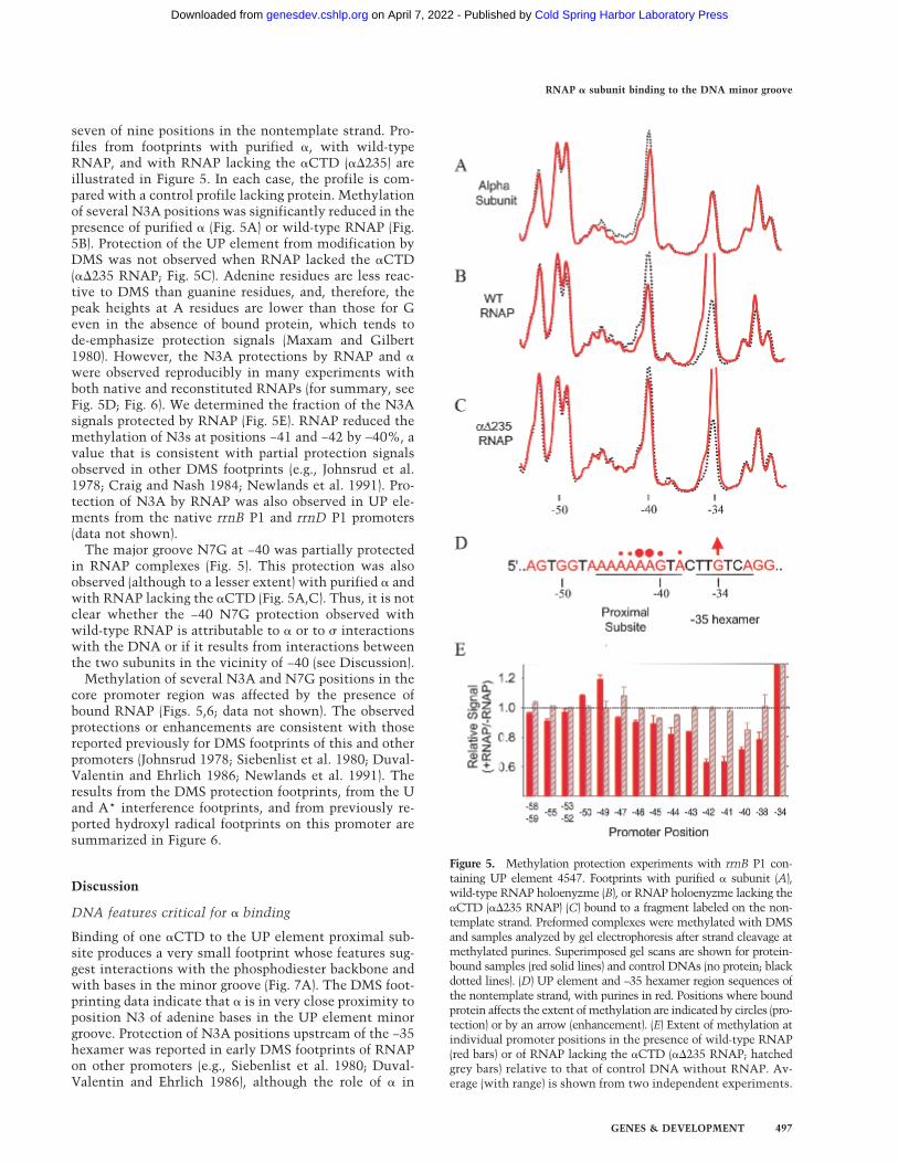
seven of nine positions in the nontemplate strand. Pro-files from footprints with purified �, with wild-typeRNAP, and with RNAP lacking the �CTD (��235) areillustrated in Figure 5. In each case, the profile is com-pared with a control profile lacking protein. Methylationof several N3A positions was significantly reduced in thepresence of purified � (Fig. 5A) or wild-type RNAP (Fig.5B). Protection of the UP element from modification byDMS was not observed when RNAP lacked the �CTD(��235 RNAP; Fig. 5C). Adenine residues are less reac-tive to DMS than guanine residues, and, therefore, thepeak heights at A residues are lower than those for Geven in the absence of bound protein, which tends tode-emphasize protection signals (Maxam and Gilbert1980). However, the N3A protections by RNAP and �were observed reproducibly in many experiments withboth native and reconstituted RNAPs (for summary, seeFig. 5D; Fig. 6). We determined the fraction of the N3Asignals protected by RNAP (Fig. 5E). RNAP reduced themethylation of N3s at positions −41 and −42 by ∼40%, avalue that is consistent with partial protection signalsobserved in other DMS footprints (e.g., Johnsrud et al.1978; Craig and Nash 1984; Newlands et al. 1991). Pro-tection of N3A by RNAP was also observed in UP ele-ments from the native rrnB P1 and rrnD P1 promoters(data not shown).
The major groove N7G at −40 was partially protectedin RNAP complexes (Fig. 5). This protection was alsoobserved (although to a lesser extent) with purified � andwith RNAP lacking the �CTD (Fig. 5A,C). Thus, it is notclear whether the −40 N7G protection observed withwild-type RNAP is attributable to � or to � interactionswith the DNA or if it results from interactions betweenthe two subunits in the vicinity of −40 (see Discussion).
Methylation of several N3A and N7G positions in thecore promoter region was affected by the presence ofbound RNAP (Figs. 5,6; data not shown). The observedprotections or enhancements are consistent with thosereported previously for DMS footprints of this and otherpromoters (Johnsrud 1978; Siebenlist et al. 1980; Duval-Valentin and Ehrlich 1986; Newlands et al. 1991). Theresults from the DMS protection footprints, from the Uand A* interference footprints, and from previously re-ported hydroxyl radical footprints on this promoter aresummarized in Figure 6.
Discussion
DNA features critical for � binding
Binding of one �CTD to the UP element proximal sub-site produces a very small footprint whose features sug-gest interactions with the phosphodiester backbone andwith bases in the minor groove (Fig. 7A). The DMS foot-printing data indicate that � is in very close proximity toposition N3 of adenine bases in the UP element minorgroove. Protection of N3A positions upstream of the −35hexamer was reported in early DMS footprints of RNAPon other promoters (e.g., Siebenlist et al. 1980; Duval-Valentin and Ehrlich 1986), although the role of � in
Figure 5. Methylation protection experiments with rrnB P1 con-taining UP element 4547. Footprints with purified � subunit (A),wild-type RNAP holoenyzme (B), or RNAP holoenyzme lacking the�CTD (��235 RNAP) (C) bound to a fragment labeled on the non-template strand. Preformed complexes were methylated with DMSand samples analyzed by gel electrophoresis after strand cleavage atmethylated purines. Superimposed gel scans are shown for protein-bound samples (red solid lines) and control DNAs (no protein; blackdotted lines). (D) UP element and −35 hexamer region sequences ofthe nontemplate strand, with purines in red. Positions where boundprotein affects the extent of methylation are indicated by circles (pro-tection) or by an arrow (enhancement). (E) Extent of methylation atindividual promoter positions in the presence of wild-type RNAP(red bars) or of RNAP lacking the �CTD (��235 RNAP; hatchedgrey bars) relative to that of control DNA without RNAP. Av-erage (with range) is shown from two independent experiments.
RNAP � subunit binding to the DNA minor groove
GENES & DEVELOPMENT 497
Cold Spring Harbor Laboratory Press on April 7, 2022 - Published by genesdev.cshlp.orgDownloaded from
promoter recognition was not known at the time ofthose experiments. X-ray structures of minor groove-binding proteins in complex with DNA have confirmedthat protection of N3A positions in DMS footprints cor-relates with minor groove contacts [e.g., Hin recombi-nase (Glasgow et al. 1989; Feng et al. 1994) and IHF(Craig and Nash 1984; Rice et al. 1996)].
Our experiments using distamycin, which bindssnugly in the minor groove of A+T-rich DNA (Coll et al.1987), support the role of minor groove interactions in �binding. Distamycin protected essentially the same setof UP element residues as � in our hydroxyl radical foot-prints (Estrem et al. 1999; Figs. 4,6) and inhibited � bind-ing to UP element DNA. Distamycin has been shownpreviously to interfere with minor groove binding ofother proteins (e.g., HMG Y/I; Reeves and Nissen 1990).
Several backbone positions on each strand in the UPelement proximal subsite are protected by RNAP fromhydroxyl radical cleavage (Estrem et al. 1999; for sum-mary, see Figs. 6 and 7A). Hydroxyl radicals react prima-rily with the hydrogens on positions C4 and C5 on theminor groove side of deoxyribose (Balasubramanian et al.1998). Therefore, the UP element base and backbone po-sitions protected by � are located in very close proximity(Fig. 7A). Although not tested in our experiments, the O2positions of thymine bases in the minor groove wouldalso be candidates for direct hydrogen bond contactswith �.
Our results suggest that � does not make significantmajor groove contacts. Substitution of U or A* for T orA, respectively, in the UP element had little if any effecton RNAP binding to the promoter (Fig. 3). Although thepartial protection of major groove position N7G at −40against DMS methylation (Fig. 5) may indicate closeproximity of RNAP to this position, other considerationssuggest that the identity of this residue is not critical toUP element function. G is not strictly conserved at −40in UP element proximal subsites (Estrem et al. 1999),and no steric hindrance to RNAP binding was observed
when A* was substituted for A at this same position inthe UP element (Fig. 3). In addition, although some de-gree of protection of N7G at −40 was observed with pu-rified �, protection was also observed with RNAP lack-ing the �CTD. Thus, it is not clear whether it is � or �that is in close proximity to the N7G at −40 in the con-text of RNAP holoenzyme. Furthermore, the reducedmethylation of the N7G at −40 could be attributable toaltered DNA structure resulting from RNAP bindingrather than from direct protein contact. The latter inter-pretation is supported by the strong enhancement ofDNase I cleavage at position −38 observed in footprintsof RNAP on rrnB P1 and other promoters (Fig. 7A;Gourse 1988; Ross et al. 1993; Craig et al. 1995).
Although our footprints indicated that � contacts onlya small subset of base surfaces, a missing base at anyposition in the UP element interfered very strongly withRNAP binding (Fig. 2). The strongest of the missing basesignals on the nontemplate strand (−41 to −43) coincidewith positions protected by RNAP in DMS footprints,suggesting that the effects of missing bases reflect, inpart, the loss of minor groove base contacts. However,distortion of the normal structure of the UP elementproximal subsite could also account for the effect of themissing bases, since structural changes can occur up to 4bp from an abasic site (Wang et al. 1997). The proximalsubsite contains a long A tract (7–8 residues), and un-usual stuctural features of the A tract (e.g., narrow minorgroove width and/or propeller twisting of bases; Shatzky-Schwartz et al. 1997) could play an important role inrecognition by �. Thus, at least some of the interferenceto RNAP binding caused by missing bases could be at-tributable to effects on a structure important to recogni-tion by �, rather than to loss of direct base contacts.
In contrast with our results, it was reported that miss-ing bases in the UP elements of the ace or the rrnB P1promoters stimulated (rather than inhibited) RNAPbinding (Negre et al. 1997). We suggest that the A+T-richUP element region containing an abasic site may have
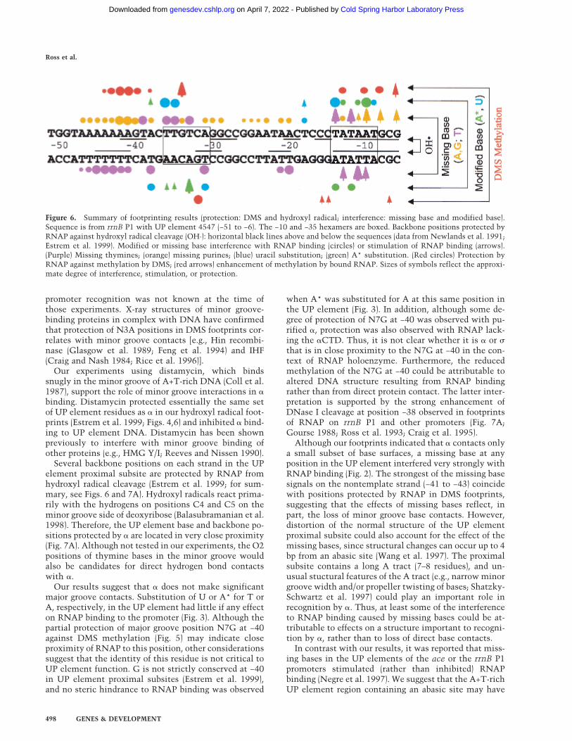
Figure 6. Summary of footprinting results (protection: DMS and hydroxyl radical; interference: missing base and modified base).Sequence is from rrnB P1 with UP element 4547 (−51 to −6). The −10 and −35 hexamers are boxed. Backbone positions protected byRNAP against hydroxyl radical cleavage (OH·): horizontal black lines above and below the sequences (data from Newlands et al. 1991;Estrem et al. 1999). Modified or missing base interference with RNAP binding (circles) or stimulation of RNAP binding (arrows).(Purple) Missing thymines; (orange) missing purines; (blue) uracil substitution; (green) A* substitution. (Red circles) Protection byRNAP against methylation by DMS; (red arrows) enhancement of methylation by bound RNAP. Sizes of symbols reflect the approxi-mate degree of interference, stimulation, or protection.
Ross et al.
498 GENES & DEVELOPMENT
Cold Spring Harbor Laboratory Press on April 7, 2022 - Published by genesdev.cshlp.orgDownloaded from

been recognized as a −10 hexamer rather than as an�-binding site, accounting for the observed stimulation
of RNAP binding in these experiments. We and others(Li and McClure 1998) have observed that missing bases
Figure 7. Positions of footprint signalsand model for �CTD binding to UP ele-ment proximal subsite 4547. (A) Posi-tions of footprint protection and interfer-ence signals in the proximal subsite andthe −35 hexamer. Backbone phosphatesare in ribbon form and deoxyribose andbases in stick form. Backbone positionsare in green (nontemplate strand) or inblue (template strand), and bases are inmagenta (UP element), yellow (−35hexamer), or white. Positions of footprintsignals (for summary, see Fig. 6) are rep-resented as spheres, and include: (1) de-oxyribose positions protected in hydroxylradical footprints (C4�, C5� in UP ele-ment and downstream edge of −35hexamer; green on nontemplate strand,blue on template strand; only a few sig-nals are labeled); (2) purines with reducedor enhanced methylation in the presenceof bound RNAP in DMS footprints (N3 ofA at −41 to −43, magenta; N7 of G at −32,yellow); (3) −35 hexamer positions whereuracil substitutions interfered withRNAP binding (shown as C5-methylgroups of thymines at −36, −35, and −31,yellow) or where A* substitution stimu-lated RNAP binding (shown as N7A at−33, yellow). Positions of enhanced cleav-age in DNase I footprints of rrnB P1, inred along the backbone ribbon (−38, non-template strand; −35, template strand;Gourse 1988; Ross et al. 1993), arethought to reflect positions of DNA dis-tortion in the RNAP–promoter complex.(B) Secondary structural features of�CTD involved in its (HhH)2 domainstructure (Shao and Grishin 2000). (Greenrectangles) � helices in HhH1; (blue rect-angles) � helices in HhH2; (white rect-angles) hairpin regions. Amino acid resi-dues comprising � helices 1–4 (or non-standard helix, NSH), are derived fromthe �CTD solution structure (Jeon et al.1995; Gaal et al. 1996). (C) Model for�CTD binding to the UP element. DNAis rotated ∼90 degrees around the helixaxis from the position in A. The �CTDsolution structure (Jeon et al. 1995) is inribbon form. � helices of the two HhHmotifs (Shao and Grishin 2000) are green(HhH1) and blue (HhH2), and hairpinloops connecting the helices are white.Four residues required for binding to theUP element (R265, G296, K298, S299;Gaal et al. 1996), and two residues impli-cated in interactions with � (D259, E261;see text) are in stick form. The �CTD isaligned with the DNA such that each
HhH motif contacts the backbone of one of the DNA strands, as suggested by features of the RuvA–DNA complex (Ariyoshi et al.2000). Side chains of R265 and K298 are shown fitting into the minor groove. Figures in A and C were prepared using Insight II software.
RNAP � subunit binding to the DNA minor groove
GENES & DEVELOPMENT 499
Cold Spring Harbor Laboratory Press on April 7, 2022 - Published by genesdev.cshlp.orgDownloaded from

in the −10 hexamer strongly stimulate RNAP binding(Figs. 2,6).
The results of our footprinting studies are consistentwith the effects on transcription of mutations in the UPelement proximal subsite (Estrem et al. 1999). Substitu-tion of C:G for A:T pairs at several UP element positions(−41 to −45) reduced transcription in vivo and in vitro,with the strongest effects (>10-fold) occurring at posi-tions −41, −42, and −43. The 2-amino group of guanine atthese positions could sterically hinder � binding in theminor groove; alternatively, effects on � binding of a C:Gsubstitution could result from disruption of A-tractstructure (Koo et al. 1986). Interestingly, a T:A ratherthan an A:T pair at position −42 (in UP element 4542;Estrem et al. 1999) did not reduce UP element function.This substitution would preserve hydrogen bonding ca-pabilities in the minor groove (with the O2 of thymine).
A model for �CTD interaction with an UP elementsubsite
Our results indicating that � binds to the UP elementminor groove are consistent with the recent observationthat the �CTD contains an (HhH)2 domain. (Shao andGrishin 2000). On the basis of this observation, on ourbiochemical data, and on our previous genetic identifi-cation of the �CTD DNA-binding surface (Gaal et al.1996; Murakami et al. 1996), we suggest a preliminarymodel for how � might interact with its binding site (Fig.7C).
The HhH motif was identified initially in the struc-ture of the DNA repair enzyme Endonuclease III (Thayeret al. 1995), and has since been identified in DNA-bind-ing proteins involved in replication, recombination, andrepair (e.g., DNA polymerases, ligases, and glycosylases;Doherty et al. 1996; Shao and Grishin 2000). The HhHmotif contains a pair of anti-parallel � helices connectedby a hairpin loop that are present in either one or twocopies [the (HhH)2 domain; Shao and Grishin 2000].DNA binding, in examples considered thus far, is notsequence specific (Doherty et al. 1996). In the rat DNApolymerase �–DNA complex (Pelletier et al. 1996) andthe E. coli RuvA–DNA complex (Ariyoshi et al. 2000),amino acid residues in the loop and in the amino-termi-nus of the second � helix make nonspecific hydrogenbond contacts to the DNA backbone. RuvA binds acrossthe minor groove of duplex DNA, with each of the re-peated HhH motifs in a RuvA monomer contacting thebackbone of one of the two DNA strands.
The RNAP �CTD contains a short nonstandard helix(NSH) followed by four � helices (Jeon et al. 1995; Gaal etal. 1996). In the (HhH)2 domain interpretation of �CTD(shown in schematic form in Fig. 7B), the first HhH motifcontains the NSH (residues ∼253–257), a loop (∼258–263),and a second � helix (264–272) referred to as � helix 1.The second HhH contains � helix 3 (286–292), a loop(∼293–297), and � helix 4 (298–309). The two HhH motifsare separated by � helix 2. The positions of substitutionsin �CTD that result in strong defects in DNA binding(Gaal et al. 1996; Murakami et al. 1996) affect residues
that, by analogy to (HhH)2 domains in other proteins(Rafferty et al. 1998; Ariyoshi et al. 2000), would be pre-dicted to make DNA contacts (e.g., R265 in the aminoterminus of the second helix in HhH1, G296 in the loop,and K298, S299 in the amino terminus of the secondhelix in HhH2).
Based on these considerations, and by analogy to theRuvA–DNA structure (Ariyoshi et al. 2000), we suggestthat each of the HhH motifs in the �CTD makes back-bone contacts with one of the DNA strands, at the po-sitions that are protected by the �CTD in hydroxyl radi-cal footprints (Fig. 7C). The length of the �-binding site issimilar to the length of DNA covered by one RuvAmonomer (Ariyoshi et al. 2000).
Unlike other HhH-motif proteins, the �CTD demon-strates sequence specificity in DNA binding. We suggestthat this sequence specificity could derive from (at least)two characteristics of the UP element DNA sequence.First, the narrow width of the minor groove in an A tract(the preferred �-binding site) could be important for si-multaneous binding of � to both DNA strands. It hasbeen proposed previously that effects of sequence onDNA structure could influence binding of HhH motifproteins (Rafferty et al. 1998).
Second, our data suggest that the specificity of � bind-ing to an A-tract sequence derives in part from base con-tacts in the minor groove (Fig. 5). These contacts (to N3Aand potentially to O2T) could occur through hydrogenbonds with the basic side chains of residues R265 andK298. Base-specific contacts in the minor groove of A+T-rich DNA sequences have been demonstrated for a num-ber of other DNA-binding proteins. Frequently these in-teractions are made by short peptide motifs containingarginine residues that contact bases in the minor grooveand the DNA backbone along the minor groove [e.g.,AT-hooks in the chromatin-associated protein HMG-I(Y)(Huth et al. 1997), and extended arm sequences in theHin recombinase (Feng et al. 1994) and in homeodo-mains (Kissinger et al. 1990)]. Because several substitu-tions in the �CTD cause defects in both specific andnonspecific DNA binding (Savery et al. 1998), we suggestthat the same DNA-binding motif is responsible for in-teractions with UP element and non-UP element DNAsequences.
In the model for the �–UP element interaction pre-sented in Figure 7C, we have chosen a specific orienta-tion for �CTD with respect to the UP element sequence,although in other contexts the orientation could be re-versed. The orientation shown is suggested by the resultsof genetic experiments that implicate � residues 259 and261 in interactions with the � subunit at promoters con-taining the UP element proximal subsite (Gourse et al.2000). This orientation was also suggested by the iden-tification of residues potentially involved in �–� inter-actions in the lac promoter–CRP–RNAP complex (Busbyand Ebright 1999; Gourse et al. 2000).
� binding to UP elements with two subsites
In the present study, we focused on interactions of �
Ross et al.
500 GENES & DEVELOPMENT
Cold Spring Harbor Laboratory Press on April 7, 2022 - Published by genesdev.cshlp.orgDownloaded from
with a single consensus binding site, the proximal sub-site. However, we have also performed footprinting ex-periments on full (i.e., two subsite) UP elements. Thedistal subsite in the full UP element responded similarlyin protection and interference experiments to the proxi-mal site by itself (W. Ross and R.L. Gourse, data notshown). However, the proximal subsite in the full UPelement responded differently from the proximal subsitein the absence of the distal subsite. In the context of thefull UP element, missing bases in the proximal subsitedid not interfere with RNAP binding (W. Ross and R.L.Gourse, data not shown), and the proximal subsite wasnot fully protected in hydroxyl radical footprints (New-lands et al. 1991; Estrem et al. 1998).
These results suggest that the proximal subsite plays adifferent role when alone than it does in a full UP ele-ment. These results are also consistent with three linesof genetic evidence: (1) Proximal subsite sequences iden-tified in binding-site selections were not identical in thepresence versus absence of the consensus distal subsite(Estrem et al. 1998, 1999). (2) Proximal subsite mutationshad larger effects on transcription when the consensusdistal subsite was absent (Estrem et al. 1998; S. Estremand R.L. Gourse, unpubl.). (3) Certain �CTD and �CTDsubstitutions reduced transcription from promoters con-taining only a proximal subsite, but not from promoterswith full UP elements (W. Ross, A. Mertens, B. Paul, D.Schneider, J. Toplin, M. Burgess, and R.L. Gourse, un-publ.).
These results suggest that the two subsites do not con-tribute equally to the binding of RNAP in promoterscontaining full UP elements. Possible explanations (seealso Estrem et al. 1999) could include: (1) Binding of�CTD to the proximal subsite could be weaker, less spe-cific, or nonspecific in the context of both subsites. (2)The proximal subsite in the context of a full UP elementcould serve primarily a structural role, rather than a rolein � binding. (3) The longer, symmetric distal subsitesequence could be a binding site for both � subunits inthe context of a full UP element, thereby reducing orexcluding occupancy of the proximal subsite by �.
RNAP contacts with the −35 hexamer
Our interference footprints yielded considerable infor-mation about the role of specific major groove base sur-faces in binding RNAP to the core promoter. These data,together with the DMS protection data, confirm and ex-tend previous information about the interaction of the �subunit with the −35 and −10 elements.
Of the 12 bases present in the −35 hexamer of rrnB P1(six on each DNA strand), information about 10 baseswas obtained with the base-specific reagents used, and ofthese 10, five appear to be in close proximity to or incontact with RNAP (Figs. 6,7). Particularly strong inter-ference signals were observed when U was substitutedfor T at positions −35 and −36, indicating probable inter-actions of � with the C5 methyl groups of these thy-
mines that are critical for RNAP binding. Because A*substitutions at the same positions on the templatestrand did not interfere with RNAP binding, we suggestthat the strong conservation of T:A pairs at these posi-tions in promoter −35 hexamers (Hawley and McClure1983) reflects interactions with the thymine bases.
These results are consistent with previous observa-tions. Mutations of the thymines in the nontemplatestrand at the upstream end of the −35 hexamer havestrong phenotypes (Youderian et al. 1982), and 5-bromo-uracil substitutions at these positions in lacUV5 are pro-tected by RNAP against UV-induced strand cleavage(Siebenlist et al. 1980). The region of �70 that interactswith the −35 hexamer (region 4.2) is thought to containa helix–turn–helix motif (HTH; Record et al. 1996; Grosset al. 1998). Although we do not know which amino acidresidues in region 4.2 interact with the thymine methylgroups at −35 and −36, they are likely to be located in theHTH recognition helix, carboxy-terminal to the arginineresidues at positions 584 and 588 that are implicated incontacting the central region of the −35 hexamer (forreview, see Gross et al. 1998). R588 was proposed to con-tact position −34. However, because the major groovesurface of −34 G does not appear to be in contact withRNAP (methylation of this G is increased, not reduced,by the presence of prebound RNAP; Fig. 5; Siebenlist etal. 1980), we suggest that the template strand C, ratherthan the nontemplate strand G, may contact RNAP andbe responsible for the conservation of the G:C pair at−34.
Our results suggest that � also contacts three templatestrand bases at the downstream end of the −35 hexamer.A* at position −33 strongly stimulated RNAP binding.This is a nonconsensus position in rrnB P1 (T:A ratherthan A:T), and the presence of the 7-nitro group on themodified base might provide a contact to RNAP thatserves as an alternative to that provided by thymine atthe same position in a consensus −35 hexamer, eventhough the nitro and methyl groups are chemically dis-similar. Additional template strand contacts in the −35hexamer are suggested by partial protection of N7G at−32 against methylation by DMS (Newlands et al. 1991;see also Johnsrud 1978; Duval-Valentin and Ehrlich1986), and by partial interference resulting from U sub-stitution at −31. Hydroxyl radical footprints indicatethat RNAP also interacts with the DNA backbone alongthe adjacent minor groove in the spacer at the down-stream edge of the −35 hexamer (Newlands et al. 1991;Figs. 6,7). Hydroxyl radical protections (Newlands et al.1991) and the effects of missing bases at several positionsin the spacer region, particularly around −20 (Fig. 6), sug-gest that the conformation of this region may be impor-tant for RNAP binding.
RNAP contacts with the −10 hexamer
In the −10 hexamer, major groove modifications at sev-eral positions interfered with the formation of a stable
RNAP � subunit binding to the DNA minor groove
GENES & DEVELOPMENT 501
Cold Spring Harbor Laboratory Press on April 7, 2022 - Published by genesdev.cshlp.orgDownloaded from
open complex at rrnB P1. Because open complex forma-tion involves recognition of the −10 region as duplexDNA, followed by strand separation and sequence-spe-cific interaction with the nontemplate strand (Robertsand Roberts 1996; Marr and Roberts 1997; Qiu and Hel-mann 1999; Fenton et al. 2000), the interference signalsobserved in our footprints could result from disruption ofcontacts with duplex DNA, single-stranded DNA, orboth. Our footprinting results should be useful for futureinterpretation and modeling of the interaction of � re-gion 2.4 with the −10 hexamer.
Specifically, we found that U substitution at two ofthe three nontemplate strand thymines reduced RNAPbinding, indicating that � may directly contact the C5methyl group of these Ts (rrnB P1 positions −14 and −9,corresponding to positions −12 and −7 in lacUV5 and�PR�; Marr and Roberts 1997; Fenton et al. 2000). A*substitution for five of the six adenines in the −10hexamer interfered with RNAP binding. Because A* con-tains a C7-nitro group in place of N7, interference couldresult from loss of direct contacts or from steric hin-drance (Min et al. 1996). In either case, our data indicatethat RNAP is in close proximity to these bases.
The inhibitory effect of A* was particularly strong atnontemplate strand position −10 (−8 in other promoters),although this is not one of the most highly conserved−10 hexamer positions (Hawley and McClure 1983). A*at −10 could conceivably sterically hinder the approachof RNAP to the adjacent highly conserved thymine ormight interfere with critical base stacking interactionsof the separated strand (Qiu and Helmann 1999). Strongeffects of A* substitutions were also observed at rrnB P1template strand positions −14 and −12 (−12 and −10 inother promoters).
Although an A at nontemplate strand position −13(−11 in other promoters) is very highly conserved andplays a critical role in promoter function, the substitu-tion of A* at −13 had a relatively small effect on RNAPbinding (Fig. 3). This A is important for single-strandedoligonucleotide binding and fork-junction binding (Marrand Roberts 1997; Guo and Gralla 1998; Qiu and Hel-mann 1999) and is thought to nucleate strand opening,perhaps through base stacking interactions with aro-matic residues in � region 2.4 (Fenton et al. 2000; Pan-aghie et al. 2000). Recognition of −13A by � apparentlydoes not involve critical contacts to the functionalgroups altered by the A* modification.
Missing bases at all but two of the positions tested inthe −10 hexamer (all except −13A, −9T) stimulatedRNAP binding, presumably by facilitating the strandseparation process. These results are consistent withthose reported previously for other promoters (Li andMcClure 1998). In addition, a missing base at templatestrand +1 strongly interfered with RNAP binding to therrnB P1 promoter (data not shown). This negative effectreflects the requirement for incorporation of the initiat-ing two nucleotides, ATP and CTP, to stabilize the rrnBP1 open complex, and is not a phenomenon general topromoters with longer-lived open complexes; that is, theeffect of a missing base at +1 of rrnB P1 does not indicate
a requirement for a specific base contact to RNAP at thisposition.
Conclusion
The work reported here provides information about thefunctional consequences of specific DNA–protein con-tacts in the initiation complex. Such data will be impor-tant for interpretation of atomic level structures of thepromoter–RNAP complex that may become available inthe future. We have shown that the � subunit of RNAPbinds to UP element DNA using minor groove as well asbackbone contacts, and we have proposed a model for�CTD binding to DNA. These data should also facilitatemodeling of �CTD interactions with DNA in promotercomplexes containing transcription activators. Our re-sults suggest further that the contacts made by � to theUP element and by �70 to the −35 hexamer are in veryclose proximity. Thus, �CTD–�70 interactions couldplay a role in the function of the UP element proximalsubsite. Finally, our results identify functional groups inthe −10 and −35 hexamers that are involved in the inter-action with the � subunit and thus increase the resolu-tion of our picture of the RNAP–core promoter interac-tion.
Materials and methods
Plasmid DNAs
DNA fragments containing rrnB P1 promoters with differentsequences in the UP element region were inserted as EcoRI (−66with respect to the transcription start site) to HindIII (+50) frag-ments into pRLG770 (Estrem et al. 1999). Plasmids containedconsensus proximal subsite 4547 [(−46) 5�-AAAAAAGTA-3� (−38)] in pRLG4213, consensus proximal subsite 4549 [(−46)5�-AAAAAAAGA-3� (−38)] in pRLG3289, full consensus UP el-ement 4192 [(−59) 5�-GGAAAATTTTTTTTCAAAAGTA-3� (−38)] in pRLG3278, or an upstream sequence lacking UP el-ement function (SUB) in pRLG4210. DNA sequences flankingthe promoters were described previously (Estrem et al. 1998,1999).
RNAP and � purification
Wild-type native RNAP was a gift from Dr. Robert Landick(University of Wisconsin). Wild-type � subunit was purified asdescribed (Gaal et al. 1996). RNAPs containing wild-type ormutant � subunits lacking the carboxy-terminal domain(��235) and containing an amino-terminal hexahistidine tagwere reconstituted from isolated subunits (Gaal et al. 1996),purified by nickel chromatography (Qiagen) and by centrifugalfiltration (Centricon YM-100; 100,000 MW cutoff). RNAPs het-erodimeric for � (containing one wild-type � and one ��235)were purified as described (Estrem et al. 1999).
Missing base interference footprinting
Promoter fragments were prepared by 15 cycles of PCR ampli-fication using primers that flanked the promoter in the vectorsequence (upstream primer 5�-CCGCGGATCCGTATCACGAGGCCCTTTCG-3�; downstream primer 5�-GCGCTACGGCGTTTCACTTC-3�). PCR reactions (50 µL) contained 0.4 µg
Ross et al.
502 GENES & DEVELOPMENT
Cold Spring Harbor Laboratory Press on April 7, 2022 - Published by genesdev.cshlp.orgDownloaded from

of plasmid DNA, 20 pmoles of each primer, 200 µM amounts ofeach dNTP (Pharmacia), 1.5 mM MgCl2, and 2.5 units of TaqDNA polymerase (Promega) in a buffer supplied by the manu-facturer. For preparation of fragments containing missing thy-mine bases, the PCR reactions also contained 2 µM dUTP (Phar-macia; Devchand et al. 1993).
PCR products were purified by use of a Qiaquick PCR Puri-fication Kit (Qiagen), digested with the appropriate restrictionenzyme [HindIII (+50) for nontemplate strand labeling or BamHI(primer-encoded) for template strand labeling], and 3�-end-la-beled with Sequenase (USB) and [�-32P]dATP for HindIII sites, or[�-32P]dGTP for BamHI sites. Labeled fragments were gel puri-fied, eluted by diffusion into 0.2 M NaCl, 20 mM Tris-Cl (pH7.4), 1 mM EDTA, and concentrated by use of Elutip D columns(Schleicher and Schuell). To prepare fragments with, on average,one missing thymine base per molecule, end-labeled fragmentscontaining dUTP were then treated with uracil DNA glyco-sylase (UDG; NEB; 0.75 units for 10 min at 37°C in 100 µL withbuffer supplied by the manufacturer) to remove the uracil base,leaving the DNA backbone intact. To prepare fragments with,on average, one missing purine base per molecule, end-labeledfragments (prepared without modified nucleotides) were treatedwith formic acid as described previously (Maxam and Gilbert1980).
To carry out the missing base interference footprint, RNAP(0.5 nM) was incubated with a 20-fold excess of end-labeledDNA fragment (10 nM) containing either a missing thyminebase or a missing purine, for 5 min at 22°C in 100 µL of 10 mMTris-Cl (pH 7.9), 30 mM KCl, 10 mM MgCl2, 1 mM DTT, 500µM ATP, 50 µM CTP. Heparin (final 10 µg/mL) was added, andRNAP–promoter complexes (∼5% of input DNA) were sepa-rated from unbound DNA by electrophoresis on a 5% acryl-amide gel in 0.5× Tris-Borate-EDTA (TBE). Because DNA waskept in excess to promote competition for RNAP, the unboundpromoter fraction did not provide useful information and wasnot analyzed. Control DNA used for comparison to the RNAP-bound population was the input DNA population without in-cubation with RNAP.
RNAP-bound DNA was eluted by diffusion, and purified byuse of Elutip D columns as described above. Then, strand cleav-age was carried out at the abasic sites by incubation in 100 µLof 1 M piperidine at 90°C for 30 min. Fragments were ethanolprecipitated, then separated by electrophoresis on 9.5% acryl-amide–7 M urea gels. Dried gels were scanned by PhosphorIm-aging using a Storm system (Molecular Dynamics). Data pointsin each lane were normalized to correct for differences in load-ing. Normalization factors were derived from comparisons ofportions of the profiles that were distant from the promoter andassumed not to be in contact with RNAP. Normalized profileswere graphed using Sigma Plot (Jandel Scientific).
Modified base interference footprinting
Promoter fragments for modified base interference footprintingwere prepared by PCR as described above, but reactions alsocontained either 2 µM dUTP (Pharmacia) for modified thyminecontaining fragments, or 30 µM 7-deaza-7-nitro dATP (dA*TP;see below for synthesis) for modified adenine containing frag-ments. Fragments were 3�-end-labeled and gel purified as de-scribed above.
RNAP–promoter fragment complexes were formed underconditions described above for missing base interference foot-printing, gel isolated and the DNA eluted and purified as de-scribed. Strand cleavage at modified adenine (A*) bases was thencarried out by incubation with 1 M piperidine, as describedabove. DNA fragments containing dUTP were treated with
UDG (above) to remove the uracil base prior to the strand cleav-age at the resulting abasic sites (see above). Then, fragmentswere analyzed by denaturing gel electrophoresis as described.
Synthesis of 7-deaza-7-nitro-2�-deoxyadenosinetriethylammonium 5�-triphosphate (dA*TP)
7-Deaza-7-nitro dATP (dA*TP) was synthesized using modifi-cations of the procedure described previously (Min et al. 1996).A suspension of 7-deaza-7-nitro-2�-deoxyadenosine (Min et al.1996; Seela et al. 1997; 23.2 mg, 78.4 µmoles, coevaporatedtwice from pyridine and dried under high vacuum overnight) intrimethylphosphate (0.32 mL) was treated at 0°C with POCl3
(8.1 µL, 86.3 µmoles), and stirred for 20 min at 0°C to produce ayellow solution. This solution was treated dropwise with 0.5 M(Bu3NH)2H2P2O7 [solution in dimethylformamide (0.78 mL,0.39 mmoles); prepared according to Moffatt 1964] and Bu3N(0.08 mL, 0.32 mmoles), stirred vigorously for 1.5 min, pouredinto 10 mL of ice-cold 1.5 M triethylammonium bicarbonate(TEAB at pH 7.5), and lyophilized. The residue was dissolved in10 mL of water and purified by ion-exchange chromatography[DEAE Sephadex A25, 9 × 2.5 cm, linear gradient of TEAB (0.05to 1.5 M at pH 7.5, 1500 mL) at a flow rate of 5 mL/min]. Thetriphosphate was eluted at 0.6–0.7 M TEAB. Lyophilization(three times from water) gave dA*TP (40.6 mg, 55%) as a yellowsolid. Analytical HPLC: tR 18.27 min. UV (H2O): 364, 276. 31P-NMR (162 MHz, D2O): −7.19 [d, J=20.1, P(�)]; −10.62 [d, J=20.9,P(�)]; −22.07 [t, J≈ 19.5, P(�)]. MS-ES (negative mode):C11H16N5O14P3 (535): 534 (100, [M − H]−).
Footprinting with DMS
Promoter fragments used in DMS protection footprints wereobtained by HindIII digestion of pRLG4213 or pRLG3289 andlabeling of the 3� end of the nontemplate strand using Sequenase(USB) and [�-32P]dATP (Newlands et al. 1991). Then, labeledDNA was digested with AatII (vector-encoded site is 75 bp up-stream of EcoRI), and the fragment was gel isolated, eluted, andpurified by use of Elutip D columns as described above.
Promoter fragments (<1 nM) were pre-incubated with wild-type or � mutant RNAP (∼10 nM) or with purified � subunit (∼2µM) for 10 min at 22°C in 50 µL of the buffer used above forinterference footprinting. Then, the DNA was methylated byaddition of 1 µL of DMS for 30 sec at 22°C, followed by additionof 25 µL of cold 1.5 M �-mercaptoethanol and 0.3 M NaOAc,extraction with 75 µL of cold phenol, precipitation with etha-nol, and reprecipitation from 0.3 M NaOAc. The DNA pelletwas resuspended in 30 µL of 10 mM sodium phosphate (pH 6.8),1 mM EDTA, and methylated purines (A,G) were released byincubation at 92°C for 15 min (Maxam and Gilbert 1980). Strandcleavage at abasic sites was carried out by addition of 3 µL of 1N NaOH and further incubation at 92°C for 30 min. Sampleswere precipitated with ethanol, and examined by electrophore-sis, gel scanning, and quantitation as described above.
Hydroxyl radical footprinting to detect distamycin-bindingsites
An end-labeled promoter fragment containing rrnB P1 with UPelement 4547 (prepared as described for DMS footprintingabove) was incubated at 22°C for 10 min with 125, 250, or 500nM distamycin A (Sigma) in 150 mM NaCl, 20 mM Tris-Cl (pH7.4), 1 mM EDTA, and 1 µg/mL sonicated calf thymus DNA.OH· cleavage was carried out as described previously (Newlands
RNAP � subunit binding to the DNA minor groove
GENES & DEVELOPMENT 503
Cold Spring Harbor Laboratory Press on April 7, 2022 - Published by genesdev.cshlp.orgDownloaded from

et al. 1991), and gel electrophoresis and scanning of gels wasperformed as described above.
Gel mobility shift assay with the � subunit
An end-labeled promoter fragment (∼0.1 nM; rrnB P1 withproximal subsite 4547) was preincubated for 10 min at 22°Cwith 15.6–1000 nM distamycin A in 10 µL of buffer containing150 mM NaCl, 20 mM Tris-Cl (pH 7.4), 1 mM EDTA, 10%glycerol, and 1 µg/mL sonicated calf thymus DNA. Purified �
subunit (2 µM) was then added for 10 min, and electrophoresiswas carried out at 4°C on prechilled 6% acrylamide gels con-taining 0.5× TBE and 10% glycerol. Bands containing the�–DNA complex were quantified by PhosphorImaging, and val-ues were expressed as a percentage of the fragment populationbound in the absence of distamycin.
In vitro transcription
Plasmid DNAs (60 ng of pRLG4213 or pRLG4210) were incu-bated with distamycin A (15.6–1000 nM) for 10 min at 22°C in25 µL of transcription buffer containing 150 mM NaCl, 10 mMTris-Cl (pH 7.9), 10 mM MgCl2, 1 mM DTT, 100 µg/mL bovineserum albumin, 500 µM ATP, 50 µM CTP, 50 µM GTP, 10 µMUTP, and 4 µCi [�-32P]UTP (NEN). Transcription was initiatedby addition of RNAP (2 nM) for 15 min, and the reaction wasterminated by addition of 25 µL of stop solution (Ross et al.1998). Samples were analyzed by electrophoresis on 5% acryl-amide–7 M urea gels and quantified after gel scanning with aPhosphorImager.
Acknowledgments
We thank Melanie Barker, Richard Ebright, Tamas Gaal, andRuth Saecker for comments on the manuscript and RichardEbright for calling dA*TP to our attention. We also thankTamas Gaal for preparation of purified � subunit and Bob Land-ick for RNAP holoenzyme. This work was supported byGM37048 to R.L.G. from the National Institutes of Health. A.E.gratefully acknowledges support from the Swiss National Sci-ence Foundation (postdoctoral fellowship, 1998–1999) and fromProfessor Gregory L. Verdine (Harvard University).
The publication costs of this article were defrayed in part bypayment of page charges. This article must therefore be herebymarked “advertisement” in accordance with 18 USC section1734 solely to indicate this fact.
References
Ariyoshi, M., Nishino, T., Iwasaki, H., Shinagawa, H., and Mori-kawa, K. 2000. Crystal structure of the Holliday junctionDNA in complex with a single RuvA tetramer. Proc. Natl.Acad. Sci. 97: 8257–8262.
Balasubramanian, B., Pogozelski, W.K., and Tullius, T.D. 1998.DNA strand breaking by the hydroxyl radical is governed bythe accessible surface areas of the hydrogen atoms of theDNA backbone. Proc. Natl. Acad. Sci. 95: 9738–9743.
Belyaeva, T.A., Rhodius, V.A., Webster, C.L., and Busby, S.J.1998. Transcription activation at promoters carrying tandemDNA sites for the Escherichia coli cyclic AMP receptor pro-tein: Organisation of the RNA polymerase alpha subunits. J.Mol. Biol. 277: 789–804.
Blatter, E.E., Ross, W., Tang, H., Gourse, R.L., and Ebright, R.H.1994. Domain organization of RNA polymerase alpha sub-unit: C-terminal 85 amino acids constitute a domain capable
of dimerization and DNA binding. Cell 78: 889–896.Brunelle, A. and Schleif, R.F. 1987. Missing contact probing of
DNA–protein interactions. Proc. Natl. Acad.Sci. 84: 6673–6676.
Busby, S.J. and Ebright, R.H. 1999. Transcription activation bycatabolite activator protein (CAP). J. Mol. Biol. 293: 199–213.
Churchill, M.E., Hayes, J.J., and Tullius, T.D. 1990. Detection ofdrug binding to DNA by hydroxyl radical footprinting. Re-lationship of distamycin binding sites to DNA structure andpositioned nucleosomes on 5S RNA genes of Xenopus. Bio-chem. 29: 6043–6050.
Coll, M., Frederick, C.A., Wang, A.H., and Rich, A. 1987. Abifurcated hydrogen-bonded conformation in the d(A.T) basepairs of the DNA dodecamer d(CGCAAATTTGCG) and itscomplex with distamycin. Proc. Natl. Acad. Sci. 84: 8385–8389.
Craig, M.L., Suh, W.-C., and Record, Jr., M.T. 1995. HO· andDNase I probing of E�70 RNA polymerase-�PR promoteropen complexes: Mg2+ binding and its structural conse-quences at the transcription start site. Biochem. 34: 15624–15632.
Craig, N.L. and Nash, H.A. 1984. E. coli integration host factorbinds to specific sites in DNA. Cell 39: 707–716.
Devchand, P.R., McGhee, J.D., and van de Sande, J.H. 1993.Uracil-DNA glycosylase as a probe for protein–DNA inter-actions. Nucleic Acids Res. 21: 3437–3443.
Doherty, A.J., Serpell, L.C., and Ponting, C.P. 1996. The helix–hairpin–helix DNA-binding motif: A structural basis fornon-sequence-specific recognition of DNA. Nucleic AcidsRes. 24: 2488–2497.
Duval-Valentin, G. and Ehrlich, R. 1986. Interaction between E.coli RNA polymerase and the tetR promoter from pSC101:Homologies and differences with other E. coli promoter sys-tems from close contact point studies. Nucleic Acids Res.14: 1967–1983.
Estrem, S.T., Gaal, T., Ross, W., and Gourse, R.L. 1998. Identi-fication of an UP element consensus sequence for bacterialpromoters. Proc. Natl. Acad. Sci. 95: 9761–9766.
Estrem, S.T., Ross, W., Gaal, T., Chen, Z.W.S., Niu, W., Ebright,R.H., and Gourse, R.L. 1999. Bacterial promoter architec-ture: Subsite structure of UP elements and interactions withthe C-terminal domain of the RNA polymerase � subunit.Genes & Dev. 13: 2134–2147.
Feng, J.A., Johnson, R.C., and Dickerson, R.E. 1994. Hin recom-binase bound to DNA: The origin of specificity in major andminor groove interactions. Science 263: 348–355.
Fenton, M.S., Lee, S.J., and Gralla, J.D. 2000. Escherichia colipromoter opening and −10 recognition: Mutational analysisof �70. EMBO J. 19: 1130–1137.
Fredrick, K., Caramori, T., Chen, Y.F., Galizzi, A., and Hel-mann, J.D. 1995. Promoter architecture in the flagellar regu-lon of Bacillus subtilis: High-level expression of flagellin bythe sigma D RNA polymerase requires an upstream pro-moter element. Proc. Natl. Acad. Sci. 92: 2582–2586.
Gaal, T., Ross, W., Blatter, E.E., Tang, H., Jia, X., Krishnan, V.V.,Assa-Munt, N., Ebright, R.H., and Gourse, R.L. 1996. DNA-binding determinants of the � subunit of RNA polymerase:Novel DNA-binding domain architecture. Genes & Dev.10: 16–26.
Glasgow, A.C., Bruist, M.F., and Simon, M.I. 1989. DNA-bind-ing properties of the Hin recombinase. J. Biol. Chem.264: 10072–10082.
Gourse, R.L. 1988. Visualization and quantitative analysis ofcomplex formation between E. coli RNA polymerase and anrRNA promoter in vitro. Nucleic
Ross et al.
504 GENES & DEVELOPMENT
Cold Spring Harbor Laboratory Press on April 7, 2022 - Published by genesdev.cshlp.orgDownloaded from
Acids Res. 16: 9789–9809.Gourse, R.L., Ross, W., and Gaal, T. 2000. UPs and downs in
bacterial transcription initiation: The role of the alpha sub-unit of RNA polymerase in promoter recognition. Mol. Mi-crobiol. 37: 687–695.
Gross, C. A., Chan, C., Dombroski, A., Gruber, T., Sharp, M.,Tupy, J., and Young, B. 1998. The functional and regulatoryroles of � factors in transcription. Cold Spring Harbor Symp.Quant. Biol. 63: 141–155.
Guo, Y. and Gralla, J.D. 1998. Promoter opening via a DNA forkjunction binding activity. Proc. Natl. Acad. Sci. 95: 11655–11660.
Hawley, D.K. and McClure, W.R. 1983. Compilation and analy-sis of Escherichia coli promoter DNA sequences. NucleicAcids Res. 11: 2237–2255.
Hayward, R.S., Igarashi, K., and Ishihama, A. 1991. Functionalspecialization within the �-subunit of Escherichia coli RNApolymerase. J. Mol. Biol. 221: 23–29.
Hochschild, A. and Dove, S.L. 1998. Protein–protein contactsthat activate and repress prokaryotic transcription. Cell92: 597–600.
Huth, J.R., Bewley, C.A., Nissen, M.S., Evans, J.N., Reeves, R.,Gronenborn, A.M., and Clore, G.M. 1997. The solutionstructure of an HMG-I(Y)-DNA complex defines a new ar-chitectural minor groove binding motif. Nature Struct. Biol.4: 657–665.
Jeon, Y. H., Negishi, T., Shirakawa, M., Yamazaki, T., Fujita, N.,Ishihama, A., and Kyogoku, Y. 1995. Solution structure ofthe activator contact domain of the RNA polymerase alphasubunit. Science 270: 1495–1497.
Jeon, Y.H., Yamazaki, T., Otomo, T., Ishihama, A., andKyogoku, Y. 1997. Flexible linker in the RNA polymerasealpha subunit facilitates the independent motion of the C-terminal activator contact domain. J. Mol. Biol. 267: 953–962.
Johnsrud, L. 1978. Contacts between Escherichia coli RNApolymerase and a lac operon promoter. Proc. Natl. Acad. Sci.75: 5314–5318.
Kissinger, C.R., Liu, B.S., Martin-Blanco, E., Kornberg, T.B., andPabo, C.O. 1990. Crystal structure of an engrailed homeodo-main-DNA complex at 2.8 A resolution: A framework forunderstanding homeodomain–DNA interactions. Cell63: 579–590.
Kolb, A., Igarashi, K., Ishihama, A., Lavigne, M., Buckle, M., andBuc, H. 1993. E. coli RNA polymerase, deleted in the C-terminal part of its �-subunit, interacts differently with thecAMP–CRP complex at the lacP1 and at the galP1 promoter.Nucleic Acids Res. 21: 319–326.
Koo, H.S., Wu, H.M., and Crothers, D.M. 1986. DNA bending atadenine-thymine tracts. Nature 320: 501–506.
Li, X.Y. and McClure, W.R. 1998. Stimulation of open complexformation by nicks and apurinic sites suggests a role fornucleation of DNA melting in Escherichia coli promoterfunction. J. Biol. Chem. 273: 23558–23566.
Marr, M.T. and Roberts, J.W. 1997. Promoter recognition asmeasured by binding of polymerase to nontemplate strandoligonucleotide. Science 276: 1258–1260.
Maxam, A.M. and Gilbert, W. 1980. Sequencing end-labeledDNA with base-specific chemical cleavages. Methods Enzy-mol. 65: 499–560.
Min, C., Cushing, T.D., and Verdine, G.L. 1996. Template-di-rected interference footprinting of protein-adenine contacts.J. Am. Chem. Soc. 118: 6116–6120.
Moffatt, J.G. 1964. A general synthesis of nucleoside-5� triphos-phates. Can. J. Chem. 42: 599–604.
Moran, C.P., Jr., Lang, N., LeGrice, S.F.J., Lee, G., Stephens, M.,
Sonenshein, A.I., Pero, J., and Losick, R. 1982. Nucleotidesequences that signal the initiation of transcription andtranslation in Bacillus subtilis. Mol. Gen. Genet. 186: 339–346.
Murakami, K., Fujita, N., and Ishihama, A. 1996. Transcriptionfactor recognition surface on the RNA polymerase alpha sub-unit is involved in contact with the DNA enhancer element.EMBO J. 15: 4358–4367.
Naryshkin, N., Revyakin, A., Kim, Y., Mekler, V. and Ebright,R.H. 2000. Structural organization of the RNA polymerase-promoter open complex. Cell 101: 601–611.
Negre, D., Bonod-Bidaud, C., Oudot, C., Prost, J.F., Kolb, A.,Ishihama, A., Cozzone, A.J., and Cortay, J.C. 1997. DNAflexibility of the UP element is a major determinant for tran-scriptional activation at the Escherichia coli acetate pro-moter. Nucleic Acids Res. 25: 713–738.
Newlands, J.T., Ross, W., Gosink, K.K., and Gourse, R.L. 1991.Factor-independent activation of Escherichia coli rRNAtranscription. II. characterization of complexes of rrnB P1promoters containing or lacking the upstream activator re-gion with Escherichia coli RNA polymerase. J. Mol. Biol.220: 569–583.
Newlands, J.T., Josaitis, C.A., Ross, W., and Gourse, R.L. 1992.Both fis-dependent and factor-independent upstream activa-tion of the rrnB P1 promoter are face of the helix dependent.Nucleic Acids Res. 20: 719–726.
Panaghie, G., Aiyar, S.E., Bobb, K.L., Hayward, R.S., and de-Haseth, P.L. 2000. Aromatic amino acids in region 2.3 ofEscherichia coli �70 participate collectively in the formationof an RNA polymerase–promoter open complex. J. Mol. Biol.299: 1217–1230.
Pelletier, H., Sawaya, M.R., Wolfle, W., Wilson, S.H., and Kraut,J. 1996. Crystal structures of human DNA polymerase �
complexed with DNA: Implications for catalytic mecha-nism, processivity, and fidelity. Biochem. 35: 12742–12761.
Pu, W.T., and Struhl, K. 1992. Uracil interference, a rapid andgeneral method for defining protein-DNA interactions in-volving the 5-methyl group of thymines: The GCN4–DNAcomplex. Nucleic Acids Res. 20: 771–775.
Qiu, J. and Helmann, J.D. 1999. Adenines at −11, −9 and −8 playa key role in the binding of Bacillus subtilis E�A RNA poly-merase to −10 region single-stranded DNA. Nucleic AcidsRes. 27: 4541–4546.
Rafferty, J.B., Ingleston, S.M., Hargreaves, D., Artymiuk, P.J.,Sharples, G.J., Lloyd, R.G., and Rice, D.W. 1998. Structuralsimilarities between Escherichia coli RuvA protein andother DNA-binding proteins and a mutational analysis of itsbinding to the holliday junction. J. Mol. Biol. 278: 105–116.
Rao, L., Ross, W., Appleman, J.A., Gaal, T., Leirmo, S., Schlax,P.J., Record, Jr., M.T., and Gourse, R.L. 1994. Factor inde-pendent activation of rrnB P1. An “extended” promoter withan upstream element that dramatically increases promoterstrength. J. Mol. Biol. 235: 1421–1435.
Record, M.T., Jr., Reznikoff, W.S., Craig, M.L., McQuade, K.L.,and Schlax, P.J. 1996. Escherichia coli RNA polymerase(E670), promoters, and the kinetics of the steps of transcrip-tion initiation. In Escherichia coli and Salmonella, cellularand molecular biology (ed. F.C. Neidhardt), pp. 792–820.ASM Press, Washington, D.C.
Reeves, R. and Nissen, M.S. 1990. The AT-DNA-binding do-main of mammalian high mobility group I chromosomalproteins. A novel peptide motif for recognizing DNA struc-ture. J. Biol. Chem. 265: 8573–8582.
Rice, P.A., Yang, S., Mizuuchi, K., and Nash, H.A. 1996. Crystalstructure of an IHF–DNA complex: A protein-induced DNAU-turn. Cell 87: 1295–1306.
RNAP � subunit binding to the DNA minor groove
GENES & DEVELOPMENT 505
Cold Spring Harbor Laboratory Press on April 7, 2022 - Published by genesdev.cshlp.orgDownloaded from

Roberts, C.W. and Roberts, J.W. 1996. Base-specific recognitionof the nontemplate strand of promoter DNA by E. coli RNApolymerase. Cell 86: 495–501.
Ross, W., Gosink, K.K., Salomon, J., Igarashi, K., Zou, C., Ishi-hama, A., Severinov, K., and Gourse, R.L. 1993. A third rec-ognition element in bacterial promoters: DNA binding bythe � subunit of RNA polymerase. Science 262: 1407–1413.
Ross, W., Aiyar, S.E., Salomon, J., and Gourse, R.L. 1998. Esch-erichia coli promoters with UP elements of differentstrength: Modular structure of bacterial promoters. J. Bacte-riol. 180: 5375–5383.
Savery, N.J., Lloyd, G.S., Kainz, M., Gaal, T., Ross, W., Ebright,R.H., Gourse, R.L., and Busby, S.J.W. 1998. Transcriptionactivation at class II CRP-dependent promoters: Identifica-tion of determinants in the C-terminal domain of the RNApolymerase alpha subunit. EMBO J. 17: 3439–3447.
Seela, F., Rosemeyer, H., Zulauf, M., Chen, Y., Kastner, G., andReuter, H. 1997. 7-Nitro-7-deaza-2�-deoxyadenosine and8-methyl-7-deaza-2�-deoxyguanosine: Pyrrolo[2,3-d]pyrimi-dine nucleosides with different sugar conformations. LiebigsAnn. Annalen-Recueil 2225–2230.
Shao, X. and Grishin, N.V. 2000. Common fold in helix–hair-pin–helix proteins. Nucleic Acids Res. 28: 2643–2650.
Shatzky-Schwartz, M., Arbuckle, N.D., Eisenstein, M., Rabi-novich, D., Bareket-Samish, A., Haran, T. E., Luisi, B.F., andShakked, Z. 1997. X-ray and solution studies of DNA oligo-mers and implications for the structural basis of A-tract-dependent curvature. J. Mol. Biol. 267: 595–623.
Siebenlist, U., Simpson, R.B., and Gilbert, W. 1980. E. coliRNA polymerase interacts homologously with two differentpromoters. Cell 20: 269–281.
Thayer, M.M., Ahern, H., Xing, D., Cunningham, R.P., andTainer, J.A. 1995. Novel DNA binding motifs in the DNArepair enzyme endonuclease III crystal structure. EMBO J.14: 4108–4120.
Wang, K.Y., Parker, S.A., Goljer, I., and Bolton, P.H. 1997. So-lution structure of a duplex DNA with an abasic site in a dAtract. Biochem. 36: 11629–11639.
Youderian, P., Bouvier, S., and Susskind, M.M. 1982. Sequencedeterminants of promoter activity. Cell 30: 843–853.
Zhang, G., Campbell, E.A., Minakhin, L., Richter, C., Sever-inov, K., and Darst, S.A. 1999. Crystal structure of Thermusaquaticus core RNA polymerase at 3.3 angstrom resolution.Cell 98: 811–824.
Ross et al.
506 GENES & DEVELOPMENT
Cold Spring Harbor Laboratory Press on April 7, 2022 - Published by genesdev.cshlp.orgDownloaded from
10.1101/gad.870001Access the most recent version at doi: 15:2001, Genes Dev.
Wilma Ross, Alexander Ernst and Richard L. Gourse subunit binding to the UP element minor groove
α RNA polymerase-promoter interactions: E. coliFine structure of
References
http://genesdev.cshlp.org/content/15/5/491.full.html#ref-list-1
This article cites 64 articles, 20 of which can be accessed free at:
License
ServiceEmail Alerting
click here.right corner of the article or
Receive free email alerts when new articles cite this article - sign up in the box at the top
Cold Spring Harbor Laboratory Press
Cold Spring Harbor Laboratory Press on April 7, 2022 - Published by genesdev.cshlp.orgDownloaded from