fluid management in hysteroscopy - hologic education · hysteroscopy can also be used in...
TRANSCRIPT

Fluid Management in Hysteroscopy

©2018 All rights reserved
Pfiedler Education, 2170 S. Parker Rd., Suite 125, Denver, CO 80231 www.pfiedler.com
Phone 720-748-6144
3
OVERVIEW Diagnostic and operative hysteroscopy requires adequate visualization of the uterine cavity by insufflating carbon dioxide gas or fluid media. This educational activity will discuss the most common indications for hysteroscopy and the advantages and disadvantages of gas and liquid distention media. Key components of fluid management systems, implications associated with each method and the nursing responsibilities associated with these procedures will be reviewed. Potential complications, such as fluid overload and dilution hyponatremia, will be discussed. Guidelines for mitigating the risk to patient safety will be identified along with nursing responsibly that can aid with achieving optimum patient outcomes.
LEARNER OBJECTIVES After completing this continuing education activity, the participant should be able to:
1. Review patient indications, required equipment, and procedure elements for hysteroscopy.
2. Discuss selection criteria for distention media in hysteroscopy. 3. Identify risks associated with fluid distention and nursing responsibilities in
hysteroscopy. 4. Describe methods and equipment used to calculate and monitor fluid deficit while
using distention media during hysteroscopy. 5. Outline evidence based recommendations to mitigate risk during hysteroscopy.
INTENDED AUDIENCE/EDUCATIONAL NEED This continuing education activity is intended for a perioperative nurse or other health care professional who wants to learn more or needs to gain knowledge and skills in fluid management in hysteroscopy.
TEACHING METHODOLOGIES The study guide is a self-paced, independent learning module. Course goals are presented, followed by corresponding content. Learners can evaluate attainment of objectives by completing the test questions and comparing with the answer key. References can be reviewed for additional information.

4
STEPS FOR SUCCESSFUL COURSE COMPLETION To earn continuing education credit, the participant must complete the following steps:
1. Read the overview and objectives to ensure consistency with your own learning needs and objectives. Review the content of the activity, paying attention to those areas that reflect the objectives. At the end of the activity, you will be assessed on the attainment of each objective.
2. Complete the Test Questions. Missed questions will offer the opportunity to re-read the question and answer choices. You may also revisit relevant content.
3. For additional information on an issue or topic, consult the references. 4. To receive credit for this activity, complete the evaluation and registration forms
and mail to: Pfiedler Enterprises
2170 S. Parker Rd., Suite 125 Denver, CO 80231
ACCREDITATION INFORMATION California Board of Registered Nursing Pfiedler Education is a provider approved by the California Board of Registered Nursing, Provider Number CEP14944, for 2.0 contact hours.
Obtaining full credit for this offering depends upon attendance, regardless of circumstances, from beginning to end. Licensees must provide their license numbers for record keeping purposes.
The certificate of course completion issued at the conclusion of this course must be retained in the participant’s records for at least four (4) years as proof of attendance.
EXPIRATION DATE This continuing education activity was planned and provided in accordance with accreditation criteria. This material was produced in July 2017 and expires after July 2019.
COMMERCIAL SUPPORT Health care organizations engaged in continuing medical, nursing and allied health education have adopted standards to promote balanced and evidence-based content. This course is supported by commercial funds and complies with the intent of these standards. Pfiedler Enterprises gratefully acknowledges educational grants provided by: Hologic.
DISCLAIMER Pfiedler Enterprises does not endorse or promote any commercial product that may be discussed in this activity.
5
ACTIVITY PLANNING COMMITTEE Melinda T. Whalen, BSN, RN, CEN Clinical Program Manager Pfiedler Enterprises Denver, CO
No conflict of interest
Judith A. Pins, RN, BSN, MBA President Pfiedler Enterprises Denver, CO
No conflict of interest
AUTHOR Dondra Tolerson, BS, MA Medical Writer Woodstock, GA
No conflict of interest
Disclosure of Relationships with Commercial Entities for Those in a Position to Control Content for this Activity Pfiedler Enterprises has a policy in place for identifying and resolving conflicts of interest for individuals who control content for a Nursing or Allied Health Professional activity. Information is provided to participants, so that a determination can be made if identified external interests or influences pose potential bias in content, recommendations or conclusions. The intent is full disclosure of those in a position to control content, with a goal of objectivity, balance, and scientific rigor in the activity. For additional information regarding Pfiedler Enterprises’ disclosure process, visit our website at: http://www.pfiedlerenterprises.com/disclosure
Disclosure includes relevant financial relationships with commercial interests related to the subject matter that may be presented in this continuing education activity. “Relevant financial relationships” are those in any amount, occurring within the past 12 months that create a conflict of interest. A commercial interest is any entity producing, marketing, reselling, or distributing health care goods or services consumed by, or used on, patients.
HIPAA COMPLIANCE Pfiedler Enterprises makes every effort to be in compliance with HIPAA. To protect patient privacy, faculty and participants have been requested to de-identify patient related material.

6

7
INTRODUCTION Advances in technology have provided means by which surgeons can effectively identify a uterine abnormality and remove the abnormality with an outpatient procedure in place of a traditional hysterectomy. This procedure is called a hysteroscopy and typically yields excellent results with minimal discomfort. Within the last four decades, higher diagnostic accuracy related to the possibility of direct endoscopic viewing of the uterine cavity combined with continuous and rapid advances in technologies and techniques, have opened up emerging possibilities for hysteroscopic modalities. Hysteroscopic procedures have become the gold standard of diagnostic and operative options in the management of intrauterine, cervical and vaginal pathologies.1 The procedure accommodates minimally invasive surgery (MIS) and uses a hysteroscope, which is a thin, lighted telescope-like device that is inserted through the vagina into the uterus. Via a digital camera, the hysteroscope provides direct examination of the cervix, cervical canal and vagina and transmits the image of the inside of the uterus, including the fallopian tubes, onto a monitor.2 Diagnostic and operative hysteroscopy requires uterine distention and irrigation to effectively visualize the uterine cavity and to remove blood and tissue debris during the procedure. Options for uterine distention include insufflation with carbon dioxide (CO2) gas, electrolytic or electrolyte-free liquid distention media.
Complications during hysteroscopy are rare although critical complications include intravasation of fluid when using a fluid distention medium, a gas embolism when using CO2 for a distention medium, or an air embolism with either method. Complications range from minor to severe and even death. It is important for the perioperative nurse to understand the various distention media available for hysteroscopy, preventative measures to mitigate complications and protocols for patient care in the event of a complication.
PATIENT INDICATIONS FOR HYSTEROSCOPY Hysteroscopy can be performed for diagnostic and operative treatment of abnormalities of the uterus or cervix. Since hysteroscopy examines the lining and interior of the uterus, it is not suitable for evaluating problems within the muscular wall or on the outer surface of the uterus. Diagnostic hysteroscopy is used to diagnose problems of the uterus or confirm results of other tests (eg, hysterosalpingography). Hysterosalpingography (HSG) is an X-ray procedure used to visualize the inside of the uterus and fallopian tubes to show if the fallopian tubes are (partly or fully) blocked or that the uterus is of a normal size and shape.3 Hysteroscopy can also be used in conjunction with procedures (ie, laparoscopy) or prior to procedures (ie, dilation and curettage). Operative hysteroscopy is used to correct abnormal conditions. If an abnormal condition was detected during the diagnostic hysteroscopy, an operative hysteroscopy can often be performed at the same time, avoiding the need for a second surgery. During operative hysteroscopy, MIS instruments are inserted through the hysteroscope to correct the condition.

8
Conditions Diagnosed and Treated with Hysteroscopy Hysteroscopy is used to diagnose or treat problems of the uterus and compared with more invasive procedures it may provide the following advantages:
• shorter hospital stay, • shorter recovery time, • less pain medication needed postsurgery, • avoidance of hysterectomy, and • potential for avoidance of open abdominal surgery.
The procedure may be recommended evaluate and remove a number of gynecological problems, including:3
• assess abnormal vaginal bleeding; • locate abnormalities in the uterine lining; • remove retained placenta or products of conception following a birth or
miscarriage; • pinpoint congenital anatomical abnormalities of the female genital tract; and • address scarring, or adhesions, from previous uterine surgery.
Unexpected bleeding, bleeding other than a normal menstrual period and even an abnormally heavy period can be a cause for alarm for a patient. Metrorrhagia refers to uterine bleeding at irregular intervals, particularly between the expected menstrual periods. Menometrorrhagia refers to excessive uterine bleeding at the usual time of menstrual periods and at other irregular intervals.4 It is important to understand exactly what is causing the bleeding, its origin (eg, cervix, uterus or vagina) and to make decisions about how to control or stop the bleeding. Evaluating and treating damage from a previous uterine procedure, such as a dilation and curettage (D&C), may also be necessary. D&C involves dilating the cervix so that the lining tissue (endometrium) of the uterus can be removed by scraping or suction. This procedure is minor surgery performed in a hospital, ambulatory surgery center (ASC) or clinic. A D&C may leave behind scaring that needs to be addressed via hysteroscopy, or it can be done in adjunct with a hysteroscopy.
Hysteroscopy can also be used to remove noncancerous growths such as polyps and fibroids. Uterine polyps are growths that occur in the endometrium and are formed by the overgrowth of endometrial tissue. They attached to the endometrium by a thin stalk or a broad base and extend inward into the uterus. Polyps may be round or oval, and range in size from a few millimeters to a few centimeters, or larger. There may be one or several polyps present. Although they are usually benign, they may cause problems with menstruation or fertility. Uterine fibroids are benign growths that are made up of the muscle and connective tissue from the wall of the uterus. Fibroids may grow as a single nodule or in clusters and may range in size from 1 mm to more than 20 cm in diameter. They may grow within the wall of the uterus or they may project into the interior cavity or toward the outer surface of the uterus. Every patient may have varying symptoms, sizes, number and location of their fibroids. Each fibroid is unique and one of a kind, which requires individualization of therapeutic options.5
9
Contraindications, Risks and Complications The contraindications noted for hysteroscopy include pregnancy, cervicitis, active pelvic infection, known cervical or endometrial cancer and comorbidities that may be exacerbated by intravascular volume expansion.6 Although hysteroscopy is a safe procedure, there is minimal risk of problems. The uterus or cervix can be punctured by the hysteroscope, unintended bleeding may occur, infection can occur, excess fluid may build up in the patient’s system, medical complications from anesthesia and, in rare cases, hysteroscopy could be potentially life threatening. A multi-facility study7 included 13,600 diagnostic and operative hysteroscopic procedures and reported a complication rate of 0.28%. Diagnostic hysteroscopy had 0.13% complication rate compared to 0.95% with operative hysteroscopy. The most common complication of both was uterine perforation (0.13% for diagnostic and 0.76% for operative hysteroscopy). Fluid intravasation occurred nearly exclusively in operative procedures and intrauterine adhesiolysis was associated with the highest incidence of complications at 4.5%.7
HYSTEROSCOPIC EQUIPMENT AND PROCEDURE Hysteroscopy can be performed in a surgeon’s office, ambulatory surgery center (ASC) or hospital operating room (OR). Most diagnostic and many operative procedures can be done in a surgeon’s office using local anesthesia and fluid distension media, while more complex operative hysteroscopic procedures require an additional instrumentation set up and appropriate knowledge and management of complications.
Equipment Hysterosocopes consists of 3 parts: an eyepiece, barrel and lens. Various types of light sources can be used and there is capacity for input and output media to control volume and visibility in managing the view of the lens.8 The distal end of hysteroscopes has different angles that allow for visualization at 0, 12, 15, 25, 30, and 70 degrees.9 Surgical instruments (eg, scissors, biopsy forceps, grasping instruments) can be used with both rigid and flexible hysteroscopes by inserting they through the operating channels of the scope.
Hysteroscopes are rigid or flexible; the primary technical difference being size and the mode of image transmission.10 There is insufficient scientific evidence to recommend preferential use of rigid or flexible hysteroscopes and the choice should be left to the discretion of the surgeon. Standard rigid hysterosocopes that were used for decades had a diameter greater than 5 mm with a 4 mm telescope. In recent years, rigid hysterosocopes with smaller diameters have been introduced. The recent reduction in the size and diameter of hysteroscopes has largely contributed to the performance of hysteroscopy as an ambulatory procedure. These smaller hysteroscopes have a diameter ranging between 1.2 mm and 3 mm and are often referred to as mini telescopes. Smaller hysteroscopes make its introduction easier, less painful compared with conventional rigid hysteroscopes, allow for less invasive procedures and have contributed to the trend of hysteroscopy in physician offices. Rigid hysteroscopes are often associated with producing better images and quicker examination time than flexible scopes. Flexible hysteroscopes with a smaller diameter have demonstrated some advantages over standard rigid scopes.11 They are less invasive because there is not a
10
need for cervical dilation, they are advantageous for maneuvering around polyps, and do not require the use of a tenaculum if the uterus is acutely anteflexed.12 Thin, flexible hysteroscopes with an outer diameter ranging from 3mm to 5mm allow for quick and comfortable inspection of the uterine cavity. The distal tip of the scope consists of a glass fiber bundle that transmits images from the lens at the distal end to the eyepiece. Both low-viscosity fluid or carbon dioxide (CO2) can be used as a distention medium based on physician preference.13 Overall, they are associated with less pain than rigid hysteroscopes.12 It is important for perioperative nurses and sterile processing personnel that flexible hysteroscopes are more fragile and require increased effort for cleaning, disinfecting and sterilizing. A semirigid minihysteroscope has been developed that consists of a 1.8 telescope with a 0 degree angle of vision and single disposable outer sheath. The sheath has an additional expanding plastic collapsible outer sheath that permits insufflation of CO2 gas and low-viscosity fluid under a continuous flow system for the uterine distention. It also allows for operative capabilities with a 7-F semiridgid instruments or 5-F bipolar electrodes.13
Patient Positioning The perioperative nurse should conduct a preoperative assessment for positioning needs, including determining the patient tolerance to the planned position, before transferring the patient to the procedure bed. The assessment should evaluate patient and intraoperative factors such as, age, body type, skin condition, nutritional status, preexisting conditions and mobility limits. Intraoperative factors include anesthesia concerns, planned length of the procedure and position required for the planned procedure.14
The hysteroscopy patient should be assisted into the dorsal lithotomy position, similar to a standard gynecological examination, on their back with the hips and knees flexed and the thighs apart. This position requires two personnel to move the patient’s legs slowly and simultaneously after the anesthesia provider has given clearance to do so.14 Use of the proper number of personnel for patient positioning decreases the risk of positioning injury. The patient’s ankles should be grasped with one hand and the calf with the other. The legs should be held together and flexed slowly at the knees as the legs are lifted and gently placed into stirrups. Care must be taken not to force flexion movements or let the hip joint externally rotate. Incorrect positioning of the patient may result in:15
• nerve injuries, • lumbosacral injuries, • soft tissue damage, and • deep vein thrombosis (DVT).
Nerve Injuries Postoperative neuropathy is a significant cause of morbidity following gynecological surgical procedures, and can also affect patients post-hysteroscopy. Neuropathy is the result of a disruption to the blood supply of the nerve caused by injury and the mechanism of intraoperative nerve injury involves a combination of nerve compression, stretching or entrapment.15 Nerves most commonly subjected to injury include brachial plexus, peroneal, femoral, sciatic and lumbosacral nerves.
11
Brachial plexus injury may result from incorrectly placed shoulder restraints or from leaving the patient's arm abducted on an arm board for too long. Injury can occur from merely 15 minutes in an improper position. Pressure on the peroneal nerve by lithotomy stirrups may result in paresthesia and foot drop. The femoral and ilioinguinal nerves can be injured by pressure and stretch in lithotomy position with excessive hip abduction and external rotation. Lithotomy position is associated with changes in intracompartmental pressure in the lower extremities depending on the type of support used for the legs. The levels of the legs in lithotomy during the procedure can vary from low to high with the legs and feet elevated into the air. Boot-style supports, commonly used for procedures where the perineum must be accessed, are preferred as they support the leg from the inferior aspect of the knee along the calf and under the length of the foot so that the leg is evenly distributed. The perioperative nurse should still be mindful to adequately pad the patient’s legs are adequately padded.
Lumbosacral Injury Lumbosacral nerve injury is related to complications of regional anesthesia, although they are rare with hysteroscopy. When spinal access is necessary, poor technique may cause paresthesia and pain as a result of needle or catheter tip trauma to nerve roots and the spinal cord or from injection of anesthesia into the incorrect spinal compartment. In most cases neurological damage in these cases is immediately apparent as the patient becomes symptomatic during the procedure.
Soft Tissue Damage The perioperative nurse should actively monitor patient body alignment and tissue integrity. Monitoring should include, the respiratory, circulatory, neurological, musculoskeletal and integumentary systems.14 The patient should be kept free from injury from moving parts of the procedure table to the patient's soft tissues. Soft tissue damage is injury of muscles, ligaments and tendons throughout the body. The nurse should also ensure that the patient’s skin is in contact with metal parts of the table because these can act as return plates for electrical energy and burns can occur at the point of contact.
Deep Vein Thrombosis (DVT) DVT is a blood clot forms in a deep vein and they typically develop in the lower leg, thigh or pelvis. Some most common symptoms and signs of DVT are acute swelling of the limb, unexplained pain or tenderness, skin that may be warm to the touch and redness of the skin. DVT can cause a life-threatening pulmonary embolisms (PE) where part or all of a clot breaks off and travels through the bloodstream and into the lungs.16 DVT can result from prolonged compression of the calves by the leg supports; therefore, the nurse should ensure that they are appropriate and well padded.
Anesthesia Hysteroscopy can be performed with local, regional or general anesthesia3 and the extent of anesthesia depends on the purpose and location (hospital, physician’s office) of the hysteroscopy. 17 The surgeon may also dilate the patient’s cervix, which generally requires administration of local cervical anesthesia.3 This does not necessarily reduce pain during the procedure; however, it may reduce the incidence of vasovagal reactions.
12
Pain is primarily produced when the speculum or tenaculum is placed for cervical dilation, passage of the hysteroscope through the cervical canal and distension of the uterus with fluid. Munro and Brooks16 suggest that due to this complex innervation, successful anesthesia requires simultaneous targeting of more than one site, including paracervical and intracervical anesthesia and topical agents in the cervical canal and endometrial cavity. However, miniaturisation of hysteroscopes and increasing use of the vaginoscopic technique may diminish any advantage of intracervical or paracervical anesthesia and routine administration of this type of anesthesia should be used with larger diameter hysteroscopes.17
To reduce pain, a no-touch approach can be performed in some patients where there is direct entry with vaginoscopy and hydrodistension of the cervix for dilation, while avoiding the use of speculum and tenaculum. Smaller caliber instruments are also used.17 Although a no-touch approach without anesthesia can be achieved in a large number of hysteroscopies, pain remains main cause of hysteroscopy failure in the conscious patient.17 To further address pain for the conscious patient, the nurse should provide emotional support as a comfort measure to calm anxiety. Depending on the patient’s comfort level, the nurse may invite the patient to observe the monitor while the nurse explains the procedure to involve the patient and potentially distract her from any pain.1
The Procedure The perioperative nurse should ensure that all necessary equipment and supplies are present in the procedure room and are in working order prior to the start of the procedure.14 Set-up should include:
• hysteroscope, • light source, • camera, • monitor, • pressure device and sterile tubing, • insufflation medium, • vaginal instruments (ie, speculum, retractors, dilators, curettes, tenaculum,
etc.), and • sterile back table supplies (ie, sterile draping, towels, etc.).
A speculum is inserted into the vagina and the hysteroscope is then inserted and gently moved through the cervical canal into the uterus. Rigid and flexible hysteroscopes are available in a number of different sizes, depending upon the type of procedure that is required. Standard rigid hysteroscopes used for decades had a diameter > 5 mm and therefore required cervical dilatation and local or general anesthesia. The total working diameters of modern diagnostic hysteroscopes are typically 2.5 to 4.0 mm. The rigid barrel has a proximal eyepiece and a distal objective lens that may be angled at 0° to allow direct viewing or offset at various angles to provide a fore-oblique view. The recent trend of reducing the size and diameter of hysteroscopes has largely contributed to the performance of hysteroscopy as an ambulatory procedure. In fact, as stated above, the miniaturization of the instruments effectively reduces the pain and challenges associated
13
with the procedure compared to more conventional hysteroscopes.1,20 Their fiber optic technology prevents miniaturization from compromising image quality. Flexible hysteroscopes with a smaller diameter have also demonstrated several advantages over the standard rigid hysteroscopes.21,22 They are less invasive because there no need for cervical dilatation, and thus less painful. On the other hand, some providers may be deterred by their use because of their higher costs for purchase and maintenance; the increased effort for cleaning, disinfection, and sterilization; a reduced image size on the monitor screen compared with full-size standard hysteroscopy; and greater frailty.1,23
Cervical dilatation and vaginal retraction is performed during hysteroscopy based on the surgeon’s preference and design of the hysteroscope. CO2 or a fluid (eg, such as saline) is inserted through the hysteroscope into the uterus to distend it and clear away blood and mucus. The amount of fluid used is carefully monitored throughout the procedure. Next, a light is shone through the hysteroscope to permit the surgeon to visualize the lining of the uterine cavity and fallopian tubes more clearly. A camera is commonly attached to the proximal end of the hysteroscope to broadcast images onto a large monitor screen.7 If surgery or a biopsy is required, surgical instruments (eg, biopsy forceps, scissors, retraction loops) are inserted through the working channels of hysteroscope.1 The length of the procedure depends on whether it is diagnostic or operative and whether an additional procedure, such as laparoscopy, is done at the same time. In general, however, diagnostic hysteroscopy takes less time than operative.
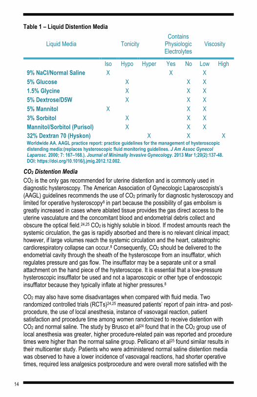
DISTENTION MEDIA Distention media is used to fill the uterus during hysteroscopy to create a large enough space for the surgeon to visualize and perform the procedure effectively. Options for distention media are selected based on the patient’s condition, the procedure to be performed and the electrosurgical device to be used. They can be categorized as gaseous or fluid, but the only gaseous medium in use is CO2. Fluid distending media can be classified according to their osmolality, their electrolyte content, and their viscosity (Table 1). Traditionally, viscosity has been used to group media types, although this has become less beneficial with the declining use of high-viscosity fluids. The goals of fluid management include:8
• choosing distention media least likely to cause complications in the event of excess absorption,
• minimizing systemic absorption, and • early recognition of excess absorption.
14
Table 1 – Liquid Distention Media
Liquid Media Tonicity Contains
Physiologic Electrolytes
Viscosity
Iso Hypo Hyper Yes No Low High 9% NaCl/Normal Saline X X X 5% Glucose X X X 1.5% Glycine X X X 5% Dextrose/D5W X X X 5% Mannitol X X X 3% Sorbitol X X X Mannitol/Sorbitol (Purisol) X X X 32% Dextran 70 (Hyskon) X X X Worldwide AA. AAGL practice report: practice guidelines for the management of hysteroscopic distending media:(replaces hysteroscopic fluid monitoring guidelines. J Am Assoc Gynecol Laparosc. 2000; 7: 167–168.). Journal of Minimally Invasive Gynecology. 2013 Mar 1;20(2):137-48. DOI: https://doi.org/10.1016/j.jmig.2012.12.002.
CO2 Distention Media CO2 is the only gas recommended for uterine distention and is commonly used in diagnostic hysteroscopy. The American Association of Gynecologic Laparoscopists’s (AAGL) guidelines recommends the use of CO2 primarily for diagnostic hysteroscopy and limited for operative hysteroscopy8 in part because the possibility of gas embolism is greatly increased in cases where ablated tissue provides the gas direct access to the uterine vasculature and the concomitant blood and endometrial debris collect and obscure the optical field.24,25 CO2 is highly soluble in blood. If modest amounts reach the systemic circulation, the gas is rapidly absorbed and there is no relevant clinical impact; however, if large volumes reach the systemic circulation and the heart, catastrophic cardiorespiratory collapse can occur.8 Consequently, CO2 should be delivered to the endometrial cavity through the sheath of the hysteroscope from an insufflator, which regulates pressure and gas flow. The insufflator may be a separate unit or a small attachment on the hand piece of the hysteroscope. It is essential that a low-pressure hysteroscopic insufflator be used and not a laparoscopic or other type of endoscopic insufflator because they typically inflate at higher pressures.8
CO2 may also have some disadvantages when compared with fluid media. Two randomized controlled trials (RCTs)24,25 measured patients’ report of pain intra- and post-procedure, the use of local anesthesia, instance of vasovagal reaction, patient satisfaction and procedure time among women randomized to receive distention with CO2 and normal saline. The study by Brusco et al24 found that in the CO2 group use of local anesthesia was greater, higher procedure-related pain was reported and procedure times were higher than the normal saline group. Pellicano et al25 found similar results in their multicenter study. Patients who were administered normal saline distention media was observed to have a lower incidence of vasovagal reactions, had shorter operative times, required less analgesics postprocedure and were overall more satisfied with the
15
procedure. These findings suggest that any advantages of CO2 are limited, although they do not preclude its use in selected circumstances with the appropriate equipment.
Fluid Distention Media The advantage of fluid over gas is its ability to distend the uterus proportionally and effectively flush blood, mucous and small tissue fragments out of the visual field.26 When selecting fluid distention media the ideal medium should allow clear visualization of the uterine cavity, be isotonic, nontoxic, hypoallergenic, non-hemolytic, be rapidly cleared by the body, readily available and inexpensive.
Liquid distension media for hysteroscopy fall into two categories: electrolyte and electrolyte-free. Electrolytes are salts or molecules that ionize completely in solution, therefore, they readily conduct electricity. For this reason they cannot be used with monopolar electrosurgical devices. AAGL recommends electrolyte fluids in hysteroscopy where mechanical, laser or bipolar electrosurgical devices are used.8 Electrolyte fluids (eg, normal saline) also maintain the osmotic gradient between extravascular and intravascular compartments within the body and their sodium concentration makes them plasma-volume expanders. Hyponatremia, where the concentration of sodium in the blood is abnormally low, cannot occur with electrolyte fluids. However, they can cause isotonic fluid overload when intravasation occurs.27,28 This can lead to pulmonary edema and congestive heart failure; therefore, patients should be strictly monitored when using electrolytic solutions as a distention fluid media.29
Electrolyte-free fluids do not dissociate into ions in solution. They are poor conductors of electricity and are recommended for use with monopolar energy. When necessary to use electrolyte-free solutions, the chemical characteristics of the medium should be known. For example, it is important to confirm glucose and fructose tolerance in patients as glucose and sorbitol (which metabolizes into fructose)29 solutions are contraindicated in patients with an intolerance. Dextran 70 can present complications such as, coagulation disorders, allergic reactions and adult respiratory distress syndrome (ARDS). It can also ruin operative hysteroscopes if remnants of the solution is left on instruments that are not cleaned promptly following the procedure.31 Excess absorption of hypotonic agents can cause cerebral edema, which appears to result in the most severe complications of excess absorption, including death.8
RISKS ASSOCIATED WITH FLUID DISTENTION MEDIA The complications of distension media during hysteroscopy are primarily related to fluid absorption and excess air or gas, and depend on the type of medium used and the complexity of the operation. The essential nature of distention media and its potential for complications make it critical that perioperative personnel understand the way they work and how to combat problems.
Air or Gas Embolism Air or gas embolism is rare but can occur during a hysteroscopy with fluid or CO2 distension media. Air can enter the uterine cavity during insertion of the hysteroscope if the inflow tubing is not primed with fluid or due to air bubbles within the distension medium itself. It is important to prime the entire tubing system with CO2 prior to the start
16
of the procedure. CO2 embolism may also occur from the combustion of gases produced when electrosurgery is involved.26 The frequency of severe adverse events is rare because CO2 is soluble in the blood and is readily eliminated from the respiratory system. However, use of inappropriate equipment such as a laparoscopic insufflator to instill CO2 during hysteroscopic surgery, has been acknowledged to cause these complications.
Fluid intravasation Hysteroscopic procedures have the risk for intravasation of uterine distention fluid. Intravasation is a process by which the pressurized irrigation fluid enters the systemic circulation. Intravasation can cause fluid overload leading to electrolyte imbalance and dilutional hypotremia32 and can lead to pulmonary edema, congestive heart failure, and other adverse health conditions. Absorption of distension media into the systemic circulation occurs by retrograde passage of the fluid through the fallopian tubes; via the endometrium; and through opened blood vessels and sinuses during resection of uterine tissue when the intrauterine pressure (IUP) is greater than the pressure in the venous sinus or blood vessel. Factors influencing absorption of distension fluid include IUP, mean arterial pressure, depth of mymetrial penetration, duration of surgery, and size of uterine cavity.8,26
IUP Systemic absorption of fluid increases considerably when IUP exceeds mean arterial pressure; therefore, the higher the pressure, the greater the degree of absorption into the body.
Mean arterial pressure The lower the mean arterial pressure, the lower the IUP required for passage of fluid into the systemic circulation. As a result, caution should be given to elderly patients and those with cardiovascular co-morbidities.
Depth of myometrial penetration When tissue damage extends into the deeper myometrium fluid can be rapidly absorbed through opened myometrial venous sinuses.
Duration of surgery Longer procedures allow more time for fluid to accumulate within the body.
Size of uterine cavity Larger cavities provide a greater endometrial surface area for fluid absorption and procedures will generally be longer.
Fluid Overload Fluid media can potentially cause complications when rapid systemic absorption and expansion of the systemic circulation leads to pulmonary edema and heart failure. Furthermore, conditions of hypoosmolality and hyponatremia can cause fluid to be absorbed into the patient’s brain cells provoking cerebral edema, neurological impairment, seizures and even death. Clinically significant fluid and electrolyte issues are more frequently observed when hypotonic and electrolyte-free distension media are used because they create an osmotic imbalance between extracellular and intracellular fluid.

17
Physiological isotonic solutions are less likely to cause such electrolyte disturbance.26 The degree of systemic fluid absorption, recorded fluid deficit and the type of the distension media used influences symptoms, type and severity of complications. Factors that impact a patient’s predilection for significant complications include osmolality, menopausal status, and cardiovascular and renal disease.
Osmolality Hypotonic electrolyte-free solutions (ie, glycine, mannitol, sorbitol) can cause hyponatremia hypervolemia. If unrecognized and untreated, bradycardia and hypertension can develop and lead to pulmonary edema, cardiovascular collapse and death.
Menopausal status In patients of reproductive age, the brain compensates with the sodium/potassium adenosine triphosphatase (Na/K ATPase) pump regulates the flow of electrolytes through the blood brain barrier. Premenopause places patients at higher risk for developing neurological complications because estrogen has suppressive effects on the Na/K ATPase pump.8
Cardiovascular and renal disease Patients with known cardiovascular disease, renal impairment and the elderly are less likely to adapt to sudden significant increases in intravascular fluid and complications from systemic fluid expansion and electrolyte imbalance are more likely at lower levels of fluid deficit.
The incidence of fluid overload during hysteroscopic surgery is generally low. Several prospective and retrospective studies have looked at the incidence of excessive fluid absorption and electrolyte disturbances during operative hysteroscopy and most report rates under 5%.26 However, collaboration and continuous communication with the surgeon, anesthesia provider and perioperative nurse is paramount. Patients should be carefully monitored during hysteroscopy and consideration should be given to terminating the procedure if fluid overload is known or suspected.8 Data evaluating fluid deficit during hysteroscopic surgery are not pervasive enough to develop a standard definition of fluid overload. A decrease in serum sodium of 10 mmol/L corresponds to an absorbed volume of about 1000 mL (when using 1.5% glycine), therefore a fluid deficit of 1000 mL is traditionally considered the threshold at which procedures should be curtailed in patients of reproductive age when using hypotonic media.8,26 Elderly patients and patients with compromised cardiovascular systems and other comorbidities, a maximum fluid deficit of 750 mL is recommended by American Association of Gynecologic Laparoscopists (AAGL).8,33 AAGL advocates for 2500 mL as a maximum limit for isotonic solutions. High-viscosity distending media should not exceed an infused volume of 500 mL and in elderly patients and those with cardiopulmonary compromise, infused volume should not exceed 300 mL. Patients demonstrating signs or symptoms of fluid overload posthysteroscopy should managed and followed up with in the same manner as symptomatic and asymptomatic patients who reach predefined threshold for fluid overload.

18
Dilutional Hyponatremia Dilutional hyponatremia is a potentially life-threatening condition caused by fluid overload and often referred to as water intoxication.8 It can manifest as mild, moderate or severe and present with an amalgam of symptoms (Table 2). It occurs due to intravasation of large amounts of electrolyte-free fluid, which occurs when the ratio between serum sodium and circulating blood volume falls below normal levels. Normal serum sodium is 135 to 142 mEq/L and values less than that can result in mild, moderate or severe hyponatremia. Patients with mild and severe hyponatremia can deteriorate quickly, resulting in seizures and respiratory arrest rapidly.34
Table 2 – Stages of Hyponatremia
Stage Sodium Level Symptoms Mild 130 – 135 mEq/L Asymptomatic or associated with subtle changes
in mental and physical function (eg, headache, nausea, vomiting, fatigue).
Moderate 125 – 130 mEq/L Nonspecific symptoms (eg, nausea, muscle cramps, confusion ).
Severe <120 mEq/L Progressive neurological symptoms (eg, delirium, restlessness, seizures) ranging from confusion to coma.
Sahay M, Sahay R. Hyponatremia: a practical approach. Indian journal of endocrinology and metabolism. 2014;18(6):760. DOI: 10.4103/2230-8210.141320. Verbrugge FH, Steels P, Grieten L, Nijst P, Tang WW, Mullens W. Hyponatremia in acute decompensated heart failure: depletion versus dilution. Journal of the American College of Cardiology. 2015;65(5):480-92. DOI: 10.1016/j.jacc.2014.12.010. Gajbhiye R, Gajbhiye R. Distension Media and Fluid Management Machines. Mastering the Techniques in Hysteroscopy. 2017:139.
Intravasation of hypotonic electrolyte-free fluids can cause the accumulation of free water, causing the body attempt homeostasis through alternate mechanisms. One of these mechanisms is osmosis, redistributing free water into extracellular and intracellular spaces, can result in cerebral edema and increased intracranial pressure. If left untreated, the persistent swelling of the brain exerts pressure against the skull and can lead to pressure necrosis of the brain. Cerebral herniation can occur if swelling exceeds 5% and immediate preventative steps are not taken. When serum sodium falls below 115 mEq/L, brain stem herniation develops in the swollen brain’s attempt to equalize interstitial and intravascular osmotic pressures. Permanent brain damage, coma, or death may result.34 Hyponatremia during hysteroscopy is especially problematic for premenopausal patients. They are 25 times at greater risk for hyponatremic encephalopathy (HE) and permanent brain damage than postmenopausal women.35 Estrogen and progesterone inhibits sodium pump activity, which protects the brain against cerebral edema. The sodium pump serves to move osmotically-active sodium cations from the brain cells, thus reducing swelling. Postmenopausal women developing dilutional hyponatremia are less likely to suffer brain damage because their low estrogen and progesterone levels allows this sodium pump to operate freely, as opposed to
19
premenopausal patients, whose sodium pump is inhibited with higher levels of estrogen and progesterone.8,36
Treatment for Hyponatremia The health care facility should have protocols designed for the care and treatment of hyponatremia; preoperative nursing assessment is essential to ensure that appropriate protocols are implemented. These protocols should include accurately monitoring and controlling the flow of distention media to avoid complications of intravasation. During the procedure the perioperative nurse should calculate the amount of distention fluid instilled minus the amount recovered (fluid deficit), identify evidence of fluid overload and decrease serum sodium concentration prior to signs of adverse effects. The nurse should observe for signs and symptoms of adverse neurological and cardiovascular effects such as muscle twitching, seizures, hypotension, and tachycardia, which can lead to pulmonary and cerebral edema, cardiovascular collapse, and death. In the even if severe symptoms prompt treatment can avoid more serious side effects of fluid overload.37,38
The treatment of hyponatremia can be divided into two steps. First, a decision must be made whether immediate treatment is required. This decision is a collaborative decision between the surgeon and anesthesia provider based on the symptoms, the degree of hyponatremia and the presence of any degree of hypotension. Second, the most appropriate method of correcting the hyponatremia must be determined.37 When intravasation occurs with isotonic solution combinations (eg, 5% mannitol or mannitol/sorbitol mixtures) specific treatment may not be necessary as these fluids have a diuretic effect and the body may expel the excess fluid, rebalancing the sodium levels. When other fluid media is used, it may be necessary to administer normal saline to stabilize serum sodium levels while avoiding the potential for overcorrection. The quantity of sodium chloride required to achieve the desired elevation in the plasma sodium concentration can be estimated by multiplying the plasma sodium deficit per liter with the total body water, which represents the osmotic space of distribution of the plasma sodium concentration. Normal values for the total body water are 0.5 the lean body weight (in kilograms) in women.38 Overcorrection of serum sodium to more than 135 mEq/L may result in adverse cerebral effects, necessitating the administration of diuretics.34 In patients with severe symptomatic hyponatremia, the rate of sodium correction should be 6 to 12 mEq per L in the first 24 hours and 18 mEq per L or less in 48 hours. An additional 8 mmol/L during every 24 hours thereafter until serum sodium concentration reaches 130 mmol/L. A bolus of 100 to 150 mL of hypertonic 3% saline can be given to correct severe hyponatremia.39
NURSING RESPONSIBILIES IN HYSTEROSCOPY Preparing a patient and the procedural team for hysteroscopy has several detailed steps that are guided by hospital, facility and perioperative department policies and procedures. The perioperative nurse should be able to identify potential patient injuries and complications that are associated with using various distention media, MIS technology and other surgical instruments. Knowledge of risk factors, monitoring practices, risk-reduction strategies, treatment for complications and safe practices can lead to optimum outcomes for patients.29 These policies, procedures and practices should be based on the standards, guidelines and recommendations developed by

20
AAGL, the Association of Operative Registered Nurses (AORN),40 and the American College of Obstetricians and Gynecologists (ACOG)41 that are developed to help the perioperative team create an environment that reduces the risk for injury to patients and the team members during hysteroscopy and MIS. The expected outcome being that the patient is free from signs and symptoms of injury or complications.
Preoperative Responsibilities The perioperative nurse is responsible for conducting a preoperative assessment to determine the patient’s individual risk factors related to hysteroscopy. This should include the patient’s age, weight, skin condition, comorbidities (congestive heart failure, kidney disorders), baseline serum sodium levels and risk factors related specifically to positioning, fluid management and medications (ie, diuretics, anticonvulsants, serotonin, norepinephrine inhibitors).29,37,42 To help minimize patient risks related to MIS procedures, the perioperative nurse should consult and collaborate with surgical team members as appropriate to implement corrective measures.
The perioperative nurse should set up the procedure room in a manner in which essential equipment and instruments are in proper working conditions and readily assessable. If an automated fluid management system is used pump settings should be verified with the surgeon before administration and continuously monitored the throughout the procedure. Alarms on the fluid management system and MIS equipment should be set so that they are audible above competing noise in the procedure room. To ensure patient safety, fluids used for irrigation and distention media should be:29
• appropriately warming and cooling fluids used for irrigation and distention media at temperatures other than room temperature,
• segregating sterile water from other irrigation solutions to reduce the risk of administering the incorrect fluid,
• following fluid manufacturers IFUs (instructions for use) on warming and cooling,
• not overheating fluids and maintaining fluid temperature according to the safe temperature range as determined by manufacturer’s IFUs,
• rotating inventory and labeling fluid containers with expiration dates to diminish waste, and
• placing a ‘do not rewarm’ label on unopened fluid containers that are taken from warming cabinets.
Additionally, endoscopic instruments can be used during hysteroscopy and may increase the risk for patient injury by perforating or by contributing to formation of an embolism. Perioperative nurses should be knowledgeable about the techniques and potential risks of any additional procedures associated with the hysteroscopy.28
There is evidence on the value of administering gonadotropin-releasing hormone (GnRH) agonists preoperatively to reduce the amount of systemic absorption of distending media and the impact of hyponatremia. GnRH agonists are a group of drugs that have been used for several decades to treat patients with endometriosis. Preoperative use of GnRH agonists has generally been associated with reduced fluid deficit among premenopausal

21
women and may decrease the morbidity associated with fluid overload of nonionic hypotonic media. Studies have shown that giving preoperative GnRH equivalents when undertaking resection of the myoma or endometrium reduces the incidence of fluid overload.43,44 Taskin et al,45 reported a statistical difference in the reduction of serum sodium concentration but not in glycine deficit in women treated preoperatively with GnRH agonists. Another study did not show a statistically significant difference in fluid deficit with the use of GnRH agonists. 46
Intraoperative Responsibilities Patients who undergo hysteroscopy may become and experience more discomfort during the procedure. As a result, the perioperative nurse should coordinate with the surgeon to implement a care plan to minimize the patient's anxiety. The nurse can provide emotional support to the patient and, if comfortable, invite the patient to view the monitor and explaining the procedure. The patient should also monitor the patient’s body temperature as continuous flow of distention media can lead to a lowering in the patient’s body temperature.29
Perioperative nurses should elevate the insufflator above the level of surgical cavity and flush the insufflator and insufflation tubing with gas or liquid before connecting the tubing. CO2 insufflators should be filtered with a single-use, hydrophobic filter compatible with the insufflator and impervious to fluids. Insufflator pressures should be monitored throughout the procedure.29 Control of the IUP intraoperatively has been shown to reduce the amount of fluid absorption by almost 85 percent.47 To visualize the uterine cavity an IUP of between 70 and 100 mmHg of distension medium is required to separate the uterine surfaces. Some studies have suggested keeping the IUP between 45 and 80 mmHg as pressures may exceed the mean arterial pressure increasing the likelihood of rapid fluid absorption.48,49 Filling pressures of up to 100 mmHg have been found to be effective and safe in some outpatient hysteroscopy procedures.50 Two trials also demonstrated that injecting dilute vasopressin into the cervix immediately before dilation can decrease fluid absorption.51,52
Precautions will be taken when using energy-generating devices during MIS. Perioperative nurses should verify the properties of the distention media to minimize risks related to use of energy-generating devices. Only nonflammable insufflation gas (eg, CO2) should be used. Electrolyte-free distention fluids should be used with monopolar electrosurgery while electrolyte distention fluids can be used with bipolar electrosurgery. The lowest power setting that achieves the desired result should always be used.29
Monitoring Fluid Deficit and Calculation The perioperative nurse has a critical role in reducing the risk of injury to patients associated with fluid used for distention media during hysteroscopy. Fluid administered to the patient should be collected in a closed container system; automated fluid management systems require the nurse to verify the pump settings with the surgeon before administration. Pump pressure, inflow and outflow volumes and fluid deficit should be reported to the anesthesia provider and surgeon. The patient should be monitored for signs and symptoms of hypervolemia and hyponatremia. 29 One nurse should be dedicated to carry out the fluid measurement, calculate the fluid balance and communicate it to the surgeon and anesthesia provider.
22
The detection of excess absorption requires accurate measurement of both media infused into the uterus cavity, fluid returned from the outflow channel of the hysteroscope and fluid expelled from the uterus from other sources. Liquid distention media can be delivered via an open or closed system. In an open system, the medium freely escapes through the cervix and the outflow channel onto a surgical drape and into a drape with a fluid reservoir or the procedure room floor making precise fluid monitoring challenging and often inaccurate. In a closed system, the liquid distention media is returned through suction to a reservoir, which also improves visibility by removing debris and blood from the uterine cavity. Even with suction, fluid can still escape through the cervix and the perineum; therefore, drapes with a fluid reservoir should still be used.
Significant fluid intravasation can occur in a very short period of time, causing very rapid changes in serum sodium levels and resulting in severe dilutional hyponatremia; therefore, accurate measurement of fluid absorption enables surgical team members to correct problems. Calculation of systemic fluid absorption is complicated by:
• It can be difficult to collect all of the media that passes out of the uterus. • The volume of media solution in 3 L infusion bags is generally more than the
labeled volume.43 • Estimating the volume of media left in a used or emptied infusion bag may be
difficult. Fluid calculations can be done using a manual or automated methods that both instills and calculates fluid deficit. When manual calculation is the only method available, it is strongly recommended that one nurse be assigned to monitor and frequently report the calculations to the surgical team and another nurse assigned to patient care during the procedure. The simplest method of monitoring involves manually subtracting the volume collected from the volume infused taking into account all sources including the hysteroscope outflow, the collection drape and fluid reservoir and the media spilled that collects on the floor.26 While conceptually simple, several challenges are associated with attempting to collect media from all sources in the procedure room. First, inaccuracy occurs because media lost onto the drapes and the floor can confound the issue, making it difficult to evaluate the intake and output accurately. Another problem is the accurate estimation of fluid used from infusion bags can be problematic and manual calculation can result in as much as a 10% margin of error.53 A study of bags of normal saline, glycine, and sorbitol found that the average overfill was between 3% and 6%,54 enabling the opportunity for undetected fluid overload. Accurate estimation of fluid within infusion bags intra- and post-procedure is poor and errors range from 4% to 50%.55 Significant bleeding during hysteroscopy can also make the calculation less reliable because the outflow can appear more than the actual value. These issues are intensified if the nurse monitoring fluid has additional duties that occupy their attention during the procedure. The limitations of manual calculation make it preferable to use automated fluid management system that take into account an exact measurement of infused volume as well as all of the potential sources of returned media.39
23
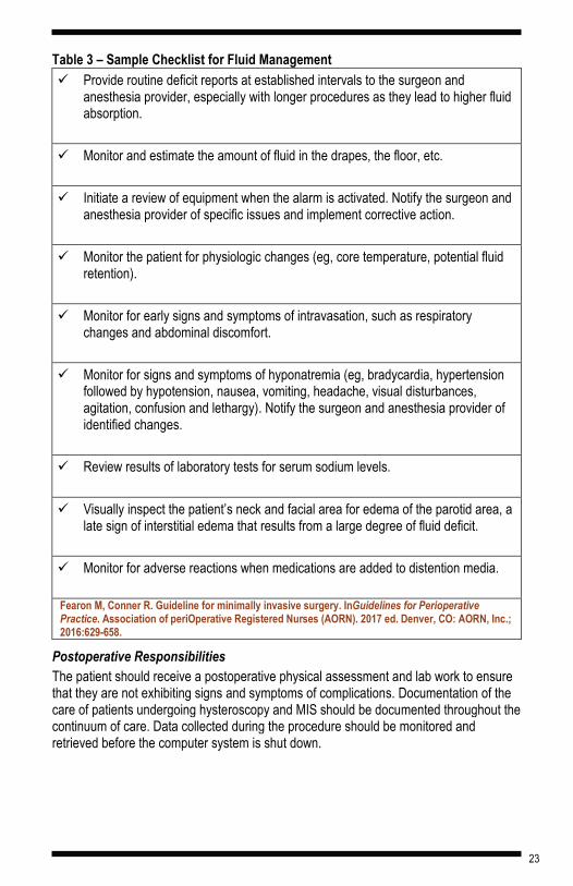
Table 3 – Sample Checklist for Fluid Management Provide routine deficit reports at established intervals to the surgeon and
anesthesia provider, especially with longer procedures as they lead to higher fluid absorption.
Monitor and estimate the amount of fluid in the drapes, the floor, etc.
Initiate a review of equipment when the alarm is activated. Notify the surgeon and anesthesia provider of specific issues and implement corrective action.
Monitor the patient for physiologic changes (eg, core temperature, potential fluid retention).
Monitor for early signs and symptoms of intravasation, such as respiratory changes and abdominal discomfort.
Monitor for signs and symptoms of hyponatremia (eg, bradycardia, hypertension followed by hypotension, nausea, vomiting, headache, visual disturbances, agitation, confusion and lethargy). Notify the surgeon and anesthesia provider of identified changes.
Review results of laboratory tests for serum sodium levels.
Visually inspect the patient’s neck and facial area for edema of the parotid area, a late sign of interstitial edema that results from a large degree of fluid deficit.
Monitor for adverse reactions when medications are added to distention media.
Fearon M, Conner R. Guideline for minimally invasive surgery. InGuidelines for Perioperative Practice. Association of periOperative Registered Nurses (AORN). 2017 ed. Denver, CO: AORN, Inc.; 2016:629-658.
Postoperative Responsibilities The patient should receive a postoperative physical assessment and lab work to ensure that they are not exhibiting signs and symptoms of complications. Documentation of the care of patients undergoing hysteroscopy and MIS should be documented throughout the continuum of care. Data collected during the procedure should be monitored and retrieved before the computer system is shut down.
24
Documentation should include:29
• type of distention media used, • amount of distention media used, • rate used to deliver the distention media, • equipment used to deliver the distention media (including equipment model
and identification number), • quantity of fluid returned, • any fluid deficit, • medication added to the distention fluid, and • relevant information about the equipment that was used (eg, insufflation,
electrosurgery, positioning).
METHODS AND EQUIPMENT USED TO CALCULATE AND MONITOR FLUID DEFICIT Intrauterine distension pressure can be maintained using gravity, manual and automated pressure delivery systems. Gravity is the simplest method and instills fluid under constant hydrostatic pressure. The fluid bag is generally hung from an intravenous (IV) pole and initially placed at a height above the patient’s uterus that creates an intrauterine pressure that is just below the patient’s mean arterial pressure.8 The pressure delivered to the inflow port of the hysteroscope’s outer sheath is the product of the inner diameter plane of the connective tubing and the elevation. Elevation of the bag will increase the intrauterine pressure and 0.3 m of height will approximate to around 25 mm of Hg.56
When the fluid is maintained at a level of 1 m to 1.5 m above the patient’s uterus, the intrauterine pressure will be between 70 to 100 mmHg.57 The height should be kept at the minimum elevation to allow sufficient distention.
An extension of the gravity system is the simple pressurized delivery system that is created by positioning a pressure cuff around the bag filled with the distending media. This approach does not allow for precise control of the pressure and lengthy cases, or those associated with intrusion of the myometrium, excessive extravasation could occur. This is particularly the case if intrauterine pressure is sustained above the mean arterial pressure. The disadvantage of these manual approaches is that they keep the flow and pressure at the inflow port constant, therefore if the pressure exceeds the mean arterial pressure it can lead to excessive fluid absorption. Irrigation of fluid is achieved by partially or fully opening the outflow tap and applying varying amounts of suction.26
Automated Methods The importance and function of automated fluid management systems are primarily to maintain patient safety and enhance procedural effectiveness.58 Without successful fluid management procedures could be stopped prior to a completion due to excessive fluid absorption or risk for complications because of fluid overload.59 A variety of infusion pumps are available, ranging from simple devices with constant flow and pressure at the inflow port to equipment that also monitors and maintains a preset intrauterine pressure. Simple pump devices continue to press fluid into the uterine cavity regardless of
25
resistance, whereas pressure-sensitive pumps titrate the flow rate when the preset level is achieved. In these systems, if the preset IUP cannot be achieved, the system should be checked for leaks. Provided there are methods for collection from all sources, the device can calculates the total weight of all the media collected by the system, which is then subtracted from the total weight of the infused volume to provide a continuous measure of systemically absorbed volume. An alarm can be set to warn when a preset volume deficit is reached. The actual measurement of the infused volume prevents underestimating the fluid deficit although it does not prevent overestimating the deficit because of fluid not recovered.8 They can minimize procedure time by rapidly achieving and holding constant distention. They offer a system pause capability as needed and the ability to replace infusion bags without using the pause function and real-time accurate deficit readings. Newer systems are designed to optimize high aspiration procedures and support the use of tissue removal devices. Automated fluid management systems are not necessary for short operative or diagnostic procedures but maybe beneficial for prolonged, operative cases such as resection of the endometrium or submucosal fibroids where endometrial and myometrial disruption occur causing bleeding and the formation of intrauterine tissue debris that can compromise visualisation of the operative field.26 Some automated fluid management systems have two integrated vacuum pumps. One pump is designated for draining the drape under the patient’s bottom and the second is capable of generating up to 500 mm Hg of suction which eliminates the need for additional tubing to connect the pump to wall suction. This allows the system to be used even when external suction available (ie, physician’s office). Some systems also offer automatic lumen calibration to insure proper inflow rates even when different caliber hysteroscopes are used.60
As with any technology, there is opportunity to improve existing automated fluid management systems. For example, many systems involve complicated set up and breakdown that is time consuming for a busy perioperative staff. Nonintuitive and complex user interfaces can make it difficult to quickly understand and troubleshoot unexpected deficit and pressure levels. Automated fluid management systems that capitalize simplicity and accuracy will go a long way in improving the ways we manage fluid absorption during hysteroscopy.61 Overall, AAGL recommends that each facility’s media management protocol includes the use of automated fluid management systems.8
SUMMARY Hysteroscopy enables visualization of the uterine cavity to diagnosis and treatment intrauterine conditions. CO2 gas distention media is mostly used for diagnostic hysteroscopy, but because bleeding during operative procedures obscures visibility, liquid media is used for operative procedures to allow continuous irrigation. During operative hysteroscopy, absorption of large volumes of liquid distension media can lead to serious complications arising from significant fluid overload. Thus, particular care is required with prolonged hysteroscopic procedures requiring continuous irrigation of fluid or where blood vessels are opened.
Perioperative nurses have an important role in preparing for and preventing serious complications from operative hysteroscopy, including correct fluid distention media and accurate monitoring of fluid absorption. Patient complications may be prevented with the

26
use of proper precautions and maintaining effective communication between members of the perioperative team. Health care facilities may consider using an automated means to measure and monitor fluid deficit, which can be more efficient and effective than manual means of calculating fluid deficit.

27
GLOSSARY
Adhesions Scars that bind together affected surfaces of the tissues inside the abdomen or uterus.
Automated fluid management system
Mechanical medical devices designed to calculate the amount of fluid dispensed to the patient compared to the amount returned to the system; alarms alert the user to fluid deficit to prompt corrective action.
Biopsy A minor surgical procedure to remove a small piece of tissue that is then examined under a microscope in a laboratory.
Cervix The opening of the uterus at the top of the vagina.
Deficit The total amount of fluid left in the patient or unaccounted for otherwise (the fluid left in the patient must be monitored).
Dilation & Curettage (D&C)
A surgical procedure in which the cervix is dilated and the endometrium of the uterus is scraped to determine the cause of abnormalities.
Electrolyte An ionized salt (such as sodium, potassium and chlorine) in blood, tissue fluids and cells that conducts electricity and provides the means by which electrochemical impulses are transmitted in nerve and muscle fibers.
Electrolytic Solution A fluid that can conduct electricity; recommended by AAGL for use in diagnostic cases and in operative cases in which mechanical, laser or bipolar energy is used. Electrolytic solutions cannot be used in conjunction with monopolar electrosurgical devices.
28
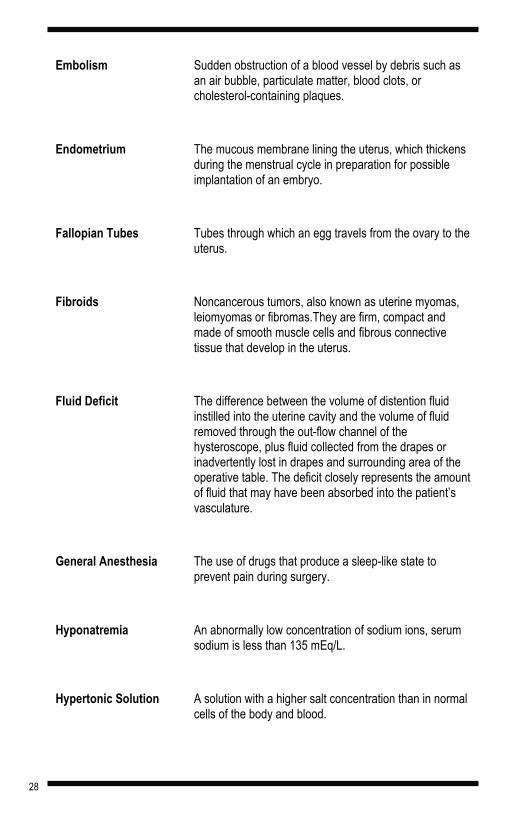
Embolism Sudden obstruction of a blood vessel by debris such as an air bubble, particulate matter, blood clots, or cholesterol-containing plaques.
Endometrium The mucous membrane lining the uterus, which thickens during the menstrual cycle in preparation for possible implantation of an embryo.
Fallopian Tubes Tubes through which an egg travels from the ovary to the uterus.
Fibroids Noncancerous tumors, also known as uterine myomas, leiomyomas or fibromas.They are firm, compact and made of smooth muscle cells and fibrous connective tissue that develop in the uterus.
Fluid Deficit The difference between the volume of distention fluid instilled into the uterine cavity and the volume of fluid removed through the out-flow channel of the hysteroscope, plus fluid collected from the drapes or inadvertently lost in drapes and surrounding area of the operative table. The deficit closely represents the amount of fluid that may have been absorbed into the patient’s vasculature.
General Anesthesia The use of drugs that produce a sleep-like state to prevent pain during surgery.
Hyponatremia An abnormally low concentration of sodium ions, serum sodium is less than 135 mEq/L.
Hypertonic Solution A solution with a higher salt concentration than in normal cells of the body and blood.

29
Hysteroscopy Visual examination of the uterine cavity with a small endoscope passed through the cervix.
Insufflation The act of blowing gas into a body cavity or the state of being distended with gas for the purpose of visual examination.
Intrauterine Device A small plastic device inserted in the uterus to prevent pregnancy.
Intrauterine Pressure The amount of pressure exerted against the walls of the uterine cavity. The IUP is influenced by the distention fluid installation pressure, which may be dictated by the infusion pump pressure setting or by gravity flow.
Isotonic Solution A solution that has the same salt concentration as the normal cells of the body and blood.
Local Anesthesia The use of drugs that prevent pain in a part of the body.
Mean Arterial Pressure The average force applied to arterial walls through the cardiac cycle. It is a function of cardiac output, systemic vascular resistance, and central venous pressure. MAP can be determined by using the formula: Mean arterial pressure = diastolic blood pressure + 1/3 pulse pressure. (Pulse pressure = systolic – diastolic blood pressure.) The usual range is 70-110.
Minimally invasive surgery (MIS)
Surgical procedures performed through one or more small incisions using endoscopic instruments, radiographic and magnetic resonance imaging, computer-assisted devices, robotics, and other emerging technologies.
30
Miscarriage Loss of pregnancy.
Osmolity A measure of the number of dissolved particles in a fluid. The osmolality test reflects the concentration of substances such as sodium.
Polyps Overgrowth of cells in endometrium.
Sodium/Potassium Adenosine Triphosphatase (ATPase) Pump
An enzyme which performs the active transport of sodium and potassium across the cell membrane.
Speculum An instrument used to open the walls of the vagina.
Sterilization A permanent method of birth control.
Tenaculum A surgical clamp with sharp hooks used to hold or pick up small pieces of tissue such as the cervix during hysteroscopy.
Uterus A muscular organ located in the female pelvis that contains and nourishes the developing fetus during pregnancy.
Vagina A tube-like structure surrounded by muscles leading from the uterus to the outside of the body.
31
REFERENCES 1. Sardo AD, Calagna G, Di Carlo C. Tips and tricks in office hysteroscopy.
Gynecology and Minimally Invasive Therapy. 2015;4(1):3-7. DOI: https://doi.org/10.1016/j.gmit.2014.12.004.
2. The American College of Obstetricians and Gynecologists (ACOG). Hysteroscopy FAQs. https://www.acog.org/Patients/FAQs/Hysteroscopy. October 2011. Accessed June 15, 2018.
3. The American College of Obstetrician and Gynecologists (ACOG). Hysterosalpingography. https://www.acog.org/Patients/FAQs/Hysterosalpingography. Accessed June 10, 2018.
4. Pandey D, Kunamneni S, Inukollu PR, Su H. Establishing patterns on hysteroscopy in abnormal uterine bleeding (AUB). Gynecol Minim Invasive Ther. 2017;6(4):178-82.DOI: https://doi.org/10.1016/j.gmit.2017.08.001.
5. Bosteels J, Kasius JC, Weyers S, Broekmans FJ, Mol BW, D'Hooghe T. Hysteroscopy for treating subfertility associated with suspected major uterine cavity abnormalities. Cochrane Database of Systematic Reviews. 2013;(1). DOI: 10.1002/14651858.CD009461.pub2.
6. Yeh J. Diagnostic hysteroscopy. Medscape. https://emedicine.medscape.com/article/1848258-overview. Updated May 22, 2018. Accessed June 17, 2018.
7. Jansen FW, Vredevoogd CB, van Ulzen K, Hermans J, Trimbos JB, Trimbos-Kemper TC. Complications of hysteroscopy: a prospective, multicenter study. Obstet Gynecol. 2000;96(2):266-70. [DOI: 10.1016/S0029-7844(00)00865-6.
8. Worldwide AA. AAGL practice report: practice guidelines for the management of hysteroscopic distending media:(replaces hysteroscopic fluid monitoring guidelines. J Am Assoc Gynecol Laparosc. 2000; 7: 167–168. J Minim Invasive Gynecol. 2013;20(2):137-48. DOI: https://doi.org/10.1016/j.jmig.2012.12.002.
9. Petrozza J. Hysteroscopy. Medscape. https://emedicine.medscape.com/article/267021-overview. Updated December 30, 2015, Accessed June 17, 2018.
10. Unfried G, Wieser F, Albrecht A, Kaider A, Nagele F. Flexible versus rigid endoscopes for outpatient hysteroscopy: a prospective randomized clinical trial. Hum Reprod. 2001;16(1):168-71. DOI: https://doi.org/10.1093/humrep/16.1.168.
11. Bettocci S, Di Spiexzo Sardo A, Ceci O. Instrumentation in office hysteroscopy: Rigid hysteroscopy. In Hysteroscopy: Office Evaluation and Management of the Uterine Cavity. Mosby Elsevier. Philadelphia, PA. 2009.

32
12. Jacobs VR, Paepke S, Schwarz-Boeger U, Fischer T, von Steinburg SP, Plattner B, Schmalfeldt B, Schaaf H, Kiechle M. Development of a thinner and more flexible type of minihysteroscope with a controlled 90-degree bendable tip for vision-guided endometrium biopsy. Journal of Minimally Invasive Gynecology. 2005;12(5):426-31. DOI: https://doi.org/10.1016/j.jmig.2005.06.013.
13. Bradley L. Instrumentation in office hysteroscopy: Flexible hysteroscopy. In Hysteroscopy: Office Evaluation and Management of the Uterine Cavity. Mosby Elsevier. Philadelphia, PA. 2009.
14. Burlingame B, Davidson J, Denholm B, et al. Guideline for positioning the patient. In: Guidelines for Perioperative Practice. 2017;1. DOI: 10.6015/psrp.17.01.e1.
15. Kuponiyi O, Alleemudder DI, Latunde‐Dada A, Eedarapalli P. Nerve injuries associated with gynaecological surgery. Obstet Gynaecol. 2014;16(1):29-36. DOI: 10.1111/tog.12064.
16. Centers for Disease Control and Prevention (CDC). Deep vein thrombosis (DVT). https://www.cdc.gov/healthcommunication/toolstemplates/entertainmented/tips/DeepVeinThrombosis.html. Updated September 15, 2017. Accessed June 17, 2018.
17. Cooper NA, Khan KS, Clark TJ. Local anaesthesia for pain control during outpatient hysteroscopy: systematic review and meta-analysis. BMJ. 2010;340:c1130. DOI: https://doi.org/10.1136/bmj.c1130.
18. Munro MG, Brooks PG. Use of local anesthesia for office diagnostic and operative hysteroscopy. Journal of Minimally Invasive Gynecology. 2010;17(6):709-18. DOI: https://doi.org/10.1016/j.jmig.2010.07.009.
19. de Carvalho Schettini JA, de Amorim MM, Costa AA, Neto LC. Pain evaluation in outpatients undergoing diagnostic anesthesia-free hysteroscopy in a teaching hospital: a cohort study. J Minim Invasive Gynecol. 2007;14(6):729-35. DOI: https://doi.org/10.1016/j.jmig.2007.05.009.
20. Campo R, Molinas CR, Rombauts L, Mestdagh G, Lauwers M, Braekmans P, Brosens I, Van Belle Y, Gordts S. Prospective multicentre randomized controlled trial to evaluate factors influencing the success rate of office diagnostic hysteroscopy. Hum Reprod. 2005 ;20(1):258-63. DOI: https://doi.org/10.1093/humrep/deh559.
21. Baxter AJ, Beck B, Phillips K. A randomized prospective trial of rigid and flexible hysteroscopy in an outpatient setting. Gynaecol Endosc. 2002;11(6):357-64. DOI: https://doi.org/10.1111/j.1365-2508.2002.00562.x.
22. Unfried G, Wieser F, Albrecht A, Kaider A, Nagele F. Flexible versus rigid endoscopes for outpatient hysteroscopy: a prospective randomized clinical trial. Hum Reprod. 2001;16(1):168-71. https://doi.org/10.1093/humrep/16.1.168.
33
23. Bettocchi S, Nappi L, Ceci O, Selvaggi L. What does ‘diagnostic hysteroscopy’mean today? The role of the new techniques. Curr Opin Obstet Gynecol. 2003;15(4):303-8. DOI: 10.1097/01.gco.0000084241.09900.c8.
24. Brusco GF, Arena S, Angelini A. Use of carbon dioxide versus normal saline for diagnostic hysteroscopy. Fertil Steril. 2003;79:993–997. DOI: https://doi.org/10.1016/S0015-0282(02)04947-6.
25. Pellicano M, Guida M, Zullo F, Lavitola G, Cirillo D, Nappi C. Carbon dioxide versus normal saline as a uterine distension medium for diagnostic vaginoscopic hysteroscopy in infertile patients: a prospective, randomized, multicenter study. Fertil Steril. 2003;79:418–421. DOI: https://doi.org/10.1016/S0015-0282(02)04681-2.
26. Umranikar S, Clark TJ, Saridogan E, Miligkos D, Arambage K, Torbe E, Campo R, Sardo AD, Tanos V, Grimbizis G. BSGE/ESGE guideline on management of fluid distension media in operative hysteroscopy. Gynecol Surg. 2016;13(4):289. DOI: https://doi.org/10.1007/s10397-016-0983-z.
27. Schafer M, Von Ungern-Sternberg BS, Wight E, Schneider MC. Isotonic fluid absorption during hysteroscopy resulting in severe hyperchloremic acidosis. Anesthesiol. 2005;103:203–204. DOI: 10.1097/00000542-200507000-00029.
28. Van Kruchten PM, Vermelis JM, Herold I, Van Zundert AA. Hypotonic and isotonic fluid overload as a complication of hysteroscopic procedures: two case reports. Minerva Anestesiol. 2010;76. 373–277. PMID: 20395900.
29. Fearon M, Conner R. Guideline for minimally invasive surgery. In: Guidelines for Perioperative Practice. Association of periOperative Registered Nurses (AORN). 2018 ed. Denver, CO: AORN, Inc.; 2018:611-639.
30. Fernández-Bañares F, Esteve M, Viver JM. Fructose-sorbitol malabsorption. Current gastroenterology reports. 2009 ;11(5):368-74. https://doi.org/10.1007/s11894-009-0056-9.
31. Bradley L. Cutting the risk of hysteroscopic complications. OBG Manag. 2004;16(1):44-52.
32. MacKenzie PP, Chandra SC, Tiffanny JL, Zaraq K. Rapid Fluid Intravasation at Time of Hysteroscopy Without Sign of Perforation. J Minim Invasive Gynecol. 2017;24(7):S191. DOI: https://doi.org/10.1016/j.jmig.2017.08.575.
33. American Association of Gynecologic Laparoscopists (AAGL). About AAGL. https://www.aagl.org/service/about-aagl/. Accessed June 17, 2018.
34. Yaprak M, Turan MN, Tamer AF, Peker N, Demirci MS, Cırpan T, Aşçı G. How quickly can acute symptomatic hyponatremia be corrected?. International urology and nephrology. 2013;45(6):1805-8. DOI: https://doi.org/10.1007/s11255-012-0291-0.
35. Sahni M, Lara-Torre E. Hysteroscopy. In:Gynecologic Care. Cambridge University Press. New York, NY. 2018;2(8):72.

34
36. Look GF. Guideline for minimally invasive surgery. AORN J. 2016; 104(5): 7-9. DOI: https://doi.org/10.1016/S0001-2092(16)30741-4.
37. Goh K. Management of hyponatremia. Am Fam Physician. 2004;69(10):2387-94. http://the-medical-dictionary.com/demeclocycline_686_article_2.htm. Accessed June 15, 2017.
38. Al-Salman J, Kemp D, Randall D. Evidence-based case reviews: hyponatremia. Western J Med. 2002;176(3):173. PMID:12016240.
39. Sahay M, Sahay R. Hyponatremia: a practical approach. Indian J Endocrinol Meta. 2014;18(6):760. DOI: 10.4103/2230-8210.141320.
40. Association of Operative Registered Nurses (AORN). Who we are. https://www.aorn.org/aorn-foundation/who-we-are. Accessed June 17, 2018.
41. American College of Obstetricians and Gynecologists (ACOG). About us. https://www.acog.org/About-ACOG/About-Us. Accessed June 17, 2018.
42. Eaton R. Detection of Hyponatremia in PACU. J Peri Anesth Nurs. 2003;186(6):392-397. DOI: https://doi.org/10.1016/j.jopan.2003.08.004.
43. Muzii L, Boni T, Bellati F, Marana R, Ruggiero A, Zullo MA, Angioli R, Panici PB. GnRH analogue treatment before hysteroscopic resection of submucous myomas: a prospective, randomized, multicenter study. Fertil Steril. 2010 ;94(4):1496-9. DOI: https://doi.org/10.1016/j.fertnstert.2009.05.070.
44. O'Connor H, Magos A. Endometrial resection for the treatment of menorrhagia. N Eng J Med. 1996;335(3):151-6. DOI: 10.1056/NEJM199607183350302.
45. Taskin O, Yalcinoglu A, Kucuk S, Burak F, Ozekici U, Wheeler JM. The degree of fluid absorption during hysteroscopic surgery in patients pretreated with goserelin. J Am Assoc Gynecol Laparosc. 1996;3(4):555–559. DOI: 10.1016/S1074-3804(05)80167-7.
46. Mavrelos D, Ben-Nagi J, Davies A, Lee C, Salim R, Jurkovic D. The value of pre-operative treatment with GnRH analogues in women with submucous fibroids: a double-blind, placebo-controlled randomized trial. Hum Reprod. 2010;25(9):2264-9. DOI: https://doi.org/10.1093/humrep/deq188.
47. Garry R, Hasham F, Kokri M, Mooney P. The effect of pressure on fluid absorption during endometrial ablation. J Gynecol Surg. 1992;8(1):1–10. DOI: 10.1089/gyn.1992.8.1.
48. Vulgaropulos SP, Haley LC, Hulka JF. Intrauterine pressure and fluid absorption during continuous flow hysteroscopy. Am J Obstet Gynecol. 1992 Aug 1;167(2):386-91. DOI: https://doi.org/10.1016/S0002-9378(11)91417-3.
49. Paschopoulos M, Kaponis A, Makrydimas G, Zikopoulos K, Alamanos Y, O'donovan P, Paraskevaidis E. Selecting distending medium for out-patient hysteroscopy. Does it really matter?. Hum Reprod. 2004;19(11):2619-25. DOI: https://doi.org/10.1093/humrep/deh464.
35
50. Shahid A, Pathak M, Gulumser C, Parker S, Palmer E, Saridogan E. Optimum uterine filling pressure for outpatient diagnostic hysteroscopy: a double-blind, randomized controlled trial. Reprod Biomed Online. 2014;28(1):86-91. DOI: https://doi.org/10.1016/j.rbmo.2013.07.018.
51. Corson SL, Brooks PG, Serden SP, Batzer FR, Gocial B. Effects of vasopressin administration during hysteroscopic surgery. J Reprod Med. 1994;39:419–423. PMID: 7932392.
52. Phillips DR, Nathanson HG, Milim SJ, Haselkorn JS, Khapra A, Ross PL. The effect of dilute vasopressin solution on blood loss during operative hysteroscopy: a randomized controlled trial. Obstet Gynecol. 1996;88:761–766. DOI: https://doi.org/10.1016/0029-7844(96)00282-7.
53. Morrison D. Management of hysteroscopic surgery complications. AORN J. 1999;69(1):194-209. DOI: https://doi.org/10.1016/S0001-2092(06)62769-5.
54. Nezhat CH, Fisher DT, Datta S. Investigation of often-reported ten percent hysteroscopy fluid overfill: is this accurate?. J Minim Invasive Gynecol. 2007;14(4):489-93. DOI: https://doi.org/10.1016/j.jmig.2007.03.007.
55. Boyd HR, Stanley C. Sources of error when tracking irrigation fluids during hysteroscopic procedures. The J Am Assoc Gynecol Laparosc. 2000;7(4):472-6. DOI: https://doi.org/10.1016/S1074-3804(05)60359-3.
56. March C. Hysteroscopic fluid monitoring guidelines. J Am Assoc Gynecol Laparosc. 2000;7(3):437-8. DOI: 10.1016/S1074-3804(05)60499-9.
57. Munro MG. Complications of hysteroscopic and uterine resectoscopic surgery. Obstet Gynecol Clinics. 2010;37(3):399-425. DOI: https://doi.org/10.1016/j.ogc.2010.05.006.
58. Gajbhiye R, Gajbhiye R. Distension media and fluid management machines. InMastering the Techniques in Hysteroscopy. Jaypee Brothers Medical Publishers. 2017;142.
59. Coomarasamy A. Use of excessive Distension Media at hysteroscopy. Gynecologic and Obstetric Surgery: Challenges and Management Options. New Dehli, IN. 2016 Jan 27:194.
60. Greenberg JA. AquilexTM Fluid Control System. Rev Obstet Gynecol. 2012; 5(3-4): e173–e174. PMCID: PMC3594854.
61. Randell M. Fluid management: A closer look at a vital element of hysteroscopy. Published March 8, 2018. Accessed June 17, 2018.
36
POST-TEST Multiple choice/True or False. Complete the Test Questions. Missed questions will offer the opportunity to re-read the question and answer choices. You may also revisit relevant content.
1. In some cases, hysteroscopy can be an alternative to hysterectomy. a. True. b. False.
2. Hysteroscopies use gas or liquid to distend the a. Vagina. b. Cervix. c. Endometrium. d. Uterus.
3. Hysteroscopy can be used in combination with minimally invasive surgery (MIS). a. True. b. False.
4. If a patient presents with a pelvic infection on the day of their hysteroscopic procedure, they surgeon will likely recommend: a. To complete the procedure under general anesthesia instead of local
anesthesia. b. To reschedule the procedure until the patient is free of infection. c. Giving the patient antibiotics intraoperatively. d. To complete the procedure as scheduled.
5. Operative hysteroscopy has a complication rate that is lower than diagnostic hysteroscopy because the patient is completely sedated. a. True. b. False.
6. When it comes to modern hysteroscopes, a. They are available as rigid and flexible. b. There is evidence that rigid hysteroscopes are superior. c. Studies have shown that surgeons prefer flexible scopes. d. Smaller hysteroscopes create more pain for the patient because they are
difficult to control.
37
7. ____________ are easier to introduce into the vaginal canal. a. Rigid hysteroscopes. b. Flexible hysteroscopes. c. Minitelescopes. d. Oval hysteroscopes.
8. During the preassessment, which of the following remarks from the patient is notable to share with the surgeon and anesthesia provider? a. I am a little nervous. b. Can you talk me through the procedure again. c. I have not had any surgeries in the last 5 years. d. I almost forgot to take my dose of chlorothiazide this morning.
9. When positioning a patient for a hysteroscopy, the nurse should place her in the a. Supine position. b. Dorsal lithotomy position. c. Lateral position. d. Trendelenburg position.
10. The perioperative nurse should remember the following considerations when positioning a patient for a hysteroscopy. a. The patient should be ready and in position before the anesthesia provider
administers anesthesia. b. To save time and resources, one nurse can easily place the patient in the
appropriate position. c. The patient’s legs should be held together and flexed slowly at the knees. d. If the patient is not flexibly, a little force can help force flex the hips.
11. Which of the following can be the result of poor patient positioning? a. Nerve injuries. b. Headache. c. Nausea. d. Pulmonary edema.
12. After the surgeon dilates the cervix, the patient will not feel any pain or discomfort from the hysteroscopy procedure. a. True. b. False.
38
13. Which occurs first during a hysteroscopy. a. Insufflation fluid is inserted into the uterine canal via the hysteroscope. b. A biopsy is completed if needed. c. The cervix is dilated as needed. d. The hysteroscopy is inserted into the vagina.
14. Which of the following labs would be important to report to the perioperative team before hysteroscopy? a. Low potassium. b. Low magnesium. c. Low phosphorous. d. Low serum sodium.
15. Which patient is less likely to adapt to sudden significant increases in intravascular fluid? a. A 20 year old woman with no children. b. A 30 year old woman with 5 children. c. A 40 year old woman with 3 children. d. A 70 year old woman with 4 children.
16. A patient has a sodium level of 130 mEq/L. What is this condition referred to? a. Hyponatremia. b. Hypernatremia. c. Normal Sodium Level. d. Hyperglycemia.
17. Severe hyponatremia involves sodium levels that are a. Greater than 120 mEq/L. b. Less than 120 mEq/L. c. Between 130 – 135 mEq/L. d. Between 140 – 150 mEq/L.
18. Which of the following is an isotonic solution used as a distention media? a. Glycerine. b. Dextran. c. Glucose. d. Normal saline.
39
19. Dilutional hyponatremia occurs due to a. Intravasation of small volumes of electrolyte fluid into the cells. b. The ratio between serum sodium and blood volume escalating above normal
levels. c. Is referred to as water intoxication from electrolyte-free fluid. d. Insufflation of distention media that is colder than room temperature.
20. Signs and symptoms of hyponatremia include a. Increased muscle tone. b. Dysuria. c. Confusion. d. Chronic cough.
21. Which of the following are reasons that a nurse might encounter difficulty with fluid calculation? a. The volume in 3 L infusion bags is generally less than labeled. b. Estimating the volume of solution left in an empty infusion bag is challenging. c. It is cost prohibitive because the calculation can only be done with an
automated fluid management system. d. All of the above.
22. Automated fluid management systems a. Have alarms that sound when preset volume deficits are reached. b. Is known to frequently underestimates fluid deficit. c. Are ideal for shorter operative procedures. d. Increase procedure time because blood and intrauterine debris interferes with
its calculation.

40
POST-TEST ANSWERS
1.A 2.D 3.A 4.B 5.B 6.A 7.C 8.D 9.B 10.C 11.A 12.B 13.C 14.D 15.D 16.A 17.B 18.D 19.C 20.C 21.B 22.A

41
EVALUATION FORM Title: Fluid Management in Hysteroscopy Activity #: 1348 Rate the following on a scale of 1-5. (1 = poor, 2 = fair, 3 = average, 4 = good, 5 = excellent) Learner Objectives:
To what extent did this course meet the following learner objectives? 1 2 3 4 5
1. Review patient indications, required equipment, and procedure elements for hysteroscopy.
2. Discuss selection criteria for distention media in hysteroscopy.
3. Identify risks associated with fluid distention and nursing responsibilities in hysteroscopy.
4. Describe methods and equipment used to calculate and monitor fluid deficit while using distention media during hysteroscopy.
5. Outline evidence based recommendations to mitigate risk during hysteroscopy.
Overall Activity To what extent… 1 2 3 4 5
1. Did the objectives meet the overall goals/purpose of this activity?
2. Was the self-assessment exercise related to the objectives?
3. Was the content accurate and current?
4. Was this learning method effective?
5. Was the subject presented at an appropriate level?
Do you intend to change or improve your practice based on information gained in this course? _____ Yes, please explain: _____ No, please explain: Additional comments:
Future topic suggestions:

42

43
REGISTRATION FORM Title: Fluid Management in Hysteroscopy Activity #: 1348 To receive 2.0 contact hour(s), please mail the completed registration and evaluation forms to: Pfiedler Education, 2170 S. Parker Rd., Suite 125, Denver, CO 80231 All blanks must be filled in.
Last Name
First Name/MI
License #
Practicing State / Other states active license is held (This information is required by some State laws when continuing education is supported by medical industry.) Please indicate type of credit needed:
Nursing Other (Please Indicate)
Email Address
Mailing Address
City/State/Zip Code
Hospital Affiliation
– –
Daytime Phone #

44