from behavior to neural dynamics: an integrated … kastner...the two most commonly used paradigms...
TRANSCRIPT
Neuron
Perspective
From Behavior to Neural Dynamics:An Integrated Theory of Attention
Timothy J. Buschman1,* and Sabine Kastner1,*1Department of Psychology, Princeton Neuroscience Institute, Princeton University, Princeton, NJ 08544, USA*Correspondence: [email protected] (T.J.B.), [email protected] (S.K.)http://dx.doi.org/10.1016/j.neuron.2015.09.017
The brain has a limited capacity and therefore needs mechanisms to selectively enhance the informationmost relevant to one’s current behavior. We refer to these mechanisms as ‘‘attention.’’ Attention acts byincreasing the strength of selected neural representations and preferentially routing them through the brain’slarge-scale network. This is a critical component of cognition and therefore has been a central topic in cogni-tive neuroscience. Here we review a diverse literature that has studied attention at the level of behavior, net-works, circuits, and neurons. We then integrate these disparate results into a unified theory of attention.
IntroductionOver 125 years ago,William James defined attention as the ‘‘tak-
ing possession by the mind.of one out of what seem simulta-
neously possible objects or trains of thought’’ (James, 1890).
James’ intuitive understanding of attention is remarkably close
to our modern definition: attention is the selective prioritization
of the neural representations that are most relevant to one’s cur-
rent behavioral goals. Such prioritization is necessary because
the brain is a limited capacity information system. Representa-
tions of external stimuli and internal thoughts compete for
access to these limited processing resources, and attention
helps to resolve that competition in favor of the information
that is currently task relevant.
Attention research has been central to the fields of cogni-
tive neuroscience, psychology, and systems neurophysiology.
This has led to the discovery of a large number of attention
effects at each of these levels of observation. In the first three
sections, we briefly review this literature, highlighting key in-
sights at the behavioral, network, and neuronal levels. Our
goal for this review is to integrate these disparate findings
into a single unified framework, which we outline in the fourth
section.
We should note that we will largely constrain our review to vi-
sual attention, as it has been the best studied. We acknowledge
the importance of extending our understanding to other sensory
modalities and to interactions between modalities, and we hope
the knowledge gained from understanding visual attention will
reveal principles of neural processing that may be fundamental
to cognition more generally.
Furthermore, even though attention is often studied in isola-
tion, a mechanism that prioritizes task-relevant information will
likely interface with many cognitive domains such as action con-
trol and decision making, motivation and emotions, memories at
different timescales, and awareness. We will review our current
knowledge of some of these interactions in the last section. Un-
derstanding the interaction of selective attention with other
cognitive domains will ultimately lay the foundation for reaching
a cohesive understanding of the general principles of cognition
and their associated neural mechanisms (Nobre and Kastner,
2014).
Behavioral Effects—Building Blocks and ShiftingConceptsClassical Attention Paradigms
The two most commonly used paradigms to study visual atten-
tion are visual spatial orienting (Posner et al., 1980) and visual
search (Treisman and Gelade, 1980).
In spatial orienting tasks, subjects are instructed by a predic-
tive cue to direct attention to a particular spatial location where
they must detect or discriminate a target stimulus. The classic
finding is that subjects benefit from the cue as they respond
faster and more accurately to stimuli occurring at the cued loca-
tion than to stimuli occurring at other locations. This facilitation
comes at the expense of other objects in the visual environment,
reflecting the competitive nature of attention.
While orienting tasks typically involve only a single target stim-
ulus, visual search tasks more closely relate to our everyday
experience, where we typically face cluttered scenes. In search
tasks, subjects are given an array of stimuli and asked to find a
particular target stimulus defined by one or more features in
the array (e.g., find the green ‘‘T’’ in an array of green and blue
‘‘T’s’’ and ‘‘L’s’’; see Figure 1A). Hence, in visual search, the
selection process is informed by features of the target (i.e.,
feature-based attention), which then guides spatial attention.
Performance in visual search tasks is affected by how many
features the target shares with other stimuli in the array. If the
target has a unique feature, such as being a different color
than the distracters, the search is completed quickly and effort-
lessly, regardless of the number of elements in the array. This
phenomenon is known as ‘‘pop-out’’ or efficient (parallel) search.
However, just by changing the distractors in the search array, the
search for the same target can be made much more difficult. For
example, if the target is defined by a conjunction of features that
each are shared by distracters (as in Figure 1A), search time in-
creases as a function of the number of elements in the array. This
is known as inefficient search, and the increase in search times is
thought to reflect a serial target search, which is mediated by a
spatial ‘‘spotlight’’ mechanism that can shift from location to
location about every 50 ms (Buschman and Miller, 2009; Wolfe
et al., 2011). However, under some circumstances, only a sub-
set of the array needs to be searched. Simple features, such
Neuron 88, October 7, 2015 ª2015 Elsevier Inc. 127

Figure 1. Behavioral Studies(A) Visual search in artificial displays versus in real-world scenes. Detecting thepresence of a green T (conjunction search) is effortful and time-consuming,such that reaction times increase as a function of display items.(B) In contrast, detecting the presence of categorical object information suchas ‘‘people’’ or ‘‘cars’’ in real-world scenes requires only a single glance,despite the large number and variety of distracter objects.(C) The detection of animals or vehicles in natural scenes does not requirefocused spatial attention. In a dual-task paradigm, subjects performed acentral discrimination task, while detecting animals in scenes presented in theperiphery. Performance is normalized to a condition when only a single taskwas performed. Performance in the peripheral animal detection task was onlymildly impaired by simultaneously performing the central discrimination task.Adapted from Li et al. (2002).(D) Selective attention has rhythmic properties. Subjects detected thedimming of a part of a rectangular object at a spatially cued location (black line;location 1 in the two-object display depicted as an example), at an uncuedlocation of the same object than the cued location (orange line; location 2), orat an uncued location of a different object than the cued location (blue line;location 3). Accuracy is plotted as a function of the cue-target interval revealingthe following rhythmic properties: at the cued location, detection performancefluctuated at 8 Hz, whereas at the same- and different-object locations acharacteristic anti-phase relationship of a 4-Hz rhythm was observed.Adapted from Fiebelkorn et al. (2013).
Neuron
Perspective
as color, can be used to guide the search to just those elements
that share a particular target feature (Wolfe et al., 1989). Search
difficulty also depends on the similarity of the target to the dis-
tracters and to the dissimilarity of the distracters to each other
(Duncan and Humphreys, 1989).
The results of studies using classical attention paradigms have
shaped our current theoretical concepts and have been founda-
tional for investigations at the neural level that we will review
below. However, attentionmechanisms have evolved to function
in real-world scenarios. Recently, there have been a growing
number of studies that have asked whether the knowledge and
concepts gained from simplified laboratory conditions translate
to more ecologically relevant situations.
128 Neuron 88, October 7, 2015 ª2015 Elsevier Inc.
Real-World Visual Search
An important first step to investigate attentional prioritization un-
der more naturalistic conditions has been to study the selection
of categorical object information from natural scenes (for an in-
depth review, see Peelen and Kastner, 2014). In daily life, we
select meaningful objects frommeaningful scenes such as look-
ing for cars when crossing a street.What would be the behavioral
prediction for detecting a car in the scene of Figure 1B on the ba-
sis of classic search paradigms? Typical scenes contain dozens
of distracter objects with highly variable appearance, and there
is not one feature that uniquely defines a target. On the basis
of classical attention theories, onewould predict a long response
time reflecting a particularly inefficient search. However, the
opposite is the case. The detection of familiar object categories
in scenes is extremely rapid (Thorpe et al., 1996), and the search
is highly efficient—adding additional items to a scene has little
cost (Wolfe et al., 2011). Furthermore, one can accurately
perform such real-world search tasks while simultaneously
performing a second, attention-demanding task at fixation
(Figure 1C; Li et al., 2002). This suggests that real-world search
of object categories does not require focused spatial attention.
Neuroimaging studies in humans have begun to investigate
the neural basis of real-world search by having subjects detect
the presence of objects from a target category in briefly pre-
sented photographs (Peelen and Kastner, 2011; Peelen et al.,
2009) or short movie segments (Cukur et al., 2013). It was found
that the pattern of neural activity in object-selective cortex
evoked by the scenes fully depended on task relevance: target
objects embedded in natural scenes were only represented
when one was actively searching for them. Responses in many
parts of the brain increased with the appearance of a stimulus
in the target category, or a semantically similar category, sug-
gesting that category-based attention may have widespread in-
fluences on brain activity. Together, these results provide neural
evidence that the attentional selection mechanism that biases
the processing of scenes acts at the level of natural categories.
Future work is needed to extend our traditional concepts of
attention to incorporate mechanisms that are optimized for natu-
ralistic conditions. The key to this will be the development of
appropriate paradigms in animal models in order to study the un-
derlying neural mechanisms in greater detail.
Rhythmic Properties of Selective Attention
Classic attention theories (Posner et al., 1980; Treisman and Ge-
lade, 1980) propose a unique and indivisible spotlight of attention
that highlights a selected item. To process an entire scene, this
spotlight was thought to be continuously moving from location
to location, shifting at a rate of approximately 20 Hz (Wolfe
et al., 2011). Previous studies suggested that this shifting may
be regular, moving the spotlight of attention in a rhythmic fashion
around a visual scene (Buschman andMiller, 2009). Surprisingly,
recent evidence shows that even when this spotlight is sustained
at one location, it is not static, but rather appears to flash rhyth-
mically. Using electroencephalogram (EEG), Busch and Van-
Rullen (2010) demonstrated that the detection of a visual target
at threshold was systematically related to the phase of an
ongoing theta oscillation (�7 Hz). This phase-behavior relation-
ship was contingent on the allocation of attentional resources
following a cue and was absent at other locations in the visual

Neuron
Perspective
field. The cue served not only to guide the deployment of atten-
tion but also caused the timing of the high- and low-excitability
states of the oscillation to align across trials (see also Lakatos
et al., 2009). Thus, it appears that the selection mechanism peri-
odically samples the attended location, with the degree of selec-
tion fluctuating with the phase of the neural rhythm. Intriguingly,
recent behavioral studies suggest that there may be at least
two concurrent spatial mechanisms: the first is the ‘‘classic’’
focusing of attention at a selected location, while the second
mechanism rhythmically monitors other locations outside this
focus (Figure 1C; Fiebelkorn et al., 2013; Landau and Fries,
2012). Such rhythmic monitoring of other locations and objects
may be an important mechanism for flexibly gating the realloca-
tion of attentional resources. It is important to note that the rhyth-
mic monitoring appears to be an automatic process that is
distinct from voluntarily splitting or dividing attentional resources
across multiple locations. Together, these findings suggest that
selective attention falls into the class of rhythmic behaviors and
is a highly dynamic and flexible resource. The neural basis of the
rhythmic properties of selective attention is unclear and awaits
future investigation.
Studies based on careful observations of behavior have pro-
vided the foundation not only for theoretical accounts of selec-
tive visual processing but also for the investigations that are
aimed at revealing its underlying neural mechanisms, as we
will discuss next.
Network Effects—From Functional Anatomy to DynamicConnectivityIn the primate brain, attentional selection is mediated by a large-
scale network of regions, including the frontal, parietal, temporal,
and occipital cortex as well as thalamic and midbrain regions
(Corbetta and Shulman, 2002; Kastner and Ungerleider, 2000).
In this section, we will review the functional anatomy of the pri-
mate attention network and its major dissociations of function.
We will particularly focus on dynamic network interactions that
ultimately drive the selection process and its associated specific
behavior. This is not a perceptual deficit as subjects will respond
if competing stimuli from the unaffected hemifield are removed.
Defining the Visual Attention Network
Early evidence that attentional selection involves a distributed
large-scale network comes from neuropsychological studies of
human patients showing that unilateral brain lesions, especially
of higher-order cortex, may cause impairment in spatially direct-
ing attention to the contralateral hemifield. This syndrome is
known as visuospatial hemineglect. In severe cases, patients
suffering from neglect will completely disregard the visual hemi-
field contralateral to the side of the lesion (e.g., Bisiach and Val-
lar, 1988). This leads to deficits in everyday behaviors; patients
will read from only one side of a book, apply makeup to only
one half of their face, or eat from only one side of a plate.
Visuospatial neglect may follow unilateral lesions at different
sites, including most frequently the temporo-parietal junction
(Mort et al., 2003) and superior temporal cortex (e.g., Karnath
et al., 2001). Neglect is also, but less frequently, observed
following damage of the frontal lobe (e.g., Damasio et al.,
1980), the anterior cingulate cortex (e.g., Janer and Pardo,
1991), other sites in parietal cortex such as the superior parietal
lobule (SPL) (Kenzie et al., 2015), the basal ganglia (e.g., Damasio
et al., 1980), and the thalamus, in particular the pulvinar (e.g.,
Karnath et al., 2002). The syndrome is not confined to cortical le-
sions, but can also result from white matter lesions that affect
structural connections between nodes of the attention network
(Lunven et al., 2015). Importantly, neglect occurs more often
with right-sided lesions than with left-sided lesions, which has
been taken as evidence for a specialized role of the right
hemisphere in attentional selection. This observed hemispheric
asymmetry led to the ‘‘hemispatial’’ theory, which proposes
that the right hemisphere directs attention to both visual hemi-
fields, whereas the left hemisphere directs attention to the right
visual field only (Heilman and Van Den Abell, 1980). Thus, while
left hemispheric damage can be compensated for by the right
hemisphere, such compensation will not be possible with right
hemispheric damage, thereby resulting in neglect of the left
visual field.
Human neuroimaging studies of the intact brain have provided
a more detailed account of the neuroanatomy of the attention
network. When subjects attend to a location in space in anticipa-
tion of the appearance of a stimulus, neural signals increase in a
fronto-parietal network consisting of regions within the SPL, the
intraparietal sulcus (IPS), the frontal eye field (FEF), and the sup-
plementary eye field (SEF; see Figure 2A for full map). This dorsal
fronto-parietal attention network has been implicated in many
visuospatial tasks, regardless of whether target stimuli were de-
tected, discriminated, or tracked in visual space (Kastner and
Ungerleider, 2000) and regardless of whether the task required
spatial attention, spatial working memory, or planning saccades
(Jerde et al., 2012).
The fronto-parietal attention network is also activated when
subjects select non-spatial information. In studies of feature-
based attention, similar activations have been found when sub-
jects shift attention from one feature to another (e.g., from color
to direction of motion in a display of colored, moving dots)
(Greenberg et al., 2010) or when subjects shift attention between
two spatially overlapping objects and perform object-based se-
lections (Serences et al., 2004). Together, these studies suggest
that the fronto-parietal network is a ‘‘domain-general’’ controller
without much functional specialization. However, it is not clear
whether distributed subpopulations within this network subserve
specific functions needed for space-, feature-, or object-based
attentional control. The different neural mechanisms associated
with the different selection modes (as described below in our
theory section) may suggest such a functional organization.
It is important to note that the fronto-parietal network consists
of a large number of topographically organized areas that coor-
dinate their functional operations (Figure 2A). Thus far, nine topo-
graphically organized areas have been found in posterior parietal
and frontal cortex, each containing a continuous representation
of the contralateral visual field (for review, see Silver and Kastner,
2009). The delineation of topographic organization in higher-or-
der cortex in individual subjects has permitted amore systematic
study of the dorsal attention network in the human brain.
In line with the topographic organization, spatial attention
increased responses more strongly when directed contra- rather
than ipsilaterally (Szczepanski et al., 2010), thus generating a
contralateral spatial biasing signal in each topographic region.
Neuron 88, October 7, 2015 ª2015 Elsevier Inc. 129

Figure 2. Fronto-Parietal Control of Attentional Selection(A) Topographic organization of areas in human frontal and parietal cortex.Using a memory-guided saccade task, several areas with a systematicrepresentation of the contralateral visual field were identified along theintraparietal sulcus (IPS0-5), along the superior parietal cortex (SPL1), and insuperior (FEF) and inferior aspects of precentral cortex. Adapted from Silverand Kastner (2009).(B) Attention-related activations within parietal and frontal cortex in a spatialattention task. There is significant overlap between attention-related activa-tions and topographic representations in higher-order cortex. Adapted fromSzczepanski et al. (2010).(C) Time series of fMRI signals in V4 and FEF. Directing attentionto a peripheral target location in the absence of visual stimulationled to an increase of baseline activity (textured blocks), which wasfollowed by a further increase after the onset of the stimuli (grayshaded blocks) in V4, but not in FEF, where the initially strongerbaseline increase was sustained, thus reflecting the attentional opera-tions of the task and not sensory processing. Adapted from Kastner et al.(1999).
130 Neuron 88, October 7, 2015 ª2015 Elsevier Inc.
Neuron
Perspective
The sum of the biasing signals across areas was of similar
magnitude across the two hemispheres, suggesting a balanced
spatial control system in the intact brain. Thus, visual space
within a hemifield appears to be largely controlled by the contra-
lateral hemisphere, contradicting the hemispatial theory of atten-
tion control. Instead, these studies have provided evidence
for an alternative model of neglect, first proposed by Kins-
bourne (1977). According to this ‘‘interhemispheric competition
account,’’ spatial attention uses an opponent processor con-
trol system, in which each hemisphere directs attention toward
the contralateral visual field. In an intact system, the two hemi-
spheric processors are balanced through mutual reciprocal
inhibition, possibly through direct callosal connections, or
through cortico-subcortical interactions of parietal cortex and
superior colliculus. The interhemispheric competition account
of attention control has received further empirical support in
transcranial magnetic stimulation studies showing that atten-
tional biasing signals can be altered in predictable ways by per-
turbing the fronto-parietal control system (Szczepanski and
Kastner, 2013).
While Kinsbourne’s original model was not able to account for
the right hemispheric dominance observed with the neglect syn-
drome, the functional brain imaging studies in the intact brain
have shown several asymmetries in the strengths of attentional
biasing signals across the nodes of the dorsal attention network
(Szczepanski et al., 2010). These asymmetries can theoretically
account for the observed right hemispheric dominance. Further
support for the Kinsbourne model comes from clinical studies in
patients suffering from hemineglect following a stroke to the right
superior temporal cortex, who show reduced activity in the right
relative to the left dorsal parietal attention network, even though
these brain regions are structurally intact (Corbetta et al., 2005).
Thus, the attentional deficits observed in these patients may
be explained by a distal impact of the lesion. This results in an
imbalance of attentional biasing signals generated by each
hemisphere and, thus, an imbalance in the ability to control
contralateral space. This imbalance is also accompanied by a
breakdown of functional connectivity within the dorsal network
between the two hemispheres (He et al., 2007).
Functional Dissociations of the Network
Thus far we have highlighted the distributed nature of attentional
processing, which is mediated by the strongly interconnected
anatomy of the brain, thereby ensuring that any information is
quickly shared between regions. In this framework, computa-
tions and behavior do not arise from a single brain region but
rather emerge through interactions between regions. However,
this does not imply that each brain region does exactly the
same computation. There are important functional dissociations
that can be drawn between regions.
One broad functional dissociation that has been made is
that higher-order fronto-parietal cortex acts as the ‘‘source’’
of modulatory attention-related signals that are fed back to sen-
sory cortex. This dissociation was observed in early human
(D) Microstimulation of FEF (below the threshold that evokes an eye move-ment) induces attention-like increases in the spikes/s of V4 neurons withreceptive fields that overlap the FEF site (black is baseline; red with micro-stimulation). Adapted from Moore and Armstrong (2003).

Neuron
Perspective
neuroimaging studies showing that when attention was directed
to the location of an upcoming stimulus, activity in frontal and
parietal cortex was sustained relative to activity in visual cortex,
reflecting the attentional operations of the task and not sensory
processing (Figure 2C; Kastner et al., 1999). To understand the
different contributions of frontal and parietal cortex in controlling
attention, we will now turn to electrophysiological studies in non-
human primates.
The large-scale fronto-parietal attention network seen in hu-
mans is generally conserved in non-human primates. For spatial
selection, important parts of the network include frontal cortex
(lateral prefrontal cortex [lPFC] and the FEFs) as well as a region
within the IPS (lateral intraparietal area [LIP]). In addition, a recent
neuroimaging study has shown evidence for a role of medial
posterior parietal cortex including areas V6 and V6A in mediating
dynamic shifts of attention across the visual field (Premereur
et al., 2015). Shifts in attention are reflected in single neuron
responses in all of these regions (e.g., FEF [Bichot and Schall,
1999], LIP [Bisley and Goldberg, 2003]).
What then distinguishes these regions? To answer this ques-
tion, Buschman andMiller (2007) used large-scale, multiple elec-
trode recording techniques to simultaneously record the activity
of neurons in lPFC, FEF, and LIP. They found that when a mon-
key’s attention was externally captured by a salient stimulus (i.e.,
by a pop-out stimulus, see above), this was reflected first in LIP
neurons and then in FEF neurons, suggesting a flow of informa-
tion from parietal to frontal cortex. In contrast, when attention
was internally directed by the memory of the target stimulus
(i.e., during a conjunction search), such voluntary control of
attention originated in frontal cortex, and information flowed
back to parietal cortex. Similar results have recently been found
in humans (Li et al., 2010).
These results suggest that frontal and parietal cortex play
different roles in guiding attention. First, parietal cortex (LIP)
encodes a ‘‘saliency’’ map of the visual scene, encoding which
locations in space are of potentially high significance. Such sa-
liency is largely defined by the properties of the stimuli. Consis-
tent with this model, LIP neurons will respond to a highly salient,
transiently flashed stimulus (Bisley and Goldberg, 2006) and
encode the saliency of stimuli in a visual scene (Arcizet et al.,
2011). In contrast, neurons in frontal cortex carry information
about task-relevant stimuli, not necessarily themost salient stim-
ulus (Hasegawa et al., 2000). Furthermore, inactivating lPFC dis-
rupts tasks requiring ‘‘top-down,’’ internal direction of attention
(Iba and Sawaguchi, 2003).
Further evidence that prefrontal cortex is the source of top-
down signals comes from the work of Moore and colleagues,
who found that electrical stimulation of the FEFs can induce
attention-like effects. Stimulation of FEF increases the animal’s
behavioral discriminability at the location of the FEF receptive
fields, as if attentional resources had been directed there
(Moore and Fallah, 2004). Furthermore, attention-like effects
were observed in V4 neurons whose receptive fields overlap-
ped with the stimulated FEF neurons (Figure 2D; Moore and
Armstrong, 2003). Causal manipulations in humans using
transcranial magnetic stimulation (TMS) have corroborated
these findings by showing qualitatively similar effects (Ruff
et al., 2006).
Dynamic Functional Connectivity
Despite these functional dissociations, it is clear that the fronto-
parietal network works as a cohesive unit to direct attention on
the basis of a multitude of factors. This then raises the following
question: how can one network dynamically adapt to changing
requirements as the situation or goals change? More globally,
how might the fronto-parietal network induce attention by
biasing connections throughout the brain? This is not likely due
to anatomical changes; changes in behavior simply happen
too quickly. Instead, changes in the effective connectivity be-
tween interconnected regions allows for the large-scale network
to adapt as needed.
Changing the synchrony of neurons is one mechanism that
may modulate effective connectivity. Theoretical and experi-
mental work has shown that increasing the synchrony of
inputs into a single neuron has a super-additive effect (Azouz
and Gray, 2000; Salinas and Sejnowski, 2001). Therefore, modu-
lating the synchrony of a population of neurons will dynami-
cally change their downstream impact. Therefore, one way to
increase the strength of an attended stimulus would be to in-
crease the synchrony of neurons representing that stimulus.
Early experimental support for such a model came from the so-
matosensory system, where Steinmetz et al. (2000) found that
attending to tactile stimuli increased the synchrony of neurons.
Studies on visual attention showed that neural synchrony
increased in a highly specific way, that is, attention increased
the high-frequency (40–80 Hz) synchronous oscillations and
decreased the low-frequency (<10Hz) oscillations in populations
of neurons representing the attended location (Fries et al., 2001;
Womelsdorf et al., 2006a).
In addition to boosting the effectiveness of local neuronal pop-
ulations, increasing synchrony between brain regions may also
change inter-areal effective connectivity. As we detail below in
the section on an integrated theory of attention, oscillations
in population activity likely reflect the ebb and flow of inhibition
in a local network. Therefore, aligning such oscillations across
regions could ensure that populations of neurons in inter-con-
nected regions will be in a co-excitable state, which is one
possible way to boost effective connectivity (Figure 3A; Bressler,
1996; Fries, 2005). There is growing evidence for such a model
(Buschman and Miller, 2007; Gregoriou et al., 2009; Saalmann
et al., 2007; Siegel et al., 2008). In particular, a recent study by
Bosman et al. (2012) demonstrated that synchrony between re-
gions can be highly selective, acting on a single visual object. By
recording simultaneously from populations of V1 neurons with
receptive fields encompassing one of two stimuli as well as
from V4 neurons whose receptive field overlapped both stimuli
(Figure 3B, middle), they showed that when attention was
directed to a single stimulus, gamma-band oscillations were
selectively synchronized between V4 and only those V1 neurons
that encoded the attended stimulus location (Figure 3B, left and
right).
These effects of synchronization are not limited to visual cor-
tex or specifically to the gamma frequency band. Buschman
and Miller (2007) found that synchrony between prefrontal and
parietal cortex differed depending on whether attention was be-
ing externally captured by a salient stimulus or internally directed
on the basis of a remembered target (Figure 3C). When attention
Neuron 88, October 7, 2015 ª2015 Elsevier Inc. 131

Figure 3. Attention Dynamically Changes the Effective Connectivitybetween Brain Regions(A) Attentionmodulates the synchrony between different brain regions. Aminorsubset of the effects of attention is outlined. Circles with letters refer to otherparts of the figure.(B) Attentional modulation is specific to selected objects: synchrony betweenV4 and V1 is specific to those regions that encode the currently attendedstimulus (red for a stimulus encoded by V1a; blue for V1b). Note that thesechanges in the pattern of synchrony overlay the underlying anatomy and canoccur rapidly, with each shift in attention. Adapted from Bosman et al. (2012).(C) There is also flexibility in the frequency of oscillations between brain re-gions. Internal (top-down) direction of attention and external (bottom-up)capture of attention emphasize different frequency bands between the samebrain regions. Synchrony between prefrontal cortex and parietal cortexchanged frequency depending on the type of attention. When attention wasexternally captured by a salient stimulus, this was reflected in neural activity inPPC first, followed by prefrontal cortex. In addition, synchrony between pre-frontal cortex and PPC was observed at high frequencies (�45 Hz; shown asnegative deflection). In contrast, when attention was internally directed by thememory of a target stimulus (as in visual search), neural activity was found first
132 Neuron 88, October 7, 2015 ª2015 Elsevier Inc.
Neuron
Perspective
was externally captured and information flowed in a ‘‘bottom-
up’’ manner (from parietal cortex to prefrontal cortex), synchrony
was observed at gamma-band frequencies (the same frequency
found in visual cortex). In contrast, when attention was internally
controlled, and information flowed top-down from prefrontal to
parietal cortex, synchrony between prefrontal and parietal cortex
was at a lower-frequency ‘‘beta’’ band. These results suggest
that attention modulates synchrony between brain regions in or-
der to guide information flow between regions in a task-specific
manner. Furthermore, these results provided the first evidence
that specific frequency bands might serve specific purposes:
beta-band oscillations may increase top-down signaling, while
gamma-band oscillations increase bottom-up signals.
Importantly, communication in cortical large-scale networks
is not only mediated through cortico-cortical interactions, but
also through thalamo-cortical interactions. In particular, there
is growing evidence that the pulvinar, the largest nucleus in the
primate thalamus, plays a key role in attention. The pulvinar is
considered a higher-order thalamic nucleus, because it forms
input-output loops almost exclusively with the cortex, thereby
forming cortico-thalamo-cortical pathways. As a general princi-
ple, directly connected cortical areaswill be indirectly connected
via the pulvinar (Jones, 2001; Shipp, 2003). This indirect connec-
tivity may be used to facilitate information transfer between
cortical areas. During spatial selection, increased synchrony
of slow-frequency oscillations in the alpha band between two
interconnected visual cortical areas (V4 and TEO) resulted from
pulvino-cortical rather than cortico-cortical communication
(Figure 3D; Saalmann et al., 2012). In addition, these slow oscil-
lations were coupled to higher-frequency oscillations in the
gamma band in each cortical region. Such cross-frequency
coupling may be an effective mechanism for coordinating long-
range communication across a network, with lower-frequency
oscillations controlling the excitability of local neural populations
in order to facilitate the coupling of higher-frequency oscillations
(Canolty et al., 2006; Lakatos et al., 2008). This mechanism may
provide a bridge between cortico-cortical and thalamo-cortical
mechanisms for large-scale communication. These studies
have begun to provide a mechanistic framework for behavioral
observations showing that pulvinar lesions or inactivations
impair orienting responses and the exploration of visual space
(Ward et al., 2002; Wilke et al., 2010).
Thus far, we have highlighted the dynamic nature of attention.
Behaviorally, attention can be internally or externally controlled,
and even when ‘‘statically’’ maintained, it vacillates between
locations. This is reflected in the dynamic nature of attentional
signals across the fronto-parietal network, including recent
in prefrontal cortex and then PPC, and synchrony between prefrontal cortexand PPC was observed at low frequencies (�25 Hz; shown as positivedeflection). Adapted from Buschman and Miller (2007).(D) Subcortical regions, such as the pulvinar, play an important role in atten-tion. In addition, they may act to coordinate activity across cortical regions:attention increased low-frequency synchrony between the pulvinar and V4(left) and between the pulvinar and TEO (right), when allocated at the receptivefield (RF) represented by the recorded neurons (ATT to RF) relative to adifferent RF location (ATT away from RF). This may organize higher-frequencyoscillations, facilitating the establishment of synchrony observed betweencortical regions (as in B and C). Adapted from Saalmann et al. (2012).

Figure 4. Attention Changes NeuralResponses(A) Attention increases the sensitivity of V4 neu-rons. Neurons increase their firing rate response tostimuli of increasing contrast, even without atten-tion (gray line). Attending to the stimulus increasesthe response to stimuli at lower contrasts (blackline; note leftward shift). Data are schematizedfrom Reynolds et al. (2000).(B) Attention reduces noise correlations in neuralactivity. Simultaneously recorded neurons oftenshare uninformative ‘‘noise’’ signals. This is sche-matized in the two model neurons below. Noise isschematized as black letters in the encoding‘‘stream,’’ and gray letters are ‘‘signal.’’ The twoneurons share much of the same noise; reducingsuch noise makes the message encoded by bothneurons clearer (‘‘toy car’’). Attention has such aneffect in the brain (top; black line is below gray line).Adapted from Cohen and Maunsell (2009).(C) Attention increases the synchrony of selectedneurons. The synchrony between individual neu-rons and the population (as measured by the localfield potential [LFP]) increases with attention allo-cated at RF (‘‘attended,’’ red) relative to away fromRF (‘‘unattended,’’ blue). This effect is specific to a‘‘gamma’’ oscillation at �50 Hz (note that syn-chrony is reduced at low-frequency, �10 Hz os-cillations). Adapted from Fries et al. (2001).(D) Attention resolves competition between stimuli.Stimuli compete for representation in V4 neurons:the response to two stimuli (purple) is approxi-mately the average of the response to eitherstimuli when presented alone (red, preferred; blue,non-preferred). Attending to a single stimulus‘‘rescues’’ this competition, causing the neuron torespond as if only the attended stimulus was pre-sented (pink dashed line). Data are schematizedfrom Reynolds et al. (1999).
Neuron
Perspective
evidence that synchrony within and between brain regions
may sculpt information flow. Next, we discuss how attention
acts on the sensory representations themselves.
Neuronal Effects—From Single Neurons to PopulationsWhen attention is allocated to a spatial location, feature, or
object, its neural representation is enhanced relative to when
attending elsewhere. This enhancement occurs inmany different
ways, ranging from changes in the responses of single neurons
to changes in the dynamics of populations of neurons.
Spatial Attention Enhances Neural Responses
In one of the first studies probing attention effects in the primate
brain, it was reported that directing spatial attention into the
receptive field of a single parietal cortex neuron increased its
response to a stimulus (Bushnell et al., 1981). Since then, studies
in monkeys and humans have shown that spatial attention in-
creases neural responses to a selected stimulus across many
levels of processing. This includes cortical visual areas, such
as V1, V2, V4, MT, MST, and IT (e.g., Chelazzi et al., 1993;
Luck et al., 1997; Spitzer et al., 1988; Treue and Maunsell,
1999), as well as subcortical regions such as the lateral genicu-
Neuron 8
late nucleus, pulvinar, reticular nucleus
of the thalamus, and superior colliculus
(McAlonan et al., 2006; O’Connor et al.,
2002; Zenon and Krauzlis, 2012). The
magnitude of the spatial attention effect increases along the
cortical hierarchy, reaching its strongest effect in associative re-
gions, such as prefrontal and parietal cortex (Rainer et al., 1998).
Similarly, spatial attention effects seem to occur first in higher
cortical regions and then cascade backward (Buffalo et al.,
2010). However, spatial attention does not simply increase the
response rate of neurons—it also increases a neuron’s sensitivity
to stimuli. For example, spatial attention shifts the contrast-
response function of single neurons in V4 and MT such that a
neuron is more sensitive to low contrast stimuli (Figure 4A; Mar-
tınez-Trujillo and Treue, 2002; Reynolds et al., 2000). By
increasing the neuronal sensitivity, the perceived contrast of a
stimulus can be increased due to attentional allocation, thus
improving behavioral performance (Carrasco et al., 2004).
Attending to Features
As noted above, attention can be directed not only to a specific
location but also to a stimulus feature. Such featural attention
influences single neuron responses in much the same way
as spatial attention, increasing the sensitivity of neurons that
respond preferentially to stimuli matching the sought-after
feature (in V1 and V4 [Haenny and Schiller, 1988] and in MT
8, October 7, 2015 ª2015 Elsevier Inc. 133

Neuron
Perspective
[Treue and Martınez Trujillo, 1999]). Similar results have been
reported in human neuroimaging studies (Saenz et al., 2002).
Selection of a desired feature also suppresses neurons with
response properties of differing selectivity (Martinez-Trujillo
and Treue, 2004) and increases baseline activity in feature-spe-
cific ways even when no stimulus is present (Serences and Boy-
nton, 2007).
Attention Changes Population Codes
Although many of the effects of attention are observed at the
level of single neurons, they also impact representations at
the population level. For example, increasing the sensitivity of
selected neurons will lead to an increase in the selectivity of
the entire population. In addition, attention also acts to directly
change theway information is represented in populations of neu-
rons. One way that attention improves the encoding of informa-
tion in a neuronal population is by decreasing noise correlations
(Cohen and Maunsell, 2009; Mitchell et al., 2009). Noise correla-
tions measure the degree to which neurons share uninformative
signals that vary from trial to trial. Since each neuron has a limited
bandwidth, correlations in their signal reduces the information
carrying capacity of the population as a whole. This is perhaps
most easily seen in the extreme: if each neuron carried the
same signal, then the amount of information carried by the entire
population would be the same as by any one neuron. Therefore,
by reducing noise correlations, attention can significantly in-
crease the information capacity of the population (Figure 4B).
However, not all correlations have a negative impact. As noted
above, attention increases the synchrony of selected popula-
tions of neurons, particularly at high frequencies (�40–50 Hz)
(Fries et al., 2001; Figure 4C). This is thought to boost the trans-
mission of information from the selected population (see above).
It is important to note that such temporal synchrony is orthogonal
to noise correlations: information is carried in the pattern of firing
across a population of neurons, and redundancy in that popula-
tion (such as in the case of noise correlations) reduces the infor-
mation capacity of a network; synchrony is local coincidence in
time and ensures the temporal precision of the firing pattern in
order to drive downstreamneurons. Exactly how such synchrony
arises remains unknown (although we propose one theory in the
section A Unified Framework for Selective Attention below).
However, it may be under top-down control. For example, micro-
stimulation of FEF induces high-frequency oscillations in parietal
cortex in a topographic manner (Premereur et al., 2012).
Attention Resolves Competition
Thus far we have largely discussed how attention impacts the
representation of isolated stimuli. However, as emphasized in
the Introduction, the need for attention is greatest when multiple
stimuli are present and thus there is competition among stimuli
for neural representation. In a now classic experiment, Moran
and Desimone (1985) demonstrated how competition is resolved
within single V4 neurons. When two stimuli were simultaneously
presented in the receptive field of a V4 neuron, they competed
with one another, reducing the overall response of the neuron
(Figure 4D, purple line). However, this effect was counteracted
by attention: when attention was directed to one of the two stim-
uli in the receptive field, the neuron responded as if only the at-
tended stimulus was presented (Figure 4D, pink dashed line).
Similar results have been found in MT and MST (Treue and
134 Neuron 88, October 7, 2015 ª2015 Elsevier Inc.
Maunsell, 1999), and corroborating evidence has been obtained
in human neuroimaging studies (reviewed in Beck and Kastner,
2009). Biasing the competition between stimuli can also be
conceived as a shift in a neuron’s selectivity: spatial attention
collapses the receptive field of neurons toward the attended
location (Connor et al., 1997; Womelsdorf et al., 2006b), while
featural attention shifts the tuning curve of neurons toward an at-
tended feature (David et al., 2008; Martinez-Trujillo and Treue,
2004).
As reviewed here, there is strong evidence that attention
impacts neural representations in several different ways. Models
of attention typically focus on an individual aspect of these
effects. However, an integrated understanding of attention will
require a unified theoretical framework that captures these
diverse effects. Next, we outline a theory that attempts to build
such an integrated understanding.
A Unified Framework for Selective AttentionAs we have reviewed thus far, attention is a complex, multi-
faceted phenomenon with a large and diverse number of associ-
ated effects, both in theway attention impacts sensory represen-
tations as well as how attentional resources are allocated in
space and time. Many different theories have been proposed
that capture specific components of these attention effects.
However, an integrated model of attention has yet to be devel-
oped. Here we outline a theoretical framework that builds upon
several existing models of attention with a focus on integrating
the disparate physiological findings reviewed above.
Basic Assertions
Before we detail our theory of attention, we will briefly outline
three basic assertions upon which our theory is built.
(1) Sensory cortex learns to represent visual objects;
these embedded representations are then used during
perception.
(2) Normalization of responses is a fundamental aspect of
neural processing in the cortex.
(3) Oscillations largely reflect rhythmic fluctuations in inhibi-
tory tone in a neural network.
Here, we will first explain the evidence for each assertion and,
where possible, propose underlying neural circuit mechanisms.
Then, we describe how these three broad observations can be
combined with top-down attention signals to explain the large
body of neurophysiological findings associated with attention.
Our first assertion states that sensory cortex encodes and
represents visual objects (Figure 5A). Although this review is
focused on visual attention, it is important to consider the com-
putations used by sensory cortex to support perception given
that visual attention affects sensory processing. Although clas-
sical models assumed that these representations were the result
of fine-tuned wiring (Hubel and Wiesel, 1959), more recent theo-
retical and experimental work suggests that these representa-
tions are learned through experience. With the help of simple
unsupervised learning rules, a ‘‘dictionary’’ can be learned that
captures the statistical regularities in the world (Simoncelli and
Olshausen, 2001). At the level of primary visual cortex, such
learning results in gabor-like representations (Olshausen and

Figure 5. Theory of Local Attention Effects(A–D) We propose many of the effects of attention to be due to local interactions within a cortical region. Attentional selection interacts with bottom-up sensorydrive (not shown) as well as object representations that have been embedded within the neural network through learning (A). Attention acts on these repre-sentations by changing interactions between excitatory neurons (green) and local inhibitory interneurons (red). In particular, pooling of responses by inhibitoryinterneurons could form the basis for normalization of excitatory responses (B). As noted in the main text, normalization likely plays a key role in perception andattention. Furthermore, rhythmic interactions between excitatory and inhibitory neurons are proposed to underlie high-frequency oscillations (C), which areincreased with attention. These oscillations may play a fundamental role to temporally organize neural activity. For example, periods of inhibition may ‘‘reset’’ thenetwork, allowing it to explore more than one attractor state (D; details in main text).
Neuron
Perspective
Field, 1996); in higher-order cortex, it likely generalizes to object-
like properties (e.g., co-linearity of line segments, correlation
of movement, parts of complex objects, etc.). In support of this
model, experiments that ‘‘re-wired’’ auditory cortex to receive
visual inputs led to neurons in auditory cortex with tuning prop-
erties that matched visual cortex (Sharma et al., 2000). In other
words, the selectivity of the neurons was not defined by a devel-
opmental plan, but rather neurons learned the representations
that best captured the variability in their inputs.
Importantly, embedding object-based representations will
ensure that the system is tolerant to noise as any input will be
transformed by the learned object dictionary: signals that match
an expected pattern will be boosted, while signals that are
orthogonal to representations in the dictionary will be ignored.
As the dictionary has been trained to optimally represent the
world, this means the system will, in effect, perform pattern
completion, settling on nearby ‘‘known’’ representations, even
when provided with a noisy input. As we detail below, this will
be crucial for allowing broad, unspecific attention signals to
become selective on the basis of the combination of a learned
dictionary and the current sensory drive.
Our second assertion is that response normalization is funda-
mental to cortical function (Figure 5B). Responses in cortex are
normalized such that a constant level of overall activity in a
region is maintained. For example, the response of V1 neurons
to multiple stimuli of varying contrasts closely fits the predictions
of a normalization model (Busse et al., 2009). The exact neural
mechanisms responsible for normalizing responses remain
unknown, although several possibilities have been discussed
(for review, see Carandini and Heeger, 2012). In particular, one
account that fits well with other observed effects of attention is
that divisive normalization is the result of lateral inhibition within
a cortical region (Figure 5B; Lee et al., 2012; Wilson et al., 2012;
but also see Nassi et al., 2014 for a role for feedback). Normali-
zation is thought to be important for cortical functioning for
several reasons. First, by bounding the overall activity level,
normalization may reduce the risk of pathologically high levels
of excitation. Second, normalization acts to sparsify responses
in a cortical region, possibly contributing to the formation of
the optimal, sparse responses described above (Schwartz and
Simoncelli, 2001). Finally, as we detail below, a wide range
of the attention effects reviewed above have been modeled
as the result of attention modulating the gain of normalization
(Reynolds and Heeger, 2009; Reynolds et al., 1999).
Our third assertion is that oscillations reflect rhythmic fluctu-
ations in inhibition (Figures 5C and 5D). Rhythmic fluctuations
in neural activity are observed throughout the brain across
a wide variety of frequency bands (for review, see Buzsaki,
2006). The exact neural mechanisms that produce oscillatory
activity in the brain remain unknown; however, there is a
general consensus that inhibitory interneurons play a key
role in the generation of rhythms. For example, a blockade
of GABA receptors reduces the high-frequency oscillations
commonly modulated by attention in the cortex (Hasenstaub
et al., 2005). Furthermore, optogenetic stimulation of parval-
bumin-positive inhibitory interneurons preferentially generates
high-frequency gamma oscillations (Cardin et al., 2009). These
results suggest that oscillations in the brain reflect the ebb and
flow of cortical excitability as inhibition is rhythmically modu-
lated.
Outline of the Theory
Wepropose thatmany of the diverse neurophysiological findings
associated with attention can be explained by combining our
three basic assertions with the mechanisms of top-down atten-
tional selection reviewed in the section on network level effects.
In particular, we propose interactions between excitatory pyra-
midal neurons and inhibitory interneurons are central to the
mechanism supporting normalization and in the generation of
Neuron 88, October 7, 2015 ª2015 Elsevier Inc. 135
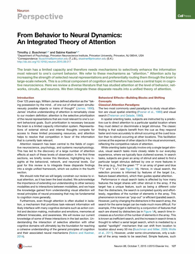
Figure 6. Cascades of Spatial and Featural Attention across theVisual Hierarchy(A–C) The brain is a densely interconnected network and so attentional se-lections, whether (A) spatial or (B) featural, propagate up and down the visualhierarchy. In this way, they will interact (C), allowing spatial attention to lead toselection of objects with similar features or featural selection to drive spatialattention.
Neuron
Perspective
synchronous oscillations. In brief, we propose that attention
works through a cascade of effects, as follows.
(1) Attention can either be (a) automatically grabbed by
salient stimuli or (b) guided by task representations in
frontal and parietal regions to specific spatial locations
or features.
(2) The pattern-completion nature of sensory cortex
sharpens the broad top-down attentional bias, restricting
it to perceptually relevant representations. Interactions
with bottom-up sensory drive will emphasize specific ob-
jects.
(3) Interneuron-mediated lateral inhibition normalizes activity
and, thus, suppresses competing stimuli. This results in
increased sensitivity and decreased noise correlations.
(4) Lateral inhibition also leads to the generation of high-fre-
quency synchronous oscillations within a cortical region.
Inter-areal synchronization follows as these local oscil-
lations synchronize along with the propagation of a bot-
tom-up sensory drive. Both forms of synchrony act to
further boost selected representations.
(5) Further buildup of inhibition acts to ‘‘reset’’ the network,
thereby restarting the process. This reset allows the
network to avoid being captured by a single stimulus
and allows a positive-only selection mechanism to move
over time.
As we detail next, many of the effects observed at the neural
level can be explained through this cascade. In addition to
noting neurophysiological observations, we will also highlight
some of the theoretical models on which our unified framework
is built.
Step 1: Direction of Attention
Attention is controlled in one of two ways. First, attention can be
captured by stimuli that are inherently salient on the basis of their
physical properties (such as their brightness, contrast, speed,
etc.) or other factors such as their associated valence. As noted
above, saliency maps capture the saliency of all objects in a
136 Neuron 88, October 7, 2015 ª2015 Elsevier Inc.
visual scene, allowing attention to be directed to stimuli in their
rank order of salience (Itti and Koch, 2001).
Second, attention can be guided toward stimuli that are rele-
vant to one’s current task. Our ability to focus our attention in
such a manner is remarkably flexible: we can attend to different
spatial locations, with seemingly different ‘‘zoom’’ levels, as
well as to both simple and complex features (i.e., ‘‘red things’’
and ‘‘cars’’). As reviewed above, such attention templates are
top-down, originating in prefrontal and parietal cortex and influ-
encing sensory cortex both through direct descending pro-
jections (e.g., from FEF to V4; Figure 6A) as well as through a
backward cascade (e.g., from prefrontal cortex to IT to V4,
etc.; Figure 6B).
Step 2: From Broad to Precise Top-Down Modulation
This then presents a conundrum: what neural mechanisms
would allow for both the flexibility and specificity of attentional
modulation? One hypothesis is that there are specific anatomical
connections that support all possible selections that could be
desired. Under this model, every form of attentional selection
would need a physiological correlate. For example, top-down
spatial attention projections would have to be distributed across
different spatial locations as well as across different spatial
scales. Although this remains a possibility, current anatomical
and physiological support for this model is limited (Anderson
et al., 2011). Instead, we argue that broad, non-specific top-
down signals are shaped by the local circuitry and activity in sen-
sory cortex (Duncan et al., 1997).
The pattern-completion nature of sensory cortex (assertion 1)
means that non-specific inputs will be transformed into some-
thing that ‘‘makes sense’’ to the network. In other words, any
energy input to the circuit that is orthogonal to its inherent rep-
resentations will be discarded, while energy along its learned
representations will be maintained. This effect will be further
amplified when the circuit is already receiving (and processing)
bottom-up inputs. In this case, sensory drive has activated a
subset of possible activity states for the network—moving
away from these would require a strong overriding input.
Instead, attention is modulatory and therefore has the greatest
impact on those representations that are already active in the
network.
To demonstrate this effect, we can imagine a simplified case
where attention is being directed to a spatial location with two
competing neurons that respond to either circular or square
stimuli. Attending to this location without visual stimulation will
broadly boost both representations (note that representations
not embedded in our network, such as triangular stimuli, will
not be boosted as our simplified network does not encode
them). However, if our spatial location begins receiving circular
visual input, this same attention signal will only be effective in
increasing the activity of the ‘‘circle’’ neuron as the ‘‘square’’
neuron will be suppressed (either in a bottom-up manner or
through competition with the circle neuron). Featural selection
would work in a similar way: attending to a car will emphasize
car components, such as circular wheels. These would be auto-
matically selected in a top-down cascade (as seen in Figure 6B).
Again, interactions with bottom-up sensory drive would collapse
that selection to a particular exemplar of a category (i.e., a BMW
versus a Ford).

Neuron
Perspective
Generalizing from this simplified example, our theory will make
a prediction about how attention selects objects. As noted above
in our first assertion, the visual system has learned the statistical
regularities of objects and has embedded this knowledge in the
connections of a distributed network (Figure 5A). Therefore,
applying attention to any part of the object representation will
cause the boosting signal to spread throughout the object.
This prediction has extensive experimental support. First, atten-
tion to an object automatically selects all components of an
object (Duncan, 1984; Egly et al., 1994; Siegel et al., 2015). In
addition, recent work suggests that attention will automatically
extend to other stimuli that follow Gestalt object rules (e.g.,
collinearity;Wannig et al., 2011). Furthermore, spatially attending
to a stimulus will also boost the representation of objects with
similar features across the visual field (McAdams and Maunsell,
2000; Treue and Martınez Trujillo, 1999).
Together, these results provide experimental support for a
model in which top-down attention is broad and non-specific
and only becomes focused through interactions with the
anatomical connectivity embedded in sensory cortex and the
bottom-up sensory drive.
Step 3: Biasing Competition through Normalization
As reviewed above, there is evidence that attention resolves
competition between stimuli in a way that boosts selected repre-
sentations while suppressing unselected ones. This finding was
captured in the highly influential ‘‘biased competition’’ theory of
attention (Desimone and Duncan, 1995). In brief, the model
proposes that stimuli are constantly competing with one another
for greater representation; attention acts to bias this competi-
tion, allowing the selected stimulus to ‘‘win.’’
Competition between stimuli is likely the result of the normal-
ization process described in our second assertion (Reynolds
et al., 1999). Recent work by Reynolds and Heeger (2009)
showed that integrating a normalization model with an atten-
tional biasing mechanism captures a wide variety of attentional
effects. First, they were able to explain how spatial attention
can increase the contrast gain of neurons (Reynolds et al.,
2000), the response gain (Williford and Maunsell, 2006), or multi-
plicatively scale responses (McAdams and Maunsell, 2000).
Second, they captured the sharpening of tuning curves with fea-
tural attention (David et al., 2008; Martinez-Trujillo and Treue,
2004). Finally, like the biased competition model, the normaliza-
tion model of attention also predicts the response to multiple
stimuli in a single receptive field (Moran and Desimone, 1985).
Lateral inhibition carried out by inhibitory interneurons is a
candidate mechanism that may instantiate the normalization
computation (Figure 5B). If so, one would expect significant
attentional modulation of the inhibitory neurons that are
computing the normalization signal. Indeed, attention has a
much larger impact on the responses of putative inhibitory inter-
neurons compared to putative pyramidal cells (Mitchell et al.,
2007). Direct evidence for top-down targeting of inhibitory inter-
neurons comes from a recent study showing that long-range
projections from cingulate cortex to visual cortex in mice in-
crease center-surround modulation via local inhibitory inter-
neuron circuits (Zhang et al., 2014).
We propose that a model that centers on lateral inhibition
has the potential to explain a diverse set of attention effects.
For example, as noted by Reynolds and Heeger (2009), such
recurrent models of normalization may capture the temporal
dynamics of attention effects (namely the lack of an attention
effect on the visual transient) or the alterations of the size and
center of receptive fields. Lateral inhibition has also been shown
to increase the sparsity of neural representations (Schwartz and
Simoncelli, 2001). As sparser signals are more likely to be inde-
pendent to one another, this will lead to a reduction in noise cor-
relations (as observed; Cohen and Maunsell, 2009).
Step 4: Synchrony Is Rhythmic Inhibition
In addition to computing the normalization effect, lateral inhibi-
tion may also underlie synchronous oscillations (Figures 5C
and 5D). As reviewed above, attention modulates local syn-
chrony, possibly to increase the gain of a selected neural repre-
sentation (Tiesinga et al., 2004) or to boost the transmission of
information from one region to the next (Fries, 2005). However,
such models that explain effects on synchronous firing in local
populations are often separated from those that explain other
effects of attention on single neurons. We propose a unifying
mechanism relying on lateral inhibition: namely, that the same
attentional modulation of inhibitory interneurons that leads to
normalization also increases synchronous high-frequency oscil-
lations.
Such a model makes several predictions about the nature
of synchronous oscillations in cortex. First, it predicts high-fre-
quency oscillations are generated by local inhibitory interneu-
rons. As noted in our third assertion, a large body of modeling
work suggests that this is true, either due to interactions between
interneurons directly (so-called ‘‘ING’’ models; Wang and Buz-
saki, 1996) or between inhibitory interneurons and excitatory
pyramidal neurons (so-called ‘‘PING’’ networks; Borgers and
Kopell, 2005). Furthermore, optogenetic stimulation of inhibitory
interneurons produces high-frequency oscillations (Cardin et al.,
2009). Second, the model predicts that attention should target
inhibitory interneurons in a way that drives synchrony. Indeed,
as noted above, experimental evidence suggests that attention
has its greatest impact on inhibitory interneurons (Mitchell
et al., 2007). More importantly, and as predicted, Vinck et al.
(2013) found inhibitory interneurons preferentially synchronized
with local populations (measured via local field potentials
[LFPs]), with a phase relationship that suggested they were
driving the high-frequency oscillations in LFP. Finally, according
to our model, attention will increase firing rates (particularly in
inhibitory interneurons) before increasing high-frequency oscilla-
tions. Although this has not been directly tested, there is some
experimental evidence that attention effects on firing rate pre-
cede modulations in high-frequency oscillations in visual (Fries
et al., 2008) and frontal cortex (Gregoriou et al., 2009).
By acting on inhibitory interneurons, attention increases local
synchrony and, thus, increases the impact of a neuronal popula-
tion on downstream brain regions (see the Dynamic Functional
Connectivity section above for details). Attention also increases
synchrony between regions, further boosting information trans-
fer. However, if high-frequency oscillations are due to the activa-
tion of local circuits, then how are they synchronized across
different brain regions? One possibility is that there is a con-
trolling input that forces synchronization across regions (and
could be modulated by attention). For example, high-frequency
Neuron 88, October 7, 2015 ª2015 Elsevier Inc. 137

Neuron
Perspective
oscillations have been found to be coupled to low-frequency
oscillations (Colgin, 2013; Schroeder and Lakatos, 2009), and
so synchronous low-frequency oscillations could organize
the temporal dynamics of higher-frequency oscillations across
regions.
Alternatively, synchronization across brain regions may be a
passive process that only requires a phase reset to initially align
local oscillations. This phase reset would occur with the onset of
a strong input into the cortex, such as the appearance of a new
stimulus in the world or an eye movement moving an existing
stimulus into a receptive field. The propagation of this stimulus
across brain regions (in a bottom-up manner) would then natu-
rally align the local oscillations across regions. This predicts
an increase in high-frequency synchrony with a strong stimulus
drive, as seen following the onset of a pop-out stimulus (as
seen by Buschman and Miller, 2007).
Step 5: Rhythmic Oscillations of Inhibition Reset the
Neural Network
Many of the above effects demonstrate how attention may in-
crease synchrony to select specific representations. However,
these effects do not strictly rely on synchrony being oscillatory
in nature. Therefore, it is not clear what mechanistic function
an oscillation may serve. We propose that oscillations modulate
the attractor dynamics of local cortex by periodically resetting
the network through strong inhibition (Figures 5C and 5D).
For example, this may be crucial to disengaging attention.
Suppose one deploys attention to a stimulus, which as a conse-
quence, wins the competition with other stimuli through lateral
inhibition. In this way, the attended stimulus has captured the
network—a state that will persist, even if attention is released
and re-deployed. One possible solution to this problem might
be a strong negative, or inhibitory, signal that can counter the
positive selection of attention. In support of this model, psycho-
physical studies have revealed a strong inhibition of return (IOR)
that inhibits re-selecting an already attended stimulus (Klein,
2000), an effect that reduces the neural representation of a pre-
viously selected stimulus (Mirpour et al., 2009). Alternatively, os-
cillations in global inhibition levels may serve this same purpose:
every cycle of an oscillation effectively resets the network, allow-
ing a new stimulus to be captured. Such a mechanism has the
advantage of not requiring a strong top-down inhibitory signal,
but rather relies on a local mechanism for generating inhibition.
If true, our theory would predict that shifts in attention should
be tied to ongoing oscillations in neural activity. Indeed, Busch-
man and Miller (2009) observed this effect during a visual search
task. Covert shifts in attention (measured behaviorally and elec-
trophysiologically) were locked to ongoing beta-band oscilla-
tions: on each cycle of the beta-band oscillation, the animal
attended to a new location in space. Similar effects have been
observed in humans, although at lower frequencies (Busch and
VanRullen, 2010; Fiebelkorn et al., 2013; Landau and Fries,
2012). Similarly, overt shifts in attention (i.e., eye movements)
are phase-locked to lower-frequency oscillations (Schroeder
et al., 2010).
Note that at the behavioral level, rhythmic attention will appear
as the classic IOR: stimuli are momentarily attended before
being inhibited for a sustained period of time (as they are never
returned to). Indeed, studies of IOR have found the onset of inhi-
138 Neuron 88, October 7, 2015 ª2015 Elsevier Inc.
bition occurs around 225 ms (Klein, 2000), which is approxi-
mately the 4 Hz observed in rhythmic fluctuations of attention
(Fiebelkorn et al., 2013). However, it remains to be seen which
is the chicken and which is the egg: do oscillations structure
the IOR or do we observe rhythmic IOR as oscillations?
Networks for Attentional Control: Interplay of Spatialand Featural AttentionDirecting attention to space of features appears to be controlled
by individual sources in the brain. Spatial attention is likely
directed by descending projections into extrastriate visual areas
(e.g., FEF to V4 projections; Figure 6A). In contrast, featural
attention is much broader, impacting the entire visual field.
Therefore, featural attention likely begins in regions with larger
receptive fields and more complex representations (Figure 6B).
A ‘‘reverse hierarchy’’ model of attention suggests selection
begins at the highest, most abstract level before filtering down
to the details of an object (Hochstein and Ahissar, 2002). Such
a model predicts the selection to begin in prefrontal and parietal
cortex, where neurons represent abstract categories (Freedman
et al., 2001), and then filters backward along the cortical hierar-
chy to ‘‘simpler’’ visual areas. Surprisingly, it also predicts that
the ease of feature-based visual search should be directly
related to whether the category of the sought-after stimulus is
‘‘natural.’’ Indeed, searching a cluttered natural scene for a com-
plex object can be highly efficient if the object is typical to our
everyday experiences, as noted above (e.g., cars; for review,
see Peelen and Kastner, 2014).
Despite their independent sources, these two forms of atten-
tion do interact with one another. A network view of selection
suggests that such interactions are mediated through the
convergence of feature and spatial attention in visual cortex
(Figure 6C). Attending to a spatial location will select an object
(or a piece of an object) at that location. This selection will prop-
agate up and down the visual hierarchy, acting to select associ-
ated representations. In turn, the more abstract, invariant repre-
sentations in higher cortical regions will lead to the automatic
selection of similar objects in the visual scene on the basis of
their featural properties. Indeed, psychophysical studies have
shown that spatial attention can drive featural attention (e.g.,
spatially attending to a single object leads to increases in atten-
tion to its properties across the entire cortex; Summerfield et al.,
2006). Conversely, featural attention can drive spatial attention
(e.g., the detection of a car in a visual scene drives spatial atten-
tion to that location). In this way, spatial and featural attention
can be flexibly combined to allow for the dynamic nature of
attention.
Future Directions
We have attempted to outline a parsimonious theoretical model
that captures the diversity of attention effects on neural activity.
In particular, we have focused on local cortical interactions as
these are the most prevalent connections in the brain and there-
fore the most likely to impact neural processing. We have also
attempted to avoid the need for precise top-down or controlling
inputs, whether it is spatially precise (in the case of a spotlight of
attention) or temporally precise (in the case of inter-areal syn-
chronization). As we hope is clear, this model relies heavily on
previous theoretical and experimental work. However, despite

Neuron
Perspective
this strong basis, amoremechanistic model is needed to test the
details of our theory.
In addition, there are many experimental details that need to
be worked out. For example, many of the observed effects
of attention can be explained by modulating the balance be-
tween excitation and inhibition of this network, particularly by
increasing the inhibitory gain in the network. However, we are
only beginning to understand how this balance is modulated in
the brain. For example, there are several (perhaps dozens) of
different types of inhibitory interneurons. Recent work is begin-
ning to unravel the relative roles of these interneurons, both in
perception and attention (Lee et al., 2012; Wilson et al., 2012;
Zhang et al., 2014), but future work must continue to detail the
respective roles of these varied cell types.
A particularly intriguing avenue for further exploration is the
role of neuromodulation in altering the computational properties
of local cortical circuitry. In particular, acetylcholine (ACh) has
been proposed to play a role in attention. Indeed, manipulating
ACh receptors changes the effect of attention on V1 neurons
(Disney et al., 2007; Herrero et al., 2008). This effect may be
mediated by cholinergic midbrain regions that represent stim-
ulus saliency (Asadollahi et al., 2010) and are themselves modu-
lated by prefrontal cortex (Sarter et al., 2005).
Finally, the theory outlined here is focused on the effects of
attention on perception (almost exclusively visual perception).
However, attention is just one small part of cognition, and it
is becoming increasingly clear that attention interacts heavily
with other cognitive domains, as we will review in the final
section.
Attention and Other Cognitive ProcessesAttention, defined as the act of selecting task-relevant informa-
tion, is a central component of cognition. Although research on
attention has been largely focused on its impact on visual pro-
cessing, there are close relationships between attention and
other cognitive processes.
Attention and Working Memory
Working memory is the ability to hold items ‘‘in mind,’’ without
relying on the external world. Working memory plays a central
role in cognition: it acts as a dynamic mental ‘‘workspace’’ in
which thoughts are processed, manipulated, and transformed.
Indeed, attention may rely on working memory workspaces to
maintain the current ‘‘search template’’ (Wolfe, 1994). Evidence
for such a model comes from studies showing that attention
can be biased by the current contents of working memory
(Soto et al., 2008). Furthermore, brain regions involved in control-
ling attention are also strongly recruited during working memory,
particularly the fronto-parietal network (Awh and Jonides, 2001).
However, the relationship between attention and working mem-
ory is complicated. Models of working memory predict the exis-
tence of a ‘‘central executive’’ that controls and manipulates the
contents of workingmemory ‘‘sketchpads’’ (Baddeley andHitch,
1974). Recent studies underline the importance of this central
executive: an individual’s general intelligence correlates highly
with how effectively the contents of working memory are
controlled (Fukuda and Vogel, 2011).
Attention may be this central executive of working memory. In
support of this idea, attention filters what enters workingmemory
(Gazzaley, 2011) and plays a role in maintaining items in memory
(Kuo et al., 2012). Attention may also pull together the distributed
brain regions necessary to support working memory (Postle,
2006). There is also growing evidence that the capacity limitation
of workingmemory is due to competition in amanner very similar
to the competition observed during perception (Buschman et al.,
2011), suggesting that some of the neural mechanisms limiting
perception may also be limiting working memory. Indeed, the
same brain regions involved in directing attention to external
stimuli are activated when attending to ‘‘internal’’ stimuli (Chun
et al., 2011; Nobre et al., 2004).
Attention and Reward Learning
Attention is also intricately related to reward processing. Attention
is attracted to salient, behaviorally relevant stimuli. Obviously,
rewarding stimuli should be salient, and therefore reward signals
are likelyclosely tied toattentionsignals.Recentelectrophysiolog-
ical evidence has begun to tease apart the relationship between
attention and reward in visual cortex, with some early evidence
for partially overlapping representations (Foley et al., 2014). In
addition, since reward information guides learning, it may aid in
learning where to direct attention (Rombouts et al., 2015).
Attention may also be critical to learning what is rewarding in
the real world. Reinforcement learning is not efficient when a
reward can be associated with too many possible sources. In
this context, attention may act to select the most likely sources
and therefore limit reinforcement learning to this subset (Niv
et al., 2015). In this way, attention can act to guide learning to-
ward task-relevant stimuli.
Together, these results suggest that attention, working mem-
ory, and reward are closely intertwined and therefore may share
many of the same underlying neural mechanisms. Future work is
needed to continue elucidating which mechanisms are shared
and which are distinct. This entwinement also highlights the
integrative nature of behavior. Attention is crucial to working
memory and reward processing because both functions rely
on selection of task-relevant stimuli. Indeed, as we review
next, the selective nature of attention may underlie cognitive
control more broadly.
Attention and Cognitive Control
Cognitive control is our ability to guide our actions on the basis of
our task, our internal goals, and the current context. Cognitive
control is thought to operate by guiding activity throughout the
brain in a task-dependent manner (Miller and Cohen, 2001). In
reality, this is just a super-set of attention: instead of only acting
upon sensory representations, cognitive control can operate
more broadly to select relevant stimulus representations, deci-
sion-making circuits, and motor planning regions (Norman and
Shallice, 1986).
Indeed, there is significant overlap between the neural mech-
anisms supporting cognitive control and attention. For example,
similar to attention, prefrontal cortex is thought to be the source
of cognitive control: lesions in prefrontal cortex disrupt cognitive
control (Barcelo and Knight, 2002) and single neurons in prefron-
tal cortex represent the current task (Wallis et al., 2001; White
and Wise, 1999). Furthermore, synchrony within prefrontal cor-
tex carves out ensembles of task-related neurons (Buschman
et al., 2012), much like in the way attention creates synchronous
ensembles in posterior cortex.
Neuron 88, October 7, 2015 ª2015 Elsevier Inc. 139

Box 1. Current Status of the Field
d Attention is used to select specific representations for
greater neural representation. This resolves competition
for limited neural resources.d Attention is controlled by a distributed fronto-parietal
network that acts upon representations in sensory cortex.d Attention increases the strength of neural responses
in several different ways to boost the attended repre-
sentation, allowing it to win the competition for neural
resources.d Attention interacts with many other cognitive domains,
including learning, short- and long-term memory, and
decision making.
Box 2. Future Directions
d Attention is highly dynamic, changing rapidly between
locations and/or features. These dynamics have been
associated with local population oscillations, yet the exact
neural mechanisms remain unknown.d The recruitment of network parts within the fronto-parietal
network is highly flexible, depending on changing behav-
ioral demands. However, the neural mechanisms utilized
to couple these parts for a given task are unknown, and it
is not clear how a network pattern produces the desired
behavior.d Attention may boost neural representations at several
different levels, from increasing single neuron responses
to boosting population selectivity. However, it is unknown
how these effects interact andwhether they share common
circuit mechanisms.d Recent work has highlighted the existence of domain-spe-
cific neural networks. However, it remains unknown how
these networks interact in order to support the integrative
nature of cognition.
Neuron
Perspective
In this light it seems that by studying attention we have been
studying one specific form of cognitive control. Therefore, it
is possible that many of the neural mechanisms underlying atten-
tion will apply more broadly. For example, cognitive control may
resolve competition betweenmotor plans in the sameway atten-
tion resolves competition between sensory stimuli (a generaliza-
tion of Figure 5B). Similarly, oscillations are observed throughout
the brain and may play a similar role in moderating cortical dy-
namics in any cortical region (Figures 5C and 5D). This would
make sense from an evolutionary perspective— once the brain
solves one problem, it might as well apply the same solution to
other, similar, problems.
ConclusionsAttention research has moved from laboratory scenes to the real
world at the behavioral level, and from the single neuron to local
populations and functional interactions across large-scale net-
works at the neural level (see Box 1. Current Status of the Field).
We have outlined a unified theory of attention that begins to inte-
grate these disparate effects. We propose broad top-down se-
lection signals interact with the inherent knowledge embedded
in sensory cortex and with bottom-up sensory drive. In addition,
we suggest that these signals interact within the local cortical
circuit to produce oscillatory synchrony. Such oscillations
temporally parse neural activity in a way that facilitates selection
of relevant representation, routes information through the brain,
and modulates attractor dynamics of a network.
Future work is needed to continue building a detailed, mecha-
nistic understanding of attention (see Box 2. Future Directions).
Detailed circuit models of attention will require continuing efforts
to identify cell types and quantify their role in attention. In addi-
tion, a unified theory must account for the dynamics of attention.
Studying these dynamics will require accurately following the
time course of neural correlates of attention across large popu-
lations of neurons throughout the brain. Such large-scale record-
ings will require continued improvements in multi-electrode
electrophysiology techniques and/or imaging approaches. This
appears particularly important as neural dynamics may be the
key to understanding the enormous flexibility of attentional re-
sources. Yet, the neural mechanisms supporting these dynamics
remain largely unknown. Finally, we must continue to integrate
attention with other cognitive domains. The parcellation of
140 Neuron 88, October 7, 2015 ª2015 Elsevier Inc.
behavior and brain into different cognitive domains has yielded
important insights into the neural mechanisms of many behav-
iors. However, cognition emerges from the interactions of these
cognitive domains; thus, a complete understanding of cognition
will require a more integrative approach.
ACKNOWLEDGMENTS
This project was supported by NIH RO1-MH64043, NIH RO1-EY017699, NIHR21-EY02565, and NSF BCS-1328270R01NS035710 to S.K. and NIH R00-MH092715, NIH DP2-EY025446, and ONR N000141410681 to T.J.B.
REFERENCES
Anderson, J.C., Kennedy, H., andMartin, K.A.C. (2011). Pathways of attention:synaptic relationships of frontal eye field to V4, lateral intraparietal cortex, andarea 46 in macaque monkey. J. Neurosci. 31, 10872–10881.
Arcizet, F., Mirpour, K., and Bisley, J.W. (2011). A pure salience response inposterior parietal cortex. Cereb. Cortex 21, 2498–2506.
Asadollahi, A., Mysore, S.P., and Knudsen, E.I. (2010). Stimulus-drivencompetition in a cholinergic midbrain nucleus. Nat. Neurosci. 13, 889–895.
Awh, E., and Jonides, J. (2001). Overlapping mechanisms of attention andspatial working memory. Trends Cogn. Sci. 5, 119–126.
Azouz, R., and Gray, C.M. (2000). Dynamic spike threshold reveals a mecha-nism for synaptic coincidence detection in cortical neurons in vivo. Proc.Natl. Acad. Sci. USA 97, 8110–8115.
Baddeley, A.D., and Hitch, G. (1974). Working memory. In Psychology ofLearning and Motivation, G.H. Bower, ed. (Academic Press), pp. 47–89.
Barcelo, F., and Knight, R.T. (2002). Both random and perseverative errors un-derlie WCST deficits in prefrontal patients. Neuropsychologia 40, 349–356.
Beck, D.M., and Kastner, S. (2009). Top-down and bottom-up mechanisms inbiasing competition in the human brain. Vision Res. 49, 1154–1165.
Bichot, N.P., and Schall, J.D. (1999). Effects of similarity and history on neuralmechanisms of visual selection. Nat. Neurosci. 2, 549–554.
Bisiach, E., and Vallar, G. (1988). Hemineglect in humans. In Handbook ofNeuropsychology, Volume 1, F. Boller, J. Grafman, G. Rizzolatti, and H. Good-glass, eds. (Elsevier Science), pp. 195–222.

Neuron
Perspective
Bisley, J.W., and Goldberg, M.E. (2003). Neuronal activity in the lateral intra-parietal area and spatial attention. Science 299, 81–86.
Bisley, J.W., and Goldberg, M.E. (2006). Neural correlates of attention anddistractibility in the lateral intraparietal area. J. Neurophysiol. 95, 1696–1717.
Borgers, C., and Kopell, N. (2005). Effects of noisy drive on rhythms in net-works of excitatory and inhibitory neurons. Neural Comput. 17, 557–608.
Bosman, C.A., Schoffelen, J.-M., Brunet, N., Oostenveld, R., Bastos, A.M.,Womelsdorf, T., Rubehn, B., Stieglitz, T., De Weerd, P., and Fries, P. (2012).Attentional stimulus selection through selective synchronization betweenmonkey visual areas. Neuron 75, 875–888.
Bressler, S.L. (1996). Interareal synchronization in the visual cortex. Behav.Brain Res. 76, 37–49.
Buffalo, E.A., Fries, P., Landman, R., Liang, H., and Desimone, R. (2010). Abackward progression of attentional effects in the ventral stream. Proc. Natl.Acad. Sci. USA 107, 361–365.
Busch, N.A., and VanRullen, R. (2010). Spontaneous EEG oscillations revealperiodic sampling of visual attention. Proc. Natl. Acad. Sci. USA 107,16048–16053.
Buschman, T.J., and Miller, E.K. (2007). Top-down versus bottom-up controlof attention in the prefrontal and posterior parietal cortices. Science 315,1860–1862.
Buschman, T.J., and Miller, E.K. (2009). Serial, covert shifts of attention duringvisual search are reflected by the frontal eye fields and correlated with popu-lation oscillations. Neuron 63, 386–396.
Buschman, T.J., Siegel, M., Roy, J.E., and Miller, E.K. (2011). Neural sub-strates of cognitive capacity limitations. Proc. Natl. Acad. Sci. USA 108,11252–11255.
Buschman, T.J., Denovellis, E.L., Diogo, C., Bullock, D., andMiller, E.K. (2012).Synchronous oscillatory neural ensembles for rules in the prefrontal cortex.Neuron 76, 838–846.
Bushnell, M.C., Goldberg, M.E., and Robinson, D.L. (1981). Behavioralenhancement of visual responses in monkey cerebral cortex. I. Modulationin posterior parietal cortex related to selective visual attention.J. Neurophysiol. 46, 755–772.
Busse, L., Wade, A.R., and Carandini, M. (2009). Representation of concurrentstimuli by population activity in visual cortex. Neuron 64, 931–942.
Buzsaki, G. (2006). Rhythms of the Brain (Oxford University Press).
Canolty, R.T., Edwards, E., Dalal, S.S., Soltani, M., Nagarajan, S.S., Kirsch,H.E., Berger, M.S., Barbaro, N.M., and Knight, R.T. (2006). High gamma poweris phase-locked to theta oscillations in human neocortex. Science 313, 1626–1628.
Carandini, M., and Heeger, D.J. (2012). Normalization as a canonical neuralcomputation. Nat. Rev. Neurosci. 13, 51–62.
Cardin, J.A., Carlen, M., Meletis, K., Knoblich, U., Zhang, F., Deisseroth, K.,Tsai, L.-H., and Moore, C.I. (2009). Driving fast-spiking cells induces gammarhythm and controls sensory responses. Nature 459, 663–667.
Carrasco, M., Ling, S., and Read, S. (2004). Attention alters appearance. Nat.Neurosci. 7, 308–313.
Chelazzi, L., Miller, E.K., Duncan, J., and Desimone, R. (1993). A neural basisfor visual search in inferior temporal cortex. Nature 363, 345–347.
Chun, M.M., Golomb, J.D., and Turk-Browne, N.B. (2011). A taxonomy ofexternal and internal attention. Annu. Rev. Psychol. 62, 73–101.
Cohen, M.R., and Maunsell, J.H.R. (2009). Attention improves performanceprimarily by reducing interneuronal correlations. Nat. Neurosci. 12, 1594–1600.
Colgin, L.L. (2013). Mechanisms and functions of theta rhythms. Annu. Rev.Neurosci. 36, 295–312.
Connor, C.E., Preddie, D.C., Gallant, J.L., and Van Essen, D.C. (1997). Spatialattention effects in macaque area V4. J. Neurosci. 17, 3201–3214.
Corbetta, M., and Shulman, G.L. (2002). Control of goal-directed and stimulus-driven attention in the brain. Nat. Rev. Neurosci. 3, 201–215.
Corbetta, M., Kincade, M.J., Lewis, C., Snyder, A.Z., and Sapir, A. (2005).Neural basis and recovery of spatial attention deficits in spatial neglect. Nat.Neurosci. 8, 1603–1610.
Cukur, T., Nishimoto, S., Huth, A.G., and Gallant, J.L. (2013). Attention duringnatural vision warps semantic representation across the human brain. Nat.Neurosci. 16, 763–770.
Damasio, A.R., Damasio, H., and Chui, H.C. (1980). Neglect following damageto frontal lobe or basal ganglia. Neuropsychologia 18, 123–132.
David, S.V., Hayden, B.Y., Mazer, J.A., and Gallant, J.L. (2008). Attention tostimulus features shifts spectral tuning of V4 neurons during natural vision.Neuron 59, 509–521.
Desimone, R., and Duncan, J. (1995). Neural mechanisms of selective visualattention. Annu. Rev. Neurosci. 18, 193–222.
Disney, A.A., Aoki, C., and Hawken, M.J. (2007). Gainmodulation by nicotine inmacaque v1. Neuron 56, 701–713.
Duncan, J. (1984). Selective attention and the organization of visual informa-tion. J. Exp. Psychol. Gen. 113, 501–517.
Duncan, J., and Humphreys, G.W. (1989). Visual search and stimulus similar-ity. Psychol. Rev. 96, 433–458.
Duncan, J., Humphreys, G., and Ward, R. (1997). Competitive brain activity invisual attention. Curr. Opin. Neurobiol. 7, 255–261.
Egly, R., Driver, J., and Rafal, R.D. (1994). Shifting visual attention betweenobjects and locations: evidence from normal and parietal lesion subjects.J. Exp. Psychol. Gen. 123, 161–177.
Fiebelkorn, I.C., Saalmann, Y.B., and Kastner, S. (2013). Rhythmic samplingwithin and between objects despite sustained attention at a cued location.Curr. Biol. 23, 2553–2558.
Foley, N.C., Jangraw, D.C., Peck, C., andGottlieb, J. (2014). Novelty enhancesvisual salience independently of reward in the parietal lobe. J. Neurosci. 34,7947–7957.
Freedman, D.J., Riesenhuber, M., Poggio, T., and Miller, E.K. (2001). Categor-ical representation of visual stimuli in the primate prefrontal cortex. Science291, 312–316.
Fries, P. (2005). A mechanism for cognitive dynamics: neuronal communica-tion through neuronal coherence. Trends Cogn. Sci. 9, 474–480.
Fries, P., Reynolds, J.H., Rorie, A.E., and Desimone, R. (2001). Modulation ofoscillatory neuronal synchronization by selective visual attention. Science 291,1560–1563.
Fries, P.,Womelsdorf, T., Oostenveld, R., andDesimone, R. (2008). The effectsof visual stimulation and selective visual attention on rhythmic neuronal syn-chronization in macaque area V4. J. Neurosci. 28, 4823–4835.
Fukuda, K., and Vogel, E.K. (2011). Individual differences in recovery time fromattentional capture. Psychol. Sci. 22, 361–368.
Gazzaley, A. (2011). Influence of early attentional modulation onworkingmem-ory. Neuropsychologia 49, 1410–1424.
Greenberg, A.S., Esterman, M., Wilson, D., Serences, J.T., and Yantis, S.(2010). Control of spatial and feature-based attention in frontoparietal cortex.J. Neurosci. 30, 14330–14339.
Gregoriou, G.G., Gotts, S.J., Zhou, H., and Desimone, R. (2009). High-fre-quency, long-range coupling between prefrontal and visual cortex duringattention. Science 324, 1207–1210.
Haenny, P.E., and Schiller, P.H. (1988). State dependent activity in monkeyvisual cortex. I. Single cell activity in V1 and V4 on visual tasks. Exp. BrainRes. 69, 225–244.
Hasegawa, R.P., Matsumoto, M., and Mikami, A. (2000). Search target selec-tion in monkey prefrontal cortex. J. Neurophysiol. 84, 1692–1696.
Neuron 88, October 7, 2015 ª2015 Elsevier Inc. 141

Neuron
Perspective
Hasenstaub, A., Shu, Y., Haider, B., Kraushaar, U., Duque, A., and McCor-mick, D.A. (2005). Inhibitory postsynaptic potentials carry synchronized fre-quency information in active cortical networks. Neuron 47, 423–435.
He, B.J., Snyder, A.Z., Vincent, J.L., Epstein, A., Shulman, G.L., and Corbetta,M. (2007). Breakdown of functional connectivity in frontoparietal networks un-derlies behavioral deficits in spatial neglect. Neuron 53, 905–918.
Heilman, K.M., and Van Den Abell, T. (1980). Right hemisphere dominance forattention: the mechanism underlying hemispheric asymmetries of inattention(neglect). Neurology 30, 327–330.
Herrero, J.L., Roberts, M.J., Delicato, L.S., Gieselmann, M.A., Dayan, P., andThiele, A. (2008). Acetylcholine contributes through muscarinic receptors toattentional modulation in V1. Nature 454, 1110–1114.
Hochstein, S., and Ahissar, M. (2002). View from the top: hierarchies andreverse hierarchies in the visual system. Neuron 36, 791–804.
Hubel, D.H., and Wiesel, T.N. (1959). Receptive fields of single neurones in thecat’s striate cortex. J. Physiol. 148, 574–591.
Iba, M., and Sawaguchi, T. (2003). Involvement of the dorsolateral prefrontalcortex of monkeys in visuospatial target selection. J. Neurophysiol. 89,587–599.
Itti, L., and Koch, C. (2001). Computational modelling of visual attention. Nat.Rev. Neurosci. 2, 194–203.
James, W. (1890). The Principles of Psychology (Holt and Company).
Janer, K.W., and Pardo, J.V. (1991). Deficits in selective attention followingbilateral anterior cingulotomy. J. Cogn. Neurosci. 3, 231–241.
Jerde, T.A., Merriam, E.P., Riggall, A.C., Hedges, J.H., and Curtis, C.E. (2012).Prioritized maps of space in human frontoparietal cortex. J. Neurosci. 32,17382–17390.
Jones, E.G. (2001). The thalamic matrix and thalamocortical synchrony.Trends Neurosci. 24, 595–601.
Karnath, H.-O., Ferber, S., and Himmelbach, M. (2001). Spatial awareness is afunction of the temporal not the posterior parietal lobe. Nature 411, 950–953.
Karnath, H.-O., Himmelbach, M., and Rorden, C. (2002). The subcortical anat-omy of human spatial neglect: putamen, caudate nucleus and pulvinar. Brain125, 350–360.
Kastner, S., and Ungerleider, L.G. (2000). Mechanisms of visual attention in thehuman cortex. Annu. Rev. Neurosci. 23, 315–341.
Kastner, S., Pinsk, M.A., De Weerd, P., Desimone, R., and Ungerleider, L.G.(1999). Increased activity in human visual cortex during directed attention inthe absence of visual stimulation. Neuron 22, 751–761.
Kenzie, J.M., Girgulis, K.A., Semrau, J.A., Findlater, S.E., Desai, J.A., andDukelow, S.P. (2015). Lesion sites associated with allocentric and egocentricvisuospatial neglect in acute stroke. Brain Connect. 5, 413–422.
Kinsbourne, M. (1977). Hemi-neglect and hemisphere rivalry. Adv. Neurol. 18,41–49.
Klein, R.M. (2000). Inhibition of return. Trends Cogn. Sci. 4, 138–147.
Kuo, B.-C., Stokes, M.G., and Nobre, A.C. (2012). Attention modulates main-tenance of representations in visual short-term memory. J. Cogn. Neurosci.24, 51–60.
Lakatos, P., Karmos, G., Mehta, A.D., Ulbert, I., and Schroeder, C.E. (2008).Entrainment of neuronal oscillations as a mechanism of attentional selection.Science 320, 110–113.
Lakatos, P., O’Connell, M.N., Barczak, A., Mills, A., Javitt, D.C., andSchroeder, C.E. (2009). The leading sense: supramodal control of neurophys-iological context by attention. Neuron 64, 419–430.
Landau, A.N., and Fries, P. (2012). Attention samples stimuli rhythmically. Curr.Biol. 22, 1000–1004.
Lee, S.-H., Kwan, A.C., Zhang, S., Phoumthipphavong, V., Flannery, J.G.,Masmanidis, S.C., Taniguchi, H., Huang, Z.J., Zhang, F., Boyden, E.S., et al.
142 Neuron 88, October 7, 2015 ª2015 Elsevier Inc.
(2012). Activation of specific interneurons improves V1 feature selectivityand visual perception. Nature 488, 379–383.
Li, F.F., VanRullen, R., Koch, C., and Perona, P. (2002). Rapid natural scenecategorization in the near absence of attention. Proc. Natl. Acad. Sci. USA99, 9596–9601.
Li, L., Gratton, C., Yao, D., and Knight, R.T. (2010). Role of frontal and parietalcortices in the control of bottom-up and top-down attention in humans. BrainRes. 1344, 173–184.
Luck, S.J., Chelazzi, L., Hillyard, S.A., and Desimone, R. (1997). Neural mech-anisms of spatial selective attention in areas V1, V2, and V4 of macaque visualcortex. J. Neurophysiol. 77, 24–42.
Lunven, M., De Schotten, M.T., Bourlon, C., Duret, C., Migliaccio, R., Rode, G.,and Bartolomeo, P. (2015). White matter lesional predictors of chronic visualneglect: a longitudinal study. Brain 138, 746–760.
Martınez-Trujillo, J., and Treue, S. (2002). Attentional modulation strength incortical area MT depends on stimulus contrast. Neuron 35, 365–370.
Martinez-Trujillo, J.C., and Treue, S. (2004). Feature-based attention increasesthe selectivity of population responses in primate visual cortex. Curr. Biol. 14,744–751.
McAdams, C.J., and Maunsell, J.H.R. (2000). Attention to both space andfeature modulates neuronal responses in macaque area V4. J. Neurophysiol.83, 1751–1755.
McAlonan, K., Cavanaugh, J., and Wurtz, R.H. (2006). Attentional modulationof thalamic reticular neurons. J. Neurosci. 26, 4444–4450.
Miller, E.K., and Cohen, J.D. (2001). An integrative theory of prefrontal cortexfunction. Annu. Rev. Neurosci. 24, 167–202.
Mirpour, K., Arcizet, F., Ong, W.S., and Bisley, J.W. (2009). Been there, seenthat: a neural mechanism for performing efficient visual search.J. Neurophysiol. 102, 3481–3491.
Mitchell, J.F., Sundberg, K.A., and Reynolds, J.H. (2007). Differential attention-dependent response modulation across cell classes in macaque visual areaV4. Neuron 55, 131–141.
Mitchell, J.F., Sundberg, K.A., and Reynolds, J.H. (2009). Spatial attentiondecorrelates intrinsic activity fluctuations in macaque area V4. Neuron 63,879–888.
Moore, T., and Armstrong, K.M. (2003). Selective gating of visual signals bymi-crostimulation of frontal cortex. Nature 421, 370–373.
Moore, T., and Fallah,M. (2004). Microstimulation of the frontal eye field and itseffects on covert spatial attention. J. Neurophysiol. 91, 152–162.
Moran, J., and Desimone, R. (1985). Selective attention gates visual process-ing in the extrastriate cortex. Science 229, 782–784.
Mort, D.J., Malhotra, P., Mannan, S.K., Rorden, C., Pambakian, A., Kennard,C., and Husain, M. (2003). The anatomy of visual neglect. Brain 126, 1986–1997.
Nassi, J.J., Gomez-Laberge, C., Kreiman, G., and Born, R.T. (2014). Cortico-cortical feedback increases the spatial extent of normalization. Front. Syst.Neurosci. 8, 105.
Niv, Y., Daniel, R., Geana, A., Gershman, S.J., Leong, Y.C., Radulescu, A., andWilson, R.C. (2015). Reinforcement learning in multidimensional environmentsrelies on attention mechanisms. J. Neurosci. 35, 8145–8157.
Nobre, A.C., Coull, J.T., Maquet, P., Frith, C.D., Vandenberghe, R., and Mesu-lam, M.M. (2004). Orienting attention to locations in perceptual versus mentalrepresentations. J. Cogn. Neurosci. 16, 363–373.
Nobre, A., and Kastner, S. (2014). The Oxford Handbook of Attention (OxfordLibrary of Psychology).
Norman, D.A., and Shallice, T. (1986). Attention to action. In Consciousnessand Self-Regulation, R.J. Davidson, G.E. Schwartz, and D. Shapiro, eds.(Springer), pp. 1–18.

Neuron
Perspective
O’Connor, D.H., Fukui, M.M., Pinsk, M.A., and Kastner, S. (2002). Attentionmodulates responses in the human lateral geniculate nucleus. Nat. Neurosci.5, 1203–1209.
Olshausen, B.A., and Field, D.J. (1996). Emergence of simple-cell receptivefield properties by learning a sparse code for natural images. Nature 381,607–609.
Peelen, M.V., and Kastner, S. (2011). A neural basis for real-world visual searchin human occipitotemporal cortex. Proc. Natl. Acad. Sci. USA 108, 12125–12130.
Peelen, M.V., and Kastner, S. (2014). Attention in the real world: toward under-standing its neural basis. Trends Cogn. Sci. 18, 242–250.
Peelen, M.V., Fei-Fei, L., and Kastner, S. (2009). Neural mechanisms of rapidnatural scene categorization in human visual cortex. Nature 460, 94–97.
Posner, M.I., Snyder, C.R., and Davidson, B.J. (1980). Attention and the detec-tion of signals. J. Exp. Psychol. 109, 160–174.
Postle, B.R. (2006). Working memory as an emergent property of the mind andbrain. Neuroscience 139, 23–38.
Premereur, E., Vanduffel, W., Roelfsema, P.R., and Janssen, P. (2012). Frontaleye field microstimulation induces task-dependent gamma oscillations in thelateral intraparietal area. J. Neurophysiol. 108, 1392–1402.
Premereur, E., Janssen, P., and Vanduffel, W. (2015). Effector specificity inmacaque frontal and parietal cortex. J. Neurosci. 35, 3446–3459.
Rainer, G., Asaad, W.F., and Miller, E.K. (1998). Selective representation ofrelevant information by neurons in the primate prefrontal cortex. Nature 393,577–579.
Reynolds, J.H., and Heeger, D.J. (2009). The normalization model of attention.Neuron 61, 168–185.
Reynolds, J.H., Chelazzi, L., and Desimone, R. (1999). Competitive mecha-nisms subserve attention in macaque areas V2 and V4. J. Neurosci. 19,1736–1753.
Reynolds, J.H., Pasternak, T., and Desimone, R. (2000). Attention increasessensitivity of V4 neurons. Neuron 26, 703–714.
Rombouts, J.O., Bohte, S.M., Martinez-Trujillo, J., and Roelfsema, P.R. (2015).A learning rule that explains how rewards teach attention. Vis. Cogn. 23,179–205.
Ruff, C.C., Blankenburg, F., Bjoertomt, O., Bestmann, S., Freeman, E.,Haynes, J.-D., Rees, G., Josephs, O., Deichmann, R., and Driver, J. (2006).Concurrent TMS-fMRI and psychophysics reveal frontal influences on humanretinotopic visual cortex. Curr. Biol. 16, 1479–1488.
Saalmann, Y.B., Pigarev, I.N., and Vidyasagar, T.R. (2007). Neural mecha-nisms of visual attention: how top-down feedback highlights relevant loca-tions. Science 316, 1612–1615.
Saalmann, Y.B., Pinsk, M.A., Wang, L., Li, X., and Kastner, S. (2012). Thepulvinar regulates information transmission between cortical areas based onattention demands. Science 337, 753–756.
Saenz, M., Buracas, G.T., and Boynton, G.M. (2002). Global effects of feature-based attention in human visual cortex. Nat. Neurosci. 5, 631–632.
Salinas, E., and Sejnowski, T.J. (2001). Correlated neuronal activity and theflow of neural information. Nat. Rev. Neurosci. 2, 539–550.
Sarter, M., Hasselmo, M.E., Bruno, J.P., and Givens, B. (2005). Unraveling theattentional functions of cortical cholinergic inputs: interactions betweensignal-driven and cognitive modulation of signal detection. Brain Res. BrainRes. Rev. 48, 98–111.
Schroeder, C.E., and Lakatos, P. (2009). Low-frequency neuronal oscillationsas instruments of sensory selection. Trends Neurosci. 32, 9–18.
Schroeder, C.E., Wilson, D.A., Radman, T., Scharfman, H., and Lakatos, P.(2010). Dynamics of Active Sensing and perceptual selection. Curr. Opin. Neu-robiol. 20, 172–176.
Schwartz, O., and Simoncelli, E.P. (2001). Natural signal statistics and sensorygain control. Nat. Neurosci. 4, 819–825.
Serences, J.T., and Boynton, G.M. (2007). Feature-based attentional modula-tions in the absence of direct visual stimulation. Neuron 55, 301–312.
Serences, J.T., Schwarzbach, J., Courtney, S.M., Golay, X., and Yantis, S.(2004). Control of object-based attention in human cortex. Cereb. Cortex 14,1346–1357.
Sharma, J., Angelucci, A., and Sur, M. (2000). Induction of visual orientationmodules in auditory cortex. Nature 404, 841–847.
Shipp, S. (2003). The functional logic of cortico-pulvinar connections. Philos.Trans. R. Soc. B Biol. Sci. 358, 1605–1624.
Siegel, M., Donner, T.H., Oostenveld, R., Fries, P., and Engel, A.K. (2008).Neuronal synchronization along the dorsal visual pathway reflects the focusof spatial attention. Neuron 60, 709–719.
Siegel, M., Buschman, T.J., and Miller, E.K. (2015). BRAIN PROCESSING.Cortical information flow during flexible sensorimotor decisions. Science348, 1352–1355.
Silver, M.A., and Kastner, S. (2009). Topographic maps in human frontal andparietal cortex. Trends Cogn. Sci. 13, 488–495.
Simoncelli, E.P., and Olshausen, B.A. (2001). Natural image statistics and neu-ral representation. Annu. Rev. Neurosci. 24, 1193–1216.
Soto, D., Hodsoll, J., Rotshtein, P., and Humphreys, G.W. (2008). Automaticguidance of attention from working memory. Trends Cogn. Sci. 12,342–348.
Spitzer, H., Desimone, R., and Moran, J. (1988). Increased attention enhancesboth behavioral and neuronal performance. Science 240, 338–340.
Steinmetz, P.N., Roy, A., Fitzgerald, P.J., Hsiao, S.S., Johnson, K.O., andNiebur, E. (2000). Attention modulates synchronized neuronal firing in primatesomatosensory cortex. Nature 404, 187–190.
Summerfield, J.J., Lepsien, J., Gitelman, D.R., Mesulam, M.M., and Nobre,A.C. (2006). Orienting attention based on long-term memory experience.Neuron 49, 905–916.
Szczepanski, S.M., and Kastner, S. (2013). Shifting attentional priorities: con-trol of spatial attention through hemispheric competition. J. Neurosci. 33,5411–5421.
Szczepanski, S.M., Konen, C.S., and Kastner, S. (2010). Mechanisms ofspatial attention control in frontal and parietal cortex. J. Neurosci. 30, 148–160.
Thorpe, S., Fize, D., and Marlot, C. (1996). Speed of processing in the humanvisual system. Nature 381, 520–522.
Tiesinga, P.H.E., Fellous, J.-M., Salinas, E., Jose, J.V., and Sejnowski, T.J.(2004). Synchronization as a mechanism for attentional gain modulation. Neu-rocomputing 58-60, 641–646.
Treisman, A.M., and Gelade, G. (1980). A feature-integration theory of atten-tion. Cognit. Psychol. 12, 97–136.
Treue, S., andMartınez Trujillo, J.C. (1999). Feature-based attention influencesmotion processing gain in macaque visual cortex. Nature 399, 575–579.
Treue, S., andMaunsell, J.H.R. (1999). Effects of attention on the processing ofmotion in macaque middle temporal and medial superior temporal visualcortical areas. J. Neurosci. 19, 7591–7602.
Vinck, M., Womelsdorf, T., Buffalo, E.A., Desimone, R., and Fries, P. (2013).Attentional modulation of cell-class-specific gamma-band synchronization inawake monkey area v4. Neuron 80, 1077–1089.
Wallis, J.D., Anderson, K.C., and Miller, E.K. (2001). Single neurons in prefron-tal cortex encode abstract rules. Nature 411, 953–956.
Wang, X.-J., and Buzsaki, G. (1996). Gamma oscillation by synaptic inhibitionin a hippocampal interneuronal network model. J. Neurosci. 16, 6402–6413.
Wannig, A., Stanisor, L., and Roelfsema, P.R. (2011). Automatic spread ofattentional response modulation along Gestalt criteria in primary visual cortex.Nat. Neurosci. 14, 1243–1244.
Neuron 88, October 7, 2015 ª2015 Elsevier Inc. 143

Neuron
Perspective
Ward, R., Danziger, S., Owen, V., and Rafal, R. (2002). Deficits in spatial codingand feature binding following damage to spatiotopic maps in the human pulvi-nar. Nat. Neurosci. 5, 99–100.
White, I.M., andWise, S.P. (1999). Rule-dependent neuronal activity in the pre-frontal cortex. Exp. Brain Res. 126, 315–335.
Wilke, M., Turchi, J., Smith, K., Mishkin, M., and Leopold, D.A. (2010). Pulvinarinactivation disrupts selection ofmovement plans. J. Neurosci. 30, 8650–8659.
Williford, T., and Maunsell, J.H.R. (2006). Effects of spatial attention oncontrast response functions in macaque area V4. J. Neurophysiol. 96, 40–54.
Wilson, N.R., Runyan, C.A., Wang, F.L., and Sur, M. (2012). Division and sub-traction by distinct cortical inhibitory networks in vivo. Nature 488, 343–348.
Wolfe, J.M. (1994). Guided Search 2.0 A revised model of visual search. Psy-chon. Bull. Rev. 1, 202–238.
Wolfe, J.M., Cave, K.R., and Franzel, S.L. (1989). Guided search: an alternativeto the feature integration model for visual search. J. Exp. Psychol. Hum.Percept. Perform. 15, 419–433.
144 Neuron 88, October 7, 2015 ª2015 Elsevier Inc.
Wolfe, J.M., Vo, M.L.-H., Evans, K.K., and Greene, M.R. (2011). Visual searchin scenes involves selective and nonselective pathways. Trends Cogn. Sci. 15,77–84.
Womelsdorf, T., Fries, P., Mitra, P.P., and Desimone, R. (2006a). Gamma-bandsynchronization in visual cortex predicts speed of change detection. Nature439, 733–736.
Womelsdorf, T., Anton-Erxleben, K., Pieper, F., and Treue, S. (2006b). Dy-namic shifts of visual receptive fields in cortical area MT by spatial attention.Nat. Neurosci. 9, 1156–1160.
Zenon, A., and Krauzlis, R.J. (2012). Attention deficits without cortical neuronaldeficits. Nature 489, 434–437.
Zhang, S., Xu, M., Kamigaki, T., Hoang Do, J.P., Chang, W.-C., Jenvay, S.,Miyamichi, K., Luo, L., and Dan, Y. (2014). Selective attention. Long-rangeand local circuits for top-down modulation of visual cortex processing. Sci-ence 345, 660–665.