function and redox state of mitochondrial localized
TRANSCRIPT

University of Nebraska - LincolnDigitalCommons@University of Nebraska - Lincoln
Biochemistry -- Faculty Publications Biochemistry, Department of
2008
Function and redox state of mitochondrial localizedcysteine-rich proteins important in the assembly ofcytochrome c oxidaseOleh KhalimonchukUniversity of Nebraska-Lincoln, [email protected]
Dennis R. WingeUniversity of Utah Health Sciences Center
Follow this and additional works at: http://digitalcommons.unl.edu/biochemfacpub
Part of the Biochemistry Commons, Biotechnology Commons, and the Other Biochemistry,Biophysics, and Structural Biology Commons
This Article is brought to you for free and open access by the Biochemistry, Department of at DigitalCommons@University of Nebraska - Lincoln. Ithas been accepted for inclusion in Biochemistry -- Faculty Publications by an authorized administrator of DigitalCommons@University of Nebraska -Lincoln.
Khalimonchuk, Oleh and Winge, Dennis R., "Function and redox state of mitochondrial localized cysteine-rich proteins important inthe assembly of cytochrome c oxidase" (2008). Biochemistry -- Faculty Publications. 285.http://digitalcommons.unl.edu/biochemfacpub/285

Function and redox state of mitochondrial localized cysteine-richproteins important in the assembly of cytochrome c oxidase
Oleh Khalimonchuk and Dennis R. WingeFrom the University of Utah Health Sciences Center, Departments of Medicine and Biochemistry,Salt Lake City, Utah 84132
SummaryThe cytochrome c oxidase (CcO) complex of the mitochondrial respiratory chain exists within themitochondrial inner membrane (IM). The biogenesis of the complex is a multi-faceted processrequiring multiple assembly factors that function on both faces of the IM. Formation of the two coppercenters of CcO occurs within the intermembrane space (IMS) and is dependent on assembly factorswith critical cysteinyl thiolates. Two classes of assembly factors exist, one group being soluble IMSproteins and the second class being proteins tethered to the IM. A common motif in the solubleassembly factors is a duplicated Cx9C sequence motif. Since mitochondrial respiration is a majorsource of reactive oxygen species, control of the redox state of mitochondrial proteins is an importantprocess. This review documents the role of these cysteinyl CcO assembly factors within the IMS andthe necessity of redox control in their function.
1. Mitochondrial OrganizationThe mitochondrion consists of a continuous reticulum that makes up nearly 10% of the cellvolume in respiring yeast cells. The tubular network is highly dynamic and changes size andshape through fission and fusion events [1]. A double membrane forming two internal spacesencloses mitochondria. The space between the two membranes is called the intermembranespace (IMS) and the volume enclosed within the inner membrane (IM) is designated as thematrix compartment. The two membranes differ significantly in their composition with the IMmore highly enriched in protein content. The outer membrane (OM) envelope contains 25 Ånm pores that allow diffusion of small ions such as glutathione. The IM is a barrier to diffusion,so passage of metabolites requires transporters. As such, the import of glutathione across theIM is believed to occur through dicarboxylate and 2-oxoglutarate transporters [2].
Mitochondria maintain their genome within the matrix compartment. This genome codes fora limited number of polypeptides, 13 and 8 in humans and yeast, respectively. All, but one, ofthese proteins are components of the oxidative phosphorylation system.
The double membrane of mitochondria is interrupted by junction points of contact betweenthe IM and OM [3]. The junction points are likely formed by protein assemblies involved inprotein import [4,5]. The mean distance across the OM and IM is 20 nm, although the distancenarrows to only about 14 nm at junction points. In cells with high respiration rates, the IM is
* Address correspondence to: Dennis Winge, University of Utah Health Sciences Center, Salt Lake City, Utah 84132; Tel: 801-585-5103;Fax: 801-585-5469; Email: [email protected]'s Disclaimer: This is a PDF file of an unedited manuscript that has been accepted for publication. As a service to our customerswe are providing this early version of the manuscript. The manuscript will undergo copyediting, typesetting, and review of the resultingproof before it is published in its final citable form. Please note that during the production process errors may be discovered which couldaffect the content, and all legal disclaimers that apply to the journal pertain.
NIH Public AccessAuthor ManuscriptBiochim Biophys Acta. Author manuscript; available in PMC 2009 April 1.
Published in final edited form as:Biochim Biophys Acta. 2008 April ; 1783(4): 618–628. doi:10.1016/j.bbamcr.2007.10.016.
NIH
-PA Author Manuscript
NIH
-PA Author Manuscript
NIH
-PA Author Manuscript

invaginated, folding into tubular structures designated cristae. Cristae are enriched in theenzyme complexes involved in oxidative phosphorylation. Electron microscopy tomographyrevealed that the cristae tubules are 30–40 nm in diameter but narrow to about 28 nm at junctionpoints with the boundary IM, designated inner boundary membrane (IBM) [3,5]. Theconstriction of the cristae junctions at the IBM resolves the soluble IMS into separate volumesthat appear to be in equilibrium only for small molecules [6]. Cristae can exist as tubularstructures or merged to form flattened lamellar compartments. Stacked lamellar cristae remainconnected to the IBM by tubular cristae junctions. The cristae junctions are believed to bedynamic and modulated by the energetics of the organelle as well as by the fusion/fissionprocess.
In this review, the lumen of the cristae will be referred to as the cristae lumen and the spacebetween the boundary IM and OM as the boundary IMS. The generic term IMS will specifythe generalized volume between the OM and IM without sub-compartmentizationspecification.
Oxidative phosphorylation occurs predominantly, but not exclusively, on the cristae membrane(CM). The enrichment of cytochrome c oxidase (CcO) in cristae is well established [7], butrecent studies confirm the abundance of complexes III (cytochrome bc1 complex) and V (ATPsynthase) within the CM [5]. Respiratory complexes I, III and IV are largely present assupercomplexes within the CM [8–11]. The yeast III/IV supercomplex consists of a dimericcomplex III species at the core with one or two CcO complexes at opposite ends [11]. Synthesisand assembly of the respiratory complexes occurs preferentially on the CM as mitochondrialribosomal proteins are associated with the CM [5].
2. Generation of reactive oxygen species in mitochondriaMitochondrial respiration is a major source of reactive oxygen species. Respiring mitochondriaconvert 1–2% of the oxygen consumed to superoxide anion [12,13]. The bulk of superoxideanion produced on a daily basis in most organisms comes from ubisemiquinone of coenzymeQ within the respiratory chain [14]. CoQ shuttles electrons from complexes I and II to complexIII. The CoQ semiquinone generated either at complex I or during the Q-cycle in complex IIIcan react with oxygen generating superoxide anions. Based on the sidedness of the Q-cycle incomplex III, superoxide is generated in both the matrix and IMS, although the bulk of thesuperoxide is generated within the matrix [15,16]. Superoxide may itself cause damage or mayreact further to yield other reactive species such as hydrogen peroxide or the hydroxyl radical.The presence of reactive oxygen species can induce oxidation events such as modification ofprotein thiols on both sides of the IM [17]. Endogenous reactive oxygen species generated atcomplex I and III were shown to modify nine distinct mitochondrial proteins [17]. Several ofthe modified proteins function in fatty acid oxidation.
The normal production of superoxide anion during respiration and subsequent generation ofhydrogen peroxide and other oxidants in both the IMS and matrix necessitates control oftransition metal ion availability to minimize Fenton chemistry to generate more potentoxidants. Copper ions used in metallation reactions are protein bound to minimize thedeleterious effects of unbound Cu(I) ions. Copper metallation of CcO and superoxidedismutase (Sod1) within the IMS occurs by metallochaperone proteins Cox17 and Ccs1,respectively. Only a small fraction of the cellular Sod1 exists within the IMS [18]. The transferreactions are protein-mediated. However, the Cu(I) sites on Ccs1 are partially solventaccessible and this is likely also true for Cox17. Although the level of copper complexes ofthese two proteins isn’t known within the IMS, it is possible that the level of Cu(I) within theIMS may be regulated to minimize chances of copper-induced oxidation reactions. The Cu(I)ions used in the IMS metallation reactions derives from a storage pool within the matrix [19,
Khalimonchuk and Winge Page 2
Biochim Biophys Acta. Author manuscript; available in PMC 2009 April 1.
NIH
-PA Author Manuscript
NIH
-PA Author Manuscript
NIH
-PA Author Manuscript

20]. It is conceivable that the Cu(I) transporter within the IM that translocates Cu(I) to the IMSis regulated such that the Cu(I) transported is coupled to the biogenesis of CcO and Sod1.
Another defense against deleterious oxidative processes is the availability of redox systems tomaintain redox homeostasis. The effectiveness of the matrix redox system is highlighted bythe observation that the apparent redox potential of the mitochondrial matrix is more negative(−360 mV) relative to the cytoplasm (−320 mV) in HeLa cells using redox-sensitive fluorescentGFP variants [21,22]. No information is available on the redox potential of the IMScompartment.
The mitochondria matrix contains well-defined redox components including glutathione,thioredoxin and glutaredoxin systems. A fraction of the cellular glutathione reductase Glr1exists within the matrix in yeast. The thioredoxin system involves the mitochondrial-specificTrx3 thioredoxin and the Trr2 thioredoxin reductase. Both monothiol (Grx5) and dithiol (Grx2)glutaredoxins exist within the matrix. Initiation at an upstream ATG in the transcripts for Grx2and Glr1 generate the mitochondrially-targeted variants. The presence of multiple proteinreductants and the overlapping functions of Glr1 and Trr2 reductases illustrates that a robustredox pathway exists within the matrix. Apart from an initial report of mammalian thioredoxinreductase-I existing within the IMS [23], it is not clear whether other redox components arepresent within the IMS compartments. The porous OM suggests that GSH may equilibrateacross the membrane, but no evidence exists for the IMS presence of Glr1 to maintain GSH/GSSG redox homeostasis.
3. Role of Cysteines in IMS ProteinsThe mitochondrial proteome is expected to contain nearly 850 proteins in yeast [24] but closerto 1000 distinct proteins in humans [25]. About 14% of the proteins are involved in oxidativephosphorylation, whereas 25% are predicted to be involved in maintaining and expressing themitochondrial genome [26]. A subset of the proteome resides within the IMS either as solubleproteins or as molecules tethered to the IM. Many of these proteins are either cysteine-rich orhave functionally important cysteine residues that can exist as within disulfide bonds or asreduced thiolates. The abundance of disulfide-containing molecules within the IMS suggeststhat redox control within this compartment differs from that within the cytoplasm wheredisulfide bond formation is rare due to the high reducing potential of the cytoplasm [27]. Theconstriction of the cristae junctions creating both cristae lumen and the boundary IMS opensthe possibility that redox pathways may be distinct within the two subcompartments.
A common structural motif of cysteine-rich proteins within the IMS is a helical hairpinconformer. The structural paradigm for the helical hairpin motif in IMS proteins is small Timproteins (Tim9/Tim10) characterized by a conserved twin Cx3C sequence motif. Tim9 andTim10 each form a helix-loop-helix conformer held together by paired disulfide bonds in thetwin Cx3C motif [28] (Fig. 1A). Tim9 and Tim10 form a heterohexameric complex thatfunctions as a chaperone for incoming proteins as they are delivered to the TIM22 complexfor IM insertion and SAM complex for OM insertion [29]. A second heterohexameric complexTim8/Tim13 exists within the IMS that also mediates import and insertion of polytopic IMproteins [30]. Within the twin Cx3C motif, one disulfide consists of the most N-terminal andC-terminal Cys residues (designated proximal pair), whereas the second disulfide consists ofthe two internal Cys residues (distal pair). The hexameric complex (Fig. 1B) is dependent onthe disulfides within each subunit [31]. Reduction of the disulfides disassembles the complex.
Yeast contains two IMS cytochrome c heme lyases, Cyc3 and Cyc7, for the assembly ofcytochrome c and cytochrome c1, respectively [32]. The two lyases attach heme to theirrespective cytochromes within the IMS. Human cells use only a single heme lyase to assemblecytochrome c and cytochrome c1. Whereas the two lyases are not cysteine-rich molecules, their
Khalimonchuk and Winge Page 3
Biochim Biophys Acta. Author manuscript; available in PMC 2009 April 1.
NIH
-PA Author Manuscript
NIH
-PA Author Manuscript
NIH
-PA Author Manuscript

substrates cytochrome c and cytochrome c1 have essential cysteinyl residues that becomecovalently attached to the bound heme. Thus, the cysteines in cytochrome c and cytochromec1 must be maintained in their reduced state for covalent attachment of hemes. The prevalenceof respiratory complexes within the cristae suggests that cytochrome maturation occurspredominantly within cristae lumen. Nothing is known how the cytochrome cysteines aremaintained in their reduced state.
Three subunits in the cytochrome bc1 complex and CcO that project into the IMS, primarilythe cristae lumen, contain disulfide bonds. The Rieske iron-sulfur protein Rip1 and Qcr2 ofthe bc1 complex contains disulfide linkages [33]. The CcO subunit VIB (yeast Cox12) containstwo disulfide bonds in the bovine CcO structure [34] (Fig. 2).
Sod1 contains a disulfide bond that is essential for its enzymatic function. The coppermetallochaperone Ccs1 activates Sod1 during its folding by inserting the catalytic Cu(I) ionand in addition catalyzes formation of the essential disulfide in Sod1 [35]. Ccs1 itself has CxCand Cx2C motifs that may be redox active. Ccs1 co-exists with Sod1 within the IMS. Thedistribution of Sod1 between the boundary IMS and cristae lumen is not known, but its presencein the cristae lumen is expected as the respiratory complexes within the cristae IM are the majorsource of superoxide anions. Recent studies on the IMS Sod1 in rat mitochondria suggest thatthe redox status of Sod1 is more complex [23,36]. Sod1 appears to exist within the IMS as aninactive, reduced enzyme. It is transiently activated either by exogenous peroxide or superoxideanions or disruption of the OM [23]. Pretreatment of mitochondria with alkylating agentsquenches activation of the enzyme. Thioredoxin reductase-I is shown to exist within the IMSand to be competent to reduce and inactivate Sod1. Inarrea et al. [23] postulate that the redoxstate, and therefore the enzymatic activity, of Sod1 is highly regulated within the IMS.
A redox proteomic study attempted to identify oxidized proteins in yeast using an affinitycapture protocol [27]. Of the 64 proteins identified, two IMS proteins Ccs1 and Sod1 wereidentified, although as mentioned, both proteins also exist within the cytoplasm. The othermitochondrial proteins identified, Prx1, Hsp60, Yhb1, Ilv5 and Aco1, are matrix proteins.
4. Role of cysteines in the MIA import pathwayCysteinyl residues in Tim proteins are additionally important for their import into the IMS.The import of Tim proteins is dependent on a disulfide relay system involving the MIAmachinery consisting of at least Mia40 and Erv1 [37–41]. Transit of the Tim proteins throughthe TOM complex in the outer membrane results in transient capture of the imported moleculesby Mia40 through disulfide bonding. Disulfide interchange between oxidized Mia40 andreduced thiolates on the imported protein generate intermolecular disulfides that trap themolecules within the IMS. The most N-terminal Cys residue in the Cx3C motif of Tim9 andTim10 was recently shown to be important for efficient capture by Mia40, whereas a mutantvariant lacking all four Cys residues in not imported at all [42]. The importance of the mostN-terminal Cys residue in both Tim9 and Tim10 suggests that recognition by Mia40 is preciseand specific [42]. Replacement of only the distal Cys pair (Cys 2 & 3 in Fig. 1A) enabledimport, but the process was inefficient. The mechanism of Tim10 release from Mia40 mayinvolve intramolecular disulfide exchange with the fourth Cys (the Cys in the proximal pair)[43]. The third Cys in Tim9 appears to be more important than the fourth Cys for Mia40 release[37]. The distal Cys disulfide pair is important for assembly of the Tim hexameric complexbut doesn’t appear essential for the release of Tim from the Mia40 import complex [43].
Erv1 is a flavin-containing sulfhydryl oxidase that generates disulfides in Mia40 for IMSprotein import [44] (Fig. 3). Erv1 has a limited substrate specificity, and Mia40 is its onlyknown in vivo substrate [45]. Erv1 has a redox-active Cx2C motif close to the FAD that formsan initial disulfide [46]. Through disulfide exchange, the disulfide is likely transferred to an
Khalimonchuk and Winge Page 4
Biochim Biophys Acta. Author manuscript; available in PMC 2009 April 1.
NIH
-PA Author Manuscript
NIH
-PA Author Manuscript
NIH
-PA Author Manuscript

N-terminal Cx2C motif for transfer to its target Mia40 [47]. In addition, Erv1 appears to containa structural disulfide in the flavin domain. In the absence of the N-terminal Cx2C motif, Erv1can induce disulfide bond formation in small molecules, but this truncate is nonfunctional invivo [47]. All three cysteine pairs are essential for normal function that also includes a role incytosolic Fe/S cluster biogenesis [48]. Oxidants for Erv1 include cytochrome c and molecularoxygen [49]. Sulfhydryl oxidases that use oxygen as the electron acceptor generate onehydrogen peroxide for every disulfide bond formed [50]. Thus, oxidative folding maycontribute to reactive oxygen species.
Protein substrates of the MIA pathway identified to date include the Tim proteins with aconserved twin Cx3C motif, Erv1 with a Cx2C motif and a series of proteins with twin Cx9Cmotifs [51]. The transient capture of Tim9/Tim10 proteins by Mia40 was recently shown to bespecific [42], suggesting that recognition may involve more than an exposed thiolate. Withinthe class of twin Cx9C motif protein are the IMS proteins Cox17, Cox19, Cox23, Mdm35,Mic14 and Mic17 (Fig. 4). Import of Sod1 may be MIA dependent as it is absent in the IMSof erv1–1ts cells cultured at the non-permissive temperature [52]. Sod1 has only two conservedcysteinyl residues separated by 88 residues. However, the import of Sod1 is dependent on itschaperone Ccs1 that itself has Cx2C and CxC motifs. Ccs1 may be imported into the IMSthrough one or both of those conserved motifs and the apparent MIA-dependency of Sod1import may arise from an indirect effect through Ccs1 [53].
The presence of a twin Cx9C sequence motif does not ensure that import is MIA-mediated,since the CcO assembly protein Pet191 has a twin Cx9C motifs and is imported in a Mia40-independent manner [53]. Likewise, Mia40 has a twin Cx9C motif yet is imported through theclassical mitochondrial presequence pathway.
Proteins transiently trapped by Mia40 during import are released by disulfide exchangereactions resulting in disulfides in the imported proteins. It is not clear whether the oxidativefolding of Tim proteins is a paradigm for the twin Cx9C motif class of IMS proteins. Forproteins with multiple disulfide bonds, Erv1 may have a role for disulfide bond formationanalogous to its oxidative role on Mia40. Alternatively, an additional mechanism may generatethe other disulfide linkages.
5. Function of cysteinyl CcO assembly factors within the IMSOne class of CcO assembly factors within the IMS consists of soluble proteins with a conservedtwin Cx9C motif. These include Cox17, Cox19, Cox23 and Pet191 (Fig. 4). Three othermembers Mic14, Mic17 and Mdm35 have no defined function in CcO biogenesis. In addition,one subunit of CcO, Cox12, projecting into the IMS contains a related twin Cx9C structuralmotif. The four cysteines in Cox12 are present in two disulfides stabilized in a helical hairpinconformation (Fig. 2). The structure of Cox17, like Cox12, exist in a helical hairpin [54,55](Fig. 5A). A second class of cysteinyl proteins important for CcO biogenesis includes the Scoprotein family and Cox11. These proteins are tethered to the IM.
5a. Cox17Cox17 is a soluble copper metallochaperone within the IMS acting as a Cu(I) donor to twoaccessory proteins Sco1 and Cox11 implicated in the copper metallation of the CuA and CuBsites of CcO, respectively [56] (Fig. 6). The importance of Cox17 in CcO assembly ishighlighted by the embryonic lethality of embryos homozygous for COX17 disruption [57].Cox17 does not form a stable interaction with either Sco1 or Cox11, yet appears to use distinctinterfaces to transfer Cu(I) to each target protein [56]. The C57Y Cox17 mutant is capable ofCu(I) transfer to Cox11, but not Sco1 [56]. One known conformer of Cu-Cox17 is a helicalhairpin monomer stabilized by two disulfide bonds with Cys residues in the twin Cx9C motif
Khalimonchuk and Winge Page 5
Biochim Biophys Acta. Author manuscript; available in PMC 2009 April 1.
NIH
-PA Author Manuscript
NIH
-PA Author Manuscript
NIH
-PA Author Manuscript

[54,55]. Cox17 molecules have two additional conserved Cys residues present just upstreamof the first Cys of the twin Cx9C motif. Those three Cys residues in a C23CxC26 sequence motifare essential for in vivo function, only Cys26 is part of the twin Cx9C motif [58]. The singlecopper is digonally coordinated by Cys23 and Cys26 or Cys23 and Cys24. The redox couple ofthe double disulfide configuration to the fully reduced state has a midpoint potential of −340mV consistent with the dual disulfide molecule being a likely species in vivo [59]. However,a mutant form of Cox17 lacking the remaining three conserved twin Cx9C motif cysteines isfunctional, suggesting that Cox17 is functional without either of the two disulfides in the twinCx9C structural motif. Cu(I) coordination is, therefore, not dependent on the disulfide-bondedhelical hairpin configuration.
The metal-free conformer is also a disulfide-bonded helical hairpin. The apo-molecule iscapable of forming three disulfides, yet the midpoint redox potential of redox couple of thetriple disulfide form to the double disulfide form is −197 mV suggesting that the triple disulfideform of Cox17 probably doesn’t stably exist in vivo [59].
A second Cu(I) conformer is an oligomeric protein complex containing reduced thiolates thatis capable of binding a polycopperthiolate cluster [60,61]. The polycopper protein is in a dimer/tetramer equilibrium, with the polycopper cluster likely existing at the dimer interface [60].The tetracopper cluster conformer necessitates that multiple cysteine residues are in the reducedthiolate state. An unresolved major question is whether the physiological state of Cox17 ismonomeric or oligomeric and what the Cu(I) binding stoichiometry is within the IMS. Cox17purified from the IMS is largely in the copper-free state (unpublished observation).
The mononuclear Cu(I) conformer was identified by in vitro Cu(I) titration studies, whereasthe polycopper conformer was identified from recombinant Cox17 purified from E. colicultured in copper-supplemented medium. In the absence of copper supplementation, therecombinant protein is devoid of bound Cu(I), but the E. coli cytoplasm has essentially no freeCu(I). The Kd value for the Cu1Cox17 was reported to be between 10−6–10−7 M, as measuredby isothermal titration calorimetry (ITC) [54]. In contrast, the dissociation constant for thetetracopper Cox17 complex was determined to be 1 × 10⁻¹⁴ M [61]. Although the twocomplexes appear to differ markedly in Cu(I) affinity, it is likely that the ITC determination isan underestimate.
5b. Cox19 and Cox23Two additional twin Cx9C proteins exist in the IMS that are relevant to CcO assembly. Cox19and Cox23 have conserved twin Cx9C structural motifs and resemble Cox17 in being solubleproteins, although both Cox17 and Cox19 are functional when tethered to the IM. Cells lackingeither Cox19 or Cox23 are respiratory deficient and have diminished CcO activity. Cox19resembles Cox17 in its ability to coordinate Cu(I) [62]. Cysteinyl residues in Cox19 areimportant for Cu(I) binding as well for the in vivo function of Cox19 in CcO assembly. Cox19isolated from the IMS contains titratable thiolates, suggesting the protein is largely in thereduced state. The correlation of its ability to bind Cu(I) and in vivo function suggests redoxcontrol of cysteines in Cox19 is important for its function [62]. If Cox19 folds into a helicalhairpin analogous to Cox17; the mutational analysis of Cox19 reveals that, as with Cox17, theprotein is functional without disulfide bonding. Mutations of cysteinyl codons that would beexpected to make a disulfide in a helical hairpin do not abrogate function. Little is known aboutthe function of Cox23 or its redox state.
5c. Pet191Another twin Cx9C motif protein localized within the IMS is Pet191. The motif in Pet191 is avariant from the motif in Cox17, Cox19 or Cox23 in that the twin motifs are separated by 22–
Khalimonchuk and Winge Page 6
Biochim Biophys Acta. Author manuscript; available in PMC 2009 April 1.
NIH
-PA Author Manuscript
NIH
-PA Author Manuscript
NIH
-PA Author Manuscript

30 residues in Pet191 molecules from different species, unlike the short 9–11 linkers in Cox17,Cox19 and Cox23. In addition, two additional conserved Cys residues exist in the candidatePet191 linker, assuming it also folds in a helical hairpin. Yeast cells lacking Pet191 arerespiratory deficient and have a specific defect in CcO assembly [63]. Steady state levels ofCox1, Cox2 and Cox3 are markedly diminished. The respiratory deficiency of pet191Δ cellscorrelates with the attenuation in the IMS protein level of Cox17, Cox19 and Cox23 [53].Although the three Cox proteins are imported into the mitochondrion by the MIA pathway, theMIA import pathway remains functional in pet191Δ cells, as the import of other MIA targetsincluding Tim13, Sod1 and Ccs1 is normal [53]. Pet191 does not have a specialized role inMIA-dependent import of the Cox proteins, as in vitro import of radiolabeled Cox19 is normalin pet191Δ cells [53]. Pet191 is tightly associated with a membrane, presumably the IM, andis facing the IMS. We are currently assessing the role of Pet191 in the maintenance of twinCx9C motif proteins within the IMS.
5d. Sco1/Sco2Sco1 is implicated in formation of the mixed valent CuA site in Cox2. Yeast lacking Sco1 aredevoid of CcO activity and show greatly attenuated Cox2 protein levels [64,65]. Human cellshave two functional Sco molecules that are required for viability [66]. Yeast have a secondSco, Sco2, but cells lacking Sco2 are not respiratory deficient. Mutations in either hSCO1 orhSCO2 lead to decreased CcO activity and early death. Patients with mutations in SCO2 havea clinical presentation distinct from that of SCO1 patients [67]. SCO2 patients present withneonatal encephalocardiomyopathy, whereas SCO1 patients exhibit neonatal hepatic failure.The distinctive clinical presentation is not a result of tissue-specific expression of the two genes,as SCO1 and SCO2 are ubiquitously expressed and exhibit a similar expression pattern indifferent human tissues. Studies with immortalized fibroblasts from SCO1 and SCO2 patientssuggest that Sco1 and Sco2 have non-overlapping but cooperative functions in CcO assembly[66].
Sco1 and Sco2 localize to the IM and are tethered by a single transmembrane helix. A globulardomain of each exhibiting a thioredoxin fold protrudes into the IMS [68,69,76]. A single Cu(I) binding site exists within the globular domain of Sco1 and Sco2 consisting of two cysteinylresidues within a Cx3C motif and a conserved histidyl residue. Mutation of the Cys or Hisresidues abrogates Cu(I) binding and leads to a non-functional CcO complex [68,69]. Thestructure predicts that the single Cu(I) ion coordinated to Sco1 is solvent-exposed and poisedfor a ligand exchange transfer reaction (Fig. 5B). The structures of the metal-free human Sco1and Cu1Sco1 complex are similar with only one loop showing significant rearrangements[70] (loop marked by arrow in Fig. 5B). The movement of this loop orients the Cu(I) bindingHis residue in the proper orientation for metal binding. Although the Cu binding site issomewhat disordered in the apo-conformation, the site is largely preformed poised for Cu(I)binding [71–73]. The structural dynamics of loop 8 suggests it may be important interface forinteractions with Cox17 and/or Cox2 [70]. In addition to binding Cu(I), Sco proteins bind Cu(II) [74]. The Cu(II) site has a higher coordination number than the three-coordinate Cu(I) site[75]. It is not clear whether Sco1 transfers both Cu(I) and Cu(II) ions to build the mixed valent,binuclear CuA site in Cox2. The human Sco2 conformer resembles human Sco1, although Sco2shows greater conformational dynamics [76]. It remains unclear whether human Sco2participates in Cu(I) transfer reactions during CcO assembly.
Cu(I) binding to Cox17 and Sco1 necessitates that the Cu(I)-binding cysteines be maintainedin the reduced state during the Cu(I) transfer reactions. Mutations that alter the redox state orCu(I) binding capacity of either protein are expected to attenuate CcO assembly. Although norespiratory deficient mutations in human COX17 have been described, missense mutationsidentified in SCO1 (P174L) and SCO2 patients (E140K and S240F) are located near the
Khalimonchuk and Winge Page 7
Biochim Biophys Acta. Author manuscript; available in PMC 2009 April 1.
NIH
-PA Author Manuscript
NIH
-PA Author Manuscript
NIH
-PA Author Manuscript
conserved Cx3C and essential His residues, suggesting that the loss of function in both proteinsmay relate to aberrant copper binding. However, the P174L substitution in hSco1 does notaffect the ability of the protein to bind and retain Cu(I) or Cu(II) [77], yet its function in vivois severely compromised as evidenced by the pronounced CcO assembly defect in SCO1 patienttissues and fibroblasts [66,78]. The molecular defect in the P174L mutant Sco1 is an impairedability to be copper metallated by Cox17 using both in vitro and in vivo assays [77,79]. Thedefect is attributed to either an attenuated interaction with Cox17 [77], or to an attenuated Cu(I) binding affinity due to a structural defect [79]. Defective Cox17-mediated coppermetallation of Sco1, and subsequent failure of CuA site maturation, is the basis for theinefficient assembly of the CcO complex in SCO1 patient fibroblasts.
Cu ions associated to Sco1 are postulated to be delivered to Cox2 thereby forming the binuclearCuA (Fig. 6). The CuA site in Cox2 is formed within a ten-stranded β-barrel and two cysteineresidues within a Cx3C motif bridging the two Cu ions [80]. The domain of Cox2 containingthe CuA site protrudes into the IMS with the CuA site 8 Å above the membrane surface [34].CuA site formation requires the cysteines to be reduced thiolates. If the two copper sites arefilled sequentially, it is conceivable that the Cys residues within the Cx3C motif in Cox2 maybe involved in ligand exchange reactions to move Cu(I) from Sco1 to Cox2. An initial Cu(I)coordination in Cox2 may be a distorted two-coodinate complex involved the two Cys residuesprior to rearrangements to form the final coordination sites.
The thioredoxin-like fold of Sco1 suggested that Sco1 may have a redox function, perhaps asa thiol:disulfide oxidoreductase to maintain the CuA site cysteines in the reduced state readyfor metallation [71,72]. Alternatively, Sco1 may function as a redox switch, in which oxidationof the two Cys residues in the Cu(I) binding Cx3C motif may faciliate Cu(I) transfer to Cox2[73]. In support of a redox role for Sco1, sco1Δ yeast cells were observed to be sensitive tohydrogen peroxide [71]. Subsequently, we showed that the hydrogen peroxide sensitivity ofsco1Δ cells arises from the transient accumulation of a pro-oxidant heme a3:Cox1 stalledintermediate and not an oxidoreductase function [81] (Fig. 7). Heme a/a3 insertion occurs inCox1 prior to the addition of Cox2 in the biogenesis of CcO. A partially accessible channelexists on the IMS side of Cox1 through which hemes a/a3 may be inserted. The candidatechannel is sterically blocked upon insertion of Cox2. The globular domain of Cox2 packs ontoCox1 occluding accessibility to the hemes. Peroxide sensitivity is also observed in cox2Δ cellsas well as cox20Δ cells that fail to process Cox2. The lack of Cox2 or the inability to fold ormature Cox2 results in varying extents of peroxide sensitivity from accumulation of the pro-oxidant Cox1 intermediate [81]. Thus, the structural similarity of Sco1 to thioredoxin may notnecessarily imply a redox function for Sco1. If oxidation of Cys residues within Sco1 facilitatesCu(I) transfer, further evidence is needed to support this postulate.
The thioredoxin family of proteins often contains a conserved cis-Pro in juxtaposition to theredox active cysteinyl residues. The cis-Pro was shown recently to preclude metal ion binding[82]. In the absence of the Pro residue, thioredoxin proteins are poised for metal ion bindingeither Fe/S cluster or Cu(I)/Zn(II) ions. The corresponding position in Sco1 is the Cu(I) ligandHis, consistent with its Cu(I) binding function. The replacement of the Pro in humanthioredoxin with a His residue enables the protein to bind Zn(II) or Cu(I) [82]. Thus, thepresence of the conserved His in Sco proteins is consistent with the postulated role of Scoproteins in Cu(I) transfer.
5e. Cox11Formation of the CuB site in Cox1 requires Cox11 [83]. CcO isolated from Rhodobactersphaeroides cox11 Δ cells lacked CuB but contained both hemes, however the environment ofheme a3 was altered [83]. S. cerevisiae cells lacking Cox11 have impaired CcO activity andhave lower levels of Cox1 [84]. Cox11 is tethered to the IM by a single transmembrane helix
Khalimonchuk and Winge Page 8
Biochim Biophys Acta. Author manuscript; available in PMC 2009 April 1.
NIH
-PA Author Manuscript
NIH
-PA Author Manuscript
NIH
-PA Author Manuscript

with a C-terminal domain protruding into the IMS [85,86]. The Cu(I) binding cysteinyl residueslie within this C-terminal domain. The cysteinyl residues in this C-terminal domain that areimportant for both Cu(I)-binding correlate with in vivo function, such that when mutatedabrogate Cu(I) binding and CcO activity. Removal of the transmembrane domain yields asoluble protein that dimerizes upon Cu(I) binding [85]. The Cu(I) sites in each monomer areclosely juxtaposed as the dimeric complexes can form a binuclear Cu(I) thiolate cluster at thedimer interface [85].
Cu(I) transfer from Cox11 to Cox1 forming the CuB site that lies 13 Å below the membranesurface appears to occur in nascent Cox1 chains during its insertion and folding within the IM[86,87]. The CuB site is a heterobimetallic site with heme a3, so CuB site formation is likelyconcurrent with heme a3 insertion. Heme a3 insertion requires Shy1 [88]. Mammalian cellscontaining a mutant Shy1 (SURF1) accumulate a Cox1, Cox4 and Cox5A subcomplexsuggesting that heme a3 insertion occurs in the subcomplex. The CuB site in Cox1 consists ofhistidyl ligands, whereas the Cu(I) site in Cox11 consists of cysteinyl residues. Thus, the redoxstate of Cox11 must be maintained within the IMS to ensure successful Cu(I) transfer.
6. OutlookRedox control of CcO assembly within the IMS is an important process. Maintanence ofreduced thiolates in Cox17, Sco1, Sco2, Cox2 and Cox11 is important for the proposed coppermetallation of CuA and CuB sites. It remains to be established whether disulfide bond formationin Cox17 or Sco1 is important to direct Cu(I) to the CuA site in Cox2. The situation may bemore complex if the initial report that the IMS Sod1 is in a dynamic redox equilibrium [23].If the Sod1 postulate is substantiated, the possibility exists that CcO assembly factors may alsoexist in redox equilibrium that serves a regulatory role in CcO biogenesis. Further work isneeded to identify the mechanism of disulfide bond formation within the IMS independent ofthe role of Erv1 in redox control of Mia40. In addition, the identity and regulation of disulfidereductants needs to be addressed. It remains to be determined whether the redox potential ofthe boundary IMS or cristae lumen differs from that of the cytoplasm or matrix. A redoxproteomic study of the two compartments would be insightful.
References1. Okamoto K, Shaw JM. Mitochondrial morphology and dynamics in yeast and multicellular eukaryotes.
Annu Rev Genet 2005;39:503–36. [PubMed: 16285870]2. Lash LH. Role of glutathione transport processes in kidney function. Toxicol Appl Pharmacol
2005;204:329–42. [PubMed: 15845422]3. Frey TG, Mannella CA. The internal structure of mitochondria. Trends in Biochem Sci 2000;25:319–
324. [PubMed: 10871882]4. Schwaiger M, Herzog V, Neupert W. Characterization of translocation contact sites involved in the
import of mitochondrial proteins. J Cell Biol 1987;105:235–46. [PubMed: 2956265]5. Vogel F, Bornhovd C, Neupert W, Reichert AS. Dynamic subcompartmentalization of the
mitochondrial inner membrane. J Cell Biol 2006;175:237–247. [PubMed: 17043137]6. Frey TG, Renken CW, Perkins GA. Insight into mitochondrial structure and function from electron
tomography. Biochim Biophys Acta 2002;1555:196–203. [PubMed: 12206915]7. Perotti ME, Anderson WA, Swift H. Quantitative cytochemistry of the diaminobenzidine cytochrome
oxidase reaction product in mitochondria of cardiac muscle and pancreas. J Histochem Cytochem1983;31:351–65. [PubMed: 6186730]
8. Cruciat CM, Brunner S, Baumann F, Neupert W, Stuart RA. The cytochrome bc1 and cytochrome coxidase complexes associate to form a single supracomplex in yeast mitochondria. J Biol Chem2000;275:18093–18098. [PubMed: 10764779]
Khalimonchuk and Winge Page 9
Biochim Biophys Acta. Author manuscript; available in PMC 2009 April 1.
NIH
-PA Author Manuscript
NIH
-PA Author Manuscript
NIH
-PA Author Manuscript
9. Schafer E, Seelert H, Reifschneider NH, Krause F, Dencher NA, Vonck J. Architecture of activemammalian respiratory chain supercomplexes. J Biol Chem 2006;281:15370–15375. [PubMed:16551638]
10. Boekema EJ, Braun HP. Supramolecular structure of the mitochondrial oxidative phosphorylationsystem. J Biol Chem 2007;282:1–4. [PubMed: 17102127]
11. Heinemeyer J, Braun H-P, Boekema EJ, Kouril R. A structural model of the cytochrome c reductasesupercomplex from yeast mitochondria. J Biol Chem 2007;282:12240–12248. [PubMed: 17322303]
12. Chance B, Sies H, Boveris A. Hydroperoxide metabolism in mammalian organs. Physiol Rev1979;59:527–605. [PubMed: 37532]
13. Lushchak VI. Oxidative stress and mechanisms of protection against it in bacteria. Biochemistry(Moscow) 2001;66:476–489. [PubMed: 11405881]
14. Maxwell DP, Wang Y, McIntosh L. The alternate oxidase lowers mitochondrial reactive oxygenproduction in plant cells. Proc Natl Acad Sci USA 1999;96:8271–8276. [PubMed: 10393984]
15. Raha S, Robinson BH. Mitochondria, oxygen free radicals, disease and ageing. Trends Biochem Sci2000;25:502–508. [PubMed: 11050436]
16. Muller FL, Liu Y, Van Remmen H. Complex III releases superoxide to both sides of the innermitochondrial membrane. J Biol Chem 2004;279:49064–73. [PubMed: 15317809]
17. Hurd TR, Prime TA, Harbour ME, Lilley KS, Murphy MP. Detection of ROS-sensitive thiol proteinsby redox-difference gel electrophoresis (Redox-DIGE): Implications for mitochondrial redoxsignaling. J Biol Chem 2007;282:22040–22051. [PubMed: 17525152]
18. Sturtz LA, Diekert K, Jensen LT, Lill R, Culotta VC. A fraction of yeast Cu,Zn-superoxide dismutaseand its metallochaperone, CCS, localize to the intermembrane space of mitochondria. a physiologicalrole for Sod1 in guarding against mitochondrial oxidative damage. J Biol Chem 2001;276:38084–38089. [PubMed: 11500508]
19. Cobine PA, Ojeda LD, Rigby KM, Winge DR. Yeast contain a non-proteinaceous pool of copper inthe mitochondrial matrix. J Biol Chem 2004;279:14447–14455. [PubMed: 14729672]
20. Cobine PA, Pierrel F, Bestwick ML, Winge DR. Mitochondrial matrix copper complex used inmetallation of cytochrome oxidase and superoxide dismutase. J Biol Chem 2006;281:36552–36559.[PubMed: 17008312]
21. Hanson GT, Aggeler R, Oglesbee D, Cannon M, Capaldi RA, Tsien RY, Remington SJ. Investigatingmitochondrial redox potential with redox-sensitive green fluorescent protein indicators. J Biol Chem2004;279:13044–53. [PubMed: 14722062]
22. Dooley CT, Dore TM, Hanson GT, Jackson WC, Remington SJ, Tsien RY. Imaging dynamic redoxchanges in mammalian cells with green fluorescent protein indicators. J Biol Chem 2004;279:22284–93. [PubMed: 14985369]
23. Inarrea P, Moini H, Han D, Rettori D, Aguilo I, Alava MA, Iturralde M, Cadenas E. Mitochondrialrespiratory chain and thioredoxin reductase regulate intermembrane Cu,Zn-superoxide dismutaseactivity: implications for mitochondrial energy metabolism and apoptosis. Biochem J 2007;405:173–179. [PubMed: 17394422]
24. Reinders J, Zahedi RP, Pfanner N, Meisinger C, Sickmann A. Toward the complete yeastmitochondrial proteome: multidimensional separation techniques for mitochondrial proteomics. JProteome Res 2006;5:1543–54. [PubMed: 16823961]
25. Ohlmeier S, Kastaniotis AJ, Hiltunen JK, Bergmann U. The yeast mitochondrial proteome, a studyof fermentative and respiratory growth. J Biol Chem 2004;279:3956–79. [PubMed: 14597615]
26. Sickmann A, Reinders J, Wagner Y, Joppich C, Zahedi R, Meyer HE, Schonfisch B, Perschil I,Chacinska A, Guiard B, Rehling P, Pfanner N, Meisinger C. The proteome of Saccharomycescerevisiae mitochondria. Proc Natl Acad Sci USA 2003;100:13207–13212. [PubMed: 14576278]
27. Le Moan N, Clement G, Le Maout S, Tacnet F, Toledano MB. The Saccharomyces cerevisiaeproteome of oxidized protein thiols: contrasted functions for the thioredoxin and glutathionepathways. J Biol Chem 2006;281:10420–30. [PubMed: 16418165]
28. Webb CT, Gorman MA, Lazarou M, Ryan MT, Gulbis JM. Crystal structure of the mitochonrialchaperone Tim9:10 reveals a six-bladed α-propeller. Mol Cell 2006;21:123–133. [PubMed:16387659]
Khalimonchuk and Winge Page 10
Biochim Biophys Acta. Author manuscript; available in PMC 2009 April 1.
NIH
-PA Author Manuscript
NIH
-PA Author Manuscript
NIH
-PA Author Manuscript

29. Wiedemann N, Truscott KN, Pfannschmidt S, Guiard B, Meisinger C, Pfanner N. Biogenesis of theprotein import channel Tom40 of the mitochondrial outer membrane: intermembrane spacecomponents are involved in an early stage of the assembly pathway. J Biol Chem 2004;279:18188–94. [PubMed: 14978039]
30. Koehler CM. The small Tim proteins and the twin Cx3C motif. Trends Biochem Sci 2004;29:1–4.[PubMed: 14729324]
31. Lu H, Allen S, Wardleworth L, Savory P, Tokatlidis K. Functional TIM10 chaperone assembly isredox-regulated in vivo. J Biol Chem 2004;279:18952–8. [PubMed: 14973127]
32. Bernard DG, Gabilly ST, Dujardin G, Merchant S, Hamel PP. Overlapping specificities of themitochondrial cytochrome c and c1 heme lyases. J Biol Chem 2003;278:49732–42. [PubMed:14514677]
33. Iwata S, Ostermeier C, Ludwig B, Michel H. Structure at 2.8 A of cytochrome c oxidase fromParacoccus denitrificans. Nature 1995;376:660–669. [PubMed: 7651515]
34. Tsukihara T, Aoyama H, Yamashita E, Tomizaki T, Yamaguichi H, Shinzawa-Itoh K, Nakashima R,Yaono R, Yoshikawa S. The whole structure of the 13-subunit oxidized cytochrome c oxidase at 2.8A. Science 1996;272:1136–1144. [PubMed: 8638158]
35. Furukawa Y, Torres AS, O’Halloran TV. Oxygen-induced maturation of SOD1: a key role fordisulfide formation by the copper chaperone CCS. EMBO J 2004;23:2872–2881. [PubMed:15215895]
36. Inarrea P, Moini H, Rettori D, Han D, Martinez J, Garcia I, Fernandez-Vizarra E, Iturralde M, CadenasE. Redox activation of mitochondrial intermembrane space Cu,Zn-superoxide dismutase. BiochemJ 2005;387:203–9. [PubMed: 15537389]
37. Rissler M, Wiedemann N, Pfannschmidt S, Gabriel K, Guiard B, Pfanner N, Chacinska A. Theessential mitochondrial protein Erv1 cooperates with Mia40 in biogenesis of intermembrane proteins.J Mol Biol 2005;353:485–492. [PubMed: 16181637]
38. Chacinska A, Pfannschmidt S, Wiedemann N, Kozjak V, Sanjuan Szklarz LK, Schulze-Specking A,Truscott KN, Guiard B, Meisinger C, Pfanner N. Essential role of Mia40 in import and assembly ofmitochondrial intermembrane space proteins. EMBO J 2004;23:3735–3746. [PubMed: 15359280]
39. Allen S, Balabanidou V, Sideris DP, Lisowsky T, Tokatidis K. Erv1 mediates the Mia40-dependentprotein import pathway and provides a functional link to the respiratory chain by shuttling electronsto cytochrome c. J Mol Biol 2005;353:937–944. [PubMed: 16185707]
40. Tokatlidis K. A disulfide relay system in mitochondria. Cell 2005;121:965–7. [PubMed: 15989945]41. Terziyska N, Grumbt B, Bien M, Neupert W, Herrmann JM, Hell K. The sulfhydryl oxidase Erv1 is
a substrate of the Mia40-dependent protein translocation pathway. FEBS Lett 2007;581:1098–102.[PubMed: 17336303]
42. Milenkovic D, Gabriel K, Guiard B, Schulze-Specking A, Pfanner N, Chacinska A. Biogenesis ofthe essential Tim9-Tim10 chaperone complex of mitochondria: site-specific recognition of cysteineresidues by the intermembrane space receptor Mia40. J Biol Chem 2007;282:22472–22480.[PubMed: 17553782]
43. Sideris DP, Tokatlidis K. Oxidative folding of small Tims is mediated by site-specific docking ontoMia40 in the mitochondrial intermembrane space. Mol Microbiol 2007;65:1360–1373. [PubMed:17680986]
44. Thorpe C, Hoober KL, Raje S, Glynn NM, Burnside J, Turi GK, Coppock DL. Sulfhydryl oxidases:emerging catalysts of protein disulfide bond formation in eukaryotes. Arch Biochem Biophys2002;405:1–12. [PubMed: 12176051]
45. Gerber J, Muhlenhoff U, Hofhaus G, Lill R, Lisowsky T. Yeast Erv2p is the first microsomal FAD-linked sulfhydryl oxidase of the Erv1p/Alrp protein family. J Biol Chem 2001;276:23486–23491.[PubMed: 11313344]
46. Vitu E, Bentzur M, Lisowsky T, Kaiser CA, Fass D. Gain of function in an ERV/ALR sulfhydryloxidase by molecular engineering of the shuttle disulfide. J Mol Biol 2006;362:89–101. [PubMed:16893552]
47. Hofhaus G, Lee JE, Tews I, Rosenberg B, Lisowsky T. The N-terminal cysteine pair of yeastsulfhydryl oxidase Erv1p is essential for in vivo activity and interacts with the primary redox centre.Eur J Biochem 2003;270:1528–35. [PubMed: 12654008]
Khalimonchuk and Winge Page 11
Biochim Biophys Acta. Author manuscript; available in PMC 2009 April 1.
NIH
-PA Author Manuscript
NIH
-PA Author Manuscript
NIH
-PA Author Manuscript
48. Lange H, Lisowsky T, Gerber J, Muhlenhoff U, Kispal G, Lill R. An essential function of themitochondrial sulfhydryl oxidase Erv1p/ALR in the maturation of cytosolic Fe/S proteins. EMBORep 2001;2:715–720. [PubMed: 11493598]
49. Senkevich TG, White CL, Koonin EV, Moss B. A viral member of the ERV1/ALR protein familyparticipates in a cytoplasmic pathway of disulfide bond formation. Proc Natl Acad Sci USA2000;97:12068–73. [PubMed: 11035794]
50. Thorpe C, Coppock DL. Generating disulfides in multicellular organisms: emerging roles for a newflavoprotein family. J Biol Chem 2007;282:13929–33. [PubMed: 17353193]
51. Gabriel K, Milenkovic D, Chacinska A, Muller J, Guiard B, Pfanner N, Meisinger C. Novelmitochondrial intermembrane space proteins as substrates of the MIA import pathway. J Mol Biol2007;365:612–620. [PubMed: 17095012]
52. Mesecke N, Terziyska N, Kozany C, Baumann F, Neupert W, Hell K, Herrmann JM. A disulfiderelay system in the intermembrane space of mitochondria that mediates protein import. Cell2005:1059–1069. [PubMed: 15989955]
53. Khalimonchuk O, Rigby K, Bestwick M, Pierrel F, Cobine PA, Winge DR. Pet191 is important formaintenance of mitochondrial intermembrane space proteins involved in cytochrome c oxidaseassembly submitted. 2007
54. Abajian C, Yatsunyk LA, Ramirez BE, Rosenzweig AC. Yeast Cox17 Solution Structure and Copper(I) Binding. J Biol Chem 2004;279:53584–53592. [PubMed: 15465825]
55. Arnesano F, Balatri E, Banci L, Bertini I, Winge DR. Folding studies of Cox17 reveal an importantinterplay of cysteine oxidase and copper binding. Structure 2005;13:713–722. [PubMed: 15893662]
56. Horng YC, Cobine PA, Maxfield AB, Carr HS, Winge DR. Specific copper transfer from the Cox17metallochaperone to both Sco1 and Cox11 in the assembly of yeast cytochrome c oxidase. J BiolChem 2004;279:35334–35340. [PubMed: 15199057]
57. Takahashi Y, Kako K, Kashiwabara S-I, Takehara A, Inada Y, Arai H, Nakada K, Kodama H, HayashiJ-I, Baba T, Munekata E. Mammalian copper chaperone Cox17p has an essential role in activationof cytochrome c oxidase and embryonic development. Mol Biol Cell 2002;22:7614–7621.
58. Heaton D, Nittis T, Srinivasan C, Winge DR. Mutational analysis of the mitochondrial coppermetallochaperone Cox17. J Biol Chem 2000;275:37582–37587. [PubMed: 10970896]
59. Voronova A, Meyer-Klaucke W, Meyer T, Rompel A, Krebs B, Kazantseva J, Sillard R, Palumaa P.Oxidative switches in functioning of mammalian copper chaperone Cox17. Biochem J. 2007epub
60. Heaton DN, George GN, Garrison G, Winge DR. The mitochondrial copper metallochaperone Cox17exists as an oligomeric polycopper complex. Biochem 2001;40:743–751. [PubMed: 11170391]
61. Palumaa P, Kangur L, Voronova A, Sillard R. Metal-binding mechanism of Cox17, a copperchaperone for cytochrome c oxidase. Biochem J 2004;382:307–314. [PubMed: 15142040]
62. Rigby K, Zhang L, Cobine PA, George GN, Winge DR. Characterization of the cytochrome c oxidaseassembly factor Cox19 of Saccharomyces cerevisiae. J Biol Chem 2007;282:10233–10242.[PubMed: 17237235]
63. McEwen JE, Hong KH, Park S, Preciado GT. Sequence and chromosomal localization of two PETgenes required for cytochrome c oxidase assembly in Saccharomyces cerevisiae. Curr Genet1993;23:9–14. [PubMed: 8381337]
64. Schulze M, Rodel G. SCO1, a yeast nuclear gene essential for accumulation of mitochondrialcytochrome c oxidase subunit II. Mol Gen Genet 1988;211:492–498. [PubMed: 2835635]
65. Krummeck G, Rödel G. Yeast SCO1 protein is required for a post-translational step in theaccumulation of mitochondrial cytochrome c oxidase subunits I and II. Curr Genet 1990;18:13–15.[PubMed: 2173976]
66. Leary SC, Kaufman BA, Pellechia G, Gguercin G-H, Mattman A, Jaksch M, Shoubridge EA. HumanSCO1 and SCO2 have independent, cooperative functions in copper delivery to cytochrome coxidase. Hum Mol Genet 2004;13:1839–1848. [PubMed: 15229189]
67. Shoubridge EA. Cytochrome c oxidase deficiency. Am J Med Genet 2001;106:46–52. [PubMed:11579424]
68. Rentzsch N, Krummeck-WeiB G, Hofer A, Bartuschka A, Ostermann K, Rodel G. Mitochondrialcopper metabolism in yeast: mutational analysis of Sco1p invovled in the biogenesis of cytochromec oxidase. Curr Genet 1999;35:103–108. [PubMed: 10079328]
Khalimonchuk and Winge Page 12
Biochim Biophys Acta. Author manuscript; available in PMC 2009 April 1.
NIH
-PA Author Manuscript
NIH
-PA Author Manuscript
NIH
-PA Author Manuscript

69. Nittis T, George GN, Winge DR. Yeast Sco1, a protein essential for cytochrome c oxidase functionis a Cu(I)-binding protein. J Biol Chem 2001;276:42520–42526. [PubMed: 11546815]
70. Banci L, Bertini I, Calderone V, Ciofi-Baffoni S, Mangani S, Martinelli M, Palumaa P, Wang S. Ahint for the function of human Sco1 from different structures. Proc Natl Acad Sci USA2006;103:8595–600. [PubMed: 16735468]
71. Williams JC, Sue C, Banting GS, Yang H, Glerum DM, Hendrickson WA, Schon EA. Crystal structureof human SCO1: implications for redox signaling by a mitochondrial cytochrome c oxidase“assembly” protein. J Biol Chem 2005;280:15202–15211. [PubMed: 15659396]
72. Abajian C, Rosenzweig AC. Cystal structure of yeast Sco1. J Biol Inorg Chem 2006;11:459–466.[PubMed: 16570183]
73. Balatri E, Banci L, Bertini I, Cantini F, Sioffi-Baffoni S. Solution structure of Sco1: a thioredoxin-like protein involved in cytochrome c oxidase. Structure 2003;11:1431–1443. [PubMed: 14604533]
74. Horng Y-C, Leary SC, Cobine PA, Young FBJ, George GN, Shoubridge EA, Winge DR. HumanSco1 and Sco2 function as copper-binding proteins. J Biol Chem 2005;280:34113–34122. [PubMed:16091356]
75. Andrruzzi L, Nakano M, Nilges MJ, Blackburn NJ. Spectroscopic studies of metal binding and metalselectivity in Bacillus subtillis BSco, a homologue of the yeast mitochondrial protein Sco1p. J AmChem Soc 2005;127:16548–16558. [PubMed: 16305244]
76. Banci L, Bertini I, Ciofi-Baffoni S, Gerothanassis IP, Leontari I, Martinelli M, Wang S. A StructuralCharacterization of Human SCO2. Structure 2007;15:1132–1140. [PubMed: 17850752]
77. Cobine PA, Pierrel F, Leary SC, Sasarman F, Horng YC, Shoubridge EA, Winge DR. The P174Lmutation in human Sco1 severely compromises Cox17-dependent metallation but does not impaircopper binding. J Biol Chem 2006;281:12270–6. [PubMed: 16520371]
78. Valnot I, Osmond S, Gigarel N, Mehaye B, Amiel J, Cormier-Daire V, Munnich A, Bonnefont J-P,Rustin P, Rotig A. Mutations of the SCO1 gene in mitochondrial cytochrome c oxidase deficiencywith neonatal-onset hepatic failure and encephalopathy. Am J Hum Genet 2000;67:1104–1109.[PubMed: 11013136]
79. Banci L, Bertini I, Ciofi-Baffoni S, Leontari I, Martinelli M, Palumaa P, Sillard R, Wang S. HumanSco1 functional studies and pathological implications of the P174L mutant. Proc Natl Acad Sci USA2007;104:15–20. [PubMed: 17182746]
80. Tsukihara T, Aoyama H, Yamashita E, Tomizaki T, Yamaguchi H, Shinzawa-Itoh K, Hakashima R,Yaono R, Yoshikawa S. Structures of metal sites of oxidized bovine heart cytochrome c oxidase at2.8A. Science 1995;269:1069–1074. [PubMed: 7652554]
81. Khalimonchuk O, Bird A, Winge DR. Evidence for a pro-oxidant intermediate in the assembly ofcytochrome oxidase. J Biol Chem 2007;282:17442–9. [PubMed: 17430883]
82. Su D, Berndt C, Fomenko DE, Holmgren A, Gladyshev VN. Conserved cis-proline precludes metalbinding by the active site thiolates in members of the thioredoxin family of proteins. Biochemistry2007;46:6903–6910. [PubMed: 17503777]
83. Hiser L, Di Valentin M, Hamer AG, Hosler JP. Cox11p is required for stable formation of the CuBand magnesium centers of Cytochrome c Oxidase. J Biol Chem 2000;275:619–623. [PubMed:10617659]
84. Tzagoloff A, Capitanio N, Nobrega MP, Gatti D. Cytochrome oxidase assembly in yeast requires theproduct of COX11, a homolog of the P. denitrificans protein encoded by ORF3. EMBO J1990;9:2759–2764. [PubMed: 2167832]
85. Carr HS, George GN, Winge DR. Yeast Cox11, a protein essential for cytochrome c oxidase assembly,is a Cu(I) binding protein. J Biol Chem 2002;277:31237–31242. [PubMed: 12063264]
86. Khalimonchuk O, Rodel G. Biogenesis of cytochrome c oxidase. Mitochondrion 2005;5:363–388.[PubMed: 16199211]
87. Carr HS, Winge DR. Assembly of cytochrome c oxidase within the mitochondrion. Acc Chem Res2003;36:309–316. [PubMed: 12755640]
88. Smith D, Gray J, Mitchell L, Antholine WE, Hosler JP. Assembly of cytochrome c oxidase in theabsence of the assembly protein Surf1p leads to loss of the active site heme. J Biol Chem2005;280:17652–17656. [PubMed: 15764605]
Khalimonchuk and Winge Page 13
Biochim Biophys Acta. Author manuscript; available in PMC 2009 April 1.
NIH
-PA Author Manuscript
NIH
-PA Author Manuscript
NIH
-PA Author Manuscript

Figure 1. Structural organization of the Tim9-Tim10 complex(A). Structure of the oxidized form of Tim9. Conserved cysteine residues forming twin Cx3Cmotif are shown in red. The two pairs are designated as the proximal pair (closest to chaintermini and distal pair (furthest from chain termini). (B). Structure of the Tim9-Tim10hexameric complex. Each subunit is shown in different color. The four conserved cysteines indisulfide linkages within the twin Cx3C motif are shown in red in one subunit colored yellow.
Khalimonchuk and Winge Page 14
Biochim Biophys Acta. Author manuscript; available in PMC 2009 April 1.
NIH
-PA Author Manuscript
NIH
-PA Author Manuscript
NIH
-PA Author Manuscript

Figure 2. Disulfide bond in subunit VIB (yeast Cox12) of cytochrome c oxidaseThe structure of bovine CcO is shown with the disulfide bonds in subunit VIB shown in red.The four cysteines are arranged in a Cx9C…..Cx10C motif. This subunit projects to the IMSside of the inner membrane. Subunit VIB is shown in grey.
Khalimonchuk and Winge Page 15
Biochim Biophys Acta. Author manuscript; available in PMC 2009 April 1.
NIH
-PA Author Manuscript
NIH
-PA Author Manuscript
NIH
-PA Author Manuscript

Figure 3. Role of the MIA machinery in the import of IMS proteins(A). Mia40 captures cysteinyl proteins imported by the TOM complex in a transientintermolecular disulfide. The Erv1 sulfhydryl oxidase generates the active disulfide in Mia40used to capture the imported protein. Disulfide exchange reactions resulting in intramoleculardisulfide bond formation within the imported protein releases it from Mia40. (B). Import andmaintenance of twin Cx9C motif proteins require the same MIA import machinery and the ill-defined role of downstream Pet191.
Khalimonchuk and Winge Page 16
Biochim Biophys Acta. Author manuscript; available in PMC 2009 April 1.
NIH
-PA Author Manuscript
NIH
-PA Author Manuscript
NIH
-PA Author Manuscript

Figure 4. Mitochondrial IMS proteins with the twin Cx9C motifsSchematic representation of the yeast IMS proteins possessing twin or quadruple Cx9C motif.All conserved cysteines and their relative spacing are shown in red. Positions of the Cx9Cmotifs are indicated.
Khalimonchuk and Winge Page 17
Biochim Biophys Acta. Author manuscript; available in PMC 2009 April 1.
NIH
-PA Author Manuscript
NIH
-PA Author Manuscript
NIH
-PA Author Manuscript

Figure 5. Structures of Cox17 and Sco1(A). Solution structure of the oxidized form of apo-Cox17. The four conserved Cys residuesof the twin Cx9C motif are shown in disulfide linkage in red. Two additional Cys residuesparticipating in Cu(I) ligation are also shown. (B). Structure of the C-terminal globular domainof human Sco1 with a bound Cu(I) ion (colored in cyan). The two liganding Cys and Hisresidues are shown in red and brown, respectively. Although the site appears on the surface,the coordinated Cu(I) is solvent shielded.
Khalimonchuk and Winge Page 18
Biochim Biophys Acta. Author manuscript; available in PMC 2009 April 1.
NIH
-PA Author Manuscript
NIH
-PA Author Manuscript
NIH
-PA Author Manuscript
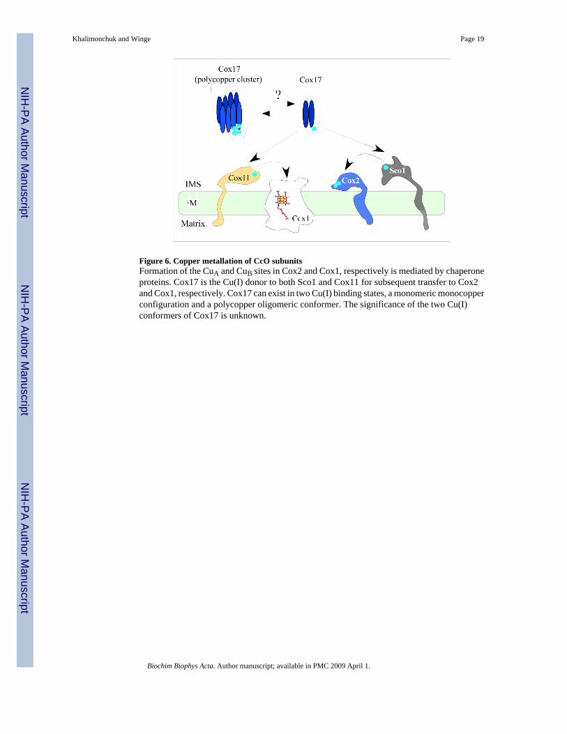
Figure 6. Copper metallation of CcO subunitsFormation of the CuA and CuB sites in Cox2 and Cox1, respectively is mediated by chaperoneproteins. Cox17 is the Cu(I) donor to both Sco1 and Cox11 for subsequent transfer to Cox2and Cox1, respectively. Cox17 can exist in two Cu(I) binding states, a monomeric monocopperconfiguration and a polycopper oligomeric conformer. The significance of the two Cu(I)conformers of Cox17 is unknown.
Khalimonchuk and Winge Page 19
Biochim Biophys Acta. Author manuscript; available in PMC 2009 April 1.
NIH
-PA Author Manuscript
NIH
-PA Author Manuscript
NIH
-PA Author Manuscript

Figure 7. A pro-oxidant intermediate in the CcO assembly pathwayA pro-oxidant intermediate consisting of heme A3-containing Cox1 accumulates when theassembly pathway is blocked subsequent to Cox1 maturation. The deleterious effects of theintermediate can be suppressed by overexpression of Cox11 or Sco1 suggesting that thesemolecules can cap the Cox1 intermediate thereby precluding peroxide access to the heme A3catalytic center. Formation of the mature core of CcO with Cox2 inserted on Cox1 resolvesthe potentially deleterious Cox1 intermediate.
Khalimonchuk and Winge Page 20
Biochim Biophys Acta. Author manuscript; available in PMC 2009 April 1.
NIH
-PA Author Manuscript
NIH
-PA Author Manuscript
NIH
-PA Author Manuscript