functional taining r and advanced rehabilitation - … · functional taining r and advanced...
TRANSCRIPT
Functional Training and Advanced
Rehabilitation Michael L. Voight, Barbara J. Hoogenboom,
Gray Cook, and Greg Rose
O B JJ E C TTT I VE S After completion of this chapter, the physical therapist should be able to do the following:
� Defi ne and discuss the importance of proprioception in the neuromuscular control process.
� Defi ne and discuss the different levels of motor control by the central nervous system and the
neural pathways responsible for transmission of afferent and efferent information at each level.
� Apply a systematic functional evaluation designed to provoke symptoms.
� Demonstrate consistency between functional and clinical testing information (combinatorial power).
� Apply a 3-step model designed to promote the practical systematic thinking required for
effective therapeutic exercise prescription and progression.
� Defi ne and discuss objectives of a functional neuromuscular rehabilitation program.
� Develop a rehabilitation program that uses various exercise techniques for development of
neuromuscular control.
513
Hoog_Ch19_0513-0546.indd 513Hoog_Ch19_0513-0546.indd 513 21/10/13 1:18 PM21/10/13 1:18 PM

Function and Functional Rehabilitation
Th e basic goal in rehabilitation is to restore and enhance function within the environ-ment and to perform specifi c activities of daily living (ADL). Th e entire rehabilitation pro-cess should be focused on improving the functional status of the patient. Th e concept of functional training is not new, nor is it limited to function related to sports. By defi nition, function means having a purpose or duty. Th erefore, functional can be defi ned as perform-ing a practical or intended function or duty. Function should be considered in terms of a spectrum because ADL encompass many diff erent tasks for many diff erent people. What is functional to one person may not be functional to another. It is widely accepted that to perform a specifi c activity better, one must practice that activity. Th erefore, the functional exercise progression for return to ADL can best be defi ned as breaking the specifi c activi-ties down into a hierarchy and then performing them in a sequence that allows acquisition or reacquisition of that skill. It is important to note that although people develop diff erent levels of skill, function, and motor control, certain fundamental tasks are common to nearly all individuals (barring pathologic conditions and disability). Lifestyle, habits, injury, and other factors can erode the fundamental components of movement without obvious altera-tions in higher-level function and skill. Ongoing higher-level function is a testament to the compensatory power of the neurologic system. Imperfect function and skill create stress in other body systems. Fundamental elements can fi rst be observed during the developmental progression of posture and motor control. Th e sequence of developmental progression can also give insight into the original acquisition of skill. Th e ability to assess retention or loss of fundamental movement patterns is therefore a way to enhance rehabilitation. Th e rehabili-tation process starts with a 2-part appraisal that creates perspective by viewing both ends of the functional spectrum:
• Th e current level of function (ADL, work, and sports/recreation) relative to the patient’s needs and goals.
• Th e ability to demonstrate the fundamental movement patterns that represent the foundation of function and basic motor control.
Objectives of Functional Rehabilitation Th e overall objective of a functional exercise program is to return patients to their preinjury level as quickly and as safely as possible by resolving or reducing the measurable dysfunc-tion within fundamental and functional movement patterns. Specifi c training activities are designed to restore both dynamic joint stability and ADL skills. 1 To accomplish this objec-tive, a basic tenet of exercise physiology is used. Th e SAID (specifi c adaptations to imposed demands) principle states that the body will adapt to the stress and strain placed on it. 2
Athletes cannot succeed if they have not been prepared to meet all the demands of their specifi c activity. 2 Reactive neuromuscular training (RNT) helps bridge the gap from tradi-tional rehabilitation via proprioceptive and balance training to promote a more functional return to activity. 2 Th e SAID principle provides constructive stress, and RNT creates oppor-tunities for input and integration. Th e main objective of the RNT program is to facilitate the unconscious process of interpreting and integrating the peripheral sensations received by the central nervous system (CNS) into appropriate motor responses. Th is approach is enhanced by the unique clinical focus on pathologic orthopedic and neurologic states and their functional representation. Th is special focus forces the clinician to consider evalu-ation of human movement as a complex multisystem interaction and the logical starting point for exercise prescription. Sometimes this will require a breakdown of the supporting mobility and stability within a pattern. Regardless of the specifi c nature of the corrective
514 Chapter 19 Functional Training and Advanced Rehabilitation
Hoog_Ch19_0513-0546.indd 514Hoog_Ch19_0513-0546.indd 514 21/10/13 1:18 PM21/10/13 1:18 PM

needs, all the functional exercises follow a simple but very specifi c path. First, the functional exercise program is driven by a functional screening or assessment that produces a baseline of movement. Th e process of screening and assessment will rate and rank patterns. It will provide valuable information about dysfunction in movement patterns such as asymmetry, diffi culty with movement, and pain. Screening and assessment will therefore identify faulty movement patterns that should not be exercised or trained until corrected. Second, the functional framework will assist in making the best possible choices for corrective catego-ries and exercises. No single exercise is best for a movement problem, but there is an appro-priate category of corrective exercises to choose from. Th ird, following the initial session of corrective exercises, the movement pattern should be rechecked for changes against the original baseline. Fourth, once an obvious change is noted in the key pattern, the screening or assessment is repeated to survey other changes in movement and identify the next prior-ity. By working on the most fundamental pattern, it is possible to see other positive changes. Th erefore, these 4 steps provide the framework that makes corrective exercise successful:
• Th e screening and assessment direct the clinician to the most fundamental movement dysfunction.
• One or 2 of the most practical corrective exercises from the appropriate category should be chosen and applied.
• Once the exercise has been taught and is being performed correctly, check for improvement in the dysfunctional basic movement pattern as revealed by specifi c tests in the screening or assessment.
Th is concept is called the functional continuum. Most patients seek care because of an obvious source of pain or dysfunction. What is not obvious is the true cause of the pain or dysfunction, ascertainment of which is the purpose of functional movement assessment (see Chapter 17). By looking at movement as a whole, all the compensations and conscious sources of pain and dysfunction can be highlighted and addressed. Patients fall into one of four phases on a functional continuum ( Table 19-1 ).
Table 19-1 Four Phases of the Functional Continuum
Phase Description
Subconscious dysfunction
This is the initial phase when most patients are fi rst seen by the clinician. Patients are totally unaware of their true dysfunction (it is in their subconscious) or are convinced that the problem lies elsewhere.
Conscious dysfunction This is what happens after a movement assessment is performed. Patients are now aware of their true dysfunction (it is in their conscious), and they can start to address the real cause.
Conscious function This phase is entered once patients can perform the correct functional pattern, but it is not automatic (it is functional only with conscious control). They still need conscious effort to perform a good pattern of movement.
Subconscious function The fi nal stage occurs when patients can perform a functional pattern automatically (it is in their subconscious control) without having to think about the correction.
Function and Functional Rehabilitation 515
Hoog_Ch19_0513-0546.indd 515Hoog_Ch19_0513-0546.indd 515 21/10/13 1:18 PM21/10/13 1:18 PM

Exercise prescription choices must continually represent the specialized training of the clinician through a consistent and centralized focus on human function and consideration of the fundamentals that make function possible. Exercise applied at any given therapeutic level must refi ne movement, not simply create general exertion in the hope of increased tolerance of movement. 3 Moore and Durstine state, “Unfortunately, exercise training to optimize functional capacity has not been well studied in the context of most chronic dis-eases or disabilities. As a result, many exercise professionals have used clinical experience to develop their own methods for prescribing exercise.” 4 Experience, self-critique, and spe-cialization produce seasoned clinicians with intuitive evaluation abilities and innovations in exercise that are sometimes diffi cult to follow and even harder to ascertain; however, common characteristics do exist. Clinical experts use parallel (simultaneous) consideration of all factors infl uencing functional movement. RNT as a treatment philosophy is inclusive and adaptable and has the ability to address a variety of clinical situations. It should also be understood that a clinical philosophy is designed to serve, not to be served. Th e treat-ment design demonstrates specifi c attention to the parts (clinical measurements and iso-lated details) with continual consideration of the whole (restoration of function). 3 Moore and Durstine follow their previous statement by acknowledging that “Experience is an acceptable way to guide exercise management, but a systematic approach would be better.” 4
We use the 3 “Rs” as a way to understand the type of treatment phases that a patient will undergo ( Table 19-2 ).
The Three-Phase Model for Prescription of Exercise Th is chapter demonstrates a practical model designed to promote the systematic thinking required for eff ective prescription of therapeutic exercise and progression at each phase of rehabilitation. 3 Th e approach is a serial (consecutive) step-by-step method that will, with practice and experience, lead to parallel thinking and multilevel problem solving. Th e intended purpose of this method is to reduce arbitrary trial-and-error attempts at prescrib-ing eff ective exercise and lessen protocol-based thinking. It will give the novice clinician a framework that will guide but not confi ne clinical exercise prescription. It will provide experienced clinicians with a system to observe their particular strengths and weaknesses in dosage and design of exercise. Inexperienced and experienced clinicians alike will
Table 19-2 Three Rs of Treatment Phases
R Description
Reset Most problems require resetting of the complete system to break them out of their dysfunctional phase. By just jumping to exercises, the results can be less than optimal. Types of treatments that would be considered a “reset” include joint mobilization, soft-tissue mobilization, and various soft-tissue techniques.
Reinforce Once the system has been reset, many dysfunctions will need support or reinforcement while proper patterns are being introduced. Types of reinforcement devices include taping, bracing, orthotics, postural devices, and static and dynamic stretching.
Reload The last phase of treatment is the exercise implementation or reload phase, in which the new software is loaded into the central nervous system and a true functional pattern of motion can be reprogrammed.
516 Chapter 19 Functional Training and Advanced Rehabilitation
Hoog_Ch19_0513-0546.indd 516Hoog_Ch19_0513-0546.indd 516 21/10/13 1:18 PM21/10/13 1:18 PM

develop practical insight by applying the model and observing the interaction of the sys-tems that produce human movement. Th e focus is specifi cally geared to orthopedic rehabil-itation and the clinical problem-solving strategies used to develop an exercise prescription through an outcome-based goal-setting process. All considerations for therapeutic exer-cise prescription will give equal importance to conventional orthopedic exercise standards (biomechanical and physiologic parameters) and neurophysiologic strategies (motor learn-ing, proprioceptive feedback, and synergistic recruitment principles). Th is 3-phase model ( Box 19-1 ) will create a mechanism that necessitates interaction between orthopedic exer-cise approaches and optimal neurophysiologic techniques. It includes a 4-principle foun-dation that demonstrates the hierarchy and interaction of the founding concepts used in rehabilitation (both orthopedic and neurologic). For all practical purposes, these 4 catego-ries help demonstrate the effi cient and eff ective continuity necessary for formulation of a treatment plan and prompt the clinician to maintain an inclusive, open-minded clinical approach.
Th is chapter is written with the clinic-based practitioner in mind. It will help the clini-cian formulate an exercise philosophy. Some clinicians will discover reasons for success that were intuitive and therefore hard to communicate to other professionals. Others will discover a missing step in the therapeutic exercise design process. Much of the confusion and frustration encountered by rehabilitation specialists is because of the vast variety of treatment options aff orded by ever-improving technology and accessibility to emerging research evidence. To eff ectively use the wealth of current information and what the future has yet to bestow, clinicians must adopt an operational framework or personal philosophy about therapeutic exercise. If a clinical exercise philosophy is based on technology, equip-ment, or protocols, the scope of problem solving is strictly confi ned. It would continually change because no universal standard or gauge exists. However, a philosophy based solely on the structure and function of the human body will keep the focus ( Box 19-2 ) uncorrupted and centralized. Technologic developments can enhance the eff ectiveness of exercise only as long as the technology, system, or protocol remains true to a holistic functional stan-dard. Known functional standards should serve as governing factors that improve the clini-cal consistency of the clinician and rehabilitation team for prescription and progression of training methods. Th e 4 principles for exercise prescription are based on human move-ment and the systems on which it is constructed ( Box 19-2 ). Th e intent of these 4 distinct categories is to break down and reconstruct the factors that infl uence functional movement and to stimulate inductive reasoning, deductive reasoning, and the critical thinking needed
Box 19-1 Three-Phase Rehabilitation Model
Box 19-2 Four Principles for Prescription of Exercise
Function and Functional Rehabilitation 517
Hoog_Ch19_0513-0546.indd 517Hoog_Ch19_0513-0546.indd 517 21/10/13 1:18 PM21/10/13 1:18 PM

to develop a therapeutic exercise progression. It is hoped that these factors will serve the intended purpose of organization and clarity, thereby giving due respect to the many insightful clinicians who have provided the foundation and substance for construction of this practical framework. 3
Proprioception, Receptors, and Neuromuscular Control
Success in skilled performance depends on how eff ectively an individual detects, perceives, and uses relevant sensory information. Knowing exactly where our limbs are in space and how much muscular eff ort is required to perform a particular action is critical for successful performance of all activities requiring intricate coordination of the various body parts. For-tunately, information about the position and movement of various body parts is available from peripheral receptors located in and around articular structures and the surrounding musculature. A detailed discussion of proprioception and neuromuscular control is also presented in Chapter 9.
Joints: Support and Sensory Function In a normal healthy joint, both static and dynamic stabilizers provide support. Th e role of capsuloligamentous tissues in the dynamic restraint of joints has been well established in the literature. 5-15 Although the primary role of these structures is mechanical in nature by providing structural support and stabilization to the joint, the capsuloligamentous tissues also play an important sensory role by detecting joint position and motion. 8,16-18 Sensory aff erent feedback from receptors in the capsuloligamentous structures projects directly to the refl ex and cortical pathways, thereby mediating reactive muscle activity for dynamic restraint. 5,6,8,17,19 Th e eff erent motor response that ensues from the sensory information is called neuromuscular control. Sensory information is sent to the CNS to be processed, and appropriate motor strategies are executed.
Physiology of Proprioception Sherrington 18 fi rst described the term proprioception in the early 1900s when he noted the presence of receptors in the joint capsular structures that were primarily refl exive in nature. Since that time, mechanoreceptors have been morphohistologically identifi ed around articular structures in both animal and human models. In addition, the well-described muscle spindle and Golgi tendon organs are powerful mechanoreceptors. Mechanorecep-tors are specialized end-organs that function as biologic transducers for conversion of the mechanical energy of physical deformation (elongation, compression, and pressure) into action nerve potentials yielding proprioceptive information. 10 Although receptor discharge varies according to the intensity of the distortion, mechanoreceptors can also be described in terms of their discharge rates. Quickly adapting receptors cease discharging shortly after the onset of a stimulus, whereas slowly adapting receptors continue to discharge while the stimulus is present. 8,10,20 Around a healthy joint, quickly adapting receptors are respon-sible for providing conscious and unconscious kinesthetic sensations in response to joint movement or acceleration, whereas slowly adapting mechanoreceptors provide continuous feedback and thus proprioceptive information related to joint position 10,20,21 (see Chapter 9 for examples of quickly and slowly adapting receptors).
Once stimulated, mechanoreceptors are able to adapt. With constant stimula-tion, the frequency of the neural impulses decreases. Th e functional implication is that
518 Chapter 19 Functional Training and Advanced Rehabilitation
Hoog_Ch19_0513-0546.indd 518Hoog_Ch19_0513-0546.indd 518 21/10/13 1:19 PM21/10/13 1:19 PM

mechanoreceptors detect change and rates of change, as opposed to steady-state condi-tions. 22 Th is input is then analyzed in the CNS to determine joint position and movement. 23
Th e status of the musculoskeletal structures is sent to the CNS so that information about static versus dynamic conditions, equilibrium versus disequilibrium, or biomechanical stress and strain relationships can be evaluated. 24,25 Once processed and evaluated, this proprioceptive information becomes capable of infl uencing muscle tone, motor execution programs, and cognitive somatic perceptions or kinesthetic awareness. 26 Proprioceptive information also protects the joint from damage caused by movement exceeding the nor-mal physiologic range of motion (ROM) and helps determine the appropriate balance of synergistic and antagonistic forces. All this information helps in generating a somatosen-sory image within the CNS. Th erefore, the soft tissues surrounding a joint serve a double purpose: they provide biomechanical support to the bony partners making up the joint by keeping them in relative anatomic alignment, and through an extensive aff erent neurologic network, they provide valuable proprioceptive information.
Central Nervous System: Integration of Motor Control
Th e response of the CNS falls into 3 categories or levels of motor control: spinal refl exes, brainstem processing, and cognitive cerebral cortex program planning. Th e goal of the rehabilitation process is to retrain the altered aff erent pathways and thereby enhance the neuromuscular control system. To accomplish this goal, the objective of the rehabilitation program should be to hyperstimulate the joint and muscle receptors to encourage maximal aff erent discharge to the respective CNS levels. 21,27-30
First-Level Response: Muscle When faced with an unexpected load, the fi rst refl exive muscle response is a burst of elec-tromyographic activity that occurs between 30 and 50 milliseconds. Th e aff erent fi bers of both the muscle spindle and the Golgi tendon organ mechanoreceptors synapse with the spinal interneurons and produce a refl exive facilitation or inhibition of the motor neurons. 28,30,31 Th e monosynaptic stretch refl ex is one of the most rapid refl exes under-lying limb control. Th e stretch refl ex occurs at an unconscious level and is not aff ected by extrinsic factors. Th ese responses can occur simultaneously to control limb position and posture. Because they can occur at the same time, are in parallel, are subconscious, and are not subject to cortical interference, they do not require attention and are thus automatic.
At this level of motor control, activities to encourage short-loop refl ex joint stabilization should dominate. 15,21,27,30 Th ese activities are characterized by sudden alterations in joint position that require refl ex muscle stabilization. With sudden alterations or perturbations, both the articular and muscular mechanoreceptors will be stimulated to produce refl ex sta-bilization. Rhythmic stabilization exercises encourage monosynaptic cocontraction of the musculature, thereby producing dynamic neuromuscular stabilization. 32 Th ese exercises serve to build a foundation for dynamic stability.
Second-Level Response: Brainstem Th e second level of motor control interaction is at the level of the brainstem. 25,28,33 At this level, aff erent mechanoreceptors interact with the vestibular system and visual input from the eyes to control or facilitate postural stability and equilibrium of the
Central Nervous System: Integration of Motor Control 519
Hoog_Ch19_0513-0546.indd 519Hoog_Ch19_0513-0546.indd 519 21/10/13 1:19 PM21/10/13 1:19 PM

body. 21,25,27-29 Aff erent mechanoreceptor input also works in concert with the muscle spindle complex by inhibiting antagonistic muscle activity under conditions of rapid lengthening and periarticular distortion, both of which accompany postural disrup-tion. 26,30 In conditions of disequilibrium in which simultaneous neural input exists, a neural pattern is generated that aff ects the muscular stabilizers and thereby returns equilibrium to the body’s center of gravity. 28 Th erefore, balance is infl uenced by the same peripheral aff erent mechanism that mediates joint proprioception and is at least partially dependent on an individual’s inherent ability to integrate joint position sense with neuromuscular control. 34
Clinical Pearl
Balance activities, both with and without visual input, will enhance motor function at the brainstem level. 28,33
It is important that these activities remain specifi c to the types of activities or skills that will be required of the athlete on return to sport. 35 Static balance activities should be used as a precursor to more dynamic skill activity. 35 Static balance skills can be initiated when the individual is able to bear weight on the lower extremity. Th e general progres-sion of static balance activities is to move from bilateral to unilateral and from eyes open to eyes closed. 21,28,35-37 With balance training, it is important to remember that the sen-sory systems respond to environmental manipulation. To stimulate or facilitate the pro-prioceptive system, vision must be disadvantaged, which can be accomplished in several ways ( Box 19-3 ).
Third-Level Response: Central Nervous System/Cognitive Appreciation of joint position at the highest or cognitive level needs to be included in an RNT program. Th ese types of activities are initiated on the cognitive level and include pro-gramming motor commands for voluntary movement. Repetitions of these movements will maximally stimulate the conversion of conscious programming to unconscious program-ming. 21,25,27-29,38 Th e term for this type of training is the forced-use paradigm. By making a task signifi cantly more diffi cult or asking for multiple tasks, the CNS is bombarded with input. Th e CNS attempts to sort and process this overload information by opening additional neu-ral pathways. When the individual goes back to a basic ADL task, the task becomes easier. Th is information can then be stored as a central command and ultimately be performed without continuous reference to conscious thought as a triggered response. 21,27-29,39 As with all training, the single greatest obstacle to motor learning is the conscious mind. We must get the conscious mind out of the act!
Box 19-3 Ways to Disadvantage Vision for Stimulation of the Proprioceptive System
520 Chapter 19 Functional Training and Advanced Rehabilitation
Hoog_Ch19_0513-0546.indd 520Hoog_Ch19_0513-0546.indd 520 21/10/13 1:19 PM21/10/13 1:19 PM

Closed-Loop, Open-Loop, and Feed-Forward Integration Why is a coordinated motor response important? When an unexpected load is placed on a joint, ligamentous damage occurs in 70 to 90 milliseconds unless an appropriate response ensues. 40-42 Th erefore, reactive muscle activity that provides suffi cient magnitude in the 40- to 80-millisecond time frame must occur after loading begins to protect the capsuloliga-mentous structures. Th e closed-loop system of CNS integration may not be fast enough to produce a response to increase muscle stiff ness. Th ere is simply no time for the system to process the information and provide feedback about the condition. Failure of the dynamic restraint system to control abnormal force will expose the static structures to excessive force. In this case, the open-loop system of anticipation becomes more important in pro-ducing the desired response. Preparatory muscle activity in anticipation of joint loading can infl uence the reactive muscle activation patterns. Anticipatory activation increases the sensitivity of the muscle spindles, thereby allowing the unexpected perturbations to be detected more quickly. 43
Very quick movements are completed before feedback can be used to produce an action to alter the course of movement. Th erefore, if the movement is fast enough, a mecha-nism such as a motor program would have to be used to control the entire action, with the movement being carried out without any feedback. Fortunately, the open-loop control sys-tem allows the motor control system to organize an entire action ahead of time. For this to occur, previous knowledge needs to be preprogrammed into the primary sensory cortex ( Box 19-4 ).
In the open-loop system, a program that sets up some kind of neural mechanism or network that is preprogrammed organizes movement in advance. A classic example of this occurs in the body as postural adjustments are made before the intended movement. When an arm is raised into forward fl exion, the fi rst muscle groups to fi re are not even in the shoulder girdle region. Th e fi rst muscles to contract are those in the lower part of the back and legs (approximately 80 milliseconds pass before noticeable activity occurs in the shoulder) to provide a stable base for movement. 44 Because the shoulder muscles are linked to the rest of the body, their contraction aff ects posture. If no preparatory compensations in posture were made, raising the arm would shift the center of gravity forward and cause a slight loss of balance. Th e feed-forward motor control system takes care of this potential problem by preprogramming the appropriate postural modifi cation fi rst rather than requir-ing the body to make adjustments after the arm begins to move.
Lee 45 demonstrated that these preparatory postural adjustments are not independent of the arm movement but rather are part of the total motor pattern. When the arm move-ments are organized, the motor instructions are preprogrammed to adjust posture fi rst and then move the arm. Th erefore, arm movement and postural control are not separate events but instead are diff erent parts of an integrated action that raises the arm while maintain-ing balance. Lee showed that these electromyographic preparatory postural adjustments
Box 19-4 Preprogrammed Information Needed for an Open-Loop System to Work
Central Nervous System: Integration of Motor Control 521
Hoog_Ch19_0513-0546.indd 521Hoog_Ch19_0513-0546.indd 521 21/10/13 1:19 PM21/10/13 1:19 PM

disappear when the individual leans against some type of support before raising the arm. Th e motor control system recognizes that advance preparation for postural control is not needed when the body is supported against the wall.
It is important to remember that most motor tasks are a complex blend of both open- and closed-loop operations. Th erefore, both types of control are often at work simultane-ously. Both feed-forward and feedback neuromuscular control can enhance dynamic stability if the sensory and motor pathways are frequently stimulated. 21 Each time a signal passes through a sequence of synapses, the synapses become more capable of transmitting the same signal. 14,46 When these pathways are “facilitated” regularly, memory of that signal is created and can be recalled to program future movements. 14,47
Conclusion: Relationship to Rehabilitation A rehabilitation program that addresses the need for restoring normal joint stability and proprioception cannot be constructed until one has total appreciation of both the mechani-cal and sensory functions of the articular structures. 27 Knowledge of the basic physiology of how these muscular and joint mechanoreceptors work together in the production of smooth, controlled coordinated motion is critical in developing a rehabilitation training program. Th is is because the role of the joint musculature extends well beyond absolute strength and the capacity to resist fatigue. With simple restoration of mechanical restraints or strengthening of the associated muscles, the smooth coordinated neuromuscular con-trolling mechanisms required for joint stability are neglected. 27 Th e complexity of joint motion necessitates synergy and synchrony of muscle fi ring patterns, thereby permitting proper joint stabilization, especially during sudden changes in joint position, which is com-mon in functional activities. Understanding of these relationships and functional implica-tions will allow the clinician greater variability and success in returning patients safely back to their playing environment.
Four Principles for Th erapeutic Exercise Prescription
Th e functional exercise program follows a linear path from basic mobility to basic stability to movement patterns. Corrective exercise falls into one of the 3 basic categories: mobil-ity, stability, and retraining of movement patterns. Mobility exercises focus on joint ROM, tissue length, and muscle fl exibility. Stability exercises focus on the basic sequencing of movement. Th ese exercises target postural control of the starting and ending positions within each movement pattern. Movement pattern retraining incorporates the use of fun-damental mobility and stability into specifi c movement patterns to reinforce coordina-tion and timing.
Th e corrective exercise progression always starts with mobility exercises. Because many poor movement patterns are associated with abnormalities in mobility, restora-tion of movement needs to be addressed fi rst. Mobility exercises should be performed bilaterally to confi rm limitation and asymmetry of mobility. Clinicians should never assume that they know the location or side in which mobility is restricted. Rather, both sides should always be checked and mobility cleared before advancing the exercise pro-gram. If the assessment reveals a limitation or asymmetry, it should be the primary focus of the corrective exercise program. Treatments that promote mobility can involve man-ual therapy, such as soft-tissue and joint mobilization and manipulation. Treatments of mobility might also include any modality that improves tissue pliability or freedom of movement. If no change in mobility is appreciated, the clinician should not proceed to
522 Chapter 19 Functional Training and Advanced Rehabilitation
Hoog_Ch19_0513-0546.indd 522Hoog_Ch19_0513-0546.indd 522 21/10/13 1:19 PM21/10/13 1:19 PM

stability work. Rather, all mobility problems should continue to be worked on until a mea-surable change is noted. Mobility does not need to become full or normal, but improve-ment must be noted before advancing. Th e clinician can proceed to a stability exercise only if the increased mobility allows the patient to get into the appropriate exercise pos-ture and position. Th e stability work should reinforce the new mobility, and the new mobility makes improved stabilization possible because the new mobility provides new sensory information. If there is any question about compromised mobility, each exercise session should always return to mobility exercises before moving to stability exercises. Th is ensures that proper tissue length and joint alignment are available for the stabiliza-tion exercises.
When no limitation or asymmetry is present during the mobility corrective exercises, one can move directly to stability corrective exercises. Once mobility has been restored, it needs to be controlled. Stability exercises demand posture, alignment, balance, and control of forces within the newly available range and without the support of compensatory stiff -ness or muscle tone. Stability exercises should be considered as challenges to posture and position, rather than being conventional strength exercises.
We propose 4 principles for therapeutic exercise prescription, which we describe as the 4 “Ps” in this section. Th ese principles serve to guide decisions for selecting, advancing, and terminating therapeutic exercise interventions. Application of these 4 principles in the appropriate sequence will allow the clinician to understand the starting point, a consistent progression, and the end point for each exercise prescription. Th is sequence is achieved by using functional activities and fundamental movement patterns as goals. By proceeding in this fashion, the clinician will have the ability to evaluate the whole before the parts and then discuss the parts as they apply. Table 19-3 lists and describes the principles for thera-peutic exercise prescription.
Table 19-3 Four Principles for Therapeutic Exercise Prescription
Principle Description
Functional evaluation and assessment in relation to dysfunction (disability) and impairment
The evaluation must identify a functional problem or limitation resulting in diagnosis of a functional problem. Observation of whole movement patterns tempered by practical knowledge of key stress points and common compensatory patterns will improve the effi ciency of evaluation.
Identifi cation and management of motor control
Rehabilitation can be greatly advanced by understanding functional milestones and fundamental movements such as those demonstrated during the positions and postures paramount to growth and development. These milestones serve as key representations of functional mobility and control, as well as play a role in the initial setup and design of the exercise program.
Identifi cation and management of osteokinematic and arthrokinematic limitations
The skills and techniques of orthopedic manual therapy are benefi cial in identifying specifi c arthrokinematic restrictions that would limit movement or impede the motor-learning process. Management of myofascial and capsular structures will improve osteokinematic movement, as well as allow balanced muscle tone between the agonist and antagonist. It will also help the clinician understand the dynamics of the impairment.
Identifi cation of current movement patterns followed by facilitation and integration of synergistic movement patterns
Once restrictions and limitations are managed and gross motion is restored, application of proprioceptive neuromuscular facilitation-type patterning will further improve neuromuscular function and control. Consideration of synergistic movement is the fi nal step in restoration of function by focusing on coordination, timing, and motor learning.
Four Principles for Therapeutic Exercise Prescription 523
Hoog_Ch19_0513-0546.indd 523Hoog_Ch19_0513-0546.indd 523 21/10/13 1:19 PM21/10/13 1:19 PM

Table 19-4 Memory Cues and Primary Questions Associated with the Four Principles for Prescription of Therapeutic Exercise
Principle Memory Cue Memory Cue Defi nition Primary Questions
Functional evaluation and assessment
Purpose Used during both the evaluation process and the exercise prescription process to keep the clinician intently focused on the greatest single factor limiting function
“ What functional activity is limited?” “ What does the limitation appear to be—a
mobility problem or a stability problem?” “ What is the dysfunction or disability?” “ What fundamental movement is limited?” “ What is the impairment?”
Identifi cation of motor control
Posture Helps the clinician remember to consider a more holistic approach to exercise prescription
“ When in the development sequence is the impairment obvious?”
“ When do the substitutions and compensations occur?”
“ When in the developmental sequence does the patient demonstrate success?”
“ When in the developmental sequence does the patient experience diffi culty?”
“ When is the best possible starting point for exercise with respect to posture?”
Identifi cation of osteokinematic and arthrokinematic limitations
Position Describes not only the location of the anatomic structure (joint, muscle group, ligament, etc) where impairment has been identifi ed but also the positions (with respect to movement and load) in which the greatest and least limitations occur
“ Where is the impairment located?” “ Where among the structures (myofascial or
articular) does the impairment have its greatest effect?”
“ Where in the range of motion does the impairment affect position the greatest?”
“ Where is the most benefi cial position for the exercise?”
Integration of synergistic movement patterns
Pattern Cues the clinician to continually consider the functional movements of the human body that occur in unifi ed patterns that occupy 3-dimensional space and cross 3 planes (frontal, sagittal, and transverse)
“ How is the movement pattern different on bilateral comparison?”
“ How can synergistic movement, coordination, recruitment and timing be facilitated?”
“ How will this affect the limitation in movement?”
“ How will this affect function?”
Clinical Pearl
The true art of rehabilitation is to understand the whole of synergistic functional movement and the therapeutic techniques that will have the greatest positive effect on that movement in the least amount of time.
The Four Ps Th e 4 Ps represent the 4 principles for therapeutic exercise: purpose, posture, position, and pattern ( Table 19-4 ). Th ey serve as quick reminders of the hierarchy, interaction, and applica-tion of each principle. Th e questions of what, when, where, and how for functional movement assessment and exercise prescription are addressed in the appropriate order ( Table 19-4 ).
524 Chapter 19 Functional Training and Advanced Rehabilitation
Hoog_Ch19_0513-0546.indd 524Hoog_Ch19_0513-0546.indd 524 21/10/13 1:19 PM21/10/13 1:19 PM

Pain Aristotle said, “We cannot learn without pain,” which is very wise because pain is usually life’s most power-ful teacher. However, pain is simply the brain’s inter-pretation of a neurologic signal normally associated with trauma, dysfunction, and instant and continu-ing damage. Pain aff ects motor control and greatly reduces the eff ectiveness of even the best corrective exercise technique.
Purpose Th e word purpose is simply a cue to be used dur-ing both the evaluation process and the exercise prescription process to keep the clinician intently focused on the greatest single factor limiting func-tion. Th e primary questions to ask for this principle appear in Table 19-4 . It is not uncommon for clini-cians to attempt to resolve multiple problems with the initial exercise prescription. However, the prac-tice of identifying the single greatest limiting factor will reduce frustration and also not overwhelm the patient. Other factors may have been identifi ed in the evaluation, but a major limiting factor or a single weak link should stand out and be the focus of the initial therapeutic exercise interven-tion. Alterations in the limiting factor may produce positive changes elsewhere, which can be identifi ed and considered before the next exercise progression.
Th e functional evaluation process should take on 3 distinct layers or levels ( Table 19-5 ). Each of the 3 levels should involve qualitative observations followed by quantitative documentation when possible. Normative data are helpful, but bilateral comparison is also eff ective and demonstrates the functional problem to the patient at each level. Many patients think that the problem is simply symptomatic and structural in nature and have no example of dysfunction outside of pain with movement. Moff roid and Zimny suggest that “Muscle strength of the right and left sides is more similar in the proximal muscles whereas we accept a 10% to 15% diff erence in strength of the distal muscles. . . . With joint fl exibility, we accept a 5% diff erence between goniometric mea-surements of the right and left sides.” 48
Th e functional activity assessment involves a reproduction of combined movements common to the patient’s lifestyle and occupation. Th ese movements usually fi t the defi ni-tion of a general or specifi c skill. Th e clinician must have the patient demonstrate a variety of positions and not just positions that correspond to the reproduction of symptoms. 49 Static postural assessment is included, as well as assessment of dynamic activity. Th e quality of control and movement is assessed. Specifi c measurement of bilateral diff erences is diffi cult, but demonstration and observation are helpful for the patient. Th e clinician should note the positions and activities that provoke symptoms, as well as the activities that illustrate poor body mechanics, poor alignment, right-left asymmetries, and inappropriate weight shifting. When the clinician has observed gross movement quality, it may be necessary to also quan-tify movement performance. Repetition of the activity for evaluation of endurance, repro-duction of symptoms, or demonstration of rapidly declining quality will create a functional baseline for bilateral comparison and documentation.
Next is the functional or fundamental movement assessment. Th e clinician must take what is learned through the observation of functional movements and break the
Table 19-5 Three Levels of Functional Evaluation
Level Name Description
I Functional activity assessment
Combined movements common to the patient’s lifestyle and occupation are reproduced. They usually fi t the defi nition of a general or specifi c skill.
II Functional or fundamental movement assessment
The clinician takes what is learned through the observation of functional movements and breaks the movements down to the static and transitional postures seen in the normal developmental sequence.
III Specifi c clinical measurement
Clinical measurements are used to identify and quantify specifi c problems that contribute to limitations in motion or control.
Four Principles for Therapeutic Exercise Prescription 525
Hoog_Ch19_0513-0546.indd 525Hoog_Ch19_0513-0546.indd 525 21/10/13 1:19 PM21/10/13 1:19 PM

movements down into the static and transitional postures seen in the normal develop-mental sequence. Th is breakdown will reduce activities to the many underlying mobi-lizing and stabilizing actions and reactions that constitute the functional activity. More simply stated, the activity is broken down into a sequence of primary movements that can be observed independently. It must be noted that these movements still involve multiple joints and muscles. 49 Assessment of individual joints and muscle groups will be performed during clinical measurements. Martin notes, “Th e developmental sequence has provided the most consistent base for almost all approaches used by physical therapists.” 48 Th is is a powerful statement, and because true qualitative measurements of normal movement in adult populations are limited, the clinician must look for universal similarities in move-ment. Changes in fundamental movements can eff ect signifi cant and prompt changes in function and must therefore be considered functional as well. Because the movement pat-terns of most adults are habitual and specifi c and thus are not representative of a full or optimal movement spectrum, the clinician must fi rst consider the nonspecifi c basic move-ment patterns common to all individuals during growth and development. Th e develop-mental sequence is predictable and universal in the fi rst 2 years of life, 50 with individual diff erences seen in the rate and quality of the progression. Th e diff erences are minimal in comparison to the variations seen in the adult population with their many habits, occu-pations, and lifestyles. In addition to diverse movement patterns, the adult population has the consequential complicating factor of a previous medical and injury history. Each medical problem or injury has had some degree of infl uence on activity and movement. Th us, evaluation of functional activities alone may hide many uneconomical movement patterns, compensations, and asymmetries that when integrated into functional activi-ties, are not readily obvious to the clinician. By using the fundamental movements of the developmental progression, the clinician can view mobility and static and dynamic stabil-ity problems in a more isolated setting. Although enormous variations in functional move-ment quality and quantity are seen in specifi c adult patient populations, most individuals have the developmental sequence in common. 50 Th e movements used in normal motor development are the building blocks of skill and function. 50 Many of these building blocks can be lost while the skill is maintained or retained at some level (though rarely optimal). We will refer to these movement building blocks as fundamental movements and consider them precursors to higher function. Bilateral comparison is helpful when the clinician identifi es qualitative diff erences between the right and left sides. Th ese movements (like functional activities) can be compared quantitatively as well.
Finally, clinical measurements will be used to identify and quantify specifi c prob-lems that are contributing to limitation of motion or limitation of control. Clinical mea-surements will fi rst classify a patient through qualitative assessment. Th e parameters that defi ne that classifi cation must then be quantifi ed to reveal impairment. Th ese classifi ca-tions are called hypermobility and hypomobility and help create guides for treatment that consider the functional status, anatomic structures, and the severity of symptoms. Th e clinician should not proceed into exercise prescription without proper identifi cation of one of these general categories. Th e success or failure of a particular exercise treatment regimen probably depends more on this classifi cation than on the choice of exercise tech-nique or protocol.
Once the appropriate clinical classifi cation is determined, specifi c quantitative mea-surements will defi ne the level of involvement within the classifi cation and set a baseline for exercise treatment. Periodic reassessment may identify a diff erent major limiting factor or a weak link that may require reclassifi cation, followed by specifi c measurement. Th e new problem or limitation would then be inserted as the purpose for a new exercise interven-tion. A simple diagram ( Figure 19-1 ) will help the clinician separate the diff erent levels of function so that intervention and purpose will always be at the appropriate level and assist in the clinical decision making related to exercise prescription. 51
526 Chapter 19 Functional Training and Advanced Rehabilitation
Hoog_Ch19_0513-0546.indd 526Hoog_Ch19_0513-0546.indd 526 21/10/13 1:19 PM21/10/13 1:19 PM
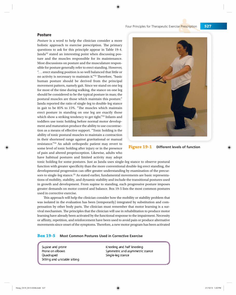
Posture Posture is a word to help the clinician consider a more holistic approach to exercise prescription. Th e primary questions to ask for this principle appear in Table 19-4 . Janda 52 stated an interesting point when discussing pos-ture and the muscles responsible for its maintenance. Most discussions on posture and the musculature respon-sible for posture generally refer to erect standing. However, “. . . erect standing position is so well balanced that little or no activity is necessary to maintain it.” 52 Th erefore, “basic human posture should be derived from the principal movement pattern, namely gait. Since we stand on one leg for most of the time during walking, the stance on one leg should be considered to be the typical posture in man; the postural muscles are those which maintain this posture.” Janda reported the ratio of single-leg to double-leg stance in gait to be 85% to 15%. “Th e muscles which maintain erect posture in standing on one leg are exactly those which show a striking tendency to get tight.” 53 Infants and toddlers use tonic holding before normal motor develop-ment and maturation produce the ability to use cocontrac-tion as a means of eff ective support. “Tonic holding is the ability of tonic postural muscles to maintain a contraction in their shortened range against gravitational or manual resistance.” 54 An adult orthopedic patient may revert to some level of tonic holding after injury or in the presence of pain and altered proprioception. Likewise, adults who have habitual postures and limited activity may adopt tonic holding for some postures. Just as Janda uses single-leg stance to observe postural function with greater specifi city than the more conventional double-leg erect standing, the developmental progression can off er greater understanding by examination of the precur-sors to single-leg stance. 55 As stated earlier, fundamental movements are basic representa-tions of mobility, stability, and dynamic stability and include the transitional postures used in growth and development. From supine to standing, each progressive posture imposes greater demands on motor control and balance. Box 19-5 lists the most common postures used in corrective exercise.
Th is approach will help the clinician consider how the mobility or stability problem that was isolated in the evaluation has been (temporarily) integrated by substitution and com-pensation by other body parts. Th e clinician must remember that motor learning is a sur-vival mechanism. Th e principles that the clinician will use in rehabilitation to produce motor learning have already been activated by the functional response to the impairment. Necessity or affi nity, repetition, and reinforcement have been used to avoid pain or produce alternative movements since onset of the symptoms. Th erefore, a new motor program has been activated
Figure 19-1 Different levels of function
Box 19-5 Most Common Postures Used in Corrective Exercise
Four Principles for Therapeutic Exercise Prescription 527
Hoog_Ch19_0513-0546.indd 527Hoog_Ch19_0513-0546.indd 527 21/10/13 1:20 PM21/10/13 1:20 PM

to manage the impairment and produce some level of function that is usually viewed as dys-function. It should be considered a natural and appropriate response of the body reacting to limitation or symptoms. Th e body will sacrifi ce quality of movement to maintain a degree of quantity of movement. Taking this into consideration, 2 distinct needs are presented.
Posture for Protection and Inhibition Th e clinician must restrict or inhibit the inap-propriate motor program. In the case of a control or stability problem, the patient must have some form of support, protection, or facilitation. Otherwise, the inappropriate pro-gram will take over in an attempt to protect and respond to the postural demand. Although most adult patients function at the necessary skill level, on evaluation, many qualitative problems are noted. Inappropriate joint loading and locking, poor tonic responses, or even tonic holding can be observed with simple activities. Some joint movements are used exces-sively, whereas others are unconsciously avoided. Many primary stability problems exist when underlying secondary mobility problems are present. Moreover, in some patients, the mobility problem precedes the stability problem. Th is is a common explanation for micro-traumatic and overuse injuries. It is also why bilateral comparison and assessment of proxi-mal and distal structures are mandatory in the evaluative process. With a mobility problem, a joint is not used appropriately because of weakness or restriction. Th e primary mobility problem may be the result of compromised stability elsewhere. Motor programs have been created to allow a patient to push on despite the mobility or stability problem. Th e prob-lems can be managed by mechanical consideration of the mobility and stability status of the patient in the fundamental postures.
For primary stability problems, mechanical support or other assistance must be pro-vided. Th is can be done simply by partial or complete reduction of stress, which may include non–weight bearing or partial weight bearing of the spine and extremities or tem-porary bracing. If the stability problem is only in a particular range of movement, that move-ment must be managed. If an underlying mobility problem is present, it must be managed and temporarily taken out of the initial exercise movement. Th e alteration in posture can eff ectively limit complete or partial motion with little need for active control by the patient. Th e patient must be trained to deal with the stability problem independently of the mobil-ity problem or be at a great mechanical advantage to avoid compensation. Th e secondary mobility problem, once managed, should be reintroduced in a nonstressful manner so that the previous compensatory pattern is not activated.
Manual articular and soft-tissue techniques, when appropriate, can be used for the pri-mary mobility problem, followed by movement to integrate any improved range and benefi t from more appropriate tone. If the limitation in mobility seems to be the result of weakness, one should make sure that the proximal structures have the requisite amount of stability before strengthening and then proceed with strengthening or endurance activities with a focus on recruitment, relaxation, timing, coordination, and reproducibility. Note that the word resistance was not used initially. Resistance is not synonymous with strengthening and is only 1 of many techniques used to improve functional movement in early move-ment reeducation. However, the later sections on position and pattern address resistance in greater detail. Posture should be used to mechanically block or restrict substitution of stronger segments and improve quality at the segment being exercised.
Posture for Recruitment and Facilitation Th e clinician must facilitate or stimulate the correct motor program, coordination, and sequence of movement. Although verbal and visual feedback is helpful through demonstration and cueing, kinesthetic feedback is para-mount to motor learning. 56 Correct body position or posture will improve feedback. Th e posture and movement that occur early in the developmental sequence require a less com-plex motor task and activate a more basic motor program. Th is creates positive feedback and reinforcement and marks the point (posture) at which appropriate and inappropriate
528 Chapter 19 Functional Training and Advanced Rehabilitation
Hoog_Ch19_0513-0546.indd 528Hoog_Ch19_0513-0546.indd 528 21/10/13 1:20 PM21/10/13 1:20 PM

actions and reactions meet. From this point, the clini-cian can manipulate frequency, intensity, and duration, or advance to a more diffi cult posture in the appropri-ate sequence.
Th e clinician must also consider developmental biomechanics by dividing movement ability into 2 cat-egories: internal forces and external forces. Internal forces include the center of gravity, base of support, and line of gravity. External forces include gravity, inertia of the body segment, and ground reaction forces. Accord-ingly, the clinician should evaluate the patient’s abilities in the same manner by fi rst observing management of the mass of the body over the particular base provided by the posture. Th e clinician then advances the patient toward more external stresses such as inertia, gravity, and ground reaction forces. Th is interaction requires various degrees of acceleration production, deceleration control, anticipatory weight shift-ing, and increased proprioception. Resistance and movement can stress static and dynamic postures, but the clinician should also understand that resistance and movement could be used to refi ne movement and stimulate appropriate reactions. 56 Postures must be chosen that reduce compensation and allow the patient to exercise below the level at which the impairment hinders movement or control. Th is is easily accomplished by creating “self-limited” exercises. 3 Such exercises require passive or active “locking” by limiting movement of the area that the patient will most likely use to substitute or “cheat” with during exercise.
To review, posture identifi es the fundamental movements used in growth and develop-ment. Th ese movements serve as steps toward the acquisition of skill and are also helpful in the presence of skill when quality is questionable. Figures 19-2 through 19-5 illustrate a few examples of these types of movements.
By following this natural sequence of movement, the clinician can observe the point at which a mobility or stability problem will fi rst limit the quality of a whole movement pat-tern. Th e specifi c posture of the body is as important as the movement that is introduced onto that posture. Clinicians may already know the movement pattern that they want to train, but they also need to consider the posture of the body as the fundamental neuro-muscular platform when making a corrective exercise choice. Th e posture is the soil and the movement is the seed. A chop pattern with the arms can be performed while supine, seated, half kneeling, tall kneeling, and standing. Each posture will require diff erent levels of stability and motor control.
When stability and motor control are the primary problems, a posture must be selected to start the corrective exercise process. A patient with a mild knee sprain or even a total knee replacement may demonstrate segmental rolling to one side, but “logroll” to the other simply to avoid using a fl exion-adduction–medial rotation movement pattern with the involved lower extremity. Th e clinician has now identifi ed where success and failure meet in the develop-mental sequence. Th e knee problem creates a dynamic stability problem in the developmental sequence long before partial or full weight bearing is an issue. Conse-quently, it must be addressed at that level. Th e patient is provided with an example of how limited knee mobility can greatly aff ect movement patterns (such as rolling) that seem to require little of the knee. However, by restor-ing the bilateral segmental rolling function, measurable
Figure 19-2 Supine bridging movement
Figure 19-3 Rolling to prone
Four Principles for Therapeutic Exercise Prescription 529
Hoog_Ch19_0513-0546.indd 529Hoog_Ch19_0513-0546.indd 529 21/10/13 1:20 PM21/10/13 1:20 PM

qualitative and quantitative improvements in many gait problems can be achieved. With use of postural progres-sion, the earliest level of functional limitation can easily be identifi ed and incorporated into the exercise program. Lim-itations can also be placed on the posture and movement (the self-limited concept) to control postural compensation and focus. If rolling from prone to supine does not present a problem, a more complex posture can be assumed. Th e obvious next choice would be to move to quadruped. From the all-fours position, alternate arms and legs can be lifted to an extended and fl exed position. Th ey can also be tucked into a fl exed and extended position by bringing the alter-nate knee to the alternate elbow. Th is causes a signifi cant motor control load by moving from 4 points of stability to 2. Th e load becomes even greater as movement of the extrem-
ities causes weight shifting, which must be managed continuously. If the movements are not compromised, the next progressive posture would be half kneeling with a narrow base. If this narrow-base half-kneeling posture demonstrates asymmetry and dysfunction, this is the pos-ture for which the corrective exercise will be developed. Slightly widening the base improves control, and as control is developed, the base can be narrowed to challenge motor control.
Clinical Pearl
The clinician must defi ne postural levels of success and failure to identify the postural level at which therapeutic exercise intervention should start. Otherwise, the clinician could potentially prescribe exercise at a postural level at which the patient makes signifi cant amounts of inappropriate compensation and substitution during exercise.
Position Th e word position describes not only the location of the anatomic structure (eg, joint, muscle group, or ligament) at which impairment has been identifi ed but also the location (with respect to movement and load) at which the greatest and least limitations occur. Th e limitations can be either reduced strength and control or restricted movement. Th e primary questions to ask for this principle appear in Table 19-4 . Orthopedic manual assessment of joints and muscles in various functional positions dem-onstrates the infl uence of the impairment and symptoms throughout the range of movement. Th e clinician will identify various defi cits. Each will be qualifi ed or quanti-fi ed through assessment and objective testing, and then addressed through the appropriate dosage and position-ing for exercise.
Purpose is the obvious reason for exercise intervention, whereas posture describes the orientation of the body in space. Position refers to the specifi c mobilizing or stabiliz-ing segment. Attention should be paid to positions of body segments not directly involved in the posture or movement pattern. For the “single-leg bridge” ( Figure 19-6 ), the hip is moving toward extension. If ROM were broken down into Figure 19-5 Half-kneeling position
Figure 19-4 Prone on elbows with reaching
530 Chapter 19 Functional Training and Advanced Rehabilitation
Hoog_Ch19_0513-0546.indd 530Hoog_Ch19_0513-0546.indd 530 21/10/13 1:20 PM21/10/13 1:20 PM

thirds, this exercise would involve only the extension third of movement. Th e fl exion third and middle third of movement are not needed because no impairment was identifi ed in those respective ranges. Not only was the hip in extension, but the knee was also in fl exion. Th is is important because the hamstring muscle will try to assist hip extension in the end range of movement when gluteal strength is not optimal. However, the hamstrings cannot assist hip extension to any signifi cant degree because of “active insuffi ciency.” Likewise, the lumbar extensors cannot assist the extension pattern because of the passive stretch placed on them via maximal passive hip fl exion. Hip extension proprioception is now void of any inappro-priate patterning or compensation from the hamstrings or spinal erectors through the positional use of active and passive insuffi ciency. 57
Qualitative measures will provide specifi c informa-tion about exercise start and fi nish position, movement speed and direction, open- and closed-chain considerations, and the need for cueing and feedback. Close observation of the osteokinematic and arthrokinematic relationships for movement and bilateral comparison is the obvious starting point. Specifi c identifi cation of the structure and position represents mobility observed by selective tension (active, passive, and resisted movements), and the end feel of the joint structures would provide specifi c information about the mechanical nature of the limitations and symptoms. 58 Assessment of positional static and dynamic control will reveal limitations in stability and provide a more specifi c starting point for exercise.
Quantitative measures will reveal a degree of defi cit, which can be recorded in the form of a percentage through bilateral comparison and compared with normative data when possible. ROM, strength, endurance, and recovery time should be considered, along with many other (quantitative) clinical parameters, to describe isolated or positional function. Th is will provide clear communication and specifi c documentation for goals, as well as be a tracking device for the eff ectiveness of treatment, information that will help defi ne the base-line for initial exercise considerations. As stated earlier, any limitation in mobility or stabil-ity requires bilateral comparison, in addition to clearing of the joints above and below. Th e proximal and distal structures must also be compared with their contralateral counterparts. Th is central point of physical examination is often overlooked. Cyriax 58 noted, “Positive signs must always be balanced by corroborative negative signs. If a lesion appears to lie at or near one joint, this region must be examined for signs identifying its site. It is equally essen-tial for the adjacent joints and the structures around them to be examined so that, by con-trast, their normality can be established. Th ese negative fi ndings then reinforce the positive fi ndings emanating elsewhere; then only can the diagnosis be regarded and established.”
After position and movement options are established, a trial exercise session should be used to observe and quantify performance before prescription of exercise. Variables, including intensity and duration, can be used to establish strength or endurance base-lines. Bilateral comparison should be used to document a defi cit in performance, which is also recorded as a percentage. A maximum repetition test (with or without resistance) to fatigue, onset of symptoms, or loss of exercise quality is a common example. Th is will allow close tracking of home exercise compliance and help to establish a rate of improve-ment. If all other factors are addressed, the rate of improvement should be quite large. Th is is the benefi t of correct dosage in prescription of exercise position and appropriate work-load. Most of the signifi cant improvement is not a result of training volume, tissue metabo-lism, or muscle hypertrophy, but of the effi cient adaptive response of neural factors. 59 Th ese
Figure 19-6 Single-leg bridge
Four Principles for Therapeutic Exercise Prescription 531
Hoog_Ch19_0513-0546.indd 531Hoog_Ch19_0513-0546.indd 531 21/10/13 1:20 PM21/10/13 1:20 PM

factors can include motor recruitment effi ciency, improved timing, increased proprio-ceptive awareness, improved agonist/antagonist coordination, appropriate phasic/tonic response to activity, task familiarity, and motor learning, as well as psychological factors. Usually, greater defi cits are associated with more drastic improvement. Treatments should be geared to stimulate these changes whenever possible.
Pattern Th e primary questions to ask for the pattern principle appear in Table 19-4 . Th e word pattern serves as a cue to the clinician to continually consider the functional movements of the human body occurring in unifi ed patterns that occupy 3-dimensional space and cross 3 planes (frontal, sagittal, and transverse). 3 Sometimes this is not easily ascertained by observing the design and use of fi xed-axis exercise equipment and the movement patterns suggested in some rehabilitation protocols. Th e basic patterns of proprioceptive neuromus-cular facilitation (PNF), for both the extremities and the spine, are excellent examples of how the brain groups movement. Muscles of the trunk and extremities are recruited in the most advantageous sequence (proprioception) to create movement (mobility) or control (stability) movement. Not only does this provide effi cient and economical function, but it also eff ectively protects the respective joints and muscles from undue stress and strain. Voss et al 60 clearly and eloquently stated, “Th e mass movement patterns of facilitation are spiral and diagonal in character and closely resemble the movements used in sports and work activities. Th e spiral and diagonal character is in keeping with the spiral rotatory char-acteristics of the skeletal system of bones and joints and the ligamentous structures. Th is type of motion is also in harmony with the topographical alignment of the muscles from origin to insertion and with the structural characteristics of the individual muscles.” When a structure within the sequence is limited by impairment, the entire pattern is limited in some way. Th e clinician should document the limited pattern, as well as the isolated segment causing the pattern to be limited. Th e isolated segment is usually identifi ed in the evalu-ation process and outlined in the “position” considerations. Th e resultant eff ect on one or more movement patterns must also be investigated. A review of the basic PNF patterns can be benefi cial to the rehabilitation specialist. Once a structure is evaluated, one should look at the basic PNF patterns involving that structure. Multiple patterns can be limited in some way, but usually one pattern in particular will demonstrate signifi cantly reduced function. Obviously, poor function in a muscle group or joint can limit the strength, endurance, and ROM of an entire PNF pattern to some degree. However, the clinician must not simply view reduced function of a PNF pattern as an output problem. It should be equally viewed as an input problem. When muscle and joint functions are not optimal, mechanoreceptor and muscle spindle functions are not optimal. Th is can create an input or proprioceptive prob-lem and greatly distort joint position and muscle tension information, which distorts the initial information (before movement is initiated), as well as feedback (once movement is in progress). Th erefore, the clinician cannot consider only functional output. Altered pro-prioception, if not properly identifi ed and outlined, can unintentionally become part of the recommended exercises and therefore be reinforced. Th e clinician must focus on synergis-tic and integrated function at all levels of rehabilitation. An orthopedic outpatient cannot aff ord to have a problem simply isolated 3 times a week for 30 minutes only to reintegrate the same problem at a subconscious level during necessary daily activities throughout the remaining week. PNF-style movement pattern exercise can often be taught as easily as an isolated movement and will produce a signifi cantly greater benefi t. Th erapeutic exercise is no longer limited by sets as repetitions of the same activity. Successive intervals of increas-ing diffi culty (although not physically stressful) that build on the accomplishment of an ear-lier task will reinforce one level of function and continually be a challenge for the next. A simple movement set focused on isolation of a problem can quickly be followed by a pattern that will improve integration. Th e integration can be followed by a familiar fundamental
532 Chapter 19 Functional Training and Advanced Rehabilitation
Hoog_Ch19_0513-0546.indd 532Hoog_Ch19_0513-0546.indd 532 21/10/13 1:20 PM21/10/13 1:20 PM

movement or functional activity that may reduce the amount of conscious and deliberate movement and give the clinician a chance to observe subcortical control of mobility and stability, as well as appropriate use of phasic and tonic responses.
Clinical Pearl
By continuously considering the pattern options, as well as pattern limitations, the clinician will be able to refi ne the exercise prescription and reduce unnecessary supplemental movements that could easily be incorporated into pattern-based exercise.
Direction, speed, and amount of resistance (or assistance) will be used to produce more refi ned patterns. Manual resistance, weighted cable or elastic resistance, weight-shifting activities, and even proprioceptive taping can improve recruitment and facilitate coordination. Th e clinician should refrain from initially discussing specifi c structural con-trol such as “pelvic tilting” or “scapular retraction.” Instead, the clinician should use posture and position to set the initial movement and design proprioceptive feedback to produce a more normal pattern whenever possible.
Reestablishing Proprioception and Neuromuscular Control
Although the concept and value of proprioceptive mechanoreceptors have been docu-mented in the literature, treatment techniques focused on improving their function have not generally been incorporated into the overall rehabilitation program. Th e neurosensory function of the capsuloligamentous structures has taken a back seat to the mechanical structural role. Th is is mainly a result of lack of information about how mechanoreceptors contribute to the specifi c functional activities and how they can be specifi cally activated. 61,62
Effects of Injury on the Proprioceptive System After injury to the capsuloligamentous structures, it is thought that partial deaff erentation of the joint occurs as the mechanoreceptors become disrupted. Th is partial deaff erentation may be caused by either direct or indirect injury. Direct eff ects of trauma include disruption of the joint capsule or ligaments, whereas posttraumatic joint eff usion or hemarthrosis 19
illustrate indirect eff ects. Whether from a direct or indirect cause, the resultant partial deaff erentation alters the
aff erent information received by the CNS and, therefore, the resulting refl ex pathways to the dynamic stabilizing structures. Th ese pathways are required by both the feed-forward and feedback motor control systems to dynamically stabilize the joint. A disruption in the proprioceptive pathway will result in an alteration in position and kinesthesia. 63,64 Barrett 65
showed that there is an increase in the threshold for detection of passive motion in a major-ity of patients with anterior cruciate ligament (ACL) rupture and functional instability. Cor-rigan et al, 66 who also found diminished proprioception after ACL rupture, confi rmed this fi nding. Diminished proprioceptive sensitivity has likewise been shown to cause giving way or episodes of instability in the ACL-defi cient knee. 67 Th erefore, injury to the capsuloliga-mentous structures not only reduces the mechanical stability of the joint but also dimin-ishes the capability of the dynamic neuromuscular restraint system. Consequently, any aberration in joint motion and position sense will aff ect both the feed-forward and feed-back neuromuscular control systems. Without adequate anticipatory muscle activity, the
Reestablishing Proprioception and Neuromuscular Control 533
Hoog_Ch19_0513-0546.indd 533Hoog_Ch19_0513-0546.indd 533 21/10/13 1:20 PM21/10/13 1:20 PM

static structures may be exposed to insult unless the reactive muscle activity can be initiated to contribute to dynamic restraint.
Restoration of Proprioception and Prevention of Reinjury Although it has been demonstrated that a proprioceptive defi cit occurs after knee injury, both kinesthetic awareness and reposition sense can be at least partially restored with sur-gery and rehabilitation. A number of studies examined proprioception after ACL recon-struction. Barrett 65 measured proprioception after autogenous graft repair and found that proprioception was better after repair than in an average patient with an ACL defi ciency but still signifi cantly worse than in a normal knee. He further noted that patients’ satisfac-tion was more closely correlated with their proprioception than with their clinical score. 65
Harter et al 68 could not demonstrate a signifi cant diff erence in the reproduction of passive positioning between the operative and nonoperative knee at an average of 3 years after ACL reconstruction. Kinesthesia has been reported to be restored after surgery, as detected by a threshold for detection of passive motion in the midrange of motion. 63 A longer thresh-old for detection of passive motion was observed in a knee with a reconstructed ACL than in the contralateral uninvolved knee when tested at the end ROM. 63 Lephart et al 69 found similar results in patients after arthroscopically assisted ACL reconstruction with a patellar-tendon autograft or allograft. Th e importance of incorporating a proprioceptive element in any comprehensive rehabilitation program is justifi ed from the results of these studies.
Methods to enhance proprioception after injury or surgery could improve function and decrease the risk for reinjury. Ihara and Nakayama 70 demonstrated a reduction in neuro-muscular lag time with dynamic joint control after a 3-week training period on an unstable board. Maintenance of equilibrium and an improvement in reaction to sudden perturba-tions on the unstable board improved neuromuscular coordination. Th is phenomenon was fi rst reported by Freeman and Wyke, in 1967, when they stated that proprioceptive defi cits could be reduced with training on an unstable surface. 51 Th ey found that proprioceptive training through stabilometry, or training on an unstable surface, signifi cantly reduced epi-sodes of giving way after ankle sprains. Tropp et al 53 confi rmed the work of Freeman and Wyke by demonstrating that the results of stabilometry could be improved with coordina-tion training on an unstable board.
Relationship of Proprioception to Function Barrett 65 demonstrated the relationship between proprioception and function. Th eir study suggested that limb function relied more on proprioceptive input than on strength dur-ing activity. Blackburn and Voight 33 also found high correlation between diminished kin-esthesia and the single-leg hop test. Th e single-leg hop test was chosen for its integrative measure of neuromuscular control because a high degree of proprioceptive sensibility and functional ability is required to successfully propel the body forward and land safely on the limb. Giove et al 71 reported a higher success rate in returning athletes to competitive sports with adequate hamstring rehabilitation. Tibone et al 72 and Ihara and Nakayama 70 found that simple hamstring strengthening alone was not adequate; it was necessary to obtain voluntary or refl ex-level control of knee instability for return to functional activities. Walla et al 73 found that 95% of patients were able to successfully avoid surgery after ACL injury when they could achieve “refl ex-level” hamstring control. Ihara and Nakayama 70 found that the refl ex arc between stressing the ACL and hamstring contraction could be shortened with training. With the use of unstable boards, the researchers were able to successfully decrease the reaction time. Because aff erent input is altered after joint injury, proprioceptive sen-sitivity to retrain these altered aff erent pathways is critical for shortening the time lag in muscular reaction to counteract the excessive strain on the passive structures and guard against injury.
534 Chapter 19 Functional Training and Advanced Rehabilitation
Hoog_Ch19_0513-0546.indd 534Hoog_Ch19_0513-0546.indd 534 21/10/13 1:21 PM21/10/13 1:21 PM
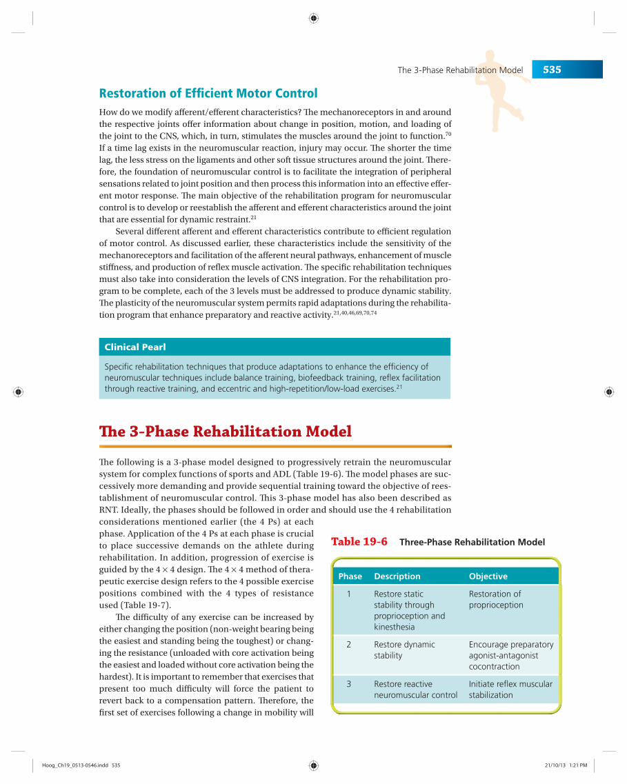
Restoration of Effi cient Motor Control How do we modify aff erent/eff erent characteristics? Th e mechanoreceptors in and around the respective joints off er information about change in position, motion, and loading of the joint to the CNS, which, in turn, stimulates the muscles around the joint to function. 70
If a time lag exists in the neuromuscular reaction, injury may occur. Th e shorter the time lag, the less stress on the ligaments and other soft tissue structures around the joint. Th ere-fore, the foundation of neuromuscular control is to facilitate the integration of peripheral sensations related to joint position and then process this information into an eff ective eff er-ent motor response. Th e main objective of the rehabilitation program for neuromuscular control is to develop or reestablish the aff erent and eff erent characteristics around the joint that are essential for dynamic restraint. 21
Several diff erent aff erent and eff erent characteristics contribute to effi cient regulation of motor control. As discussed earlier, these characteristics include the sensitivity of the mechanoreceptors and facilitation of the aff erent neural pathways, enhancement of muscle stiff ness, and production of refl ex muscle activation. Th e specifi c rehabilitation techniques must also take into consideration the levels of CNS integration. For the rehabilitation pro-gram to be complete, each of the 3 levels must be addressed to produce dynamic stability. Th e plasticity of the neuromuscular system permits rapid adaptations during the rehabilita-tion program that enhance preparatory and reactive activity. 21,40,46,69,70,74
Clinical Pearl
Specifi c rehabilitation techniques that produce adaptations to enhance the effi ciency of neuromuscular techniques include balance training, biofeedback training, refl ex facilitation through reactive training, and eccentric and high-repetition/low-load exercises. 21
Th e 3-Phase Rehabilitation Model
Th e following is a 3-phase model designed to progressively retrain the neuromuscular system for complex functions of sports and ADL ( Table 19-6 ). Th e model phases are suc-cessively more demanding and provide sequential training toward the objective of rees-tablishment of neuromuscular control. Th is 3-phase model has also been described as RNT. Ideally, the phases should be followed in order and should use the 4 rehabilitation considerations mentioned earlier (the 4 Ps) at each phase. Application of the 4 Ps at each phase is crucial to place successive demands on the athlete during rehabilitation. In addition, progression of exercise is guided by the 4 × 4 design. Th e 4 × 4 method of thera-peutic exercise design refers to the 4 possible exercise positions combined with the 4 types of resistance used ( Table 19-7 ).
Th e diffi culty of any exercise can be increased by either changing the position (non-weight bearing being the easiest and standing being the toughest) or chang-ing the resistance (unloaded with core activation being the easiest and loaded without core activation being the hardest). It is important to remember that exercises that present too much diffi culty will force the patient to revert back to a compensation pattern. Th erefore, the fi rst set of exercises following a change in mobility will
Table 19-6 Three-Phase Rehabilitation Model
Phase Description Objective
1 Restore static stability through proprioception and kinesthesia
Restoration of proprioception
2 Restore dynamic stability
Encourage preparatory agonist-antagonist cocontraction
3 Restore reactive neuromuscular control
Initiate refl ex muscular stabilization
The 3-Phase Rehabilitation Model 535
Hoog_Ch19_0513-0546.indd 535Hoog_Ch19_0513-0546.indd 535 21/10/13 1:21 PM21/10/13 1:21 PM

give all the information that one needs to know by producing 1 of 3 responses:
• It is too easy. Th e patient can perform the movement for more than 30 repetitions with good quality.
• It is challenging, but possible. Th e patient can perform the movement 8 to 15 times with good quality of movement and no signs of stress. Between 5 and 15 repetitions, however, there is a sharp decline in quality as demonstrated by a limited ability to maintain full ROM, balance, stabilization, and coordination, or the patient just becomes physically fatigued.
• It is too diffi cult. Th e patient has sloppy, stressful, poorly coordinated movement from the beginning, and it only gets worse.
Using this as a corrective exercise base, the clinician can observe the response and act accordingly. If the initial choice of exercise is too diffi cult, decrease the diffi culty, observe the response to the next set, and repeat the process. If the initial exercise is too easy, increase the diffi culty, observe the response to the next set, and repeat the process. Increasing dif-fi culty rarely means increased resistance. A more advanced posture, a smaller base of sup-port, or a more complex or involved movement pattern is usually indicated to increase the diffi culty. A typical example is some form of activity with a rolling movement pattern mov-ing to a quadruped exercise, then going to a half-kneeling activity, and fi nally progressing to movement with a single-leg stance.
Phase I: Restore Static Stability Through Proprioception and Kinesthesia Functional neuromuscular rehabilitation activities are designed to both restore functional stability about the joint and enhance motor control skills. Th e RNT program is centered on stimulation of both the peripheral and central refl ex pathways to the skeletal muscles. Th e fi rst objective that should be addressed in the RNT program is restoration of propriocep-tion. Reliable kinesthetic and proprioceptive information provides the foundation on which dynamic stability and motor control is based. It has already been established that altered aff erent information received by the CNS can alter the feed-forward and feedback motor control systems. Th erefore, the fi rst objective of the RNT program is to restore the neurosen-sory properties of the damaged structures while at the same time enhancing the sensitivity of the secondary peripheral aff erents. 69
To facilitate appropriate kinesthetic and proprioceptive input into the CNS, joint repo-sition exercises should be used to provide maximal stimulation of the peripheral mecha-noreceptors. Th e use of closed-kinetic-chain activities creates axial loads that maximally stimulate the articular mechanoreceptors via the increase in compressive force. 10,55 Th e use of closed-chain exercises not only enhances joint congruency and neurosensory feedback but also minimizes shearing stress about the joint. 75 At the same time, the muscle receptors are facilitated by the change in both length and tension. 10,55 Th e objective is to induce unantici-pated perturbations and thereby stimulate refl ex stabilization. Persistent use of these path-ways will decrease the response time when an unanticipated joint load occurs. 76 In addition to weightbearing exercises, active and passive joint-repositioning exercises can be used to enhance the conscious appreciation of proprioception. Rhythmic stabilization exercises can
Table 19-7 Four-by-Four Method for Design of Therapeutic Exercise
The 4 Positions The 4 Types of Resistance
Non-weight bearing (supine or prone)
Unloaded with core activation
Quadruped Unloaded without core activation
Kneeling (half kneeling or tall kneeling)
Loaded with core activation
Standing (lunge, split, squat, single leg)
Loaded without core activation
536 Chapter 19 Functional Training and Advanced Rehabilitation
Hoog_Ch19_0513-0546.indd 536Hoog_Ch19_0513-0546.indd 536 21/10/13 1:21 PM21/10/13 1:21 PM

Table 19-8 Upper-Extremity Neuromuscular Exercises
Phase I: Proprioception and Kinesthesia
Phase II: Dynamic Stabilization
Phase III: Reactive Neuromuscular Control
Goals
Normalize motion Restore proprioception and kinesthesia Establish muscular balance Diminish pain and infl ammation
Enhance dynamic functional stability Reestablish neuromuscular control Restore muscular balance Maintain normalized motion
Improve reactive neuromuscular abilities Enhance dynamic stability Improve power and endurance Gradual return to activities/throwing
Stability Exercises
Joint repositioning Movement awareness RS RI SRH PNF D 2 Flex/Ext PNF D 2 Flex/Ext RS, SRH, RI Side-lying RS, SRH, RI Weight bearing (axial compression) Weightbearing RS, RI Standing while leaning on hands Quadruped position Tripod position Biped position Axial compression with ball on wall OTIS
PNF D 2 Flex/Ext Supine Side-lying Seated Standing PNF D 2 Flex/Ext at end range 90 degrees/90 ER at end range Scapular strengthening Scapular PNF—RS, SRH ER/IR at 90 degree abduction—eyes
closed PNF D 2 Flex/Ext—eyes closed Balance beam PNF D 2 Flex/Ext—balance beam Slide board—side to side Slide board pushups Axial compression—side to side Axial compression—unstable surfaces Plyometrics—two handed (light
and easy) Two-handed chest throw Two-handed underhand throw
PNF D2 Flex/Ext RS with T-band Perturbation RS Perturbation RS—eyes closed 90 degree/90 degree ER at end-range RS ER Conc/Ecc ER Conc/Ecc RS ER/IR Conc/Ecc ER/IR Conc/Ecc RS Eyes closed Standing on one leg Reactive plyoballs Pushups on unstable surface UE plyometrics Two-handed overhead throw Side-to-side overhead throw One-handed baseball throw Endurance Wall dribble Wall baseball throw Axial compression circles Axial compression—side/side Sports specifi c Underweighted throwing Overweighted throwing Oscillating devices Boing Body blade
Conc, concentric; Ecc, eccentric; ER, external rotation; Ext, extension; Flex, fl exion; IR, internal rotation; OTIS, oscillating techniques for isometric stabilization; PNF, proprioceptive neuromuscular facilitation; RI, reciprocal isometrics; RS, rhythmic stabilization; SRH, slow-reversal-hold; UE, upper extremity.
be included early in the RNT program to enhance neuromuscular coordination in response to unexpected joint translation. Th e intensity of the exercises can be manipulated by increasing either the weight loaded across the joint or the size of the perturbation ( Tables 19-8 and 19-9 ). Th e addition of a compressive sleeve, wrap, or taping about the joint can also provide addi-tional proprioceptive information by stimulating the cutaneous mechanoreceptors. 21,65,77,78
Figures 19-7 through 19-10 provide examples of exercises that can be begun in this phase.
The 3-Phase Rehabilitation Model 537
Hoog_Ch19_0513-0546.indd 537Hoog_Ch19_0513-0546.indd 537 21/10/13 1:21 PM21/10/13 1:21 PM

Table 19-9 Lower-Extremity Neuromuscular Exercises
Phase I: Proprioception and Kinesthesia
Phase II: Dynamic Stabilization
Phase III: Reactive Neuromuscular Control
Goals
Normalize motion Restore proprioception and kinesthesia Establish muscular balance Diminish pain and infl ammation Develop static control and posture
Enhance dynamic functional stability
Reestablish neuromuscular control Restore muscular balance Maintain normalized motion
Improve reactive neuromuscular abilities Enhance dynamic stability Improve power and endurance Gradual return to activities, running,
jumping, cutting
Stability Exercises
Bilateral to unilateral Eyes open to eyes closed Stable to unstable surfaces Level surfaces Foam pad Controlled to uncontrolled PNF Rhythmic stabilization Rhythmic isometrics Slow reversal hold
OTIS AWS PWS MWS LWS Chops/lifts ITIS PACE PNF Rhythmic stabilization Rhythmic isometrics Slow-reversal-hold Stable to unstable surface Rocker board Wobble board BAPS Balance beam Foam rollers DynaDisc
Squats Assisted AWS PWS MWS LWS Chops/lifts Lunges (front and lateral) AWS PWS MWS LWS Stationary walking with unidirectional WS Stationary running PWS MWS LWS AWS Mountain climber CKC side to side Fitter Slide board Plyometrics Jumps in place Standing jumps Bounding Multiple hops and bounds Hops with rotation Bounds with rotation Resisted lateral bounds Box jumps Depth jumps Multidirectional training Lunges Rock wall Clock drill Step-tos Four-square Agility training
AWS, anterior weight shift; BAPS, biomechanical ankle platform system; CKC, closed-chain kinetic; ITIS, impulse techniques for isometric stabilization; LWS, lateral weight shift; MWS, medial weight shift; OTIS, oscillating techniques for isometric stabilization; PACE, partial-arc controlled exercise; PNF, proprioceptive neuromuscular facilitation; PWS, posterior weight shift; WS, weight shift.
538 Chapter 19 Functional Training and Advanced Rehabilitation
Hoog_Ch19_0513-0546.indd 538Hoog_Ch19_0513-0546.indd 538 21/10/13 1:21 PM21/10/13 1:21 PM
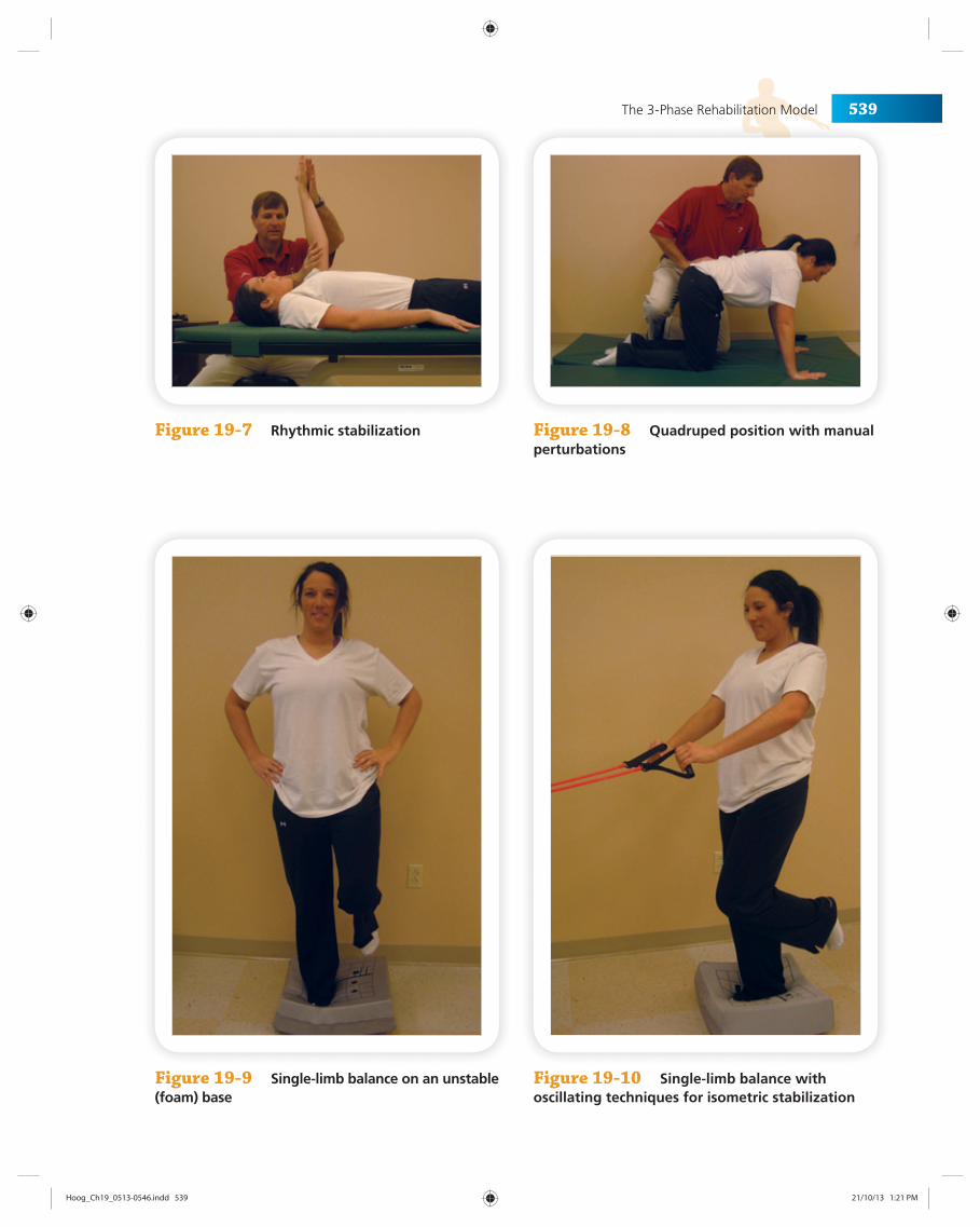
Figure 19-7 Rhythmic stabilization
Figure 19-9 Single-limb balance on an unstable (foam) base
Figure 19-10 Single-limb balance with oscillating techniques for isometric stabilization
Figure 19-8 Quadruped position with manual perturbations
The 3-Phase Rehabilitation Model 539
Hoog_Ch19_0513-0546.indd 539Hoog_Ch19_0513-0546.indd 539 21/10/13 1:21 PM21/10/13 1:21 PM
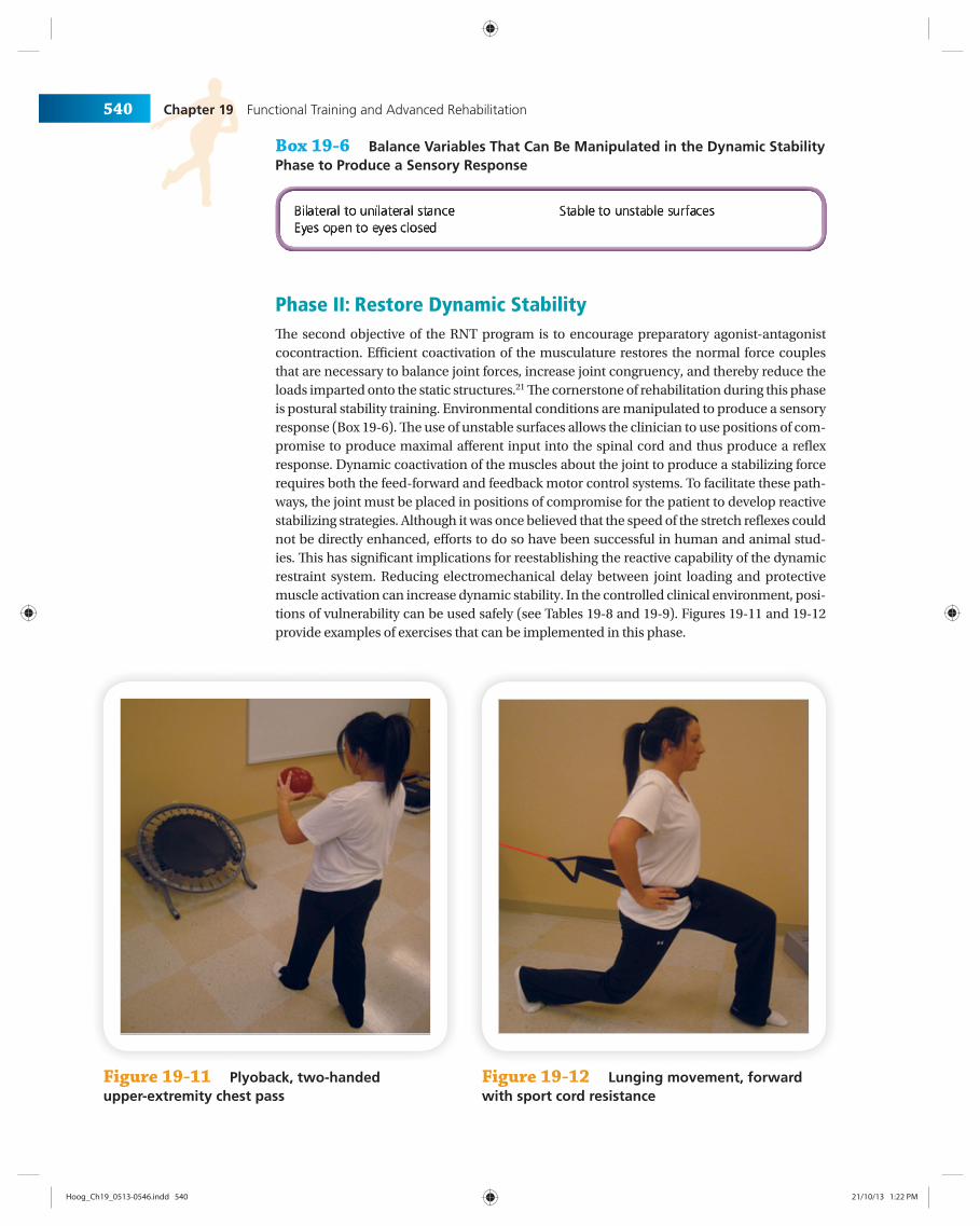
Phase II: Restore Dynamic Stability Th e second objective of the RNT program is to encourage preparatory agonist-antagonist cocontraction. Effi cient coactivation of the musculature restores the normal force couples that are necessary to balance joint forces, increase joint congruency, and thereby reduce the loads imparted onto the static structures. 21 Th e cornerstone of rehabilitation during this phase is postural stability training. Environmental conditions are manipulated to produce a sensory response ( Box 19-6 ). Th e use of unstable surfaces allows the clinician to use positions of com-promise to produce maximal aff erent input into the spinal cord and thus produce a refl ex response. Dynamic coactivation of the muscles about the joint to produce a stabilizing force requires both the feed-forward and feedback motor control systems. To facilitate these path-ways, the joint must be placed in positions of compromise for the patient to develop reactive stabilizing strategies. Although it was once believed that the speed of the stretch refl exes could not be directly enhanced, eff orts to do so have been successful in human and animal stud-ies. Th is has signifi cant implications for reestablishing the reactive capability of the dynamic restraint system. Reducing electromechanical delay between joint loading and protective muscle activation can increase dynamic stability. In the controlled clinical environment, posi-tions of vulnerability can be used safely (see Tables 19-8 and 19-9 ). Figures 19-11 and 19-12 provide examples of exercises that can be implemented in this phase.
Box 19-6 Balance Variables That Can Be Manipulated in the Dynamic Stability Phase to Produce a Sensory Response
Figure 19-11 Plyoback, two-handed upper-extremity chest pass
Figure 19-12 Lunging movement, forward with sport cord resistance
540 Chapter 19 Functional Training and Advanced Rehabilitation
Hoog_Ch19_0513-0546.indd 540Hoog_Ch19_0513-0546.indd 540 21/10/13 1:22 PM21/10/13 1:22 PM

Proprioceptive training for functionally unstable joints after injury has been docu-mented in the literature. 38,53,70,79 Tropp et al 53 and Wester et al 80 reported that ankle disk training signifi cantly reduced the incidence of ankle sprains. Concerning the mechanism of the eff ects, Tropp et al 53 suggested that unstable surface training reduced the pro-prioceptive defi cit. Sheth et al 79 demonstrated changes in healthy adults in patterns of contraction of the inversion and eversion musculature before and after training on an unstable surface. Th ey concluded that the changes would be supported by the concept of reciprocal Ia inhibition via the mechanoreceptors in the muscles. Konradsen and Ravin 81 also suggested from their work that aff erent input from the calf musculature was responsi-ble for dynamic protection against sudden ankle inversion stress. Pinstaar et al 82 reported that postural sway was restored after 8 weeks of ankle disk training when performed 3 to 5 times a week. Tropp and Odenrick also showed that postural control improved after 6 weeks of training when performed 15 minutes per day. 39 Bernier and Perrin, 54 whose program consisted of balance exercises progressing from simple to complex sessions (3 times a week for 10 minutes each time), also found that postural sway was improved after 6 weeks of training. Although each of these training programs do have some diff er-ences, postural control improved after 6 to 8 weeks of proprioceptive training in subjects with functional instability of the ankle.
Phase III: Restore Reactive Neuromuscular Control Dynamic reactive neuromuscular control activities should be initiated into the overall reha-bilitation program after adequate healing and dynamic stability have been achieved. Th e key objective is to initiate refl ex muscular stabilization.
Progression of these activities is predicated on the athlete satisfactorily completing the activities that are considered prerequisites for the activity being considered. With this in mind, progression of activities must be goal oriented and specifi c to the tasks that will be expected of the athlete.
Th e general progression of activities to develop dynamic reactive neuromuscular con-trol is from slow-speed to fast-speed activities, from low-force to high-force activities, and from controlled to uncontrolled activities. Initially, these exercises should evoke a balance reaction or weight shift in the lower extremities and ultimately progress to a movement pat-tern. A sudden alteration in joint position induced by either the clinician or the athlete may decrease the response time and serve to develop reactive strategies to unexpected events. Th ese reactions can be as simple as static control with little or no visible movement or as complex as a dynamic plyometric response requiring explosive acceleration, deceleration, or change in direction. Th e exercises will allow the clinician to challenge the patient by using visual or proprioceptive input, or both, via tubing (oscillating techniques for isometric sta-bilization) and other devices (eg, medicine balls, foam rolls, or visual obstacles). Although these exercises will improve physiologic parameters, they are specifi cally designed to facili-tate neuromuscular reactions. Th erefore, the clinician must be concerned with the kines-thetic input and quality of the movement patterns rather than the particular number of sets and repetitions. When fatigue occurs, motor control becomes poor and all training eff ects are lost. Th erefore, during the exercise progression, all aspects of normal function should be observed, including isometric, concentric, and eccentric muscle control; articular loading and unloading; balance control during weight shifting and changes in direction; controlled acceleration and deceleration; and demonstration of both conscious and unconscious con-trol (see Tables 19-7 and 19-8 ). Figures 19-13 through 19-15 are examples of exercises that can be implemented in this phase.
When dynamic stability and refl ex stabilization have been achieved, the focus of the neuromuscular rehabilitation program is to restore ADL and sport-specifi c skills. It is
The 3-Phase Rehabilitation Model 541
Hoog_Ch19_0513-0546.indd 541Hoog_Ch19_0513-0546.indd 541 21/10/13 1:22 PM21/10/13 1:22 PM

essential that the exercise program be specifi c to the patient’s needs. Th e most important factor to consider during reha-bilitation of patients is that they should be performing func-tional activities that simulate their ADL requirements. Th is rule applies not only to the specifi c joints involved but also to the speed and amplitude of movement required in ADL. Exercise and training drills that will refi ne the physiologic parameters required for return to preinjury levels of func-tion should be incorporated into the program. Th e progres-sion should be from straight plane to multiplane movement patterns. ADL movement does not occur along a single joint or plane of movement. Th erefore, exercise for the kinetic chain must involve all 3 planes simultaneously. Emphasis in the RNT program must be placed on progression from simple to complex neuromotor patterns that are specifi c to the demands placed on the patient during function. Th e function progression breaks an activity down into its com-ponent parts so that they can be performed in a sequence that allows acquisition or reacquisition of the activity. Basic conditioning and skill acquisition must be achieved before advanced conditioning and skill acquisition. Th e training program should begin with simple activities, such as walking/running, and then progress to highly complex motor skills requiring refi ned neuromuscular mechanisms, including proprioceptive and kinesthetic awareness, which provides refl ex joint stabilization. A signifi cant amount
Figure 19-14 Dynamic training; Body Blade, elevated position
Figure 19-15 Elevated wall dribble
Figure 19-13 Dynamic training; Body Blade, low position
542 Chapter 19 Functional Training and Advanced Rehabilitation
Hoog_Ch19_0513-0546.indd 542Hoog_Ch19_0513-0546.indd 542 21/10/13 1:22 PM21/10/13 1:22 PM

of “controlled chaos” should be included in the program. Unexpected activities during ADL are by nature unstable. Th e more patients rehearse in this type of environment, the better they will react under unrehearsed conditions. Th e clinician needs to learn how to categorize, prioritize, and plan eff ectively because corrective exercises will evolve and equipment will change. Th e clinician’s professional skill must be based in a systematic approach. Just being great at a technique is not good enough. Technical aspects of exer-cise will change. Th e clinician should not worry. Th is system is not based on exercise. It is based on human movement, not equipment, techniques, or trends. Th e fi nal and most important consideration of this phase is to make the rehabilitation program fun. Th e fi rst 3 letters of functional are FUN. If the program is not fun, compliance will suff er and so will the results.
SUMMARY
1. Increased attention has been devoted to the development of balance, proprioception, and neuromuscular control in the rehabilitation and reconditioning of athletes after injury.
2. It is believed that injury results in altered somatosensory input, which infl uences neu-romuscular control.
3. If static and dynamic balance and neuromuscular control are not reestablished after injury, the patient will be susceptible to recurrent injury and performance may decline.
4. Th e 3-phase model for RNT may be an excellent method to assist athletes in regaining optimal neuromuscular performance and high-level function after injury or surgery.
5. Th e 3-phase model consists of restoring static stability through proprioception and kinesthesia, dynamic stability, and reactive neuromuscular control.
6. Current information has been synthesized to produce a new perspective for therapeu-tic exercise decisions. Th is new perspective was specifi cally designed to improve treat-ment effi ciency and eff ectiveness and have a focus on function.
7. Th e 4 principles of purpose, posture, position, and pattern assist problem solving by providing a framework that categorizes clinical information in a hierarchy.
8. Th e 4 principles serve as quick reminders of the hierarchy, interaction, and applica-tion for each therapeutic exercise prescription principle. Th e questions of what, when, where, and how for functional movement assessment and exercise prescription are an-swered in the appropriate order.
a. Functional evaluation and assessment = purpose.
b. Identifi cation of motor control = posture.
c. Identifi cation of osteokinematic and arthrokinematic limitations = position.
d. Integration of synergistic movement patterns = pattern.
9. Th e clinician should always ask whether the program makes sense. If it does not make sense, it is probably not functional and therefore not optimally eff ective.
10. Clinical wisdom is the result of experience and applied knowledge. Intense familiarity and practical observation improve application. To be of benefi t, the knowledge available must be organized and tempered by an objective and inclu-sive framework. It is hoped that this framework will provide a starting point to better organize and apply each clinician’s knowledge and experience of functional exercise prescription.
The 3-Phase Rehabilitation Model 543
Hoog_Ch19_0513-0546.indd 543Hoog_Ch19_0513-0546.indd 543 21/10/13 1:22 PM21/10/13 1:22 PM
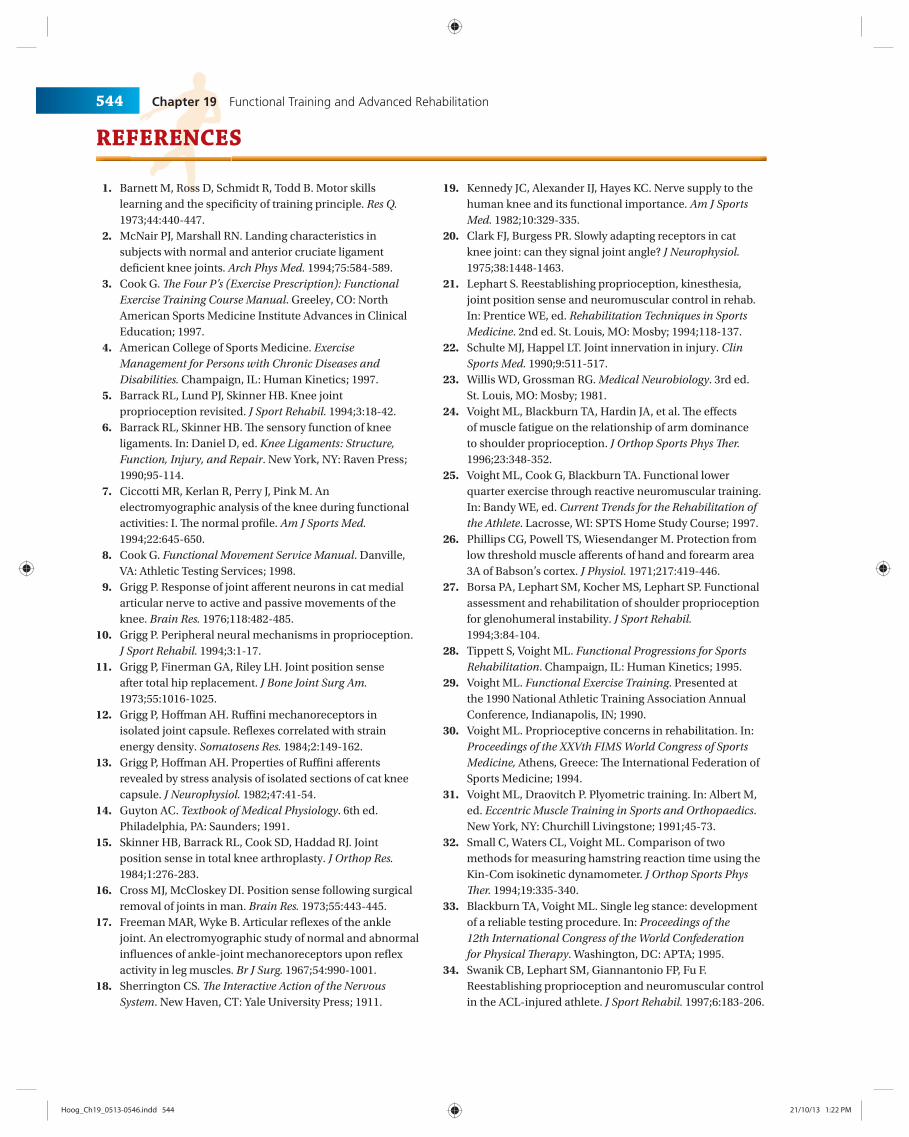
REFERENCES
1. Barnett M, Ross D, Schmidt R, Todd B. Motor skills learning and the specifi city of training principle. Res Q. 1973;44:440-447.
2. McNair PJ, Marshall RN. Landing characteristics in subjects with normal and anterior cruciate ligament defi cient knee joints. Arch Phys Med. 1994;75:584-589.
3. Cook G. Th e Four P’s (Exercise Prescription): Functional Exercise Training Course Manual . Greeley, CO: North American Sports Medicine Institute Advances in Clinical Education; 1997.
4. American College of Sports Medicine. Exercise Management for Persons with Chronic Diseases and Disabilities. Champaign, IL: Human Kinetics; 1997.
5. Barrack RL, Lund PJ, Skinner HB. Knee joint proprioception revisited. J Sport Rehabil. 1994;3:18-42.
6. Barrack RL, Skinner HB. Th e sensory function of knee ligaments. In: Daniel D, ed. Knee Ligaments: Structure, Function, Injury, and Repair . New York, NY: Raven Press; 1990;95-114.
7. Ciccotti MR, Kerlan R, Perry J, Pink M. An electromyographic analysis of the knee during functional activities: I. Th e normal profi le. Am J Sports Med. 1994;22:645-650.
8. Cook G. Functional Movement Service Manual . Danville, VA: Athletic Testing Services; 1998.
9. Grigg P. Response of joint aff erent neurons in cat medial articular nerve to active and passive movements of the knee. Brain Res. 1976;118:482-485.
10. Grigg P. Peripheral neural mechanisms in proprioception. J Sport Rehabil. 1994;3:1-17.
11. Grigg P, Finerman GA, Riley LH. Joint position sense after total hip replacement. J Bone Joint Surg Am. 1973;55:1016-1025.
12. Grigg P, Hoff man AH. Ruffi ni mechanoreceptors in isolated joint capsule. Refl exes correlated with strain energy density. Somatosens Res. 1984;2:149-162.
13. Grigg P, Hoff man AH. Properties of Ruffi ni aff erents revealed by stress analysis of isolated sections of cat knee capsule. J Neurophysiol. 1982;47:41-54.
14. Guyton AC. Textbook of Medical Physiology . 6th ed. Philadelphia, PA: Saunders; 1991.
15. Skinner HB, Barrack RL, Cook SD, Haddad RJ. Joint position sense in total knee arthroplasty. J Orthop Res. 1984;1:276-283.
16. Cross MJ, McCloskey DI. Position sense following surgical removal of joints in man. Brain Res. 1973;55:443-445.
17. Freeman MAR, Wyke B. Articular refl exes of the ankle joint. An electromyographic study of normal and abnormal infl uences of ankle-joint mechanoreceptors upon refl ex activity in leg muscles. Br J Surg. 1967;54:990-1001.
18. Sherrington CS. Th e Interactive Action of the Nervous System . New Haven, CT: Yale University Press; 1911.
19. Kennedy JC, Alexander IJ, Hayes KC. Nerve supply to the human knee and its functional importance. Am J Sports Med. 1982;10:329-335.
20. Clark FJ, Burgess PR. Slowly adapting receptors in cat knee joint: can they signal joint angle? J Neurophysiol. 1975;38:1448-1463.
21. Lephart S. Reestablishing proprioception, kinesthesia, joint position sense and neuromuscular control in rehab. In: Prentice WE, ed. Rehabilitation Techniques in Sports Medicine . 2nd ed. St. Louis, MO: Mosby; 1994;118-137.
22. Schulte MJ, Happel LT. Joint innervation in injury. Clin Sports Med. 1990;9:511-517.
23. Willis WD, Grossman RG. Medical Neurobiology . 3rd ed. St. Louis, MO: Mosby; 1981.
24. Voight ML, Blackburn TA, Hardin JA, et al. Th e eff ects of muscle fatigue on the relationship of arm dominance to shoulder proprioception. J Orthop Sports Phys Th er. 1996;23:348-352.
25. Voight ML, Cook G, Blackburn TA. Functional lower quarter exercise through reactive neuromuscular training. In: Bandy WE, ed. Current Trends for the Rehabilitation of the Athlete . Lacrosse, WI: SPTS Home Study Course; 1997.
26. Phillips CG, Powell TS, Wiesendanger M. Protection from low threshold muscle aff erents of hand and forearm area 3A of Babson’s cortex. J Physiol. 1971;217:419-446.
27. Borsa PA, Lephart SM, Kocher MS, Lephart SP. Functional assessment and rehabilitation of shoulder proprioception for glenohumeral instability. J Sport Rehabil. 1994;3:84-104.
28. Tippett S, Voight ML. Functional Progressions for Sports Rehabilitation . Champaign, IL: Human Kinetics; 1995.
29. Voight ML. Functional Exercise Training . Presented at the 1990 National Athletic Training Association Annual Conference, Indianapolis, IN; 1990.
30. Voight ML. Proprioceptive concerns in rehabilitation. In: Proceedings of the XXVth FIMS World Congress of Sports Medicine, Athens, Greece: Th e International Federation of Sports Medicine; 1994.
31. Voight ML, Draovitch P. Plyometric training. In: Albert M, ed. Eccentric Muscle Training in Sports and Orthopaedics . New York, NY: Churchill Livingstone; 1991;45-73.
32. Small C, Waters CL, Voight ML. Comparison of two methods for measuring hamstring reaction time using the Kin-Com isokinetic dynamometer. J Orthop Sports Phys Th er. 1994;19:335-340.
33. Blackburn TA, Voight ML. Single leg stance: development of a reliable testing procedure. In: Proceedings of the 12th International Congress of the World Confederation for Physical Th erapy . Washington, DC: APTA; 1995.
34. Swanik CB, Lephart SM, Giannantonio FP, Fu F. Reestablishing proprioception and neuromuscular control in the ACL-injured athlete. J Sport Rehabil. 1997;6:183-206.
544 Chapter 19 Functional Training and Advanced Rehabilitation
Hoog_Ch19_0513-0546.indd 544Hoog_Ch19_0513-0546.indd 544 21/10/13 1:22 PM21/10/13 1:22 PM
35. Rine RM, Voight ML, Laporta L, Mancini R. A paradigm to evaluate ankle instability using postural sway measures [abstract]. Phys Th er. 1994;74:S72.
36. Voight ML, Rine RM, Apfel P, et al. Th e eff ects of leg dominance and AFO on static and dynamic balance abilities [abstract]. Phys Th er. 1993;73:S51.
37. Voight ML, Rine RM, Briese K, Powell C. Comparison of sway in double versus single leg stance in unimpaired adults [abstract]. Phys Th er. 1993;73:S51.
38. Tropp H, Askling C, Gillquist J. Prevention of ankle sprains. Am J Sports Med. 1985;13:259-262.
39. Tropp H, Odenrick P. Postural control in single limb stance. J Orthop Res. 1988;6:833-839.
40. Beard DJ, Dodd CF, Trundle HR, et al. Proprioception after rupture of the ACL: an objective indication of the need for surgery? J Bone Joint Surg Br. 1993;75:311.
41. Pope MH, Johnson DW, Brown DW, Tighe C. Th e role of the musculature in injuries to the medial collateral ligament. J Bone Joint Surg Am. 1972;61:398-402.
42. Wojtys E, Huston L. Neuromuscular performance in normal and anterior cruciate ligament-defi cient lower extremities. Am J Sports Med. 1994;22:89-104.
43. Dunn TG, Gillig SE, Ponser ES, Weil N. Th e learning process in biofeedback: is it feed-forward or feedback? Biofeedback Self Regul. 1986;11:143-155.
44. Belen’kii VY, Gurfi nkle VS, Pal’tsev YI. Elements of control of voluntary movements. Biofi zika. 1967;12:135-141.
45. Lee WA. Anticipatory control of postural and task muscles during rapid arm fl exion. J Mot Behav. 1980;12:185-196.
46. Hodgson JA, Roy RR, DeLeon R, et al. Can the mammalian lumbar spinal cord learn a motor task? Med Sci Sports Exerc. 1994;26:1491-1497.
47. Schmidt RA. Motor Control and Learning . Champaign, IL: Human Kinetics; 1988.
48. Scully R, Barnes M. Physical Th erapy . Philadelphia, PA: Lippincott; 1989.
49. Cook G, Fields K. Functional Training for the Torso . Colorado Springs, CO: National Strength and Conditioning Association; 1997:14-19.
50. Sullivan PE, Markos PD, Minor MD. An Integrated Approach to Th erapeutic Exercise: Th eory and Clinical Application . Reston, VA: Reston Publishing; 1982.
51. Freeman MAR, Wyke B. Articular contributions to limb refl exes. Br J Surg. 1966;53:61-69.
52. Janda V. Muscles and motor control in low back pain: assessment and management. In: Twomey L, ed. Physical Th erapy of the Low Back . New York, NY: Churchill Livingstone; 1987:253-278.
53. Tropp H, Ekstrand J, Gillquist J. Factors aff ecting stabilometry recordings of single leg stance. Am J Sports Med. 1984;12:185-188.
54. Bernier JN, Perrin DH. Eff ect of coordination training on proprioception of the functionally unstable ankle. J Orthop Sports Phys Th er. 1998;27:264-275.
55. Clark FJ, Burgess RC, Chapin JW, Lipscomb WT. Role of intramuscular receptors in the awareness of limb position. J Neurophysiol. 1985;54:1529-1540.
56. Voight ML, Cook G. Clinical application of closed kinetic chain exercise. J Sport Rehabil. 1996;5:25-44.
57. Kendall FP, McCreary KE, Provance PG. Muscle Testing and Function . 4th ed. Baltimore, MD: Williams & Wilkins; 1993.
58. Cyriax J. Textbook of Orthopedic Medicine . Vol. I. Diagnosis of Soft Tissue Lesions. 8th ed. London, UK: Bailliere Tindall; 1982.
59. Baechle TR. Essentials of Strength Training and Conditioning . Champaign, IL: Human Kinetics; 1994.
60. Voss DE, Ionta MK, Myers BJ. Proprioceptive Neuromuscular Facilitation: Patterns and Techniques . 3rd ed. Philadelphia, PA: Harper & Row; 1985.
61. Gandevia SC, McCloskey DI. Joint sense, muscle sense and their contribution as position sense, measured at the distal interphalangeal joint of the middle fi nger. J Physiol. 1976;260:387-407.
62. Glenncross D, Th ornton E. Position sense following joint injury. Am J Sports Med. 1981;21:23-27.
63. Barrack RL, Skinner HB, Buckley SL. Proprioception in the anterior cruciate defi cient knee. Am J Sports Med. 1989;17:1-6.
64. Skinner HB, Wyatt MP, Hodgdon JA, et al. Eff ect of fatigue on joint position sense of the knee. J Orthop Res. 1986;4:112-118.
65. Barrett DS. Proprioception and function after anterior cruciate reconstruction. J Bone Joint Surg Br. 1991;3:833-837.
66. Corrigan JP, Cashman WF, Brady MP. Proprioception in the cruciate defi cient knee. J Bone Joint Surg Br. 1992;74:247-250.
67. Borsa PA, Lephart SM, Irrgang JJ, et al. Th e eff ects of joint position and direction of joint motion on proprioceptive sensibility in anterior cruciate ligament defi cient athletes. Am J Sports Med. 1997;25:336-340.
68. Harter RA, Osternig LR, Singer SL, et al. Long-term evaluation of knee stability and function following surgical reconstruction for anterior cruciate ligament insuffi ciency. Am J Sports Med. 1988;16:434-442.
69. Lephart SM, Pincivero DM, Giraldo JL, Fu F. Th e role of proprioception in the management and rehabilitation of athletic injuries. Am J Sports Med. 1997;25:130-137.
70. Ihara H, Nakayama A. Dynamic joint control training for knee ligament injuries. Am J Sports Med. 1986;14:309-315.
71. Giove TP, Miller SJ, Kent BE, et al. Non-operative treatment of the torn anterior cruciate ligament. J Bone Joint Surg Am. 1983;65:184-192.
72. Tibone JE, Antich TJ, Funton GS, et al. Functional analysis of anterior cruciate ligament instability. Am J Sports Med. 1986;14:276-284.
73. Walla DJ, Albright JP, McAuley E, et al. Hamstring control and the unstable anterior cruciate ligament-defi cient knee. Am J Sports Med. 1985;13:34-39.
The 3-Phase Rehabilitation Model 545
Hoog_Ch19_0513-0546.indd 545Hoog_Ch19_0513-0546.indd 545 21/10/13 1:22 PM21/10/13 1:22 PM
74. Wojtys E, Huston LJ, Taylor PD, Bastian SD. Neuromuscular adaptations in isokinetic, isotonic, and agility training programs. Am J Sports Med. 1996;24:187-192.
75. Voight ML, Bell S, Rhodes D. Instrumented testing of tibial translation during a positive Lachman’s test and selected closed-chain activities in anterior cruciate defi cient knees. J Orthop Sports Phys Th er. 1992;15:49.
76. Ognibene J, McMahon K, Harris M, et al. Eff ects of unilateral proprioceptive perturbation training on postural sway and joint reaction times of healthy subjects. In: Proceedings of the National Athletic Training Association Annual Meeting . Champaign, IL: Human Kinetics; 2000.
77. Matsusaka N, Yokoyama S, Tsurusaki T, et al. Eff ect of ankle disk training combined with tactile stimulation to the leg and foot in functional instability of the ankle. Am J Sports Med . 2001;29:25-30.
78. Perlau RC, Frank C, Fick G. Th e eff ects of elastic bandages on human knee proprioception in the uninjured population. Am J Sports Med. 1995;23:251-255.
79. Sheth P, Yu B, Laskowski ER, et al. Ankle disk training infl uences reaction times of selected muscles in a simulated ankle sprain. Am J Sports Med. 1997;25:538-543.
80. Wester JU, Jespersen SM, Nielsen KD, et al. Wobble board training after partial sprains of the lateral ligaments of the ankle: a prospective randomized study. J Orthop Sports Phys Th er. 1996;23:332-336.
81. Konradsen L, Ravin JB. Prolonged peroneal reaction time in ankle instability. Int J Sports Med. 1991;12:290-292.
82. Pinstaar A, Brynhildsen J, Tropp H. Postural corrections after standardized perturbations of single limb stance: eff ect of training and orthotic devices in patients with ankle instability. Br J Sports Med. 1996;30:151-155.
546 Chapter 19 Functional Training and Advanced Rehabilitation
Hoog_Ch19_0513-0546.indd 546Hoog_Ch19_0513-0546.indd 546 21/10/13 1:22 PM21/10/13 1:22 PM