garcia-pravia et al. - plosone 2013
TRANSCRIPT

Overexpression of COL11A1 by Cancer-AssociatedFibroblasts: Clinical Relevance of a Stromal Marker inPancreatic CancerCarmen García-Pravia1,2, José A. Galván2,3, Natalia Gutiérrez-Corral4, Lorena Solar-García4, Eva García-Pérez3, Marcos García-Ocaña2,5, Jokin Del Amo-Iribarren6, Primitiva Menéndez-Rodríguez1, Juan García-García1, Juan R. de los Toyos2,7, Laureano Simón-Buela8, Luis Barneo2,3,4*
1 Pathological Anatomy Service, Hospital Universitario Central de Asturias (HUCA), Oviedo, Spain, 2 Instituto Universitario de Oncología del Principado deAsturias (IUOPA), Oviedo, Spain, 3 Department of Surgery, School of Medicine and Health Sciences, University of Oviedo, Oviedo, Spain, 4 General SurgeryService, Hospital Universitario Central de Asturias (HUCA), Oviedo, Spain, 5 Biotechnological and Biomedical Assays Unit, Technical-Scientific Services,Oviedo, Spain, 6 Progenika Biopharma, S.A., Science and Technology Park of Zamudio, Derio, Spain, 7 Immunology Area, School of Medicine and HealthSciences, University of Oviedo, Oviedo, Spain, 8 Oncomatrix, S.L., Science and Technology Park of Zamudio, Derio, Spain
Abstract
Background: The collagen11A1 (COL11A1) gene is overexpressed in pancreatic cancer. The expression ofCOL11A1 protein could be involved in desmoplastic events in pancreatic cancer, but an antibody that specificallystains the COL11A1 protein is not currently available.Methods and findings: A total of 54 pancreatic ductal adenocarcinomas (PDAC), 23 chronic pancreatitis (CP)samples, and cultured peritumoral stromal cells of PDAC (passages 3-6) were studied. Normal human pancreastissue samples were obtained through a cadaveric organ donation program.
1) Validation of COL11A1 gene overexpression by q-RT-PCR. Findings: the expression of COL11A1 gene issignificantly increased in PDAC samples vs. normal and CP samples.
2) Analysis of COL11A1 by immunohistochemistry using highly specific anti-proCOL11A1 antibodies. Findings:anti-proCOL11A1 stains stromal cells/cancer-associated fibroblasts (CAFs) of PDAC but it does not stain chronicbenign condition (chronic pancreatitis) stromal cells, epithelial cells, or normal fibroblasts.
3) Evaluation of the discrimination ability of the antibody. Findings: anti-proCOL11A1 immunostaining accuratelydiscriminates between PDAC and CP (AUC 0.936, 95% CI 0.851, 0.981).
4) Phenotypic characterization of proCOL11A1+ stromal cells co-staining with mesenchymal, epithelial and stellatecell markers on pancreatic tissue samples and cultured peritumoral pancreatic cancer stromal cells. Findings:ProCOL11A1+ cells present co-staining with mesenchymal, stellate and epithelial markers (EMT phenotype) indifferent proportions.Conclusions/Significance: Detection of proCOL11A1 through immunostaining with this newly-developed antibodyallows for a highly accurate distinction between PDAC and CP. Unlike other available antibodies commonly used todetect CAFs, anti-proCOL11A1 is negative in stromal cells of the normal pancreas and almost absent in benigninflammation. These results strongly suggest that proCOL11A1 is a specific marker for CAFs, and thus, anti-proCOL11A1 is a powerful new tool for cancer research and clinical diagnostics.
Citation: García-Pravia C, Galván JA, Gutiérrez-Corral N, Solar-García L, García-Pérez E, et al. (2013) Overexpression of COL11A1 by Cancer-Associated Fibroblasts: Clinical Relevance of a Stromal Marker in Pancreatic Cancer . PLoS ONE 8(10): e78327. doi:10.1371/journal.pone.0078327
Editor: Francisco X. Real, Centro Nacional de Investigaciones Oncológicas (CNIO), Spain
Received April 28, 2013; Accepted September 11, 2013; Published October 23, 2013
Copyright: © 2013 García-Pravia et al. This is an open-access article distributed under the terms of the Creative Commons Attribution License, whichpermits unrestricted use, distribution, and reproduction in any medium, provided the original author and source are credited.
Funding: This research has been co-financed by ERDF Funds from the EuropeanUnion; by INNPACTO-ONCOPAN IPT-010000-2010-31 Project; byFISS-09- PS09/01911 Project, Ministry of Science and Innovation, Spain; by FC-11-PC10-23 Project, FICYT, Axe 1 of the 2007-2013 ERDF OperationalFramework Programme of the Principality of Asturias, Spain; and by Progenika Biopharma, S.A. and Oncomatrix, S.L. Derio, Spain. The proCOL11A1 mAbhas been patented by Oncomatrix, S.L. (PCT/ES2012/070616; WO 2013/021088 A2). The funders had no role in study design, data collection andanalysis, decision to publish, or preparation of the manuscript.
Competing interests: The antibody detailed in this study is under a patent filed by Drs. Luis Barneo, Carmen García-Pravia, Juan R. de los Toyos, MarcosGarcía-Ocaña, Jokin Del Amo-Iribarren, Laureano Simón-Buela and others titled: METHODS AND PRODUCTS FOR IN VITRO DIAGNOSIS, IN VITROPROGNOSIS AND THE DEVELOPMENT OF DRUGS AGAINST INVASIVE CARCINOMAS (PCT/ES2012/070616; WO 2013/021088 A2). Jokin Del Amo-Iribarren works for Progenika Biopharma, S.A. and Laureano Simón-Buela works for Oncomatrix, S.L. This study was partly funded by ProgenikaBiopharma, S.A. and Oncomatrix, S.L. There are no other patents, products in development or marketed products to declare. This does not alter theauthors' adherence to all the PLOS ONE policies on sharing data and materials.
* E-mail: [email protected]
PLOS ONE | www.plosone.org 1 October 2013 | Volume 8 | Issue 10 | e78327

Introduction
Pancreatic ductal adenocarcinoma (PDAC) represents thefourth leading cause of death from cancer in men and women.The 5-year survival rate is less than 5% and average survivaltime is 6 months after the initial diagnosis. Even in patients whoundergo resection, long-term survival rates remain extremelypoor [1]. At the present time, there are no early diagnosismethods or effective therapies for use against this type oftumor. Despite progress having been made in its diagnosis andtreatment, pancreatic cancer continues to have the worstprognosis of all solid malignant tumors. Pancreatic cancer isthe paradigm of advanced neoplastic disease: independently ofthe TNM stage, the majority of patients present withdisseminated disease in the early phases [2]. Furthermore,pancreatic cancer is resistant to chemo- and radiotherapy [3].
Pancreatic carcinoma is characterized by a desmoplasticreaction involving cellular and acellular components, such asfibroblasts (activated or resting), myofibroblasts, pericytes,pancreatic stellate cells, immune cells, blood vessels, theextracellular matrix, and soluble proteins such as cytokines andgrowth factors [4,5]. This heterogeneous stroma influencesmultiple aspects of PDAC and seems to promote tumor growth,invasion, and resistance to chemotherapy [6-9].
Chronic pancreatitis is an inflammatory diseasecharacterized by irreversible and progressive destruction of theorgan, resulting in exocrine and endocrine insufficiency. Thelost parenchyma is replaced by dense fibrous tissue withinfiltrating leukocytes and ductular hyperplasia. Chronicpancreatitis significantly increases the risk of developingpancreatic cancer [10-12], which suggests that chronicinflammation within the pancreas may be a predisposing factorto the development of cancer. The causative link betweenchronic inflammation and cancer was described two centuriesago by Marjolin [13], but the inflammatory mediators that leadto the development of cancer remain undefined.
Among the tumor-associated matrix collagens, fibrillarcollagens are the most conspicuous. Collagens aresynthesized as procollagens by fibroblasts. These procollagenshave a main central triple-helical domain, designated as α1, α2,and α3, and coded by specific gene sequences. Once secretedto the extracellular milieu, these procollagens are cleaved, andthen the mature collagen molecules assemble extracellularly infibrils. In normal tissues, collagen types I, II and III are the mainmajor fibrillar collagens, while collagens V and XI are lessabundant minor fibrillar collagens [14]. Collagens V and XIshare a 75% homology at their amino acid sequence level.Procollagens α1 of types V and XI are coded by COL5A1 andCOL11A1 genes, respectively.
Studies of the fibroblasts in the vicinity of the tumor, the so-called cancer-associated fibroblasts (CAF), have demonstratedtheir role in stimulating tumor progression [15-20]. Thecharacteristics of the CAFs have been investigated in depth,showing that their genotypic expression, growth pattern,migratory behavior and secretion of growth factors differ fromthose of normal fibroblasts [15,21]. However, researchers donot have specific tools to differentiate CAFs from inflammatoryfibroblasts. Vimentin (VIM) and alpha-smooth muscle actin
(αSMA) are often used to identify CAFs, but these bio-markersare not specific, since they stain inflammatory fibroblasts andother cells as well. We have previously identified 116 genesthat were overexpressed in PDAC using DNA microarrays [22](see File S2). We found genes of the extracellular matrixwhose expression was increased compared to normal andchronic pancreatitis (CP) tissues. One of the most significantlyand consistently overexpressed genes was COL11A1 (TableS1 in File S1). Given the lack of a reliable commercial antibody,we generated a rabbit polyclonal antiserum to a highly specificamino acid stretch of human proCOL11A1 in order to assessthe protein level expressed in pancreatic cancer [23].Subsequently, we developed a monoclonal antibody [24],highly specific for human proCOL11A1, which clearly marksthese peritumoral stromal cells.
In this study we attempted to confirm the overexpression ofCOL11A1 gene in PDAC. In addition, we evaluated the clinicalutility of the immunodetection of proCOL11A1 in PDAC and CPtissues. Finally, we explored the phenotypic characteristics ofCOL11A1-expressing cells within the pancreatic stroma.
Materials and Methods
Patient characteristics and tissue samplingThe immunohistochemistry studies with anti-proCOL11A1
pAb were performed on 54 PDAC and 23 CP (20 alcoholic, 2autoimmune, 1 biliary) historical paraffin-embedded samplesobtained from surgical specimens from patients who underwentsurgery at the Hospital Universitario Central de Asturias,Oviedo, Spain. The mean age of the patients with PDAC was65 ± 9 years (46-81 years old) and, 56 ± 12 years (25-73 yearsold) of the patients with CP. The gender ratio (M/F) was 37/17for the PDAC patients, while male predominance was evenhigher in the CP patients (22/1). The distribution of PDACtumor stage according to the TNM classification (AJCC CancerStaging Manual. 7th ed. 2010) was: IA, 13%; IB, 17%; IIA,19%; IIB, 37%; III, 6%; IV, 9%. The neoplastic grading was:G1, 20%; G2, 66%; G3, 12%; G4, 2%. The anti-proCOL11A1mAb was applied to 69 (51 PDAC and 18 CP) of the previouscases. Freshly removed PDAC, CP and normal pancreastissue samples were immediately snap-frozen in liquid nitrogenin the operating room and stored at -80°C until processing. Thenormal human pancreas tissue samples were obtained througha cadaveric organ donation program (6 cases, all 41-76 yearold males) and were used for q-RT-PCR. Fresh samples wereused to isolate and culture the fibroblasts. Q-RT-PCR studieswere applied to frozen PDAC and CP samples. This studycomplies with the Helsinki Declaration and was approved bythe Hospital Universitario Central de Asturias ethical committee(Project n° 42/12). All patients signed consent forms indicatingtheir willingness to participate and their understanding of theprocedure and general aim of the study.
Quantitative RT-PCR of COL11A1Total RNA was isolated from pancreas biopsies using TRIzol
reagent (Life Technologies), and cDNA was synthesized withthe reverse transcriptase enzyme SuperScript II RNAse (LifeTechnologies). All PCR reactions were run in duplicate on a
Overexpression of COL11A1 in Pancreatic Cancer
PLOS ONE | www.plosone.org 2 October 2013 | Volume 8 | Issue 10 | e78327
LightCycler 2.0 (Roche) using the FastStart DNA master SYBRGreen I kit (Roche). Primer sequences were as follows:COL11A1 (target gene): forward 5’ TGGTGATCAGAATCAGAAGTTCG 3’, reverse 5’AGGAGAGTTGAGAATTGGGAATC 3’; Ribosomal protein L10(reference gene): forward 5’ TGCGATGGCTGCACACA 3’,reverse 5’ TCCCTTAGAGCAACCCATACAAC 3’. Theefficiency of the PCR reactions was calculated using referencecurves with serial dilutions of cDNA. The ratios of normalizedgene expression values were determined using the relativequantification equation which corrects for differences in PCRefficiency [25]. Finally, gene expression data were comparedbetween tumor and control samples using the Mann-Whitney Utest.
Rabbit polyclonal antiserum to the variable region ofhuman procollagen 11A1 (anti-proCOL11A1 pAb)
We have previously described the generation of thisantiserum [23]. Briefly, after the recombinant COL11A1-TGSTfusion protein was constructed, expressed, and purified, a NewZealand white rabbit was injected intramuscularly at 2-weekintervals with 2 ml of immunogen emulsion of the purifiedCOL11A1-T-GST fusion protein (see File S2). The antiserumobtained by cardiac puncture was depleted of the anti_GSTreactivity. The IgG fraction was purified using ProteinASepharose.
Mouse monoclonal antibody to human procollagen11A1 (anti-proCOL11A1 mAb)
The methodology for generating the anti-humanproCOL11A1 monoclonal antibody and for the characterizationthereof has been published [24]. In summary, the purifiedCOL11A1-T-GST fusion protein was used to hyperimmunizeBALB/c mice. Using Sp2/0 myeloma cells as fusion partner, B-cell hybridomas were generated by standard methods. Theantibody, DMTX1, was supplied by Oncomatrix, S.L., Derio,Spain (Ref# P0002, lot#200212).
Establishment of cell culturesSamples were obtained from the tumor area, peritumoral
area and normal area using different surgical blades to avoidcontamination. Representative aliquots were histologicallyexamined. The samples were transported in RPMI-1640Medium (Gibco, Invitrogen) supplemented with amikacin andvancomycin (Normon Laboratories, Madrid, Spain, both at 40μg/ml) to the culture laboratory where they were cut intosmaller fragments using surgical scissors. The fragmentsobtained were enzymatically digested with collagenase (Type I2 mg/ml, Sigma) for 1-2 hours. After the digestion, thecollagenase solution was centrifuged at 400 g for 10 minutes.The pellet was resuspended in a fibroblast culture medium(Dulbecco’s modified Eagle’s medium (Gibco, Invitrogen)supplemented with 10% fetal calf serum (FCS, Gibco,Invitrogen), amikacin and vancomycin (both at 40 μg/ml). Thetissue fragments that had not been digested with collagenaseunderwent a second digestion with 0.05% trypsin and 0.02%EDTA (T/E, Gibco, Invitrogen, Barcelona, Spain) for 30-60minutes. The removed T/E was inactivated with serum-
containing culture medium (DMEM + 10% FCS) andcentrifuged at 400 g for 10 minutes. The pellet wasresuspended in fibroblast culture medium. Cells obtained fromcollagenase digestion and trypsin digestion were seeded in six-well plates using fibroblast culture medium and maintained at37°C in a 5% CO2 incubator. The medium was changed every3 days. When the primary culture was confluent, the cells werewashed twice with PBS and treated with T/E until they weredetached. Then, the T/E was neutralized with culture mediumand centrifuged at 400 g for 10 minutes to recover the cells. Allstromal cells were used at early passages (passages 3-6). Thecell purity of stromal cells was assessed by morphology and byimmunostaining for vimentin.
Immunohistochemistry and double immunostaining inpancreatic tissue resection
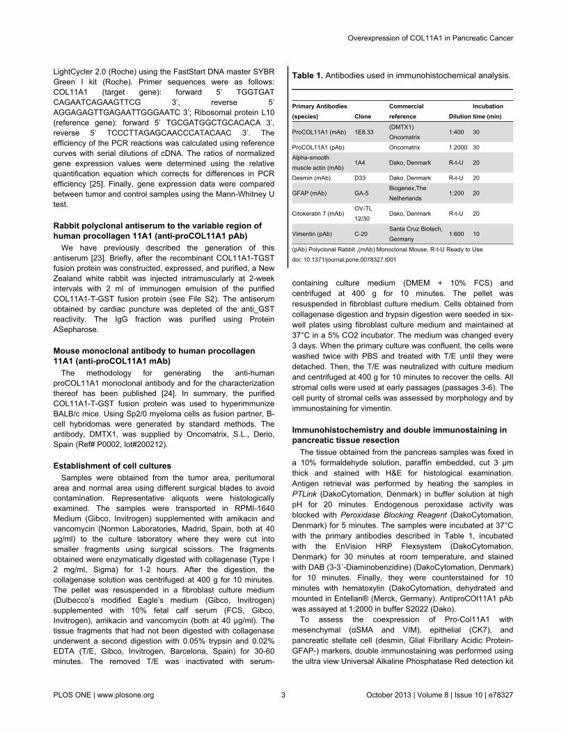
The tissue obtained from the pancreas samples was fixed ina 10% formaldehyde solution, paraffin embedded, cut 3 μmthick and stained with H&E for histological examination.Antigen retrieval was performed by heating the samples inPTLink (DakoCytomation, Denmark) in buffer solution at highpH for 20 minutes. Endogenous peroxidase activity wasblocked with Peroxidase Blocking Reagent (DakoCytomation,Denmark) for 5 minutes. The samples were incubated at 37°Cwith the primary antibodies described in Table 1, incubatedwith the EnVision HRP Flexsystem (DakoCytomation,Denmark) for 30 minutes at room temperature, and stainedwith DAB (3-3´-Diaminobenzidine) (DakoCytomation, Denmark)for 10 minutes. Finally, they were counterstained for 10minutes with hematoxylin (DakoCytomation, dehydrated andmounted in Entellan® (Merck, Germany). AntiproCOl11A1 pAbwas assayed at 1:2000 in buffer S2022 (Dako).
To assess the coexpression of Pro-Col11A1 withmesenchymal (αSMA and VIM), epithelial (CK7), andpancreatic stellate cell (desmin, Glial Fibrillary Acidic Protein-GFAP-) markers, double immunostaining was performed usingthe ultra view Universal Alkaline Phosphatase Red detection kit
Table 1. Antibodies used in immunohistochemical analysis.
Primary Antibodies(species) Clone
Commercialreference Dilution
Incubationtime (min)
ProCOL11A1 (mAb) 1E8.33(DMTX1)Oncomatrix
1:400 30
ProCOL11A1 (pAb) Oncomatrix 1.2000 30Alpha-smoothmuscle actin (mAb)
1A4 Dako, Denmark R-t-U 20
Desmin (mAb) D33 Dako, Denmark R-t-U 20
GFAP (mAb) GA-5Biogenex,TheNetherlands
1:200 20
Citokeratin 7 (mAb)OV-TL12/30
Dako, Denmark R-t-U 20
Vimentin (pAb) C-20Santa Cruz Biotech,Germany
1:600 10
(pAb) Polyclonal Rabbit ,(mAb) Monoclonal Mouse, R-t-U Ready to Usedoi: 10.1371/journal.pone.0078327.t001
Overexpression of COL11A1 in Pancreatic Cancer
PLOS ONE | www.plosone.org 3 October 2013 | Volume 8 | Issue 10 | e78327

(Ventana Medical Systems, Tucson, AZ) as red chromogenand DAB as brown chromogen. Previously, the samples hadbeen incubated at 37°C with the primary antibodies describedin Table 1.
Immunocytofluorescence of cultured stromal cells andConfocal Microscopy
Cells were fixed in acetone (-20°C) for 10 minutes in thechamber slide. The cells were dried at room temperature andthen were introduced into the wash buffer (Dako) for 30minutes. The samples were incubated with the anti-proCOl11A1 mAb, (DMTX1, Oncomatrix) with Cytokeratin 7(CK7) antibody and VIM antibody, to room temperature, underthe conditions specified in Table 1. The secondary antibodiesused were green anti-rabbit Alexa-488 (1:500, Invitrogen) andred anti-rabbit Alexa-546 (1:500, Invitrogen), for 1 hour at roomtemperature. Finally, the sections were mounted with mountingmedium containing DAPI (Vector Labs).
The colocalization (proCOL11A1 vs. VIM, proCOL11A1 vs.Ck7, proCOL11A1 vs. αSMA and VIM vs. CK7) was visualizedand photographed using a Leica TCS SP2 confocalmicroscope with 63X and 100X oil immersion objectives, usingthe following sources of illumination for each fluorochromeexcitation: Argon/Krypton laser (488 nm), Helium/Neon laser(546 nm) and blue-violet Diode (405 nm).
PDAC vs. CP immunohistochemistry assessmentAssessment was carried out in four regions selected as the
best representatives of the desmoplastic reaction. Cases thatpresented 4 positive fields through the 10X objective wereassigned the maximum score of 4, while negative fields wereassigned the minimum score of 0. For the staining assessment,a 20X field in the largest area with the most intense staining(“hot spot”) was chosen. Cases with less than 1% positive cellsin relation to the stromal surface were assigned a score of 0,cases with between 1 and 10% were assigned a score of 1,cases with between 10% and 50% were assigned a score of 2,and cases with more than 50% positive cells were assigned ascore of 3. Total variation in staining was defined by multiplyingthe number of positive fields (0-4) by the field’s stainingintensity (0-3), yielding a total score of 0-12. Theimmunostaining was double-blind scored by two pathologists(CGP and JGG). A series of 24 PDAC and 16 CPimmunostained slides were also quantified by means of theQWin image analysis program (Leica) in order to comparethem to the pathologists’ scores. Images of the largest areawith the most intense staining were taken through the 20Xobjective of an Olympus BX61 microscope and recorded on anOlympus Dp70 camera.
Statistical data analysisAssuming unequal variances, we applied the Welch test to
determine the significance of the differences in the image databetween PDAC and chronic pancreatitis samples. The ANOVAtest was also applied. The correlation between various imagingparameters was assessed using the Spearman Rankcorrelation test. The sensitivity and specificity of proCOL11A1was depicted as a receiver operating characteristic (ROC)
analysis. The accuracy (i.e. correctly classified cases) wascalculated. The position of the cut-off in the curve willdetermine the number of true positives, true negatives, falsepositives and false negatives. The criterion value is the cut-offcorresponding to the highest accuracy (minimal false negativeand false positive results). The sample size used in ourimmunohistochemical study (n=77) allowed us to achievestatistical significance (alpha=0.05) for an AUC=0.9 under thenull hypothesis of an AUC=0.5, with a statistical power of 90%.All the analyses were implemented using the StatisticalPackage for the Social Sciences (SPSS 13) or MedCalcv9.4.1.0.
Results
Q-RT-PCR of COL11A1 and immunohistochemistry ofpancreatic tissues with anti-proCOL11A1
Pancreatic cancer cells express high levels of COL11A1mRNA, as shown by quantitative RT-PCR (Figure 1). Theresults confirmed a significantly increased expression of theCOL11A1 gene in PDAC samples vs. normal and CP samples(fold change: 174; SLR: 7).
All PDAC samples tested with anti-proCOL11A1 mAb andwith antiproCOL11A1 pAb showed strong intracytoplasmaticlabeling of tumor-surrounding desmoplastic stromal CAFs(Figure 2E; Figure S1D in File S1). The staining was notwidespread but rather localized in restricted peritumoral areas,even where tumor cells were not seen. The morphologicalfeatures of these fibroblast-like stromal cells were compatiblewith those of myofibroblasts (Figure SlE, F in File S1).Extracellular staining was never observed. In contrast, theexpression of proCOL11A1 in chronic pancreatitis samples waseither absent (Figure 2A; Figure S1B in File S1) or very lowand restricted to a few stromal cells. CP stroma showed fibroticand inflammatory changes with no remarkable myofibroblasticpopulation. Normal pancreas and epithelial tumor cells, on theother hand, did not show any staining at all with anti-proCol11A1 (Figure S2 in File S1). Normal pancreas showedstaining with VIM and αSMA, but did not stain with GFAP. Themajority of CP (Figures 2C, D) and PDAC (Figures 2G, H)stromal cells showed strong intracellular staining with VIM andαSMA. The staining with desmin was intense in PDAC (Figure2F) samples but nonexistent in CP (Figure 2B) samples. FigureS3 in File S1 shows positive staining with anti-proCOL11A1mAb (score 4) and desmin in a case of autoimmunepancreatitis; in addition, intense positivity with VIM and αSMA,and negativity with GFAP is observed.
Characterization of pancreatic CAFsTo characterize the CAFs, pancreatic tissue sections were
double stained for proCOL11A1/desmin, proCOL11A1/αSMA,proCOL11A1/VIM, proCOL11A1/GFAP, proCOL11A1/CK7 andCK7/VIM (Figure 3). A very high number of mesenchymal cellswere strongly labeled for VIM and αSMA. Immunostaining withGFAP was negative. As a proportion a small number of cellswere proCOL11A1+ and desmin+. Co-staining of VIM or CK7with proCOL11A1 identified a subset of CAFs with themesenchymal phenotype (proCOL11A1+/VIM+) and very few
Overexpression of COL11A1 in Pancreatic Cancer
PLOS ONE | www.plosone.org 4 October 2013 | Volume 8 | Issue 10 | e78327

cells with the epithelial phenotype (proCOL11A1+/CK7+)(Figures 3 and E, respectively). Some desmin or αSMA cellsco-stained with proCOL11A1 (Figure 3 A and B, respectively).However, CP stroma samples did not show staining withproCOL11A1, desmin or GFAP, and there was no co-staining(Figure 4).
The cell distribution of cultured CAFs was analyzed bycolocalization with immunocytofluorescence. The resulting dataare depicted in Table 2 (Figure 5). The interpretation of thetable is as follows: when a double staining was applied to thecultured pancreatic CAFs, for instance proCOL11A1 and CK7,a total of 188 cells were stained; of these, 82 cells were labeledwith anti-proCOL11A1, 60 cells were CK7 positive, and 46 cellswere stained with both antibodies. The same logic applies tothe rest of the stainings. According to the data shown in Table2, among proCOL11A1 cells, 36% are CK7+, 49% are VIM+and 48% are αSMA+; among the cells with the VIM phenotype,21% are proCOL11A1+ and only 8% are CK7+; 33% of αSMAcells share the proCOL11A1 phenotype; and lastly, 45% ofCK7 cells have the VIM+ phenotype and 43% have the
proCOL11A1 phenotype. The distribution of the proCOL11A1,CK7 and desmin phenotypes in tissue samples is shown inTable S2 in File S1 . It was not possible to analyze those fieldsin which cells are VIM+ and αSMA+ due to the high number ofstained cells, which makes it complicated to individualize andcount them. According to the data shown in Table S2 in FileS1, 18% of proCOl11A1 cells are CK7+, and 21% are desmin+;45% of those cells staining with desmin have the proCOL11A1phenotype, and 50% of CK7 cells are proCOL11A1.
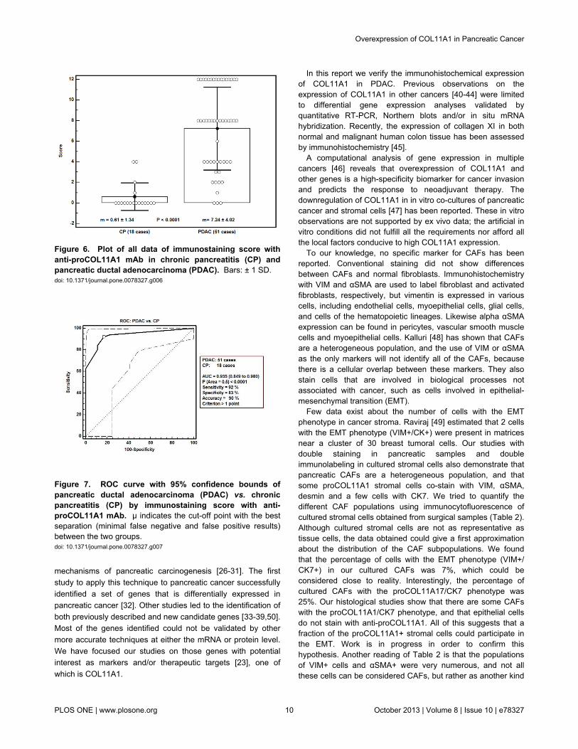
PDAC vs. CPThe characteristics of each patient and his/her pathologist’s
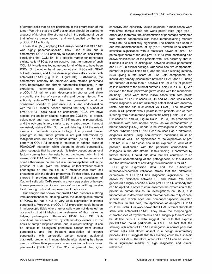
proCOL11A1 immunostaining score are shown in Table S3 inFile S1. There was a statistically significant difference betweenPDAC and chronic pancreatitis in the mAb score (mean ± SD:7.33 ± 4.04 vs. 0.61 ± 1.33; P < 0.0001). The data are shown inFigure 6. The AUC of ROC curves PDAC vs. CP was 0.936(0.851 to 0.981), P < 0.0001 (Figure 7). The immunostainingshowed a sensitivity of 92% and a specificity of 83% indiscriminating PDAC from CP, with an accuracy of 90%. The
Figure 1. Normalized gene expression levels (log2-transformed fold change) in pancreatic ductal adenocarcinoma(PDAC) samples relative to normal pancreas (NP) and chronic pancreatitis (CP). doi: 10.1371/journal.pone.0078327.g001
Overexpression of COL11A1 in Pancreatic Cancer
PLOS ONE | www.plosone.org 5 October 2013 | Volume 8 | Issue 10 | e78327

Figure 2. Comparative immunohistochemical profile of stromal cells of chronic pancreatitis (CP) and pancreatic ductaladenocarcinoma (PDAC) in serial sections. Stromal cells in CP were negative for anti-proCOL11A1 mAb (A) and desmin (B)whereas a substantial number of stromal cells of PDAC expressed anti-proCOL11A1 mAb (E) and desmin (F). In CP and PDAC thestain of stromal cells for αSMA (C and G, respectively) and VIM (D and H, respectively) were diffuse and non-selective. Anti-proCOL11A1 mAb (A and E), desmin (B and F), αSMA (C and G) and VIM (D and H) (all photomicrographs at ×400, Scale bar 50μm).doi: 10.1371/journal.pone.0078327.g002
Overexpression of COL11A1 in Pancreatic Cancer
PLOS ONE | www.plosone.org 6 October 2013 | Volume 8 | Issue 10 | e78327

Figure 3. Co-staining of anti-proCOL11A1 mAb with different fibroblastic markers and CK 7, in Pancreatic DuctalAdenocarcinoma. A, anti-proCOL11A1 (brown) vs. desmin (magenta); B, anti-proCOL11A1 (brown) vs. αSMA (magenta); C, anti-proCOL11A1 (brown) vs. VIM (magenta); D, anti-proCOL11A1 (brown) vs. GFAP (not staining); E, anti-proCOL11A1 (brown) vs.CK7 (magenta); F, CK7 (epithelial tumor cells: brown) vs. VIM (magenta) (all photomicrographs at ×200, Scale bar 200 μm; insetX1000).doi: 10.1371/journal.pone.0078327.g003
Overexpression of COL11A1 in Pancreatic Cancer
PLOS ONE | www.plosone.org 7 October 2013 | Volume 8 | Issue 10 | e78327

Figure 4. Co-staining of anti-proCOL11A1 mAb with different fibroblastic markers and CK 7, in Chronic Pancreatitis. A,anti-proCOL11A1 (brown) vs. desmin (magenta); B, Anti-proCOL11A1 (brown) vs. αSMA (magenta); C, anti-proCOL11A1 (brown)vs. VIM (magenta); D, anti-proCOL11A1 (brown) vs. GFAP (not staining); E, anti-proCOL11A1 (brown) vs. CK7 (magenta); F, CK7(epithelial tumor cells: brown) vs. VIM (magenta).(all photomicrographs at ×200, Scale bar 200 μm). .doi: 10.1371/journal.pone.0078327.g004
Overexpression of COL11A1 in Pancreatic Cancer
PLOS ONE | www.plosone.org 8 October 2013 | Volume 8 | Issue 10 | e78327

use of pAb showed a similar outcome discriminating PDACfrom CP (Table S4 in File S1). Observational concordancebetween pathologists was >90%. The staining score did not
Table 2. Quantitative analysis of cell distribution in culturedperitumoral pancreatic cancer fibroblasts (passage 3)*.
proCOL11A1/CK7(DI) proCOL11A1/VIM (DI)
proCOL11A1/αSMA(DI)
VIM/CK7(DI)
proCOL11A1+ only82 (43%)
proCOL11A1+ only106 (18%)
proCOL11A1+ only73 (23%)
VIM+ only364 (85%)
CK7+ only 60 (32%) VIM+ only 370 (64%)αSMA+ only 138(50%)
CK7+ only36 (8%)
proCOL11A1+/CK7+ 46 (25%)
proCOL11A1+/VIM+101 (18%)
proCOL11A1+/αSMA+ 67 (24%)
VIM+/CK7+30 (7%)
Total 188 (100%) Total 577 (100%) Total 278 (100%)Total 430(100%)
*. One patient sample, valuation on five fields for each double immunostaining (DI)
experiment. Cells stained with both Ab in bold.doi: 10.1371/journal.pone.0078327.t002
correlate to the patients’ age, sex, tumor stage or grade. Theassociation with patient overall survival was not investigatedbecause the number of PDAC patients with a low score wasvery low (4/51 cases).
Immunostaining was also evaluated using the QWin imageanalysis software. The results are shown in Table S5 in FileS1. There is a statistical difference between PDAC and CP inthe number of positive cells in the stained surface and in thereference area. Within the same series, there was a highconcordance between the pathologist score and the QWin datasuch as the surface area of the stained cells and the number ofpositive cells (Spearman’s rank correlation coefficient = 0.825and 0.825, respectively). ROC curve AUC data were obtainedfor the six image-analysis parameters (Table S6 in File S1). Allparameters permit discrimination between PDAC and chronicpancreatitis. The most relevant parameters were the number ofpositive cells and the surface area of the stained cells.
Discussion
DNA microarrays permit simultaneous analysis of theexpression level of thousands of genes and could clarify the
Figure 5. Confocal microscopy of pancreatic cultured CAFs. Double fluorescence stain illustrates the presence of cellsproCOL11A1+/CK7+, proCOL11A1+/αSMA+ and proCOL11A1+/VIM+. Red: proCOL11A1; green: CK7, αSMA and VIM,respectively; blue: nuclei. Insets: randomly taken high power fields of culture. Scale bar 100 μm (X200) and 20 μm (X630).doi: 10.1371/journal.pone.0078327.g005
Overexpression of COL11A1 in Pancreatic Cancer
PLOS ONE | www.plosone.org 9 October 2013 | Volume 8 | Issue 10 | e78327
mechanisms of pancreatic carcinogenesis [26-31]. The firststudy to apply this technique to pancreatic cancer successfullyidentified a set of genes that is differentially expressed inpancreatic cancer [32]. Other studies led to the identification ofboth previously described and new candidate genes [33-39,50].Most of the genes identified could not be validated by othermore accurate techniques at either the mRNA or protein level.We have focused our studies on those genes with potentialinterest as markers and/or therapeutic targets [23], one ofwhich is COL11A1.
Figure 6. Plot of all data of immunostaining score withanti-proCOL11A1 mAb in chronic pancreatitis (CP) andpancreatic ductal adenocarcinoma (PDAC). Bars: ± 1 SD.doi: 10.1371/journal.pone.0078327.g006
Figure 7. ROC curve with 95% confidence bounds ofpancreatic ductal adenocarcinoma (PDAC) vs. chronicpancreatitis (CP) by immunostaining score with anti-proCOL11A1 mAb. µ indicates the cut-off point with the bestseparation (minimal false negative and false positive results)between the two groups.doi: 10.1371/journal.pone.0078327.g007
In this report we verify the immunohistochemical expressionof COL11A1 in PDAC. Previous observations on theexpression of COL11A1 in other cancers [40-44] were limitedto differential gene expression analyses validated byquantitative RT-PCR, Northern blots and/or in situ mRNAhybridization. Recently, the expression of collagen XI in bothnormal and malignant human colon tissue has been assessedby immunohistochemistry [45].
A computational analysis of gene expression in multiplecancers [46] reveals that overexpression of COL11A1 andother genes is a high-specificity biomarker for cancer invasionand predicts the response to neoadjuvant therapy. Thedownregulation of COL11A1 in in vitro co-cultures of pancreaticcancer and stromal cells [47] has been reported. These in vitroobservations are not supported by ex vivo data; the artificial invitro conditions did not fulfill all the requirements nor afford allthe local factors conducive to high COL11A1 expression.
To our knowledge, no specific marker for CAFs has beenreported. Conventional staining did not show differencesbetween CAFs and normal fibroblasts. Immunohistochemistrywith VIM and αSMA are used to label fibroblast and activatedfibroblasts, respectively, but vimentin is expressed in variouscells, including endothelial cells, myoepithelial cells, glial cells,and cells of the hematopoietic lineages. Likewise alpha αSMAexpression can be found in pericytes, vascular smooth musclecells and myoepithelial cells. Kalluri [48] has shown that CAFsare a heterogeneous population, and the use of VIM or αSMAas the only markers will not identify all of the CAFs, becausethere is a cellular overlap between these markers. They alsostain cells that are involved in biological processes notassociated with cancer, such as cells involved in epithelial-mesenchymal transition (EMT).
Few data exist about the number of cells with the EMTphenotype in cancer stroma. Raviraj [49] estimated that 2 cellswith the EMT phenotype (VIM+/CK+) were present in matricesnear a cluster of 30 breast tumoral cells. Our studies withdouble staining in pancreatic samples and doubleimmunolabeling in cultured stromal cells also demonstrate thatpancreatic CAFs are a heterogeneous population, and thatsome proCOL11A1 stromal cells co-stain with VIM, αSMA,desmin and a few cells with CK7. We tried to quantify thedifferent CAF populations using immunocytofluorescence ofcultured stromal cells obtained from surgical samples (Table 2).Although cultured stromal cells are not as representative astissue cells, the data obtained could give a first approximationabout the distribution of the CAF subpopulations. We foundthat the percentage of cells with the EMT phenotype (VIM+/CK7+) in our cultured CAFs was 7%, which could beconsidered close to reality. Interestingly, the percentage ofcultured CAFs with the proCOL11A17/CK7 phenotype was25%. Our histological studies show that there are some CAFswith the proCOL11A1/CK7 phenotype, and that epithelial cellsdo not stain with anti-proCOL11A1. All of this suggests that afraction of the proCOL11A1+ stromal cells could participate inthe EMT. Work is in progress in order to confirm thishypothesis. Another reading of Table 2 is that the populationsof VIM+ cells and αSMA+ were very numerous, and not allthese cells can be considered CAFs, but rather as another kind
Overexpression of COL11A1 in Pancreatic Cancer
PLOS ONE | www.plosone.org 10 October 2013 | Volume 8 | Issue 10 | e78327
of stromal cells that do not participate in the progression of thetumor. We think that the CAF designation should be applied toa subset of fibroblast-like stromal cells in the peritumoral regionthat influence cancer growth and are identified by the anti-proCOL11A1 antibody.
Erkan et al. [50], applying DNA arrays, found that COL11A1was highly pancreas-specific. They used αSMA and acommercial COL11A1 antibody to evaluate the co-localization,concluding that COL11A1 is a specific marker for pancreaticstellate cells (PSCs), but we observe that the number of suchCOL11A1+ cells was too numerous for all of them to have beenPSCs. On the other hand, we cannot stain PSCs with GFAPbut with desmin, and those desmin positive cells co-stain withanti-proCOL11A1 (Figure 2F; Figure 3D). Furthermore, thecommercial antibody he employed also stained pancreaticacini, hepatocytes and chronic pancreatitis fibroblasts. In ourexperience, commercial antibodies other than anti-proCOL11A1 fail to stain desmoplastic stroma and showunspecific staining of cancer cells and benign tissue. Theantibody anti-proCOL11A1 that we generated could beconsidered specific to pancreatic CAFs, and co-localizationwith the PSC marker desmin showed that only a subset ofproCOL11A1+ cells could be considered PSCs. We haveapplied the antibody against human pro-COL11A1 to breast,colon, neck and head tumors [51-53] (papers in preparation),and the outcome is very similar to pancreatic cancer: it almostexclusively stains CAFs. Our current data highlight the role ofstroma in pancreatic cancer biology. The present cancerparadigm is that tumor growth is not just determined bymalignant cells, but also by tumor stroma [54,55]. The focalpattern of COL11A1 staining is restricted to defined areas ofPDAC/CAF interaction while absent in chronic pancreatitis,which suggests that its expression is highly dependent on localfactors coming from the neighboring carcinoma cells. In thatsense, COL11A1 and CK7 co-expression in the same cellcould either mean that the cell is a tumoral epithelial cell in theprocess of EMT (with the double epithelial/mesenchymalphenotype) or that the cell is a mesenchymal stem cellpresenting with the double phenotype. To this effect, our teamshowed in previous reports [56,57] that the association ofCapan-1 cells with CAFs results in a very aggressive orthotopichuman pancreatic carcinoma xenograft model, with aggressivelocal tumor growth and the presence of metastasis.
Our analysis has shown that proCOL11A1 presents a strongimmunohistochemical staining within the desmoplastic stromaof PDAC, but has a null or very weak expression in chronicpancreatitis. Moreover, proCOL11A1 expression could be seenin microscopic fields where tumor cells were not detected, anobservation that highlights the usefulness of this marker inhelping pathologists differentiate PDAC from CP. Bothconditions are characterized by inflammatory events. On thebasis of sample biopsies alone, and in clinical settings, it canbe difficult to distinguish pancreatic cancer from chronicpancreatitis, and the frequent association of chronicpancreatitis with pancreatic cancer causes additionaldiagnostic problems. Immunohistochemical staining has beenused to differentiate pancreatic adenocarcinoma from chronicpancreatitis (Table S7 in File S1). In general, the higher
sensitivity and specificity values obtained in most cases werewith small sample sizes and weak power tests (high type IIerror), and therefore, the differentiation of pancreatic carcinomafrom chronic pancreatitis with those immunostaining markerswould not be statistically significant. The sample size used inour immunohistochemical study (n=78) allowed us to achievestatistical significance with a statistical power of 90%. Thepathologist score of the anti-proCOL11A1 immunostaining alsoallows classification of the patients with 90% accuracy; that is,it makes it easier to distinguish between chronic pancreatitisand PDAC in clinical settings. Our score has two components:number of positive fields (0-4) and the field’s staining intensity(0-3), giving a total score of 0-12. Both components canindividually already discriminate between PDAC and CP, usingthe criterion of more than 1 positive field, or ≥ 1% of positivecells in relation to the stromal surface (Table S8 in File S1). Wereviewed the false positive/negative cases with the monoclonalantibody. There were three PDAC cases with zero points(Table S3 in File S1: cases 41, 44 and 54); one was a casewhose diagnosis was not ultimately established with accuracy(distal common bile duct cancer vs. PDAC). The maximumscore in CP patients was 4 points in two patients, one of themsuffering from autoimmune pancreatitis (AIP) (Table S3 in FileS1: cases 15 and 31, Figure S3 in File S1). Its preoperativeusefulness with core needle biopsy, as we demonstrated inbreast cancer [51,52], needs to be investigated in pancreaticcancer. Whether proCOL11A1 can be useful as a differentialdiagnosis marker using non-invasive techniques must beexplored as well. The significance of the expression of pro-Col11A1 in our AIP case should be explored in view of itspossible relationship with the particular composition ofcollagens in the AIP stroma. If this finding is confirmed byfurther studies, it could add data that will contribute to animproved understanding of the pathogenesis of this diseaseand the development of new diagnostic biomarkers for AIP.
Our gene expression data and the subsequentimmunohistochemical validation stress that the differentialexpression of COL11A1 has diagnostic significance, as itallows for distinction between CP and PDAC. We havegenerated a highly specific human proCOL11A1- antibody thatcan be applied in order to immunoscreen the expression of theprotein in human tissues. In investigations on CAFs, it isfundamental to determine which stromal cells are truly cancer-specific and which ones are non-cancer-specific activatedfibroblasts. In this field, the application of anti-proCOL11A1could be useful. Our work shows that PDAC stromal fibroblastsstain with anti-proCOL11A1. They have the morphologicalcharacteristics of myofibroblasts and a subgroup thereof couldbe stellate cells. Our data suggest that cells that expressproCOL11A1 could participate in EMT. The fact that thestaining with anti-proCOL11A1 is negative in normal pancreasstromal cells and almost absent in a benign inflammatoryprocess like CP suggests that this staining could be a specificmarker for CAFs. Therefore, anti-proCOL11A1 can be seen tobe a significant marker of high diagnostic and clinicalrelevance.
Overexpression of COL11A1 in Pancreatic Cancer
PLOS ONE | www.plosone.org 11 October 2013 | Volume 8 | Issue 10 | e78327

Supporting Information
File S1. Supporting Figures and Tables.Figure S1, Immunohistochemical staining with anti-proCOL11A1 pAb of Chronic Pancreatitis (CP) and PancreaticDuctal Adenocarcinoma (PDAC) . A: CP (H&E). B: CP negativeanti-proCOL11A1 stain. C: PDAC (H&E). D: PDAC positiveanti-proCOL11A1 stain. E and F: Detail of anti-proCOL11A1expression in stromal cells of PDAC. H & E indicatesHematoxilin and Eosin (all photomicrographs at ×400). FigureS2, Immunohistochemical staining of normal pancreas withdifferent fibroblastic markers. A, anti-proCOL11A1 mAb,positive control (inset): cell line A204; B, desmin, positivecontrol (inset): appendix; C, alpha-Smooth Muscle Actin,positive control (inset): appendix; D, vimentin, positive control(inset) : appendix) ; E, GFAP, positive control (inset) :astrocytoma). Serial sections (all photomicrographs at ×200,Scale bar 200 μm). Figure S3, Immunohistochemical stainingof autoimmune pancreatitis with different fibroblastic markers.A, anti-proCOL11A1 mAb , positive control (inset): cell lineA204); B, desmin, positive control (inset): appendix); C, alpha-Smooth Muscle Actin , positive control (inset) : appendix) ; D,vimentin , positive control (inset): appendix) ; E, GFAP, positivecontrol (inset) : astrocytoma). Serial sections (allphotomicrographs at ×200, Scale bar 200 μm). Table S1,Comparison of gene expression data from microarray analysis(Affymetrix GeneChips). Table S2, Quantitative analysis of celldistribution in peritumoral pancreatic cancer tissue. Table S3,Patient characteristics and immunohistochemistry score withanti-proCOL11A1 pAb and mAb. Table S4, Discriminationbetween PDAC (pancreatic ductal adenocarcinoma) and CP
(chronic pancreatitis) using pathologist score. Table S5,Summary statistics of immunohistochemical analyses. TableS6, Area under the ROC curve (AUC) of image analysisparameters (pancreatic ductal adenocarcinoma) PDAC vs. CP(chronic pancreatitis). Table S7, Discrimination between PDAC(pancreatic ductal adenocarcinoma) and CP (chronicpancreatitis) using various diagnostic markers in tissues. TableS8, Discrimination PDAC (pancreatic ductal adenocarcinoma)vs. CP (chronic pancreatitis) with anti-proCOL11A1 mAb bycomponents of score and total score.(DOCX)
File S2. Global Gene Expression Analysis. Generation of arabbit polyclonal antiserum to the variable region of humanprocollagen 11A1 (anti-proCOL11A1 pAb). SDS-PAGE andWestern-blots.(DOCX)
Acknowledgements
The excellent technical assistance of Laura Suárez-Fernándezis greatly acknowledged.
Author Contributions
Conceived and designed the experiments: LB CGP JRT LSB.Performed the experiments: LB MGO JRT JDAI JAG EGP.Analyzed the data: LB CGP JGG PMR NGC LSG. Contributedreagents/materials/analysis tools: LSB CGP PMR JRT. Wrotethe manuscript: LB CGP JRT JAG JDAI.
References
1. Siegel R, Naishadham D, Jemal A (2013) Cancer statistics, 2013. CACancer J Clin 63: 11-30. doi:10.3322/caac.21166. PubMed: 23335087.
2. Lim JE, Chien MW, Early CC (2003) Prognostic factors followingcurative resection for pancreatic adenocarcinoma. A population-based,linked database analysis of 396 patients. Ann Surg 237: 74-85. doi:10.1097/00000658-200301000-00011. PubMed: 12496533.
3. Kollmannsberger C, Peters HD, Fink U (1998) Chemotherapy inadvanced pancreatic adenocarcinoma. Cancer Treat Rev 24: 133-156.doi:10.1016/S0305-7372(98)90079-2. PubMed: 9728423.
4. Imamura T, Iguchi H, Manabe T, Ohshio G, Yoshimura T et al. (1995)Quantitative analysis of collagen and collagen subtypes I, III, an V inhuman pancreatic cancer, tumor-associated chronic pancreatitis, andalcoholic chronic pancreatitis. Pancreas 11: 357-364. doi:10.1097/00006676-199511000-00007. PubMed: 8532652.
5. Linder S, Castanos-Velez E, Von Rosen A, Biberfeld P (2001)Immunohistochemical expression of extracellular matrix proteins andadhesion molecules in pancreatic carcinoma. Hepatogastroenterology48: 1321-1327. PubMed: 11677955.
6. Feig C, Gopinathan A, Neesse A, Chan DS, Cook N et al. (2012) ThePancreas Cancer Microenvironment. Clin Cancer Res 18: 4266-4276.doi:10.1158/1078-0432.CCR-11-3114. PubMed: 22896693.
7. Erkan M, Hausmann S, Michalski CW, Fingirle AA, Dobritz M, Jet al(2012) The role of stroma in pancreatic cancer: diagnostic andtherapeutic implications. Nat. Rev Gastroenterol Hepatol 9: 454–467doi:10.1038/nrgastro.2012.115.
8. Sadlonova A, Bowe DB, Novak Z, Mukherjee S, Duncan VE et al.(2009) Identification of molecular distinctions between normal breast-associated fibroblasts and breast cancer-associated fibroblasts. CancerMicroenviron 2: 9-21. doi:10.1007/s12307-008-0017-0. PubMed:19308679.
9. Luciani MG, Seok J, Sayeed A, Champion S, Goodson WH et al.(2011) Distinctive responsiveness to stromal signaling accompanies
histologic grade programming of cancer cells. PLOS ONE 6: e20016.doi:10.1371/journal.pone.0020016. PubMed: 21625507.
10. Howes N, Neoptolemos JP (2002) Risk of pancreatic ductaladenocarcinoma in chronic pancreatitis. Gut 51: 765-766. doi:10.1136/gut.51.6.765. PubMed: 12427771.
11. Lowenfels AB, Maisonneuve P, Cavallini G, Ammann RW, Lankisch PGet al. (1993) Pancreatitis and the risk of pancreatic cancer. InternationalPancreatitis Study Group. N Engl J Med 328: 1433-1437. doi:10.1056/NEJM199305203282001. PubMed: 8479461.
12. Otsuki M (2003) Chronic pancreatitis in Japan: epidemiology,prognosis, diagnostic criteria, and future problems. J Gastroenterol 38:315-326. doi:10.1007/s005350300058. PubMed: 12743770.
13. Marjolin J-N (1828) Ulcere. Dictionnaire Medecine. Vol 21. Pratique14. Shoulders MD, Rainez RT (2009) Collagen structure and stability. Annu
Rev Biopchem 78: 929-958.15. Olumi AF, Grossfeld GD, Hayward SW, Carroll PR, Tlsty TD et al.
(1999) Carcinoma-associated fibroblasts direct tumor progression ofinitiated human prostatic Epithelium. Cancer Res 59: 5002-5011.PubMed: 10519415.
16. Mahadevan D, Von Hoff DD (2007) Tumor-stroma interactions inpancreatic ductal adenocarcinoma. Mol Cancer Ther 6: 1186-1197. doi:10.1158/1535-7163.MCT-06-0686. PubMed: 17406031.
17. Desmoulière A, Guyot C, Gabbiani G (2004) The stroma reactionmyofibroblast: a key player in the control of tumor cell behavior. Int JDev Biol 48: 509-517. doi:10.1387/ijdb.041802ad. PubMed: 15349825.
18. Kore M (1997) Pancreatic cancer-associated stroma production. Am JSurg 194: S84-S86.
19. Hwang RF, Moore T, Arumugam T, Ramachandran V, Amos KD et al.(2008) Cancer-associated stromal fibroblasts promote pancreatic tumorprogression. Cancer Res 68: 918-926. doi:10.1158/0008-5472.CAN-07-5714. PubMed: 18245495.
Overexpression of COL11A1 in Pancreatic Cancer
PLOS ONE | www.plosone.org 12 October 2013 | Volume 8 | Issue 10 | e78327

20. Hanahan D, Weinberg RA (2011) Hallmarks of cancer: The nextgeneration. Cell 144: 646-674. doi:10.1016/j.cell.2011.02.013. PubMed:21376230.
21. Sato N, Maehara N, Goggins M (2004) Gene expression profiling oftumor stromal interactions between pancreatic cancer cells and stromalfibroblasts. Cancer Res 64: 6950-6956. doi:10.1158/0008-5472.CAN-04-0677. PubMed: 15466186.
22. Del Amo J, de los Toyos JR, García C, Pérez M, Ochoa G, Vázquez Let al. (2005) Target identification and validation in pancreatic cancer.Eur J Cancer: s3: 54
23. Barneo L, Del Amo J, García-Pravia C, de los Toyos JR, Pérez-Basterrechea M et al. (2006) Identification of specific genes bymicroarrays, validation and use of polyclonal antibodies in pancreaticcancer: preliminary results. In: 41st Congress of the European Societyfor Surgical Research-ESSR. Bologna; Italy, Vollmar Brigitte (ed)Medimond, International Proceedings; 27-35
24. García-Ocaña M, Vázquez F, García-Pravia C, Fuentes-Martínez N,Menéndez-Rodríguez P et al. (2012) Characterization of a novel mousemonoclonal antibody, clone 1E8.33, highly specific for humanprocollagen 11A1, a tumor associated stromal component. Int J Oncol,40: 1447-1454. PubMed: 22322826.
25. Pfaffl MW (2001) A new mathematical model for relative quantificationin real time RT-PCR. Nucleic Acids Res, 29: e45. PubMed: 11328886.
26. Missiaglia E, Blaveri E, Terris B, Wang YH, Costello E et al. (2004)Analysis of gene expression in cancer cell lines identifies candidatemarkers for pancreatic tumorigenesis and metastasis. Int J Cancer,112: 100-112. PubMed: 15305381.
27. Buchholz M, Boeck W, Fensterer H, Müller F, Wenger C et al. (2001)Use of DNA arrays/microarrays in pancreatic research. Pancreatology1: 581-586. doi:10.1159/000055867. PubMed: 12120240.
28. Crnogorac-Jurcevic T, Efthimiou E, Capelli P, Blaveri E, Baron A et al.(2001) Gene expression profiles of pancreatic cancer and stromaldesmoplasia. Oncogene 20: 7437-7446. doi:10.1038/sj.onc.1204935.PubMed: 11704875.
29. Han H, Bearss D, Browne L, Calaluce R, Nagle RB et al. (2002)Identification of differentially expressed genes in pancreatic cancercells using cDNA microarray. Cancer Res 62: 2890-2896. PubMed:12019169.
30. Iacobuzio-Donahue CA, Maitra A, Shen-Ong GL, van Heek T, Ashfaq Ret al. (2002) Discovery of novel tumor markers of pancreatic cancerusing global gene expression technology. Am J Pathol 160: 1239-1249.doi:10.1016/S0002-9440(10)62551-5. PubMed: 11943709.
31. Crnogorac-Jurcevic T, Missiaglia E, Blaveri E, Gangeswaran R, JonesM et al. (2003) Molecular alterations in pancreatic carcinoma:expression profiling shows that dysregulated expression of S100 genesis highly prevalent. J Pathol 201: 63-74. doi:10.1002/path.1418.PubMed: 12950018.
32. Friess H, Ding J, Kleeff J, Fenkell L, Rosinski JA et al. (2003)Microarray-based identification of differentially expressed growth- andmetastasis-associated genes in pancreatic cancer. Cell Mol Life Sci 60:1180-1199. PubMed: 12861384.
33. Iacobuzio-Donahue C, Maitra A, Olsen M, Lowe AW, van Heek NT etal. (2003) Exploration of global gene expression patterns in pancreaticadenocarcinoma using cDNA microarrays. Am J Pathol 162:1151-1162. doi:10.1016/S0002-9440(10)63911-9. PubMed: 12651607.
34. Logsdon C, Simeone D, Binkley C, Arumugam T, Greenson JK et al.(2003) Molecular profiling of pancreatic adenocarcinoma and chronicpancreatitis identifies multiple genes differentially regulated inpancreatic cancer. Cancer Res 63: 2649-2657. PubMed: 12750293.
35. Tan Z, Hu X, Cao G, Tang Y (2003) Analysis of gene expression profileof pancreatic carcinoma using cDNA microarray. World J Gastroenterol9: 818-823. PubMed: 12679940.
36. Grützmann R, Pilarsky C, Ammerpohl O, Lüttges J, Böhme A et al.(2004) Gene expression profiling of microdissected pancreatic ductalcarcinomas using high density DNA microarrays. Neoplasia 6: 611-622.doi:10.1593/neo.04295. PubMed: 15548371.
37. Nakamura T, Furukawa Y, Nakagawa H, Tsunoda T, Ohigashi H et al.(2004) Genome-wide cDNA microarray analysis of gene expressionprofiles in pancreatic cancers using populations of tumor cells andnormal ductal epithelial cells selected for purity by lasermicrodissection. Oncogene 23: 2385-2400. doi:10.1038/sj.onc.1207392. PubMed: 14767473.
38. Jin G, Hu X, Ying K, Tang Y, Liu R et al. (2005) Discovery and analysisof pancreatic adenocarcinoma genes using cDNA microarrays. World JGastroenterol 11: 6543-6548. PubMed: 16425432.
39. Pilarsky C, Ammerpohl O, Sipos B, Dahl E, Hartmann A et al. (2008)Activation of Wnt signalling in stroma from pancreatic cancer identifiedby gene expression profiling. J Cell Mol Med 12: 2823-2835. doi:10.1111/j.1582-4934.2008.00289.x. PubMed: 18298655.
40. Fisher H, Salahshor S, Stenling R, Björk J, Lindmark G et al. (2001)COL11A1 in FAP polyps and in sporadic colorectal tumors. BMCCancer 1: 17, Epub October 29. doi:10.1186/1471-2407-1-17. PubMed:11707154.
41. Binkley CE, Zhang L, Greenson JK, Giordano TJ, Kuick R et al. (2004)The molecular basis of pancreatic fibrosis: common stromal geneexpression in chronic pancreatitis and pancreatic adenocarcinoma.Pancreas 29: 254-263. doi:10.1097/00006676-200411000-00003.PubMed: 15502640.
42. Sok JC, Kuriakose MA, Mahajan VB, Pearlman AN, DeLacure MD et al.(2003) Tissue-specific gene expression of head and neck squamouscell carcinoma in vivo by complementary DNA microarray analysis.Arch Otolaryngol Head Neck Surg 129: 760-770. doi:10.1001/archotol.129.7.760. PubMed: 12874079.
43. Schmalbach CE, Chepeha DB, Giordano TJ, Rubin MA, Teknos TN etal. (2004) Molecular profiling and the identification of genes associatedwith metastatic oral cavity/pharynx squamous cell carcinoma. ArchOtolaryngol Head Neck Surg 130: 295-302. doi:10.1001/archotol.130.3.295. PubMed: 15023835.
44. Chong IW, Chang MY, Chang HC, Yu YP, Sheu CC et al. (2006) Greatpotential of a panel of multiple hMTH1, SPD, ITGA11 and COL11A1markers for diagnosis of patients with non-small cell lung cancer. OncolRep 16: 981-988. PubMed: 17016581.
45. Bowen KB, Reimers AP, Luman S, Kronz JD, Fyffe WE et al. (2008)Immunohistochemical localization of collagen type XI alpha1 andalpha2 chains in human colon tissue. J Histochem Cytochem 56: 275–283. PubMed: 18040076.
46. Kim H, Watkinson J, Varadan V, Anastassiou D (2010) Multi-cancercomputational analysis reveals invasion-associated variant ofdesmoplastic reaction involving INHBA, THBS2 and COL11A1. BMCMed Genomics 3: 51-61. doi:10.1186/1755-8794-3-51. PubMed:21047417.
47. Sato N, Maehara N, Goggins M (2004) Gene expression profiling oftumor-stromal interactions between pancreatic cancer cells and stromalfibroblasts. Cancer Res 64: 6950-6956. doi:10.1158/0008-5472.CAN-04-0677. PubMed: 15466186.
48. Sugimoto H, Mundel TR, Kieran MW, Kalluri R (2006) Identification offibroblast heterogeneity in the tumor microenvironment. Cancer BiolTher 5: 1640-1646. doi:10.4161/cbt.5.12.3354. PubMed: 17106243.
49. Raviraj V, Zhang H, Chien HY, Cole L, Thompson EW et al. (2012)Dormant but migratory tumour cells in desmoplastic stroma of invasiveductal carcinomas. Clin Exp Metastasis 29: 273-292. doi:10.1007/s10585-011-9450-4. PubMed: 22271313.
50. Erkan M, Weis N, Pan Z, Schwager C, Samkharadze T et al. (2010)Organ-, inflammation- and cancer specific transcriptional fingerprints ofpancreatic and hepatic stellate cells. Mol Cancer 9: 88. doi:10.1186/1476-4598-9-88. PubMed: 20416094.
51. García Pravia C, Fuentes Martínez N, García Ocaña M, Del Amo J, Delos Toyos JR et al. (2009) Anti-proCOL11A1, a new marker ofinfiltrating breast cancer. Br J Surg 96 (S5): 11.
52. Fuentes Martínez Nelson (2009) Colágeno 11: Nuevo marcador en elcáncer de mama. Ph.D. Thesis, Universidad de Oviedo, Spain.
53. Fuentes N, Pravia CG, Rodríguez PM, De los Toyos JR, Ocaña MG etal. (2010) Anticol11a1 a marker of infiltration in bronchioalveolar lungcarcinoma. Virchows Arch 457: 230.
54. Kalluri R (2003) Basement membranes: structure, assembly and role intumour angiogenesis. Nat Rev Cancer 3: 422-443. doi:10.1038/nrc1094. PubMed: 12778132.
55. Kalluri R, Zeisberg M (2006) Fibroblasts in cancer. Nat Rev Cancer 6:392-401. doi:10.1038/nrc1877. PubMed: 16572188.
56. García-Pérez E, López-Arévalo CA, García-Pravia C, Suarez-Fernández L, Fernando-Macías E et al. (2013) Isolation and culture offibroblasts associated to cancer (CAFs): co-culture with tumor cells. BrJ Surg 100 (Suppl. 1): 6-7.
57. Porrero-Guerrero B, López-Arévalo CA, Fernando-Macías E, Ugalde-Serrano P, García-Pérez E et al. (2013) A new model of orthotopichuman pancreatic carcinoma xenograft. Br J Surg 100 (Suppl. 1): 7.
Overexpression of COL11A1 in Pancreatic Cancer
PLOS ONE | www.plosone.org 13 October 2013 | Volume 8 | Issue 10 | e78327