general physiology - cardiovascular sys
TRANSCRIPT

Cardiovascular system
Dr. Tasneem .I. Al Rbaihat

Cardiac muscle fibers :
- Arranged in a latticework, with the fibers dividing, recombining, and then spreading again.
-They are also striated in the same manner as in skeletal muscle.
- Further, cardiac muscle has typical myofibrils that contain actin and myosin filaments, almost identical to those found in skeletal muscle; these filaments lie side by side and slide along one another during contraction in the same manner as occurs in skeletal muscle. But in other ways, cardiac muscle is quite different from skeletal muscle.
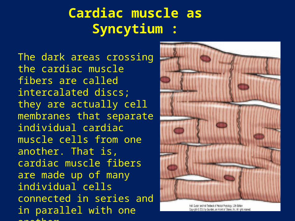
Cardiac muscle as Syncytium :
The dark areas crossing the cardiac muscle fibers are called intercalated discs; they are actually cell membranes that separate individual cardiac muscle cells from one another. That is, cardiac muscle fibers are made up of many individual cells connected in series and in parallel with one another.

- At each intercalated disc the cell membranes fuse together by permeable "communicating" junctions (gap junctions) that allow rapid diffusion of ions. so that action potentials travel easily from one cardiac muscle cell to the next, past the intercalated discs.
- Thus, cardiac muscle is a syncytium of many heart muscle cells in which the cardiac cells are so interconnected that when one of these cells becomes excited, the action potential spreads to all of them.

-The heart actually is composed of two syncytiums: 1- The atrial syncytium, which constitutes the walls of the two atria. 2- The ventricular syncytium, which constitutes the walls of the two ventricles.
•The atria are separated from the ventricles by fibrous tissue that surrounds the atrioventricular (A-V) valvular openings between the atria and ventricles.•Normally, potentials are not conducted from the atrial syncytium into the ventricular syncytium directly through this fibrous tissue. Instead, they are conducted only by the A-V bundle.
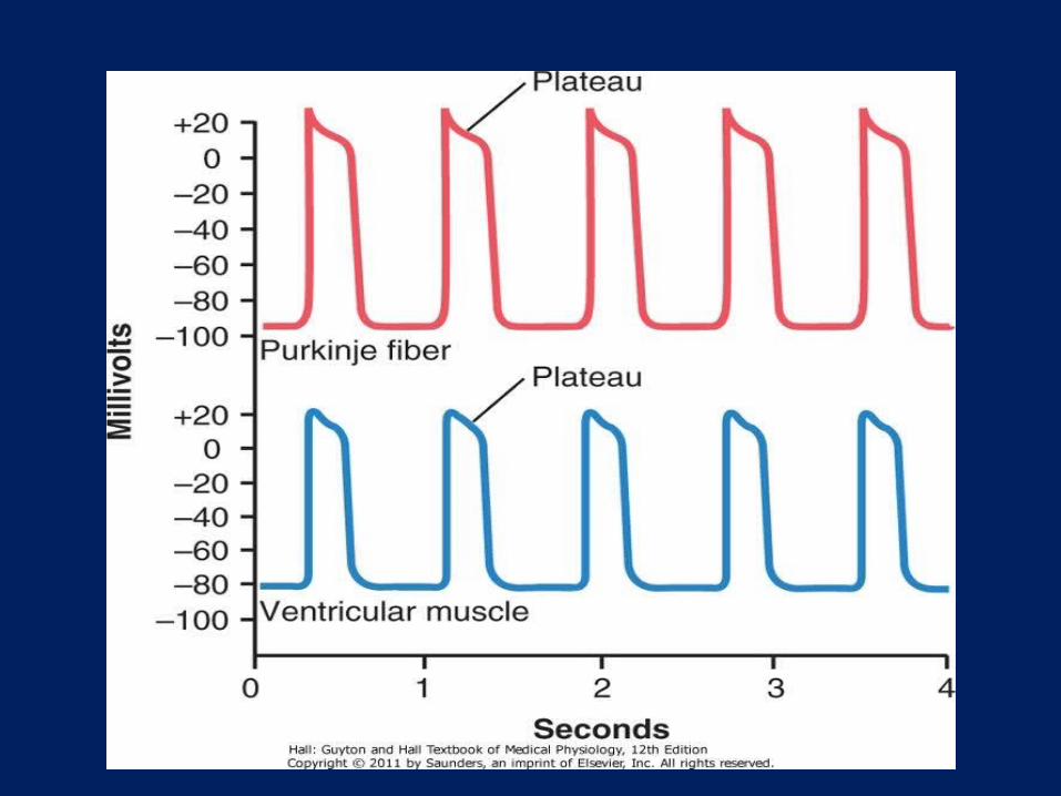

Action potential in cardiac muscle:-The action potential recorded in a ventricular muscle fiber averages about 105 millivolts, which means that the intracellular potential rises from a very negative value, about -85 millivolts, between beats to a slightly positive value, about +20 millivolts, during each beat.-After the initial spike, the membrane remains depolarized for about 0.2 second, exhibiting a plateau , followed at the end of the plateau by abrupt repolarization .-The presence of this plateau in the action potential causes ventricular contraction to last as much as 15 times as long in cardiac muscle as in skeletal muscle.

What causes the long Action Potential and the plateau in cardiac
muscle ?
At least two major differences between the membrane properties of cardiac and skeletal muscle account for this :

** First :the action potential of skeletal muscle is caused almost entirely by sudden opening of large numbers of so-called fast sodium channels .They remain open for only a few thousandths of a second and then abruptly close. At the end of this closure, repolarization occurs, and the action potential is over within another thousandth of a second or so.
In cardiac muscle, the action potential is caused by opening of two types of channels: (1) the same fast sodium channels as those in skeletal muscle and (2) slow calcium channels, which are also called calcium-sodium channels. they are slower to open and remain open for several tenths of a second. During this time, a large quantity of both calcium and sodium ions flows through these channels to the interior of the cardiac muscle fiber, and this maintains a prolonged period of depolarization, causing the plateau in the action potential. Further, the calcium ions that enter during this plateau phase activate the muscle contractile process, while the calcium ions that cause skeletal muscle contraction are derived from the intracellular sarcoplasmic reticulum.
**Second :-Immediately after the onset of the action potential, the permeability of the cardiac muscle membrane for potassium ions decreases about fivefold,(this does not occur in skeletal muscle), and may result from the excess calcium influx through the calcium channels .- Regardless of the cause, the decreased potassium permeability greatly decreases the outflux of positively charged potassium ions during the action potential plateau and thereby prevents early return of the action potential voltage to its resting level. When the slow calcium-sodium channels do close at the end of 0.2 to 0.3 second and the influx of calcium and sodium ions ceases, the membrane permeability for potassium ions also increases rapidly - This rapid loss of potassium from the fiber immediately returns the membrane potential to its resting level, thus ending the action potential.

Velocity of signal conduction in cardiac muscle
• The velocity of conduction of the excitatory action potential signal along both atrial and ventricular muscle fibers is about 0.3 to 0.5 m/sec, or about 1/250 the velocity in very large nerve fibers and about 1/10 the velocity in skeletal muscle fibers.
• The velocity of conduction in the specialized heart conductive system-in the Purkinje fibers-is as great as 4 m/sec in most parts of the system, which allows reasonably rapid conduction of the excitatory signal to the different parts of the heart.
Refractory period of cardiac muscle:
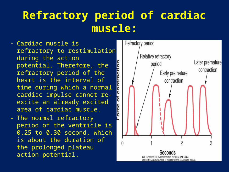
- Cardiac muscle is refractory to restimulation during the action potential. Therefore, the refractory period of the heart is the interval of time during which a normal cardiac impulse cannot re-excite an already excited area of cardiac muscle.
- The normal refractory period of the ventricle is 0.25 to 0.30 second, which is about the duration of the prolonged plateau action potential.

• There is an additional (relative refractory period) of about 0.05 second during which the muscle is more difficult than normal to excite but nevertheless can be excited by a very strong excitatory signal, as demonstrated by the early "premature" contraction in the second example of the figure.
• The refractory period of atrial muscle is much shorter than that for the ventricles (about 0.15 second for the atria compared with 0.25 to 0.30 second for the ventricles).

Excitation-Contraction Coupling-Function of Calcium Ions and the Transverse Tubules:

• The term "excitation-contraction coupling" refers to the mechanism by which the action potential causes the myofibrils of muscle to contract.
• When an action potential passes over the cardiac muscle membrane, the action potential spreads to the interior of the cardiac muscle fiber along the membranes of the transverse (T) tubules.
• The T tubule action potentials in turn act on the membranes of the longitudinal sarcoplasmic tubules to cause release of calcium ions into the muscle sarcoplasm from the sarcoplasmic reticulum. In another few thousandths of a second, these calcium ions diffuse into the myofibrils and catalyze the chemical reactions that promote sliding of the actin and myosin filaments along one another; this produces the muscle contraction.

- In addition to the calcium ions that are released into the sarcoplasm from the cisternae of the sarcoplasmic reticulum, calcium ions also diffuse into the sarcoplasm from the T tubules themselves.- without these extra calcium, the strength of the cardiac muscle contraction would be reduced.- The strength of contraction of cardiac muscle depends to a great extent on the concentration of calcium ions in the extracellular fluids.- At the end of the plateau of the cardiac action potential, the influx of calcium ions to the interior of the muscle fiber is suddenly cut off, and the calcium ions in the sarcoplasm are rapidly pumped back out of the muscle fibers into both the sarcoplasmic reticulum and the T tubule-extracellular fluid space.

-Transport of calcium back into the sarcoplasmic reticulum is achieved with the help of a calcium- ATPase pump.
-Calcium ions are also removed from the cell by a sodium-calcium exchanger.
-The sodium that enters the cell during this exchange is then transported out of the cell by the sodium-potassium ATPase pump.
- As a result, the contraction ceases until a new action potential comes along.

Duration of contraction:
• Cardiac muscle begins to contract a few milliseconds after the action potential begins and continues to contract until a few milliseconds after the action potential ends.
• Therefore, the duration of contraction of cardiac muscle is mainly a function of the duration of the action potential, including the plateau- about 0.2 second in atrial muscle and 0.3 second in ventricular muscle.

Conduction velocity:
• Reflects the time required for the excitation to spread throughout the cardiac tissue.
• Depends on the size of the inward current during the upstroke of the action potential. The larger the inward current , the higher the conduction velocity.
• Is fastest in Perkinje system.• Is slowest in AV node ( seen as PR interval on the ECG),
allowing time for ventricular filling before ventricular contraction. If conduction velocity through the AV node is increased , ventricular filling maybe compromised.

Summery of action potential :
- The resting membrane potential is determined by the conductance to K + and approaches the K+ equilibrium potential.
- Inward current brings +ve charge into the celland depolarize the membrane potential.
- Outward current takes positive charge out of the cell and hyperpolarizes the membrane potential.
- The role of Na+ , K+-adenosine triphosphatase(ATPase) is to maintain ionic gradients across cell membrane.
** ventricles, Atria, and the purkinje sys.:- Have stable resting membrane potentials of about -90 mV .
This value approaches the K+ equilibrium potential.- Action potentials are of long duration, esp in purkinje fibers,
where they last 300 msec.

A- phase 0 : Is the upstroke of action potential. Is caused by transient increase in Na+ conductance ( this increase results in an inward Na+ current that depolarizes the memb.) . At the peak of the action potential the membrane potential approaches the Na+ equilibrium potentail.
B- phase 1 : Is a brief period of initial repolarization. Initial repolarization is caused by an outward current, in part because of movement of K+ ions(favored by both chemical and electrical gradients) out of the cell and in part because of a decrease in Na+ conductance.
C- phase 2 : Is the plateau. Is increased by transient increase in Ca+ conductance which results in an inward Ca+ current and by increase in K+ conductance. Outward and inward currents are approximately equal so the membrane potential is stable at the plateau level.
D- phase 3 : Is repolarization. Here , Ca+ conductance decreases and K+ conductance increases and therefore predominates. The high K+ conductance results in a large outward K+ current which hyperpolarize the membrane back toward the K+ equilibrium potential.
E- phase 4: Is the resting membrane potential. Is a period during which inward and outward current are equal and the membrane potential approaches the K+ equilibrium potential.

** sinoatrial node ( SA node) :-Is normally the pacemaker of the heart.- has an unstable resting potential.- exhibits phase 4 depolarization or automaticity.- the AV node and the His-Purkinje systems are latent pacemakers that may exhibit automaticity and override the SA node if it is suppressed.- the interinsic rate of phase 4 depolarization ( and heart rate ) is fastest in the SA node and slowest in the His-Purkijne system.
** AV node:Upstroke of the action potential in the AV node is the result of an inward Ca+ current ( as in the SA node )
The normal electrocardiogram( ECG )

What is ECG ?
When the cardiac impulse passes through the heart, electrical current also spreads from the heart into the adjacent tissues . A small portion of the current spreads all the way to the surface of the body. If electrodes are placed on the skin on opposite sides of the heart, electrical potentials generated by the current can be recorded; the recording is known as an electrocardiogram.
The normal characteristics of the ECG
• The normal ECG is composed of a P wave, a QRS complex, and a T wave. The QRS complex is often, but not always, three separate waves: the Q wave, the R wave, and the S wave.

• The P wave is caused by electrical potentials generated when the atria depolarize before atrial contraction begins.
• The QRS complex is caused by potentials generated when the ventricles depolarize before contraction, that is, as the depolarization wave spreads through the ventricles.
• Therefore, both the P wave and the components of the QRS complex are depolarization waves

PR interval :- Is the interval between the beginning of the P wave and the beginning of the Q wave ( the initial depolarization of the ventricles).-It varies according to the conduction velocity through the atrioventricular node (AV node ), e.g if AV nodal conduction decreases , the PR interval increases.

-The T wave is caused by potentials generated as the ventricles recover from the state of depolarization. This process normally occurs in ventricular muscle 0.25 to 0.35 second after depolarization, and the T wave is known as a repolarization wave.
Thus, the ECG is composed of both depolarization and repolarization waves.

Recording depolarization wave and repolarization wave from a cardiac
muscle fiber :

Relationship of Atrial and Ventricular Contraction to the Waves of the ECG
• Before contraction of muscle can occur, depolarization must spread through the muscle to initiate the chemical processes of contraction.
• the P wave occurs at the beginning of contraction of the atria, and the QRS complex of waves occurs at the beginning of contraction of the ventricles. The ventricles remain contracted until after repolarization has occurred, that is, until after the end of the T wave.

- The atria repolarize about 0.15 to 0.20 second after termination of the P wave. This is also approximately when the QRS complex is being recorded in the ECG.
- Therefore, the atrial repolarization wave, known as the atrial T wave, is usually obscured by the much larger QRS complex.For this reason, an atrial T wave seldom is observed in the ECG
** The ventricular repolarization wave is the T wave of the normal ECG.
-Ordinarily, ventricular muscle begins to repolarize in some fibers about 0.20 second after the beginning of the depolarization wave (the QRS complex), but in many other fibers, it takes as long as 0.35 second.
- Thus, the process of ventricular repolarization extends over a long period, about 0.15 second. For this reason, the T wave in the normal ECG is a prolonged wave, but the voltage of the T wave is considerably less than the voltage of the QRS complex, partly because of its prolonged length.

Rate of Heartbeat as Determined from the Electrocardiogram
- The rate of heartbeat can be determined easily from an electrocardiogram because the heart rate is the reciprocal of the time interval between two successive heartbeats. If the interval between two beats as determined from the time calibration lines is 1 second, the heart rate is 60 beats per minute. The normal interval between two successive QRS complexes in the adult person is about 0.83 second. This is a heart rate of 60/0.83 times per minute, or 72 beats per minute.
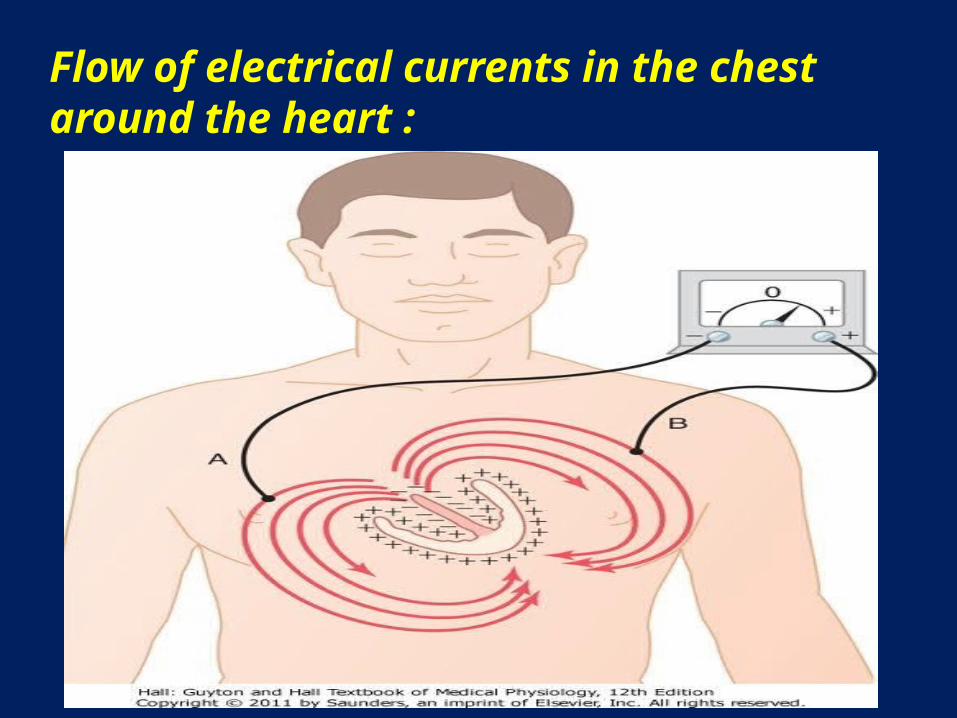
Flow of electrical currents in the chest around the heart :

- The cardiac impulse first arrives in the ventricles in the septum and shortly thereafter spreads to the inside surfaces of the remainder of the ventricles, as shown by the red areas and the negative signs.- This provides electronegativity on the insides of the ventricles and electropositivity on the outer walls of the ventricles, with electrical current flowing through the fluids surrounding the ventricles along elliptical paths, as demonstrated by the curving arrows in the figure.- If one algebraically averages all the lines of current flow (the elliptical lines), one finds that the average current flow occurs with negativity toward the base of the heart and with positivity toward the apex.

ECG Leads

-Three bipolar limb leads- chest leads ( precordial leads )- augmented unipolar limb leads
** Einthoven’s triangle.

Three bipolar Limb Leads- The figure shows electrical connections between the
patient's limbs and the electrocardiograph for recording ECG from the so-called standard bipolar limb leads.
- The term "bipolar" means that the ECG is recorded from two electrodes located on different sides of the heart-in this case, on the limbs. Thus, a "lead" is not a single wire connecting from the body but a combination of two wires and their electrodes to make a complete circuit between the body and the electrocardiograph.

Lead I
- In recording limb lead I, the negative terminal of the electrocardiograph is connected to the right arm and the positive terminal to the left arm.- Therefore, when the point where the right arm connects to the chest is electronegative with respect to the point where the left arm connects, the electrocardiograph records positively, that is, above the zero voltage line in the electrocardiogram.- When the opposite is true, the electrocardiograph records below the line.

Lead II
To record limb lead II, the negative terminal of the electrocardiograph is connected to the right arm and the positive terminal to the left leg. Therefore, when the right arm is negative with respect to the left leg, the electrocardiograph records positively.
Lead III
To record limb lead III, the negative terminal of the electrocardiograph is connected to the left arm and the positive terminal to the left leg. This means that the electrocardiograph records positively when the left arm is negative with respect to the left leg.

** Einthoven's Triangle :the triangleis drawn around the area of the heart. This illustrates that the two arms and the left leg form apices of a triangle surrounding the heart.** Einthoven's Law : Einthoven's law states that if the electrical potentials of any two of the three bipolar limb electrocardiographic leads are known at any given instant, the third one can be determined mathematically by simply summing the first two. Note, however, that the positive and negative signs of the different leads must be observed when making this summation.
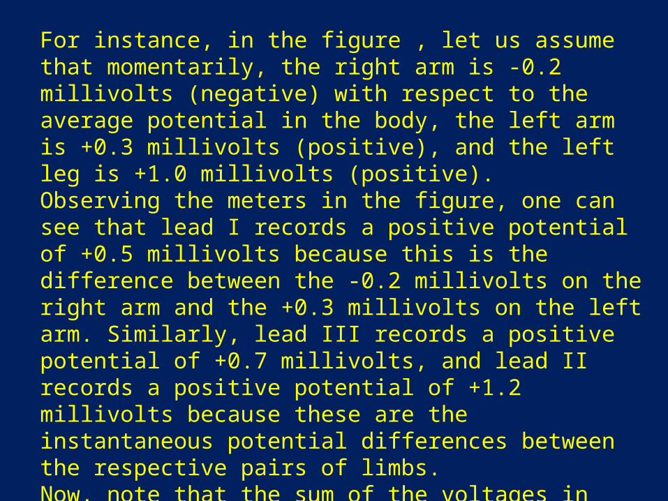
For instance, in the figure , let us assume that momentarily, the right arm is -0.2 millivolts (negative) with respect to the average potential in the body, the left arm is +0.3 millivolts (positive), and the left leg is +1.0 millivolts (positive).Observing the meters in the figure, one can see that lead I records a positive potential of +0.5 millivolts because this is the difference between the -0.2 millivolts on the right arm and the +0.3 millivolts on the left arm. Similarly, lead III records a positive potential of +0.7 millivolts, and lead II records a positive potential of +1.2 millivolts because these are the instantaneous potential differences between the respective pairs of limbs.Now, note that the sum of the voltages in leads I and III equals the voltage in lead II; that is, 0.5 plus 0.7 equals 1.2. Mathematically, this principle, called Einthoven's law, holds true at any given instant while the three "standard" bipolar ECG are being recorded.

Chest Leads (Precordial Leads)Often ECGs are recorded with one electrode placed on the anterior surface of the chest directly over the heart . This electrode is connected to the positive terminal of the ECG, and the negative electrode, called the indifferent electrode, is connected through equal electrical resistances to the right arm, left arm, and left leg all at the same time, Usually six standard chest leads are recorded, one at a time, from the anterior chest wall, the chest electrode being placed sequentially at the six points shown here . The different recordings are known as leads V1, V2, V3, V4, V5, and V6.

- Because the heart surfaces are close to the chest wall , any minute abnormalities in the ventricles, particularly in the anterior ventricular wall, can cause marked changes in the ECGs recorded from chest leads.- In leads V1 and V2, the QRS recordings of the normal heart are mainly negative because, as shown in the figure, the chest electrode in these leads is nearer to the base of the heart than to the apex, and the base of the heart is the direction of electronegativity during most of the ventricular depolarization process. Conversely, the QRS complexes in leads V4, V5, and V6 are mainly positive because the chest electrode in these leads is nearer the heart apex, which is the direction of electropositivity during most of depolarization.

Augmented Unipolar Limb Leads:
In this type of recording, two of the limbs are connected through electrical resistances to the negative terminal of the electrocardiograph, and the third limb is connected to the positive terminal. When the positive terminal is on the right arm, the lead is known as the aVR lead; when on the left arm, the aVL lead; and when on the left leg, the aVF lead.

Nerveous regulation of the circulation , and rapid control of the arterial
pressure

** The nervous system controls the circulation almost entirely through the autonomic nervous system.
Autonomic Nervous System :By far the most important part of the autonomic nervous system for regulating the circulation is the sympathetic nervous system. The parasympathetic nervous system, however, contributes importantly to regulation of heart function.

Sympathetic Nervous System : - Sympathetic vasomotor nerve fibers leave the spinal cord through all the thoracic spinal nerves and through the first one or two lumbar spinal nerves.-They then pass immediately into a sympathetic chain, one of which lies on each side of the vertebral column.- Next, they pass by two routes to the circulation:(1) through specific sympathetic nerves that innervate
mainly the vasculature of the internal viscera and the heart.
(2) almost immediately into peripheral portions of the spinal nerves distributed to the vasculature of the peripheral areas.

Sympathetic Innervation of the Blood Vessels :
- In most tissues all the vessels except the capillaries are innervated.- Precapillary sphincters and metarterioles are innervated in some tissues- The innervation of the small arteries and arterioles allows sympathetic stimulation to increase resistance to blood flow and thereby to decrease rate of blood flow through the tissues.- The innervation of the large vessels, particularly of the veins, makes it possible for sympathetic stimulation to decrease the volume of these vessels. This can push blood into the heart and thereby play a major role in regulation of heart pumping.

Sympathetic Nerve Fibers to the Heart
-Sympathetic fibers also go directly to the heart.
- It should be recalled that sympathetic stimulation markedly increases the activity of the heart, both increasing the heart rate and enhancing its strength and volume of pumping.

Parasympathetic Control of Heart Function, Especially Heart Rate
- It plays only a minor role in regulation of vascular function in most tissues.- Its most important circulatory effect is to control heart rate by way of parasympathetic nerve fibers to the heart in the vagus nerves, from the brain medulla directly to the heart.
- It causes a marked decrease in heart rate and a slight decrease in heart muscle contractility.

Sympathetic Vasoconstrictor System and Its Control by the Central Nervous System :
- The sympathetic nerves carry tremendous numbers of vasoconstrictor nerve fibers and only a few vasodilator fibers.-The vasoconstrictor fibers are distributed to essentially all segments of the circulation, but more to some tissues than others.-This sympathetic vasoconstrictor effect is especially powerful in the kidneys, intestines, spleen, and skin but much less potent in skeletal muscle and the brain.
Vasomotor Center in the Brain and Its Control of the Vasoconstrictor System :
-Located bilaterally mainly in the reticular substance of the medulla and of the lower third of the pons is an area called the vasomotor center.
-This center transmits parasympathetic impulses through the vagus nerves to the heart and transmits sympathetic impulses through the spinal cord and peripheral sympathetic nerves to virtually all arteries, arterioles, and veins of the body.
Continuous Partial Constriction of the Blood Vessels Is Normally Caused by Sympathetic
Vasoconstrictor Tone
- Under normal conditions, the vasoconstrictor area of the vasomotor center transmits signalscontinuously to the sympathetic vasoconstrictor nerve fibers over the entire body, causing slow firing of these fibers at a rate of about one half to two impulses per second.-This continual firing is called sympathetic vasoconstrictor tone.-These impulses normally maintain a partial state of contraction in the blood vessels, called vasomotor tone.

Norepinephrine - The Sympathetic Vasoconstrictor Transmitter
Substance
The substance secreted at the endings of the vasoconstrictor nerves is almost entirely norepinephrine , which acts directly on the alpha adrenergic receptors of the vascular smooth muscle to cause vasoconstriction.

Adrenal Medullae and Their Relation to the Sympathetic Vasoconstrictor System
• Sympathetic impulses are transmitted to the adrenal medullae at the same time that they are transmitted to the blood vessels.
• They cause the medullae to secrete both epinephrine and• norepinephrine.• These two hormones are carried in the blood stream to all
parts of the body, where they act directly on all blood vessels, usually to cause vasoconstriction.
• In a few tissues epinephrine causes vasodilation because it also has a "beta" adrenergic receptor stimulatory effect, which dilates rather than constricts certain vessels.

Emotional Fainting -Vasovagal Syncope
- Is vasodilatory reaction occurs in people who experience intense emotional disturbances that cause fainting.
- In this case, the muscle vasodilator system becomes activated, and at the same time, the vagal cardio-inhibitory center transmits strong signals to the heart to slow the heart rate markedly.
- The arterial pressure falls rapidly, which reduces blood flow to the brain and causes the person to lose consciousness.
- This overall effect is called vasovagal syncope.
- The pathway probably then goes to the vasodilatory center of the anterior hypothalamus next to the vagal centers of the medulla, to the heart through the vagus nerves, and also through the spinal cord to the sympathetic vasodilator nerves of the muscles

Role of the Nervous System in Rapid Control of Arterial Pressure
- One of the most important functions of nervous control of the circulation is its capability to cause rapid increases in arterial pressure.
- For this purpose, the entire vasoconstrictor and cardioaccelerator functions of the sympathetic nervous system are stimulated together.
- At the same time, there is reciprocal inhibition of parasympathetic vagal inhibitory signals to the heart.

-Thus, three major changes occur simultaneously, each of which helps to increase arterial pressure. They are as follows: 1. Most arterioles of the systemic circulation are constricted. This greatly increases the total peripheral resistance, thereby increasing the arterial pressure. 2. The veins are strongly constricted. This displaces blood out of the large peripheral blood vessels toward the heart, thus increasing the volume of blood in the heart chambers. The stretch of the heart then causes the heart to beat with far greater force and therefore to pump increased quantities of blood. This, too, increases the arterial pressure. 3. Finally, the heart itself is directly stimulated by the autonomic nervous system, further enhancing cardiac pumping. In addition, sympathetic nervous signals have a significant direct effect to increase contractile force of the heart muscle, this, too, increasing the capability of the heart to pump larger volumes of blood.

-An especially important characteristic of nervous control of arterial pressure is its rapidity of response, beginning within seconds and often increasing the pressure to two times normal within 5 to 10 seconds.
** Baroreceptor Arterial Pressure Control System-Baroreceptor Reflexes :By far the best known of the nervous mechanisms for arterial pressure control is the baroreceptor reflex. Basically, this reflex is initiated by stretch receptors, called either baroreceptors or pressoreceptors, located at specific points in the walls of several large systemic arteries. A rise in arterial pressure stretches the baroreceptors and causes them to transmit signals into the central nervous system. "Feedback" signals are then sent back through the autonomic nervous system to the circulation to reduce arterial pressure downward toward the normal level

Summery of the autonomic effects on the heart and the vessels :
-sympathetic sys will increase the heart rate , the conduction velocity ( AV node ) , and the contractility , all by B 1 recepter.
- parasympathetic sys will decrease the heart rate , the conduction velocity ( AV node ) , and the contractility of atria only , all by muscarinic recepter.

Thank you