invited review comparative cardiovascular physiology...
TRANSCRIPT
INVITED REVIEW
Comparative cardiovascular physiology: future trends,
opportunities and challenges
W. W. Burggren,1 V. M. Christoffels,2 D. A. Crossley II,1 S. Enok,3 A. P. Farrell,4
M. S. Hedrick,1 J. W. Hicks,5 B. Jensen,2,3 A. F. M. Moorman,2 C. A. Mueller,1 N. Skovgaard,3
E. W. Taylor6 and T. Wang3
1 Developmental Integrative Biology Cluster, Department of Biological Sciences, University of North Texas, Denton, TX, USA
2 Department of Anatomy, Embryology & Physiology, Academic Medical Centre, Amsterdam, The Netherlands
3 Zoophysiology, Department of Bioscience, Aarhus University, Aarhus, Denmark
4 Department of Zoology and Faculty of Land and Food Systems, University of British Columbia, Vancouver, BC, Canada
5 Department of Ecology and Evolutionary Biology, University of California, Irvine, CA, USA
6 School of Biosciences, University of Birmingham, Birmingham, UK
Received 13 June 2013,
revision requested 16 July 2013,
revision received 27 August 2013,
accepted 12 September 2013
Correspondence: W. Burggren,
Department of Biological Sciences,
University of North Texas, 1155
Union Circle #311190, Denton,
TX 76203-5015, USA.
E-mail: [email protected]
Abstract
The inaugural Kjell Johansen Lecture in the Zoophysiology Department of
Aarhus University (Aarhus, Denmark) afforded the opportunity for a
focused workshop comprising comparative cardiovascular physiologists to
ponder some of the key unanswered questions in the field. Discussions were
centred around three themes. The first considered function of the vertebrate
heart in its various forms in extant vertebrates, with particular focus on the
role of intracardiac shunts, the trabecular (‘spongy’) nature of the ventricle
in many vertebrates, coronary blood supply and the building plan of the
heart as revealed by molecular approaches. The second theme involved the
key unanswered questions in the control of the cardiovascular system,
emphasizing autonomic control, hypoxic vasoconstriction and developmen-
tal plasticity in cardiovascular control. The final theme involved poorly
understood aspects of the interaction of the cardiovascular system with the
lymphatic, renal and digestive systems. Having posed key questions around
these three themes, it is increasingly clear that an abundance of new analyti-
cal tools and approaches will allow us to learn much about vertebrate
cardiovascular systems in the coming years.
Keywords cardiovascular, comparative physiology, heart.
The legacy of Kjell Johansen
On 15 March 2012, the Zoophysiology Department of
Aarhus University, Denmark, held the inaugural Kjell
Johansen Lecture, the first in an annual physiology lec-
ture series established to commemorate Professor Kjell
Johansen’s seminal contributions to comparative physi-
ology, and to bring comparative physiologists to Aar-
hus to discuss current trends and future challenges. The
inaugural lecture, held almost exactly 25 years after
Kjell Johansen’s death, was presented by Professor
Warren Burggren. The lecture provided a personal
account of Kjell Johansen’s career, from his graduate
studies at the University of Oslo, through his faculty
position at the University of Washington, Seattle
(USA), before he was called to Aarhus University where
he created the Department of Zoophysiology and pre-
sided as head of department until his untimely and tra-
gic death in 1987 (Linzen 1987).
Among Kjell Johansen’s many remarkable attributes
was his legendary ability to generate complex ideas
and hypotheses in rapid-fire fashion. Many who
© 2013 Scandinavian Physiological Society. Published by John Wiley & Sons Ltd, doi: 10.1111/apha.12170 1
Acta Physiol 2013
worked with him recall discussions that left the dis-
cussant with an overwhelming sense of how little we
know (and especially how little the discussant knew!)and how much experimentation and interpretation
there was yet to be done. The senior author recalls
how one of Kjell Johansen’s long-standing ‘pet pro-
jects’ suddenly appeared in a journal, published by
another research group. While a lesser scientist might
have shown distress at being ‘scooped’, Kjell Johansen
instead looked almost delighted and commented
‘Good! I now know the answer, and that is one less
experiment I have to do!’ Kjell Johansen was intent
on sharing his ideas, seeding comparative physiology
much like a farmer seeds a field, hoping and anticipat-
ing that new ideas would rise from the fertile minds
with which he surrounded himself.
Here, we review current concepts regarding the evo-
lution of the vertebrate heart and pose a series of key
questions that we feel are ripe for study. These ideas
and questions were discussed during the course of a
focused workshop immediately after the Kjell Johan-
sen Lecture. In the spirit of Kjell Johansen, we hope
that the ideas and proposed experiments will result in
fewer experiments we have to do ourselves.
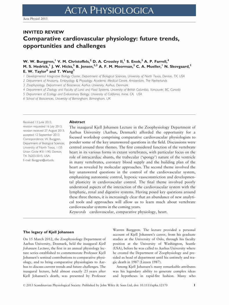
Strategies to study cardiovascular evolution
A variety of investigative strategies at all levels of bio-
logical organization, in conjunction with a compara-
tive and evolutionary approach, are required to
understand the major evolutionary transitions of the
vertebrate heart. Such approaches – some time-hon-
oured, others new – are outlined below and repre-
sented in Figure 1.
Phylogenetic approaches
A comparative analysis of the cardiovascular charac-
ters can when placed in a phylogenetic context – be
used to identify when a given character evolved. Addi-
tionally, the fossil record and other palaeontological
evidence can provide insight into cardiovascular struc-
ture through speculation on heart–head distances, etc.
(although direct fossil evidence of cardiovascular
structure in dinosaurs is lacking – see Cleland et al.
2011). From these, it is possible to construct likely
evolutionary scenarios to explain links between physi-
ological or behavioural traits.
Developmental physiology
Natural selection drives evolutionary change in mor-
phological structures and physiological processes in
developing organisms, as in adults (Burggren & War-
burton 1994). Consequently, as Burggren (1992)
notes, ‘Developmental studies of physiological traits…
should be included wherever possible in studies of
physiological adaptation’. The most comprehensive
understanding of the evolution of physiological pro-
cess will thus come from a study of the evolutionary
change in the entire life cycle of animal lineages, not
just by studying the adults (Burggren 1991).
Figure 1 Interdisciplinary strategies for investigating cardiovascular function. Embryonic turtle is modified from Agassiz
(1857); oxygen transport model is modified from Wang & Hicks (2002); proximal function original drawing by J.W. Hicks,
schematic heart is modified from Eme et al. (2010) and the molecular/genetic heart is modified from Zina Deretsky, National
Science Foundation after Benoit Bruneau, the Gladstone Institute of Cardiovascular Disease.
© 2013 Scandinavian Physiological Society. Published by John Wiley & Sons Ltd, doi: 10.1111/apha.121702
Trends in cardiovascular physiology · W W Burggren et al. Acta Physiol 2013

Mathematical modelling
The vertebrate oxygen transport system is accurately
described by a series of mass transfer equations as
well as hemodynamic relationships. The resulting
mathematical models are instructive for quantitative
evaluations of the functional significance of particular
cardiovascular traits.
Proximal function
Most physiological studies have largely been restricted
to the laboratory, where complex multifactorial, envi-
ronmental stressors are difficult to accurately repro-
duce, and thus the functional benefits of given traits
are difficult to quantify. The recent advent of minia-
turized electronics and computer-assisted data acquisi-
tion (biologging; telemetry) provides the potential to
correlate cardiovascular performance with natural
feeding, physical activity, basking, diving, etc. in unre-
strained and even free-ranging animals.
Phenotypic manipulation
By surgical or pharmacological manipulations of given
cardiovascular traits, it is possible to quantify how rel-
evant performance variables are affected and hence
provide insight into the functional significance of the
trait (Sinervo & Basolo 1996). A challenge of such
studies is to perform the phenotypic manipulation and
then expose the animal to relevant environmental con-
ditions. This approach benefits from being combined
with field measurements.
Molecular genetics
It is now clear that cardiac development is coordinated
by a set of very similar gene regulatory networks in all
vertebrates, and the common developmental patterns
will allow for an understanding of how given traits,
such as the conduction system, the left atrium or the
ventricular septum, appeared in specific lineages
(Moorman & Christoffels 2003, Olson 2006, Koshiba-
Takeuchi et al. 2009, Jensen et al. 2012). As the
genomes of more and more species are being sequenced
(e.g. Shaffer et al. 2013), it will be much easier to
design probes for in situ hybridization and generate
antibodies for expression and molecular studies.
Form and function of vertebrate hearts
The hearts of the earliest chordates were, in all likeli-
hood, merely contractile vessels where peristaltic move-
ments, initiated by simple pacemakers, accounted for
the propulsion of blood through a vascular tree with
minimal endothelial function (see Burggren & Johansen
1986, Burggren & Reiber 2007, Xavier-Neto et al.
2010, Farmer 2011 for reviews). The cardiovascular
systems of vertebrates therefore have undergone
marked anatomical and functional changes with forma-
tion of proper cardiac chambers, evolution of a pulmo-
nary circulation and, finally, the emergence of the four-
chambered heart among the endothermic vertebrates.
The basic heart plan
In all of the ectothermic vertebrates (hagfish through
fishes, amphibians and reptiles), the cardiac ventricle is
a trabecular, sponge-like chamber. This contrasts with
the single-lumen ventricles composed of compact walls
of the endothermic mammals and birds. It is tempting
to correlate this transition in ventricular anatomy with
the rise in cardiac output and blood pressures that are
characteristics of endothermic birds and mammals, but
the advantages of single-lumen ventricles with compact
walls are not obvious. For example, ventricular ejection
fraction of mammals and birds is approximately 50%,
whereas the typical ejection fraction of the trabecular
ectothermic ventricles is close to 100% (Franklin &
Davie 1992, Burggren et al. 1997). Because the shorten-
ing of each cardiomyocyte is around 20% in all verte-
brates (e.g. Shiels & White 2008), it is possible that the
architectural arrangement of the cardiomyocytes within
a trabecular ventricle allows for the higher ejection frac-
tion because most blood resides in the many miniscule
cavities of the trabecular walls, allowing for a more effi-
cient contraction according to the law of Laplace
(Johansen & Burggren 1980, Van Mierop & Kutsche
1985).
Some ectotherms such as tunas, varanid lizards,
pythons and crocodiles have high mammalian-like
blood pressures (70–100 mmHg; Burggren & Johan-
sen 1982, Jones et al. 1993, Hicks 1998, Brill &
Bushnell 2001, Wang et al. 2003), and their stroke
volume resembles that of mammals (around
1 mL kg�1; Farrell 1991, 1996, Korsmeyer et al.
1997, Hicks et al. 2000, Secor et al. 2000, Seymour
& Blaylock 2000, Brill & Bushnell 2001, Clark et al.
2005). These high-performance ectotherms have
retained a trabecular layer within the ventricle, but
their maximal heart rates are considerably lower than
similar-sized mammals (rarely above 120 min�1; e.g.
Wang et al. 1997, Lillywhite et al. 1999, Hicks et al.
2000, Brill & Bushnell 2001, Clark et al. 2005). A
key, unanswered question, then, is:
Did the evolution of high heart rates driven by the high
endothermic metabolism favour a compact ventricle?
A scenario for the rise in cardiac output by elevated
heart rate to accommodate the high endothermic
© 2013 Scandinavian Physiological Society. Published by John Wiley & Sons Ltd, doi: 10.1111/apha.12170 3
Acta Physiol 2013 W W Burggren et al. · Trends in cardiovascular physiology

metabolism is illustrated in Figure 2. The tachycardia
shortens the time available for filling and emptying,
and we hypothesize that the transition from a trabecu-
lar to a single-lumen compact-walled ventricle yields a
lower viscous resistance to inflows and outflows of the
ventricle. Supporting this conjecture, the human and
murine pathological condition of non-compaction
with heavily trabeculated ventricles often leads to
heart failure (e.g. Dyson et al. 1995, Freedom et al.
2005, Engberding et al. 2007). At the same time, the
elevation of cardiac output, needed to sustain the rise
in metabolism, was associated with a rise in blood
pressure, adding additional work on the heart and
requiring a thicker ventricular wall.
Figure 2 also includes a number of other changes in
cardiac structure and physiology resulting from the
rise in heart rate, including the need for a specialized
cardiac conduction system to ensure fast and coordi-
nated contractions of the cardiac chambers (Jensen
et al. 2012, 2013a), improved calcium handling by
the sarcoplasmic reticulum to ensure swift activation
and deactivation of the contractile apparatus (Galli &
Shiels 2012), increased coronary supply for the thicker
and more compact myocardium, and finally a fully
divided ventricle.
The evolutionary scenario outlined above is not
readily investigated through testable hypotheses, but a
detailed comparison of cardiac function and anatomy
in birds and mammals with their reptilian ancestors
would provide evidence for convergent evolution of
functional solutions resulting in both higher heart rate
and blood pressure.
The four-chambered heart and the functional role of
cardiac shunts
The formation of the four-chambered heart with a fully
divided ventricle appeared at least twice independently:
Transition from trabeculateto compact myocardium
Atrial filling of the ventricle
Coronarysupply
Ventriclularmass
Elimination ofCardiac shunts
Development of specialised
conduction system
Dependence on sarcoplasmic reticulum for Ca2+ handling
Ejectionfraction
Cardiac output
Mean arterial pressure
Filling time
Ejection time
Resistanceto flow
Myocardial oxygen consumption
Oxygen consumption
Heart rate Stroke volume
Cardiac work
Figure 2 The complex inter-relationships of factors determining cardiac function and their potential relationship to the evolu-
tion of a greater dependence on compact rather than trabecular myocardium.
© 2013 Scandinavian Physiological Society. Published by John Wiley & Sons Ltd, doi: 10.1111/apha.121704
Trends in cardiovascular physiology · W W Burggren et al. Acta Physiol 2013

once in the vertebrate group that gave rise to crocodil-
ians and birds (archosaurs) and once in the ancestral
group of mammals (synapsids) (Goodrich 1930, Jensen
et al. 2010b, 2014). The fully divided ventricle allows
for high systemic arterial blood pressures, while keep-
ing low blood pressures within the pulmonary circula-
tion, thus allowing for a thinner blood–gas barrier (e.g.
West 2009). A fully divided ventricle also avoids
admixture of oxygen-rich and oxygen-poor blood
within the heart and hence provides for a more effec-
tive oxygen transport cascade. Collectively, these char-
acteristics are necessary to support the increased
metabolism associated with endothermy that appears
directly linked with the rapid expansion and success of
mammals and birds (Burggren et al. 1997, Hicks &
Wang 2012).
Although cardiac shunts in reptiles and amphibians
reduce arterial oxygen saturation, the mixing of oxy-
gen-rich and oxygen-poor blood is a normal, func-
tional characteristic of their hearts (see Johansen &
Burggren 1985, Burggren & Bemis 1990, Farmer
1999, Hicks & Wang 2012), and it is relevant to ask
whether there are distinct benefits to such an arrange-
ment. Such shunting may allow for putative energy
savings by reducing overall cardiac work and may
enhance ventricular oxygenation in species lacking a
coronary circulation. Yet, in adult birds and mam-
mals, cardiac shunts that result from congenital car-
diac defects are invariably detrimental and reduce
performance. Given that cardiac shunting has been
regarded as beneficial in ectothermic vertebrates, but
detrimental in endothermic vertebrates, a major ques-
tion in comparative cardiovascular physiology is:
Are cardiac shunts an adaptive trait – that is, a trait that
conveys unique physiological advantages to ectothermic
vertebrates?
Despite more than a century of anatomical and
physiological studies, there is no convincing evidence
that the lack of the ability to shunt blood reduces
physiological performance and/or reproductive fitness
(see Wang et al. 1998, Farmer et al. 2008, Hicks &
Wang 2012). Thus, it is equally plausible that the
unique features of the reptilian heart are embryonic or
ancestral characters that, with no negative impacts on
overall animal fitness, have simply not been selected
against. Quantifying fitness in reptiles or any of the
other vertebrates with cardiac shunts is very difficult.
Phylogenetic studies often require broad surveys of
closely related species and given the substantial techni-
cal demands required to quantify cardiac shunts in a
single species, such an approach would seem implausi-
ble. As an alternative, morphological studies on the
ventricular anatomy may predict the ability of a given
species to alter their cardiac shunts. Specifically, the
size of the muscular ridge, the partial ventricular sep-
tum that separates the pulmonary and systemic cava
in the ventricle, may be an important indicator of the
capacity for cardiac shunting in reptiles. In some rep-
tiles, the muscular ridge is relatively small and not
well developed (Hicks 1998) and the ventricle func-
tions as a single pressure pump during the entire
cardiac cycle (Burggren 1986). Under these conditions,
the size and direction of the cardiac shunts are influ-
enced by changes in pulmonary and systemic vascular
resistances (Hicks 1998, Wang & Hicks, 2002). In
some reptiles (varanids and pythons), the muscular
ridge is enlarged, resulting in a dual pressure pump
during systole (Burggren, 1986; Hicks 1998, Jensen
et al. 2010b,c). In these animals, mixing of deoxygen-
ated and oxygenated blood can occur during diastole.
Recently, Jensen et al. (2014) standardized morpho-
logical procedures to quantitate the muscular ridge
and ventricular cava. By applying such techniques to a
number of vertebrate species with different ventricular
anatomies, it will be possible to target physiological
experimentation in particular species of interest.
There have been few studies on cardiac function
during development in reptiles (Eme et al. 2011a,b,c).
A more detailed understanding of cardiac function
during embryonic development may reveal whether
cardiac shunts are critical for normal development. In
addition, an understanding of the developmental ori-
gins of cardiac shunts may also reveal whether cardiac
shunts play different roles (quantitatively or qualita-
tively) in hatchlings, juveniles or adult animals.
The effects of cardiac shunting on oxygen transport
are easily modelled using well-known mass transfer
equations. Such approaches provide testable hypothe-
sis for the role of shunts on VO2 max, blood oxygen
and carbon dioxide regulation, aerobic dive limita-
tions and temperature regulation (Wood & Hicks
1985, White & Hicks 1987, Wang & Hicks 1996,
Wang et al. 1997).
Recent studies on alligators and rattlesnakes have
exploited phenotypic manipulation to investigate the
functional role of right-to-left shunts (Eme et al. 2009,
2010, Leite et al. 2013). These approaches reveal that
eliminating the capacity for R-L shunts in both species
did not influence growth rates relative to normal ani-
mals. Functional studies of animals with cardiac malfor-
mations may provide similar insight. For example,
pythons with septal defects did not appear to behave or
grow in an abnormal fashion (Jensen &Wang 2009).
Coronary circulation and O2 consumption of the
vertebrate heart
Proper cardiac function requires continuous ATP pro-
duction and consequently most vertebrate hearts need
© 2013 Scandinavian Physiological Society. Published by John Wiley & Sons Ltd, doi: 10.1111/apha.12170 5
Acta Physiol 2013 W W Burggren et al. · Trends in cardiovascular physiology

a continuous oxygen supply. Cardiac oxygen supply
and consumption are tightly coupled with cardiac
work, that is, cardiac output and arterial blood pres-
sure (Farrell 1987, 2002, Farrell & Stecyk 2007,
Duncker & Bache 2008). Although there is compre-
hensive understanding of the importance of cardiac
oxygen supply in mammalian and avian hearts (Dunc-
ker & Bache 2008), a key area for future experimen-
tation is to understand the evolution of the various
forms of cardiac oxygen supply among vertebrates
and to correlate these forms to the habitats, behav-
iours and metabolic status of animals.
The array of cardiac designs is remarkably diverse
across vertebrates, but none are as dependent on coro-
nary circulation as adult mammals and birds, where a
coronary circulation supplies a compact wall with its
dense arrangement of cardiomyocytes. This is in con-
trast to the ancestral vertebrate heart (possibly repre-
sented in extant cyclostomes) where the trabecular
arrangement of cardiomyocytes receives no coronary
circulation, and where cardiac oxygen supply is pro-
vided entirely by the oxygen-poor venous blood return-
ing to the heart (Davie & Farrell 1991, Farrell et al.
2012). Certain teleosts and all amphibians also have an
entirely trabecular ventricular myocardium, while
others wrap the trabecular myocardium to varying
degrees with a layer or layers of condensed myocardium
with a distinct coronary circulation (Santer 1985). Even
so, myocardial compaction and associated coronary
vessels apparently appeared at the outset of vertebrate
evolution, because all elasmobranchs studied have a
coronary circulation (Tota 1989, De Andres et al.
1990, 1992, Farrell et al. 2012). Other teleosts, and
perhaps all reptiles, have compact myocardium, but
never as much as mammals (Farrell et al. 2012). Some
teleosts even have coronary vessels in their ventricular
trabeculation, as in elasmobranchs, birds and mam-
mals. Clearly, the phylogeny of the coronary circulation
is far from simple (Fig. 3). The importance of cardiac
work rate and hypoxia in coronary development seems
intuitive, their relative importance has still to be
defined, and the selection pressures that result in the
diversity of coronary arrangements remains unknown.
The phylogenetic and ontogenetic shifts from the
trabecular heart to compact heart are relevant to the
patterns observed in the coronary circulation. A key
question is:
What is the evolutionary advantage of retaining a par-
tially trabecular heart, the ancient, pre-vertebrate solution
to cardiac oxygen supply?
Finding answers to this and other related questions
requires a three-pronged experimental approach. Fore-
most, the diversity of cardiac anatomy must be stud-
ied in a much larger number of species in a wide
phylogenetic context to reveal whether phenotypic
responses relate to natural selective pressures. For
example, we still lack a clear understanding of the
exact distribution of the coronary circulation in elas-
mobranchs, lungfishes and reptiles, and it remains
very uncertain whether the poor coronary supply in
anuran amphibians represents an evolutionary loss.
Future studies should estimate capillary density, a
determinant of oxygen supply, and hence an indirect
proxy for cardiac work.
The second prong of the experimental approach is
to determine how anatomy of the coronary arterial
tree and the venous oxygen supply relate to the envi-
ronmental conditions and behaviours of a given spe-
cies. This will require comprehensive measurements of
relevant routine and maximum physiological variables
including cardiac output, arterial blood pressure, myo-
cardial oxygen consumption and coronary blood flow.
These measurements are currently feasible, but have
been made in relatively few species and concurrently
in even fewer species.
The molecular building plan of the heart and the
evolution of the conduction system
The high heart rates of mammals and birds require an
electrical conduction system to generate and conduct
the electrical impulses that ensure proper activation of
the cardiac chambers. Errors in development or func-
tion of the chambers, septa or conduction system fre-
quently result in congenital defects or arrhythmias in
humans (e.g. Christoffels & Moorman 2009).
Comparative development is instructive because the
profound differences between the fully formed hearts of
ectotherms and endotherms are not evident in their
embryonic stages. For example, the trabecular heart of
ectotherms has a much simpler conduction system than
birds and mammals, although the electrical activation
pattern of the hearts of ectotherms and endotherms is
strikingly similar (see Burggren 1978). Therefore, by
comparing the hearts of endotherms and ectotherms,
the basic building blocks of the conduction system can
be revealed, and cardiac specializations evolved in the
endotherms to support their high metabolic state and
physically separated blood flows can be deduced. Gen-
erally, in vertebrates, an adult type of electrocardio-
gram can be detected in the early stages of chamber
formation, even though a morphologically distinct con-
duction system has not yet developed (Christoffels et al.
2010). This electrical pattern indicates a shared cardiac
building plan among vertebrates.
A major focus in cardiovascular development is to
elucidate the common mechanisms that underlie the
developmental control of the growth and patterning
of the vertebrate heart. Major questions include:
© 2013 Scandinavian Physiological Society. Published by John Wiley & Sons Ltd, doi: 10.1111/apha.121706
Trends in cardiovascular physiology · W W Burggren et al. Acta Physiol 2013

How do the cardiac precursor cells become organized
into an evolutionary conserved cardiac tube?
What controls the formation of cardiac chambers, and
which components are minimally required to establish
the conserved electrical pattern?
Which precursor populations and molecular signalling
systems have been evolved to support the specific features
of the hearts of endotherms?
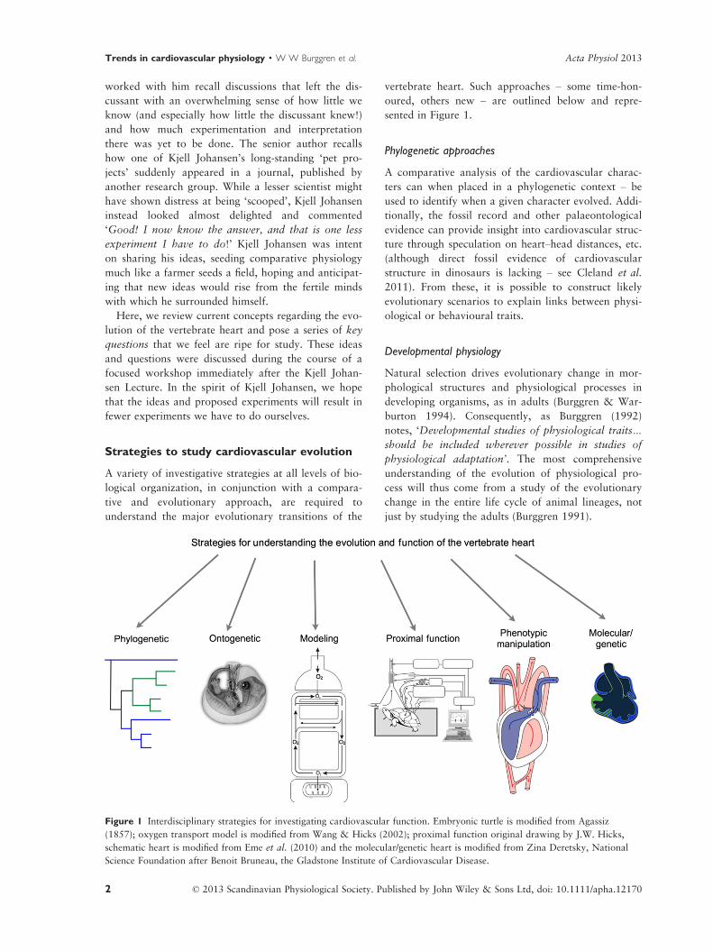
We now know that the primary heart tube evident
in embryonic vertebrates grows by recruitment of pre-
cursor cells. Chamber differentiation and expansion
occurs at specific locations within the tube, which
develop fast depolarization and strongly increased
intercellular conductivity (Fig. 4). This allows the
chambers to rapidly propagate the electrical impulse,
which is seen as the P wave (atrial excitation) and
QRS complex (ventricular excitation) in the electro-
cardiogram (Fig. 4). The sinus venosus, which con-
tains the principal pacemaker in all vertebrate hearts
(Burggren et al. 1997), the atrioventricular canal and
the outflow tract initially do not differentiate into
chamber-type myocardium. Rather, they retain the
slow proliferative and conductive properties found in
the original embryonic heart tube (Christoffels et al.
2010). The delay in impulse propagation in the atrio-
ventricular canal is seen between the P wave and QRS
complex in the electrocardiogram.
Heart development seems to be conserved across
species and is driven by transcription factors including
T-box, homeobox and GATA zinc-finger transcription
factors (Olson 2006). In all species examined to date
(lampreys, fish, frog, birds and mammals), Tbx2 is
Chordate ancestortubular heart
with pacemaker
Cartilagenousfishes
Jawlesschordates
Hagfishes &lampreys
Jawedchordates
Basal boneyfishes
Boneyfishes
Amphibians
Reptiles
Birds &mammals
Lungfishes
Compact myocardium &associated coronary vessels
Outflow vessel
Conal compact myocardiumwrapping the outflow vessel
Trabecular myocardium withoutor with ( ) coronary vessels
Tetrapods
Figure 3 A schematic representation and generalization of the oxygen supply routes with the chordate phylum. The archetype
avascular ventricle that relies on oxygen contained in venous blood is exemplified by cyclostomes. A coronary oxygen supply to
the conal myocardium and a variable amount of compact and trabecular myocardium is seen in the elasmobranchs. Cardiac
oxygen supply is highly variable among teleosts. The exact distribution of the coronary circulation in lungfish is unknown.
Reptiles have a variable proportion of compact myocardium, and again the exact distribution of the coronary capillaries is
unknown. The ventricle of birds and mammals is almost entirely compact myocardium, and the few trabeculations that are pres-
ent contain coronary capillaries. These generalizations for lower vertebrates are based on anatomical studies for a relatively
small number of the over 50 000 species.
© 2013 Scandinavian Physiological Society. Published by John Wiley & Sons Ltd, doi: 10.1111/apha.12170 7
Acta Physiol 2013 W W Burggren et al. · Trends in cardiovascular physiology
specifically expressed in, and controls the formation
of, the atrioventricular regions (Christoffels et al.
2010, Kokubo et al. 2010). The development of the
sinus venosus, which includes the primary pacemaker,
is under control of Tbx18 in mouse, a process that
may be evolutionary conserved (Christoffels et al.
2010). The transcription factor Isl1 is expressed in the
developing and mature pacemaker in both mammals
and zebrafish (see Tessadori et al. 2012), and pace-
maker function is disrupted in the absence of Isl1. Isl1
expression, for the first time, reveals the presence of a
specialized conduction system tissue in endotherms as
well as ectotherms.
The following are key specific features of mamma-
lian and avian hearts: the development of a right ven-
tricular component and interventricular septum,
formation of the compact ventricular wall, the devel-
opment of atrioventricular connective tissue and
appearance of discrete conduction system components.
Markers for these components or their progenitors
have been identified in the mouse and chicken
(Fig. 4b). Studying these markers in species in which
specific features are not present or present in a less-
developed form allows us to identify the origin and
development of these features and their required path-
ways. For example, ventricular septation has been
studied in the anole lizard (no interventricular septum)
and a freshwater turtle (controversially described as
having a small interventricular ridge) using the pattern
of Tbx5 during embryonic development (Koshiba-
Takeuchi et al. 2009). Similarly, mammals and birds
express family member Tbx3 in the conduction system
(Hoogaars et al. 2004). Using Tbx3 and other con-
served genetic markers to identify conduction system
components, we found that the conduction system
design of lizard (Anolis), frog (Xenopus) and zebrafish
(a)
(b)
Figure 4 Cardiac development and gene expression in vertebrates. (a) Embryonic heart tube (purple), local development of
chambers (grey). Chambers are characterized by fast activation and an adult-like electrocardiogram can be monitored (p, atrial
activation; qrs, ventricular activation). The mature configuration of the cardiac conduction system, as seen in mammals and
birds only, has still to develop. (b) Conserved transcriptional programmes including broadly expressed activators and locally
expressed suppressors determine the position of chamber formation, atrioventricular development and electrical patterning.
Modified from Christoffels et al. 2010.
© 2013 Scandinavian Physiological Society. Published by John Wiley & Sons Ltd, doi: 10.1111/apha.121708
Trends in cardiovascular physiology · W W Burggren et al. Acta Physiol 2013
adults is strikingly similar to that of embryos of mam-
mals and chicken (Jensen et al. 2012, 2013a). Under-
standing of the evolution and development of these
specializations can be improved by comparisons to
hearts of ectotherms – particularly to species that have
anatomically and physiologically exceptional hearts
such as tunas, varanid lizards, pythons and crocodiles
(Burggren et al. 1997, Jensen et al. 2010a,b,c, 2014).
Control of the cardiovascular system
Autonomic control
Neural control of the vertebrate cardiovascular system
is primarily achieved through the autonomic nervous
system (Taylor et al. 1999). A coordinated regulation
of visceral functions to maintain homeostasis must
have been of paramount importance already in the
earliest vertebrates, and the autonomic nervous system
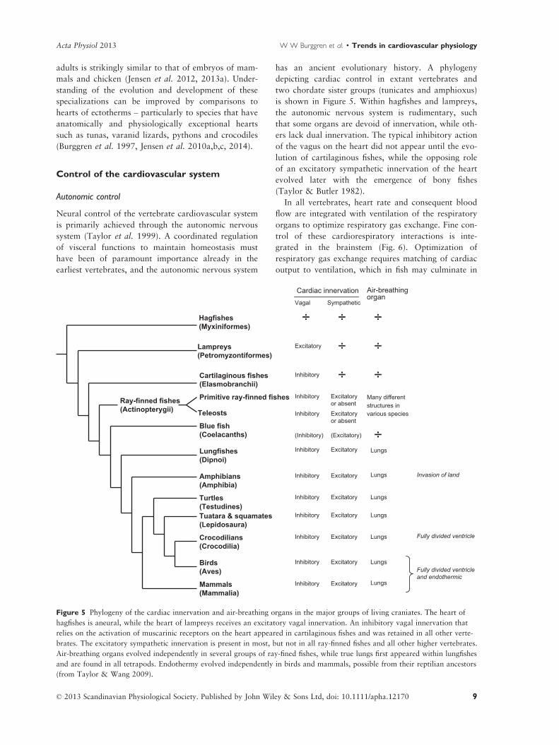
has an ancient evolutionary history. A phylogeny
depicting cardiac control in extant vertebrates and
two chordate sister groups (tunicates and amphioxus)
is shown in Figure 5. Within hagfishes and lampreys,
the autonomic nervous system is rudimentary, such
that some organs are devoid of innervation, while oth-
ers lack dual innervation. The typical inhibitory action
of the vagus on the heart did not appear until the evo-
lution of cartilaginous fishes, while the opposing role
of an excitatory sympathetic innervation of the heart
evolved later with the emergence of bony fishes
(Taylor & Butler 1982).
In all vertebrates, heart rate and consequent blood
flow are integrated with ventilation of the respiratory
organs to optimize respiratory gas exchange. Fine con-
trol of these cardiorespiratory interactions is inte-
grated in the brainstem (Fig. 6). Optimization of
respiratory gas exchange requires matching of cardiac
output to ventilation, which in fish may culminate in
Hagfishes(Myxiniformes)
Lampreys(Petromyzontiformes)
Cartilaginous fishes(Elasmobranchii)
Blue fish(Coelacanths)
Ray-finned fishes(Actinopterygii)
Lungfishes(Dipnoi)
Amphibians(Amphibia)
Mammals(Mammalia)
Turtles(Testudines)Tuatara & squamates(Lepidosaura)
Crocodilians(Crocodilia)
Birds(Aves)
Primitive ray-finned fishes
Teleosts
Cardiac innervationVagal Sympathetic
Air-breathingorgan
Excitatory
Inhibitory
Inhibitory
Inhibitory
(Inhibitory)
Inhibitory
Inhibitory
Inhibitory
Inhibitory
Inhibitory
Inhibitory
Inhibitory
Excitatory or absent
Excitatory or absent
(Excitatory)
Excitatory
Excitatory
Excitatory
Excitatory
Excitatory
Excitatory
Excitatory
Many different structures in various species
Lungs
Lungs
Lungs
Lungs
Lungs
Lungs
Lungs
Invasion of land
Fully divided ventricle
Fully divided ventricleand endothermic
÷
÷
÷
÷ ÷
÷
÷
÷
Figure 5 Phylogeny of the cardiac innervation and air-breathing organs in the major groups of living craniates. The heart of
hagfishes is aneural, while the heart of lampreys receives an excitatory vagal innervation. An inhibitory vagal innervation that
relies on the activation of muscarinic receptors on the heart appeared in cartilaginous fishes and was retained in all other verte-
brates. The excitatory sympathetic innervation is present in most, but not in all ray-finned fishes and all other higher vertebrates.
Air-breathing organs evolved independently in several groups of ray-fined fishes, while true lungs first appeared within lungfishes
and are found in all tetrapods. Endothermy evolved independently in birds and mammals, possible from their reptilian ancestors
(from Taylor & Wang 2009).
© 2013 Scandinavian Physiological Society. Published by John Wiley & Sons Ltd, doi: 10.1111/apha.12170 9
Acta Physiol 2013 W W Burggren et al. · Trends in cardiovascular physiology
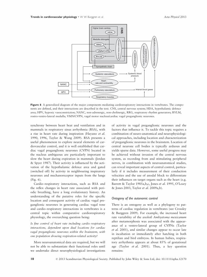
synchrony between heart beat and ventilation and in
mammals in respiratory sinus arrhythmia (RSA), with
a rise in heart rate during inspiration (Hayano et al.
1990, 1996, Taylor & Wang 2009). RSA presents a
useful phenomenon to explore neural elements of car-
diovascular control, and it is well established that car-
diac vagal preganglionic neurones (CVPN) located in
the nucleus ambiguous are particularly important to
slow the heart during expiration in mammals (Jordan
& Spyer 1987). Their activity is influenced by the acti-
vation of the hypothalamic defence area and gated
(switched off) by activity in neighbouring inspiratory
neurones and mechanoreceptor inputs from the lungs
(Fig. 6).
Cardio–respiratory interactions, such as RSA and
the reflex changes in heart rate associated with peri-
odic breathing, have a long evolutionary history. An
understanding of the putative roles for the specific
location and consequent activity of cardiac vagal pre-
ganglionic neurones in generating cardiac vagal tone
and cardio–respiratory interactions in vertebrates is a
central topic within comparative cardiorespiratory
physiology, the overarching question being:
Is fine control of heart rate including cardio–respiratory
interactions, dependent upon dual locations for cardiac
vagal preganglionic neurones within the brainstem, with
one population showing respiration-related activity?
More neuroanatomical data are required, but we will
not be able to substantiate their functional roles until
we undertake direct neurophysiological investigations
of activity in vagal preganglionic neurones and the
factors that influence it. To tackle this topic requires a
combination of neuro-anatomical and neurophysiologi-
cal approaches, including location and characterization
of preganglionic neurones in the brainstem. Location of
central neurone cell bodies is typically arduous and
yields sparse data. However, some useful progress may
be achieved without invasion of the central nervous
system, as recording from and stimulating peripheral
nerves, in combination with neuroanatomical studies,
can reveal important aspects of central control, particu-
larly if it includes measurement of their conduction
velocities and the use of anodal block to differentiate
their influences on target organs such as the heart (e.g.
Barrett & Taylor 1985a,b,c, Jones et al. 1995, O’Leary
& Jones 2003, Taylor et al. 2009a,b).
Ontogeny of the autonomic control
There is an ontogeny as well as a phylogeny to pat-
terns of cardiac regulation in vertebrates (see Crossley
& Burggren 2009). For example, the increased heart
rate variability of the axolotl Ambystoma mexicanum
after metamorphosis was associated with the appear-
ance of a ventro-lateral group of CVPNs (Taylor
et al. 2001), and similar changes appear to occur late
in incubation or immediately after hatching in both
reptilian and bird embryos. In human babies, respira-
tory arrhythmia appears at about 85% of gestational
age (Taylor et al. 2001). Thus, a key question
remains:
Arterial blood gases
Systemic blood pressure
Systemic vasculature
Heart - rate and contractility
Pulmonary vasculature
Respiratory muscles
Mec
hano
rece
ptor
s in
lung
s/gi
lls
Cen
tral a
nd p
erip
hera
l ch
emor
ecep
tors
Bar
orec
epto
rs
CN
S
Higher brain centres
Medulla oblongata
HDA
RRG
RVLM
VMN/CVPN
Sympathetic chain
NANC
HPV
Nuc
leus
tract
usso
litar
ius
Figure 6 A generalized diagram of the major components mediating cardiorespiratory interactions in vertebrates. The compo-
nents are defined, and their interactions are described in the text. CNS, central nervous system; HDA, hypothalamic defence
area; HPV, hypoxic vasoconstriction; NANC, non-adrenergic, non-cholinergic, RRG, respiratory rhythm generators; RVLM,
rostro-ventro-lateral medulla; VMN/CVPN, vagal motor nucleus/cardiac vagal preganglionic neurones.
© 2013 Scandinavian Physiological Society. Published by John Wiley & Sons Ltd, doi: 10.1111/apha.1217010
Trends in cardiovascular physiology · W W Burggren et al. Acta Physiol 2013

What is the functional relationship between developmen-
tal morphological changes taking place in the central ner-
vous system and the onset of autonomic control of the
cardiovascular system?
This question is being addressed by a combination
of neuroanatomical and electrophysiological tech-
niques applied to reptilian embryos that represent a
key group in the evolution of tetrapods (Taylor &
Wang 2009, Taylor et al. 2010). The important matu-
rational changes that result in central control of
cardio–respiratory interactions may be migration of
vagal preganglionic neurones within the brainstem,
myelination of peripheral nerves and completion of
their innervation of target organs such as the heart.
Developmental phenotypic plasticity in cardiovascular
control
Phenotypic plasticity is the capacity for a given geno-
type to give rise to multiple phenotypic traits depen-
dent on the internal or external environmental milieu
(Bateson et al. 2004, Garland & Kelly 2006, Pigliucci
et al. 2006). The basic evolutionary concept of ‘plas-
ticity’ or phenotypic flexibility within a genotype rep-
resents a powerful means of adaptation, maximizing
advantageous traits for a given environment (Dewitt
& Scheiner 2004, Pigliucci et al. 2006), but plasticity
during the developmental period may also produce
maladaptive traits, such as the increased incidences of
cardiovascular diseases through the mechanism of
‘foetal programming’ hypothesis underlying adult
onset human diseases (Barker 2000). Normal mamma-
lian foetal development may require a relatively nar-
row range of abiotic conditions, but the effects of
altered environmental conditions for non-mammalian
vertebrates and the potentially deleterious conse-
quences of cardiovascular developmental plasticity are
poorly understood. This leads to two key unanswered
questions:
In natural environments, does developmental cardiovascu-
lar plasticity convey advantageous or deleterious pheno-
typic changes in non-mammalian vertebrates?
Does developmental cardiovascular plasticity in non-
mammalian vertebrates provide insight to the evolution-
ary constraints on developmental biology?
Egg-laying reptiles are useful models to understand
the impact of the developmental environment on car-
diovascular maturation because it is easy to manipu-
late the environment where they develop and to relate
these environmental perturbations and the natural
environment. Manipulation such as changes in gas
composition, temperature or water content can be
used to investigate phenotypic plasticity. Growth rates
of several reptilian species are related to food avail-
ability, which is an advantage when conducting longi-
tudinal studies of cardiovascular performance.
Reptiles also differ in the embryonic onset, importance
and magnitude of autonomic or central nervous sys-
tem control. This feature makes reptiles a potentially
rich group to investigate cardiovascular homeostasis
in an embryonic system and assess the plasticity of
regulatory control mechanisms. To date, studies of
developmental plasticity in non-mammalian amniotes
have been restricted primarily to the embryonic phase
(Chan & Burggren 2005, Crossley & Altimiras 2005,
2012, Eme et al. 2011a,b, Crossley et al. 2012, Eme
et al. 2012).
Hypoxic responses of systemic and pulmonary vessels
In addition to autonomic regulation, the peripheral
circulation is affected by numerous local factors,
including metabolites and oxygen levels. Physiologists
have generally been interested in understanding how
organisms sense and respond to oxygen. Comparative
approaches reveal that hypoxia constricts the pulmo-
nary vasculature, an intrinsic phenomenon termed
‘hypoxic pulmonary vasoconstriction’ (von Euler &
Liljestrand 1946, Sommer et al. 2008). This response
is thought to be adaptive in that it diverts pulmonary
blood flow from inadequately ventilated and hypoxic
parts of the lung to more highly ventilated areas.
Thus, hypoxic pulmonary vasoconstriction is impor-
tant for local matching of blood perfusion to ventila-
tion by improving pulmonary gas exchange efficiency
and, consequently, maintaining arterial oxygenation
(Skovgaard & Wang 2006, Sylvester et al. 2012).
Hypoxic vasoconstriction is an ancient and highly
conserved response expressed in the respiratory organs
of all vertebrates, including lungs of mammals, birds
and reptiles, amphibian skin and fish gills.
In contrast to the pulmonary circulation, hypoxia is
well known to elicit vasodilation in the systemic vas-
culature of most vertebrates, an equally adaptive
response that allows for increased perfusion of oxy-
gen-deprived regions in the various organs. While it
may seem intuitive that the vasodilatory response pre-
cedes the constriction in evolutionary terms, a number
of recent studies document that hypoxia causes con-
striction of vascular smooth muscles in cyclostomes, a
phylogenetically early group of vertebrates, and that
this response is intrinsic to the vasculature (Olson
et al. 2001, 2008, Russell et al. 2008). Hypoxic vaso-
constriction thus appears to be an ancient vascular
response to hypoxia that has been embellished with
secondary regulatory factors as vertebrates evolved to
be more responsive to hypoxia. Thus, hypoxic pulmo-
© 2013 Scandinavian Physiological Society. Published by John Wiley & Sons Ltd, doi: 10.1111/apha.12170 11
Acta Physiol 2013 W W Burggren et al. · Trends in cardiovascular physiology
nary vasoconstriction in tetrapods has evolved to
become a multifactorial process associated with sev-
eral signalling pathways. This leads to a key question:
What are the signalling pathways involved in hypoxic
vasomotor activity?
O2 sensing by vascular tissues has been intensely
studied in mammals for more than 50 years. Numer-
ous mechanisms have been proposed to explain how
blood vessels sense low PO2 and transduce this signal
into dilation or constriction, but none have received
unequivocal support and the O2-sensor and sensor/
transduction cascade(s) remain unresolved (Ward
2008, L�opez-Barneo et al. 2010, Clanton et al.
2013). Nevertheless, there is now evidence that a
mechanism involving hydrogen sulphide (H2S) metab-
olism may explain both hypoxic vasoconstriction and
hypoxic vasodilatation (Olson et al. 2006, 2010).
The mechanisms of the vascular oxygen sensor are
more likely to be unravelled in phylogenetically
ancient groups of vertebrates without the secondary
regulatory factors. Accordingly, a comparative
approach is likely to provide novel insights. Studies
of respiratory vessels from primitive (i.e. early) air-
breathers, such as lungfish, as well as reptiles and
mammals, may reveal an evolutionary progression in
complexity of the oxygen sensor and mechanisms
underlying hypoxic vasoconstriction.
Cardiovascular interactions with other organ
systems
Physiologists have tended to focus on individual sys-
tems (e.g. cardiovascular, gas exchange, ion exchange,
endocrine, neural systems), viewing homeostasis
through the eyes of regulation of the system they
study. While there is, of course, broad appreciation
that all of these systems operate in an integrated fash-
ion to achieve homeostasis, studies specifically
designed to look at the interactions between systems
are relatively uncommon. Below we explore as exam-
ples three sets of interactions – with the lymphatic,
renal and digestive systems – and how the compara-
tive approach will provide model approaches to inves-
tigate these interactions.
Cardiovascular–lymphatic interactions
Maintenance of plasma volume is critical for cardiovas-
cular homeostasis. There is tremendous diversity in
both plasma volume as a fraction of body mass and the
regulatory mechanisms that maintain plasma volume,
with no clear phylogenetic pattern (Takei 2000).
Plasma volume regulation entails balancing fluid
exchange between the vascular and interstitial compart-
ments. Regulation of plasma volume (Vp) and the fluid
exchange between vascular and interstitial compart-
ments are governed by a combination of short-term and
long-term effector feedback loops. Long-term regula-
tion of Vp is accomplished by renal mechanisms. Short-
term, transient changes in plasma volume occur
through transcapillary fluid flux or through changes in
lymphatic fluid flux. Compared with mammals, ecto-
thermic vertebrates have substantially greater transcap-
illary fluid flux rates, which promote a high rate of
lymph formation (Hedrick et al. 2013).
Recent investigations on anurans have shed some
light on this process. Plasma turnover rates in anurans
are 3–5% of Vp min�1 and much higher compared
with either fish (0.8–0.9%) or mammals (<0.1%)
(Hillman et al. 2004). The reason for the much higher
plasma turnover in anurans is 10-fold higher intersti-
tial compliance (Cist) in anurans, where the extensive
subcutaneous lymph sacs provide considerable lym-
phatic storage capacity with little interstitial pressure
development to counterbalance the hydrostatic
pressure in the capillary. Thus, the net balance of
forces favours the loss of plasma from the vascular
space to the interstitium under most physiological
conditions, even including dehydration and haemor-
rhage (Hillman et al. 1987). Filtered plasma is then
returned to the vascular space via the lymphatic sys-
tem. This comparison points out the extreme impor-
tance of the interactions between cardiovascular–
lymphatic function in anurans, but the synergy
between these two systems is almost completely
unstudied in fish, reptiles and birds. The extremely
high rates of lymph formation and plasma turnover
in anurans beg the question:
How do anurans maintain cardiovascular homeostasis
given their unusual lymph and plasma dynamics?
Anurans have two pairs of spinally innervated
lymph hearts that actively pump lymph into the
venous renal portal system (Crossley & Hillman
2010). These lymph hearts are under feedback control
of arterial baroreceptors (Crossley & Hillman 1999),
but are also under hormonal control (De Grauw &
Hillman 2004) and through the volume of lymph pro-
vided by the various lymph sacs (Hillman et al. 2010).
Lymph hearts are also found in some reptiles and a
few birds, but their role in cardiovascular homeostasis
is unknown. The critical issue for anurans is that any
lymph formed moves gravitationally to the ventral
regions of the animal, but must be moved to the dor-
sally located lymph hearts before it can be returned to
the vascular space (Hillman et al. 2004).
Anurans overcome this morphological challenge
using a variety of mechanisms including differential
lymph sac compliance, contraction of specialized
© 2013 Scandinavian Physiological Society. Published by John Wiley & Sons Ltd, doi: 10.1111/apha.1217012
Trends in cardiovascular physiology · W W Burggren et al. Acta Physiol 2013

skeletal muscles and lung ventilation to move lymph
(Hillman et al. 2005, 2010, Drewes et al. 2007,
2013, Hedrick et al. 2007, 2011). The finding that
lung ventilation is involved in lymph movement (Hed-
rick et al. 2007), in addition to its traditional role in
gas exchange, is a novel finding and may explain the
evolutionary origin of the well-known link between
blood pressure and ventilation in mammals (McMu-
llan & Pilowsky 2010).
A summary of the various factors involved in mov-
ing lymph in anurans, which may be applicable to
other vertebrates, is shown in Figure 7. Whether these
processes to move lymph are unique to anurans is
unclear and so comprise key areas for future study. In
this regard, imaging techniques such as computed
tomography (CT) and magnetic resonance imaging
(MRI) should prove very useful for visualizing path-
ways and providing non-invasive techniques for evalu-
ating lymphatic function.
Other taxa in addition to amphibians may offer
interesting insights that can help us understand cardio-
vascular–lymphatic interactions. For example, fish
have a ‘secondary circulation’ that is connected to the
arterial system via small anastomoses referred to as
arterial-lymphatic conduits (Jensen et al. 2009).
Whether the secondary circulation represents a primi-
tive lymphatic system has been the source of much
speculation and controversy (Isogai et al. 2009, Vogel
2010, Hedrick et al. 2013). Molecular control of lym-
phatic vessel generation (lymphangiogenesis) is con-
served in fish and mammals (Yaniv et al. 2006),
suggesting that common molecular mechanisms are
involved in development of the lymph system in verte-
brates. The use of molecular tools will continue to be
important in lymphatic studies, but should be applied
more broadly to examine unique or conserved features
in a comparative context.
Birds also present a particularly interesting physio-
logical problem, having mammalian-like arterial blood
pressures coupled with a low plasma oncotic pressure.
This situation should favour high transcapillary filtra-
tion rates, yet birds tolerate dehydration and haemor-
rhage to a much greater extent than mammals
(Djojusugito et al. 1968, Carmi et al. 1994). This
differential capacity remains unexplained, but a role
for lymph mobilization is implied within this problem.
Clearly there is much investigation to be completed in
the future to determine the proximate and evolution-
ary aspects of cardiovascular–lymphatic interactions in
vertebrates.
Cardiovascular–renal interactions during development
As in adults, a developing animal increasingly func-
tions as a collective of highly interactive systems that
respond to environmental challenge in a tightly inte-
grated fashion, rather than as distinct units. Conse-
quently, a major question in physiology is:
When does the co-dependency of major organ systems
first develop and how does it then mature?
The nexus of the cardiovascular and renal system
provides a logical starting point for beginning an
intersystems approach in developmental physiology.
The adult cardiovascular system depends upon the
osmoregulatory system to maintain blood pressure
and flow, as well as blood fluid volume, via hormonal
regulation. Likewise, changes in cardiovascular func-
tion, particularly blood pressure, can influence blood
supply to the nephrons of the kidneys, heavily influ-
encing renal function and therefore salt and water bal-
ance through the renin-angiotensin system (RAS)
pressure and act through negative feedback, inhibiting
further renin release (Vander 1980).
Blood pressure and fluid volume regulatory systems,
such as the RAS, have been extensively studied in
adult vertebrates, especially mammals (for a recent
review see Crowley & Coffman 2012). However, our
knowledge of such intersystem physiological interac-
tions during development of the vertebrate embryo/
foetus is scarce at best and many unanswered ques-
tions remain:
Lymph heart output
Stroke volume Lymph heart rate
Transcapillaryfiltra on
Fc Pvas Pist
Lymph sacs compliance (volume/pressure)
Lung volume Lymph skeletal muscles
Spinal nerves
Hormones
Venous pressure
End diastolic volume
Lymph heart diastolic pressure
Ven latory muscles
Figure 7 Schematic diagram of the various forces involved
in moving lymph in anurans. Dorsally located lymph hearts
pump lymph into the venous circulation. The various lymph
sacs are combined into a single box (shaded) for clarity.
Lymph sac compliance (DVolume/DPressure) in the various
lymph sacs is affected by transcapillary filtration, ventilation
acting on lung volume and a variety of skeletal lymph mus-
cles. Transcapillary filtration is influenced by the whole body
filtration coefficient (Fc), vascular pressure (Pvas) and intersti-
tial pressure (Pint) as described by Tanaka (1979). See Hill-
man et al. (2004) for additional details.
© 2013 Scandinavian Physiological Society. Published by John Wiley & Sons Ltd, doi: 10.1111/apha.12170 13
Acta Physiol 2013 W W Burggren et al. · Trends in cardiovascular physiology
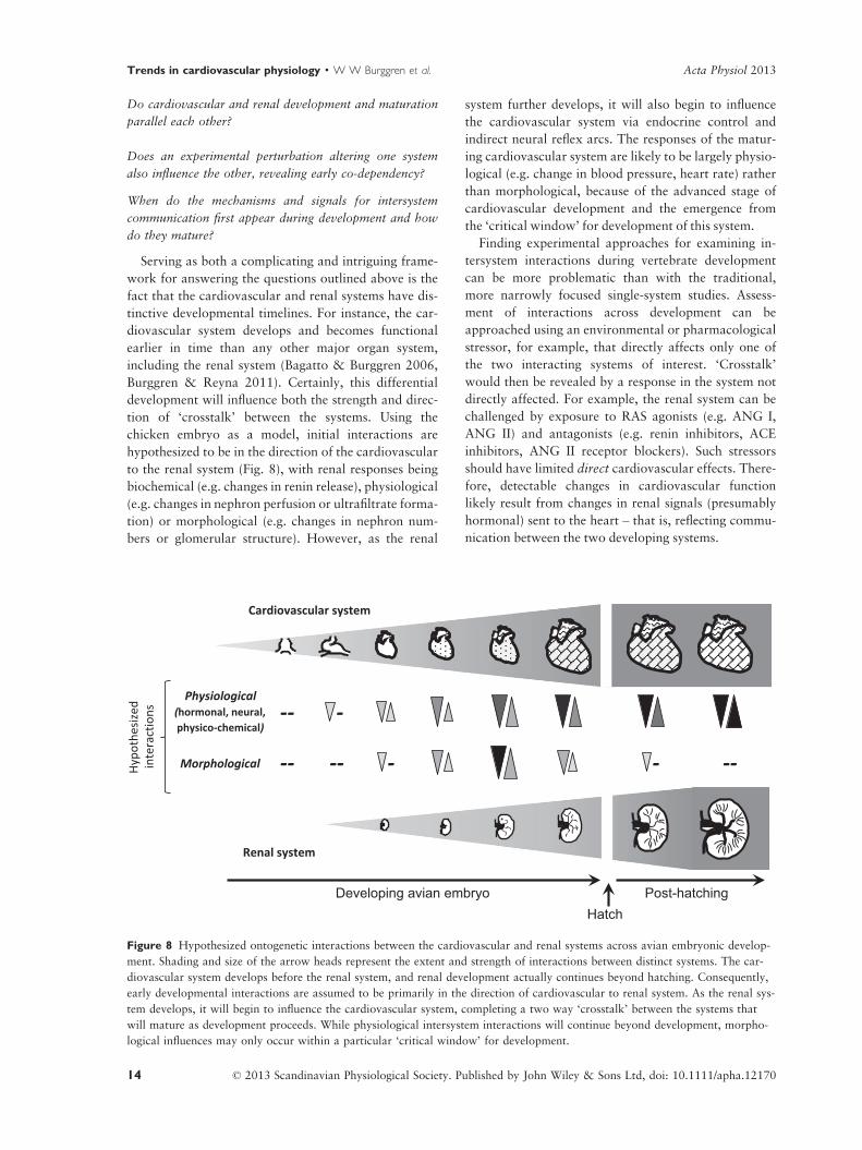
Do cardiovascular and renal development and maturation
parallel each other?
Does an experimental perturbation altering one system
also influence the other, revealing early co-dependency?
When do the mechanisms and signals for intersystem
communication first appear during development and how
do they mature?
Serving as both a complicating and intriguing frame-
work for answering the questions outlined above is the
fact that the cardiovascular and renal systems have dis-
tinctive developmental timelines. For instance, the car-
diovascular system develops and becomes functional
earlier in time than any other major organ system,
including the renal system (Bagatto & Burggren 2006,
Burggren & Reyna 2011). Certainly, this differential
development will influence both the strength and direc-
tion of ‘crosstalk’ between the systems. Using the
chicken embryo as a model, initial interactions are
hypothesized to be in the direction of the cardiovascular
to the renal system (Fig. 8), with renal responses being
biochemical (e.g. changes in renin release), physiological
(e.g. changes in nephron perfusion or ultrafiltrate forma-
tion) or morphological (e.g. changes in nephron num-
bers or glomerular structure). However, as the renal
system further develops, it will also begin to influence
the cardiovascular system via endocrine control and
indirect neural reflex arcs. The responses of the matur-
ing cardiovascular system are likely to be largely physio-
logical (e.g. change in blood pressure, heart rate) rather
than morphological, because of the advanced stage of
cardiovascular development and the emergence from
the ‘critical window’ for development of this system.
Finding experimental approaches for examining in-
tersystem interactions during vertebrate development
can be more problematic than with the traditional,
more narrowly focused single-system studies. Assess-
ment of interactions across development can be
approached using an environmental or pharmacological
stressor, for example, that directly affects only one of
the two interacting systems of interest. ‘Crosstalk’
would then be revealed by a response in the system not
directly affected. For example, the renal system can be
challenged by exposure to RAS agonists (e.g. ANG I,
ANG II) and antagonists (e.g. renin inhibitors, ACE
inhibitors, ANG II receptor blockers). Such stressors
should have limited direct cardiovascular effects. There-
fore, detectable changes in cardiovascular function
likely result from changes in renal signals (presumably
hormonal) sent to the heart – that is, reflecting commu-
nication between the two developing systems.
--
Cardiovascular system
Renal system
Developing avian embryo
Physiological(hormonal, neural, physico-chemical)
MorphologicalHyp
othe
size
din
tera
ctio
ns
Post-hatching
--
-
--
--
--
Hatch
Figure 8 Hypothesized ontogenetic interactions between the cardiovascular and renal systems across avian embryonic develop-
ment. Shading and size of the arrow heads represent the extent and strength of interactions between distinct systems. The car-
diovascular system develops before the renal system, and renal development actually continues beyond hatching. Consequently,
early developmental interactions are assumed to be primarily in the direction of cardiovascular to renal system. As the renal sys-
tem develops, it will begin to influence the cardiovascular system, completing a two way ‘crosstalk’ between the systems that
will mature as development proceeds. While physiological intersystem interactions will continue beyond development, morpho-
logical influences may only occur within a particular ‘critical window’ for development.
© 2013 Scandinavian Physiological Society. Published by John Wiley & Sons Ltd, doi: 10.1111/apha.1217014
Trends in cardiovascular physiology · W W Burggren et al. Acta Physiol 2013
To fully understand the onset and ontogenetic
changes in cardiovascular–renal interactions, we also
must search for and identify ‘critical windows’ during
development. Critical windows are those specific peri-
ods during development when a tissue or system is most
sensitive to perturbation (e.g. Burggren & Fritsche
1995, Burggren 1998, Burggren & Reyna 2011). Criti-
cal windows have been employed within the framework
of the development of individual organs and systems,
but we can also apply this concept to cardiovascular–
renal interactions. This will allow us not only to address
whether intersystem communication occurs during
development but also to determine when and for how
long it is most important.
Cardiovascular–digestive system interactions
Comparative studies on autonomic control of the car-
diovascular system have emphasized the role of adren-
ergic and cholinergic system as well as endothelial
function, while the role of alternative factors has
received considerably less attention. Metabolic rate
increases during digestion in most vertebrates due to
digestive and anabolic processes, and the increased
oxygen demand is met by an increase in both heart
rate and stroke volume (e.g. Secor et al. 2000), as well
as redistribution of flows to the gastrointestinal organs
(Farrell et al. 2001). This regulation is governed by
the ANS, primarily by release of parasympathetic tone
on the heart. However, in addition to autonomic regu-
lation, a non-cholinergic-non-adrenergic (NANC) fac-
tor acts to stimulate heart rate during digestion in
snakes (Wang et al. 2001, Skovgaard et al. 2009,
Enok et al. 2012). There also seems to be a NANC
factor that stimulates the heart during digestion in
frogs and humans with transplanted hearts continue
to increase heart rate in response to feeding despite
the lack of cardiac innervation (Waaler et al. 2002).
This leads to the question:
What is the importance and mechanisms behind NANC
cardiovascular regulation during digestion?
One possible NANC factor is histamine, which has
a direct chronotropic effect on heart rate during the
initial phase of digestion in pythons (Skovgaard et al.
2009). Mast cells are a major store of histamine in
vertebrates, and mast cells are distributed throughout
the body, including cardiac tissues, and provide a
likely site of release. However, other NANC factors
may also contribute.
Synthesis and conclusions
Comparative cardiovascular physiology has never been
at a more exciting position in the physiological sci-
ences. New and additional conceptual frameworks,
such as ‘evo-devo’ and comparative developmental
physiology (Burggren & Crossley 2002, Burggren &
Warburton 2005, Warburton et al. 2006), have helped
define our current view of the evolution and function
of vertebrate cardiovascular systems. The expansion
of additional animal models, along with the contin-
uing exploitation of existing models (Burggren 2000,
Burggren & Warburton 2007), has extended our
experimental reach. Experimental paradigms involving
phylogenetic, mathematical and developmental strate-
gies (Hicks & Wang 2012) are accelerating our acqui-
sition of new understanding about how cardiovascular
systems evolved, and how they currently function.
Further accelerating our progress is the wide availabil-
ity of expanded tools spanning from molecular levels
(e.g. Moorman & Christoffels 2003, Christoffels et al.
2010, Jensen et al. 2013a,b) to whole animal levels
(e.g. nuclear magnetic resonance, echocardiography
and other imaging technologies (Jensen et al. 2010b).
In this essay, we have not tried to present an inclusive
list of promising areas for future comparative cardio-
vascular research. Rather, we have each tried to shed
light on some of the key, unanswered questions and
unmet challenges in cardiovascular biology as we see
them through our own lenses. We have additionally
attempted to indicate how a comparative approach can
help advance not just our basic understanding, but can
lead to novel insights into clinically relevant malforma-
tions and cardiac anomalies, as well. This is, after all,
the very approach that would be advocated by Kjell Jo-
hansen – Viking and Physiologist.
Conflict of interest
None.
References
Agassiz, L. 1857. Contributions to the Natural History of
the United States of America. Little, Brown and Company,
Boston.
Bagatto, B. & Burggren, W. 2006. A three-dimensional func-
tional assessment of heart and vessel development in the
larva of the zebrafish (Danio rerio). Physiol Biochem Zool
79, 194–201.
Barker, D.J.P. 2000. In utero programming of cardiovascular
disease. Theriogenology 53, 555–574.
Barrett, D.J. & Taylor, E.W. 1985a. Spontaneous efferent
activity in branches of the vagus nerve controlling heart
rate and ventilation in the dogfish. J Exp Biol 117, 433–
448.
Barrett, D.J. & Taylor, E.W. 1985b. The location of cardiac
vagal preganglionic neurones in the brainstem of the
dogfish. J Exp Biol 117, 449–458.
© 2013 Scandinavian Physiological Society. Published by John Wiley & Sons Ltd, doi: 10.1111/apha.12170 15
Acta Physiol 2013 W W Burggren et al. · Trends in cardiovascular physiology
Barrett, D.J. & Taylor, E.W. 1985c. The characteristics of
cardiac vagal preganglionic motoneurones in the dogfish.
J Exp Biol 117, 459–470.
Bateson, P., Barker, D.J.P., Clutton-Brock, T., Deb, D.,
D’Udine, B., Foley, R.A., Gluckman, P., Godfrey, K.M.,
Kirkwood, T., Lahr, M.M., McNamara, J., Metcalfe, N.B.,
Monaghan, P., Spencer, H.G. & Sultan, S.E. 2004. Devel-
opmental plasticity and human health. Nature 430,
419–421.
Brill, R.W. & Bushnell, P.G. 2001. The cardiovascular
system of tunas. In: B. Block & E. Stevens (eds) Fish
Physiology Tuna: Physiology, Ecology, and Evolution,
pp. 79–120. Academic Press, New York.
Burggren, W.W. 1978. Influence of intermittent breathing on
ventricular depolarization patterns in chelonian reptiles.
J Physiol 278, 349–364.
Burggren, W.W. 1986. Form and function in reptilian circu-
lations. Amer Zool 27, 5–19.
Burggren, W.W. 1991. Does comparative respiratory physiol-
ogy have a role in evolutionary biology (and vice versa)?
In: A.J. Woakes, M.K. Grieshaber & C.R. Bridges (eds)
Comparative Insights into Strategies for Gas Exchange and
Metabolism, pp. 1–13. Cambridge University Press,
Cambridge.
Burggren, W.W. 1992. The importance of an ontogenetic
perspective in physiological studies. Amphibian cardiology
as a case study. In: S.C. Wood, R.E. Weber, A.R. Hargens
& R.W. Millard (eds) Physiological Adaptations in Verte-
brates: Respiration, Circulation and Metabolism, pp. 235–
253. Marcel Dekker, New York, NY.
Burggren, W.W. 1998. Studying physiological development:
past, present and future. Biol Bull 33, 71–84.
Burggren, W.W. 2000. Developmental physiology, animal
models, and the August Krogh principle. Zool Anal Com-
plex Syst 102, 148–156.
Burggren, W.W. & Bemis, W.E. 1990. Studying physiological
evolution: paradigms and pitfalls. In: M.H. Nitecki (ed.)
Evolutionary Innovations: Patterns and Processes,
pp. 191–228. Oxford University Press, Oxford.
Burggren, W. & Crossley, D.A. II 2002. Comparative cardio-
vascular development: improving the conceptual frame-
work. Comp Biochem Physiol A 132, 661–674.
Burggren, W.W. & Fritsche, R. 1995. Amphibian cardiovas-
cular development. In: W.W. Burggren & B. Keller (eds)
Development of Cardiovascular Systems: Molecules to
Organisms, pp. 166–182. University of Cambridge Press,
New York.
Burggren, W. & Johansen, K. 1982. Ventricular hemodynam-
ics in the monitor lizard Varanus exanthematicus - pulmo-
nary and systemic pressure separation. J Exp Biol 96,
343–354.
Burggren, W. & Johansen, K. 1986. Circulation and respira-
tion in lungfishes. J Morph Suppl 1, 217–236.
Burggren, W.W. & Reiber, C.L. 2007. Evolution of cardio-
vascular systems. In: W.C. Aird (ed.) Endothelial Biomedi-
cine, pp. 29–49. Cambridge University Press, Cambridge.
Burggren, W.W. & Reyna, K. 2011. Developmental trajec-
tories, critical windows and phenotypic alteration during
cardio-respiratory development. Respiratory Physiology
and Neurobiology 178, 13–21.
Burggren, W.W. & Warburton, S.J. 1994. Patterns of form
and function in developing hearts: contributions from non-
mammalian vertebrates. Cardioscience 5, 183–191.
Burggren, W.W. & Warburton, S. 2005. Comparative devel-
opmental physiology: an interdisciplinary convergence.
Annu Rev Physiol 67, 203–223.
Burggren, W.W. & Warburton, S. 2007. Amphibians as ani-
mal models for laboratory research in physiology. ILAR J
48, 260–269.
Burggren, W.W., Farrell, A.P. & Lillywhite, H.B. 1997. Ver-
tebrate cardiovascular systems. In: W. Dantzler (ed.) Hand-
book of Comparative Physiology, pp. 215–308. Oxford
University Press, Oxford, UK.
Carmi, N., Pinshow, B. & Horowitz, M. 1994. Plasma vol-
ume conservation in pigeons: effects of air temperature
during dehydration. Am J Physiol 267, R1449–R1453.
Chan, T. & Burggren, W.W. 2005. Hypoxic incubation cre-
ates differential morphological effects during specific devel-
opmental critical windows in the embryo of the chicken
(Gallus gallus). Respir Physiol Neurobiol 145, 251–263.
Christoffels, V.M. & Moorman, A.F. 2009. Development of
the cardiac conduction system: why are some regions of
the heart more arrhythmogenic than others? Circ
Arrhythm Electrophysiol 2, 195–207.
Christoffels, V.M., Smits, G.J., Kispert, A. & Moorman, A.F.
2010. Development of the pacemaker tissues of the heart.
Circ Res 106, 240–254.
Clanton, T.L., Hogan, M.C. & Gladden, L.B. 2013. Regula-
tion of cellular gas exchange, oxygen sensing, and meta-
bolic control. Comprehen Physiol 3, 1135–1190.
Clark, T.D., Wang, T., Butler, P.J. & Frappell, P.B. 2005.
Factorial scopes of cardio-metabolic variables remain con-
stant with changes in body temperature in the varanid liz-
ard, Varanus rosenbergi. Am J Physiol 288, R992–R997.
Cleland, T.P., Stoskopf, M.K. & Schweitzer, M.H. 2011.
Histological, chemical, and morphological reexamination
of the “heart” of a small Late Cretaceous Thescelosaurus.
Naturwissenschaften 98, 203–211.
Crossley, D.A. & Altimiras, J. 2005. Cardiovascular develop-
ment in embryos of the American alligator Alligator missis-
sippiensis: effects of chronic and acute hypoxia. J Exp Biol
208, 31–39.
Crossley, D.A. & Altimiras, J. 2012. Effect of selection for
commercially productive traits on the plasticity of cardio-
vascular regulation in chicken breeds during embryonic
development. Poult Sci 91, 2628–2636.
Crossley, D.A. II & Burggren, W.W. 2009. Development of
cardiac form and function in ectothermic sauropsids.
J Morphol 270, 1400–1412.
Crossley, D.A. & Hillman, S.S. 1999. The role of pulmocuta-
neous baroreceptors in the control of lymphatic heart rate
in the toad Bufo marinus. Physiol Biochem Zool 72,
109–115.
Crossley, D.A. & Hillman, S.S. 2010. Posterior lymph
heart function in two species of anurans: analysis based
on both in vivo pressure-volume relationships by conduc-
© 2013 Scandinavian Physiological Society. Published by John Wiley & Sons Ltd, doi: 10.1111/apha.1217016
Trends in cardiovascular physiology · W W Burggren et al. Acta Physiol 2013

tance manometry and ultrasound. J Exp Biol 213,
3710–3716.
Crossley, D.A., Tate, K.B., Elfwing, M. & Eme, J. 2012.
Chronic developmental hypoxia alters the cardiovascular
baroreflex phenotype of embryonic common snapping
turtles. FASEB J 26, 1071.11.
Crowley, S.D. & Coffman, T.M. 2012. Recent advances
involving the renin-angiotensin system. Exp Cell Res 318,
1049–1056.
Davie, P.S. & Farrell, A.P. 1991. The coronary and luminal
circulations of the myocardium of fishes. Can J Zool 69,
1993–2001.
De Andres, A.V., Mu~noz-Chapuli, R., Sans-Coma, V. &
Garcia-Garrido, L. 1990. Anatomical studies of the coro-
nary system in elasmobranchs: I. Coronary arteries in lam-
noid sharks. Am J Anat 187, 303–310.
De Andres, A.V., Mu~noz-Chapuli, R., Sans-Coma, V. & Gar-
cia-Garrido, L. 1992. Anatomical studies of the coronary
system in elasmobranchs: II. Coronary arteries in hexanch-
oid, squaloid, and carcharhinoid sharks. Anat Rec 233,
429–439.
De Grauw, E.A. & Hillman, S.S. 2004. General function and
endocrine control of the posterior lymph hearts in Bufo
marinus and Rana catesbeiana. Physiol Biochem Zool 77,
594–600.
Dewitt, T.J. & Scheiner, S.M. 2004. Phenotypic Plasticity
Functional and Conceptual Approaches. Oxford University
Press, Oxford.
Djojusugito, M., Folkow, B. & Kovach, G.B. 1968. The mech-
anism behind the rapid blood volume restoration after hem-
orrhage in birds. Acta Physiol Scand 74, 114–122.
Drewes, R.C., Hedrick, M.S., Hillman, S.S. & Withers, P.C.
2007. Unique role of skeletal muscle contraction in vertical
lymph movement in anurans. J Exp Biol 210, 3931–3939.
Drewes, R.C., Hillman, S.S., Withers, P.C. & Hedrick, M.S.
2013. Evolutionary implications of the distribution and
variation of the skeletal muscles of the anuran lymphatic
system. Zoomorphology 132, 339–349.
Duncker, D.J. & Bache, R.J. 2008. Regulation of coronary
blood flow during exercise. Physiol Rev 88, 1009–1086.
Dyson, E., Sucov, H.M., Kubalak, S.W., Schmid-Schonbein,
G.W., DeLano, F.A., Evans, R.M., Ross, J. Jr & Chien,
K.R. 1995. Atrial-like phenotype is associated with embry-
onic ventricular failure in retinoid X receptor alpha -/-
mice. Proc Natl Acad Sci 92, 7386–7390.
Eme, J., Gwalthney, J., Blank, J.M., Owerkowicz, T., Barron,
G. & Hicks, J.W. 2009. Surgical removal of right-to-left
cardiac shunt in the American alligator (Alligator mississip-
piensis) causes ventricular enlargement but does not alter
apnoea or metabolism during diving. J Exp Biol 212,
3553–3563.
Eme, J., Gwalthney, J., Owerkowicz, T., Blank, J.M. &
Hicks, J.W. 2010. Turning crocodilian hearts into bird
hearts: growth rates are similar for alligators with and
without cardiac shunt. J Exp Biol 213, 2673–2680.
Eme, J., Crossley, D.A. & Hicks, J.W. 2011a. Role of the left
aortic arch and blood flows in embryonic American alliga-
tor (Alligator mississippiensis). J Comp Physiol B 181,
391–401.
Eme, J., Altimiras, J., Hicks, J.W. & Crossley, D.A. II
2011b. Hypoxic alligator embryos: chronic hypoxia, cate-
cholamine levels and autonomic responses of in ovo alliga-
tors. Comp Biochem Physiol A 160, 412–420.
Eme, J., Hicks, J.W. & Crossley, D.A. II 2011c. Chronic
hypoxic incubation blunts a cardiovascular reflex loop in
embryonic American alligator (Alligator mississippiensis).
J Comp Physiol B 181, 981–990.
Eme, J., Tate, K.B., Slay, C.E., Kohl, Z.F., Hicks, J.W. &
Crossley, D.A. 2012. Cardiovascular plasticity during hyp-
oxic development in snapping turtle and alligator embryos.
Integr Comp Biol 52(suppl 1), 54.
Engberding, R., Yelbuz, T.M. & Breithardt, G. 2007. Iso-
lated noncompaction of the left ventricular myocardium -
a review of the literature two decades after the initial case
description. Clin Res Cardiol 96, 481–488.
Enok, S., Simonsen, L.S., Pedersen, S.V., Wang, T. & Skovg-
aard, N. 2012. Humoral regulation of heart rate during
digestion in the Burmese python (Python molurus). Am J
Physiol 302, R1176–R1183.
von Euler, U.S. & Liljestrand, G. 1946. Observations on the
pulmonary arterial blood pressure in the cat. Acta Physiol
Scand 12, 301–320.
Farmer, C.G. 1999. Evolution of the vertebrate cardio-pul-
monary system. Annu Rev Physiol 61, 573–592.
Farmer, C.G. 2011. On the evolution of arterial vascular pat-
terns of tetrapods. J Morphol 272, 1325–1341.
Farmer, C.G., Uriona, T.J., Olsen, D.B., Steenblik, M. &
Sanders, K. 2008. The right-to-left shunt of crocodilians
serves digestion. Physiol Biochem Zool 81, 125–137.
Farrell, A.P. 1987. Coronary flow in a perfused rainbow
trout heart. J Exp Biol 129, 107–123.
Farrell, A.P. 1991. From hagfish to tuna - a perspective on
cardiac-function in fish. Physiol Zool 64, 1137–1164.
Farrell, A.P. 1996. Features heightening cardiovascular per-
formance in fishes, with special reference to tunas. Comp
Biochem Physiol 113, 61–67.
Farrell, A.P. 2002. Cardiorespiratory performance in salmo-
nids during exercise at high temperature: insights into car-
diovascular design limitations in fishes. Comp Biochem
Physiol A 132, 797–810.
Farrell, A.P. & Stecyk, J.A. 2007. The heart as a working model
to explore themes and strategies for anoxic survival in ecto-
thermic vertebrates. Comp Biochem Physiol A 147, 300–312.
Farrell, A.P., Thorarensen, H., Axelsson, M., Crocker, C.E.,
Gamperl, A.K. & Cech, J.J. Jr 2001. Gut blood flow in fish
during exercise and severe hypercapnia. Comp Biochem
Physiol A 128, 551–563.
Farrell, A.P., Farrell, N.D., Jourdan, H. & Cox, G.K. 2012.
A perspective on the evolution of the coronary circulation
in fishes and the transition to terrestrial life. In: D. Sedmer-
a & T. Wang (eds) Ontogeny and Phylogeny of the Verte-
brate Heart, pp 75–102. Springer, New York.
Franklin, C.E. & Davie, P.S. 1992. Dimensional analysis of
the ventricle of an in situ perfused trout heart using echo-
cardiography. J Exp Biol 166, 47–60.
Freedom, R.M., Yoo, S.J., Perrin, D., Taylor, G., Petersen, S.
& Anderson, R.H. 2005. The morphological spectrum of
ventricular noncompaction. Cardiol Young 15, 345–364.
© 2013 Scandinavian Physiological Society. Published by John Wiley & Sons Ltd, doi: 10.1111/apha.12170 17
Acta Physiol 2013 W W Burggren et al. · Trends in cardiovascular physiology

Galli, G.L.J. & Shiels, H.A. 2012. The sarcoplasmic reticu-
lum in the vertebrate heart. In: D. Sedmera & T. Wang
(eds) Ontogeny and Phylogeny of the Vertebrate Heart,
pp. 103–123. Springer, New York.
Garland, T. Jr & Kelly, S.A. 2006. Phenotypic plasticity and
experimental evolution. J Exp Biol 209, 2344–2361.
Goodrich, E.S. 1930. Studies on the Structure and Develop-
ment of Vertebrates. Macmillan and Company, London.
Hayano, J., Sakakibara, Y., Yamada, M., Ohte, N., Fujinam-
i, T. & Yokoyama, K. 1990. Decreased magnitude of heart
rate spectral components in coronary artery disease. Its
relation to angiographic severity. Circulation 81, 1217–
1224.
Hayano, J., Yasuma, F., Okada, A., Mukai, S. & Fujinami,
T. 1996. Respiratory sinus arrhythmia. A phenomenon
improving pulmonary gas exchange and circulatory effi-
ciency. Circulation 94, 842–847.
Hedrick, M.S., Drewes, R.C., Hillman, S.S. & Withers, P.C.
2007. Lung ventilation contributes to vertical lymph move-
ment in anurans. J Exp Biol 210, 3940–3945.
Hedrick, M.S., Hillman, S.S., Drewes, R.C. & Withers, P.C.
2011. Pulmonary compliance and lung volume varies with
ecomorphology in anuran amphibians: implications for
ventilatory-assisted lymph flux. J Exp Biol 214, 3279–
3285.
Hedrick, M.S., Hillman, S.S., Drewes, R.C. & Withers, P.C.
2013. Lymphatic regulation in non-mammalian verte-
brates. J Appl Physiol 115, 297–308.
Hicks, J.W. 1998. Cardiac shunting in reptiles: mechanisms,
regulation and physiological function. In: C. Gans & A.S.
Gaunt (eds) Biology of the Reptilia, vol. G., The Visceral
Organs, pp. 425–483. Society for the Study of Amphibians
and Reptiles, Ithaca, New York.
Hicks, J.W. & Wang, T. 2012. The functional significance of
the reptilian heart: new insights into an old question. In:
D. Sedmra & T. Wang (eds) Ontogeny and Phylogeny of
the Vertebrate Heart, pp. 207–227. Springer, New York.
Hicks, J.W., Wang, T. & Bennett, A.F. 2000. Patterns of car-
diovascular and ventilatory response to elevated metabolic
states in the lizard Varanus exanthematicus. J Exp Biol
203, 2437–2445.
Hillman, S.S., Zygmunt, A. & Baustian, M. 1987. Transcap-
illary fluid forces during dehydration in two amphibians.
Physiol Zool 60, 339–345.
Hillman, S.S., Hedrick, M.S., Drewes, R.C. & Withers, P.C.
2004. Sump pumps in the attic, lymph pools in the base-
ment: the anuran dilemma for lymph movement. Physiol
Biochem Zool 77, 161–173.
Hillman, S.S., Withers, P.C., Hedrick, M.S. & Drewes, R.C.
2005. Functional roles for the compartmentalization of the
subcutaneous lymphatic sacs in terrestrial anuran amphibi-
ans. Physiol Biochem Zool 78, 515–523.
Hillman, S.S., Hedrick, M.S., Drewes, R.C. & Withers, P.C.
2010. Lymph flux rates from various lymph sacs in the
cane toad Rhinella marina: an experimental analysis of the
roles of compliance, skeletal muscles and lungs in the
movement of lymph. J Exp Biol 213, 3161–3166.
Hoogaars, W.M.H., Tessari, A., Moorman, A.F.M., de Boer,
P.A.J., Hagoort, J., Soufan, A.T., Campione, M. &
Christoffels, V.M. 2004. The transcriptional repressor
Tbx3 delineates the developing central conduction system
of the heart. Cardiovasc Res 62, 489–499.
Isogai, S., Hitomi, J., Yaniv, K. & Weinstein, B.M. 2009. Ze-
brafish as a new animal model to study lymphangiogenesis.
Anat Sci Int 84, 102–111.
Jensen, B. & Wang, T. 2009. Hemodynamic consequences of
cardiac malformations in two juvenile ball pythons
(Python regius). J Zoo Wildlife Med 40, 752–756.
Jensen, L.D.E., Cao, R., Hedlund, E.-M., Sӧll, I., Lundberg,
J.O., Hauptmann, G., Steffensen, J.F. & Cao, Y. 2009.
Nitric oxide permits hypoxia-induced lymphatic perfusion
by controlling arterial-lymphatic conduits in zebrafish and
glass catfish. Proc Nat Acad Sci 106, 18408–18413.
Jensen, B., Abe, A.S., Andrade, D.V., Nyengaard, J.R. &
Wang, T. 2010a. The heart of the South American rattle-
snake, Crotalus durissus. J Morphol 271, 1066–1077.
Jensen, B., Nielsen, J.M., Axelsson, M., Pedersen, M., Lof-
man, C. & Wang, T. 2010b. How the python heart sepa-
rates pulmonary and systemic blood pressures and blood
flows. J Exp Biol 213, 1611–1617.
Jensen, B., Nyengaard, J.R., Pedersen, M. & Wang, T.
2010c. Anatomy of the python heart. Anat Sci Int 85,
194–203.
Jensen, B., Boukens, B.J.D., Postma, A.V., Gunst, Q.D., van
den Hoff, M.J.B., Moorman, A.F.M., Wang, T. & Chris-
toffels, V.M. 2012. Identifying the evolutionary building
blocks of the cardiac conduction system. PLoS ONE 7,
e44231.
Jensen, B., Wang, T., Christoffels, V.M. & Moorman,
A.F.M. 2013a. Evolution and development of the building
plan of the vertebrate heart. Biochim Biophys Acta – Mol
Cell Res 4, 783–794.
Jensen, B., Gert van den Berg, G., van den Doel, R., Oostra,
R.-J., Wang, T. & Moorman, A.F.M. 2013b. Development
of the hearts of lizards and snakes and perspectives to
cardiac evolution. PLoS ONE 8, e63651.
Jensen, B., Moorman, A.F.M. & Wang, T. 2013. Structure
and function of the hearts of lizards and snakes. Biol Rev
DOI: 10.1111/brv.12056 (in press).
Johansen, K. & Burggren, W.W. 1980. Cardiovascular func-
tion in the lower vertebrates. In: G. Bourne (ed.) Hearts
and Heart-Like Organs, vol. 1, pp. 61–117. Academic
Press, New York.
Johansen, K. & Burggren, W.W. 1985. (eds) Cardiovascular
Shunts: Phylogenetic, Ontogenetic and Clinical Aspects.
Munksgaard, Copenhagen.
Jones, D.R., Brill, R.W. & Bushnell, P.G. 1993. Ventricular
and arterial dynamics of anaesthetised and swimming tuna.
J Exp Biol 182, 97–112.
Jones, J.F.X., Wang, Y. & Jordan, D. 1995. Heart rate
responses to selective stimulation of cardiac vagal C fibres
in anaesthetized cats, rats and rabbits. J Physiol 489,
203–214.
Jordan, D. & Spyer, K.M. 1987. Central mechanisms mediat-
ing respiratory-cardiovascular interactions. In: E.W. Taylor
(ed.) Neurobiology of the Cardiorespiratory System,
pp. 322–341. Manchester University Press, Manchester.
© 2013 Scandinavian Physiological Society. Published by John Wiley & Sons Ltd, doi: 10.1111/apha.1217018
Trends in cardiovascular physiology · W W Burggren et al. Acta Physiol 2013
Kokubo, N., Matsuura, M., Onimaru, K., Tiecke, E.,
Kuraku, S., Kuratani, S. & Tanaka, M. 2010. Mechanisms
of heart development in the Japanese lamprey, Lethenteron
japonicum. Evol Dev 12, 34–44.
Korsmeyer, K.E., Lai, N.C., Shadwick, R.E. & Graham, J.B.
1997. Heart rate and stroke volume contribution to
cardiac output in swimming yellowfin tuna: response to
exercise and temperature. J Exp Biol 200, 1975–1986.
Koshiba-Takeuchi, K., Mori, A.D., Kaynak, B.L., Cebra-Tho-
mas, J., Sukonnik, T., Georges, R.O., Latham, S., Beck, L.,
Henkelman, R.M., Black, B.L. et al. 2009. Reptilian heart
development and the molecular basis of cardiac chamber
evolution. Nature 461, 95–98.
Leite, C.A.C., Taylor, E.W., Wang, T., Abe, A.S. & de And-
rade, D.O.V. 2013. Ablation of the ability to control the
Right-to-Left cardiac shunt does not affect oxygen uptake,
specific dynamic action or growth in rattlesnakes, Crotalus
durissus. J Exp Biol 216, 1881–1889.
Lillywhite, H.B., Zippel, K.C. & Farrell, A.P. 1999. Resting
and maximal heart rates in ectothermic vertebrates. Comp
Biochem Physiol A 124, 369–382.
Linzen, B. 1987. In memoriam. Kjell Johansen 1932–1987.
J Comp Physiol B 157, 403–404.
L�opez-Barneo, J., Nurse, C.A., Nilsson, G.E., Buck, L.T.,
Gassmann, M. & Bogdanova, A.Y. 2010. First aid kit for
hypoxic survival: sensors and strategies. Physiol Biochem
Zool 83, 753–763.
McMullan, S. & Pilowsky, P.M. 2010. The effects of barore-
ceptor stimulation on respiratory drive: a review. Respir
Physiol Neurobiol 174, 37–42.
Moorman, A.F.M. & Christoffels, V.M. 2003. Cardiac
chamber formation: development, genes, and evolution.
Physiol Rev 83, 1223–1267.
O’Leary, D.M. & Jones, J.F.X. 2003. Discharge patterns of
preganglionic neurones with axons in a cardiac vagal
branch in the rat. Exp Physiol 88, 711–723.
Olson, E.N. 2006. Gene regulatory networks in the evolution
and development of the heart. Science 313, 1922–1927.
Olson, K.R., Russell, M.J. & Forster, M.E. 2001. Hypoxic
vasoconstriction of cyclostome systemic vessels: the ante-
cendent of hypoxic pulmonary vasoconstriction? Am J
Physiol 280, R198–R206.
Olson, K.R., Dombkowski, R.A., Russell, M.J., Doellman,
M.M., Head, S.K., Whitfield, N.L. & Madden, J.A. 2006.
Hydrogen sulfide as an oxygen sensor/transducer in verte-
brate hypoxic vasoconstriction and hypoxic vasodilation. J
Exp Biol 209, 4011–4023.
Olson, K.R., Forgan, L.G., Dombkowski, R.A. & Forster,
M.E. 2008. Oxygen dependency of hydrogen sulfide-medi-
ated vasoconstriction in cyclostome aortas. J Exp Biol 211,
2205–2213.
Olson, K.R., Whitfield, N.L., Bearden, S.E., St Leger, J.,
Nilson, E., Gao, Y. & Madden, J.A. 2010. Hypoxic pul-
monary vasodilation: a paradigm shift with a hydrogen
sulfide mechanism. Am J Physiol Regul Integr Comp Phys-
iol 298, R51–R60.
Pigliucci, M., Murren, C.J. & Schlichting, C.D. 2006. Pheno-
typic plasticity and evolution by genetic assimilation.
J Exp Biol 209, 2362–2367.
Russell, M.J., Dombkowski, R.A. & Olson, K.R. 2008.
Effects of hypoxia on vertebrate blood vessels. J Exp Zool
A 309, 55–63.
Santer, R.M. 1985. Morphology and innervation of the fish
heart. Adv Anat Embryol Cell 89, 1–99.
Secor, S.M., Hicks, J.W. & Bennett, A.F. 2000. Ventilatory
and cardiovascular responses of a python (Python molurus)
to exercise and digestion. J Exp Biol 203, 2447–2454.
Seymour, R.S. & Blaylock, A.J. 2000. The principle of
Laplace and scaling of ventricular wall stress and blood
pressure in mammals and birds. Physiol Biochem Zool 73,
389–405.
Shaffer, H.B., Minx, P., Warren, D.E., Shedlock, A.M.,
Thompson, R.C., Valenzuela, N., Abramyan, J., Amemiya,
C.T., Badenhorst, D., Biggar, K.K. et al. 2013. The wes-
tern painted turtle genome, a model for the evolution of
extreme physiological adaptations in a slowly evolving
lineage. Genome Biol 14, R28.
Shiels, H.A. & White, E. 2008. The Frank-Starling mecha-
nism in vertebrate cardiac myocytes. J Exp Biol 211,
2005–2013.
Sinervo, B. & Basolo, A.L. 1996. Testing adaptation with
phenotypic manipulations. In: M.R. Rose & G.V. Lauder
(eds) Evolutionary Biology of Adaptation, pp. 149–185.
Academic Press, New York.
Skovgaard, N. & Wang, T. 2006. Local control of pulmo-
nary blood flow and lung structure in reptiles: implications
for ventilation perfusion matching. Respir Physiol Neuro-
biol 154, 107–117.
Skovgaard, N., Møller, K., Gesser, H. & Wang, T. 2009.
Histamine induces postprandial tachycardia through a
direct effect on cardiac H2-receptors in pythons. Am J
Physiol 296, R774–R785.
Sommer, N., Dietrich, A., Schermuly, R.T., Ghofrani, H.A.,
Gudermann, T., Schulz, R., Seeger, W., Grimminger, F. &
Weissmann, N. 2008. Regulation of hypoxic vasoconstric-
tion: basic mechanisms. Euro Respir J 52, 1639–1651.
Sylvester, J.T., Shimoda, L.A., Aaronson, P.I. & Ward, J.P.
2012. Hypoxic pulmonary vasoconstriction. Physiol Rev
92, 367–520.
Takei, Y. 2000. Comparative physiology of body fluid regu-
lation in vertebrates with special reference to thirst regula-
tion. Jpn J Physiol 50, 171–186.
Tanaka, Y. 1979. Whole body transvascular filtration coeffi-
cient and interstitial space capacities. Jpn J Physiol 29,
181–193.
Taylor, E.W. & Wang, T. 2009. Control of the heart and
cardiorespiratory interactions in ectothermic vertebrates.
In: M.L. Glass & S.C. Wood (eds) Cardio-Respiratory
Control in Vertebrates, pp. 285–315. Springer, Heidel-
berg.
Taylor, E.W., Jordan, D. & Coote, J.H. 1999. Central con-
trol of the cardiovascular and respiratory systems and
their interactions in vertebrates. Physiol Rev 79, 855–
916.
Taylor, E.W., Al-Ghamdi, M.A.D., Ihmied, Y.I., Wang, T. &
Abe, A.S. 2001. The neuranatomical basis of central con-
trol of cardiorespiratory interactions in vertebrates. Exp
Phys 86, 781–786.
© 2013 Scandinavian Physiological Society. Published by John Wiley & Sons Ltd, doi: 10.1111/apha.12170 19
Acta Physiol 2013 W W Burggren et al. · Trends in cardiovascular physiology
Taylor, E.W., Andrade, D.V., Abe, A.S., Leite, C.A.C. &
Wang, T. 2009a. The unequal influences of the left and
right vagi on the control of the heart and pulmonary artery
in the rattlesnake, Crotalus durissus. J Exp Biol 212,
145–151.
Taylor, E.W., Leite, C.A.C. & Levings, J.J. 2009b. Central
control of cardiorespiratory interactions in fish. Acta His-
tochem 111, 257–267.
Taylor, E.W., Leite, C.A.C. & Skovgaard, N. 2010. Auto-
nomic control of cardiorespiratory interactions in fish,
amphibians and reptiles. Braz J Med Biol Res 43, 600–
610.
Tessadori, F., van Weerd, J.H., Burkhard, S.B., Verkerk,
A.O., de Pater, E., Boukens, B.J., Vink, A., Christoffels,
V.M. & Bakkers, J. 2012. Identification and functional
characterization of cardiac pacemaker cells in zebrafish.
PLoS ONE 7, e47644.
Tota, B. 1989. Myoarchitecture and vascularization of the
elasmobranch heart ventricle. J Exp Zool 252, 122–135.
Van Mierop, L.H.S. & Kutsche, L.M. 1985. Some aspects of
comparative anatomy of the heart. In: K. Johansen &
W.W. Burggren (eds) Cardiovascular Shunts, pp. 38–56.
Munksgaard, Copenhagen.
Vander, A.J. 1980. Renal Physiology. McGraw-Hill Book
Company, New York.
Vogel, W.O.P. 2010. Zebrafish and lymphangiogenesis: a
reply. Anat Sci Int 85, 118–119.
Waaler, B.A., Hisdal, J. & Eriksen, M. 2002. Circulatory
responses to a meal in patients with a newly transplanted
heart. Acta Physiol Scand 174, 101–108.
Wang, T. & Hicks, J.W. 1996. The interaction of pulmonary
ventilation and the right-left shunt on arterial oxygen
levels. J Exp Biol 199, 2121–2129.
Wang, T. & Hicks, J.W. 2002. An integrative model to pre-
dict maximum O2 uptake in animals with central vascular
shunts. Zoology 105, 45–53.
Wang, T., Carrier, D.R. & Hicks, J.W. 1997. Ventilation
and gas exchange in lizards during treadmill exercise.
J Exp Biol 200, 2629–2639.
Wang, T.B., Smits, A.W. & Burggren, W.W. 1998. Pulmo-
nary function in reptiles. In: C. Gans & A.S. Gaunt (eds)
Biology of the Reptilia, vol. G., The Visceral Organs,
pp. 297–374. Society for the Study of Amphibians and
Reptiles, Ithaca, New York.
Wang, T., Taylor, E.W., Andrade, D. & Abe, A.S. 2001.
Autonomic control of heart rate during forced activity and
digestion in the snake Boa constrictor. J Exp Biol 204,
3553–3560.
Wang, T., Altimiras, J., Klein, W. & Axelsson, M. 2003.
Ventricular haemodynamics in Python molurus: separation
of pulmonary and systemic pressures. J Exp Biol 206,
4241–4245.
Warburton, S., Burggren, W.W., Pelster, B., Reiber, C. &
Spicer, J. 2006. Comparative Developmental Physiology.
Oxford University Press, New York.
Ward, J.P. 2008. Oxygen sensors in context. Biochim
Biophys Acta 1777, 1–14.
West, J.B. 2009. Comparative physiology of the pulmonary
blood-gas barrier: the unique avian solution. Am J Physiol
Regul Integr Comp Physiol 297, R1625–R1634.
White, F.N. & Hicks, J.W. 1987. Cardiovascular implica-
tions of the transition from aquatic to aerial respiration.
In: P. Dejours, L. Bolis, C.R. Taylor & E.R. Weibel (eds)
Comparative Physiology: Life in Water and on Land, pp.
93–106. Fidia Research Series, Liviana Press, Padova.
Wood, S.C. & Hicks, J.W. 1985. Oxygen homeostasis in
vertebrates with cardiovascular shunts. In: K. Johansen &
W. Burggren (eds) Cardiovascular Shunts: Phylogenetic,
Ontogenetic, and Clinical Aspects, pp. 354–367. Munksg-
aard, Copenhagen.
Xavier-Neto, J., Davidson, B., Simoes-Costa, M.S., Castro,
R.A., Castillo, H.A., Sampaio, A.C. & Azambuja, A.P.
2010. Evolutionary origins of hearts. In: N. Rosenthal &
R.P. Harvey (eds) Heart Development and Regeneration,
pp. 3–45. Elsevier, New York.
Yaniv, K., Isogai, S., Castranova, D., Dye, L., Hitomi, J. &
Weinstein, B.M. 2006. Live imaging of lymphatic develop-
ment in the zebrafish. Nat Med 12, 711–716.
© 2013 Scandinavian Physiological Society. Published by John Wiley & Sons Ltd, doi: 10.1111/apha.1217020
Trends in cardiovascular physiology · W W Burggren et al. Acta Physiol 2013