gilt accelerates autoimmunity to the melanoma … accelerates autoimmunity to the melanoma antigen...
TRANSCRIPT

of June 28, 2018.This information is current as
Protein 1Melanoma Antigen Tyrosinase-Related GILT Accelerates Autoimmunity to the
HastingsNicholas P. Restifo, Peter Cresswell and K. Taraszka Matthew P. Rausch, Kari R. Irvine, Paul A. Antony,
http://www.jimmunol.org/content/185/5/2828doi: 10.4049/jimmunol.1000945July 2010;
2010; 185:2828-2835; Prepublished online 28J Immunol
MaterialSupplementary
5.DC1http://www.jimmunol.org/content/suppl/2010/07/28/jimmunol.100094
Referenceshttp://www.jimmunol.org/content/185/5/2828.full#ref-list-1
, 38 of which you can access for free at: cites 50 articlesThis article
average*
4 weeks from acceptance to publicationFast Publication! •
Every submission reviewed by practicing scientistsNo Triage! •
from submission to initial decisionRapid Reviews! 30 days* •
Submit online. ?The JIWhy
Subscriptionhttp://jimmunol.org/subscription
is online at: The Journal of ImmunologyInformation about subscribing to
Permissionshttp://www.aai.org/About/Publications/JI/copyright.htmlSubmit copyright permission requests at:
Email Alertshttp://jimmunol.org/alertsReceive free email-alerts when new articles cite this article. Sign up at:
Print ISSN: 0022-1767 Online ISSN: 1550-6606. Immunologists, Inc. All rights reserved.Copyright © 2010 by The American Association of1451 Rockville Pike, Suite 650, Rockville, MD 20852The American Association of Immunologists, Inc.,
is published twice each month byThe Journal of Immunology
by guest on June 28, 2018http://w
ww
.jimm
unol.org/D
ownloaded from
by guest on June 28, 2018
http://ww
w.jim
munol.org/
Dow
nloaded from
The Journal of Immunology
GILT Accelerates Autoimmunity to the Melanoma AntigenTyrosinase-Related Protein 1
Matthew P. Rausch,*,† Kari R. Irvine,‡ Paul A. Antony,‡,1 Nicholas P. Restifo,‡
Peter Cresswell,x,{ and K. Taraszka Hastings*,†,‖
Melanocyte differentiation Ags, including tyrosinase-related protein (TRP) 1, are relevant to both autoimmune skin depigmentation
(vitiligo) and tumor immunity, because they are expressed by both benign melanocytes and many malignant melanomas. Melanoma
patients generate CD4+ T cells that specifically recognize these proteins. TRP1 contains internal disulfide bonds and is presented
by MHC class II molecules. g-IFN–inducible lysosomal thiol reductase (GILT) facilitates the generation of class II-binding
peptides by the endocytic reduction of protein disulfide bonds. We show in this study that GILT is required for efficient MHC
class II-restricted processing of a TRP1 epitope in vitro and accelerates the onset of vitiligo in TRP1-specific TCR transgenic mice.
The presence of GILT confers a small increase in the percentage of autoreactive T cells with an effector memory phenotype that
may contribute to earlier disease onset. The onset of vitiligo is associated with a greater increase in the percentage of autoreactive
T cells with an effector memory phenotype. Given that many self and tumor Ags have disulfide bonds and are presented on MHC
class II, GILT is likely to be important in the pathogenesis of other CD4+ T cell-mediated autoimmune diseases and for the
development of effective cancer immunotherapy. The Journal of Immunology, 2010, 185: 2828–2835.
Gamma-IFN inducible lysosomal thiol reductase (GILT) isexpressed in APCs, where it localizes to MHC classII-loading compartments (1–5). Its expression can be in-
duced by IFN-g in other cell types, including melanomas (1, 4, 6).GILT is synthesized as a precursor and targeted via the mannose-6phosphate receptor to the endocytic pathwaywhereN- andC-terminalpropeptides are removed to generate the mature form (7) found inmultivesicular late endosomes and multilamellar lysosomes (1, 5).A minor amount of enzymatically active precursor is secreted asa disulfide-linked dimer (7). A thioredoxin-like CXXC motif con-stitutes the active site (1) of the enzyme, which facilitates the gen-eration of MHC class II-restricted epitopes from disulfide bond-containing Ags, such as hen egg lysozyme (HEL), HIV-1 envelope
protein, and a cysteinylated peptide from Ig k (5, 6, 8, 9). Despite the
fact that not all HEL epitopes are dependent on GILT, the CD4+
T cell recall response to HEL in GILT2/2 mice is about one-tenthof that seen in wild-type mice (5). Similar reductions in recallresponses are seen upon immunization with other Ags containingdisulfide bonds (5).Melanocyte differentiation Ags, such as tyrosinase, tyrosinase-
related protein (TRP) 1 (also called gp75), and TRP2, are melano-somal integral membrane proteins involved in melanin pigmentsynthesis. These Ags contain a dileucine-based sorting signal thattargets them to the endosomal system where they can be processedfor MHC class II-restricted presentation (10). Tyrosinase and TRPshave 16–19 cysteine residues (11), and internal disulfide bonds arepresent based on biochemical analyses and homology with plantcatechol oxidase (11, 12). Moreover, posttranslational disulfidebond formation is required for TRP1 transport and maturation(12). Melanocyte differentiation Ags are important for both theautoimmune destruction of melanocytes, which results in depig-mentation or vitiligo, and the antimelanoma immune response.Abs (13) and CTLs (14–17) specific for melanocyte differentiationAgs have been found in vitiligo patients, and CD4+ and CD8+
T cells from melanoma patients recognize multiple epitopes frommelanocyte differentiation Ags (www.cancerimmunity.org/peptidedatabase/differentiation.htm). Melanocyte differentiation Ags arelikely to be substrates for GILT, given that they contain disulfidebonds and are presented on MHC class II. In fact, the processingof an HLA-DR4–restricted epitope of tyrosinase has been shownto be partially dependent on GILT in vitro (6). However, it is notknown how GILT would influence the development of immune responses to these Ags in vivo.Muranski et al. (18) developed a model of CD4+, class II-restricted
antimelanoma immunity directed against TRP1, an endogenous selfand tumor Ag. By immunizing TRP1Bwmice (19), which lack func-tional TRP1 protein, with murine TRP1, an I-Ab–restricted TRP1-specific T cell hybridoma and a TRP1-specific TCR transgenic(TRP1tg) mouse strain were generated (18). CD4+ T cells fromTRP1BwRAG2/2TRP1tg mice cause severe vitiligo and have anti-melanoma activity (18, 20, 21).
*Department of Basic Medical Sciences, College of Medicine, University of Arizona,Phoenix, AZ 85004; †Arizona Cancer Center and ‖Department of Immunobiology,College of Medicine, University of Arizona, Tucson, AZ 85724; ‡Center for CancerResearch, National Cancer Institute, National Institutes of Health, Bethesda, MD20892; and xHoward Hughes Medical Institute and {Department of Immunobiology,School of Medicine, Yale University, New Haven, CT 06520
1Current address: Department of Pathology, Program in Oncology, University ofMaryland Greenebaum Cancer Center, School of Medicine, University of Maryland,Baltimore, MD.
Received for publication March 23, 2010. Accepted for publication June 21, 2010.
This work was supported by a Dermatology Foundation Dermatologist InvestigatorResearch Fellowship, National Institutes of Health Grants T32-AR007016, R25-CA078447, and K08-AR054388, the Arizona Cancer Center, an American CancerSociety Institutional Cancer Research grant, a Melanoma Research Foundation CareerDevelopment Research Award (to K.T.H.), and National Institutes of Health GrantsR37-AI23081 (to P.C.) and T32 CA09213 (to M.P.R.).
Address correspondence and reprint requests to Dr. K. Taraszka Hastings or Dr. PeterCresswell, College of Medicine, University of Arizona, 425 North 5th Street, Phoe-nix, AZ 85004 (K.T.H.) or School of Medicine, Yale University, New Haven, CT06520 (P.C.). E-mail addresses: [email protected] (K.T.H.) and [email protected] (P.C.)
The online version of this article contains supplemental material.
Abbreviations used in this paper: DC, dendritic cell; GILT, g-IFN–inducible lysosomalthiol reductase;HEL,hen egg lysozyme;Mel,melanoma;SCC, squamous cell carcinoma;Treg, T regulatory; TRP, tyrosinase-related protein; TRP1tg, TRP1-specific TCR trans-genic.
www.jimmunol.org/cgi/doi/10.4049/jimmunol.1000945
by guest on June 28, 2018http://w
ww
.jimm
unol.org/D
ownloaded from

In this study, we identify TRP1 as a new disease-relevant Ag thatrequires GILT for efficient class II-restricted processing, demon-strate that GILT accelerates CD4+ T cell-mediated vitiligo, andevaluate GILT’s role in vivo in the development and function ofTRP1-specific CD4+ T cells. Induction of autoimmunity is used asa surrogate for an antitumor response as autoimmunity to mela-nocyte differentiation Ags and other self Ags improves antimela-noma immune responses in animal models (18, 22) and in patientsundergoing immunotherapy (23).
Materials and MethodsCell lines
The 95-10, a subclone of 7A6 (18), is an I-Ab–restricted T cell hybridomarecognizing murine TRP1. The minimal epitope is murine TRP1113–126CRPGWRGAACNQKI (18). Two murine H-2b–derived tumor cell lineswere used as follows: B16.F10 melanoma line (American Type CultureCollection, Manassas, VA) and PDV (24), a keratinocyte-derived squa-mous cell carcinoma (SCC) line provided by M. Girardi (Yale University,New Haven, CT).
Mice and onset of vitiligo analysis
C57BL/6 (wild-type) mice were obtained from The Jackson Laboratory(Bar Harbor, ME). GILT2/2, TRP1tg, and TRP1BwRAG2/2TRP1tg micehave been described (5, 18, 21). TRP1tg mice were backcrossed on theGILT2/2 background. Thymi, spleens, and skin-draining lymph nodes (in-guinal, axillary, and cervical) were harvested. Thymocytes, splenocytes,and lymph node cells were liberated by mechanical dissociation; spleno-cytes were additionally depleted of RBCs using 0.15 M NH4Cl, 1 MKHCO3, and 0.1 mM Na2EDTA. Mice bearing the TRP1tg were visuallyinspected each week for the appearance of depigmented fur. The minimumcriteria used to establish the onset of vitiligo was a 2-mm2 patch ofdepigmented fur on the dorsal aspect of the mouse, excluding the ventralaspect or extremities where the fur has less pigmentation. Scattered indi-vidual white hairs, which could be a result of aging, were not scored asvitiligo. Mice were housed in a pathogen-free facility. These studies wereapproved by the institutional review committee.
Immunoblotting
Cells were lysed with 1% Triton X-100 in TBS for 30 min on ice. A totalof 2 3 105 cell equivalents of postnuclear supernatants was resolved bySDS-PAGE (8% w/v acrylamide) and electrophoretically transferred toImmobilon-P membrane (Millipore, Bedford, MA). The membrane wasblocked in PBS with 0.2% Tween 20 and 5% dehydrated milk, and thenincubatedwith primaryAb. TRP1was immunoblottedwithmouse anti-TRP1mAb supernatant (clone TA-99; American Type Culture Collection undernonreducing conditions. Rat anti-GRP94 mAb (1:5000; StressGen Biotech-nologies, Victoria, Canada) was used as a loading control. The membraneswere then washed, incubated with HRP-conjugated goat anti-mouse or ratIgG (1:5000; Jackson ImmunoResearch Laboratories) and enhanced chemi-luminescent substrate (SuperSignal West Pico; Pierce, Rockford, IL), andexposed to film.
Flow cytometry
FcgRIII/II was blocked by preincubating cells with rat anti-mouse CD16/CD32 mAb (1 mg per million cells, mouse BD Fc block; BD Biosciences,San Jose, CA) in PBS with 1% BSA and 0.05% sodium azide. Cells werestained with FITC-, PE-, PerCP-, or allophycocyanin-conjugated mAbsagainst murine CD45R (B220; clone RA3-6B2), I-Ab (AF6-120.1), CD11c(N418), CD80 (16-10A1), CD4 (RM4-5), CD8 (53-6.7), Vb14 (14-2), Va2(B20.1), Va3.2 (RR3-16), Va8 (B21.14), Va11.1/11.2 (RR8-1), CD44(IM7), CD62L (MEL-14), CD25 (PC61.5), FoxP3 (FJK-16s), and corre-sponding isotype controls (BD Biosciences; eBioscience, San Diego, CA);the TCR Vb screening panel was purchased from BD Biosciences. For in-tracellular staining of FoxP3, cells were fixed and permeabilized using theFoxP3 staining buffer set (eBioscience). Cell-associated fluorescence wasmeasured using anLSRII flowcytometer (BDBiosciences), and analysiswasperformed using FlowJo software (Tree Star, Ashland, OR).
Class II Ag-processing assay
A total of 13 105 TRP1-specific T cells was cocultured with 53 105 wild-type or GILT2/2 APCs and B16 melanoma lysate, murine TRP1109–130
peptide: NCGTCRPGWRGAACNQKILTVR (10 mg/ml) or PDV SCC ly-sate per well of a 96-well plate at 37˚C for 24 h with 95-10 T cell hybridomaor 48 h with primary CD4+ TRP1-specific T cells. Tumor cell lysates weregenerated by five cycles of freeze-thaw at 1 3 107 cells/ml in completemedia. Splenic B cells were isolated by negative selection (AutoMACS;Miltenyi Biotec, Auburn, CA). Bone marrow-derived dendritic cells (DCs)were generated, as described (25). Primary CD4+ TRP1-specific T cellswere isolated from pooled lymph node and spleen cells from TRP1Bw
RAG2/2TRP1tg mice using the EasySep mouse CD4-positive selectionkit (StemCell Technologies, Vancouver, Canada). The IL-2 concentrationin culture supernatants was determined by ELISA (mouse IL-2 ELISA, BDOptEIA; BD Biosciences).
Adoptive transfer
Primary CD4+ TRP1-specific T cells were isolated as for the class II-processing assay. A total of 2.5 3 105 CD4+ T cells was injected i.v. intothe tail vein of RAG2/2 or GILT2/2RAG2/2 mice. Mice were visuallyinspected eachweek for thedevelopment of depigmented fur and eye changes.
Statistical analyses
Comparisons between two groups were performed using independentsample t tests. Comparisons among three groups were performed withANOVA. Pairwise analysis of the time to onset curves between threegroups was performed by the log rank test. In each case, p # 0.05 wasconsidered to be significant. Adjustment for multiple comparisons wasmade using the Bonferroni correction.
ResultsGILT is required for efficient MHC class II-restrictedprocessing of TRP1
As a source of properly folded TRP1 protein potentially susceptibleto GILT-mediated reduction, we used freeze-thaw lysates of mela-noma cell line B16. Immunoblot analysis confirmed that TRP1was expressed in lysates of B16 cells, but not the PDV keratino-cyte-derived SCC line (Fig. 1A). Initially, resting B cells fromwild-type and GILT2/2 mice were used as APCs. Their puritywas 96% based on CD45R (B220) staining, and I-Ab surfaceexpression was equivalent on wild-type and GILT2/2 B cells (Fig.1B). Ag processing was evaluated by coculturing I-Ab–restrictedTRP1-specific T cells with APCs and melanoma lysate and measur-ing IL-2 production by ELISA (Fig. 1C). SCC lysate served asa negative control, and 10 mg/ml TRP1 peptide served as a positivecontrol (Fig. 1C).Wild-type B cells were able to process and presentTRP1 in the melanoma lysate, inducing T cell hybridoma 95-10 toproduce IL-2 in the range of 600–840 pg/ml (Fig. 1C). In contrast,GILT2/2 B cells were entirely unable to process TRP1 over the 8-fold range of tumor cell equivalents used (6.1253 104 to 53 105)(Fig. 1C). Comparison of the averages across all melanoma lysatedoses using an independent sample t test showed a statistically sig-nificant difference between the ability of wild-type and GILT2/2
B cells to present the TRP1 epitope (p , 0.0001). Coculture ofwild-type andGILT2/2B cells with TRP1 peptide resulted in equiv-alent IL-2 production (p = 0.061; Fig. 1C).Next,wewent on to evaluate the ability ofwild-type andGILT2/2
DCs to process the TRP1 epitope and stimulate primary CD4+
TRP1-specific T cells. The DC preparations were 73% pure basedon CD11c staining and had equivalent I-Ab and CD80 surfaceexpression (Fig. 2A). Primary CD4+ TRP1-specific T cells isolatedfrom TRP1BwRAG2/2TRP1tg mice showed 95% purity (Fig. 2B).T cells from TRP1tg mice on the TRP1Bw and RAG12/2 back-ground were used to ensure the absence of tolerance mechanismsand the specificity of the TCR, respectively. Wild-type DCs effi-ciently processed and presented TRP1 from the melanoma lysateto stimulate primary CD4+ TRP1-specific T cells generating a max-imal IL-2 concentration of 5680 pg/ml with 105 melanoma cellequivalents (Fig. 2C). GILT2/2 DCs required �100-fold more Agthan wild-type DCs to detect TRP1 processing (Fig. 2C). At the
The Journal of Immunology 2829
by guest on June 28, 2018http://w
ww
.jimm
unol.org/D
ownloaded from

highest levels of melanoma cell equivalents (105–106), GILT2/2
DCs exhibited modest processing of the TRP1 epitope with a max-imal IL-2 concentration of 1430 pg/ml (25% of the maximum withwild-type DCs; Fig. 2C). Comparison of the averages across allmelanoma lysate doses using an independent sample t test demon-strated a statistically significant difference between the ability ofwild-type andGILT2/2DCs to present the TRP1 epitope (p# 0.05).Taken together, the data show that GILT is necessary for efficientclass II-restricted processing of this TRP1 epitope in vitro.
GILT accelerates the onset of vitiligo mediated by TRP1-specific CD4+ T cells
Because GILT strongly influenced the class II-restricted presenta-tion of this TRP1 epitope in vitro, we sought to determine whetherGILT would impact the development of a TRP1-specific autoim-mune response in vivo. TRP1tg mice on the C57BL/6 backgrounddeveloped patches of hair graying, which progressed to involvegreater portions of the coat over time (Fig. 3A). We refer to thisdepigmentation as vitiligo (22). Similar to the clinical presentationof vitiligo in humans, TRP1tg mice commonly developed diseasearound the eyes and at sites of trauma (e.g., venipuncture; data notshown). Because GILT is required for efficient processing of theTRP1 epitope in vitro, we predicted that vitiligo would developmore slowly or with less severity in GILT2/2TRP1tg mice. TRP1tgmice on thewild-type (n=127), GILT+/2 (n= 56), andGILT2/2 (n=76) backgrounds were visually inspected each week for the appear-ance of patches of depigmentation (Fig. 3B). Vitiligo spontaneouslydeveloped over a range of 6–31 wk of age (Fig. 3B). Wild-type andGILT+/2 TRP1tg mice spontaneously developed vitiligo with a me-dian onset of 12 wk, whereas on the GILT2/2 background the
median onset was delayed to 19 wk (Fig. 3B). This delay in onsetwas highly significant (p , 0.0001). Although the development ofspontaneous vitiligo in GILT-deficient TRP1tg mice was delayed,GILT-deficient TRP1tg mice eventually developed vitiligo with thesame pattern and severity (number, size, and distribution of lesions)as GILT-expressing TRP1tg mice (data not shown). The data showthat GILT plays a significant role in the onset of this TRP1-specificautoimmune response.Todemonstrate thatGILTinAPCs is necessary for the accelerated
onset of vitiligo, we adoptively transferred CD4+ TRP1-specificT cells into RAG2/2 and GILT2/2RAG2/2 hosts. As above,TRP1-specific T cells from TRP1-deficient and RAG-deficientTRP1tg mice were used to ensure that the T cells were not subjectto tolerance mechanisms, that the T cells were uniformly naive, andthat all T cells were specific for TRP1. Adoptive transfer of TRP1-specific CD4+ T cells into RAG2/2 hosts produced large patches ofdepigmented fur and ocular damage with a median onset of 4 wk(Fig. 4A, top), consistent with prior studies and the expression ofTRP1 in melanocytes located in hair follicles and in the retinalpigment epithelium (18). In contrast, the severity of the vitiligowas dramatically reduced with adoptive transfer of the sameT cells into GILT2/2RAG2/2 hosts. In GILT-deficient hosts, adop-tive transfer of TRP1-specific CD4+ T cells caused sparse individualwhite hairs and did not promote ocular damage during 15 wk ofobservation (Fig. 4A, middle). In addition, the median onset of dis-ease was significantly delayed from 4 wk in GILT-expressing hoststo 6 wk in GILT-deficient hosts (p# 0.001; Fig. 4B), consistent withthe spontaneous vitiligo model above. Therefore, GILT in APCsincreases the severity and accelerates the onset of vitiligo.
Increased percentage of TRP1-specific transgenic T cells inGILT-deficient mice
We hypothesized that GILT could accelerate autoimmunity to TRP1through altered T cell development or increased peripheral activation
FIGURE 1. GILTis necessary for class II-restricted processing ofTRP1 in
B cells. A, Immunoblot analysis demonstrated TRP1 expression in the B16
Mel cell line, but not in the PDV keratinocyte-derived SCC line. GRP94 was
used as a loading control. B, Flow cytometric analysis of primary wild-type
(solid line) and GILT2/2 (dashed line) splenic B cells isolated by magnetic
bead negative selection demonstrated 96% purity based on CD45R (B220)
staining and equivalent I-Ab surface expression. C, Stimulation of I-Ab–
restricted TRP1-specific T cell hybridoma 95-10 in response to coculture
with wild-type or GILT2/2 primary B cells, and TRP1 peptide (10 mg/ml),
Mel lysate, or SCC lysate. A range of tumor cell equivalents from 6.125 3104 to 5 3 105 was used. T cell stimulation was assessed by measuring the
IL-2 concentration in culture supernatants by ELISA. Columns and error
bars represent the mean 6 SEM for triplicates in one experiment. The data
are representative of two or more experiments. Mel, melanoma.
FIGURE 2. Class II-restricted processing of TRP1 in DCs is strongly
GILT dependent. A, Flow cytometric analysis of bone marrow-derived
DCs from wild-type (solid line) and GILT2/2 (dashed line) mice revealed
comparable purity based on CD11c staining and equivalent expression of
I-Ab and CD80. B, Flow cytometric analysis of CD4+ T cells isolated from
lymphnodes and spleens ofTRP1BwRAG2/2TRP1tgmice bymagnetic bead
positive selection showed 95% purity. C, Stimulation of I-Ab–restricted
TRP1-specific primary T cells from TRP1BwRAG2/2TRP1tg mice in re-
sponse to coculture with wild-type or GILT2/2 bone marrow-derived DCs
andMel or SCC lysate in the range of 103–106 tumor cell equivalents/well. T
cell stimulationwas assessed by IL-2 releasemeasured byELISA. Points and
error bars represent the mean6 SEM for triplicates in one experiment. The
data are representative of three experiments. Mel, melanoma.
2830 GILT ACCELERATES AUTOIMMUNITY TO TRP1
by guest on June 28, 2018http://w
ww
.jimm
unol.org/D
ownloaded from

of TRP1-specific T cells. First, we investigated T cell development.Total cell numbers isolated from thymi, spleens, and lymph nodesof the GILT-positive and -negative strains were identical (Fig. 5A).Both CD4+ and CD8+ T cells developed (Fig. 5B). No statisticallysignificant differences in the CD4:CD8 T cell ratio were observed in
the thymus, spleen, or lymph node between TRP1tg and GILT2/2
TRP1tg mice (Fig. 5B). The transgenic TCR is composed of Va3.3
and Vb14 (18). Because a specific tetramer, clonotypic Ab, or Va3.3
Ab is not available, transgenic T cells were identified by Vb14 and
CD4 staining. CD4+Vb14+ T cells were present in the spleen and
lymph node cells of TRP1tg and GILT2/2TRP1tg mice (Fig. 5C).
No significant TCR down-modulation was observed, as measured by
binding with the anti-Vb14mAb (data not shown). TherewereVb14+
CD42cells (Fig. 5C) thatwereCD8+ (SupplementalFig. 1), suggesting
that the transgenic TCR or a TCR composed of the transgenic b-chain
and an endogenous a-chain may also be able to select CD8+ T cells.
Comparison of the percentage of CD4+CD82Vb14+ cells in the thy-
mus and CD4+Vb14+ cells in the spleen and lymph nodes of TRP1tg
and GILT2/2 TRP1tg mice revealed an increased percentage of trans-
genic T cells in the thymus, spleen, and lymph nodes in the GILT2/2
background (Fig. 5D). The increased percentage of peripheral autor-
eactive T cells in GILT2/2TRP1tg mice suggested possible defects
in tolerance when TRP1 is not efficiently presented.Barjaktarevic and colleagues (26) have described increased
proliferation of GILT2/2 T cells. To exclude the absence of GILT in
T cells as the cause of increased T cells, we evaluated the per-centage of nontransgenic T cells. If the absence of GILT in T cellswas responsible for the increased percentage of T cells, one wouldexpect an increase in all T cell specificities. However, GILT did notalter the percentage of nontransgenic CD4+Vb142 T cells (Fig.5E). To further investigate GILT’s impact on T cell developmentand evaluate the expression of endogenous TCR chains in RAG-expressing TRP1tg mice, suggested by the presence of Vb142
peripheral T cells (Fig. 5C), lymph node cells from TRP1tg andGILT2/2TRP1tg mice were analyzed for Va and Vb expression.A diverse array of endogenous TCR Va- and Vb-chains wasexpressed (Fig. 5F), which is not surprising given that ab TCRtransgenic mice commonly express endogenous TCR chains (27–29). No differences in TCR usage were detected between TRP1tgand GILT2/2TRP1tg mice, other than the increased percentage ofCD4+Vb14+ T cells in the GILT2/2 background identified above(Fig. 5D). Taken together, the data show that the expansion ofT cells is limited to the TRP1-specific CD4+Vb14+ T cells.T regulatory (Treg) cells play an established role in containing
autoimmunity and have been shown to delay vitiligo in this model(21). To reconcile the apparent contradiction of increased TRP1-specific T cells in GILT-deficient animals that have diminishedTRP1-mediated autoimmunity, we next evaluated whether therewere increased Treg cells in GILT-deficient TRP1tg mice. Nodifferences in the percentage of CD4+CD25+FoxP3+ T cells inthe lymph node cells of TRP1tg and GILT2/2TRP1tg mice wereobserved (Fig. 5G). In addition, no differences in the mean fluo-rescence intensity of FoxP3 expression in CD4+CD25+FoxP3+
FIGURE 3. GILT accelerates the onset of spontaneous vitiligo in
TRP1tg mice. A, TRP1tg mice spontaneously developed vitiligo. Mice
shown were 2, 4, 8, and.12 mo old (clockwise from top). B, TRP1tg mice
on the wild-type (n = 127), GILT+/2 (n = 56), and GILT2/2 (n = 76)
backgrounds were visually inspected each week for the appearance of
depigmented fur. The minimum criteria used to establish the onset of
vitiligo was a 2-mm2 patch of depigmented fur. No difference in the
onset of spontaneous vitiligo in TRP1tg and GILT+/2TRP1tg mice was
observed (p = 0.66); the median onset in both groups was 12 wk. In the
GILT2/2TRP1tg mice, the median onset of vitiligo was significantly de-
layed to 19 wk (p , 0.0001), demonstrating an important role for GILT in
the development of a TRP1-specific immune response in vivo. TRP1tg,
GILT+/2TRP1tg, and GILT2/2TRP1tg mice born during a 9-mo interval
were included in this study and followed for an additional 8 mo. A similar
delay in vitiligo onset in GILT-deficient mice compared with GILT-
expressing mice was observed in an independent experiment.
FIGURE 4. GILT in host APCs increases the severity and accelerates the
onset of vitiligo following adoptive transfer of TRP1-specific CD4+
T cells. CD4+ T cells from TRP1BwRAG2/2TRP1tg mice were i.v. injected
into the tail vein of GILT-expressing or GILT-deficient RAG2/2 recipients.
A, Adoptive transfer of TRP1-specific T cells into GILT-expressing recip-
ients produced large confluent patches of vitiligo and ocular damage (top).
In contrast, adoptive transfer of TRP1-specific T cells into GILT-deficient
recipients produced sparse individual white hairs and did not cause ocular
damage (middle). An unaffected mouse is shown for comparison (bottom).
B, The median onset of vitiligo was significantly delayed from 4 wk in
GILT-expressing recipients to 6 wk in GILT-deficient recipients compared
by the log rank test (p , 0.001). The data represent three pooled experi-
ments with a total of n = 11 GILT-expressing recipients and n = 10 GILT-
deficient recipients.
The Journal of Immunology 2831
by guest on June 28, 2018http://w
ww
.jimm
unol.org/D
ownloaded from

T cells were seen; the mean fluorescence intensities 6 SEM inTRP1tg and GILT2/2TRP1tg mice were 4306 100 and 3606 11,respectively. The data show an increased percentage of peripheralautoreactive T cells, but no change in the percentage of Treg cells,in TRP1tg mice on the GILT2/2 background, prompting us toevaluate T cell activation in disease development.
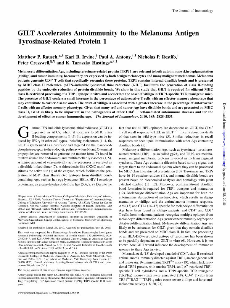
Effector memory cells are increased in the presence of GILTand following development of vitiligo
Because a direct consequence of Ag presentation is T cell activation,we anticipated diminished activation of TRP1-specific T cells inGILT-deficient animals. We examined markers of naive and Ag-experienced T cells in TRP1tg and GILT2/2TRP1tg mice. Withinthe CD4+Vb14+ population, the percentages of CD62L2CD44+
(effector memory), CD62L+CD44+ (central memory), and CD62L+
CD442 (naive) populations were determined in the skin-draininglymph nodes. Initially, we evaluated 10- to 20-wk-old TRP1tg micewith and without GILT, because we would have anticipated thegreatest difference in T cell phenotype in this age range based onthe onset to vitiligo analysis (Fig. 3). However, the results in thisage range were highly variable (data not shown), most likely due tothe extremely variable onset of spontaneous disease. When we re-stricted the analysis to mice ,10 wk old for a more uniform pop-ulation, we observed a decrease in effector memory T cells in GILT-deficient mice and an increase in effectormemory T cells correlatingwith vitiligo onset (Fig. 6). Themajority of the TRP1-specific T cellsin GILT2/2TRP1tgmice and TRP1tgmice (with or without vitiligo)were naive (Fig. 6). Comparison of GILT2/2TRP1tg with allTRP1tg mice showed an increase in effector memory cells from8.0 to 18% and a corresponding decrease in naive cells from 81 to
FIGURE 5. Increased percentage of TRP1-specific T cells in GILT-deficient mice. A, Thymus, spleen, and lymph node cells were enumerated from TRP1tg
and GILT2/2TRP1tg mice. No differences in lymphoid organ size were observed (p = 0.80, 0.75, and 0.23, respectively). B and C, Representative dot plots of
forward scatter and side scatter gates of TRP1tg mice on the wild-type and GILT2/2 backgrounds. B, Similar percentages of both CD4+ and CD8+ T cells
developed inTRP1tg andGILT2/2TRP1tgmice.C, CD4+Vb14+ T cellswere present in the peripheral lymphoid organs.Not all CD4+ cells expressedVb14, and
Vb14+CD42 cells were present (see Supplemental Fig. 1).D, Comparison of the percentage of CD4+ single-positive Vb14+ cells in the thymus and CD4+Vb14+
cells in the spleen and lymph node of TRP1tg andGILT2/2TRP1tgmice relative to the total cell number. An increased percentage of transgenic T cells was seen
in the thymus (pppp#0.001), spleen (ppp#0.01), and lymphnode (pppp#0.001) in theGILT2/2background.E, Comparison of thepercentage ofCD4+ single-
positive Vb142 cells in the thymus and CD4+Vb142 cells in the spleen and lymph node relative to the total cell number. No differences in the percentage of
nontransgenicCD4+Tcellswere observed. The data inD andE represent six pooledexperiments ofn=19mice per group,whichwere comparedwith two sample
independent t tests. F, Comparison of the percentage of CD4+TCRV+ T cells in the forward scatter and side scatter gates of lymph node cells from TRP1tg and
GILT2/2TRP1tgmice. Both strains expressed a diverse array of endogenous TCR chains; therewas an increased percentage of CD4+Vb14+ T cells in theGILT-
deficientmice (ppp# 0.01). The data represent two pooled experiments of n = 6mice per group, whichwere compared by two sample independent t tests.G, No
difference in the percentage ofCD25+FoxP3+ T cells in theCD4+ gate of lymphnode cells fromTRP1tg andGILT2/2TRP1tgmicewas observed (p= 0.58). The
data represent two pooled experiments of n = 6 mice per group, which were compared by two sample independent t tests.
2832 GILT ACCELERATES AUTOIMMUNITY TO TRP1
by guest on June 28, 2018http://w
ww
.jimm
unol.org/D
ownloaded from
68%, respectively (Fig. 6B, left). These differences are both statis-tically significant (p , 0.01 for each). Therefore, although there isa greater percentage of TRP1-specific T cells in GILT-deficientmice, a smaller percentage of these cells have an effector memoryphenotype. The reduction in effectormemory cells in GILT-deficientmice suggests that the delay in vitiligo onset in the absence of GILTmay reflect delayed activation of TRP1-specific T cells.A secondary analysis was performed dividing the TRP1tg group
into micewith andwithout vitiligo (Fig. 6B, right). GILT2/2TRP1tgmice with vitiligo were not included in the analysis because duringthe time course of this experiment, no GILT2/2TRP1tg mice de-veloped vitiligo by 9 wk of age. In GILT-expressing TRP1tg micewith vitiligo, a mean of 51% of the transgenic T cells were naive anda mean of 34%were effector memory cells, whereas inmicewithoutvitiligo 73% were naive and 13% were effector memory cells (Fig.6B, right). Thus, effector memory cells were increased, whereasnaive cells were reduced in mice with vitiligo. The differences
between the TRP1tg mice with and without vitiligo in both thepercentage of naive (p # 0.001) and effector memory TRP1-specific transgenic T cells (p# 0.001) were statistically significant.Therefore, increased T cell activation as measured by an increasedpercentage of effectormemory cells correlates with the developmentof vitiligo. In summary, these data support the idea that GILT facil-itates class II-restricted processing of TRP1, thereby enhancingT cell activation and accelerating vitiligo.
DiscussionTRP1 is a melanocyte differentiation Ag that is relevant to bothautoimmune skin depigmentation (vitiligo) and melanoma, be-cause it is expressed by both benign melanocytes and many ma-lignant melanomas. TRP1 contains disulfide bonds (11, 12), ispresented on MHC class II (10, 18, 22, 30, 31), and, thus, could besubject to GILT-mediated reduction in the late endosomes andlysosomes prior to class II-restricted presentation. The data pre-sented in this study show that GILT is necessary for efficientgeneration of an I-Ab–restricted TRP1 epitope in vitro, identi-fying a new GILT-dependent epitope in a disease-relevant Ag(Figs. 1, 2). GILT is required for detection of class II-restrictedpresentation of this epitope in B cells (Fig. 1C). However, in thepresence of a large amount of Ag, a small amount of GILT-independent processing is observed in DCs (Fig. 2C). The differ-ential abilities of GILT2/2 DCs and B cells to process largeamounts of TRP1 might be anticipated given the superior Agpresentation capability of DCs (32). Given that other melanocytedifferentiation Ags, including tyrosinase, TRP2, and gp100/Pmel17, contain disulfide bonds (11, 12, 33) and are presentedon MHC class II (30, 34–37), and that prior studies demonstratedthat an epitope of human tyrosinase is partially GILT dependentin vitro (6), GILT will most likely be of general importance in theMHC class II-restricted presentation of this group of melanomaand melanocyte Ags.The TRP1tg mouse strain generated by Muranski et al. (18)
allowed us to evaluate the role of GILT in the in vivo autoimmuneresponse. Because this strain expresses a TCR specific for a natu-rally occurring epitope of an endogenously expressed Ag, it rep-resents an ideal model to study the development and function ofT cells relevant to the pathogenesis of autoimmune disease andantitumor responses to differentiation Ags. Remarkably, GILT ex-pression accelerated the development of an in vivo TRP1-mediatedimmune response, resulting in spontaneous vitiligo (Fig. 3B), whichis the first demonstration that GILT can exacerbate an autoimmunedisease. Consistent with severely diminished Ag processing of theTRP1 epitope observed in GILT2/2 APCs in vitro (Figs. 1, 2),GILT2/2TRP1tg mice have a significant delay in the onset of spon-taneous vitiligo (Fig. 3B). Interestingly, GILT-deficient TRP1tgmice eventually develop spontaneous vitiligowith the same severityas those expressing GILT, suggesting that the low level of epitopegeneration in the absence of GILT is sufficient to produce diseaseover time. Because GILT is also expressed in T cells (26), we per-formed adoptive transfer experiments to determine whether GILTexpression in APCs was necessary for the exacerbated autoimmu-nity. Transfer of TRP1-specific T cells into GILT-deficient RAG2/2
compared with GILT-expressing RAG2/2 hosts resulted in de-creased vitiligo severity as well as delayed onset, demonstratingdefinitively that Ag processing in the absence of GILT attenuatesTRP1-mediated vitiligo. Therefore, GILT can play a substantial rolein the in vivo development of an autoimmune disease, which in thiscase is also a surrogate for an antimelanoma response (18, 22, 23).The progression of other CD4+ T cell-mediated diseases in whichthe primary target Ag contains disulfide bonds may similarly bemodulated by GILT.
FIGURE 6. Effector memory cells are increased in the presence of GILT
and following development of vitiligo. Cervical, axillary, and inguinal
lymph node cells from age-matched, 4- to 9-wk-old TRP1tg and GILT2/2
TRP1tg micewere analyzed. Staining with CD44 and CD62Lwas evaluated
in the CD4+Vb14+ population. A, Representative dot plots demonstrating
CD44 and CD62L staining in CD4+Vb14+ cells in GILT2/2TRP1tg mice
without vitiligo, TRP1tg mice without vitiligo, and TRP1tg mice with vit-
iligo. B, Bar graphs summarizing the mean percentage of TRP1-specific
transgenic T cells with a CD62L+CD442 naive and CD62L2CD44+ effector
memory phenotype 6 SEM for seven pooled experiments comparing
GILT2/2TRP1tg mice (n = 14) with TRP1tg mice (n = 19, consisting of
14 mice without vitiligo and 5 mice with vitiligo). Comparison of GILT2/2
TRP1tg mice and all TRP1tg mice by independent sample t tests showed
a statistically significant decrease in the percentage of naive cells (ppp #
0.01) and increase in the percentage of effector memory cells (ppp # 0.01)
in the presence of GILT. A secondary analysis was performed dividing the
TRP1tg mice into mice with or without vitiligo. A one-way ANOVA was
performed separately for the percentage of naive and the percentage of
effector memory T cells in the three groups of mice (GILT2/2TRP1tg mice
without vitiligo, TRP1tg mice without vitiligo, and TRP1tg mice with vit-
iligo). There was an increase in the percentage of TRP1-specific effector
memory T cells (pppp# 0.001) and a corresponding decrease in naive cells
(pppp # 0.001) in TRP1tg mice with vitiligo compared with TRP1tg mice
without vitiligo. There were significant differences between GILT2/2
TRP1tg mice and TRP1tg mice with vitiligo in both naive cells (pppp #
0.001) and effector memory cells (pppp# 0.001). No significant differences
were observed between TRP1tg mice without disease and GILT2/2TRP1tg
mice for either naive (p = 0.20) or effector memory cells (p = 0.54).
The Journal of Immunology 2833
by guest on June 28, 2018http://w
ww
.jimm
unol.org/D
ownloaded from

GILT expression in T cells could, in theory, affect the de-velopment of the autoimmune phenotype in the TRP1tg system.The absence of GILT in T cells results in increased T cell pro-liferation and cytotoxic function (26), which may be due to de-creased superoxide dismutase 2 activity and increased levels ofreactive oxygen species (38). GILT expression increases withT cell maturation from double-positive to single-positive thymo-cytes to peripheral T cells (39) and may raise the threshold forT cell sensitivity to self Ags. Indeed, GILT2/2 T cells induce moresevere hyperglycemia following streptozotocin injection, a CD8+
T cell-mediated model of diabetes that excludes GILT’s role inclass II-restricted Ag processing (39). In contrast, we observe di-minished severity and delayed onset of vitiligo in GILT-deficienthosts in our model, which most likely reflects GILT’s role in Agprocessing of TRP1 and decreased peripheral epitope generation.GILT could exacerbate autoimmunity by altering T cell devel-
opment or enhancing peripheral activation of autoreactive T cells.Unexpectedly, GILT2/2TRP1tg mice generated increased percen-tages of TRP1-specific T cells (Fig. 5C, 5D). Although we cannotentirely exclude a minor role for GILT in T cells that leads to in-creased T cell numbers, GILT’s expression in T cells is unlikely toaccount for our findings. Enhanced proliferation of all T cells lack-ing GILT would be expected to lead to global T cell expansion.However, we demonstrated that the expansion was restricted tothe TRP1-specific Vb14+ T cells (Fig. 5D–F). These findings sug-gest that the absence of GILT influenced the development of T cellsspecific for a GILT-dependent epitope in a manner similar to Agdeficiency (i.e., because TRP1 cannot be efficiently presented asa self Ag in the absence of GILT, more TRP1-specific T cells de-velop). For example, in class I-restricted, tyrosinase-specific TCRtransgenic mice and in dual transgenic mice expressing a class II-restricted, HEL-specific TCR and a membrane-bound form of HELunder the TRP2 promoter to create a melanocyte-specific neoanti-gen, autoreactive T cells are increased inAg-deficient mice (40, 41).Possible explanations for the apparent contradiction of increased
autoreactive T cells in GILT-deficient animals with delayed autoim-munity are increased Treg cells or diminished T cell activation inGILT-deficient animals. Treg cells have been shown to control auto-immunity in this and related models. For example, FoxP3-deficientTRP1tg mice develop spontaneous vitiligo earlier than FoxP3-expressing TRP1tg mice (21). Likewise, development of vitiligoin chimeric class I-restricted, tyrosinase-specific TCR transgenicmice is accelerated with anti-CD25 mAb treatment (42). In anothermousemodelwith vitiligo,mice expressing aHEL-specific, class II-restricted TCR have an increased percentage of clonotype-specificT cells that are CD4+CD25+ in HEL-expressing mice comparedwith HEL-deficient mice (40). Although Treg cells are importantin controlling autoimmunity and vitiligo in particular, there were nodifferences in the percentages of CD4+CD25+FoxP3+ Treg cellsbetween TRP1tg and GILT2/2TRP1tg mice (Fig. 5G), suggestingthat GILT did not affect the development of Treg cells, and a differ-ence in the percentage of Treg cells did not contribute to the delayedonset of vitiligo in the absence of GILT.We demonstrated that an increase in TRP1-specific cells with an
effector memory phenotype correlates with vitiligo onset, and thatthere is a decrease in effector memory T cells in GILT-deficientmice (Fig. 6B). Thus, although more TRP1-specific T cells de-velop in the absence of GILT, a smaller percentage becomes ac-tivated. The changes observed in the absence of GILT are similarto those seen in the absence of Ag. A recent study observed T cellswith an effector memory phenotype in Ag-expressing TRP1tgmice, but not in Ag-deficient TRP1BwRAG2/2TRP1tg mice(21). Similarly, in HEL-specific, class II-restricted TCR transgenicmice, CD4+CD44+ T cells are increased in the lymph node in the
presence of melanocyte-restricted HEL expression (40). Presum-ably, inefficient class II-restricted processing of TRP1 in GILT-deficient mice diminishes T cell activation, and thus delays theonset of vitiligo.Recent studies using adoptive transfer of TRP1BwRAG2/2
TRP1tg T cells to treat established melanoma tumors have elu-cidated the mechanism of melanoma destruction (20, 21). Follow-ing adoptive transfer into Ag-expressing recipients, TRP1-specificT cells that developed in the absence of Ag and RAG secrete IFN-g,express granzyme B and Fas ligand, and demonstrate cytotoxicity;furthermore, melanoma destruction is not dependent on NKT orNK cells (20, 21). A similar mechanism may be responsible formelanocyte destruction, resulting in spontaneous vitiligo in Ag-expressing, RAG-expressing TRP1tg mice. TRP1tg T cells that de-velop in the presence of Ag and RAG are also capable of producingIFN-g (M. Rausch and K. Hastings, unpublished data). Similar tomelanoma cells, melanocytes can upregulate class II expression inresponse to IFN-g and present class II-restricted Ags (43–48), sug-gesting that they may become sensitized to class II-restricted cyto-toxicity by IFN-g–secreting, melanocyte-specific CD4+ T cells (20,37, 49, 50).The TRP1tg model has revealed a role for GILT in the de-
velopment and function of T cells important in autoimmunity andanti-cancer immunity. Given that many melanocyte differentiationAgs have multiple disulfide bonds and are presented on MHC classII, additional Ags in this group are likely to be GILT dependent.Therefore, GILT is likely to have a more general role in the devel-opment of an effective antimelanoma immune response.
AcknowledgmentsWe thank Drs. Denise Roe and Bradley Appelhans for help with statistical
analyses.
DisclosuresThe authors have no financial conflicts of interest.
References1. Arunachalam, B., U. T. Phan, H. J. Geuze, and P. Cresswell. 2000. Enzymatic
reduction of disulfide bonds in lysosomes: characterization of a gamma-interferon-inducible lysosomal thiol reductase (GILT). Proc. Natl. Acad. Sci.USA 97: 745–750.
2. Honey, K., M. Duff, C. Beers, W. H. Brissette, E. A. Elliott, C. Peters, M. Maric,P. Cresswell, and A. Rudensky. 2001. Cathepsin S regulates the expression ofcathepsin L and the turnover of g-interferon-inducible lysosomal thiol reductasein B lymphocytes. J. Biol. Chem. 276: 22573–22578.
3. Lackman, R. L., A. M. Jamieson, J. M. Griffith, H. Geuze, and P. Cresswell.2007. Innate immune recognition triggers secretion of lysosomal enzymes bymacrophages. Traffic 8: 1179–1189.
4. Luster, A. D., R. L. Weinshank, R. Feinman, and J. V. Ravetch. 1988. Molecularand biochemical characterization of a novel g-interferon-inducible protein. J.Biol. Chem. 263: 12036–12043.
5. Maric, M., B. Arunachalam, U. T. Phan, C. Dong, W. S. Garrett, K. S. Cannon,C. Alfonso, L. Karlsson, R. A. Flavell, and P. Cresswell. 2001. Defective antigenprocessing in GILT-free mice. Science 294: 1361–1365.
6. Haque, M. A., P. Li, S. K. Jackson, H. M. Zarour, J. W. Hawes, U. T. Phan,M. Maric, P. Cresswell, and J. S. Blum. 2002. Absence of g-interferon-induciblelysosomal thiol reductase in melanomas disrupts T cell recognition of selectimmunodominant epitopes. J. Exp. Med. 195: 1267–1277.
7. Phan, U. T., B. Arunachalam, and P. Cresswell. 2000. Gamma-interferon-induciblelysosomal thiol reductase (GILT): maturation, activity, and mechanism of action. J.Biol. Chem. 275: 25907–25914.
8. Hastings, K. T., R. L. Lackman, and P. Cresswell. 2006. Functional requirementsfor the lysosomal thiol reductase GILT in MHC class II-restricted antigen pro-cessing. J. Immunol. 177: 8569–8577.
9. Sealy, R., W. Chaka, S. Surman, S. A. Brown, P. Cresswell, and J. L. Hurwitz.2008. Target peptide sequence within infectious human immunodeficiency virustype 1 does not ensure envelope-specific T-helper cell reactivation: influences ofcysteine protease and gamma interferon-induced thiol reductase activities. Clin.Vaccine Immunol. 15: 713–719.
10. Wang, S., S. Bartido, G. Yang, J. Qin, Y. Moroi, K. S. Panageas, J. J. Lewis, andA. N. Houghton. 1999. A role for a melanosome transport signal in accessing the
2834 GILT ACCELERATES AUTOIMMUNITY TO TRP1
by guest on June 28, 2018http://w
ww
.jimm
unol.org/D
ownloaded from

MHC class II presentation pathway and in eliciting CD4+ T cell responses. J.Immunol. 163: 5820–5826.
11. Garcıa-Borron, J. C., and F. Solano. 2002. Molecular anatomy of tyrosinase andits related proteins: beyond the histidine-bound metal catalytic center. PigmentCell Res. 15: 162–173.
12. Negroiu, G., R. A. Dwek, and S. M. Petrescu. 2000. Folding and maturation oftyrosinase-related protein-1 are regulated by the post-translational formation ofdisulfide bonds and by N-glycan processing. J. Biol. Chem. 275: 32200–32207.
13. Naughton, G. K., M. Eisinger, and J. C. Bystryn. 1983. Antibodies to normalhuman melanocytes in vitiligo. J. Exp. Med. 158: 246–251.
14. Lang, K. S., C. C. Caroli, A. Muhm, D. Wernet, A. Moris, B. Schittek,E. Knauss-Scherwitz, S. Stevanovic, H. G. Rammensee, and C. Garbe. 2001.HLA-A2 restricted, melanocyte-specific CD8+ T lymphocytes detected in viti-ligo patients are related to disease activity and are predominantly directedagainst MelanA/MART1. J. Invest. Dermatol. 116: 891–897.
15. Mandelcorn-Monson, R. L., N. H. Shear, E. Yau, S. Sambhara, B. H. Barber,D. Spaner, and M. A. DeBenedette. 2003. Cytotoxic T lymphocyte reactivity togp100, MelanA/MART-1, and tyrosinase, in HLA-A2-positive vitiligo patients.J. Invest. Dermatol. 121: 550–556.
16. Ogg, G. S., P. Rod Dunbar, P. Romero, J. L. Chen, and V. Cerundolo. 1998. Highfrequency of skin-homing melanocyte-specific cytotoxic T lymphocytes in au-toimmune vitiligo. J. Exp. Med. 188: 1203–1208.
17. Palermo, B., R. Campanelli, S. Garbelli, S. Mantovani, E. Lantelme, V. Brazzelli,M. Ardigo, G. Borroni, M. Martinetti, C. Badulli, et al. 2001. Specific cytotoxicT lymphocyte responses against Melan-A/MART1, tyrosinase and gp100 in vitiligoby the use of major histocompatibility complex/peptide tetramers: the roleof cellular immunity in the etiopathogenesis of vitiligo. J. Invest. Dermatol. 117:326–332.
18. Muranski, P., A. Boni, P. A. Antony, L. Cassard, K. R. Irvine, A. Kaiser,C. M. Paulos, D. C. Palmer, C. E. Touloukian, K. Ptak, et al. 2008. Tumor-specific Th17-polarized cells eradicate large established melanoma. Blood 112:362–373.
19. Smyth, I. M., L. Wilming, A. W. Lee, M. S. Taylor, P. Gautier, K. Barlow, J. Wallis,S. Martin, R. Glithero, B. Phillimore, et al. 2006. Genomic anatomy of the Tyrp1(brown) deletion complex. Proc. Natl. Acad. Sci. USA 103: 3704–3709.
20. Quezada, S. A., T. R. Simpson, K. S. Peggs, T. Merghoub, J. Vider, X. Fan,R. Blasberg, H. Yagita, P. Muranski, P. A. Antony, et al. 2010. Tumor-reactiveCD4+ T cells develop cytotoxic activity and eradicate large established mela-noma after transfer into lymphopenic hosts. J. Exp. Med. 207: 637–650.
21. Xie, Y., A. Akpinarli, C. Maris, E. L. Hipkiss, M. Lane, E. K. Kwon, P. Muranski,N. P. Restifo, and P. A. Antony. 2010. Naive tumor-specific CD4+ T cells differ-entiated in vivo eradicate established melanoma. J. Exp. Med. 207: 651–667.
22. Overwijk, W. W., D. S. Lee, D. R. Surman, K. R. Irvine, C. E. Touloukian,C. C. Chan, M. W. Carroll, B. Moss, S. A. Rosenberg, and N. P. Restifo. 1999.Vaccination with a recombinant vaccinia virus encoding a “self” antigen inducesautoimmune vitiligo and tumor cell destruction in mice: requirement for CD4+
T lymphocytes. Proc. Natl. Acad. Sci. USA 96: 2982–2987.23. Gogas, H., J. Ioannovich, U. Dafni, C. Stavropoulou-Giokas, K. Frangia, D. Tsoutsos,
P. Panagiotou, A. Polyzos, O. Papadopoulos, A. Stratigos, et al. 2006. Prognosticsignificance of autoimmunity during treatment of melanoma with interferon. N. Engl.J. Med. 354: 709–718.
24. Fusenig, N. E., S. M. Amer, P. Boukamp, and P. K. Worst. 1978. Characteristicsof chemically transformed mouse epidermal cells in vitro and in vivo. Bull.Cancer 65: 271–279.
25. Inaba, K., M. Inaba, N. Romani, H. Aya, M. Deguchi, S. Ikehara, S. Muramatsu,and R. M. Steinman. 1992. Generation of large numbers of dendritic cells frommouse bone marrow cultures supplemented with granulocyte/macrophage colony-stimulating factor. J. Exp. Med. 176: 1693–1702.
26. Barjaktarevic, I., A. Rahman, S. Radoja, B. Bogunovic, A. Vollmer, S. Vukmanovic,and M. Maric. 2006. Inhibitory role of IFN-gamma-inducible lysosomal thiol re-ductase in T cell activation. J. Immunol. 177: 4369–4375.
27. von Boehmer, H. 1990. Developmental biology of T cells in T cell-receptortransgenic mice. Annu. Rev. Immunol. 8: 531–556.
28. Borgulya, P., H. Kishi, Y. Uematsu, and H. von Boehmer. 1992. Exclusion andinclusion of alpha and beta T cell receptor alleles. Cell 69: 529–537.
29. McGargill,M.A., D.Mayerova,H. E. Stefanski, B.Koehn, E.A. Parke, S. C. Jameson,A. Panoskaltsis-Mortari, and K. A. Hogquist. 2002. A spontaneous CD8 T cell-dependent autoimmune disease to an antigen expressed under the human keratin 14promoter. J. Immunol. 169: 2141–2147.
30. Robbins, P. F., M. El-Gamil, Y. F. Li, G. Zeng, M. Dudley, and S. A. Rosenberg.2002. Multiple HLA class II-restricted melanocyte differentiation antigens arerecognized by tumor-infiltrating lymphocytes from a patient with melanoma. J.Immunol. 169: 6036–6047.
31. Touloukian, C. E., W. W. Leitner, P. F. Robbins, Y. F. Li, X. Kang, R. Lapointe,P. Hwu, S. A. Rosenberg, and N. P. Restifo. 2002. Expression of a “self-”antigen
by human tumor cells enhances tumor antigen-specific CD4+ T-cell function.Cancer Res. 62: 5144–5147.
32. Delon, J., N. Bercovici, G. Raposo, R. Liblau, and A. Trautmann. 1998. Antigen-dependent and -independent Ca2+ responses triggered in T cells by dendriticcells compared with B cells. J. Exp. Med. 188: 1473–1484.
33. Berson, J. F., D. C. Harper, D. Tenza, G. Raposo, and M. S. Marks. 2001. Pmel17initiates premelanosome morphogenesis within multivesicular bodies. Mol. Biol.Cell 12: 3451–3464.
34. Kobayashi, H., T. Kokubo, K. Sato, S. Kimura, K. Asano, H. Takahashi,H. Iizuka, N. Miyokawa, and M. Katagiri. 1998. CD4+ T cells from peripheralblood of a melanoma patient recognize peptides derived from nonmutated ty-rosinase. Cancer Res. 58: 296–301.
35. Parkhurst, M. R., J. P. Riley, P. F. Robbins, and S. A. Rosenberg. 2004. Inductionof CD4+ Th1 lymphocytes that recognize known and novel class II MHC re-stricted epitopes from the melanoma antigen gp100 by stimulation with recom-binant protein. J. Immunother. 27: 79–91.
36. Topalian, S.L., M.I. Gonzales, M. Parkhurst, Y.F. Li, S. Southwood, A. Sette,S.A. Rosenberg, and P.F. Robbins. 1996. Melanoma-specific CD4+ T cellsrecognize nonmutated HLA-DR-restricted tyrosinase epitopes. J. Exp. Med.183:1965–1971.
37. Touloukian, C. E., W. W. Leitner, S. L. Topalian, Y. F. Li, P. F. Robbins,S. A. Rosenberg, and N. P. Restifo. 2000. Identification of a MHC class II-restricted human gp100 epitope using DR4-IE transgenic mice. J. Immunol. 164:3535–3542.
38. Bogunovic, B., M. Stojakovic, L. Chen, and M. Maric. 2008. An unexpectedfunctional link between lysosomal thiol reductase and mitochondrial manganesesuperoxide dismutase. J. Biol. Chem. 283: 8855–8862.
39. Maric,M., I.Barjaktarevic,B.Bogunovic,M.Stojakovic,C.Maric, andS.Vukmanovic.2009. Cutting edge: developmental up-regulation of IFN-gamma-inducible lysosomalthiol reductase expression leads to reduced T cell sensitivity and less severe autoimmu-nity. J. Immunol. 182: 746–750.
40. Lambe, T., J. C. Leung, T. Bouriez-Jones, K. Silver, K. Makinen, T. L. Crockford,H. Ferry, J. V. Forrester, and R. J. Cornall. 2006. CD4 T cell-dependent autoim-munity against a melanocyte neoantigen induces spontaneous vitiligo and dependsupon Fas-Fas ligand interactions. J. Immunol. 177: 3055–3062.
41. Nichols, L. A., Y. Chen, T. A. Colella, C. L. Bennett, B. E. Clausen, andV. H. Engelhard. 2007. Deletional self-tolerance to a melanocyte/melanoma anti-gen derived from tyrosinase is mediated by a radio-resistant cell in peripheraland mesenteric lymph nodes. J. Immunol. 179: 993–1003.
42. Gregg, R. K., L. Nichols, Y. Chen, B. Lu, and V. H. Engelhard. 2010. Mecha-nisms of spatial and temporal development of autoimmune vitiligo in tyrosinase-specific TCR transgenic mice. J. Immunol. 184: 1909–1917.
43. Tsujisaki, M., M. Igarashi, K. Sakaguchi, M. Eisinger, M. Herlyn, and S. Ferrone.1987. Immunochemical and functional analysis of HLA class II antigens inducedby recombinant immune interferon on normal epidermal melanocytes. J. Immunol.138: 1310–1316.
44. Aubock, J., D. Niederwieser, N. Romani, P. Fritsch, and C. Huber. 1985. Humaninterferon-gamma induces expression of HLA-DR on keratinocytes and mela-nocytes. Arch. Dermatol. Res. 277: 270–275.
45. Hedley, S. J., R. Metcalfe, D. J. Gawkrodger, A. P. Weetman, and S. Mac Neil.1998. Vitiligo melanocytes in long-term culture show normal constitutive andcytokine-induced expression of intercellular adhesion molecule-1 and majorhistocompatibility complex class I and class II molecules. Br. J. Dermatol.139: 965–973.
46. Herlyn, M., D. Guerry, and H. Koprowski. 1985. Recombinant gamma-interferoninduces changes in expression and shedding of antigens associated with normalhuman melanocytes, nevus cells, and primary and metastatic melanoma cells. J.Immunol. 134: 4226–4230.
47. Houghton, A. N., T. M. Thomson, D. Gross, H. F. Oettgen, and L. J. Old. 1984.Surface antigens of melanoma and melanocytes: specificity of induction of Iaantigens by human gamma-interferon. J. Exp. Med. 160: 255–269.
48. Le Poole, I. C., T. Mutis, R. M. van den Wijngaard, W. Westerhof, T. Ottenhoff,R. R. de Vries, and P. K. Das. 1993. A novel, antigen-presenting function ofmelanocytes and its possible relationship to hypopigmentary disorders. J. Immu-nol. 151: 7284–7292.
49. Manici, S., T. Sturniolo,M.A. Imro, J. Hammer, F. Sinigaglia, C.Noppen,G. Spagnoli,B. Mazzi, M. Bellone, P. Dellabona, and M. P. Protti. 1999. Melanoma cells presenta MAGE-3 epitope to CD4+ cytotoxic T cells in association with histocompatibilityleukocyte antigen DR11. J. Exp. Med. 189: 871–876.
50. Zarour, H. M., J. M. Kirkwood, L. S. Kierstead, W. Herr, V. Brusic, C. L. Slingluff,Jr., J. Sidney, A. Sette, and W. J. Storkus. 2000. Melan-A/MART-1(51-73) repre-sents an immunogenic HLA-DR4-restricted epitope recognized by melanoma-reactive CD4+ T cells. Proc. Natl. Acad. Sci. USA 97: 400–405.
The Journal of Immunology 2835
by guest on June 28, 2018http://w
ww
.jimm
unol.org/D
ownloaded from