glutathione, ascorbate, and cellular protection1 · glutathione is exported by the liver to the...
TRANSCRIPT
ICANCÕRRESEARCH(SUPPL.)54,
Glutathione, Ascorbate, and Cellular Protection1
Alton Meister
Department of Biochemistry, Cornell University Medical College, New York, New York 10021
Introduction
Glutathione. a tripeptide thiol present in virtually all animal cells, issynthesized within many cells from its constituent amino acids (glutamate, cystcine, glycine); these "nonessential" amino acids can be
synthesized within the body and are also obtained from the diet.Glutathione is also synthesized by tumors, some of which (notablydrug- and radiation-resistant tumors) exhibit high cellular levels of
glutathione and high capacity for the synthesis of glutathione. Wereported at previous conferences in this series on the increase ofcellular radiosensitivity that occurs after administration of an inhibitorof glutathione synthesis (1, 2) and on the effects of modulation ofglutathione metabolism (3, 4). These and related topics (5, 6) will besummarized here with emphasis on some current developments.
Glutathione is probably the most important cellular antioxidant.Interestingly, Fahey and Sundquist (7) found strong evidence for anevolutionary link between glutathione and aerobic eukaryotic metabolism; the findings indicate that glutathione evolved as a molecule thatprotects cells against oxygen toxicity. Although there is currentlymuch interest in the hypothesis that oxidative phenomena may lead toa variety of pathological states, and that antioxidants may play asignificant protective role, the important role that glutathione plays inthe protection of cells has sometimes been insufficiently appreciated.Cells that are deprived of glutathione typically suffer severe oxidativedamage associated with mitochondria! degeneration. Analogous effects are not always found when there is a deficiency of certain othercellular components that are thought to act as antioxidants. It has longbeen known that the antioxidant ascorbate is required in the diet ofhumans and certain other animals such as the guinea pig (but not bymany other animals, including some commonly used in laboratoryexperiments; e.g., mice, rats, rabbits). The ascorbate deficiency syndrome, scurvy, which is associated with oxidative inactivation ofcertain enzymes, can be prevented in humans by administration of aslittle as 10 mg/day of ascorbate. The officially recommended dailydose of ascorbate for humans is 30-100 mg/day (depending upon the
country); although much larger doses of ascorbate than this are takenby some individuals, it is estimated that a substantial proportion of thehuman population takes in relatively small amounts. The question asto whether larger doses of ascorbate and also of other "antioxidants"
would have beneficial effects has often been discussed but remainsunsettled.
Experimental findings summarized here that are relevant to thisquestion include: (a) the observation that glutathione deficiency inanimals that are unable to synthesize ascorbate (newborn rats, guineapigs) is lethal and that death can be prevented by giving high doses ofascorbate; and (b) the onset of scurvy in guinea pigs that are fed a dietdeficient in ascorbate is substantially delayed by giving glutathionemonoethyl ester, a glutathione delivery agent (6, 8).
Various questions about the functions of putative antioxidant compounds need to be considered in relation to the functions of cellularglutathione. As discussed here, one such function, shown in vivo (9),is to reduce dehydroascorbate to ascorbate. Glutathione also keeps
1 Presented at the 4th International Conference on Anticarcinogenesis & Radiation
Protection. April 1H-23, 1993, Baltimore. MD. The research described here that wascarried out in the author's laboratory was supported in part by NIH Grant 2 R37 DK12034
from the United States Public Health Service.
a-tocopherol in its reduced form, either by a direct reaction or by apathway involving ascorbate (10-17). Glutathione. which has the
important function of maintaining the reducing milieu of cells, isundoubtedly involved in the reduction of many cellular components;e.g., other tocopherols and ß-carotenearc apparently also maintainedby glutathione-mediated reactions (e.g.. Réf.18).
An interesting aspect of glutathione metabolism and function relates to drug-resistant and radiation-resistant tumors that have high
levels of glutathione or exhibit high capacity for glutathione synthesis.Such tumors have a greater requirement for glutathione than do manynormal tissues and this provides a promising chemotherapeutic approach, which is considered below.
Biochemistry of Glutathione: Enzymology and TransportPhenomena
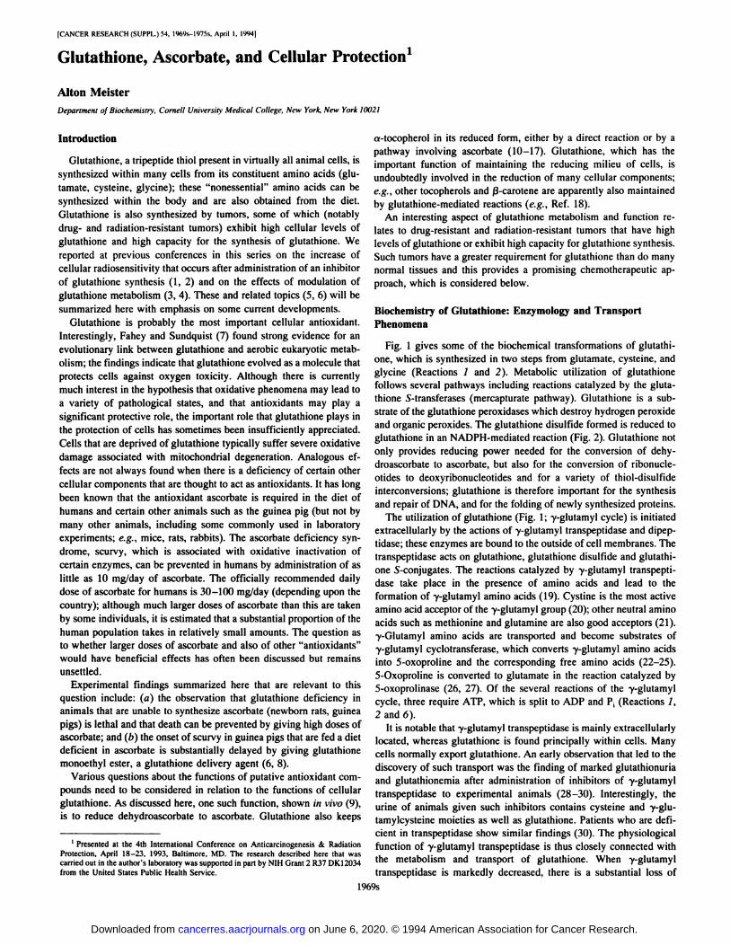
Fig. 1 gives some of the biochemical transformations of glutathione, which is synthesized in two steps from glutamate, cystcine, andglycine (Reactions / and 2). Metabolic utilization of glutathionefollows several pathways including reactions catalyzed by the glutathione S-transferases (mercapturate pathway). Glutathione is a sub
strate of the glutathione peroxidases which destroy hydrogen peroxideand organic peroxides. The glutathione disulfide formed is reduced toglutathione in an NADPH-mediated reaction (Fig. 2). Glutathione not
only provides reducing power needed for the conversion of dehydroascorbate to ascorbate, but also for the conversion of ribonucle-otides to deoxyribonucleotides and for a variety of thiol-disulfide
interconversions; glutathione is therefore important for the synthesisand repair of DNA, and for the folding of newly synthesized proteins.
The utilization of glutathione (Fig. 1; y-glutamyl cycle) is initiatedextracellularly by the actions of y-glutamyl transpeptidase and dipep-
tidase; these enzymes are bound to the outside of cell membranes. Thetranspeptidase acts on glutathione. glutathione disulfide and glutathione 5-conjugates. The reactions catalyzed by y-glutamyl transpepti
dase take place in the presence of amino acids and lead to theformation of y-glutamyl amino acids (19). Cystine is the most activeamino acid acceptor of the y-glutamyl group (20); other neutral amino
acids such as methionine and glutamine are also good acceptors (21).y-Glutamyl amino acids are transported and become substrates ofy-glutamyl cyclotransferase, which converts y-glutamyl amino acidsinto 5-oxoproline and the corresponding free amino acids (22-25).5-Oxoproline is converted to glutamate in the reaction catalyzed by5-oxoprolinase (26, 27). Of the several reactions of the y-glutamyl
cycle, three require ATP, which is split to ADP and P¡(Reactions /,2 and 6).
It is notable that y-glutamyl transpeptidase is mainly extracellularly
located, whereas glutathione is found principally within cells. Manycells normally export glutathione. An early observation that led to thediscovery of such transport was the finding of marked glutathionuriaand glutathionemia after administration of inhibitors of y-glutamyltranspeptidase to experimental animals (28-30). Interestingly, theurine of animals given such inhibitors contains cysteine and y-glu-
tamylcysteine moieties as well as glutathione. Patients who are deficient in transpeptidase show similar findings (30). The physiologicalfunction of y-glutamyl transpeptidase is thus closely connected withthe metabolism and transport of glutathione. When y-glutamyl
transpeptidase is markedly decreased, there is a substantial loss of
1969s
on June 6, 2020. © 1994 American Association for Cancer Research. cancerres.aacrjournals.org Downloaded from
GLUTATHIONE, ASCORBATE, AND CELLULAR PROTECTION
OXIDATION-REDUCTIONPATHWAYS
GSSG
MERCAPTURATEPATHWAY
2HJÃ )/ Glutamate
Y-Glu-AA
Fig. 1. Overall pathway of glutathione (GSH) metabolism. /. y-glutamylcysteinesynthetase; 2, glulathione synthetase; 3, y-glutamyl transpeptidase; 4, cysteinyl glycinehydrolases; 5, y-glutamyl cyclotransfera.se; 6, 5-oxoprolinase; 7, glutathione 5-trans-ferases; 8, transport and reduction of y-Glu-(Cys)2; 9, see Fig. 2. Reactions 1, 2, and 6
involve cleavage of ATP to ADP and P,.
-GSSG
Deoxyribonucleottd NADPH, H
NADP
V-GLU CYCLE
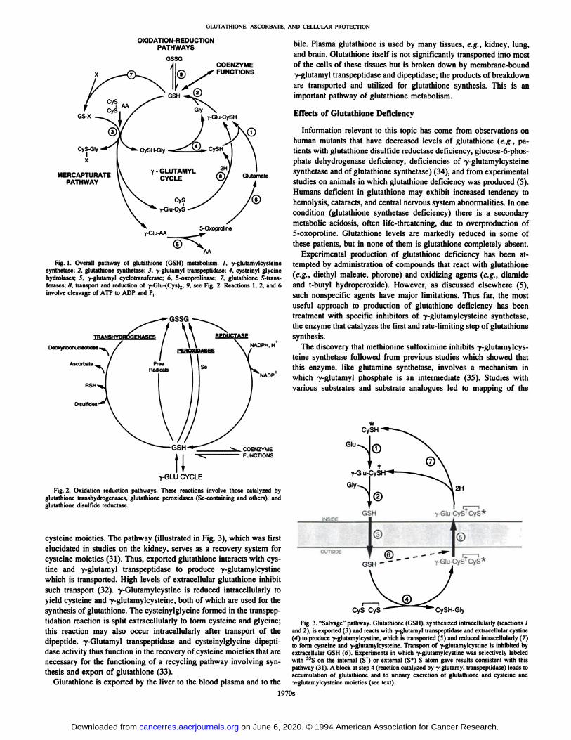
Fig. 2. Oxidation reduction pathways. These reactions involve those catalyzed byglutathione transhydrogenases, glutathione peroxidases (Se-containing and others), and
glutathione disulfidc reducÃase.
cysteine moieties. The pathway (illustrated in Fig. 3), which was firstelucidated in studies on the kidney, serves as a recovery system forcysteine moieties (31). Thus, exported glutathione interacts with cys-tine and y-glutamyl transpeptidase to produce y-glutamylcystine
which is transported. High levels of extracellular glutathione inhibitsuch transport (32). y-Glutamylcystine is reduced intracellularly toyield cysteine and y-glutamylcysteine, both of which are used for thesynthesis of glutathione. The cysteinylglycine formed in the transpep-
tidation reaction is split extracellularly to form cysteine and glycine;this reaction may also occur intracellularly after transport of thedipeptide. y-Glutamyl transpeptidase and cysteinylglycine dipepti-
dase activity thus function in the recovery of cysteine moieties that arenecessary for the functioning of a recycling pathway involving synthesis and export of glutathione (33).
Glutathione is exported by the liver to the blood plasma and to the
bile. Plasma glutathione is used by many tissues, e.g., kidney, lung,and brain. Glutathione itself is not significantly transported into mostof the cells of these tissues but is broken down by membrane-boundy-glutamyl transpeptidase and dipeptidase; the products of breakdown
are transported and utilized for glutathione synthesis. This is animportant pathway of glutathione metabolism.
Effects of Glutathione Deficiency
Information relevant to this topic has come from observations onhuman mutants that have decreased levels of glutathione (e.g., patients with glutathione disulfide reducÃasedeficiency, glucose-6-phos-phate dehydrogenase deficiency, deficiencies of y-glutamylcysteine
synthetase and of glutathione synthetase) (34), and from experimentalstudies on animals in which glutathione deficiency was produced (5).Humans deficient in glutathione may exhibit increased tendency tohemolysis, cataracts, and central nervous system abnormalities. In onecondition (glutathione synthetase deficiency) there is a secondarymetabolic acidosis, often life-threatening, due to overproduction of5-oxoproline. Glutathione levels are markedly reduced in some of
these patients, but in none of them is glutathione completely absent.Experimental production of glutathione deficiency has been at
tempted by administration of compounds that react with glutathione(e.g., diethyl maléate,phorone) and oxidizing agents (e.g., diamideand t-butyl hydroperoxide). However, as discussed elsewhere (5),
such nonspecific agents have major limitations. Thus far, the mostuseful approach to production of glutathione deficiency has beentreatment with specific inhibitors of y-glutamylcysteine synthetase,the enzyme that catalyzes the first and rate-limiting step of glutathione
synthesis.The discovery that methionine sulfoximine inhibits y-glutamylcys
teine synthetase followed from previous studies which showed thatthis enzyme, like glutamine synthetase, involves a mechanism inwhich y-glutamyl phosphate is an intermediate (35). Studies with
various substrates and substrate analogues led to mapping of the
CyS CyS - CySH-Gly
Fig. 3. "Salvage" pathway. Glutathione (GSH), synthesized intracellularly (reactions /
and 2), is exported (3) and reacts with y-glutamyl transpeptidase and extracellular cystine(4) to produce y-glutamylcystine, which is transported (5) and reduced intracellularly (7)to form cysteine and y-glutamylcysteine. Transport of y-glutamylcystine is inhibited byextracellular GSH (6). Experiments in which y-glutamylcystine was selectively labeledwith 15S on the internal (Sf) or external (S*) S atom gave results consistent with this
pathway (31). A block at step 4 (reaction catalyzed by y-glutamyl transpeptidase) leads to
accumulation of glutathione and to urinary excretion of glutathione and cysteine andy-glutamylcysteine moieties (see text).
1970s
on June 6, 2020. © 1994 American Association for Cancer Research. cancerres.aacrjournals.org Downloaded from
OLUTATHIONE, ASCORBATE. AND CELLULAR PROTECTION
active site ofglutamine synthetase and to design of selective inhibitorsof glutamine synthetase (a-ethyl methionine sulfoximine) and ofy-glutamylcysteine synthetase [prothionine sulfoximine, BSO,2 and
others (35-40)]. BSO and related compounds have very little orvirtually no effect on glutamine synthetase but inhibit y-glutamylcys
teine synthetase very effectively. BSO, now commercially available,has been widely used in in vitro experiments and in vivo. The effectsof glutathione deficiency induced by administration of BSO have beenextensively examined (see Ref. 5) and include the following:
1. Glutathione deficiency sensitizes cells to the effects of radiation,oxidative reactions, and to various toxic compounds. These effectshave been applied in chemotherapy and radiation systems (5, 41-44).
Tumor cell resistance associated with overproduction of glutathione isreversed by treatment with BSO in animal models and is currentlybeing tested clinically (45). Depletion of glutathione by treatment withbuthionine sulfoximine sensitizes cells to the toxic effects of heavymetals (46, 47), nitrogen mustards (48, 49), radiation (1, 2, 5),cisplatin (50), cyclophosphamide (51, 52), morphine (53), compoundsthat produce oxidative cytolysis (54), and others (55).
2. Glutathione deficiency leads to oxidative stress in many tissues(5). Mitochondria! and associated cell damage is found in mice treatedwith BSO. Mitochondria do not synthesize glutathione but obtain it bytransport from the cytosol. Several tissues of adult mice are affectedby administration of BSO, but in newborn rats and guinea pigs moreextensive damage is found and there is early mortality due to multi-
organ failure (56). Glutathione deficiency in these experimental systems serves as a model of endogenously produced oxidative stress.Mortality and tissue damage are significantly decreased by administration of glutathione esters or of ascorbate (5, 9).
3. Deficiency of glutathione leads to decreased reduction of dehy-
droascorbate to ascorbate in vivo. This is observed in newborn rats andguinea pigs, animals that cannot synthesize ascorbate, and also inadult mice, which can (6). In adult mice, glutathione deficiency leadsto induction of ascorbate synthesis in the liver (57), and this explainswhy BSO has less damaging effects on adult mice than it does onnewborn rats and guinea pigs.
4. Glutathione deficiency in newborn rats and mice leads to formation of ocular cataracts (9, 58, 59). Cataracts have also been foundin some patients with inherited glutathione disulfidc reducÃasedeficiency.
5. Treatment of peripheral blood mononuclear cells with BSO wasfound to markedly inhibit their proliferation (4), and later work hasconfirmed that glutathione deficiency decreases lectin-induced prolif
eration of lymphocytes (60). Although glutathione is required forproliferation, the mechanism of its function in this system is stillunsettled.
Application of Glutathione Depletion to the Treatment ofTumors: Sensitization of Tumors to Chemotherapyand to Radiation
Early studies showed that tumor cells that are resistant to alkylatingagents have increased levels of nonprotein thiol, later shown to beglutathione (see Ref. 5). The resistance of leukemic cells to phenyl-
alanine mustard was found not to be related to an effect on uptake orefflux of the mustard, but to be closely related to the cellular level ofglutathione (48, 49). The resistant cells converted phenylalanine mustard to a nontoxic compound in a glutathione-dependent dehydrochlo-
rination reaction.After development of the amino acid sulfoximine inhibitors of
y-glutamylcysteinc synthetase, it was suggested that treatment with
~ The abbreviation used is: BSO. buthionine sulfoximine.
these agents might make tumor cells more sensitive to chemotherapyand to radiation treatment (41 ). The potential usefulness of BSO in thesensitization of cells to radiation was first directly shown in studies onseveral human lymphoid cell lines (1, 2). Cells that had 4-5% of the
control levels of glutathione were found to be much more sensitivethan the controls to the effects of y-irradiation. It was found that
treatment with BSO of mice bearing B16 melanomas sensitizes thisradio-resistant tumor to radiation (61, 62). Treatment with BSO alone
did not affect tumor growth, but treatment with BSO and radiation ledto significant decrease in the size of the tumor and to an increase in thelongevity of the tumor-bearing mice. It was also found that treatment
of resistant Icukemias with BSO led to sensitization of the tumors tophenylalanine mustard (48. 49). In studies with mice bearing suchresistant tumors, i.p. infusion of BSO led to sensitization of the tumorsand to an increase in the life span of the treated animals. Additionalstudies on the relationship between glutathione levels and the expression of primary drug resistance and cross-resistance in human ovarian
cancer cell lines, together with studies in which an i.p. model ofhuman ovarian cancer was developed in nude mice, were of importance because they led to a clinical trial of BSO which is now inprogress (44, 45). In these studies it was also found that resistance ofthe tumors to phenylalanine mustard is associated with resistance toother drugs such as Adriamycin. Of interest, these drug-resistant cells
are also resistant to radiation. It seems probable that at least one typeof multidrug resistance is associated with overproduction of glutathione; however, other cellular mechanisms can also lead to drug resistance. Much attention has been given to multidrug resistance associated with a novel membrane glycoprotein; this type of resistance isassociated with decreased accumulation within the tumor of a numberof structurally unrelated drugs. However, this P-glycoprotein-related
system does not appear to confer radiation resistance.Cellular levels of glutathione may determine the degree of drug
resistance or radiation resistance of a particular tumor, but the capacity of a tumor cell to synthesize glutathione may also be an importantfactor in resistance. The ability of a cell to synthesize glutathionerapidly in response to a stress may be as important or perhaps moreimportant than the initial cellular level of glutathione. This idea issupported by model studies on a strain of Escherichia coli enriched inits content of y-glutamylcysteine synthetase and glutathione syn
thetase by recombinant DNA techniques (63). Recent studies ontumors that are resistant and sensitive to Adriamycin are consistentwith this idea (5, 64).
Studies on human ovarian tumor cell lines that are resistant tocisplatin showed that cellular glutathione levels are greatlyincreased (13- to 50-fold) as compared with the sensitive cells oforigin (65). The cell lines examined exhibited up to 1000-fold
increases in resistance to cisplatin. Cisplatin resistance wasassociated with increased expression of mRNAs for y-glutamylcysteine synthetase and y-glutamyl transpeptidasc and withincreased activities of these enzymes. Thus, y-glutamylcysteinesynthetase and y-glutamyl transpeptidase appear to contribute to the
development of cisplatin resistance. It is notable that there was asignificant increase in the levels of both subunits of y-
glutamylcysteine synthetase. The heavy subunit (Mr 72,614) of thisenzyme (66) contains the binding sites for the substrates (ATP,glutamate, cysteine) and is feedback inhibited by glutathione (67).However, the light subunit (Mr 30,548) of this enzyme is requiredfor optimal activity and for physiologically appropriate feedbackinhibition (68). It seems therefore to function in a regulatorymanner.
Because the effects of glutathione deficiency induced in experimental animals by administration of BSO can be reversed to a significant extent by administration of large amounts of ascorbate (sec
1971s
on June 6, 2020. © 1994 American Association for Cancer Research. cancerres.aacrjournals.org Downloaded from
OLUTATHIONE. ASCORBATE, AND CELLULAR PROTECTION
below), it is relevant to consider the ascorbate status of patients treatedwith BSO (see Ref. 69). It is possible that administration of largedoses of ascorbate to such patients would negate the desired effects ofBSO administration.
On the Essentiality of Glutathione: Mitochondria! Function
When BSO is administered to rats or mice there is rapid decline ofthe glutathione levels of the liver and kidney to an apparent limitingvalue of 15-20% of the total glutathione initially present. Further
treatment with BSO leads to additional and gradual decline of cellularglutathione. This biphasic decrease in the cellular glutathione level ledto further investigations which showed that a substantial fraction ofcellular glutathione is sequestered in the mitochondria. BSO is nottransported significantly into mitochondria (70). However, mitochondria were found to lack the enzymes required for glutathione synthesis; therefore, the failure of BSO to enter mitochondria is not relevant,but the absence of the synthetases from mitochondria showed thatmitochondrial glutathione must arise from the cytosol (71). Studies onisolated rat liver mitochondria indicate that mitochondrial glutathionehomeostasis is regulated by a multicomponent transport system whichappears to explain the remarkable ability of mitochondria to take upand to retain glutathione (72). Evidence was found for two transporters with apparent Km values of 60 ¡JLMand 5.4 mM. Extramitochondrialglutathione promotes mitochondrial uptake and exchange, and theintermembranous space appears to function as a recovery zone thatfacilitates efficient cycling of matrix glutathione. Decreased levels ofglutathione produced by administration of BSO decrease the netexport of glutathione from mitochondria to the cytosol. That the netefflux of glutathione from mitochondria is very slow when there arelow levels of extramitochondrial glutathione is consistent with amechanism that conserves mitochondrial glutathione during periodsof cytosolic glutathione depletion.
A significant fraction of the oxygen utilized by mitochondria (about2-5%) is converted, apparently through Superoxide, to hydrogen
peroxide (73). When glutathione levels are greatly decreased, hydrogen peroxide accumulates, and this leads to extensive mitochondrialdamage. Other antioxidants may be involved in the protection ofmitochondria, but glutathione appears to be the principal functionalone. Mitochondria do not contain catalase and are therefore largely, ifnot entirely, dependent upon glutathione and glutathione peroxidases.
Electron microscopy has revealed that mitochondrial damage is animportant consequence of glutathione deficiency in many tissues.These effects, which are produced without application of externalstress, develop after glutathione is depleted by administration of BSO(5). Not only mitochondria but other types of cellular damage werefound, including nuclear effects, and in the lungs, effects on thelamellar bodies. There appears to be a relationship between the extentof mitochondrial depletion of glutathione and cellular damage, asestimated by determinations of citrate synthetase and electron microscopy. In studies on adult mice, degeneration of skeletal muscle wasfound when the mitochondrial glutathione levels were decreased toabout 20% of the controls. Similarly, mitochondrial and lamellar bodydamage in lung type II cells were found when the mitochondrialglutathione levels were about 21% of the controls. Jejunal mucosaldamage was found with mitochondrial glutathione levels of about13% of the controls. In newborn rats, cataracts appeared when the lensmitochondrial glutathione levels were about 20% of the controls.
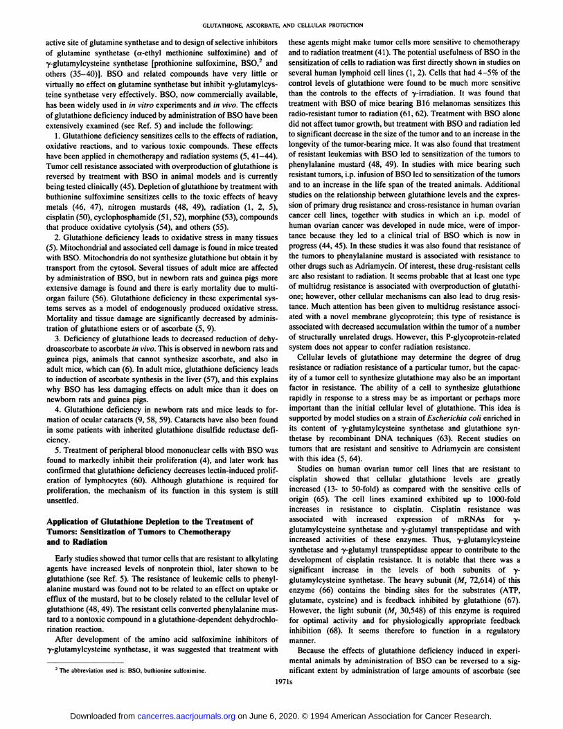
A summary of the effects of glutathione deficiency induced byadministration of BSO is given in Table 1. In adult mice, prolongedtreatment with BSO did not produce cellular damage in the liver,heart, or kidneys. In both newborn rats and guinea pigs, treatment withBSO led to death within 4-6 days, whereas adult mice did not exhibit
Table 1 Effects of glutathiurie deficiency
EffectDeath,
4-6daysCell
damage'LiverKidneyLungBrainLensSkeletal
muscleJejunum,colonHeartStomachLymphocytes"
+, cell damage ordeathMainly
mitochondrial.Newborn
rats Guineapigs-f-a++
++++
+ +++0+0II
0Ãœas
indicated.Adult
mice000+M0-+IIII+
early mortality. The lethal effects of glutathione deficiency in animalssuch as newborn rats and guinea pigs (which do not synthesizeascorbate) appear to be related to multiorgan failure involving mitochondrial and other damage in liver, kidney, and lung (56). In liverthere is focal necrosis: in kidney, there is proximal tubular damage;and in lung, there is lamellar body degeneration. The lungs of adultmice showed damage, although less than in newborn rats.
The mitochondrial and other cell damage seen in newborn animalsand in adults were prevented by administration of glutathione esters orby administration of ascorbate (see below).
Function of Glutathione in the Reduction of Dehydroascorbate
A function of glutathione in the reduction of dehydroascorbate wassuspected by early investigators. Borsook et al. (74) concluded thatglutathione is involved in the reduction of dehydroascorbate by animal tissues, but Guzman-Barron. another early pioneer in this field,
considered this unlikely (75). Hopkins and Morgan (76) studied thisreaction in plants. The reduction of dehydroascorbate to ascorbate wasexamined by a number of investigators in in vitro animal systems (seeRef. 6), and purified preparations of glutaredoxin and protein disulfideisomerase were found to exhibit substantial glutathione-dependent
dehydroascorbate reducÃaseactivity (77).Convincing evidence linking glutathione to the reduction of dehy
droascorbate in vivo has recently been obtained (6, 9). In the first ofthese studies, newborn rats treated with BSO were found to havemarked depletion of tissue (liver, kidney, lung, brain, eye) ascorbate.Both the levels of ascorbate and total ascorbate (ascorbate plus dehydroascorbate) were decreased (6). It is of interest that when ascorbatewas also given to these newborn rats, the levels of glutathione in thetissues and in their mitochondria were increased significantly, indicating that ascorbate can spare glutathione. Findings closely similar tothose made on newborn rats were made in adult guinea pigs (78). Inthis species also, tissue damage and early mortality due to glutathionedeficiency are greatly decreased or prevented by administration ofascorbate. Treatment with ascorbate spares mitochondrial glutathioneas found also in newborn rats (9).
Although glutathione deficiency is lethal to newborn rats andguinea pigs, adult mice are able to survive because they can synthesizeascorbate. Treatment of adult mice with BSO actually leads to aninitial increase of the ascorbate level in the liver (57). Within 4 h afterBSO administration, the level of ascorbate in the liver increases about2-fold and then decreases with concomitant accumulation of dehy
droascorbate. In other tissues, the ascorbate levels decreased and thelevels of dehydroascorbate increased. Therefore, an early effect ofglutathione deficiency in adult mice appears to be an induction ofascorbate synthesis in the liver. Such induction does not occur innewborn rats or in guinea pigs, findings consistent with the view that
1972s
on June 6, 2020. © 1994 American Association for Cancer Research. cancerres.aacrjournals.org Downloaded from
OLUTATHIONE, ASCORBATE. AND CELLULAR PROTECTION
GLU+CySH
GLY
>- GSSG
—--GLUTAREDOXIN ""
DEHYDROASCORBATE ASCORBATE
H202
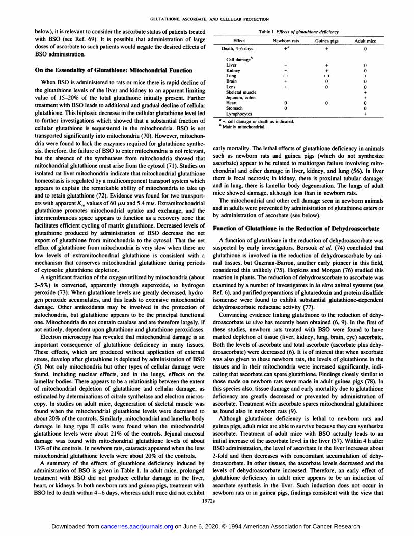
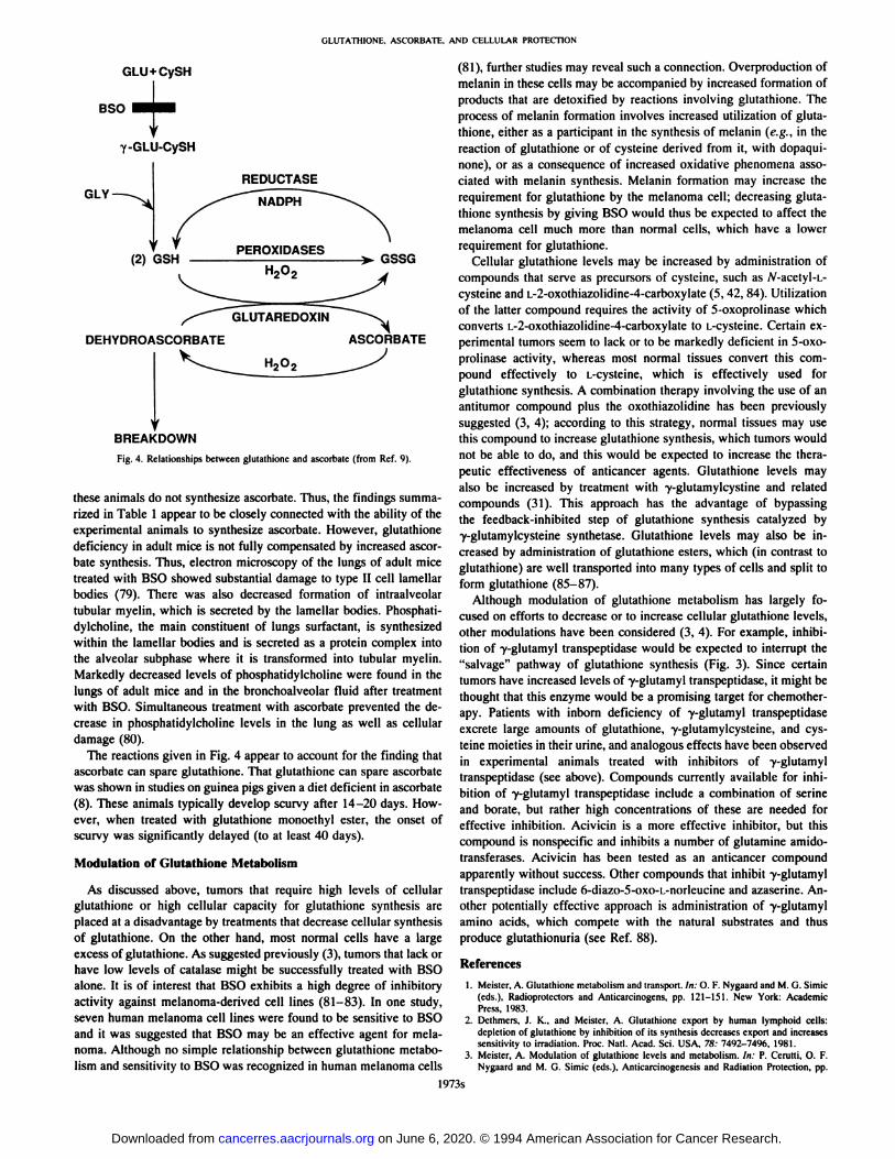
BREAKDOWNFig. 4. Relationships between glutathione and ascorbatc (from Ref. l>).
these animals do not synthesize ascorbate. Thus, the findings summarized in Table 1 appear to be closely connected with the ability of theexperimental animals to synthesize ascorbate. However, glutathionedeficiency in adult mice is not fully compensated by increased ascorbate synthesis. Thus, electron microscopy of the lungs of adult micetreated with BSO showed substantial damage to type II cell lamellarbodies (79). There was also decreased formation of intraulvcolartubular myelin, which is secreted by the lamellar bodies. Phosphati-
dylcholine. the main constituent of lungs surfactant, is synthesizedwithin the lamellar bodies and is secreted as a protein complex intothe alveolar subphase where it is transformed into tubular myelin.Markedly decreased levels of phosphatidylcholine were found in thelungs of adult mice and in the bronchoalveolar fluid after treatmentwith BSO. Simultaneous treatment with ascorbate prevented the decrease in phosphatidylcholine levels in the lung as well as cellulardamage (80).
The reactions given in Fig. 4 appear to account for the finding thatascorbate can spare glutathione. That glutathione can spare ascorbatewas shown in studies on guinea pigs given a diet deficient in ascorbate(8). These animals typically develop scurvy after 14-20 days. How
ever, when treated with glutathione monoethyl ester, the onset ofscurvy was significantly delayed (to at least 40 days).
Modulation of Glutathione Metabolism
As discussed above, tumors that require high levels of cellularglutathione or high cellular capacity for glutathione synthesis areplaced at a disadvantage by treatments that decrease cellular synthesisof glutathione. On the other hand, most normal cells have a largeexcess of glutathione. As suggested previously (3), tumors that lack orhave low levels of catalase might be successfully treated with BSOalone. It is of interest that BSO exhibits a high degree of inhibitoryactivity against melanoma-derived cell lines (81-83). In one study,
seven human melanoma cell lines were found to be sensitive to BSOand it was suggested that BSO may be an effective agent for melanoma. Although no simple relationship between glutathione metabolism and sensitivity to BSO was recognized in human melanoma cells
(81), further studies may reveal such a connection. Overproduction ofmelanin in these cells may be accompanied by increased formation ofproducts that are detoxified by reactions involving glutathione. Theprocess of melanin formation involves increased utilization of glutathione, either as a participant in the synthesis of melanin (e.g., in thereaction of glutathione or of cysteine derived from it, with dopaqui-
none), or as a consequence of increased oxidative phenomena associated with melanin synthesis. Melanin formation may increase therequirement for glutathione by the melanoma cell; decreasing glutathione synthesis by giving BSO would thus be expected to affect themelanoma cell much more than normal cells, which have a lowerrequirement for glutathione.
Cellular glutathione levels may be increased by administration ofcompounds that serve as precursors of cysteine, such as A'-acetyl-L-cysteine and L-2-oxothiazolidine-4-carboxylate (5, 42, 84). Utilizationof the latter compound requires the activity of 5-oxoprolinase whichconverts i.-2-oxothiazolidine-4-carboxylate to i.-cysteine. Certain experimental tumors seem to lack or to be markedly deficient in 5-oxo
prolinase activity, whereas most normal tissues convert this compound effectively to L-cysteine, which is effectively used for
glutathione synthesis. A combination therapy involving the use of anantitumor compound plus the oxothiazolidine has been previouslysuggested (3, 4); according to this strategy, normal tissues may usethis compound to increase glutathione synthesis, which tumors wouldnot be able to do, and this would be expected to increase the therapeutic effectiveness of anticancer agents. Glutathione levels mayalso be increased by treatment with y-glutamylcystinc and related
compounds (31). This approach has the advantage of bypassingthe feedback-inhibited step of glutathione synthesis catalyzed byy-glutamylcysteine synthetase. Glutathione levels may also be in
creased by administration of glutathione esters, which (in contrast toglutathione) are well transported into many types of cells and split toform glutathione (85-87).
Although modulation of glutathione metabolism has largely focused on efforts to decrease or to increase cellular glutathione levels,other modulations have been considered (3, 4). For example, inhibition of y-glutamyl transpeptidase would be expected to interrupt the"salvage" pathway of glutathione synthesis (Fig. 3). Since certain
tumors have increased levels of y-glutamyl transpeptidase. it might be
thought that this enzyme would be u promising target for chemotherapy. Patients with inborn deficiency of y-glutamyl transpeptidaseexcrete large amounts of glutathione, y-glutamylcysteine, and cys
teine moieties in their urine, and analogous effects have been observedin experimental animals treated with inhibitors of y-glutamyl
transpeptidase (see above). Compounds currently available for inhibition of y-glutamyl transpeptidase include a combination of serine
and borate, but rather high concentrations of these are needed foreffective inhibition. Acivicin is a more effective inhibitor, but thiscompound is nonspecific and inhibits a number of glutamine amido-
transferases. Acivicin has been tested as an anticancer compoundapparently without success. Other compounds that inhibit y-glutamyltranspeptidase include 6-diazo-5-oxo-i.-norlcucine and azaserine. Another potentially effective approach is administration of y-glutamyl
amino acids, which compete with the natural substrates and thusproduce glutathionuria (see Ref. 88).
References
1. Meister. A. Glutathione metabolism and transport. In: O. F. Nygaard and M. G. Simic(eds.), Radioprotectors and Anticarcinogens. pp. 121-151. New York: AcademicPress, 1983.
2. Dethmers, J. K.. and Meister. A. Glutathione export hy human lymphoid cells:depletion of glutathione by inhibition of its synthesis decreases export and increasessensitivilv to irradiation. Proc. Nati. Acad. Sci. USA, 78: 7492-7496, 1981.sensitivity to irrauiauon. rroc. pian. /\cau. ^ci. u;>/\. in: m**¿—/HVO, ivni.
3. Meister, A. Modulation of glulathione levels and metabolism, in: P. Cerutti, O. F.Nygaard and M. G. Simic (eds.). Anticarcinogenesis and Radiation Protection, pp.
1973s
on June 6, 2020. © 1994 American Association for Cancer Research. cancerres.aacrjournals.org Downloaded from

OLUTATHIONE, ASCORBATE. AND CELLULAR PROTECTION
361-372. New York: Plenum Publishing Corp., 1988.
4. Meister, A. Selective modification of glutathionc metabolism. Science (WashingtonDC), 220.- 471-477, 1983. 35.
5. Meister, A. Glutathione deficiency produced by inhibition of its synthesis and itsreversal; applications in research and therapy. Pharmacol. Ther., 5/: 155-194, 1991. 36.
6. Meister. A. On the antioxidant effects of ascorbic acid and glutathione. Biochem.Pharmacol., 44: 1905-1915, 1992.
7. Fahcy. R. C., and Sundquist. A. R. Evolution of glutathione metabolism. Adv.Enzymol., 64: 1-53, 1991. 37.
8. Mârtensson,J. M., Han. J., Griffith. O. W.. and Meister, A. Glulathinne ester delaysthe onset of scurvy in ascorbate-deficient guinea pigs. Proc. Nail. Acad. Sci. USA. °0:317-321. 1993. 38.
9. Mârtensson.J. M.. and Meister. A. Glutathione deficiency decreases tissue ascorbatelevels in newborn rats: Ascorbate spares glutathionc and protects. Proc. Nail. Acad.Sci. USA, 88: 4656-4660. 1991. 39.
10. Packer. J. E.. Slater, T. F., and Wilson, R. L. Direct observation of a free radicalinteraction between vitamin E and vitamin C. Nature (Lond.), 278: 737-738, 40.1979.
11. Niki, E., Tsuchiya. J., Tanimura, R.. and Kamiya. Y. Regeneration of vitamin E froma-chromanoyl radical by glutathione and vitamin C. Chem. Lett. Jpn., 23: 789-792, 41.
1982.12. Reddy, C. C., Scholz. R. W.. Thomas, C. E.. and Massaro, E. J. Vitamin E dependent
reduced glutathione inhibition of rat liver microsomal lipid peroxidation. Life Sci., 42.31: 571-576, 1982.
13. Doha, T., Burton. B. W., and Ingold. K. U. Antioxidant and co-antioxidant activity ofvitamin C. The effect of vitamin C, either alone or in the presence of vitamin E or a 43.water-soluble vitamin E analogue, upon the peroxidation of aqueous multi-lamellarphospholipid liposomes. Biochim. Biophys. Acta, 835: 298-303, 1985.
14. Lecdlc. R. A., and Aust, S. D. The effect of glutathione on the vitamin E requirementfor inhibition of liver chromosomal lipid peroxidation. Lipids, 25: 241-245, 1990. 44.
15. Graham, K. S.. Reddy, C. C., and Scholz, R. W. Reduced glulalhione effects onor-tocopherol concentration of rat liver microsomes undergoing NADPH-dcpendentlipid peroxidation. Lipids, 24: 909-914. 1989.
16. Scholich. H., Murphy. M. E.. and Sies. H. Antioxidant activity of dihydrolipoatc 45.against microsomal lipid peroxidation and its dependence on a-tocopherol. Biochim.Biophys. Acia, IOOI: 256-261, 1989.
17. Wefers. H.. and Sics. H. The protection by ascorbate and glulathione against micro- 46.stimai lipid peroxidation is dependent on vitamin E. Eur. J. Biochem.. 174: 353-357,
1988. 47.18. h.il.il I., and Grundy. S. M. Preservation of the endogenous antioxidants in low
density lipoprotcin by ascorbate but not probucol during oxidative modification.J. Clin. Invest.. 87: 597-601, 1991. 48.
19. Allison. R. D.. and Meister, A. Evidence thai (ranspcptidation is a significant functionof y-glulamyl transpeplidase. J. Biol. Chem., 256: 2988-2992, 1981.
211. Thompson, G. A., and Meisler, A. Utilization of L-cystine by the -y-glutamyl Irans- 49.pt'ptidasc-y-glulamvl cyclotransferase pathway. Proc. Nati. Acad. Sci. USA, 72:1985-1988, 1975.
21. Täte.S. S., and Meister. A. Interaction of y-glulamyl transpeplidase with amino acids. 50.dipeptides, and derivatives and analogs of glutathione. J. Biol. Chem.. 249: 7593-
7602, 1974. 51.22. Meister. A. On the enzymology of amino acid transport. Science (Washington DC).
180: 33-39. 1973.23. Orlowski, M.. and Meister. A. y-GIutamyl cyclotransferase; distribution, isozymic 52.
forms, and specificity. J. Biol. Chem., 24«:2836-2844, 1973.24. Griffith. O. W., Bridges, R. J., and Meister, A. Evidence that the y-glulamyl cycle
functions in r/wj using intracellular glutathione: effects of amino acids and selective 53.inhibition of enzymes. Proc. Nati. Acad. Sci. USA, 75: 5405-5408. 1978.
25. Taniguchi. N., and Meisler. A. y-Glulamyl cyclotransferase from rat kidney: sulfhy- 54.
dryl groups and isolation of a stable form of the enzyme. J. Biol. Chem., 253:1799-1806. 1978. 55.
26. Van der Werf. P.. Orlowski. M.. and Meisler, A. Enzymatic conversion of 5-oxo-L-proline (I-pyrrolidonc carhoxylate) to L-glutamate coupled with ATP cleavage toADP: a reaction in the y-glutamyl cycle. Proc. Nail. Acad. Sci. USA, 68: 2982-2985. 56.1971.
27. Scddon. A. P.. and Meislcr. A. Trapping of an intermediale in the reaclion catalyzedby 5-oxoprolina.se. J. Biol. Chem., 267: 11538-11541, 1986. 57.
28. Griffith, O. W.. and Meister. A. Translocalion of intracellular glulathione to membrane-bound y-glulamyl Iranspeplidase as a discrete step in the y-glutamyl cycle; 58.glutathionuria after inhibition of transpeptidase. Proc. Nati. Acad. Sci. USA, 76:268-272. 1979.
29. Griffith. O. W., Novogrodsky, A., and Meister. A. Translocation of glulalhione from 59.lymphoid cells that have markedly different y-glutamyl transpeplidase activities.Proc. Nail. Acad. Sci. USA. 76: 2249-2252, 1979.
30. Griffith. O. W.. and Meister. A. Excrelion of cysleine and y-glulamylcysleine 60.moielies in human and experimental animai y-glutamyl transpeptidase deficiency.Proc. Nail. Acad. Sci. USA. 77: 3384-3387, 1980.
31. Anderson, M. E., and Meister, A. Transpon and direct utilization of y-glutamylcys-t(e)ine for glutathione synthesis. Proc. Nati. Acad. Sci. USA. 80: 707-711, 1983. hi.
32. Griffith. O. W.. Bridges, R. J., and Meister, A. Transport of y-glutamyl amino acids;role of glutalhione and y-glulamyl Iranspeptidase. Proc. Nati. Acad. Sci. USA. 76: 62.6319-6322, 1979.
33. Meister. A. Metabolism and funclion of glulathione. In: D. Dolphin, R. Poulson. andO. Avramovic (eds.). Glulathione: Chemical. Biochemical and Medical Aspects, pp. 63.367-374. New York: John Wiley and Sons. 1989.
34. Meister, A., and Larsson. A. Glulalhione synlhetase deficiency and other disorders ofthe y-glutamyl cycle. In: C. R. Scriver. A. L. Beaudet, W. S. Sly, and D. Valle (eds.),
1974s
The Metabolic Basis of Inheriled Disease. Ed. 6. pp. 855-868. New York: McGraw-Hill. 19X9.Meisler, A. A Irail of research: from glulamine synlhelase lo selective inhibition ofglulalhione synlhesis. ChemTracls-Biochem. Molec. Biol.. .ÃŽ:75-106, 1992.Meister, A. Inhibition of glutamine synlhelase and y-glutamylcysteine synthctase by
melhionine sulfoximine and relaled compounds. In: N. Seiler. M. J. Jung, and J.Koch-Weser (eds.). Enzyme-aclivated Irreversible Inhibitors, pp. 187-211. Amsterdam: Elsevier-North Holland BiomédicalPress, 1978.
Griffith, O. W., Anderson, M. E., and Meisler. A. Inhibition of glutathione biosynthesis by prolhionine sulfoximine (S-n-propyl-homocysleine sulfoximine). a sclecliveinhibitor of y-glulamylcysteinc synlhetase. J. Biol. Chem.. 254: 1205-1210, 1979.Griffith. O. W.. and Meisler. A. Polent and specific inhibition of glulalhione synthesisby huthionine sulfoximine (S-/i-butyl homocysteine sulfoximine). J. Biol. Chem.,254: 7558-7560, 1979.
Griffith, O. W. Bulhionine sulfoximine and its higher homologs. J. Biol. Chem., 257:13704-13712. 1982.
Meister, A. On the biochemistry of glutalhione. in: N. Taniguchi et al. (eds.),Glutathione Centennial: Molecular Properties and Clinical Implications, pp. 3—21.New York: Academic Press, 1989.Meister, A., and Griffilh. O. W. Effects of methionine sulfoximine analogs on thesynthesis of glutamine and glutalhione; possible chemotherapeutic implicalions.Cancer Treal. Rep.. 6.?: 1115-1121, 1979.Meisler. A. Novel drugs thai affect glutalhione melabolism. In: P. V. Woolley andK. D. Tew (eds.). Mechanisms of Drug Resistance in Ncoplaslic Cells, Bristol MyersSymposium No. 9. Chapter 7, pp 99-127. New York: Academic Press, 1988.Vistica, D. T., and Ahmad. S. Acquired resistance of tumor cells to i -phenylalaninemustard: implications for the design of a clinical trial involving glutathione depletion.In: N. Taniguchi et al. (eds.). Glutathione Centennial Molecular Perspectives andClinical Implications, Chaplcr 21, pp. 301-305. New York: Academic Press. 1989.Ozols, R. F.. Hamilton. T. C. Masuda. H.. and Young. R. C. Manipulation of cellularthiols lo influence drug resistance. In: P. V. Woolley and K. D. Tew (eds.). Mechanisms of Drug Resistance in Neoplastic Cells, Bristol Myers Symposium No. 9.Chapter 19, pp. 289-305. New York: Academic Press, 1988.Hamilton. T.. O'Dwyer, P.. Young, R., Tew, K., Padavic, K., Comis, R., and Ozols.
R. Phase 1 trial of bulhionine sulfoximine (BSO) plus melphalan (l-PAM) in palientswith advanced cancer. Proc. A. Meet. Am. Soc. Clin. Oncol.. °:A2KI. 1990.
Singhal. R. K., Anderson. M. E.. and Meister, A. Glutalhione. a first line of defenseagainst cadmium loxicity. FASEB J., /: 220-223, 1987.
Naganuma. A.. Anderson. M. E.. and Meister. A. Cellular glutalhione is a determinantof sensitivity to mercuric chloride loxicity: prevention of toxicity by giving glutalhione monoeslcr. Biochcm. Pharmacol., 40: 693-697, 1990.
Suzukake, K.. Petro, B. J.. and Vislica. D. T. Reduction in glulalhione content of1--PAM resistant L1210 cells confers drug sensitivily. Biochem. Pharmacol.. 31:121-124, 1982.Suzukake, K.. Vislica. B. P.. and Vislica. D. T. Dechlorination of i -phenylalaninemustard by sensitive and resistant tumor cells and its relationship to intracellularglutathione contenÃ.Biochem. Pharmacol., 32: 165-167, 1983.
Anderson, M. E.. Naganuma. A., and Meisler. A. Proteclion against cisplatin toxicilyby adminislration of glutathione ester. FASEB J., 4: 3251-3255, 1990.Ishikawa. M.. Sasaki. K-L, and Takayanagi. Y. Injurious effect of huthionine sulfoximine. an inhibitor of glulalhione biosynthesis, on Ihe lethality and uroloxicity ofcyclophosphamide in mice. J. Pharmacol. Jap., 5/: 146-149, 1989a.Ishikawa. M.. Takayanagi. Y.. and Sasaki. K-l. Modification of cydophosphamidc-induced urotoxicily by buthionine sulfoximine and disulfiram in mice. Res. Commun.Palh. Pharmacol., 65: 265-268, 1989b.
McCarlney. M. A. Effect of glutathione depiction on morphine toxicily in mice.Biochem. Pharmacol., 38: 207-209. 1989.
Arrick. B. A.. Nathan. C. F.. Griffilh, O. W.. and Cohn, Z. A. Glulalhione depletionsensitizes tumor cells to oxidative cytolysis. J. Biol. Chem., 257: 1231-1237, 1982.
Perez, R. P.. Hamilton. T. C.. and Ozols, R. F. Resistance to alkylaling agents andeisplatin: insighls from ovarian carcinoma model systems. Pharmacol. Ther.. 48:19-27, 1990.
Mârtensson,J. M., Jain, A., Stole, E., Frayer, W., Auld, P. A. M., and Meister. A.Inhibilion of glutathione synthesis in the newborn rat: a model of cndogenously-produced oxidative stress. Proc. Nati. Acad. Sci. USA, 88: 9360-9364, 1991.
Mârtensson.J. M.. and Meister. A. Glulalhione deficiency increases hepalic ascorbicacid synlhesis in aduli mice. Proc. Nati. Acad. Sci. USA'. 89: 11566-11568, 1992.
Calvin. H. L, Medvedovsky. C., and Worgul, B. V. Near-total glutalhione depletionand age-specific cataracts induced by buthionine sulfoximine in mice. Science (Washington DC), 233: 553-555, 1986.
Mârtensson,J. M., Sleinherz. R.. Jain, A., and Meisler, A. Glulathione ester prevenÃsbuthionine sulfoximinc-induced cataracts and lens epithelial cell damage. Proc. Nati.Acad. Sci. USA, 86: 8727-8731, 1989.Suthanlhiran. M., Anderson. M. E.. Sharma. V. K.. and Meisler, A. Glutalhioneregulates activation-dependent DNA synthesis in highly purified normal human Tlymphocytes stimulated via the CD2 and CD3 antigens. Proc. Nati. Acad. Sci. USA,87: 3343-3347. 1990.
Griffith. O. W. Glutalhione and cell survival. In: S. Ebraski (ed.). Cellular Regulationand Malignanl Growih, pp. 292-300. Tokyo: Japan Societies Press, Springer. 1985.Meister. A. Modulation of intracellular levels of glutathionc. in: F. Valeriote and !..Baker (eds.). Biochemical Modulation of Anticancer Agents: Experimental ClinicalApproaches, pp. 245-275. Boston. MA: Marlinus Nijhoff. 1986.Moore. W. R., Anderson. M. E.. Meisler, A.. Murala. K.. and Kimura. A. Increasedcapacity for glutathione synthesis enhances resistance to radiation in Escherichia coli:a possible model for mammalian cell protection. Proc. Nail. Acad. Sci. USA, 86:1461-1464. 1989.
on June 6, 2020. © 1994 American Association for Cancer Research. cancerres.aacrjournals.org Downloaded from
OLUTATHIONE. ASCÃœRBATE. AND CELLULAR PROTECTION
64. Lee, F. Y. F.. Siemann, D. W., and Sutherland, R. M. Changes ¡ncellular glutathionecontent during adriamycin treatment in human ovarian cancer-a possible indicator ofchemosensitivity. Br. J. Cancer, 60: 291-298, 1989.
65. Godwin, A. K., Meister, A., O'Dwyer, P. J., Hamilton, T. C., Huang, C-S., and
Anderson, M. E. High resistance to cisplatin in human ovarian cancer cell lines isassociated with marked increase of glutathione synthesis. Proc. Nati. Acad. Sci. USA.89: 3070-3074. 1992.
66. Yan, N.. and Meister. A. Amino acid sequence of rat kidney y-glutamylcystcinesynlhetase. J. Biol. Chem., 265: 1588-1593, 1990.
67. Huang, C-S., Chang, L-S., Anderson, M. E., and Meister, A. Catalytic and regulatoryproperties of the heavy subunit of rat kidney y-glutamylcystcine synthctase. J. Biol.Chem., 26«:19675-19678, 1993.
68. Huang, C-S., Anderson. M. E.. and Meister, A. Amino acid sequence function of thelight subunit of rat kidney •y-glutamylcysteinesynthetase. J. Biol. Chem.. 268:20578-20583, 1993.
69. Meister, A. Depletion of glutathione in normal and malignant human cells in riva byt.-huthioninc sulfoximine: possible interaction with ascorbatc. J. Nail. Cancer Inst.,84: 1601-1602, 1992.
70. Meister, A., and Griffith, O. W. Effects of BSO and related compounds on mito-
chondrial glutathione levels (Abstract). Federation Proc., 42: 2642. 1983.71. Griffith, O. W., and Meister, A. Origin and turnover of mitochondrial glulathionc.
Proc. Nati. Acad. Sci. USA, 82: 4668-4672. 1985.72. Mârtensson,J. M., Lai, J. C'. K., and Meister, A. High affinity transport of glutathione
is part of a multicomponent system essential for mitochondrial function. Proc. Nad.Acad. Sci. USA, 87: 7185-7189, 1990.
73. Boveris. A., Oshino, N., and Chance, B. The cellular production of hydrogenperoxide. Biochem. J.. 128: 617-630, 1972.
74. Borsook, H., Davenport, H. W., Jeffreys, C. E. P., and Warner. R. C. The oxidationof ascorbic acid and its reduction in \-iiro and m vìvo.J. Biol. Chem.. 117: 237-279,
1937.75. Guzman-Barron. E. Thiol groups of biological importance. Adv. Enzymol.. 11:
201-266. 1951.
76. Hopkins. F. G., and Morgan, E. J. Some relations between ascorbic acid andglutathionc. Biochem. J.. 30: 1446-1462, 1936.
77. Wells, W. W., Xu. D. P., Yang, Y., and Rocque, P. A. Mammalian Ihioltransferase(glutarcdoxin) and protein disulfide isomcrase have dchydroascorbate reducÃaseactivity. J. Biol. Chem., 265: 15361-15364, 1990.
78. Griffith, O. W., Han. J.. and Mârtensson.J. M. Vitamin C' protects adult guinea pigs
against tissue damage and lethality caused by BSO-mediated glutathione depletion
(Abstract 4708). FASEB J.. 5: 1991.7°.Mârtensson,J. M., Jain. A.. Frayer. W., and Meister, A. Glutathione metabolism in
the lung: inhibition of its synthesis leads to lamellar body and mitochondrial defects.Proc. Nati. Acad. Sci. USA, «6:5296-5300, 1989.
80. Jain. A.. Mârtensson,J. M., Menta, T., Krauss, A. N.. Auld, P. A. M., and Meister,A. Ascorbic acid prevents oxidative stress in glutathione-deficient mice; effects onlung type-2 cell lamellar bodies, lung surfactant, and skeletal muscle. Proc. Nati.Acad. Sci. USA, #9: 5093-5097, 1992.
81. Kable, E. P. W., Favier, D., and Parsons, P. G. Sensitivity of human melanoma cellsto i.-dopa and m.-buthioninc(S,R)-sulfoximine. Cancer Res., 4V: 2327-2331, 1989.
82. Dorr, R. T., Liddil, J. D., and Sohle, M. J. Cytotoxic effects of glutathione synthesisinhibition by i -buthioninc-(SR)-sulfoximine on human and murine tumor cells.Invest. New Drugs, 4: 305-313. 1986.
83. Peinado, P., Martinez-Liarte. J. H.. del Marmol. V., Solano. F.. and Lozano. J. A.
Glulalhionc depletion in mouse melanoma cells increases their sensitivity to oxidativelysis. Cancer J., 5: 348-353, 1992.
84. Williamson. J. M.. and Meister, A. Stimulation of hepatic glutathione formation byadministration of l-2-oxt)thiazolidine-4-carboxylate, a 5-oxo-L-prolinase substrate.Proc. Nati. Acad. Sci. USA, 78: 936-939, 1981.
85. Puri, R. N., and Meister. A. Transport of glutathione as y-glutamylcysteinylglycylester, into liver and kidney. Proc. Nati. Acad. Sci. USA, KO: 5258-5260, 1983.
86. Anderson, M. E., and Meister, A. Glutathione monoesters. Anal. Biochem., 183:16-20, 1989.
87. Levy, E. J., Anderson, M. E.. and Meister, A. Transport of glutathione diethyl esterinto human cells. Proc. Nati. Acad. Sci. USA, W: 9171-9175, 1993.
88. Anderson. M. E., and Meister, A. Inhibition of y-glutamyl transpeptidase and gluta-thionuria produced by y-glutamyl amino acids. Proc. Nati. Acad. Sci. USA, 83:5029-5032, 1986.
1975s
on June 6, 2020. © 1994 American Association for Cancer Research. cancerres.aacrjournals.org Downloaded from
1994;54:1969s-1975s. Cancer Res Alton Meister Glutathione, Ascorbate, and Cellular Protection
Updated version
http://cancerres.aacrjournals.org/content/54/7_Supplement/1969s.citation
Access the most recent version of this article at:
E-mail alerts related to this article or journal.Sign up to receive free email-alerts
Subscriptions
Reprints and
To order reprints of this article or to subscribe to the journal, contact the AACR Publications
Permissions
Rightslink site. Click on "Request Permissions" which will take you to the Copyright Clearance Center's (CCC)
.http://cancerres.aacrjournals.org/content/54/7_Supplement/1969s.citationTo request permission to re-use all or part of this article, use this link
on June 6, 2020. © 1994 American Association for Cancer Research. cancerres.aacrjournals.org Downloaded from