h-reflex as method to probe spinal circuitry
TRANSCRIPT

Journal of Neuroscience Methods 171 (2008) 1–12
Contents lists available at ScienceDirect
Journal of Neuroscience Methods
journa l homepage: www.e lsev ier .com/ locate / jneumeth
Invited review
The H-reflex as a probe: Pathways and pitfalls
Maria Knikoua,b,c,∗
a Health Sciences Doctoral Programs, City University of New York, Staten Island, NY 10314, USAb Department of Physical Medicine and Rehabilitation, Feinberg Medical School, Northwestern University, Chicago, IL 60611, USAc Sensory Motor Performance Program, Rehabilitation Institute of Chicago, Chicago, IL 60611, USA
a r t i c l e i n f o
Article history:Accepted 26 February 2008
Keywords:H-reflexHumanMethodsNeuronal pathwaysProtocolPresynaptic–postsynaptic inhibitionSpinal circuits
a b s t r a c t
The Hoffmann (or H) reflex is considered a major probe for non-invasive study of sensorimotor integrationand plasticity of the central nervous system in humans. The first section of this paper reviews the neu-rophysiological properties of the H-reflex, which if ignored create serious pitfalls in human experimentalstudies. The second section reviews the spinal inhibitory circuits and neuronal pathways that can be indi-rectly assessed in humans using the H-reflex. The most confounding factor is that reciprocal, presynaptic,and Ib inhibition do not act in isolation during movement. Therefore, characterization of these spinalcircuits should be more comprehensive, especially in cases of a neurological injury because neurophysi-ological findings are critical for the development of successful rehabilitation protocols. To conclude, theH-reflex is a highly sensitive reflex with an amplitude that is the result of complex neural mechanismsthat act synchronously. If these limitations are recognized and addressed, the H-reflex constitutes one ofthe major probes to assess excitability of interneuronal circuits at rest and during movement in humans.
© 2008 Elsevier B.V. All rights reserved.
Contents
1. Introduction . . . . . . . . . . . . . . . . . . . . . . . . . . . . . . . . . . . . . . . . . . . . . . . . . . . . . . . . . . . . . . . .2. Neurophysiological characteristics of the H-reflex . . . . . . . . . . . . . . . . . . . . . . . .3. The H-reflex as a probe to study spinal neuronal pathways and mechanis
3.1. Monosynaptic Ia excitation and homosynaptic depression. . . . . . . .3.2. Presynaptic inhibition of Ia afferents . . . . . . . . . . . . . . . . . . . . . . . . . . . . . . .
3.2.1. Protocol for studying presynaptic inhibition in humans .3.3. Reciprocal Ia inhibition . . . . . . . . . . . . . . . . . . . . . . . . . . . . . . . . . . . . . . . . . . . . .
3.3.1. Protocol for studying reciprocal Ia inhibition in humans .3.4. Non-reciprocal (or Ib) inhibition . . . . . . . . . . . . . . . . . . . . . . . . . . . . . . . . . . .
3.4.1. Protocol for studying Ib inhibition in humans . . . . . . . . . . . .3.5. Recurrent inhibition. . . . . . . . . . . . . . . . . . . . . . . . . . . . . . . . . . . . . . . . . . . . . . . .
3.5.1. Protocol for studying recurrent inhibition in humans . . . .4. Clinical implications . . . . . . . . . . . . . . . . . . . . . . . . . . . . . . . . . . . . . . . . . . . . . . . . . . . . . . . .5. Conclusions . . . . . . . . . . . . . . . . . . . . . . . . . . . . . . . . . . . . . . . . . . . . . . . . . . . . . . . . . . . . . . . . .
Acknowledgements . . . . . . . . . . . . . . . . . . . . . . . . . . . . . . . . . . . . . . . . . . . . . . . . . . . . . . . .References . . . . . . . . . . . . . . . . . . . . . . . . . . . . . . . . . . . . . . . . . . . . . . . . . . . . . . . . . . . . . . . . . .
1. Introduction
The Hoffmann (or H) reflex is one of the most studied reflexesin humans and is the electrical analogue of the monosynaptic
∗ Correspondence address: Health Sciences Doctoral Programs, The Graduate Cen-ter, City University of New York, 2800 Victory Boulevard, Building 5N-207, StatenIsland, NY 10314, USA. Tel.: +1 718 982 3316; fax: +1 718 982 2984.
E-mail addresses: [email protected], [email protected].
0165-0270/$ – see front matter © 2008 Elsevier B.V. All rights reserved.doi:10.1016/j.jneumeth.2008.02.012
. . . . . . . . . . . . . . . . . . . . . . . . . . . . . . . . . . . . . . . . . . . . . . . . . . . . . . . . . . . . . . . . . . . . . . . . . . . . 1
. . . . . . . . . . . . . . . . . . . . . . . . . . . . . . . . . . . . . . . . . . . . . . . . . . . . . . . . . . . . . . . . . . . . . . . . . . . . 2ms . . . . . . . . . . . . . . . . . . . . . . . . . . . . . . . . . . . . . . . . . . . . . . . . . . . . . . . . . . . . . . . . . . . . . . . 3
. . . . . . . . . . . . . . . . . . . . . . . . . . . . . . . . . . . . . . . . . . . . . . . . . . . . . . . . . . . . . . . . . . . . . . . . . . . . 3. . . . . . . . . . . . . . . . . . . . . . . . . . . . . . . . . . . . . . . . . . . . . . . . . . . . . . . . . . . . . . . . . . . . . . . . . . . 3
. . . . . . . . . . . . . . . . . . . . . . . . . . . . . . . . . . . . . . . . . . . . . . . . . . . . . . . . . . . . . . . . . . . . . . . . . . . . 4. . . . . . . . . . . . . . . . . . . . . . . . . . . . . . . . . . . . . . . . . . . . . . . . . . . . . . . . . . . . . . . . . . . . . . . . . . . 5
. . . . . . . . . . . . . . . . . . . . . . . . . . . . . . . . . . . . . . . . . . . . . . . . . . . . . . . . . . . . . . . . . . . . . . . . . . . 6. . . . . . . . . . . . . . . . . . . . . . . . . . . . . . . . . . . . . . . . . . . . . . . . . . . . . . . . . . . . . . . . . . . . . . . . . . . . 7. . . . . . . . . . . . . . . . . . . . . . . . . . . . . . . . . . . . . . . . . . . . . . . . . . . . . . . . . . . . . . . . . . . . . . . . . . . 7. . . . . . . . . . . . . . . . . . . . . . . . . . . . . . . . . . . . . . . . . . . . . . . . . . . . . . . . . . . . . . . . . . . . . . . . . . . . 8. . . . . . . . . . . . . . . . . . . . . . . . . . . . . . . . . . . . . . . . . . . . . . . . . . . . . . . . . . . . . . . . . . . . . . . . . . . 8. . . . . . . . . . . . . . . . . . . . . . . . . . . . . . . . . . . . . . . . . . . . . . . . . . . . . . . . . . . . . . . . . . . . . . . . . . 10. . . . . . . . . . . . . . . . . . . . . . . . . . . . . . . . . . . . . . . . . . . . . . . . . . . . . . . . . . . . . . . . . . . . . . . . . . 10. . . . . . . . . . . . . . . . . . . . . . . . . . . . . . . . . . . . . . . . . . . . . . . . . . . . . . . . . . . . . . . . . . . . . . . . . . 10. . . . . . . . . . . . . . . . . . . . . . . . . . . . . . . . . . . . . . . . . . . . . . . . . . . . . . . . . . . . . . . . . . . . . . . . . . 10
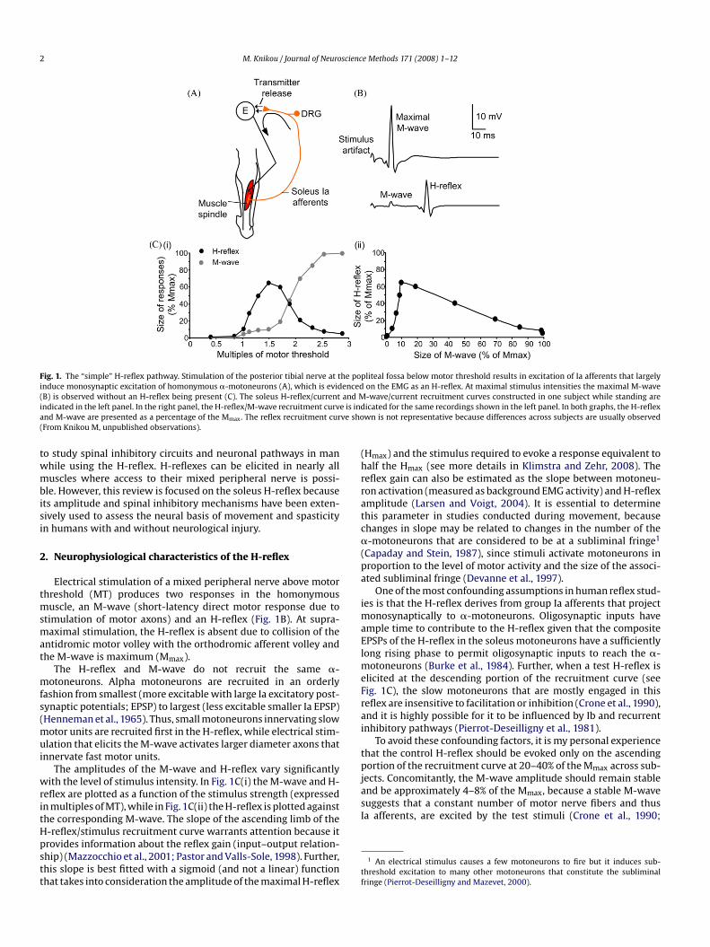
stretch reflex. The H-reflex is evoked by low-intensity electri-cal stimulation of the afferent nerve, rather than a mechanicalstretch of the muscle spindle, that results in monosynaptic exci-tation of �-motoneurons (Fig. 1A). Hence, the H-reflex bypassesthe muscle spindle and the fusimotor activity that may influencethe sensitivity of the Ia afferents to engage a ‘simple’ reflex cir-cuit.
The purpose of this brief review is to discuss specific neurophys-iological characteristics of the H-reflex and the methods available
2 M. Knikou / Journal of Neuroscience Methods 171 (2008) 1–12
he poencedand M
e is indve sho
ated subliminal fringe (Devanne et al., 1997).One of the most confounding assumptions in human reflex stud-
ies is that the H-reflex derives from group Ia afferents that projectmonosynaptically to �-motoneurons. Oligosynaptic inputs have
Fig. 1. The “simple” H-reflex pathway. Stimulation of the posterior tibial nerve at tinduce monosynaptic excitation of homonymous �-motoneurons (A), which is evid(B) is observed without an H-reflex being present (C). The soleus H-reflex/currentindicated in the left panel. In the right panel, the H-reflex/M-wave recruitment curvand M-wave are presented as a percentage of the Mmax. The reflex recruitment cur(From Knikou M, unpublished observations).
to study spinal inhibitory circuits and neuronal pathways in manwhile using the H-reflex. H-reflexes can be elicited in nearly allmuscles where access to their mixed peripheral nerve is possi-ble. However, this review is focused on the soleus H-reflex becauseits amplitude and spinal inhibitory mechanisms have been exten-sively used to assess the neural basis of movement and spasticityin humans with and without neurological injury.
2. Neurophysiological characteristics of the H-reflex
Electrical stimulation of a mixed peripheral nerve above motorthreshold (MT) produces two responses in the homonymousmuscle, an M-wave (short-latency direct motor response due tostimulation of motor axons) and an H-reflex (Fig. 1B). At supra-
maximal stimulation, the H-reflex is absent due to collision of theantidromic motor volley with the orthodromic afferent volley andthe M-wave is maximum (Mmax).The H-reflex and M-wave do not recruit the same �-motoneurons. Alpha motoneurons are recruited in an orderlyfashion from smallest (more excitable with large Ia excitatory post-synaptic potentials; EPSP) to largest (less excitable smaller Ia EPSP)(Henneman et al., 1965). Thus, small motoneurons innervating slowmotor units are recruited first in the H-reflex, while electrical stim-ulation that elicits the M-wave activates larger diameter axons thatinnervate fast motor units.
The amplitudes of the M-wave and H-reflex vary significantlywith the level of stimulus intensity. In Fig. 1C(i) the M-wave and H-reflex are plotted as a function of the stimulus strength (expressedin multiples of MT), while in Fig. 1C(ii) the H-reflex is plotted againstthe corresponding M-wave. The slope of the ascending limb of theH-reflex/stimulus recruitment curve warrants attention because itprovides information about the reflex gain (input–output relation-ship) (Mazzocchio et al., 2001; Pastor and Valls-Sole, 1998). Further,this slope is best fitted with a sigmoid (and not a linear) functionthat takes into consideration the amplitude of the maximal H-reflex
pliteal fossa below motor threshold results in excitation of Ia afferents that largelyon the EMG as an H-reflex. At maximal stimulus intensities the maximal M-wave-wave/current recruitment curves constructed in one subject while standing areicated for the same recordings shown in the left panel. In both graphs, the H-reflexwn is not representative because differences across subjects are usually observed
(Hmax) and the stimulus required to evoke a response equivalent tohalf the Hmax (see more details in Klimstra and Zehr, 2008). Thereflex gain can also be estimated as the slope between motoneu-ron activation (measured as background EMG activity) and H-reflexamplitude (Larsen and Voigt, 2004). It is essential to determinethis parameter in studies conducted during movement, becausechanges in slope may be related to changes in the number of the�-motoneurons that are considered to be at a subliminal fringe1
(Capaday and Stein, 1987), since stimuli activate motoneurons inproportion to the level of motor activity and the size of the associ-
ample time to contribute to the H-reflex given that the compositeEPSPs of the H-reflex in the soleus motoneurons have a sufficientlylong rising phase to permit oligosynaptic inputs to reach the �-motoneurons (Burke et al., 1984). Further, when a test H-reflex iselicited at the descending portion of the recruitment curve (seeFig. 1C), the slow motoneurons that are mostly engaged in thisreflex are insensitive to facilitation or inhibition (Crone et al., 1990),and it is highly possible for it to be influenced by Ib and recurrentinhibitory pathways (Pierrot-Deseilligny et al., 1981).
To avoid these confounding factors, it is my personal experiencethat the control H-reflex should be evoked only on the ascendingportion of the recruitment curve at 20–40% of the Mmax across sub-jects. Concomitantly, the M-wave amplitude should remain stableand be approximately 4–8% of the Mmax, because a stable M-wavesuggests that a constant number of motor nerve fibers and thusIa afferents, are excited by the test stimuli (Crone et al., 1990;
1 An electrical stimulus causes a few motoneurons to fire but it induces sub-threshold excitation to many other motoneurons that constitute the subliminalfringe (Pierrot-Deseilligny and Mazevet, 2000).
scienc
M. Knikou / Journal of NeuroBoorman et al., 1996). However, it is possible for the H-reflex toreach an amplitude of 50–60% of the Mmax when the M-wave isabsent. Nonetheless, the M-wave amplitude during an experimentis an important point of reference and should be monitored so itssize is similar under all tested conditions. This will ensure thatthe observed reflex modulation pattern (when present) is not dueto changes in the composition of the test afferent volley but tomechanisms that act to depress or facilitate the H-reflex (see nextsection).
It is widely held that the H-reflex measures motoneuron poolexcitability. Based on this assumption, it was suggested that theratio of Hmax/Mmax estimates motoneuron pool excitability (Funaseet al., 1994). However, since the magnitude of the Hmax depends onpre- and post-synaptic events (see next section on spinal inhibitorymechanisms), and the amount of concomitant antidromic activityelicited in the motor nerve axons (Misiaszek, 2003), it is certainthat this ratio cannot estimate motoneuron pool excitability.
The state of excitability of the �-motoneuron pool plays asignificant role in determining the H-reflex magnitude. In orderto maintain stable motoneuron excitability and minimize post-synaptic effects, H-reflex recordings should be conducted duringvoluntary, sustained, homonymous muscle contractions and notat rest (see more details about muscle contraction and H-reflexin Stein and Thompson, 2006). More specifically, H-reflex record-ings are conducted at 5–10% (or higher) of the maximal voluntarycontraction (MVC) of the homonymous muscle. If the H-reflex isrecorded during voluntary sustained contraction of the homony-mous muscle, the amplitude of the H-reflex may be affected bychanges of �-motoneuron excitability due to descending excitation,contraction-associated sensory feedback, decrease of Ib inhibition(Marchand-Pauvert et al., 2002), and changes in recurrent inhibi-tion or even presynaptic inhibition (Hultborn et al., 1987a).
When reflex recordings are conducted with subjects at rest,influences from supraspinal centers are anticipated to be mini-mal and spinal inhibitory interneurons such as Renshaw cells andIa–Ib inhibitory interneurons, that are affected by muscle contrac-tion, might be less active. However, there is no way to assess theexcitability state of the cells or their sub-threshold excitability level,which may vary within and across subjects. This is the most signif-icant drawback when reflex recordings are conducted while thehomonymous muscle is in a relaxed state.
A major issue that needs attention is that during various motortasks, low threshold motoneurons (the ones that are mostly asso-ciated with human H-reflex studies) not involved in the muscle
contraction might be equally excited (Pierrot-Deseilligny, 1997). Tocounteract this phenomenon, several studies have proposed thatthe reflex gain should be similar across tasks (Capaday and Stein,1987; Larsen and Voigt, 2004). However, the gain should also beestimated for muscles not involved in the motor task, becausethe reflex gain of the antagonistic muscles differs during concen-tric and eccentric contraction of the agonist muscle (Sekiguchi etal., 2003). The input/output properties of the H-reflex are basedon the number of sensory fibers that are synchronously excitedto make motoneurons discharge (DeBruin et al., 2006). However,when a movement is attempted a marked shift in the relationshipof Ia input/motoneuron output has been observed (DeBruin et al.,2006). The H-reflex might have a different recruitment gain for var-ious motor tasks even in the same subject (Kernell and Hultborn,1990).Taken altogether, we may assume that motoneuron excitabil-ity and actions of spinal interneurons are minimal when reflexrecordings are conducted during different tasks at similar low-muscle contraction levels. However, it should be noted that sensoryafferent input might combine differently during different motortasks.
e Methods 171 (2008) 1–12 3
3. The H-reflex as a probe to study spinal neuronalpathways and mechanisms
Changes in H-reflex amplitude following a conditioning stimu-lus are usually employed to assess post-synaptic events or changesin the amount of the presynaptic inhibition acting on Ia afferentterminals. This is because the amplitude of the test reflex dependson the motoneuron excitability and the ongoing presynaptic inhi-bition of Ia fibres that mediate the test afferent volley. Thus, theH-reflex can be used as a probe to study spinal neuronal pathwaysand mechanisms at rest and during movement in humans.
3.1. Monosynaptic Ia excitation and homosynaptic depression
The amplitude of the H-reflex depends on the history of pre-viously activated Ia afferents, even when variables that influencethe H-reflex amplitude are kept constant. This phenomenon isknown as homosynaptic or post-activation depression and occursat the level of the synapse between the soleus Ia afferents and�-motoneurons (see Fig. 1A).
In cats, it has been shown that depressed subsequent motoneu-ron Ia EPSPs elicited at low frequency (e.g. 3/s) are associated witha decrease in the amount of neurotransmitter released (Kuno,1964). The depression of the EPSP, driven mostly by the previouslyactivated Ia afferents in humans, has been attributed to similarmechanisms as those described in cats (Crone and Nielsen, 1989a;Hultborn et al., 1996). This depression is likely localized at thepresynaptic terminal (Crone and Nielsen, 1989a; Kohn et al.,1997; Voigt and Sinkjaer, 1998) although not related to the classicGABAergic presynaptic inhibition, which is discussed in the nextsection.
Homosynaptic depression is reported to be of functional sig-nificance in the neonatal rat spinal cord (Lev-Tov and Pinco, 1992)and depends on the size of the H-reflex with respect to the Mmax
(Floeter and Kohn, 1997). It is reduced during voluntary contractionof the homonymous muscle and is abolished in standing humansubjects when the soleus muscle is contracting at 15–20% of MVC(Stein et al., 2007). This depression is dramatic when consecutiveH-reflexes are elicited at short inter-stimulus intervals of 1–2 sand decreases progressively as the interval increases. However,an inter-stimulus interval as long as 10 s is required for this reflexdepression to vanish completely (Aymard et al., 2000; Crone andNielsen, 1989a). Thus, the longer the inter-stimulus interval thesmaller the homosynaptic depression.
Post-activation H-reflex depression may arise from passivestretch or voluntary contraction of the tested muscle, and Ia affer-ent discharges elicited by the stretch of the tested muscle duringcontraction of its antagonist (Crone and Nielsen, 1989a; Hultborn etal., 1996). In order to eliminate the effects of movement-mediatedpost-activation depression, test and conditioning stimuli have to betriggered at the very onset of movement or at least 8 s after the endof the preceding movement (Crone and Nielsen, 1989a).
Reduced homosynaptic depression may underlie muscle stiff-ness and spasticity in people with neurological injury at the spinalor supraspinal level (Aymard et al., 2000; Grey et al., 2008), or inrats with mid-thoracic spinal contusions (Thompson et al., 1998).Unfortunately, the functional role of homosynaptic depression inhumans is poorly understood.
3.2. Presynaptic inhibition of Ia afferents
Afferent input flows constantly to the spinal cord from var-ious sources including skin, muscles, tendons, and joints. Thissensory feedback needs to be controlled (through inhibitionor dis-facilitation) for a motor task to be executed. The point
scienc
4 M. Knikou / Journal of Neurowhere sensory feedback from the periphery might be effectivelycontrolled is at the presynaptic inhibitory synapses of afferent ter-minals on �-motoneurons.
Frank and Fuortes (1957) described depression in the size ofthe EPSP without any detectable changes in the resting mem-brane potential or the excitability of postsynaptic cells in the cat.This presynaptic inhibition was accompanied by primary affer-ent depolarization and caused by axo-axonal gamma-aminobutyric(GABA) synapses that reduced the size of the presynaptic impulsethat led to decreased liberation of excitatory transmitters andconsequently decreased the monosynaptic transmission of the Iaexcitatory effects (Rudomin and Schmidt, 1999). Presynaptic inhi-bition and primary afferent depolarization are generally consideredto be mediated by the same interneurons. These interneurons areactivated by group I afferents, inhibited by flexor reflex afferents,and controlled by descending tracts (Jankowska, 1992).
Presynaptic inhibition can arise from a number of sources andconstitutes an inhibitory mechanism associated with modulationof monosynaptic reflexes under numerous conditions. Changes inthe amount of presynaptic inhibition acting on Ia afferent terminalshas been associated with soleus H-reflex modulation during ipsi-
lateral or bilateral passive leg movements in humans (Brooke et al.,1993; Knikou, 2006; Knikou and Rymer, 2002), passive ankle dorsiflexion (Morita et al., 2001), and standing (Katz et al., 1988). Further,presynaptic inhibition has been suggested to account partly for thedifferences observed in the soleus H-reflex amplitude at equivalentEMG levels during walking, standing, and running (Capaday andStein, 1987; Morin et al., 1982). This finding supports the notionthat presynaptic inhibition is capable of changing the reflex ampli-tude during a motor task regardless the excitation level of the�-motoneurons. It is worth noting that conclusions regarding mod-ulation and functional significance of the ankle stretch reflex cannotbe made on evidence derived from H-reflex studies because thesoleus H-reflex is more sensitive to presynaptic inhibition than themechanically evoked ankle stretch reflex (Andersen and Sinkjaer,1999; Morita et al., 1998).At the onset of voluntary contraction in the human lower limb,the amount of presynaptic inhibition acting on Ia afferent terminalsin the contracting muscle is decreased (Hultborn et al., 1987b; Ilesand Roberts, 1987), probably due to descending control (Meunierand Pierrot-Deseilligny, 1998; Nielsen and Kagamihara, 1993). Itis widely accepted that different subsets of interneurons trans-
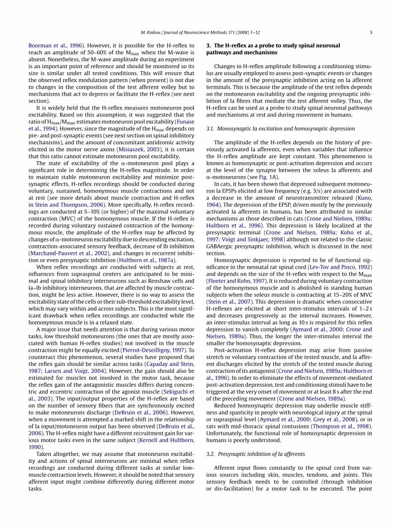
Fig. 2. Presynaptic inhibition of Ia afferents induced by a conditioning afferent volley. Comtibial nerve stimulation to establish based on the amplitude of the conditioned soleus H-rand average size of the soleus H-reflex conditioned by CP nerve stimulation at C–T intervfrom Knikou and Taglianetti, 2006).
e Methods 171 (2008) 1–12
mit presynaptic inhibition to Ia terminals projecting to variousmotoneuron pools. Thus at the onset of voluntary contraction, thereis a differential control of presynaptic inhibition of Ia afferent ter-minals projecting to motoneurons of the contracting muscle and toother motor nuclei (Hultborn et al., 1987b), particularly the antago-nist motoneuron pools (Meunier and Morin, 1989). This differentialcontrol could be due to cortical control since transcranial stim-ulation increases heteronymous Ia facilitation and decreases D1inhibition, while the decrease in presynaptic inhibition of Ia affer-ents to motoneurons of the contracting muscle persists after anischaemic blockage of group I afferents from the contracting muscle(Meunier and Pierrot-Deseilligny, 1998).
To conclude, presynaptic inhibition is critical to neural controlof movement since it gates sensory afferent feedback to the spinalcord to assist in smooth execution of a movement or motor task.Adjustment of presynaptic inhibition of Ia afferents to motoneu-rons that are (or not) involved in contraction may contribute to theproduction of muscle synergy or movement patterns suited to themotor task performed.
3.2.1. Protocol for studying presynaptic inhibition in humans
Different methods have been proposed for assessing presynapticinhibition in humans, including comparison between changes inEMG activity and H-reflex amplitude (Schieppati and Crenna, 1984),and soleus H-reflex depression following tibialis anterior, bicepsfemoris or Achilles tendon vibration (Burke et al., 1976; Hultbornet al., 1987a; Morin et al., 1984).
In this section, details will be provided for protocols that use thesoleus H-reflex as a test reflex. A conditioning afferent volley to thecommon peroneal (CP) (train of 3–5 shocks, 1–1.4× MT) or radialnerve (single shock, 0.7–1× MT) evokes several phases of H-reflexdepression in the antagonist muscles, e.g. soleus and flexor carpiradialis (FCR) (Berardelli et al., 1987; Crone et al., 1987). The earlysoleus H-reflex depression involves the reciprocal Ia inhibitorypathway and is exerted at a postsynaptic level, which is discussedin the next section. The soleus H-reflex depression that appearsat conditioning–test (C–T) intervals that range from 6 to 30 ms iscalled D1 inhibition, and is believed to be mediated by presynapticinhibition of soleus Ia afferents (Crone and Nielsen, 1994; Pierrot-Deseilligny, 1997) (Fig. 2A). In Fig. 2B, soleus H-reflex depression byCP nerve stimulation at a MT level is indicated for C–T intervals of60–120 ms. However, CP nerve stimulation results in a long lasting
mon peroneal (CP) nerve stimulation at low intensities is delivered before posterioreflex the amount of presynaptic inhibition acting on soleus Ia afferent terminals (A)als ranged from 60 to 120 ms for 10 seated subjects (B) (data adopted and modified
scienc
M. Knikou / Journal of Neuro(up to 200 ms) soleus H-reflex depression, similar to that observedin the cat (Eccles, 1964; Eccles et al., 1962).
In humans, the same conditioning afferent volleys do not modifycortical-evoked responses in the soleus or FCR muscles (Berardelliet al., 1987; Faist et al., 1996), which supports the presynaptic natureof the soleus and FCR H-reflex depression. In this context, D1 soleusH-reflex depression may reflect an increased excitability of the pri-mary afferent depolarization interneurons that might be drivenby strong afferent inputs generated by movement (Capaday et al.,1995). Under this scenario, the circuits involved in the presynapticinhibition might become saturated and stop responding to the con-ditioning afferent volley, as reported during human walking (Faistet al., 1996).
A method to study presynaptic inhibition of Ia afferents inhumans was proposed and developed by Hultborn et al. (1987a).This method involves the assessment of heteronymous Ia facili-tation exerted by quadriceps afferents onto soleus motoneurons,and relies on the heteronymous monosynaptic projections from thequadriceps to soleus motoneurons. This method was demonstratedby motor unit studies and verified in animals by direct intracellular
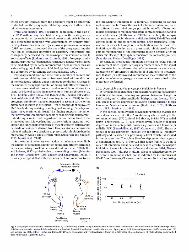
recordings (Fournier et al., 1986; Hultborn et al., 1987a).The femoral nerve (conditioning stimulus) is stimulated with asingle pulse (1-ms duration) through a monopolar ball electrodeplaced at the femoral triangle, while the indifferent electrode isplaced on the gluteus maximus muscle. The stability of the condi-tioning stimulation strength is verified by a stable M-wave in thevastus lateralis muscle. Femoral nerve stimulation is delivered afterposterior tibial nerve because the former is closer to the spinal cordand thus the C–T intervals are negative by convention. The involvedneuronal pathway and the time course of the heteronymous soleusH-reflex facilitation are shown in Fig. 3.
During the first 0.5 ms of femoral nerve induced soleus H-reflexfacilitation, the monosynaptic Ia excitation is not contaminated byany other events and the soleus H-reflex facilitation depends onthe size of the conditioning Ia monosynaptic EPSP (Fournier et al.,1986; Hultborn et al., 1987a). Thus, the changes observed on theconditioned soleus H-reflex amplitude during the first half ms thatthe soleus H-reflex facilitation is established reflect modificationsof the on-going presynaptic inhibition of heteronymous Ia afferentterminals. Simply stated, the larger the conditioned soleus H-reflexfacilitation the smaller the on-going presynaptic inhibition. Under
Fig. 3. Presynaptic inhibition of Ia afferents reflected by changes of heteronymous Ia facstimulation at low intensities delivered after posterior tibial nerve stimulation induces meronymous Ia facilitation reflect modulation of the on-going presynaptic inhibition actin(B) time course of soleus H-reflex facilitation by FN stimulation in one seated subject andH-reflex following FN stimulation at −7.8 ms are shown. Note that the heteronymous Ia(data adopted and modified from Knikou, 2006).
e Methods 171 (2008) 1–12 5
the same principles, the on-going presynaptic inhibition can beindirectly assessed by examining the amount of soleus H-reflexfacilitation by inferior soleus nerve stimulation in the first half ms(Pierrot-Deseilligny et al., 1981).
At this point it should be made clear that heteronymousmonosynaptic Ia facilitation and D1 inhibition differ significantly.The former tests the on-going presynaptic inhibition exerted on Iaafferents involved in the conditioning afferent volley, and the lattermethod tests the effects of activation of the presynaptic networkby a conditioning (electrical or mechanical) afferent volley.
Regardless of the method used, the above assumptions canbe made only when the recruitment gain of the �-motoneuronsremains stable, especially if the test is conducted during voluntarymuscle contraction. To confidently demonstrate changes in on-going presynaptic inhibition, similar findings should be observed inmotor unit studies using the post-stimulus time histogram method(Katz et al., 1988; Nielsen and Kagamihara, 1993). When this is notpossible, a change in the recruitment gain can be eliminated whenthe amount of monosynaptic facilitation and vibration-inducedreflex depression occur in the opposite direction.
Further, the amplitude of the conditioned H-reflex should besimilar to that of the unconditioned (or test) reflex, since its sensi-tivity to facilitation and inhibition depends on the size of the controlH-reflex (Crone et al., 1990). Therefore, when the conditioning stim-ulus is delivered and the reflex size increases or decreases theintensity should be adjusted appropriately. However, this adjust-ment may affect the recruitment reflex gain (input–output relationof the test reflex) (Kernell and Hultborn, 1990), which in turn mayinfluence low- and high-threshold motoneurons differently. Thus,to ensure that the effects are not due to changes in the reflex recruit-ment gain, the H-reflex should be accompanied by a stable M-waveor more direct methods, such as motor unit studies.
3.3. Reciprocal Ia inhibition
Ia afferents participate in neuronal pathways that inhibit theantagonist � motoneurons subserving a reciprocal activation pat-tern between the agonist and corresponding antagonist musclesduring movement in humans. The existence of this neural pathwaywas first postulated in humans by Hoffmann who showed thatthe soleus H-reflex is decreased when the pretibial (antagonistic)
ilitation. (A) Sketch illustrates the spinal circuit during which femoral nerve (FN)onosynaptic excitation of soleus �-motoneurons. Changes in the amount of het-
g on the Ia afferents of the conditioning afferent volley (quadriceps Ia afferents),(C) full-wave waveform rectified averages (n = 20) of the control and conditioned
reflex facilitation occurred without a significant change in the size of the M-wave
scienc
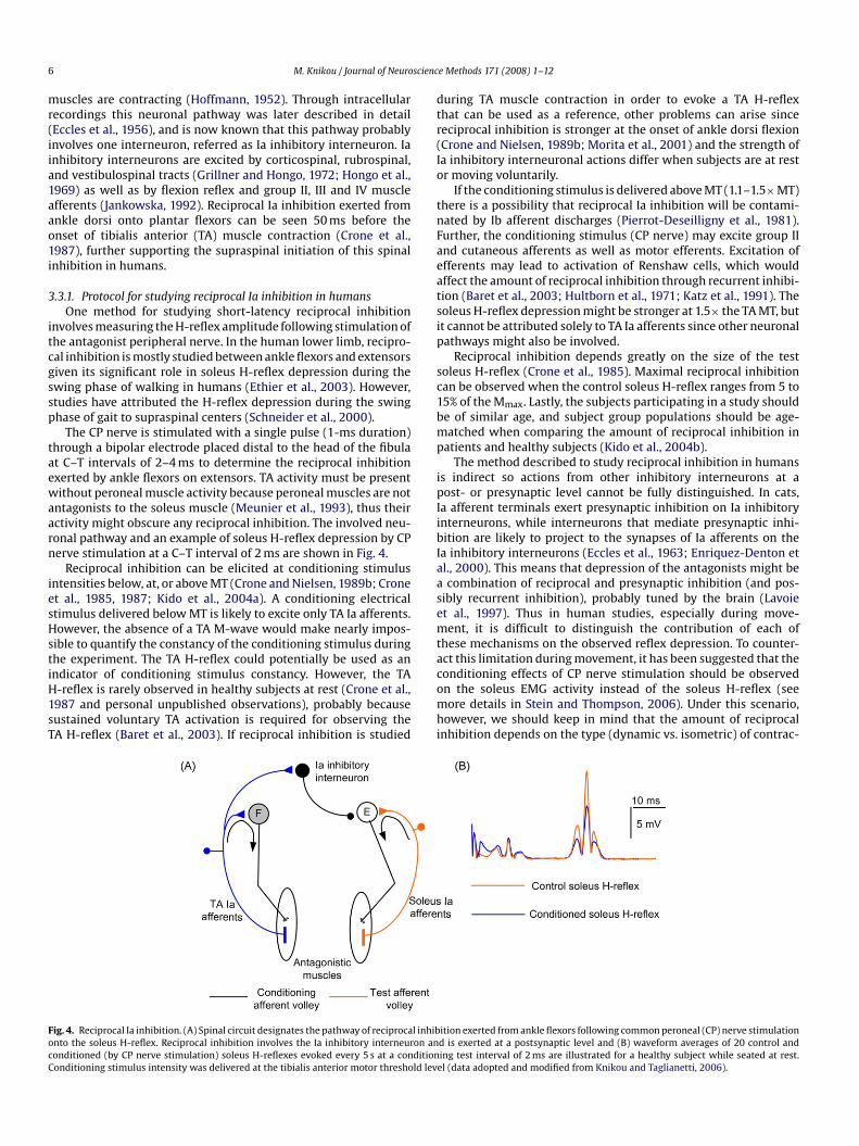
6 M. Knikou / Journal of Neuromuscles are contracting (Hoffmann, 1952). Through intracellularrecordings this neuronal pathway was later described in detail(Eccles et al., 1956), and is now known that this pathway probablyinvolves one interneuron, referred as Ia inhibitory interneuron. Iainhibitory interneurons are excited by corticospinal, rubrospinal,and vestibulospinal tracts (Grillner and Hongo, 1972; Hongo et al.,1969) as well as by flexion reflex and group II, III and IV muscleafferents (Jankowska, 1992). Reciprocal Ia inhibition exerted fromankle dorsi onto plantar flexors can be seen 50 ms before theonset of tibialis anterior (TA) muscle contraction (Crone et al.,1987), further supporting the supraspinal initiation of this spinalinhibition in humans.
3.3.1. Protocol for studying reciprocal Ia inhibition in humansOne method for studying short-latency reciprocal inhibition
involves measuring the H-reflex amplitude following stimulation ofthe antagonist peripheral nerve. In the human lower limb, recipro-cal inhibition is mostly studied between ankle flexors and extensorsgiven its significant role in soleus H-reflex depression during theswing phase of walking in humans (Ethier et al., 2003). However,studies have attributed the H-reflex depression during the swingphase of gait to supraspinal centers (Schneider et al., 2000).
The CP nerve is stimulated with a single pulse (1-ms duration)through a bipolar electrode placed distal to the head of the fibulaat C–T intervals of 2–4 ms to determine the reciprocal inhibitionexerted by ankle flexors on extensors. TA activity must be presentwithout peroneal muscle activity because peroneal muscles are notantagonists to the soleus muscle (Meunier et al., 1993), thus theiractivity might obscure any reciprocal inhibition. The involved neu-ronal pathway and an example of soleus H-reflex depression by CPnerve stimulation at a C–T interval of 2 ms are shown in Fig. 4.
Reciprocal inhibition can be elicited at conditioning stimulusintensities below, at, or above MT (Crone and Nielsen, 1989b; Croneet al., 1985, 1987; Kido et al., 2004a). A conditioning electricalstimulus delivered below MT is likely to excite only TA Ia afferents.However, the absence of a TA M-wave would make nearly impos-sible to quantify the constancy of the conditioning stimulus duringthe experiment. The TA H-reflex could potentially be used as anindicator of conditioning stimulus constancy. However, the TAH-reflex is rarely observed in healthy subjects at rest (Crone et al.,1987 and personal unpublished observations), probably becausesustained voluntary TA activation is required for observing theTA H-reflex (Baret et al., 2003). If reciprocal inhibition is studied
Fig. 4. Reciprocal Ia inhibition. (A) Spinal circuit designates the pathway of reciprocal inhibonto the soleus H-reflex. Reciprocal inhibition involves the Ia inhibitory interneuron anconditioned (by CP nerve stimulation) soleus H-reflexes evoked every 5 s at a conditionConditioning stimulus intensity was delivered at the tibialis anterior motor threshold lev
e Methods 171 (2008) 1–12
during TA muscle contraction in order to evoke a TA H-reflexthat can be used as a reference, other problems can arise sincereciprocal inhibition is stronger at the onset of ankle dorsi flexion(Crone and Nielsen, 1989b; Morita et al., 2001) and the strength ofIa inhibitory interneuronal actions differ when subjects are at restor moving voluntarily.
If the conditioning stimulus is delivered above MT (1.1–1.5× MT)there is a possibility that reciprocal Ia inhibition will be contami-nated by Ib afferent discharges (Pierrot-Deseilligny et al., 1981).Further, the conditioning stimulus (CP nerve) may excite group IIand cutaneous afferents as well as motor efferents. Excitation ofefferents may lead to activation of Renshaw cells, which wouldaffect the amount of reciprocal inhibition through recurrent inhibi-tion (Baret et al., 2003; Hultborn et al., 1971; Katz et al., 1991). Thesoleus H-reflex depression might be stronger at 1.5× the TA MT, butit cannot be attributed solely to TA Ia afferents since other neuronalpathways might also be involved.
Reciprocal inhibition depends greatly on the size of the testsoleus H-reflex (Crone et al., 1985). Maximal reciprocal inhibitioncan be observed when the control soleus H-reflex ranges from 5 to15% of the Mmax. Lastly, the subjects participating in a study shouldbe of similar age, and subject group populations should be age-
matched when comparing the amount of reciprocal inhibition inpatients and healthy subjects (Kido et al., 2004b).The method described to study reciprocal inhibition in humansis indirect so actions from other inhibitory interneurons at apost- or presynaptic level cannot be fully distinguished. In cats,Ia afferent terminals exert presynaptic inhibition on Ia inhibitoryinterneurons, while interneurons that mediate presynaptic inhi-bition are likely to project to the synapses of Ia afferents on theIa inhibitory interneurons (Eccles et al., 1963; Enriquez-Denton etal., 2000). This means that depression of the antagonists might bea combination of reciprocal and presynaptic inhibition (and pos-sibly recurrent inhibition), probably tuned by the brain (Lavoieet al., 1997). Thus in human studies, especially during move-ment, it is difficult to distinguish the contribution of each ofthese mechanisms on the observed reflex depression. To counter-act this limitation during movement, it has been suggested that theconditioning effects of CP nerve stimulation should be observedon the soleus EMG activity instead of the soleus H-reflex (seemore details in Stein and Thompson, 2006). Under this scenario,however, we should keep in mind that the amount of reciprocalinhibition depends on the type (dynamic vs. isometric) of contrac-
ition exerted from ankle flexors following common peroneal (CP) nerve stimulationd is exerted at a postsynaptic level and (B) waveform averages of 20 control anding test interval of 2 ms are illustrated for a healthy subject while seated at rest.el (data adopted and modified from Knikou and Taglianetti, 2006).
scienc
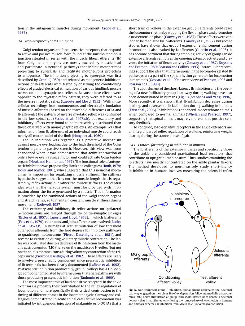
Pearson et al., 1998).The abolishment of the short-latency Ib inhibition and the open-
ing of a new facilitatory group I pathway during walking have alsobeen demonstrated in humans (Fig. 5) (Stephens and Yang, 1996).More recently, it was shown that Ib inhibition decreases duringloading, and reverses to Ib facilitation during walking in humans(Faist et al., 2006). Ib inhibition is enhanced in decerebrate animalswhen compared to normal animals (Whelan and Pearson, 1997),suggesting that spinal animals may rely more on this positive sen-sory feedback.
To conclude, load-sensitive receptors in the ankle extensors arean integral part of reflex regulation of walking, reinforcing weightbearing during the stance phase of gait.
3.4.1. Protocol for studying Ib inhibition in humansThe Ib afferents of the extensor muscles and specifically those
of the ankle are considered gravitational load receptors thatcontribute to upright human posture. Thus, studies examining theIb effects have mostly concentrated on the ankle plantar flexors.The method developed to non-invasively study short-latencyIb inhibition in humans involves measuring the soleus H-reflex
M. Knikou / Journal of Neuro
tion in the antagonistic muscles during movement (Crone et al.,1987).
3.4. Non-reciprocal (or Ib) inhibition
Golgi tendon organs are force-sensitive receptors that respondto active and passive muscle force found at the muscle-tendinousjunction situated in series with the muscle fibers. Afferents (Ib)from Golgi tendon organs are mostly excited by muscle loadand participate in neuronal pathways that inhibit motoneuronsprojecting to synergists and facilitate motoneurons projectingto antagonists. The inhibition projecting to synergists was firstdescribed by Granit (1950) and referred as autogenetic inhibition.Actions of Ib afferents were tested by observing the conditioningeffects of graded electrical stimulation of various hindlimb musclenerves on monosynaptic test reflexes. Because these effects wereopposite to the myotatic reflex pattern, they were referred to asthe inverse myotatic reflex (Laporte and Lloyd, 1952). With intra-cellular recordings from motoneurons and electrical stimulationof muscle afferents (based on the threshold differences of Ia andIb afferents) the pattern of inverse myotatic reflex was confirmedin the low spinal cat (Eccles et al., 1957a,b), but excitatory andinhibitory effects were found to be more widely distributed thanthose observed with monosynaptic reflexes. An example was thatinformation from Ib afferents of an individual muscle could reachnearly all motor nuclei of the limb (Hongo et al., 1969).
The Ib inhibition was regarded as a protective mechanismagainst muscle overloading due to the high threshold of the Golgitendon organs to passive stretch. However, this view was soonabandoned when it was demonstrated that active contraction ofonly a few or even a single motor unit could activate Golgi tendonorgans (Houk and Henneman, 1967). The functional role of autoge-netic inhibition was proposed by Houk and colleagues (Houk, 1979;Houk and Rymer, 1981), who suggested that this neuronal mech-anism is important for regulating muscle stiffness. The stiffnesshypothesis suggests that it is not the muscle length that is regu-lated by reflex actions but rather the muscle stiffness. The centralidea was that the nervous system must be provided with infor-mation about the force generated by a muscle. This informationis provided by the combined actions of the Golgi tendon organsand stretch reflex, so to maintain constant muscle stiffness duringmovement (Rothwell, 1987).
The excitatory and inhibitory Ib reflex actions on ipsilateral�-motoneurons are relayed through di- or tri-synaptic linkages
(Eccles et al., 1957a; Laporte and Lloyd, 1952), in which Ia afferents(Fetz et al., 1979), cutaneous, and joint afferents are involved (Eccleset al., 1957a,b). In humans at rest, stimulation of low thresholdcutaneous afferents from the foot depress Ib inhibitory pathwaysto quadriceps motoneurons (Pierrot-Deseilligny et al., 1981), andreverse to excitation during voluntary muscle contraction. The lat-ter was postulated due to a decrease of Ib inhibition from the medi-alis gastrocnemius (MG) nerve on the quadriceps H-reflex (but noton the soleus motoneurons) during voluntary contraction of the tri-ceps surae (Pierrot-Deseilligny et al., 1982). These effects are likelyto involve a presynaptic component since presynaptic inhibitionof Ib terminals has been clearly documented (Lafleur et al., 1992).Postsynaptic inhibition produced by group I volleys has a GABAer-gic component mediated by interneurons that share pathways withthose producing presynaptic inhibition (Rudomin et al., 1990).The most important role of load-sensitive receptors in the ankleextensors is probably their contribution to the reflex regulation oflocomotion and more specifically their critical contribution to thetiming of different phases in the locomotor cycle. Conway and col-leagues demonstrated in acute spinal cats (fictive locomotion wasinitiated by intravenous injection of nialamide or L-DOPA) that a
e Methods 171 (2008) 1–12 7
short train of volleys in the extensor group I afferents could resetthe locomotor rhythm by stopping the flexion phase and promotinga new extension phase (Conway et al., 1987). These effects were ver-ified to be mediated by Ib afferents (Conway et al., 1987), but recentstudies have shown that group I extension enhancement duringlocomotion is also evoked by Ia afferents (Guertin et al., 1995). Ithas become pertinent that during stepping, activity of group I ankleextensor afferents reinforces the ongoing extensor activity and pre-vents the initiation of flexor activity (Conway et al., 1987; Duysensand Pearson, 1980; Pearson and Collins, 1993). Intracellular record-ings support the idea that interneurons in the locomotor related Ibpathways are a part of the spinal rhythm generator for locomotionin mammals (Gossard et al., 1994; see reviews of Pearson, 1995 and
Fig. 5. Non-reciprocal group I inhibition. Spinal circuit designates the neuronalpathway engaged on the soleus H-reflex depression following medialis gastrocne-mius (MG) nerve stimulation at group I threshold. Dotted lines denote a neuronalnetwork that is manifested only during the stance phase of locomotion in humansand animals, whereas Ib inhibition from MG to soleus reverses to excitation.

scienc
8 M. Knikou / Journal of Neurofollowing stimulation of the synergist MG nerve at a C–T interval of6 ms. Because the MG nerve in non-human primates contains moreIb than Ia afferents (Hongo et al., 1984), it was proposed that MGnerve actions on the soleus H-reflex are mediated by Ib inhibitionconsistent with a disynaptic pathway (Pierrot-Deseilligny et al.,1979, 1981).
The conditioning stimulation electrode is placed 7–10 cm distaland medial to the cathode electrode for the posterior tibial nervewhere a clear contraction of the MG muscle can be seen. The stimu-lus anode for the MG nerve is placed over the anterolateral portionof the leg just distal to the patella. Ib inhibition is obtained at con-ditioning stimulus intensities below MT to ensure that the effectsobserved are not contaminated by recurrent inhibition (Rossi et al.,1994). Thus, the MG MT needs to be checked throughout the exper-iment to ensure that the stimulus intensity is sub-threshold, andthat the MG MT level does not change during the experiment. Theconditioning stimulus to the MG nerve can be either a single (1 ms)or multiple pulses. Multiple conditioning pulses are more effec-tive in generating the short-latency soleus H-reflex depression inhumans (Bouaziz et al., 1975; Pierrot-Deseilligny et al., 1979).
3.5. Recurrent inhibition
Inhibitory neurons participate in neuronal circuits that subservemovement. One of the first identified inhibitory neurons was the
Renshaw cell located in the ventral horn medial to the motor nuclei(Renshaw, 1946). Renshaw cells are excited by axon collaterals frommotoneurons and provide recurrent inhibition of �-motoneuronsthat project to the same or synergistic muscles. Activity in segmen-tal afferents may influence Renshaw cells apart from the indirectexcitation that is produced by motoneuronal reflex discharge.Polysynaptic excitation has been described after stimulation of dor-sal roots, ipsilateral group II and III muscle afferents, cutaneousafferents, and contralateral flexor reflex afferents (see referencesin Baldissera et al., 1981), while Renshaw cells receive inhibitionfrom ipsilateral and contralateral segmental afferents. In additionto their well known projection to �-motoneurons, Renshaw cellsconnect with �-motoneurons, the interneurons mediating recip-rocal Ia inhibition, other Renshaw cells, and receive inputs fromboth primary afferents and descending tracts (Hultborn et al., 1971;Mazzocchio et al., 1994; see extensive review of Baret et al., 2003;Katz and Pierrot-Deseilligny, 1998) (Fig. 6). The wide convergencefrom a number of segmental reflex pathways suggests that thelocal feedback regulation provided by recurrent inhibition is notstereotyped and hard-wired but versatile in nature.Fig. 6. Recurrent inhibition. Spinal circuit denotes the neuronal pathway of Renshawcells and their connections to �- and �-motoneurons, and Ia inhibitory interneu-rons between ankle flexors and extensors. Renshaw cells depress the activity of �–�motoneurons, and Ia inhibitory interneurons. Broken lines indicate parallel controlof �-motoneurons, Ia inhibitory interneurons, and Renshaw cells by the brain; closedcircles: inhibition, closed triangles: facilitation.
e Methods 171 (2008) 1–12
Recurrent inhibition has mostly been described in terms of astabilizing or limiting feedback mechanism that reduces the sensi-tivity of neurons to changes in their excitatory drive and decreasesthe frequency of their discharge to a given input when com-pared to a system without recurrent feedback. However, recurrentinhibition restricts motoneuron discharge by inhibiting motoneu-rons in the subliminal fringe (Brooks and Wilson, 1959), stabilizesthe discharge frequency from tonically firing motoneurons (Granitet al., 1960), inhibits motoneurons to slow contracting musclefibers during rapid contractions (Eccles et al., 1961), synchro-nizes motoneuron discharge patterns (Mattei et al., 2003), andincreases short-term synchronization of �-motoneuron discharges(Uchiyama and Windhorst, 2007).
Further, since there is Renshaw facilitation (increasing recur-rent inhibition) during a weak tonic voluntary contraction, butRenshaw cell inhibition (suppressing recurrent inhibition) duringa strong contraction, it has been suggested that recurrent inhibi-tion may operate as a gain regulator of motor output (Hultbornand Pierrot-Deseilligny, 1979b). For example, when Renshaw cellsare facilitated during a weak muscle contraction (Hultborn andPierrot-Deseilligny, 1979a,b) the slope of the input–output relationis reduced thus providing a mechanism to control motor output.In contrast, Renshaw cell inhibition during strong contractionsensures a high input–output gain for the motoneuron pool favor-ing a large muscle force output. In addition, recurrent inhibition isreduced during soleus muscle contraction but is enhanced duringvoluntary contraction of the TA muscle when the subject is stand-ing without support (Pierrot-Deseilligny et al., 1977), and whenreciprocal inhibition of the antagonists is required (Mazzocchioet al., 1994). These findings strongly suggest that recurrent inhi-bition is engaged in motor tasks when equilibrium is threatened,and plays a role during selection of “appropriate” muscle synergypatterns.
Renshaw cell activity during rhythmic motor tasks such as loco-motion has been studied in the cat using extracellular recordings(Pratt and Jordan, 1987) and during locomotor-like activity in spinalcord preparations in vitro (Nishimaru et al., 2006). These studieshave shown that Renshaw cells are rhythmically active and thattheir firing properties are modulated by motoneurons as well asby ipsilateral and contralateral locomotor networks. To conclude, itis apparent that recurrent inhibition plays a significant role in theneural control of movement.
3.5.1. Protocol for studying recurrent inhibition in humans
Heteronymous recurrent inhibition in humans can be assessedby establishing the effects of femoral nerve stimulation on thesoleus H-reflex or EMG activity. As previously described, femoralnerve stimulation at group I level induces facilitation of the soleusH-reflex at negative C–T intervals. The soleus H-reflex facilitationhowever is followed by a depression that is believed to be mediatedby recurrent inhibition because it increases with the conditioningH-reflex, it has a short central delay, and lasts up to 40 ms (Busseland Pierrot-Deseilligny, 1977). By analogy, heteronymous recurrentinhibition can be demonstrated using the quadriceps H-reflex pre-ceded by a conditioning stimulus delivered to the inferior soleusnerve at a C–T interval of 22 ms (Iles et al., 2000).
The method for demonstrating homonymous recurrent inhibi-tion was proposed by Pierrot-Deseilligny and colleagues (Busseland Pierrot-Deseilligny, 1977; Pierrot-Deseilligny and Bussel, 1975)and relies on activating Renshaw cells with a conditioning soleusH-reflex discharge. More specifically, due to collision in motoraxons between the orthodromic conditioning discharge and theantidromic motor volley from the strong test stimulus, theexcitability is assessed only for motoneurons that have already firedin the first conditioning discharge.
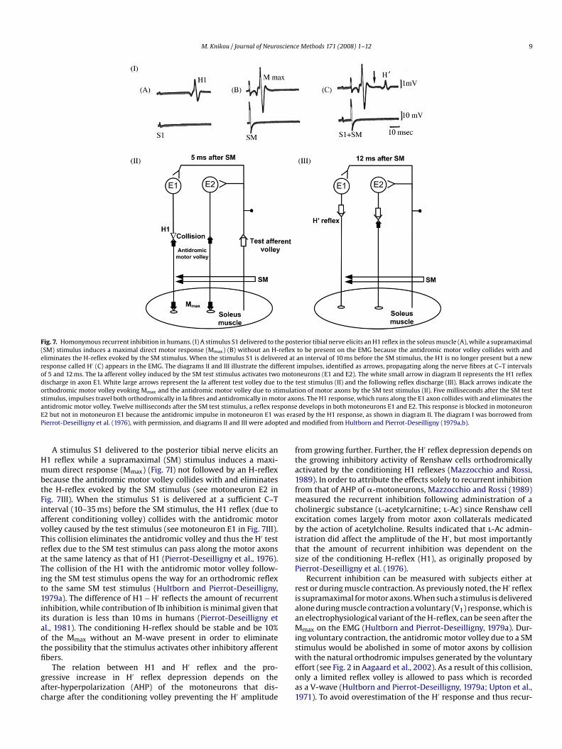
M. Knikou / Journal of Neuroscience Methods 171 (2008) 1–12 9
e posreflexred aterentmoto
o theimulattor axoponseas erated an
Fig. 7. Homonymous recurrent inhibition in humans. (I) A stimulus S1 delivered to th(SM) stimulus induces a maximal direct motor response (Mmax) (B) without an H-eliminates the H-reflex evoked by the SM stimulus. When the stimulus S1 is deliveresponse called H′ (C) appears in the EMG. The diagrams II and III illustrate the diffof 5 and 12 ms. The Ia afferent volley induced by the SM test stimulus activates twodischarge in axon E1. White large arrows represent the Ia afferent test volley due torthodromic motor volley evoking Mmax and the antidromic motor volley due to ststimulus, impulses travel both orthodromically in Ia fibres and antidromically in moantidromic motor volley. Twelve milliseconds after the SM test stimulus, a reflex resE2 but not in motoneuron E1 because the antidromic impulse in motoneuron E1 wPierrot-Deseilligny et al. (1976), with permission, and diagrams II and III were adop
A stimulus S1 delivered to the posterior tibial nerve elicits anH1 reflex while a supramaximal (SM) stimulus induces a maxi-mum direct response (Mmax) (Fig. 7I) not followed by an H-reflexbecause the antidromic motor volley collides with and eliminatesthe H-reflex evoked by the SM stimulus (see motoneuron E2 in
Fig. 7III). When the stimulus S1 is delivered at a sufficient C–Tinterval (10–35 ms) before the SM stimulus, the H1 reflex (due toafferent conditioning volley) collides with the antidromic motorvolley caused by the test stimulus (see motoneuron E1 in Fig. 7III).This collision eliminates the antidromic volley and thus the H′ testreflex due to the SM test stimulus can pass along the motor axonsat the same latency as that of H1 (Pierrot-Deseilligny et al., 1976).The collision of the H1 with the antidromic motor volley follow-ing the SM test stimulus opens the way for an orthodromic reflexto the same SM test stimulus (Hultborn and Pierrot-Deseilligny,1979a). The difference of H1 − H′ reflects the amount of recurrentinhibition, while contribution of Ib inhibition is minimal given thatits duration is less than 10 ms in humans (Pierrot-Deseilligny etal., 1981). The conditioning H-reflex should be stable and be 10%of the Mmax without an M-wave present in order to eliminatethe possibility that the stimulus activates other inhibitory afferentfibers.The relation between H1 and H′ reflex and the pro-gressive increase in H′ reflex depression depends on theafter-hyperpolarization (AHP) of the motoneurons that dis-charge after the conditioning volley preventing the H′ amplitude
terior tibial nerve elicits an H1 reflex in the soleus muscle (A), while a supramaximalto be present on the EMG because the antidromic motor volley collides with andan interval of 10 ms before the SM stimulus, the H1 is no longer present but a newimpulses, identified as arrows, propagating along the nerve fibres at C–T intervalsneurons (E1 and E2). The white small arrow in diagram II represents the H1 reflextest stimulus (II) and the following reflex discharge (III). Black arrows indicate theion of motor axons by the SM test stimulus (II). Five milliseconds after the SM testns. The H1 response, which runs along the E1 axon collides with and eliminates thedevelops in both motoneurons E1 and E2. This response is blocked in motoneuron
sed by the H1 response, as shown in diagram II. The diagram I was borrowed fromd modified from Hultborn and Pierrot-Deseilligny (1979a,b).
from growing further. Further, the H′ reflex depression depends onthe growing inhibitory activity of Renshaw cells orthodromicallyactivated by the conditioning H1 reflexes (Mazzocchio and Rossi,1989). In order to attribute the effects solely to recurrent inhibitionfrom that of AHP of �-motoneurons, Mazzocchio and Rossi (1989)
measured the recurrent inhibition following administration of acholinergic substance (l-acetylcarnitine; l-Ac) since Renshaw cellexcitation comes largely from motor axon collaterals medicatedby the action of acetylcholine. Results indicated that l-Ac admin-istration did affect the amplitude of the H′, but most importantlythat the amount of recurrent inhibition was dependent on thesize of the conditioning H-reflex (H1), as originally proposed byPierrot-Deseilligny et al. (1976).Recurrent inhibition can be measured with subjects either atrest or during muscle contraction. As previously noted, the H′ reflexis supramaximal for motor axons. When such a stimulus is deliveredalone during muscle contraction a voluntary (V1) response, which isan electrophysiological variant of the H-reflex, can be seen after theMmax on the EMG (Hultborn and Pierrot-Deseilligny, 1979a). Dur-ing voluntary contraction, the antidromic motor volley due to a SMstimulus would be abolished in some of motor axons by collisionwith the natural orthodromic impulses generated by the voluntaryeffort (see Fig. 2 in Aagaard et al., 2002). As a result of this collision,only a limited reflex volley is allowed to pass which is recordedas a V-wave (Hultborn and Pierrot-Deseilligny, 1979a; Upton et al.,1971). To avoid overestimation of the H′ response and thus recur-

scienc
10 M. Knikou / Journal of Neurorent inhibition, the V1 responses should be subtracted from thecorresponding H′ reflexes.
4. Clinical implications
The H-reflex can be utilized to assess modulation of spinalinhibitory interneuronal circuits, but attention is needed to the fac-tors previously discussed that affect Ia transmission. The H-reflex isnot hard-wired but is dramatically modulated during various motortasks (task dependence) or during different phases of a cyclicalmovement (e.g. cycling and walking), and can be affected by sev-eral factors that must be acknowledged to avoid misinterpretationof the data. This is especially important when these mechanismsare assessed in people with a neurological injury.
All of the spinal inhibitory circuits described above (homosy-naptic depression, reciprocal Ia inhibition, presynaptic inhibition,Ib inhibition, and recurrent inhibition) have been correlated tosome degree with spasticity (for references see Nielsen et al., 2007),especially when the lesion occurs at the spinal cord. Presynapticand postsynaptic inhibition might be differentially organized atcervical and lumbar levels so a characterization of these circuits isnecessary to understand the organization of these circuits follow-ing spinal cord lesions at different segmental levels. Further, giventhe numerous factors that affect spinal reflex excitability, neuro-physiological differences driven by the type or extent of the lesion,and spinal excitability state across patients, it is clear that a moresystematic characterization of these interneuronal circuits shouldbe conducted.
This is extremely important in light of new evidence. For exam-ple, homosynaptic depression can be increased after just a singlecycling session in spinal-intact subjects (Meunier et al., 2007), andafter 10 treadmill training sessions in one SCI subject capable ofambulation (Trimble et al., 1998), suggesting that the H-reflex canbe used as a tool to study short- and long-term plasticity of thenervous system. This is further supported by the elegant studiesconducted by Wolpaw and colleagues (for references see Wolpaw,2007) whereas up- or down H-reflex conditioning training resultedin changes of motoneuron properties. In this line, conditioning H-reflex protocols that lead to locomotor recovery in rats (Chen et al.,2006) may be developed for people with neurological injuries inthe near future.
5. Conclusions
The H-reflex has been utilized as a probe to study neuronalpathways and spinal inhibitory control systems that are tightlycoupled with the neural control of movement in health and neu-rological disorders. However, the reflex magnitude can changedramatically during contraction or stretch of agonist and antago-nist muscles. Given the differences in motoneuron excitability stateacross subjects, differential supraspinal control of spinal inhibitoryinterneurons, and our inability to distinguish the relative con-tribution of each spinal inhibitory mechanism to motoneuronalexcitability during a motor task or condition, it is clear that greatattention should be paid to all of the limiting factors discussedin this review and be taken into consideration when data areinterpreted. If these limitations are recognized and addressed, theH-reflex will remain one of the major probes for studying sensori-motor integration and training-induced neural adaptation in healthand neural pathology.
Acknowledgements
This work was supported by the New York State Department ofHealth, Spinal Cord Injury Research Program, Contract No. C020933.
e Methods 171 (2008) 1–12
I thank the anonymous reviewers for their valuable and construc-tive comments and Dr. Elizabeth Kay for scrutinizing the English.
References
Aagaard P, Simonsen EB, Andersen JL, Magnuson P, Dyhre-Poulsen P. Neural adapta-tion to resistance training: changes in evoked V-wave and H-reflex responses. JAppl Physiol 2002;92:2309–18.
Andersen JB, Sinkjaer T. The stretch reflex and H-reflex of the human soleus muscleduring walking. Motor Control 1999;3:151–7.
Aymard C, Katz R, Lafitte C, Lo E, Penicaud A, Pradat-Diehl P, et al. Presynap-tic inhibition and homonymous depression: a comparison between lower andupper limbs in normal human subjects and patients with hemiplegia. Brain2000;123:1688–702.
Baldissera F, Hultborn H, Illert M. Integration in spinal neuronal systems. Handbookof physiology. The nervous system. Bethesda: American Physiological Society;1981. pp. 509–95.
Baret M, Katz R, Lamy JC, Penicaud A, Wargon I. Evidence for recurrent inhibitionof reciprocal inhibition from soleus to tibialis anterior in man. Exp Brain Res2003;152:133–6.
Berardelli A, Day BL, Marsden CD, Rothwell JC. Evidence favouring presynaptic inhi-bition between antagonist muscle afferents in the human forearm. J Physiol Lond1987;391:71–83.
Boorman GI, Hoffer JA, Kallesoe K, Viberg D, Mah C. A measure of peripheralnerve stimulation efficacy applicable to H-reflex studies. Can J Neurol Sci1996;23:264–70.
Bouaziz Z, Bouaziz M, Hugon M. Modulation of soleus electromyogram during elec-trical stimulation of medial gastrocnemius nerve in man. ElectroencephalogrClin Neurophysiol 1975;15:31–42.
Brooke JD, Misiaszek JE, Cheng J. Locomotor-like rotation of either hip orknee inhibits soleus H reflexes in humans. Somatosens Mot Res 1993;10:357–64.
Brooks VB, Wilson VJ. Recurrent inhibition in the cat’s spinal cord. J Physiol Lond1959;146:380–91.
Burke RE, Rudomin P, Zajac FE. The effect of activation history on tension productionby individual muscle units. Brain Res 1976;109:515–29.
Burke D, Gandevia SC, McKeon B. Monosynaptic and oligosynaptic contributions tohuman ankle jerk and H-reflex. J Neurophysiol 1984;52:435–48.
Bussel B, Pierrot-Deseilligny E. Inhibition of human motoneurones, probably ofRenshaw origin, elicited by an orthodromic motor discharge. J Physiol Lond1977;269:319–39.
Capaday C, Stein RB. Difference in the amplitude of the human soleus H reflex duringwalking and running. J Physiol Lond 1987;392:513–22.
Capaday C, Lavoie BA, Comeau F. Differential effects of a flexor nerve input on thehuman soleus H-reflex during standing versus walking. Can J Physiol Pharmacol1995;73:436–49.
Chen Y, Chen XY, Jakeman LB, Chen L, Stokes BT, Wolpaw JR. Operant conditioningof H-reflex can correct a locomotor abnormality after spinal cord injury in rats.J Neurosci 2006;26:12537–43.
Conway BA, Hultborn H, Kiehn O. Proprioceptive input resets central locomotorrhythm in the spinal cat. Exp Brain Res 1987;68:643–56.
Crone C, Nielsen J. Methodological implications of the post activation depression ofthe soleus H-reflex in man. Exp Brain Res 1989a;78:28–32.
Crone C, Nielsen J. Spinal mechanisms in man contributing to reciprocal inhibition
during voluntary dorsiflexion of the foot. J Physiol 1989b;416:255–72.Crone C, Nielsen J. Central control of disynaptic reciprocal inhibition in humans. ActaPhysiol Scand 1994;152:351–63.
Crone C, Hultborn H, Jespersen B. Reciprocal Ia inhibition from the peroneal nerveto soleus motoneurones with special reference to the size of the test reflex. ExpBrain Res 1985;59:418–22.
Crone C, Hultborn H, Jespersen B, Nielsen J. Reciprocal Ia inhibition between ankleflexors and extensors in man. J Physiol Lond 1987;389:163–85.
Crone C, Hultborn H, Mazieres L, Morin C, Nielsen J, Pierrot-Deseilligny E. Sensi-tivity of monosynaptic test reflexes to facilitation and inhibition as a functionof the test reflex size: a study in man and the cat. Exp Brain Res 1990;81:35–45.
DeBruin H, Fu W, Galea V, McComas A. Speculations surrounding a spinal reflex. JNeurol Sci 2006;242:75–82.
Devanne H, Lavoie BA, Capaday C. Input–output properties and gain changes in thehuman corticospinal pathways. Exp Brain Res 1997;114:329–38.
Duysens J, Pearson GK. Inhibition of flexor burst generation by loading ankle extensormuscles in walking cats. Brain Res 1980;187:321–32.
Eccles JC. Presynaptic inhibition in the spinal cord. Prog Brain Res 1964;12:65–91.Eccles JC, Fatt P, Landgren S. Central pathway for direct inhibitory action of impulses
in largest afferent nerve fibres to muscle. J Neurophysiol 1956;19:75–98.Eccles JC, Eccles RM, Lundberg A. The convergence of monosynaptic excitatory
afferents on to many different species of alpha motoneurones. J Physiol Lond1957a;137:22–50.
Eccles JC, Eccles RM, Lundberg A. Synaptic actions on motoneurones caused byimpulses in Golgi tendon organ afferents. J Physiol Lond 1957b;138:227–52.
Eccles JC, Eccles RM, Magni F. Potential changes recorded inside primary afferentfibres within the spinal cord. J Physiol Lond 1961;159:147–66.
scienc
M. Knikou / Journal of NeuroEccles JC, Schmidt RF, Willis WD. Presynaptic inhibition of the spinal monosynapticreflex pathway. J Physiol Lond 1962;161:282–97.
Eccles JC, Schmidt R, Willis WD. Pharmacological studies on presynaptic inhibition.J Physiol Lond 1963;168:500–30.
Enriquez-Denton M, Nielsen J, Perreault MC, Morita H, Petersen N, Hultborn H.Presynaptic control of transmission along the pathway mediating disynapticreciprocal inhibition in the cat. J Physiol Lond 2000;526:623–37.
Ethier C, Imbeault M-A, Ung V, Capaday C. On the soleus H-reflex modulation patternduring walking. Exp Brain Res 2003;151:420–5.
Faist M, Dietz V, Pierrot-Deseilligny E. Modulation, probably presynaptic in origin, ofmonosynaptic Ia excitation during human gait. Exp Brain Res 1996;109:441–9.
Faist M, Hoefer C, Hodapp M, Dietz V, Berger W, Duysens J. In humans Ib facilitationdepends on locomotion while suppression of Ib inhibition requires loading. BrainRes 2006;1076:87–92.
Fetz EE, Jankowska E, Johannisson T, Lipski J. Autogenetic inhibition of motoneu-rones by impulses in group Ia muscle spindle afferents. J Physiol Lond1979;293:173–95.
Floeter MK, Kohn AF. H-reflexes of different sizes exhibit differential sensi-tivity to low frequency depression. Electroencephalogr Clin Neurophysiol1997;105:470–5.
Fournier E, Meunier S, Pierrot-Deseilligny E, Shindo M. Evidence for interneuronallymediated Ia excitatory effects to human quadriceps motoneurones. J PhysiolLond 1986;377:143–69.
Frank K, Fuortes MGF. Presynaptic and postsynaptic inhibition of monosynapticreflexes. Fed Proc 1957;16:39–40.
Funase K, Imanaka K, Nishihira Y. Excitability of the soleus motoneuron pool revealedby the developmental slope of the H-reflex as reflex gain. Electromyogr ClinNeurophysiol 1994;34:477–89.
Gossard J-P, Brownstone RM, Barajon I, Hultborn H. Transmission in a locomotor-related group Ib pathway from hindlimb extensor muscles in the cat. Exp BrainRes 1994;98:213–28.
Granit R. Autogenetic inhibition. Electroencephalogr Clin Neurophysiol1950;2:417–24.
Granit R, Haase J, Rutledge LT. Recurrent inhibition in relation to frequency of fir-ing and limitation of discharge rate of extensor motoneurones. J Physiol Lond1960;154:308–28.
Grey MJ, Klinge K, Crone C, Lorentzen J, Biering-Sorensen F, Ravnborg M, et al. Post-activation depression of soleus stretch reflexes in healthy and spastic humans.Exp Brain Res 2008;185:189–97.
Grillner S, Hongo T. Vestibulospinal effects on motoneurones and interneurones inthe lumbosacral cord. Prog Brain Res 1972;37:243–62.
Guertin P, Angel MJ, Perreault MC, McCrea DA. Ankle extensor group I afferents exciteextensors throughout the hindlimb during fictive locomotion in the cat. J PhysiolLond 1995;487:197–209.
Henneman E, Somjen G, Carpenter DO. Excitability and inhibitability of motoneuronsof different sizes. J Neurophysiol 1965;28:599–620.
Hoffmann P. Reflex inhibition and voluntary action of normal human muscles. J NervMent Dis 1952;116:579–84.
Hongo T, Jankowska E, Lundberg A. The rubrospinal tract. II. Facilitation ofinterneuronal transmission in reflex paths to motoneurones. Exp Brain Res1969;7:365–91.
Hongo T, Lundberg A, Phillips CG, Thompson RF. The pattern of monosynaptic Ia-connections to hindlimb motor nuclei in the baboon: a comparison with the cat.Proc R Soc Lond B 1984;221:261–89.
Houk JC. Regulation of stiffness by skeletomotor reflexes. Ann Rev Physiol1979;41:99–114.
Houk JC, Henneman E. Responses of Golgi tendon organs to active contractions of
the soleus muscle of the cat. J Neurophysiol 1967;30:466–81.Houk JC, Rymer WZ. Neural control of muscle length and tension. In: Brooks VB,editor. Handbook of Physiology, 2. Baltimore: Williams and Wilkins; 1981. p.257–323.
Hultborn H, Pierrot-Deseilligny E. Changes in recurrent inhibition during voluntarysoleus contractions in man studied by an H-reflex technique. J Physiol Lond1979a;297:229–51.
Hultborn H, Pierrot-Deseilligny E. Input–output relations in the pathway of recurrentinhibition to motoneurones in the cat. J Physiol Lond 1979b;297:267–87.
Hultborn H, Jankowska E, Lindstrom S. Recurrent inhibition from motor axon col-laterals of transmission in the Ia inhibitory pathway to motoneurones. J PhysiolLond 1971;215:591–612.
Hultborn H, Meunier S, Pierrot-Deseilligny E, Shindo M. Changes in presynaptic inhi-bition of Ia fibres at the onset of voluntary contraction in man. J Physiol Lond1987a;389:757–72.
Hultborn H, Meunier S, Morin C, Pierrot-Deseilligny E. Assessing changes in presy-naptic inhibition of Ia fibres: a study in man and the cat. J Physiol Lond1987b;389:729–56.
Hultborn H, Illert M, Nielson J, Paul A, Ballegaard M, Wiese H. On the mechanism ofthe post-activation depression of the H-reflex in human subjects. Exp Brain Res1996;108:450–62.
Iles JF, Roberts RC. Inhibition of monosynaptic reflexes in the human lower limb. JPhysiol Lond 1987;385:69–87.
Iles JF, Ali A, Pardoe J. Task-related changes of transmission in the pathway ofheteronymous spinal recurrent inhibition from soleus to quadriceps motor neu-rones in man. Brain 2000;123:2264–72.
Jankowska E. Interneuronal relay in spinal pathways from proprioceptors. Prog Neu-robiol 1992;38:335–78.
e Methods 171 (2008) 1–12 11
Katz R, Pierrot-Deseilligny E. Recurrent inhibition in humans. Prog Neurobiol1998;57:325–55.
Katz R, Meunier S, Pierrot-Deseilligny E. Changes in presynaptic inhibition of Ia fibresin man while standing. Brain 1988;111:417–37.
Katz R, Penicaud A, Rossi A. Reciprocal Ia inhibition between elbow flexors andextensors in the human. J Physiol Lond 1991;437:269–86.
Kernell D, Hultborn H. Synaptic effects on recruitment gain: a mechanism ofimportance for the input–output relations of motoneurone pools? Brain Res1990;507:176–9.
Kido A, Tanaka N, Stein RB. Spinal reciprocal inhibition in human locomotion. J ApplPhysiol 2004a;96:1969–77.
Kido A, Tanaka N, Stein RB. Spinal excitation and inhibition decrease as humans age.Can J Physiol Pharmacol 2004b;82:238–48.
Klimstra M, Zehr EP. A sigmoid function is the best fit for the ascending limb of theHoffmann reflex recruitment curve. Exp Brain Res 2008;186:93–105.
Knikou M. Effects of changes in hip position on actions of spinal inhibitory interneu-rons in humans. Int J Neurosci 2006;116:945–61.
Knikou M, Rymer WZ. Effects of changes in hip joint angle on H-reflex excitabilityin humans. Exp Brain Res 2002;143:149–59.
Knikou M, Taglianetti C. On the methods employed to record and measure the humansoleus H-reflex. Somatosens Motor Res 2006;23:55–62.
Kohn A, Floeter MK, Hallett M. Presynaptic inhibition compared with homosynapticdepression as an explanation for soleus H-reflex depression in humans. ExpBrain Res 1997;116:375–80.
Kuno M. Quantal components of excitatory synaptic potentials in spinal motoneu-rones. J Physiol Lond 1964;175:81–99.
Lafleur J, Zytnicki D, Horchole-Bossavit G, Jami L. Depolarization of Ib afferent axonsin the cat spinal cord during homonymous muscle contraction. J Physiol Lond1992;445:345–54.
Laporte Y, Lloyd DPC. Nature and significance of the reflex connections establishedby large afferent fibres of muscles. Am J Physiol 1952;169:609–21.
Larsen B, Voigt M. Changes in the gain of the soleus H-reflex with changesin the motor recruitment level and/or movement speed. Eur J Appl Physiol2004;93:19–29.
Lavoie BA, Devanne H, Capaday C. Differential control of reciprocal inhibition duringwalking versus postural and voluntary motor tasks in humans. J Neurophysiol1997;78:429–38.
Lev-Tov A, Pinco M. In vitro studies of prolonged synaptic depression in the neonatalrat spinal cord. J Physiol Lond 1992;447:149–69.
Marchand-Pauvert V, Nicolas G, Burke D, Pierrot-Deseilligny E. Suppression of Hreflex by disynaptic autogenetic inhibitory pathways activated by the test volleyin humans. J Physiol Lond 2002;542:963–76.
Mattei B, Schmied A, Mazzocchio R, Decchi B, Rossi A, Vedel J-P. Pharmacologicallyinduced enhancement of recurrent inhibition in humans: effects on motoneu-rone discharge patterns. J Physiol Lond 2003;548:615–29.
Mazzocchio R, Rossi A. Further evidence for Renshaw inhibition in man. Acombined electrophysiological and pharmacological approach. Neurosci Lett1989;106:131–6.
Mazzocchio R, Rossi A, Rothwell JC. Depression of Renshaw recurrent inhibition byactivation of corticospinal fibres in human upper and lower limb. J Physiol Lond1994;481:487–98.
Mazzocchio R, Scarfo GB, Mariottini A, Muzii VF, Palma L. Recruitment curve ofthe soleus H-reflex in chronic back pain and lumbosacral radiculopathy. BMCMusculoskelet Disord 2001;2:4–11.
Meunier S, Morin C. Changes in presynaptic inhibition of Ia fibres to soleusmotoneurones during voluntary dorsiflexion of the foot. Exp Brain Res 1989;76:510–8.
Meunier S, Pierrot-Deseilligny E. Cortical control of presynaptic inhibition of Ia affer-ents in humans. Exp Brain Res 1998;119:415–26.
Meunier S, Pierrot-Deseilligny E, Simonetta-Moreau M. Pattern of monosynap-tic heteronymous Ia connections in the human lower limb. Exp Brain Res1993;96:533–44.
Meunier S, Kwon J, Russmann H, Ravindran S, Mazzocchio R, Cohen L. Spinal use-dependent plasticity of synaptic transmission in humans after a single cyclingsession. J Physiol Lond 2007;579:375–88.
Misiaszek JE. The H-reflex as a tool in neurophysiology: its limitations and uses inunderstanding nervous system function. Muscle Nerve 2003;28:144–60.
Morin C, Katz R, Mazieres L, Pierrot-Deseilligny E. Comparison of soleus H reflexfacilitation at the onset of soleus contractions produced voluntarily and duringthe stance phase of human gait. Neurosci Lett 1982;33:47–53.
Morin C, Pierrot-Deseilligny E, Hultborn H. Evidence for presynaptic inhibition ofmuscle spindle Ia afferents in man. Neurosci Lett 1984;44:137–42.
Morita H, Petersen N, Christensen LO, Sinkjaer T, Nielsen J. Sensitivity of H-reflexes and stretch reflexes to presynaptic inhibition in humans. J Neurophysiol1998;80:610–20.
Morita H, Crone C, Christenhuis D, Petersen NT, Nielsen JB. Modulation of presynapticinhibition and disynaptic reciprocal Ia inhibition during voluntary movement inspasticity. Brain 2001;124:826–37.
Nielsen J, Kagamihara Y. The regulation of presynaptic inhibition during co-contraction of antagonistic muscles in man. J Physiol Lond 1993;464:575–93.
Nielsen JB, Crone C, Hultborn H. The spinal pathophysiology of spasticity from abasic science point of view. Acta Physiol 2007;189:171–80.
Nishimaru H, Restrepo CE, Kiehn O. Activity of Renshaw cells during locomotor-like rhythmic activity in the isolated spinal cord of neonatal mice. J Neurosci2006;26:5320–8.
scienc
12 M. Knikou / Journal of NeuroPastor P, Valls-Sole J. Recruitment curve of the soleus H reflex in patients withneurogenic claudication. Muscle Nerve 1998;21:985–90.
Pearson KG. Proprioceptive regulation of locomotion. Curr Opin Neurobiol1995;5:786–91.
Pearson KG, Collins DF. Reversal of the influence of group Ib afferents from plan-taris on activity in medial gastrocnemius muscle during locomotor activity. JNeurophysiol 1993;70:1009–17.
Pearson KG, Misiaszek JE, Fouad K. Enhancement and resetting of locomotor activityby muscle afferents. Ann NY Acad Sci 1998;860:203–15.
Pierrot-Deseilligny E. Assessing changes in presynaptic inhibition of Ia afferentsduring movement in humans. J Neurosci Methods 1997;74:189–99.
Pierrot-Deseilligny E, Bussel B. Evidence for recurrent inhibition by motoneuronesin human subjects. Brain Res 1975;88:105–8.
Pierrot-Deseilligny E, Mazevet D. The monosynaptic reflex: a tool to investigatemotor control in humans. Interest and limits. Neurophysiol Clin 2000;30:67–80.
Pierrot-Deseilligny E, Bussel B, Held JP, Katz R. Excitability of human motoneuronesafter discharge in a conditioning reflex. Electroencephalogr Clin Neurophysiol1976;40:279–87.
Pierrot-Deseilligny E, Morin C, Katz R, Bussel B. Influence of voluntary movementand posture on recurrent inhibition in human subjects. Brain Res 1977;124:427–36.
Pierrot-Deseilligny E, Katz R, Morin C. Evidence for Ib inhibition in human subjects.Brain Res 1979;166:176–9.
Pierrot-Deseilligny E, Bergego C, Katz R, Morin C. Cutaneous depression of Ib reflexpathways to motoneurons in man. Exp Brain Res 1981;42:351–61.
Pierrot-Deseilligny E, Bergego C, Katz R. Reversal in cutaneous control of Ib pathwaysduring human voluntary contraction. Brain Res 1982;233:400–3.
Pratt CA, Jordan LM. Ia inhibitory interneurons and Renshaw cells as contributors tothe spinal mechanisms of fictive locomotion. J Neurophysiol 1987;57:56–71.
Renshaw B. Central effects of centripetal impulses in axons of spinal ventral roots. JNeurophysiol 1946;9:191–204.
Rossi A, Zalaffi A, Decchi B. Heteronymous recurrent inhibition from gastrocnemiusmuscle to soleus motoneurons in humans. Neurosci Lett 1994;169:141–4.
Rothwell JC. Control of human voluntary movement. Chapman & Hall; 1987.Rudomin P, Schmidt RF. Presynaptic inhibition in the vertebrate spinal cord revisited.
Exp Brain Res 1999;129:1–37.
e Methods 171 (2008) 1–12
Rudomin P, Jimbnez I, Quevedo J, Solodkin M. Pharmacologic analysis of inhi-bition produced by last-order intermediate nucleus interneurons mediatingnonreciprocal inhibition of motoneurons in cat spinal cord. J Neurophysiol1990;63:147–60.
Schieppati M, Crenna P. From activity to rest: gating of excitatory autogenetic affer-ences from the relaxing muscle in man. Exp Brain Res 1984;56:448–57.
Schneider C, Lavoie BA, Capaday C. On the origin of the soleus H-reflex modulationpattern during human walking and its task-dependent differences. J Neurophys-iol 2000;83:2881–90.
Sekiguchi H, Nakazawa K, Akai M. Recruitment gain of antagonistic motoneuronsis higher during lengthening contraction than during shortening contraction inman. Neurosci Lett 2003;342:69–72.
Stein RB, Thompson AK. Muscle reflexes in motion: how, what, and why? Exerc SportSci Rev 2006;34:145–53.
Stein RB, Estabrooks KL, McGie S, Roth MJ, Jones KE. Quantifying the effects of volun-tary contraction and inter-stimulus interval on the human soleus H-reflex. ExpBrain Res 2007;182:309–19.
Stephens MJ, Yang JF. Short latency, non-reciprocal group I inhibition is reducedduring the stance phase of walking in humans. Brain Res 1996;743:24–31.
Thompson FJ, Parmer R, Reier PJ. Alteration in rate modulation of reflexes to lumbarmotoneurons after midthoracic spinal cord injury in the rat. I. Contusion injury.J Neurotrauma 1998;15:495–508.
Trimble MH, Kukulka CG, Behrman AL. The effect of treadmill gait training on low-frequency depression of the soleus H-reflex: comparison of a spinal cord injuredman to normal subjects. Neurosci Lett 1998;246:186–8.
Uchiyama T, Windhorst U. Effects of spinal recurrent inhibition on motoneuronshort-term synchronization. Biol Cybern 2007;96:561–75.
Upton ARM, McComas AJ, Sica REP. Potentiation of late responses evoked in musclesduring effort. J Neurol Neurosurg Psychiatry 1971;34:699–711.
Voigt M, Sinkjaer T. The H-reflex in the passive human soleus muscle ismodulated faster than predicted from post-activation depression. Brain Res1998;783:332–46.
Whelan PJ, Pearson KG. Comparison of the effects of stimulating extensor group Iafferents on cycle period during walking in conscious and decerebrate cats. ExpBrain Res 1997;117:444–52.
Wolpaw JR. Spinal cord plasticity in acquisition and maintenance of motor skills.Acta Physiol Scand 2007;189:155–69.