heart surgeries
TRANSCRIPT
8/3/2019 Heart Surgeries
http://slidepdf.com/reader/full/heart-surgeries 1/16
S u r g i c a l P r o c e d u r e s
Surgical Procedures For Treatment of Congenitaland Acquired Heart DefectsCardiovascular surgical procedures or operations may have one of several goals.
Anatomically corrective procedures are required to make heart structures nor-mal. Physiologically corrective procedures are required to separate and/or redi-rect the blue and pink blood circulations. These corrective procedures range from
the relatively simple, such as closing or patching a hole or tying off a vessel, to thecomplex, such as switching vessels or rerouting blood streams. Most correctiveoperations require the use of a heart-lung machine, a cardio-pulmonary bypasspump. Some corrective surgical procedures provide complete correction of thenormal physiology of the heart. Some corrective procedures provide an altered typeof physiology of the heart and provide permanent palliation, such as the Fontan pro-cedure.
Palliative procedures are required to improve, but not correct, an abnormal heartfunction. Palliative operations are performed to improve the heart function, usually in children too young for corrective surgery. The aim is to lessen cyanosis, to con-
trol heart failure, or to prepare the circulation for later repair.
Few heart operations are truly corrective; usually some lifetime follow up is required.Most surgeries can restore the heart and vessels to near normal performance andextend the life span.
Cardiovascular operations require the chest to be opened under general anesthesia.The approach is either through the midline of the chest, called a median sternoto-my, or from either side of the chest, called a lateral thoracotomy. Minimally invasive
heart surgery using small incisions is gaining popularity and doctors are hopeful thatendoscopic surgery will soon be available for some cardiac defects. Endoscopic sur-gery uses a viewing tube to examine and treat internal structures without opening
the body surgically.
105
It’s My Heart Chapter Six

8/3/2019 Heart Surgeries
http://slidepdf.com/reader/full/heart-surgeries 2/16
Arterial SwitchThe Arterial Switch is the preferred anatomically corrective operation for both sim-ple and complex forms of d-transposition of the great arteries because it restores
the structure and the function of the heart to normal. The aorta and pulmonary artery are severed from their transposed origins and reconnected in such a way that
they receive the correct blood from the correct ventricle. That is, after repair, the LVdirects blood to the aorta and the RV sends blood to the LV. The arterial switchrequires a separate transfer of the coronary arteries from the originally right-sidedaortic stump to the new left-sided aortic root, the former pulmonary trunk.
Some of the better-known heart operations are described below for quick refer-
ence. (See chapter on Congenital Heart Defects for the currently recommendedsurgical procedure appropriate to a specific defect.)
It’s My Heart Surgical Procedures
106
Postoperative Arterial Switch Repair of Transposition of the Great Arteries
8/3/2019 Heart Surgeries
http://slidepdf.com/reader/full/heart-surgeries 3/16
The Blalock-Hanlon Procedure(Creation of an Atrial Septal Defect)This is a closed-heart operation aimed at improving the bluish cyanosis color in cer-
tain conditions such as transposition or tricuspid atresia. While complete separationof blue and pink circulations is normally desirable, in transposition and several other anomalies, the two circulations must mix within the heart for the child to do well. Ahole in the atrial septum is an efficient way to accomplish such mixing. Eventually,anatomic or physiologic repair will follow.
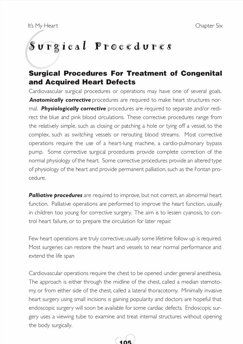
The Blalock-Taussig or BT ShuntShunts are surgical connections, or anastomoses, between two arteries or betweena vein and an artery. The Blalock-Taussig shunt connects a branch of the aorta, usu-ally the subclavian artery, to the pulmonary artery directly or by using a plastic tube
to increase the pulmonary blood flow.
107
It’s My Heart Surgical Procedures
Postoperative Subclavian Arteryto Pulmonary Artery Anastomosis(Blalock-Taussig Shunt) for Tetralogy of Fallot
8/3/2019 Heart Surgeries
http://slidepdf.com/reader/full/heart-surgeries 4/16
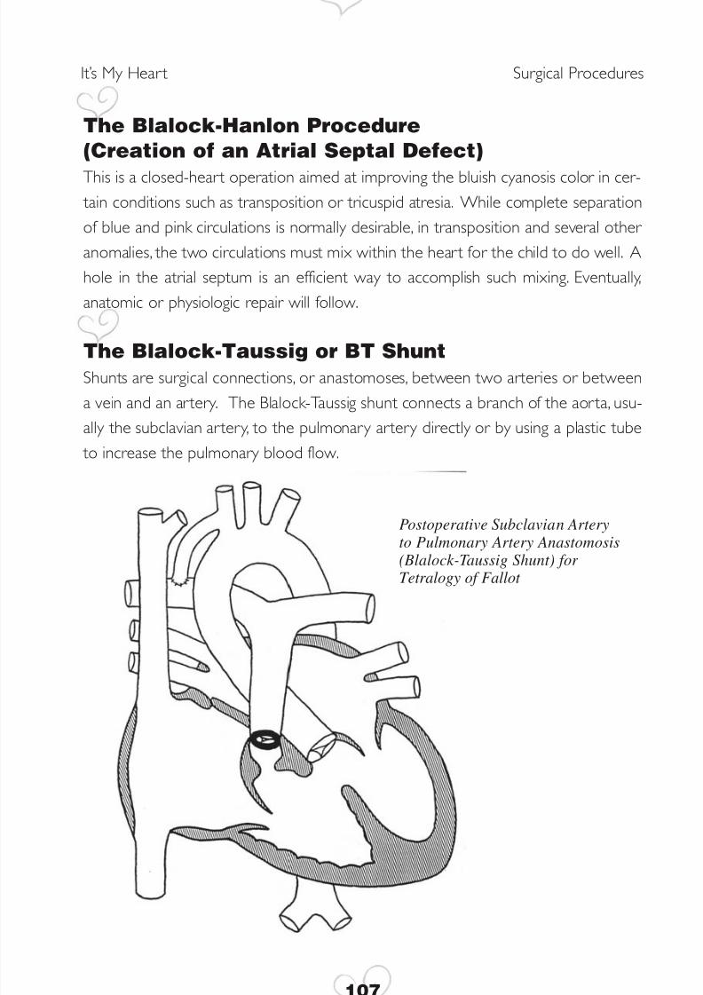
Closure of Septal DefectsSmaller atrial and ventricular septal defects can be closed with sutures or stitches.
Larger defects require the use of a plastic patch. Occasionally, in complex defects, the patch is used not only to close the hole between the ventricles but also to chan-nel left ventricular blood through the right ventricle into the aorta through an intrac-ardiac tunnel.
It’s My Heart Surgical Procedures
108
Postoperative Patch Repair of Ventricular Septal Defect
8/3/2019 Heart Surgeries
http://slidepdf.com/reader/full/heart-surgeries 5/16
Correction of Narrow ValvesThe four valves in the heart can be narrow, stenosed, or regurgitant, leaky, or both.
They can be either repaired or replaced, depending on the nature of the problem.The most common aortic and pulmonic stenosis is caused by congenital fusion of one or more moving valve leaflets. Repair is done using cardiopulmonary bypass andby cutting the fused leaflets apart. Occasionally, however, the seat of the valve, theannulus, is small, a condition called hypoplastic. Enlargement of the pulmonic annu-lus is accomplished by cutting it and placing a trans-annular patch across it. Theresulting valve leak is usually well tolerated for a decade or more. A small aortic
annulus cannot be enlarged without valve replacement because it would causeexcessive regurgitation, or leaking. A regurgitant aortic valve can sometimes berepaired by tightening the loose leaflets in a procedure called a valvuloplasty). Narrowmitral or tricuspid valves can rarely be enlarged and usually need to be replaced.Regurgitant mitral or tricuspid valves can frequently be repaired by valvuloplasty.
Valves can be replaced by biological or prosthetic material. Biological valves are
either homograft, taken from a human cadaver donor, or porcine, taken from a pig.There is a variety of prosthetic valves available. Many issues need to be consideredwhen choosing the most suitable valve. Mechanical prosthetic valves will require alifetime of clot-preventing medication, such as Coumadin or aspirin, and may not beideal for females desiring to become pregnant. At this writing, an ideal valve has notyet been designed and most prosthetic valves will eventually need replacement.
109
It’s My Heart Surgical Procedures
8/3/2019 Heart Surgeries
http://slidepdf.com/reader/full/heart-surgeries 6/16
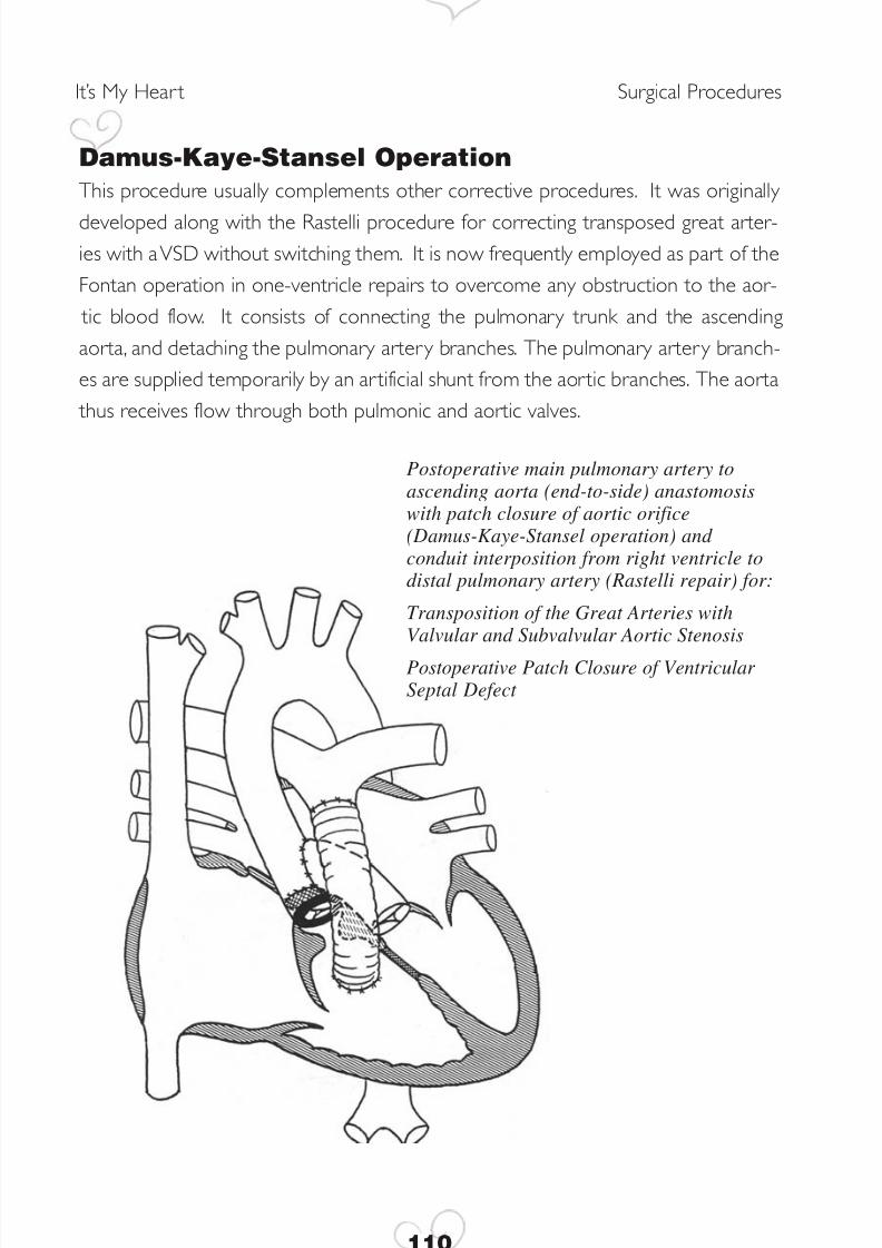
Damus-Kaye-Stansel OperationThis procedure usually complements other corrective procedures. It was originally
developed along with the Rastelli procedure for correcting transposed great arter-ies with a VSD without switching them. It is now frequently employed as part of theFontan operation in one-ventricle repairs to overcome any obstruction to the aor-
tic blood flow. It consists of connecting the pulmonary trunk and the ascendingaorta, and detaching the pulmonary artery branches. The pulmonary artery branch-es are supplied temporarily by an artificial shunt from the aortic branches. The aorta
thus receives flow through both pulmonic and aortic valves.
It’s My Heart Surgical Procedures
110
Postoperative main pulmonary artery toascending aorta (end-to-side) anastomosiswith patch closure of aortic orifice(Damus-Kaye-Stansel operation) and conduit interposition from right ventricle todistal pulmonary artery (Rastelli repair) for:
Transposition of the Great Arteries withValvular and Subvalvular Aortic Stenosis
Postoperative Patch Closure of Ventricular Septal Defect
8/3/2019 Heart Surgeries
http://slidepdf.com/reader/full/heart-surgeries 7/16
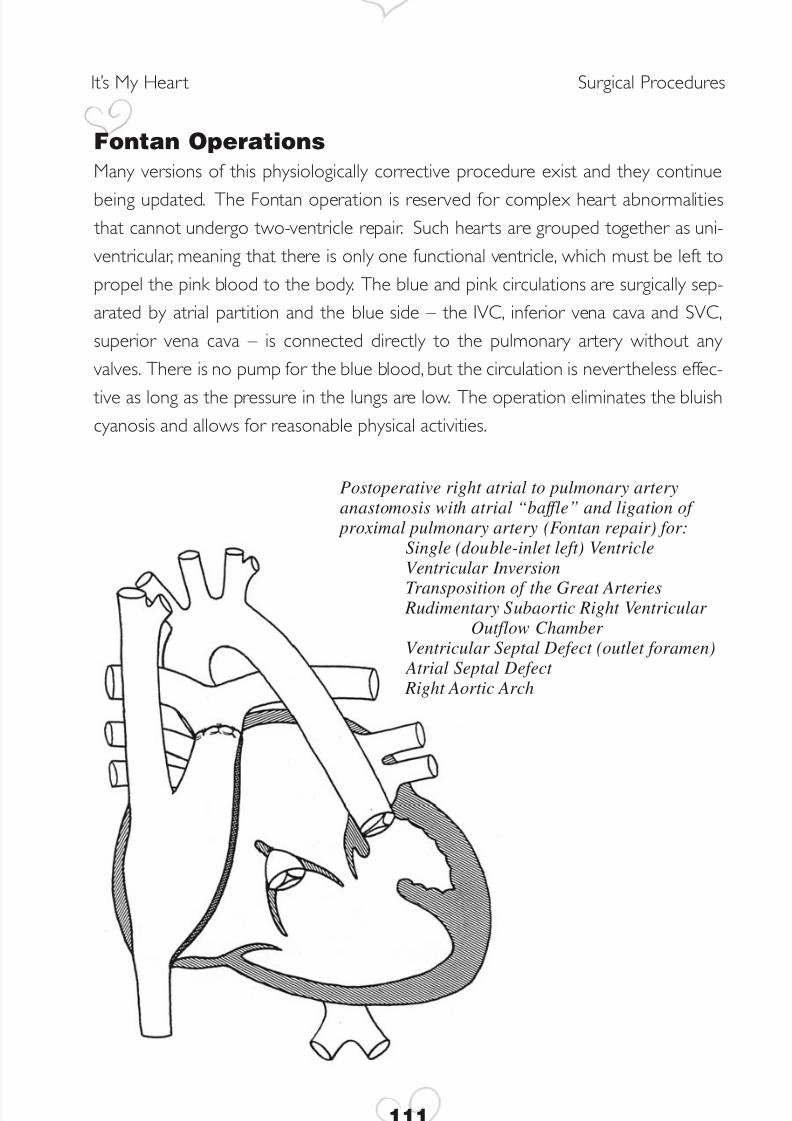
Fontan OperationsMany versions of this physiologically corrective procedure exist and they continue
being updated. The Fontan operation is reserved for complex heart abnormalities that cannot undergo two-ventricle repair. Such hearts are grouped together as uni-ventricular, meaning that there is only one functional ventricle, which must be left topropel the pink blood to the body. The blue and pink circulations are surgically sep-arated by atrial partition and the blue side – the IVC, inferior vena cava and SVC,superior vena cava – is connected directly to the pulmonary artery without any valves. There is no pump for the blue blood, but the circulation is nevertheless effec-
tive as long as the pressure in the lungs are low. The operation eliminates the bluishcyanosis and allows for reasonable physical activities.
111
It’s My Heart Surgical Procedures
Postoperative right atrial to pulmonary arteryanastomosis with atrial “baffle” and ligation of
proximal pulmonary artery (Fontan repair) for:Single (double-inlet left) VentricleVentricular Inversion
Transposition of the Great Arteries Rudimentary Subaortic Right Ventricular
Outflow Chamber Ventricular Septal Defect (outlet foramen)
Atrial Septal Defect Right Aortic Arch
8/3/2019 Heart Surgeries
http://slidepdf.com/reader/full/heart-surgeries 8/16
Kawashima OperationThis intraventricular tunnel repair is used for anatomic correction of double outlet
right ventricle where both the aorta and the pulmonary ar tery originate from the rightventricle and the only exit from the left ventricle is the ventricular septal defect. Theventricular septal defect is left open and sometimes even enlarged to serve as themouth into a tunnel leading from the left ventricle through the right ventricle to theaorta. This tunnel is called an intracardiac conduit because it occurs completely with-in the heart. This tunnel is created in such a way as to separate the LV to the aortaand the RV flow to the pulmonary ar tery. This operation avoids the use of an extrac-
ardiac pulmonary conduit.
Ligation (Division) of Patent Ductus ArteriosusThis is one of the truly corrective operations. Once the ductus is successfully closed,no further surgery is required. Ductal closure is a closed-heart procedure consist-ing of cutting the ductus and sewing up the two stumps. In tiny premature babies,ligation, or tying off, the ductus is preferred, although the ductus infrequently reopens.
It’s My Heart Surgical Procedures
112
8/3/2019 Heart Surgeries
http://slidepdf.com/reader/full/heart-surgeries 9/16
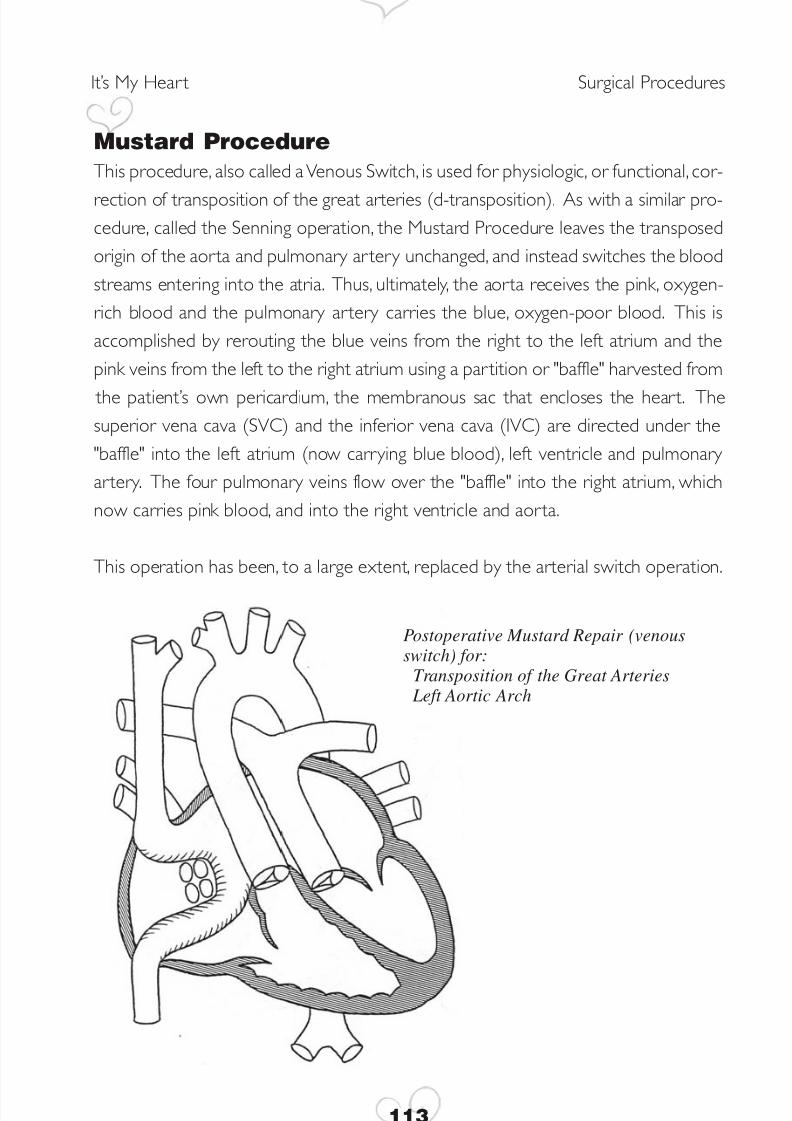
Mustard ProcedureThis procedure, also called a Venous Switch, is used for physiologic, or functional, cor-
rection of transposition of the great arteries (d-transposition). As with a similar pro-cedure, called the Senning operation, the Mustard Procedure leaves the transposedorigin of the aorta and pulmonary ar tery unchanged, and instead switches the bloodstreams entering into the atria. Thus, ultimately, the aorta receives the pink, oxygen-rich blood and the pulmonary artery carries the blue, oxygen-poor blood. This isaccomplished by rerouting the blue veins from the right to the left atrium and thepink veins from the left to the right atrium using a partition or "baffle" harvested from
the patient’s own pericardium, the membranous sac that encloses the heart. Thesuperior vena cava (SVC) and the inferior vena cava (IVC) are directed under the"baffle" into the left atrium (now carrying blue blood), left ventricle and pulmonary artery. The four pulmonary veins flow over the "baffle" into the right atrium, whichnow carries pink blood, and into the right ventricle and aorta.
This operation has been, to a large extent, replaced by the arterial switch operation.
113
It’s My Heart Surgical Procedures
Postoperative Mustard Repair (venousswitch) for:
Transposition of the Great Arteries Left Aortic Arch
8/3/2019 Heart Surgeries
http://slidepdf.com/reader/full/heart-surgeries 10/16
Norwood ProceduresThese procedures are used to treat hypoplastic left heart syndrome, a group of
defects in which the left ventricle is very small or absent.
Norwood 1 is a palliative operation – a fix, not a cure – performed in newborns asan emergency procedure using cardiopulmonary bypass pump. It converts the func-
tionally single right ventricle to act as the left ventricle while the pulmonary trunk issurgically joined with the tiny aorta to form a large new aorta, using a modifiedDamus-Kaye-Stansel procedure. The aortic arch is reconstructed as well, if neces-
sary. The pulmonary artery branches are detached and connected to the new aortaby a small plastic tube, a procedure known as a modified Blalock-Taussig anastomo-sis. Norwood 1 enables an infant to grow to 4 to 10 months of age, when the sec-ond stage palliative operation can be undertaken.
Norwood 2 converts the Blalock-Taussig anastomosis to a bi-directional Glenn or aHemi-Fontan anastomosis in preparation for the eventual Fontan procedure, which
is the corrective repair. The Glenn and the Hemi-Fontan, a version of the Glenn, con-sist of connecting both pulmonary artery branches to the superior vena cava,enabling the blue blood to reach the pulmonary circulation directly without having
to pass through the heart chambers.
At a suitable age, around 18-24 months, the child becomes eligible for the Fontanoperation, which eliminates cyanosis by directing the inferior vena cava to the pul-
monary ar tery and par titioning the two atria.
It’s My Heart Surgical Procedures
114
8/3/2019 Heart Surgeries
http://slidepdf.com/reader/full/heart-surgeries 11/16
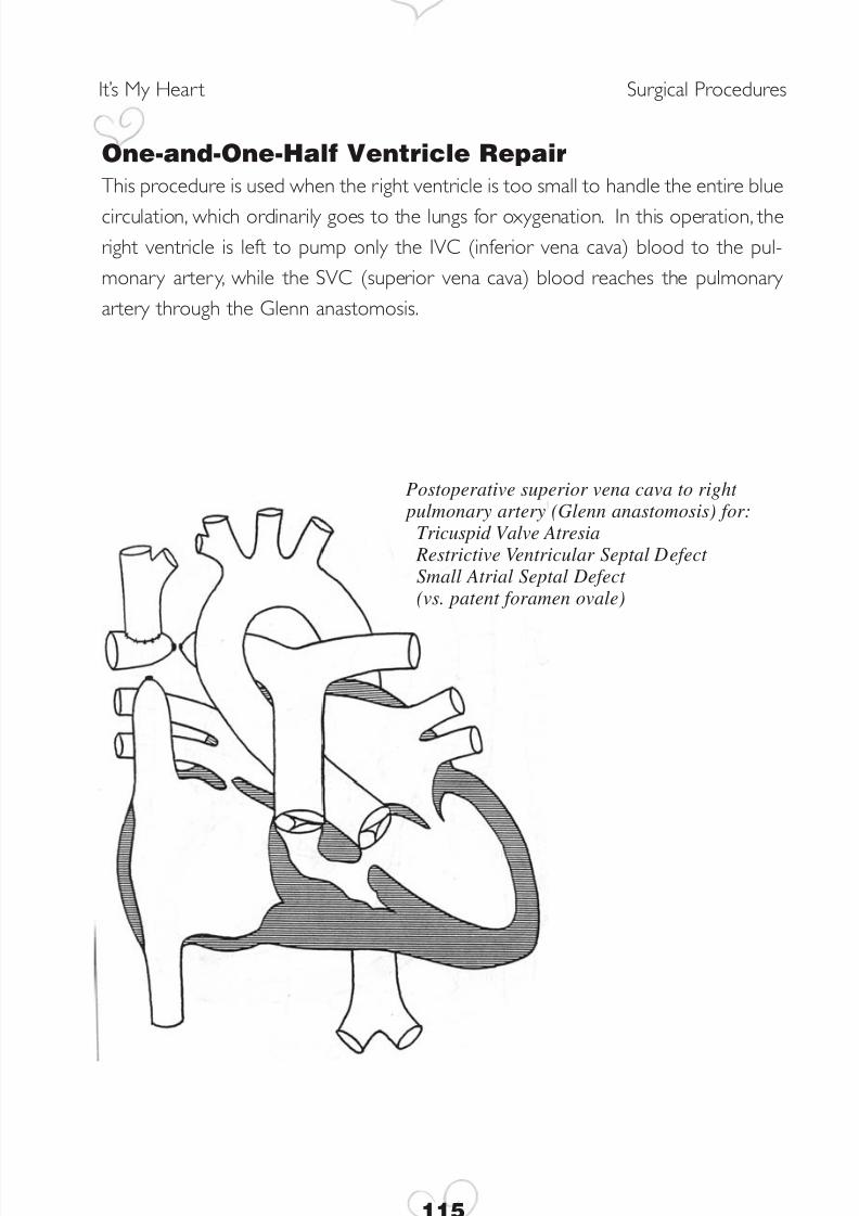
One-and-One-Half Ventricle RepairThis procedure is used when the right ventricle is too small to handle the entire blue
circulation, which ordinarily goes to the lungs for oxygenation. In this operation, theright ventricle is left to pump only the IVC (inferior vena cava) blood to the pul-monary artery, while the SVC (superior vena cava) blood reaches the pulmonary artery through the Glenn anastomosis.
115
It’s My Heart Surgical Procedures
Postoperative superior vena cava to right pulmonary artery (Glenn anastomosis) for:
Tricuspid Valve Atresia Restrictive Ventricular Septal Defect Small Atrial Septal Defect (vs. patent foramen ovale)
8/3/2019 Heart Surgeries
http://slidepdf.com/reader/full/heart-surgeries 12/16
Pulmonary Artery BandingThis is a temporary, palliative procedure that reduces excessive flow and pressure in
the pulmonary artery. The pulmonary artery is surgically constricted using a wide tape to the point where heart failure due to excessive pulmonary blood flow is con- trolled. As the child grows, the banded artery remains the same size, causing thechild’s color to become bluer. At this point a corrective surgery may be carried out,or occasionally, a shunt will be placed to restore the pink color.
It’s My Heart Surgical Procedures
116
8/3/2019 Heart Surgeries
http://slidepdf.com/reader/full/heart-surgeries 13/16
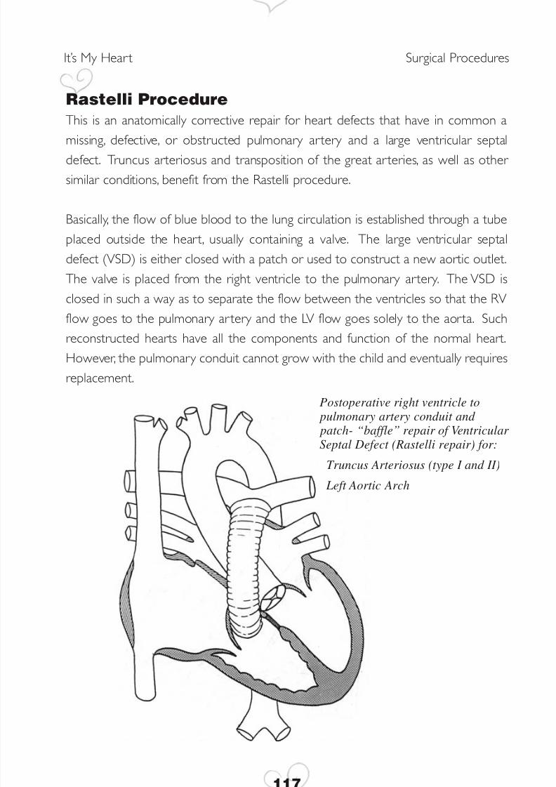
Rastelli ProcedureThis is an anatomically corrective repair for heart defects that have in common a
missing, defective, or obstructed pulmonary artery and a large ventricular septaldefect. Truncus arteriosus and transposition of the great arteries, as well as other similar conditions, benefit from the Rastelli procedure.
Basically, the flow of blue blood to the lung circulation is established through a tubeplaced outside the heart, usually containing a valve. The large ventricular septaldefect (VSD) is either closed with a patch or used to construct a new aortic outlet.
The valve is placed from the right ventricle to the pulmonary artery. The VSD isclosed in such a way as to separate the flow between the ventricles so that the RVflow goes to the pulmonary ar tery and the LV flow goes solely to the aorta. Suchreconstructed hearts have all the components and function of the normal heart.However, the pulmonary conduit cannot grow with the child and eventually requiresreplacement.
117
It’s My Heart Surgical Procedures
Postoperative right ventricle to pulmonary artery conduit and patch- “baffle” repair of Ventricular Septal Defect (Rastelli repair) for:
Truncus Arteriosus (type I and II)
Left Aortic Arch
8/3/2019 Heart Surgeries
http://slidepdf.com/reader/full/heart-surgeries 14/16
Repair of Anomalous Pulmonary Venous Return Whether some or all four pulmonary veins are draining anomalously into a wrong
cardiac structure, the aim of surgery is to re-connect them to the left atrium. If the veins are neither obstructed nor narrow before surgery, the result can beexcellent. Obstructed veins, however, tend to re-narrow and may require addi-
tional operations.
Repair of Coarctation of the AortaThis procedure usually does not require cardio- pulmonary bypass. A variety of sur-
gical methods are currently in use. Resection, or removal, of the narrowing and anend-to-end anastomosis, a surgical connection of ar teries to form a passage, is rarely used because of high recurrence rate. Instead, an extended end-to-end anastomo-sis offers good long-term repair by removing all the abnormal wall. The subclavianpatch repair uses part of the left subclavian artery to enlarge the aortic narrowing,but a plastic patch can be used instead. Rarely is the narrow segment bypassed by a conduit or tube graft.
Repair of Common Atrio-Ventricular (AV) CanalThis anomaly consists of a large confluent atrial and ventricular septal defect and anundivided, or common, inlet valve. Although surgical techniques vary, repair requirespatch closure of the two defects and separation of the common valve into a tricus-pid and a mitral valve, called the two-ventricle repair. Rarely, when one of the ven-
tricles is too small, a condition called an unbalanced AV canal, a one-ventricle repair,
or Fontan operation, is preferable. A partial AV canal, also called an ostium primumdefect, consists of a large atrial septal defect and a cleft, or regurgitant, mitral valve.Repair is accomplished by patch closure of the defect and suturing the cleft.
It’s My Heart Surgical Procedures
118
8/3/2019 Heart Surgeries
http://slidepdf.com/reader/full/heart-surgeries 15/16
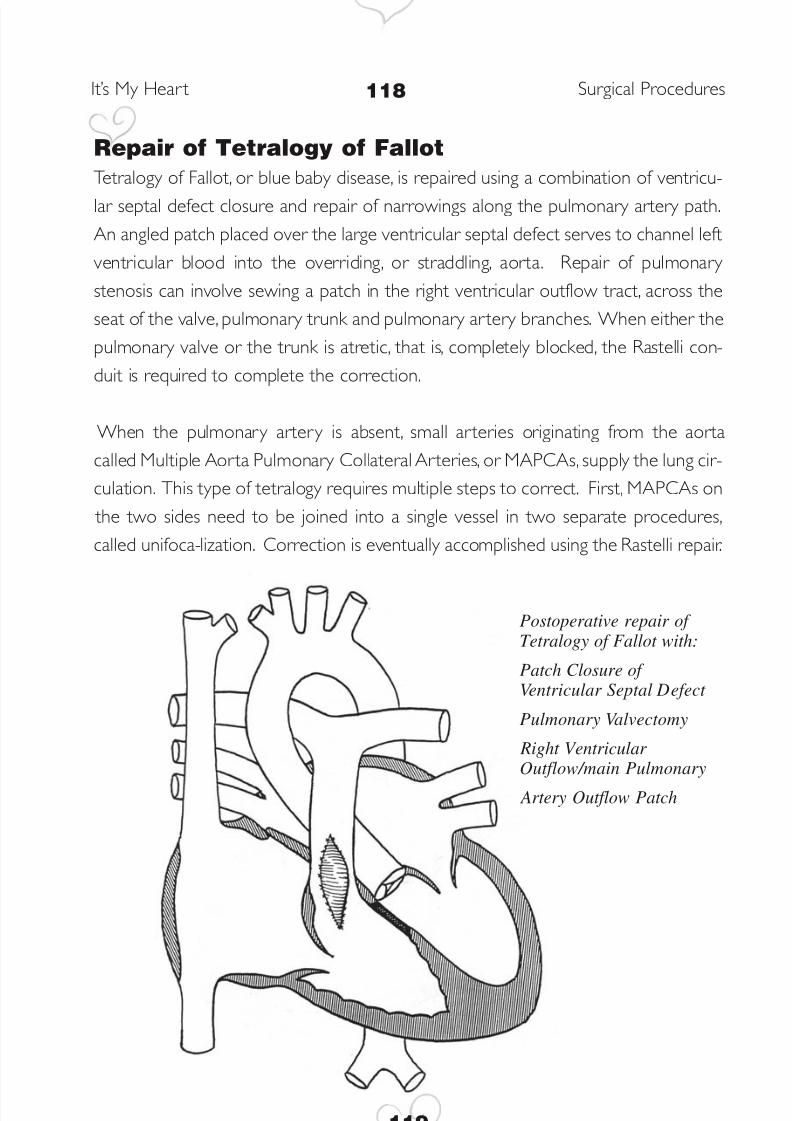
Repair of Tetralogy of FallotTetralogy of Fallot, or blue baby disease, is repaired using a combination of ventricu-
lar septal defect closure and repair of narrowings along the pulmonary artery path.An angled patch placed over the large ventricular septal defect serves to channel leftventricular blood into the overriding, or straddling, aorta. Repair of pulmonary stenosis can involve sewing a patch in the right ventricular outflow tract, across theseat of the valve, pulmonary trunk and pulmonary artery branches. When either thepulmonary valve or the trunk is atretic, that is, completely blocked, the Rastelli con-duit is required to complete the correction.
When the pulmonary artery is absent, small arteries originating from the aortacalled Multiple Aorta Pulmonary Collateral Arteries, or MAPCAs, supply the lung cir-culation. This type of tetralogy requires multiple steps to correct. First, MAPCAs on
the two sides need to be joined into a single vessel in two separate procedures,called unifoca-lization. Correction is eventually accomplished using the Rastelli repair.
119
It’s My Heart Surgical Procedures
Postoperative repair of Tetralogy of Fallot with:
Patch Closure of Ventricular Septal Defect
Pulmonary Valvectomy
Right Ventricular Outflow/main Pulmonary
Artery Outflow Patch
8/3/2019 Heart Surgeries
http://slidepdf.com/reader/full/heart-surgeries 16/16
Ross ProcedureThis procedure consists of replacing a faulty aortic valve with the patient’s own
healthy pulmonic valve. It requires re-implantation of the coronary arteries into thereconstructed aortic root. The pulmonic valve is replaced with a biologic valve, either homograft taken from a human cadaver donor, or porcine, taken from a pig. Thisoperation can be part of the Ross-Konno procedure employed when there is addi-
tional narrowing below the aortic valve. In the Konno procedure, the narrow left ven- tricular outlet is approached through the right ventricle, the septum is cut open, andfilled with a large patch in such a way as to enlarge the LV outflow tract.
It’s My Heart Surgical Procedures
120