high-density lipoproteins: vascular guards against ... · organismo, ya que estudios recientes...
TRANSCRIPT

CorSalud 2013 Oct-Dec;5(4):366-378
RNPS 2235-145 © 2009-2013 Cardiocentro Ernesto Che Guevara, Villa Clara, Cuba. All rights reserved. 366
Cuban Society of Cardiology ________________________
Review Article
High-density lipoproteins: vascular guards against atherosclerosis
Yosit Ponce Gutiérreza, MD; Arik Ponce Gutiérreza, MD; Arnaldo Rodríguez Leónb, MD, MSc; and Carlos Llanes Álvareza, MD
a Juan B. Contreras Fowler Polyclinic. Ranchuelo, Villa Clara, Cuba. b Celestino Hernández Robau University Hospital. Santa Clara, Villa Clara, Cuba.
Este artículo también está disponible en español ARTICLE INFORMATION Received: October 26, 2012 Modified: December 18, 2012 Accepted: January 31, 2013 Competing interests The authors declare no competing interests
Acronyms CETP: cholesteryl esters transfer protein eNOS: endothelial nitric oxide synthase HDL: high-density lipoprotein LCAT: lecithin-cholesterol acyltransferase LDL: low-density lipoprotein LPL: lipoprotein lipase PON: paraoxonase RCT: reverse cholesterol transport VLDL: very low density lipoproteins On-Line Versions: Spanish - English Y Ponce Gutiérrez Calle Camilo Cienfuegos Nº 63, e/ Carmen Rivero y Federico Escobar. Ranchuelo, Villa Clara, Cuba. E-mail address: [email protected]
ABSTRACT Increasing levels of high-density lipoproteins and their potential benefits in Atheros-clerosis motivated us to write this article in order to update knowledge on this topic. The structure of these lipoproteins is described, as well as their atheroprotective vas-cular effects and the reverse cholesterol transport. New strategies to increase their concentration in the body are presented, as recent studies indicate that they stabilize atherosclerotic plaques in an expedited manner. Therefore, it constitutes a novel therapeutic alternative in high risk patients. This will update the knowledge on high-density lipoproteins, in order to provide a better quality care in the prevention, con-trol and treatment of this common disease. Palabras clave: High-density lipoprotein, HDL, Atherosclerosis, Atherosclerotic plaque Las lipoproteínas de alta densidad: protectoras vasculares contra la aterosclerosis RESUMEN El incremento de los niveles de lipoproteínas de alta densidad y sus potenciales bene-ficios en la aterosclerosis ha sido motivo para la realización de este artículo, en el que se efectúa una revisión sobre la información médica más reciente que existe sobre el tema y su posterior actualización. Se describen la estructura de estas lipoproteínas, sus efectos vasculares ateroprotectores, el transporte reverso de colesterol, y se ex- ponen las nuevas estrategias que permiten incrementar sus concentraciones en el organismo, ya que estudios recientes indican que estabilizan las placas de ateroma de una manera acelerada, por lo que constituyen una novedosa alternativa terapéutica en los pacientes de alto riesgo. De esta forma se consolidan los conocimientos sobre las lipoproteínas de alta densidad, con el fin de brindar una atención de más calidad en la prevención, control y tratamiento de esta frecuente enfermedad. Palabras clave: Lipoproteínas de alta densidad, HDL, Aterosclerosis, Placa de ateroma INTRODUCTION Atherosclerosis is a disease characterized by the deposition and infiltration of fatty material on the inner walls of large and medium arteries. It is the most
Ponce Gutiérrez Y, et al.
CorSalud 2013 Oct-Dec;5(4):366-378 367
common form of arteriosclerosis. It causes an inflam-matory response and the proliferation and migration of smooth muscle cells in the wall, which leads to a narrowing of the arterial lumen. The concrete thicken-ings are called atherosclerotic plaques, and thrombo-sis is their most common complication1.
This disease is the leading cause of death in de-veloped countries, that is, North America, Europe and Australia, associated with an unhealthy lifestyle. Approximately 76 % of fatal coronary thrombi are caused by the rupture of a complicated atherosclerotic plaque2.
Atherosclerosis is the result of an imbalance between deposition and removal of cholesterol in the arterial wall, with a predominance of the former. The main responsible for the deposition of cholesterol in the arterial wall is low-density lipoprotein (LDL), while the main responsible for its removal is high-density lipoprotein (HDL). Both, high systemic concentrations of LDL cholesterol and low concentration of HDL have been consistently associated with the development of atherosclerosis3.
Numerous therapeutic strategies have been tested in an attempt to prevent the development of the disease by lowering LDL or increasing HDL. While LDL reduction, mainly with the use of statins4, has been established as a standard therapy for primary and secondary prevention of atherothrombotic events, the increase in HDL predicts a hopeful future. That is why the vital importance of studying it and the therapeutic prospects for its use in such a common disease, which still does not have a full and effective control.
HDL STRUCTURE HDL is a macromolecular pseudomicellar complex composed of amphipathic lipids (phospholipids and free cholesterol), non-polar lipids (triglyceride and cholesteryl esters) and proteins called apolipoproteins (apo). Amphipathic lipids are arranged in a monolayer on the surface of the complex, and have their polar groups toward the aqueous medium. The stability of this monolayer is guaranteed by the apo. Non-polar lipids are insoluble in aqueous media such as plasma and, consequently, are located within lipoproteins, thus avoiding interactions with polar groups that would be physicochemically unfavorable. Therefore, the transport of lipids in plasma is guaranteed5.
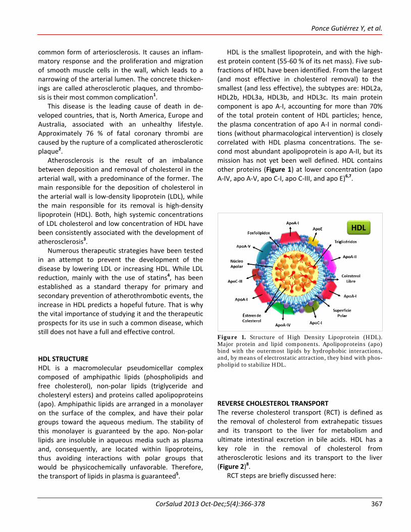
HDL is the smallest lipoprotein, and with the high-est protein content (55-60 % of its net mass). Five sub-fractions of HDL have been identified. From the largest (and most effective in cholesterol removal) to the smallest (and less effective), the subtypes are: HDL2a, HDL2b, HDL3a, HDL3b, and HDL3c. Its main protein component is apo A-I, accounting for more than 70% of the total protein content of HDL particles; hence, the plasma concentration of apo A-I in normal condi-tions (without pharmacological intervention) is closely correlated with HDL plasma concentrations. The se-cond most abundant apolipoprotein is apo A-II, but its mission has not yet been well defined. HDL contains other proteins (Figure 1) at lower concentration (apo A-IV, apo A-V, apo C-I, apo C-III, and apo E)6,7. Figure 1. Structure of High Density Lipoprotein (HDL). Major protein and lipid components. Apolipoproteins (apo) bind with the outermost lipids by hydrophobic interactions, and, by means of electrostatic attraction, they bind with phos-pholipid to stabilize HDL.
REVERSE CHOLESTEROL TRANSPORT The reverse cholesterol transport (RCT) is defined as the removal of cholesterol from extrahepatic tissues and its transport to the liver for metabolism and ultimate intestinal excretion in bile acids. HDL has a key role in the removal of cholesterol from atherosclerotic lesions and its transport to the liver (Figure 2)8.
RCT steps are briefly discussed here:

High-density lipoproteins: vascular guards against atherosclerosis
CorSalud 2013 Oct-Dec;5(4):366-378 368
HDL synthesis The liver and intestine synthesize and secrete apo A-I (the main component of HDL). The liver produces 75% of the human apo A-I. Both tissues are responsible for the lipidation of newly secreted lipid-poor apo A-I via ATP-binding cassette transporter A1 (ABCA1), where the nascent HDL is formed (which is also called lipid-poor apo A-I)3.9.
Cholesterol uptake by nascent HDL (cholesterol efflux) This process can be ac-complished by different mechanisms and eventual-ly results in the formation of discoidal HDL particles: Aqueous diffusion: This
passive mechanism is fulfilled by a simple diffusion process, so that the movement of cholesterol may be bi-directional, and the di-rection is determined solely by its concentra-tion gradient. It occurs in all cells and is a slow and rather inefficient process (takes hours)3.
ABCA1-mediated free cholesterol efflux: this movement of free cho-lesterol is unidirectio-nal, only from the cells to the lipid-poor apoli-poproteins 3,9,10.
Scavenger receptor class B type I (SR-BI): the flux of free choles-terol mediated by SR-BI only takes place to-ward phospholipid-containing acceptors (that is, HDL and lipi-dated apolipoproteins), and it is bidirectional, depending on the con-
centration gradient on both sides of the mem-brane 3,11.
ABCG1 and ABCG4: provide an alternative pathway for the transport of free cholesterol from macro-phages to mature HDL, but never to nascent HDL (lipid-poor apo A-I)3,12.
HDL maturation Nascent HDL particles undergo a process of intravas-
Figure 2. Graphical representation of the RCT. Apo A-I, the main protein of HDL, is synthesized in the liver and intestine where, via ABCA1 receptor, receives a small amount of phospholipids and becomes lipid-poor apo A-I (nascent HDL). The apo A-I leads the nascent HDL to extrahepatic tissues, mainly to macrophages, from which it receives free cholesterol via ABCA1 receptor (nascent HDL with preβ1 migration). Through the action of the enzyme LCAT, free cholesterol is turned into cholesteryl esters, and so it is transformed into spherical mature HDL (HDL3 and HDL2), which receives cholesterol from peripheral tissues via the receptor SR-BI or ABCG1, increasing its size and its esterified cholesterol content. The RCT is completed in two ways: a) hepatic uptake of mature HDL-C via SR-BI receptor; and b) the CETP catalyzes the transfer of cholesteryl ester to LDL-C, which in turn will be taken up by the liver through the LDL receptor. Finally, from the liver, free cholesterol may be poured directly into the bile or turned into bile acids (the enzyme responsible for this reaction is the 7α-hydroxylase), prior to the occurrence of biliary excretion in the intestine. Legend: Apolipoprotein (apo); HDL cholesterol (HDL-C); LDL cholesterol (LDL-C); free cholesterol (FC); Enzyme 7- α hydroxylase (CYP7A1); Cholesteryl esters (CE); Cholesterol ester hydrolase (CEH); Phospholipids (PL); Lecithin-cholesterol acyltransferase (LCAT); Endothelial lipase (EL); Hepatic lipase (HL); Lipoprotein lipase (LPL); Cholesteryl ester transfer protein (CETP); LDL receptor (LDL-R); Scavenger receptor BI (SR-BI); Triglycerides (TG).

Ponce Gutiérrez Y, et al.
CorSalud 2013 Oct-Dec;5(4):366-378 369
cular transformation by the action of several enzymes: The enzyme called lecithin-cholesterol acyltrans-
ferase (LCAT): inside the discoidal nascent HDL particle, LCAT catalyzes the transfer of 2-acyl group from lecithin to free cholesterol captured from the macrophages, thus generating cholesteryl esters and lysolecithin. Cholesteryl esters are more hydro-phobic than free cholesterol, so they move into the core of the lipoprotein particle, thus forming a mature HDL particle, which is large and spheric-al3,13.
Cholesteryl esters transfer protein (CETP): it is a hydrophobic glycoprotein synthesized by liver and adipose tissue which circulates in the plasma bound to lipoproteins. CETP promotes the transfer of cholesteryl esters from HDL particles to apo B-containing lipoproteins [LDL, chylomicrons and very low density lipoproteins (VLDL)] in exchange for triglycerides; that is, it transfers triglycerides con-versely from VLDL, chylomicrons and LDL to HDL, thus, there is migration of cholesteryl esters back to LDL and a reduction in the size of the HDL particle3,14,15.
Other proteins involved are: the phospholipid transfer protein and several lipases [lipoprotein lipase (LPL), hepatic lipase and endothelial lipase]. The triglycerides in mature HDL are hydrolyzed by hepatic lipase. This hydrolysis, in association with the activity of phospholipid transfer protein, re-duces the size of mature HDL, and turns them into nascent HDL, poor in phospholipids, which can re-start the uptake cycle3,16.
Catabolism of HDL The most decisive factor determining HDL and apo A-I plasma concentrations is the apo A-I clearance rate. The kidneys, liver and steroidogenic organs (adrenals, ovaries and testes) are the main sites for catabolism of HDL. This catabolism may be effected by: a) endo-cytosis and lysosomal degradation of the whole particle (including apo A-I), which occur both in the liver and kidneys, and b) selective cholesterol uptake, that is, removal of cholesterol and other lipids from the particle without affecting the protein content. The mechanism that has been best characterized is hepatic uptake by SR-BI. Free cholesterol that is transported in HDL may be directly excreted into the bile or become
bile acids, prior to bile excretion (the enzyme respon-sible for this reaction is 7α-hydroxylase)3,17. HDL ATHEROPROTECTIVE ACTIONS WHICH ARE NOT RELATED TO RCT Vascular protection given by high concentrations of HDL against atherosclerosis is not only limited to the effect of RCT, but includes other atheroprotective actions, such as antioxidant activity, protection of endothelial function, anti-inflammatory and anti-apop-totic activity, inactivation of the complement system, regulation of the endothelium secretory activity, as well as antithrombotic and fibrinolytic effects (Figure 3). Antioxidant activity Due to its proinflammatory properties, oxidized LDL in the subendothelial space is involved in the formation of the atherosclerotic plaque. In this context, the antiatherogenic role of HDL is due its antioxidant quality. Several of its elements are involved in this quality, including its apolipoproteins, and particularly paraoxonase (PON) 2, an enzyme physically associated with plasma HDL. This PON is synthesized in mamma-lians’ liver, circulates in blood bound to the apo A-I and apo J of HDL, and its expression is inhibited by proatherogenic stimuli5,18.
The molecular bases for the inverse relation between PON and atherosclerosis have been found in the enzyme capacity to eliminate the lipoperoxides that are involved in plaque formation. The onset and progression of the atherosclerotic plaque in the ar-terial wall is largely dependent on peroxidation of lipoproteins mediated by free radicals, in particular from LDL19.
These biological effects are associated with the PON’s ability to remove peroxides that are linked to lipoproteins, leading to the corresponding alcohols, which are inactive derivatives from the viewpoint of peroxidation, chemotaxis and the inflammatory pro-cess in general. PON activity varies among individuals due to genetic and pathophysiological factors. Indeed, the human gene for this enzyme has two polymor-phisms (M55L and Q192R) affecting its activity. More-over, a decreased PON activity has been found in hyperlipidemic and insulin-dependent diabetic sub-

High-density lipoproteins: vascular guards against atherosclerosis
CorSalud 2013 Oct-Dec;5(4):366-378 370
Figure 3. Summary chart of HDL atheroprotective function. 1) Inactivation of complement: HDL prevents the formation of C5-C9 complex in the activation cascade of the complement system. 2) Regulation of inflammation: HDL inhibits the attraction of circulating monocytes and lymphocytes to the endothelium by downregulating the expression of MCP-1, VCAM-1 and ICAM-1, which are mediators of the adhesion, and of E-selectin, which allows the anchorage and sliding on the surface of endothelial cells. 3) Fibrinolysis: HDL promotes the conversion of plasminogen into plasmin (fibrinolytic enzyme) by upregulation of t-PA and downregulation of PAI-1. 4) Antithrombolitic: HDL prevents platelet activation by decreasing the platelet activating factor and thromboxane A2 and by increasing the synthesis of nitric oxide and prostacyclin. The sphingosine of HDL limits the procoagulant interactions between factors Xa and Va of the coagulation cascade. Decreased production of thrombin is due to improved activity of activated protein C and protein S, and upregulation of endothelial thrombomodulins. The decrease in endothelial activation occurs because it prevents the apoptosis of endothelial cells and the formation of microparticles of adhesion, promotes the inhibition of tissue factor, P-selectin and the expression of E-selectin, and increases the production of nitric oxide. 5) Regulation of endothelial secretory activity: HDL stimulates the production of PGI2, as it supplies the endothelial cells with arachidonic acid, the main substrate for the synthesis of PGI2, and stimulates the synthesis of cyclooxygenase in endothelial cells and vascular smooth muscle cells, and inhibits that of endothelin-1. 6) Protection of endothelial function: HDL inactivates the harmful effects of ox-LDL at nitric oxide production level, and increase eNOS activity. 7) Anti-apoptosis: HDL prevents the apoptosis of endothelial cells, macrophages and foam cells because it prevents the sustained increase in intracellular calcium that is induced by pro-apoptotic agents, such as ox-LDL, preventing the activation of caspase-3 and -9 proteins, and the antagonism of TNF-α. This is due to the fact that the HDL stimulation of migration and proliferation of endothelial cells is calcium-dependent and mediated by multiple kinases cascades. 8) Antioxidant: HDL inactivates ox-LDL through the enzyme PON-1, apo A-I and phospholipids. HDL also reduces the uptake of lysophosphatidylcholine, one of the products derived from the LDL oxidation process. 9) Efflux of cholesterol: HDL receives cholesterol from extrahepatic tissues, mainly macrophages and vascular foam cells, for its subsequent transport to the liver and excretion into the bile in the intestine, as part of the RCT. Legend: Plasminogen activator inhibitor type 1 (PAI-1); tissue plasminogen activator (t-PA); Apolipoprotein (apo); Endothelial cells (EC); Tumor necrosis factor alpha (TNF-α); Tissue factors (TF); Oxidized LDL ( ox-LDL ); Vascular cell adhesion molecule-1 (VCAM-1); Intercellular adhesion molecule-1 (ICAM-1); nitric oxide (NO); Endothelial nitric oxide synthase (eNOS); Monocyte chemoattractant protein-1 (MCP-1). ↑ Increase. ↓ decrease.

Ponce Gutiérrez Y, et al.
CorSalud 2013 Oct-Dec;5(4):366-378 371
jects. In addition, there is a positive correlation between its concentration and that of apo A-I5. Protection of endothelial function It has been postulated that the mechanisms respon-sible for the preservation of endothelial function mediated by HDL are related to its ability to inactivate the harmful effects of oxidized LDL (ox-LDL) at nitric oxide production level20.
HDL promotes the production of this oxide by means of the enzyme endothelial nitric oxide synthase (eNOS) by different mechanisms. 1- HDL regulates the subcellular distribution of eNOS.
The eNOS protein is localized in the enriched cholesterol of the caveolae of the plasma mem-brane, as a result of the myristoylation and palmi-toylation of the protein. HDL regulates lipid en-vironment within caveolae and ox-LDL, allowing preservation of the signaling module of eNOS21,22.
2- Based on mimetic studies of apo A-I, HDL prevents decoupling of eNOS by LDL, which favors the pro-duction of nitric oxide over that of superoxide anion (O2
-)21,23 3- HDL activates the signal mechanisms of the
membrane that stimulate the activity of eNOS. The binding of apo A-I from HDL with SR-BI, causes a rapid activation of Src receptor of tyrosine kinase, triggering the activation of phosphatidylinositol 3-kinase, and activating consecutively protein kinase B alpha (Akt) and mitogen-activated protein kinase (MAPK), which produced an increase in eNOS ac-tivity. Although apolipoproteins and phospholipids of HDL are sufficient to activate the signaling, it can also occur through lysophospholipids present in HDL such as sphingosylphosphorylcholine (SPC), sphingosine-1-phosphate (S1P) and lysosulfatides acting via the lysophospholipid receptor S1P-3.
4- HDL regulates the abundance of eNOS. Besides mo-dulating the acute response of the activation of phosphatidylinositol 3-kinase pathway, Akt and MAPK, HDL also causes increased eNOS 24-26.
HDL inactivates the ox-LDL not only through the
PON, as it was previously discussed, but also by means of apo A-I, its phospholipids and lysophosphatidyl-choline uptake, one of the products derived from LDL oxidative process21,23.
Regulation of inflammatory response Attraction and adhesion of leukocytes to endothelial cells and their interaction with smooth muscle cells play a central role in the development of atheros-clerotic plaque. The interaction of leukocytes with endothelial cells is mediated by adhesion molecules that are located on the luminal surface of the endo-thelium27.
HDL inhibits the attraction of monocytes to the endothelium by downregulating the expression of monocyte chemoattractant protein-1 (MCP-1). Among the molecules involved in leukocyte adhesion to the endothelium are the vascular cell adhesion molecule-1 (VCAM-1), the intercellular adhesion molecule-1 (ICAM-1) and E-selectin. The VCAM-1 and ICAM-1 mediate in the adhesion of circulating lymphocytes and monocytes, while the E-selectin allows its anchorage and sliding on the surface of endothelial cells. Furthermore, these three molecules are abun-dantly expressed in atherosclerotic plaques, most likely to recruit specific cells into the subendothelial space by the inflammatory process triggered by ox-LDL28.
In vitro studies with human endothelial cells have shown that physiological concentrations of HDL inhibit the expression of VCAM-1, ICAM-1 and E-selectin29. This effect seems to be related to the inhibition of tumor necrosis factor alpha (TNF-α) and its impact on intracellular second messengers resulting in the syn-thesis of adhesion molecules. In addition, it is inde-pendent of the elimination, mediated by HDL, of free radicals that are generated in the atherosclerotic lesion5,21. Prevention of endothelial cell apoptosis Multiple proatherogenic factors promote apoptosis in the endothelium, including ox-LDL, TNF-α, homocys-teine and angiotensin II. The anti-apoptotic actions of HDL include preventing a sustained increase in intra-cellular calcium, induced by pro-apoptotic agents such as ox-LDL, which prevents activation of caspase-3 and 9 proteins, and the antagonism of a variety of other pro-apoptotic mechanisms. This is due to the fact that the HDL stimulation of migration and proliferation of endothelial cells is calcium-dependent and mediated by multiple kinases cascades involving phosphatidyl-inositol 3-kinase, p38 and p42/44 MAPK, and Rho ki-

High-density lipoproteins: vascular guards against atherosclerosis
CorSalud 2013 Oct-Dec;5(4):366-378 372
nase. TNF-α, which also induces endothelial cell death, is inhibited by HDL through the attenuated induction of caspase-3, a major component of all primary apop-totic pathways30-33. Inactivation of the complement system When the initial inflammatory process begins in the early stages of atheroma formation, the complement causes damage to endothelial cells, which culminates in tissue necrosis. HDL, through its apo A-I, binds with complement factor C9, which inhibits the formation of C5a-C9 complex, and, consequently, avoids the com-plement harmful effects on vascular endothelium in the atherosclerotic process5. Regulation of secretory activity of the endothelium Prostacyclin PGI2, produced by the action of the cyclo-oxygenase from endothelial cells, has potent vaso-relaxing effect, and reduces the release of growth factors that stimulate local proliferation of smooth muscle cells involved in the development of atheroma. In this context, the HDL stimulates the production of PGI2 by two mechanisms: 1) providing arachidonic acid for the endothelial cell, the main substrate for the synthesis of PGI2, and 2) stimulating the synthesis of cyclooxygenase in endothelial cells and vascular smooth muscle cells. Endothelin-1 is another com-pound whose synthesis is affected by HDL. This effect is likely to originate at the level of post-transcriptional regulation of the synthesis of endothelin-134-37. Antithrombotic activity HDL has multiple antithrombotic properties that in-volve an increase in blood flow, a reduction in throm-bin generation and endothelial and platelet activation. HDL increases blood flow by increasing nitric oxide and prostacyclin production. The decrease in endothelial activation occurs when it prevents the apoptosis of endothelial cells and the formation of microparticles of adhesion, promotes the inhibition of tissue factor, P-selectin and the expression of E-selectin, and in-creases nitric oxide production. Decreased production of thrombin by HDL is due to improvement in the activity of activated protein C and protein S, and the upregulation of endothelial thrombomodulins. The
antagonism of HDL, on platelet activation, occurs by a downregulation of the release of the platelet acti-vating factor and thromboxane A2 synthesis, and by increased synthesis of nitric oxide and prostacyclin38-40.
HDL transports various sphingolipids that are pre-sent in plasma at micromolar range. It has been de-termined that at least four types of sphingolipids in HDL may directly or indirectly contribute to the anti-thrombotic activity. - The glycosphingolipids and glucosylceramide that
are associated with it are lipid cofactors of the anticoagulant activities of activated protein C and protein S.
- Sphingosine inhibits the activation of prothrombin in platelet aggregation and seems to limit procoa-gulant interactions between factors Xa and Va of the coagulation cascade, and may also directly downregulate the production of thrombin.
- The above mentioned sphingolipids and various lysosphingolipids exert potent cellular effects by means of the group of receptors coupled to G-protein, and HDL is the major transporter of S1P. As the S1P and others lysosphingolipids are related to vasoactive and anti-apoptotic activity, and the apoptosis of endothelial cells leads to thrombosis, then the anti-apoptotic activity of HDL, mediated by both lysosphingolipids and nitric oxide, may reduce the risk of thrombosis.
- As the interaction of endothelial cells with procoa-gulants, inflammatory leukocytes and micropar-ticles derived from cells involve adhesive reactions, then, the lysosphingolipids that are associated with HDL may have an antithrombotic effect by inhi-biting the synthesis of adhesive endothelial mole-cules21,41.
Fibrinolytic activity Fibrinolysis reactions exert a proteolytic action on fibrin, and lysis of fibrin rich thrombi by plasmin, which is formed by the activation of plasminogen. Hypofi-brinolysis contributes to arterial thrombosis, rather than venous thrombosis. The HDL can promote fibri-nolysis by a downregulation of plasminogen activator inhibitor type-1 (PAI-1) and an upregulation of the tissue plasminogen activator (t-PA). HDL oxidation alters their effect on fibrinolysis because it oxidizes HDL3, and, in the other subtypes of HDL, promotes the

Ponce Gutiérrez Y, et al.
CorSalud 2013 Oct-Dec;5(4):366-378 373
expression of PAI-1 suppressing, therefore, fibrinolytic activity42,43. THERAPIES TO RAISE HDL CHOLESTEROL CONCENTRATIONS The most effective way of preventing atherosclerosis and cardiovascular diseases is decreasing LDL concen-trations and raising HDL (Figure 4). NON-DRUG THERAPIES Dietary measures Diets rich in monounsaturated and polyunsaturated fatty acids (oily fish, nuts, olive oil) raise HDL levels and
reduce cardiovascular risk. The consumption of sa-turated fatty acids reduces the anti-inflammatory potential of HDL, whereas polyunsaturated acids im-prove it44. Aerobic exercise Frequent aerobic exercise increases HDL about 5%45,46. This effect is early (within 2 months), and seems to be linked to the frequency, intensity and duration of exercise47. Weight loss A recent meta-analysis has shown that in obese indivi-
Figura 4: Therapies for raising HDL cholesterol levels. Apolipoprotein (apo); Diacylglycerol acyltransferase 2 (DGAT2); 1, 2-Dimyristoyl-sn-glycero-3-phosphocholine (DMPC); Large unilamellar liposomes (LUV); Lecithin-cholesterol acyltransferase (LCAT); Lipoprotein lipase (LPL); Cholesteryl ester transfer protein (CETP); Peroxisome proliferator-activated receptor (PPAR); Liver X receptors (LXR); Reverse cholesterol transport (RCT).

High-density lipoproteins: vascular guards against atherosclerosis
CorSalud 2013 Oct-Dec;5(4):366-378 374
duals the loss of each kilogram of body weight is asso-ciated with a 0.35 mg/dl increase in HDL48. Cese del hábito tabáquico It increases HDL levels in 5 mg/dl, even in periods as short as two weeks after cessation49,50. Moderate alcohol intake It increases HDL concentrations between 5-15% and reduces cardiovascular risk51 (30-40 g daily, it is recommended 2 drinks for men and one for women). Apparently, ethyl alcohol per se causes the increase, so any alcoholic beverage could do it52; however, the benefits must be weighed against the risks before recommending alcohol intake. STANDARD PHARMACOTHERAPIES Niacin/nicotinic acid Niacin reduces HDL uptake by the liver and the amount of apo A-I that is extracted, giving rise to apo A-I-rich HDL particles (very efficient in RCT)53,54. It also reduces the activity of CETP, lipolysis and the release fatty acids into the liver, resulting in decreased VLDL production. It is the most effective treatment to raise HDL (20-35%). It reduces total cholesterol by 10-15%, LDL cholesterol by 15-20%, triglycerides by 30-50 % and is the only one that reduces lipoprotein (a)54. Statins Statins raise HDL by 5-10%47 (rosuvastatin is the one inducing the biggest increases55), by increasing the synthesis of apo A-I and decreasing CETP activity56. Their effects depend on the initial levels of HDL; the lower the levels the bigger the effect. Fibrates They are agonists of peroxisome proliferator-activated receptor (PPAR)-alpha. They Increase the expression of apo A-I, apo A-II and LPL, and reduce apo C-III and CETP activity. They also reduce VLDL levels by in-creasing fatty acid oxidation in the liver, reducing lipogenesis and stimulating fatty acid uptake by muscles. They increase HDL by 10-20%, and reduce
triglyceride levels by 20-50% and LDL cholesterol by 10-15%. Fenofibrate and bezafibrate have a higher impact in LDL cholesterol reduction, while gemfibrozil is more effective in reducing triglycerides. Their impact depends on baseline lipid levels; the increase in HDL is more pronounced when baseline concentrations of triglycerides are high57. Thiazolidinediones Used in the treatment of diabetes mellitus type 2, they are PPAR-gamma agonists which act to increase insulin sensitivity in adipose tissue and liver. They promote glucose uptake and decrease both its hepatic pro-duction and the concentration of circulating free fatty acids. Pioglitazone and rosiglitazone have shown a similar hypoglycemic activity (both reduce glyco-sylated hemoglobin by 1.5%). However, pioglitazone is superior in cardiovascular effects, because it increases HDL by 10% and reduces, to a larger extent, trigly-cerides, although it has no effect on LDL cholesterol; while rosiglitazone raises it by 10%58. According Lincoff et al59, they may trigger heart failure. Cannabinoid type 1 receptor antagonists Rimonabant was the first selective cannabinoid type 1 receptor. It had marked anorectic properties and could increase HDL and lower triglycerides, but its use was stopped in 2009 by the European Medicines Agency because of the risk of serious psychiatric disorders and suicide in patients who used it60. PHARMACOLOGICAL TARGETS BEING DEVELOPED CETP inhibitors CETP catalyzes the transfer of cholesteryl esters from HDL to LDL-VLDL in exchange of triglycerides53. According Badimóm54, this strategy of increasing HDL by pharmacological inhibition of CETP was truncated when, in the ILLUMINATE study, torcetrapib showed a significant increase in cardiovascular events and mor-tality despite increases in HDL by 72% and decreases in LDL by 25%. Despite the failure of torcetrapib, there were two other CETP inhibitors, anacetrapib and dalcetrapib, which showed good results.

Ponce Gutiérrez Y, et al.
CorSalud 2013 Oct-Dec;5(4):366-378 375
LXR receptor agonists Liver X receptors (LXR) function as nuclear trans-cription factors that are associated with the retinoid X receptors and induce the expression of certain genes54,61. Their agonists increase RCT by increasing the expression of ABCA1 and ABCG1 (they transport cholesterol from macrophage to immature and mature HDL, respectively), of ABCG5/ABCG8 (excretion of cholesterol from the liver into the bile) and 7α-hydroxylase (the rate-limiting enzyme for bile acid synthesis). They also improve glucose tolerance in animal models and have anti-apoptotic and anti-inflammatory properties61. ApoA-I Milano The ApoA-1 Milano is a protein discovered in some Italian families due to a mutation in the apo A-I gene (the substitution of cysteine for arginine at position 173). Surprisingly, in these people, cardiovascular risk is low, despite having low levels of HDL and apoA-I, and high TG levels 54.
Repeated administrations of recombinant ApoA-1 Milano/phospholipid complex (ETC-216) achieved a decrease of atherosclerosis in mice54. These findings have been confirmed in humans, in patients with ACS who achieved a decrease of coronary atherosclerosis (4.5%) measured by intracoronary ultrasound54,62. Other therapies involving apo A-I Direct infusion of reconstituted HDL (rHDL: apo A-I combined with phospholipids) has shown to improve RCT54, when using biochemical end-points63,64. Another strategy is the use of mimetics of apo A-I which are ad-ministered intravenously, and have shown to reduce the progression of atherosclerosis in mice, without altering the lipid profile54,65,66.
Also, it was found that weekly injections of auto-logous delipidated HDL (i.e. apolipoproteins only) caused a 12% reduction in the plaque volume, assessed by intracoronary ultrasound in 28 patients who had suffered an acute coronary syndrome, compared to a 3 % increase in the plaque of control patients54. Finally, continue Badimón et al54, the phos-pholipids are part of the HDL particle. In mice, they raise HDL and reduce atherosclerosis; and in 16 normolipemic volunteers, phosphatidylinositol, a deri-
vative of soy lecithin, increased HDL concentrations by 13-18%. CONCLUSIONS In the RCT, HDL has the leading role in the removal of cholesterol from atherosclerotic lesions and its transport to the liver, for subsequent metabolism and intestinal excretion with bile in feces. The vascular-protective effect of HDL, unrelated to RCT, is that it has antioxidant, anti-inflammatory, anti-apoptotic, antithrombotic and fibrinolytic properties, and acts on the complement system, regulates the secretory activity of the endothelium and protects endothelial function. High serum levels of HDL have been co-rrelated with a decrease in the size of atherosclerotic plaques, as well as stabilization in high-risk plaques, thereby diminishing the incidence of arterial throm-botic accidents. Therefore, the possibility of increasing HDL with both pharmacological and nonpharma-cological therapies is an important therapeutic target in atherosclerosis. REFERENCES 1. Badimón L, Vilahur G. Enfermedad aterotrombótica
coronaria: avances en el tratamiento antiplaque-tario. Rev Esp Cardiol. 2008;61(5):501-13.
2. Falk E. Pathogenesis of Atherosclerosis. J Am Coll Cardiol. 2006;47(8):7-12.
3. Badimón JJ, Ibañez B. Incremento de las HDL como arma terapéutica en la aterotrombosis. Rev Esp Cardiol. 2010;63(3):323-33.
4. Nissen SE, Nicholls SJ, Sipahi I, Libby P, Raichlen JS, Ballantyne CM, et al. Effect of very high-intensity statin therapy on regression of coronary atheros-clerosis: the ASTEROID trial. JAMA. 2006;295(13): 1556-65.
5. Pérez Méndez O. Lipoproteínas de alta densidad (HDL). ¿Un objetivo terapéutico en la prevención de la aterosclerosis? Arch Cardiol Mex. 2004;74(1): 53-67.
6. Toth P. The "good cholesterol": high-density lipo-protein. Circulation. 2005;111(5):e89-91.
7. Vaisar T, Pennathur S, Green PS, Gharib SA, Hoof-nagle AN, Cheung MC, et al. Shotgun proteomics implicates protease inhibition and complement ac-tivation in the antiinflammatory properties of HDL.

High-density lipoproteins: vascular guards against atherosclerosis
CorSalud 2013 Oct-Dec;5(4):366-378 376
J Clin Invest. 2007;117(3):746-56. 8. Lewis GF, Rader DJ. New insights into the regul-
ation of HDL metabolism and reverse cholesterol transport. Circ Res. 2005;96:1221-32.
9. Brunham LR, Kruit JK, Iqbal J, Fievet C, Timmins JM, Pape TD, et al. Intestinal ABCA1 directly contributes to HDL biogenesis in vivo. J Clin Invest. 2006;116(4): 1052-62.
10. Yokoyama S. ABCA1 and biogenesis of HDL. J Athe-roscler Thromb. 2006;13(1):1-15.
11. Yvan-Charvet L, Pagler TA, Wang N, Senokuchi T, Brundert M, Li H, et al. SR-BI inhibits ABCG1-stimulated net cholesterol efflux from cells to plas-ma HDL. J Lipid Res. 2008;49(1):107-14.
12. Wang N, Collins HL, Ranalletta M, Fuki IV, Billhei-mer JT, Rothblat GH, et al. Macrophage ABCA1 and ABCG1, but not SR-BI, promote macrophage re-verse cholesterol transport in vivo. J Clin Invest. 2007;117(8):2216-24.
13. Curtiss LK, Volenta DT, Hime NJ, Rye KA. What is so special about apolipoprotein AI in reverse choles-terol transport? Arterioscler Thromb Vasc Biol. 2006;26(1):12-9.
14. Cuchel M, Rader DJ. Macrophage reverse choles-terol transport: key to the regression of Atheros-clerosis? Circulation. 2006;113(21):2548-55.
15. Shah PK. Inhibition of CETP as a novel therapeutic strategy for reducing the risk of atherosclerotic disease. Eur Heart J. 2007;28(1):5-12.
16. Barter PJ, Caulfield M, Eriksson M, Grundy SM, Kastelein JJ, Komajda M, et al. Effects of torcetrapib in patients at high risk for coronary events. N Engl J Med. 2007;357(21):2109-22.
17. Rader DJ. Molecular regulation of HDL metabolism and function: implications for novel therapies. J Clin Invest. 2006;116(12):3090-100.
18. Mackness B, Hine D, Liu Y, Mastorikou M, Mack-ness M. Paraoxonase-1 inhibits oxidised LDL-induced MCP-1 production by endothelial cells. Bio-chem Biophys Res Commun. 2004;318(3):680-3.
19. Tomás M, Latorre G, Sentí M, Marrugat J. Función antioxidante de las lipoproteínas de alta densidad: un nuevo paradigma en la arteriosclerosis. Rev Esp Cardiol. 2004;57(6):557-69.
20. Assmann G, Gotto AM. HDL Cholesterol and Pro-tective Factors in Atherosclerosis. Circulation. 2004; 109(23 Suppl 1):III8-14.
21. Mineo C, Deguchi H, Griffin JH, Shaul PW. Endo-
thelial and antithrombotic actions of HDL. Circ Res. 2006;98(11):1352-64.
22. Gharavi NM, Baker NA, Mouillesseaux KP, Yeung W, Honda HM, Hsieh X, et al. Role of endothelial nitric oxide synthase in the regulation of SREBP acti-vation by oxidized phospholipids. Circ Res. 2006; 98(6): 768-76.
23. Shaul PW, Mineo C. HDL action on the vascular wall: is the answer NO? J Clin Invest. 2004;113(4): 509-13.
24. Drew BG, Fidge NH, Gallon-Beaumier G, Kemp BE, Kingwell BA. High-density lipoprotein and apolipo-protein AI increase endothelial NO synthase activity by protein association and multisite phosphoryl-ation. Proc Natl Acad Sci USA. 2004;101(18):6999-7004.
25. Mineo C, Yuhanna IS, Quon MJ, Shaul PW. High density lipoprotein-induced endothelial nitric-oxide synthase activation is mediated by Akt and MAP kinases. J Biol Chem. 2003;278(11):9142-9.
26. Nofer JR, van der Giet M, Tölle M, Wolinska I, von Wnuck Lipinski K, Baba HA, et al. HDL induces NO-dependent vasorelaxation via the lysophospholipid receptor S1P3. J Clin Invest. 2004;113(4):569-81.
27. Blankenberg S, Barbaux S, Tiret L. Adhesion mole-cules and atherosclerosis. Atherosclerosis. 2003; 170(2):191-203.
28. Hausenloy DJ, Yellon DM. Targeting residual cardiovascular risk: raising high-density lipoprotein cholesterol levels. Heart. 2008;94(6):706-14.
29. Calabresi L, Franceschini G, Sirtori CR, De Palma A, Saresella M, Ferrante P, et al. Inhibition of VCAM-1 expression in endothelial cells by reconstituted high density lipoproteins. Biochem Biophys Res Commun. 1997;238(1):61-5.
30. Sugano M, Tsuchida K, Makino N. High-density li-poproteins protect endothelial cells from tumor necrosis factor-alpha-induced apoptosis. Biochem Biophys Res Commun. 2000;272(3):872-6.
31. Dimmeler S, Haendeler J, Zeiher AM. Regulation of endothelial cell apoptosis in atherothrombosis. Curr Opin Lipidol. 2002;13(5):531-6.
32. Choy JC, Granville DJ, Hunt DW, McManus BM. Endothelial cell apoptosis: biochemical character-istics and potential implications for atherosclerosis. J Mol Cell Cardiol. 2001;33(9):1673-90.
33. Nofer JR, Levkau B, Wolinska I, Junker R, Fobker M, von Eckardstein A, et al. Suppression of endothelial

Ponce Gutiérrez Y, et al.
CorSalud 2013 Oct-Dec;5(4):366-378 377
cell apoptosis by high density lipoproteins (HDL) and HDL-associated lysosphingolipids. J Biol Chem. 2001;276(37):34480-5.
34. Caughey GE, Cleland LG, Penglis PS, Gamble JR, James MJ. Roles of cyclooxygenase (COX)-1 and COX-2 in prostanoid production by human endo-thelial cells: selective up-regulation of prostacyclin synthesis by COX-2. J Immunol. 2001;167(5):2831-8.
35. Linton MF, Fazio S. Cyclooxygenase-2 and atheros-clerosis. Curr Opin Lipidol. 2002;13(5):497-504.
36. Norata GD, Callegari E, Inoue H, Catapano AL. HDL3 induces cyclooxygenase-2 expression and prostacy-clin release in human endothelial cells via a p38 MAPK/CRE-dependent pathway: effects on COX-2/PGI-synthase coupling. Arterioscler Thromb Vasc Biol. 2004;24(5):871-7.
37. Tall AR, Yvan-Charvet L, Terasaka N, Pagler T, Wang N. HDL, ABC transporters, and cholesterol efflux: implications for the treatment of atheros-clerosis. Cell Metabolism. 2008;7(5):365-75.
38. Assmann G, Nofer JR. Atheroprotective effects of high-density lipoproteins. Annu Rev Med. 2003;54: 321-41.
39. Durand E, Scoazec A, Lafont A, Boddaert J, Al Haj-zen A, Addad F, et al. In vivo induction of endo-thelial apoptosis leads to vessel thrombosis and endothelial denudation: a clue to the under-standing of the mechanisms of thrombotic plaque erosion. Circulation. 2004;109(21):2503-6.
40. Polgar J, Matuskova J, Wagner DD. The P-selectin, tissue factor, coagulation triad. J Thromb Haemost. 2005;3(8):1590-6.
41. Deguchi H, Yegneswaran S, Griffin JH. Sphingolipids as bioactive regulators of thrombin generation. J Biol Chem. 2004;279(13):12036-42.
42. Ridker PM, Brown NJ, Vaughan DE, Harrison DG, Mehta JL. Established and emerging plasma bio-markers in the prediction of first atherothrombotic events. Circulation. 2004;109(25 Suppl 1):IV6-19.
43. Norata GD, Banfi C, Pirillo A, Tremoli E, Hamsten A, Catapano AL, et al. Oxidised-HDL3 induces the expression of PAI-1 in human endothelial cells. Role of p38MAPK activation and mRNA stabilization. Br J Haematol. 2004;127(1):97-104.
44. Nicholls SJ, Lundman P, Harmer JA, Cutri B, Griffiths KA, Rye KA, et al. Consumption of saturated fat impairs the anti-inflammatory properties of high-
density lipoproteins and endothelial function. J Am Coll Cardiol. 2006;48(4):715-20.
45. Kraus WE, Houmard JA, Duscha BD, Knetzger KJ, Wharton MB, McCartney JS, et al. Effects of the amount and intensity of exercise on plasma lipo-proteins. N Engl J Med. 2002;347(19):1483-92.
46. Kodama S, Tanaka S, Saito K, Shu M, Sone Y, Oni-take F, et al. Effect of aerobic exercise training on serum levels of high-density lipoprotein choles-terol: a meta-analysis. Arch Intern Med. 2007; 167(10):999-1008.
47. Singh IM, Shishehbor MH, Ansell BJ. High-density lipoprotein as a therapeutic target: a systematic review. JAMA. 2007;298(7):786-98.
48. Dattilo AM, Kris-Etherton PM. Effects of weight re-duction on blood lipids and lipoproteins: a meta-analysis. Am J Clin Nutr. 1992;56(2):320-8.
49. Garrison RJ, Kannel WB, Feinleib M, Castelli WP, McNamara PM, Padgett SJ. Cigarette smoking and HDL cholesterol: the Framingham offspring study. Atherosclerosis. 1978;30(1):17-25.
50. Maeda K, Noguchi Y, Fukui T. The effects of cessation from cigarette smoking on the lipid and lipoprotein profiles: a meta-analysis. Prev Med. 2003;37(4):283-90.
51. Gaziano JM, Buring JE, Breslow JL, Goldhaber SZ, Rosner B, VanDenburgh M, et al. Moderate alcohol intake, increased levels of high-density lipoprotein and its subfractions, and decreased risk of myocar-dial infarction. N Engl J Med. 1993;329(25):1829-34.
52. Mukamal KJ, Conigrave KM, Mittleman MA, Camargo CA, Stampfer MJ, Willett WC, et al. Roles of drinking pattern and type of alcohol consumed in coronary heart disease in men. N Engl J Med. 2003;348(2):109-18.
53. Santos-Gallego CG, Ibanez B, Badimon JJ. HDL-cholesterol: is it really good? Differences between apoA-I and HDL. Biochem Pharmacol. 2008;76(4): 443-52.
54. Badimón JJ, Santos-Gallego CG, Badimón L. Impor-tancia del colesterol HDL en la aterotrombosis. ¿De dónde venimos? ¿Hacia dónde vamos? Rev Esp Cardiol. 2010;63(2):20-35.
55. Jones PH, Davidson MH, Stein EA, Bays HE, Mc-Kenney JM, Miller E, et al. Comparison of the efficacy and safety of rosuvastatin versus atorvas-tatin, simvastatin, and pravastatin across doses
High-density lipoproteins: vascular guards against atherosclerosis
CorSalud 2013 Oct-Dec;5(4):366-378 378
(STELLAR* Trial). Am J Cardiol. 2003;92(2):152-60. 56. Schaefer JR, Schweer H, Ikewaki K, Stracke H, Sey-
berth HJ, Kaffarnik H, et al. Metabolic basis of high density lipoproteins and apolipoprotein A-I in-crease by HMG-CoA reductase inhibition in healthy subjects and a patient with coronary artery disease. Atherosclerosis. 1999;144(1):177-84.
57. Chapman MJ, Le Goff W, Guerin M, Kontush A. Cholesteryl ester transfer protein: at the heart of the action of lipid-modulating therapy with statins, fibrates, niacin, and cholesteryl ester transfer pro-tein inhibitors. Eur Heart J. 2010;31(2):149-64.
58. Yki-Jarvinen H. Thiazolidinediones. N Engl J Med. 2004;351(11):1106-18.
59. Lincoff AM, Wolski K, Nicholls SJ, Nissen SE. Piogli-tazone and risk of cardiovascular events in patients with type 2 diabetes mellitus: a meta-analysis of randomized trials. JAMA. 2007;298(10):1180-8.
60. Willemen MJ, Mantel-Teeuwisse AK, Buggy Y, Lay-ton D, Straus SM, Leufkens HG, et al. Reasons for and time to discontinuation of rimonabant therapy: a modified prescription-event monitoring study. Drug Saf. 2012;35(12):1147-58.
61. Zelcer N, Tontonoz P. Liver X receptors as inte-grators of metabolic and inflammatory signaling. J
Clin Invest. 2006;116(3):607-14. 62. Nissen SE, Tsunoda T, Tuzcu EM, Schoenhagen P,
Cooper CJ, Yasin M, et al. Effect of recombinant ApoA-I Milano on coronary atherosclerosis in pa-tients with acute coronary syndromes: a random-ized controlled trial. JAMA. 2003;290(17):2292-300.
63. Tardif JC, Gregoire J, L'Allier PL, Ibrahim R, Lesper-ance J, Heinonen TM, et al. Effects of reconstituted high-density lipoprotein infusions on coronary atherosclerosis: a randomized controlled trial. JAMA. 2007;297(15):1675-82.
64. Shaw JA, Bobik A, Murphy A, Kanellakis P, Blombery P, Mukhamedova N, et al. Infusion of reconstituted high-density lipoprotein leads to acute changes in human atherosclerotic plaque. Circ Res. 2008; 103(10):1084-91.
65. Hausenloy DJ, Yellon DM. Enhancing cardiovascular disease risk reduction: raising high-density lipo-protein levels. Curr Opin Cardiol. 2009;24(5):473-82.
66. Bloedon LT, Dunbar R, Duffy D, Pinell-Salles P, No-rris R, DeGroot BJ, et al. Safety, pharmacokinetics, and pharmacodynamics of oral apoA-I mimetic peptide D-4F in high-risk cardiovascular patients. J Lipid Res. 2008;49(6):1344-52.