hiro: changing the image of imaging in clinical researchchanging the image of imaging in clinical...
TRANSCRIPT

Human Imaging Research Office (HIRO): Human Imaging Research Office (HIRO): Changing the Image of Imaging in Changing the Image of Imaging in
Clinical ResearchClinical Research
Department of RadiologyDepartment of RadiologyImaging Research Institute, Biological Sciences DivisionImaging Research Institute, Biological Sciences DivisionThe University of ChicagoThe University of Chicago
Samuel G. Samuel G. ArmatoArmato III, Nicholas P. III, Nicholas P. GruszauskasGruszauskas5 November 20105 November 2010
Human Imaging Research OfficeImaging Research Institute, BSDThe University of Chicago

Background
• Medical imaging is often an integral part of clinical research.
•• Clinical trialsClinical trials often use imaging to:– Determine patient eligibility– Measure response to treatment– Determine if an endpoint has been met
•• Research studiesResearch studies may use imaging to: – Assess disease characteristics– Investigate novel imaging parameters and/or
systems

Background
• Acquiring, interpreting and distributing medical images in accordance with a research protocol’s requirements is critical and not a trivial task.
• This responsibility often falls on the clinical Radiology Department by default.

Motivation• Managing and coordinating research imaging can
be complicated and time-consuming, particularly in a large academic medical center with multiple studies in progress simultaneously.
• Research imaging requirements often differ from standard clinical imaging protocols. These may include:– Unique and specific acquisition parameters and
sequences– Need for phantom/calibration pre-studies– Altered contrast administration regimens– Modified lesion measurement paradigms– Off-site distribution of de-identified image data

Motivation• Medical imaging is not an “off-the-shelf,”
turn-key application; imaging professionals and specialists must often be involved during the early phases of study planning and execution.

The Human Imaging Research Office
• The Human Imaging Research OfficeHuman Imaging Research Office (or HIROHIRO) was created at our institution to coordinate, consolidate, harmonize and streamline all aspects of research involving human imaging.
• This includes (but is not limited to):– Site validation and image quality verification– Scan scheduling– Standardized reporting forms– Assistance with billing and research billing codes– Integration with enterprise IT infrastructure– Distribution of de-identified images

The Human Imaging Research Office
• Our goal is to improve the research imaging process and to provide investigators with accurate and compliant imaging data in a timely and efficient manner.

An Example: De-Identifying Images
• All computer files have a “headerheader.”
• The header of a file contains special information about the file like the date it was created, its size, the user that created it, etc.
• Headers are generally not viewable by the user without special software.
Header information for a Microsoft Word doc file.

An Example: De-Identifying Images
• The headers in digital image files contain a wide variety of information.
• Some of the information stored in an image header may be surprising, however…
Sculpture near the Ellis Ave entrance,photo taken 10/29/2010
GPS location& time stamp!

An Example: De-Identifying Images
• Medical images are stored in “dicomdicom” format.
• The headers of dicom-formatted images contain a substantial amount of information about the imaging parameters and the patient imaged.
• Medical image viewing programs will often display information in the header “on top of” the image (circled here in pink).

An Example: De-Identifying Images
• Some medical images, like ultrasound images, contain patient information on the image itself.
• This information is “burned into” the image and is not part of the image header.
Retrieved from the Internet via Google Image Search
An Example: De-Identifying Images
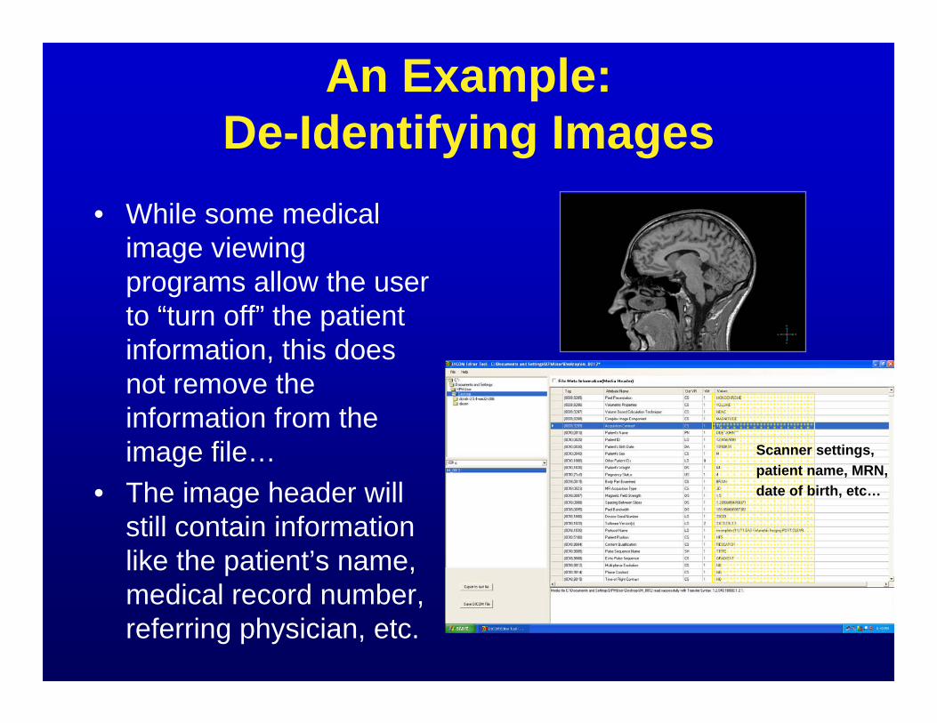
• While some medical image viewing programs allow the user to “turn off” the patient information, this does not remove the information from the image file…
• The image header will still contain information like the patient’s name, medical record number, referring physician, etc.
Scanner settings,patient name, MRN, date of birth, etc…

An Example: De-Identifying Images
• Properly de-identifying medical images is often a complicated task.– HIPAA requires that specific information be removed from the
image headers, but the images must retain enough information to be considered “dicom-compliant” or image viewing programs will not read them.
– Specialized software is often required to remove/replace image header information; using the scanner to delete a patient’s nameand MRN is not enough.
– Removing “burned in” information is time consuming and manually intensive.
• Many clinical trial sponsors require that Protected Health Information (PHI) be removed and replaced with trial-specific information (substituting a subject number for the patient’s name, for example).

An Example: De-Identifying Images
• De-identifying medical images requires time, resources and expertise.
• HIPAA defines certain minimum standards, but research protocols often have specific de-identification requirements.
• There are currently no standardized de-identification methods and/or software.
• How can our Radiology Department coordinate and manage the de-identification protocols for all of the clinical trials and research studies currently in progress?

Organization of the Human Imaging Research Office
ImageAcquisition
ResearchSystemsImaging
ClinicalSystemsImaging
Research MRScheduling
Research MRScan Protocols
ClinicalRadiologyScheduling
Research ScanProtocols
Clinical ScanProtocols
ResponseAssessment/
Image Analysis
MeasurementProtocols
RECISTMeasurements
ResearchMeasurements
Image DatabaseCollection
RadiologyPACS
Non-ImagingDatabases
ResearchPACS
HIROInvestigator Imaging Research InstituteFaculty Advisory Committee
Clinical Trials Review Committee
• The HIRO has three primary arms.• Each arm provides a variety of
targeted services.

Services
•• The Image Acquisition ArmThe Image Acquisition ArmThis arm is responsible for coordinating issues related to the planning and execution of research-related human imaging.
ImageAcquisition
ResearchSystemsImaging
ClinicalSystemsImaging
Research MRScheduling
Research MRScan Protocols
ClinicalRadiologyScheduling
Research ScanProtocols
Clinical ScanProtocols
ResponseAssessment/
Image Analysis
MeasurementProtocols
RECISTMeasurements
ResearchMeasurements
Image DatabaseCollection
RadiologyPACS
Non-ImagingDatabases
ResearchPACS

Services
• Services include:– Protocol preparation and review with the principal investigator and
his/her team, imaging parameters coordination with clinical and research radiology staff, scan scheduling and billing coordination, resource and data management, and quality assurance.
ImageAcquisition
ResearchSystemsImaging
ClinicalSystemsImaging
Research MRScheduling
Research MRScan Protocols
ClinicalRadiologyScheduling
Research ScanProtocols
Clinical ScanProtocols
ResponseAssessment/
Image Analysis
MeasurementProtocols
RECISTMeasurements
ResearchMeasurements
Image DatabaseCollection
RadiologyPACS
Non-ImagingDatabases
ResearchPACS

Services
•• The Image Measurement and Analysis ArmThe Image Measurement and Analysis ArmThis arm is responsible for providing investigators with tumor and lesion measurement data in a consistent and efficient manner.
ImageAcquisition
ResearchSystemsImaging
ClinicalSystemsImaging
Research MRScheduling
Research MRScan Protocols
ClinicalRadiologyScheduling
Research ScanProtocols
Clinical ScanProtocols
ResponseAssessment/
Image Analysis
MeasurementProtocols
RECISTMeasurements
ResearchMeasurements
Image DatabaseCollection
RadiologyPACS
Non-ImagingDatabases
ResearchPACS

Services
• Services include:– Ensuring that measurements are made in accordance with the study
protocol's specific requirements (e.g., RECIST, modified RECIST,IWCLL, etc.), providing consistent measurements for follow-up scans, and ensuring that required measurement data is completed and delivered in a timely fashion.
ImageAcquisition
ResearchSystemsImaging
ClinicalSystemsImaging
Research MRScheduling
Research MRScan Protocols
ClinicalRadiologyScheduling
Research ScanProtocols
Clinical ScanProtocols
ResponseAssessment/
Image Analysis
MeasurementProtocols
RECISTMeasurements
ResearchMeasurements
Image DatabaseCollection
RadiologyPACS
Non-ImagingDatabases
ResearchPACS

Services
•• The Image Collection and Database ArmThe Image Collection and Database ArmThis arm is responsible for managing and distributing clinical image data for research studies in an IRB- and HIPAA-compliant manner.
ImageAcquisition
ResearchSystemsImaging
ClinicalSystemsImaging
Research MRScheduling
Research MRScan Protocols
ClinicalRadiologyScheduling
Research ScanProtocols
Clinical ScanProtocols
ResponseAssessment/
Image Analysis
MeasurementProtocols
RECISTMeasurements
ResearchMeasurements
Image DatabaseCollection
RadiologyPACS
Non-ImagingDatabases
ResearchPACS

Services
• Services include:– Providing researchers with "de-identified" consented patient
information and DICOM-compatible medical images, and creating customized image databases tailored to an investigator’s criteria.
ImageAcquisition
ResearchSystemsImaging
ClinicalSystemsImaging
Research MRScheduling
Research MRScan Protocols
ClinicalRadiologyScheduling
Research ScanProtocols
Clinical ScanProtocols
ResponseAssessment/
Image Analysis
MeasurementProtocols
RECISTMeasurements
ResearchMeasurements
Image DatabaseCollection
RadiologyPACS
Non-ImagingDatabases
ResearchPACS

Staffing Levels
1 full1 full--time stafftime staffResearch Imaging CoordinatorResearch Imaging Coordinator
3 part3 part--time technical stafftime technical staffMR PhysicistMR PhysicistResearch MR TechnologistResearch MR TechnologistResearch CT TechnologistResearch CT Technologist
ImageAcquisition
ResearchSystemsImaging
ClinicalSystemsImaging
Research MRScheduling
Research MRScan Protocols
ClinicalRadiologyScheduling
Research ScanProtocols
Clinical ScanProtocols
ResponseAssessment/
Image Analysis
MeasurementProtocols
RECISTMeasurements
ResearchMeasurements
Image DatabaseCollection
RadiologyPACS
Non-ImagingDatabases
ResearchPACS
2 part2 part--time stafftime staffResearch RadiologistResearch RadiologistImage Analysis Software Image Analysis Software
DeveloperDeveloper
1 full1 full--time stafftime staffResearch Coordinator & Data Research Coordinator & Data
ManagerManager
1 part1 part--time stafftime staffApplications and Web Applications and Web
DeveloperDeveloper
HIRO LeadershipHIRO Leadership1 Faculty Director (science)1 Faculty Director (science)1 Faculty Co1 Faculty Co--Director (clinical)Director (clinical)1 Technical Director1 Technical Director

The HIRO Process Model1. The HIRO performs an imaging review and sign-
off for protocols submitted to the CTRC:– Ensure required imaging technologies exist.– Verify validity of proposed imaging protocol.– Identify a “primary radiologist” collaborator to oversee
implementation of imaging protocol.Studies may come to the HIRO through other mechanisms
2. The HIRO establishes a workflow and addresses imaging prerequisites.
3. The HIRO creates template materials for study staff.

The HIRO Process Model4. The HIRO monitors the scheduling, billing, and
scanning processes.5. The HIRO assists with imaging-related
paperwork required for the study.6. The HIRO reviews and/or provides appropriate
lesion measurements.7. The HIRO delivers appropriately de-identified
image data to the investigator (or submits it directly to an external study sponsor).

The HIRO Process Model• Investigators can utilize any or all of the
HIRO’s services for their studies.• The HIRO is also capable of providing large
amounts of HIPAA-compliant de-identified image data for basic science, epidemiology and other retrospective studies.
• The HIRO’s costs are recovered via a chargeback model that can be included in grants and contracts.

Web Tools
• To manage workflow and provide interactive tools for HIRO staff, imaging staff and investigators, a comprehensive web-based system was developed and deployed.

Web Tools• This web system allows
users to:– Register their studies with
the HIRO– Request access to HIRO
services and IT systems for their team
– Submit requests for de-identified copies of images and related data
– View imaging-related documentation and materials for their studies

An Example:Requesting a De-Identified Scan
1. Create an account on the HIRO Website.2. Register your IRB Protocol and obtain Image
Request access (usually done by the Principal Investigator).
3. Click the “Request Images” link.4. Select the IRB Protocol and the type of request
you’d like to make.5. Fill out the request form with the scan’s details
and press the Submit button.Note that Step 1 only needs to be done once for each user, and Step 2 only needs to be done once for each study/protocol.

Current Usage• After nearly two years of operation:
– The Image Acquisition ArmImage Acquisition Arm has assisted in more than 50 clinical trials from 10 different clinical departments.
– The Image Measurement and Analysis ArmImage Measurement and Analysis Armhas assisted in 5 clinical trials and has performed quality assessments for each trial.
– The Image Collection and Database ArmImage Collection and Database Arm has fulfilled over 400 unique requests for clinical images, delivering over 27.1 million images and related reports to investigators for research purposes.

Costs• There are currently no fees associated
with our Image Acquisition services and our Image Measurement services. Once we have finalized the fee structure for these services, we will make it widely available.
• The fees for our Image Collection and Distribution services are intended to recover our operational costs. They are charged on a per-exam basis.

Costs
Additional $5/exam(max. $200 per request)
"Rush" Requests*
Additional $5/examImage-based Anonymization*
Additional $5/examCustomized Anonymization*
$25/examIndustry
$10/examInternal/Federal Grant
No ChargeAcademic/ClinicalStandard image anonymization/distribution
CostCostFunding SourceFunding SourceServiceService
* This service is considered an addition to our standard image anonymization/distribution service, so scans that require this service will be charged the standard anonymization fee plus the indicated surcharge.

Experiences•• The HIRO is becoming the institutional The HIRO is becoming the institutional
resource for the spectrum of research resource for the spectrum of research imaging needs.imaging needs.
• Early collaboration between research groups and the HIRO is highly beneficial.– Early radiology involvement in research studies
and clinical trials that require medical imaging is key to a successful imaging experience.

Future Goals•• Based on our experiences, we have Based on our experiences, we have
identified several key issues that must be identified several key issues that must be addressed to ensure further success:addressed to ensure further success:– The “imaging review” of a research protocol
would be best achieved at an earlier stage.– Further integration with HIS/RIS* for the
purposes of automation will relieve burden on both HIRO staff and investigators.
*HIS/RIS: Health Information Systems/Radiology Information Systems

Future Goals•• Based on our experiences, we have Based on our experiences, we have
identified several key issues that must be identified several key issues that must be addressed to ensure further success:addressed to ensure further success:– Continued deployment of web-based services
will provide investigators with more features and easy-to-use access to HIRO services.
– The Image Measurement and Analysis arm is a highly desired asset within the clinical research community but is logistically challenging; continued development on a prospective measurement program is necessary.

Questions?
• The Human Imaging Research OfficeHuman Imaging Research Office is always happy to answer any research imaging-related questions you may have!

Thank You!Thank You!The The HIROHIRO’’ss faculty and staff:faculty and staff:
Samuel G. Armato III, Ph.D. Heber MacMahon, M.D.Faculty DirectorFaculty Director Faculty CoFaculty Co--DirectorDirector
Nick Gruszauskas, Ph.D. Maryellen L. Giger, Ph.D.Technical DirectorTechnical Director DirectorDirector, Imaging Research Institute, Imaging Research Institute
Michael D. Torno, D.Sc. Caileigh PudelaResearch Coordinator & Data ManagerResearch Coordinator & Data Manager Research Imaging CoordinatorResearch Imaging Coordinator
Feng Li, M.D., Ph.D. Milica Medved, Ph.D.Research RadiologistResearch Radiologist MR PhysicistMR Physicist
Roger Engelmann, M.S. Adam StarkeyImage Analysis Software DeveloperImage Analysis Software Developer Applications and Web DeveloperApplications and Web Developer
Human Imaging Research OfficeImaging Research InstituteBiological Sciences DivisionThe University of Chicago
• Visit us at http://http://hiro.bsd.uchicago.eduhiro.bsd.uchicago.edu