histopathological changes related to chemical ... · pdf filedisorders related to the general...
TRANSCRIPT
Histopathological changes related to chemical
p.
Vol. 46: 101-107, 1988
contamination in Mytilus edulis from field and experimental conditions
MARINE ECOLOGY - PROGRESS SERIES Mar. Ecol. Prog. Ser.
M. Auffret
Published June 30
Pathology Laboratory, Faculty of Medicine. Universite d e Bretagne Occidentale, F-29283 Brest Cedex, France
ABSTRACT: This comparative histopathological study, carried out at the GEEP Workshop, assesses tissue changes in mussels Mytilus edulis from a contaminated Norwegian fjord and from mesocosm basins where a contaminant gradient had been simulated. Parasitism by larval trematodes was observed in both circumstances, but was not related to contaminant levels. Mussels from the field exhibited granulocytomas in their interstitial tissues, with a greater incidence at polluted sites than at a reference site, suggesting that granulocytomas could be a consequence of chronic pollutant exposure. This pathological condition was not observed in experimentally treated mussels, which nevertheless had severe tissue alterations, especially in digestive tubules and gills, under exposure to high levels of a diesel oil and copper mixture. It is concluded that this type of histopathological analysis can provide useful information on the health of mussels, and that this information can be used successfully in the comparison of field samples.
INTRODUCTION MATERIAL AND METHODS
Pathology is now a standard part of environmental monitoring programmes on pollution effects (Yevich & Barszcz 1976, Balouet & Poder 1981, Couch 1985). Based on an easily reproducible technique, his- topathological studies yield basic information on tissue disorders related to the general state of organisms, and assess the host's susceptibility to infectious diseases and parasitic infestation. Some of these parameters may serve as indicators of the effects of xenobiotic contamination in marine animals (Sindermann 1980). Among the diseases and pathological changes that can be measured in terms of prevalence and geographical distribution, it is essential to select parameters that are related to pollution, rather than other environmental stressors, in order to determine efficient tools for monitoring programmes (Sindermann et al. 1980).
The present study was undertaken both on field and experimentally exposed Mytilus edulis. Its aim was to ascertain the ability of tissue and organ changes, and the prevalence of parasitism and other diseases, to determine the effects of xenobiotics at various concen- trations.
Mytilus edulis were collected during August 1986 from field sites in Langesundfjord (Norway), an area contaminated by metals and organic xenobiotics of industrial origin (Follum & Moe 1988), and from NIVA mesocosm facilities at Solbergstrand (near Oslo), where a contaminant gradient had been simulated in experi- mental basins (Bakke et al. 1988). Briefly, the M. edulis were sampled according to the following schedule.
The 4 field sites in Langesundfjord (designated Sites 1 to 4 ) were chosen to represent a gradient of increasing water contamination towards the contami- nant source. Tissue levels for selected contaminants - such as polycyclic aromatic hydrocarbons (PAHs), polychlorinated biphenyls (PCBs) and trace metals - were measured by Klungseryr et al. (1988) and Abdullah & Steffenak (1988); see also Appendix 1.
The 4 experimental basins were dosed for 15 wk from April 1986 with differing concentrations of the same mixture of diesel oil (water accommodated frac- tion) and copper (C: control, L: low, M: medium and H: high dose), see Bakke et al. (1988). High dose levels were chosen to impact the organisms strongly, while C,
0 Inter-Research/Printed in F. R . Germany 0171-8630/88/0046/0101/$ 03.00
102 GEEP WORKSHOP: CELLULAR- AND HISTOPATHOLOGY
Source Shell length Body weight
Site 1 46.6 + 1.0 4.1 f 0.2 2 56.3 + 1.1 6.8 f 0.5 3 45.4 + 1.2 3.5 t 0.2 4 46.9 + 1.3 3.1 t 0.3
Basin
C 54.2 + 1.3 7.6 t 0.5 L 51.4 + 2.1 6.1 + 0.8 M 52.9 f 1.3 4.9 + 0.4 CH 59.0 f 2.1 8.9 f 0.9 H 51.8 f 1.3 5.0
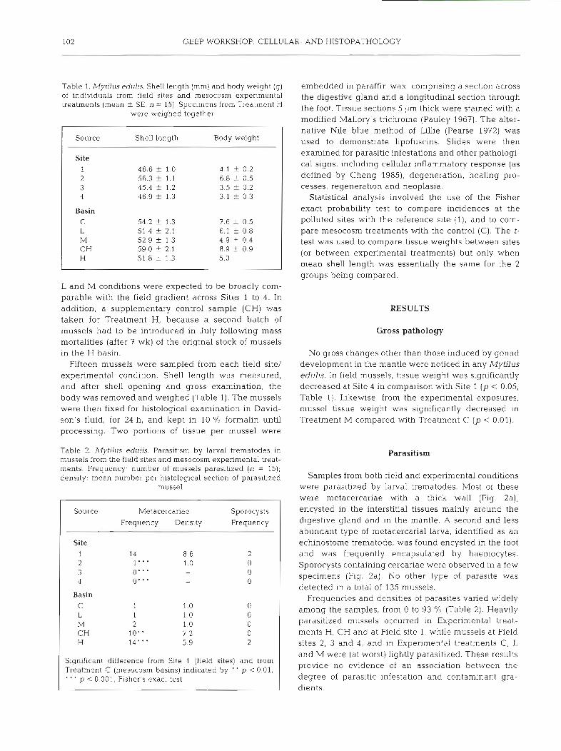
Table 1. ~llytdus edulis. Shell length (mm) and body weight (g) embedded in paraffin wax, comprising a section across of individuals from field sites and mesocosm experimental the digestive gland and a longitudinal section through treatments (mean +. SE, n = 15). Specimens from Treatment H the foot. Tissue sections pm thick were stained with a
were weighed together modified Mallory's trichrome (Pauley 1967). The alter- native Nile blue method of Lillie (Pearse 1972) was used to demonstrate lipofuscins. Slides were then examined for parasitic infestations and other pathologi- cal signs, including cellular inflammatory response (as defined by Cheng 1985), degeneration, healing pro- cesses, regeneration and neoplasia.
Statistical analysis involved the use of the Fisher exact probability test to compare incidences at the polluted sites with the reference site ( l ) , and to com- pare mesocosm treatments with the control (C). The t- test was used to compare tissue weights between sites (or between experimental treatments) but only when mean shell length was essentially the same for the 2 groups being compared.
L and M conditions were expected to be broadly com- parable with the field gradient across Sites 1 to 4. In addition, a supplementary control sample (CH) was RESULTS taken for Treatment H, because a second batch of mussels had to be introduced in July following mass Gross pathology mortalities (after 7 wk) of the original stock of mussels in the H basin. No gross changes other than those induced by gonad
Fifteen mussels were sampled from each field site/ development in the mantle were noticed in any Mytilus experimental condition. Shell length was measured, edulis. In field mussels, tissue weight was significantly and after shell opening and gross examination, the decreased at Site 4 in comparison with Site 1 (p < 0.05, body was removed and weighed (Table 1). The mussels Table 1). Likewise, from the experimental exposures, were then fixed for histological examination in David- mussel tissue weight was significantly decreased in son's fluid, for 24 h, and kept in 10 % formalin until Treatment M compared with Treatment C (p < 0.01). processing. Two portions of tissue per mussel were
Source Metacercariae Sporocysts
Frequency Density Frequency
Site 1 14 8.6 2 2 1"' 1.0 0 3 0" ' - 0 4 0" ' - 0
Basin
C 1 1 .o 0 L 1 1.0 0 M 2 1.0 0 CH 10" 7.2 0 H 14"' 5.9 2
Significant difference from Site 1 (field s~ tes ) and from Treatment C (mesocosm basins) indicated by " p < 0.01. ' ' ' p < 0.001, Fisher's exact test
Table 2. Mytilus edulis. Parasitism by larval trematodes in Parasitism mussels from the field sites and mesocosm experimental treat- ments. Frequency. number of mussels parasltized (n = 15); density: mean number per histological section of parasitized Samples from both field and experimental conditions
mussel were parasitized by larval trematodes. Most of these were metacercariae with a thick wall (Fig. 2a), encysted in the interstitial tissues mainly around the digestive gland and in the mantle. A second and less abundant type of metacercanal larva, identified as an echinostome trematode, was found encysted in the foot and was frequently encapsulated by haemocytes. Sporocysts containing cercariae were observed in a few specimens (Fig. 2a). No other type of parasite was detected in a total of 135 mussels.
Frequencies and densities of parasites varied widely among the samples, from 0 to 93 YO (Table 2). Heavily parasitized mussels occurred in Experimental treat- ments H, CH and at Field site 1, while mussels at Field sites 2, 3 and 4, and in Experimental treatments C, L and M were (at worst) lightly parasitized. These results provide no evidence of an association between the degree of parasitic infestation and contaminant gra- dients.

Auffret: Histopathology of mussels 103
Table 3. Mytilus edulis. Incidence of main histological characteristics and pathological changes (not related to parasitism) in individuals from field sites and mesocosm experimental treatments (%, n = 15). (Interst: interstitial tissues, muscles and foot; Circ: circulatory system; CBC' clusters of brown cell-i; DEG: degenerative changes; CIR: cellular inflammatory response; BCL: brown
coloured lysosomes; G: granulocytomas; HA: haemocyte aggregates; subscripts: M, males; F, females
Source Stomach Intestine Ducts Tubules Kidney Interst Circ Gill Gonad CBC DEG CIR CBC DEG CIR DEG BCL DEC CBC DEG CIR G HA DEG DEGpCIRpCIRM G
Site 1 73 13 0 13 0 0 27 2 100 7 0 60"' 7 0 0 3 53 0 7 20 0 7 0 4 38 0 0 47' 7 0
Basin
C 67 53 0 33 27 G 0 0 7 0 u 0 0 0 7 1 0 0 0 L 93 7" 7 13 0 13 7 0 20 0 0 1 0 0 0 80 0 0 0 M 60 15' 0 47 0 7 0 0 13 0 0 0 0 0 0 53 0 0 0 H 80 20 33 27 33 40 60 0 60"' 0 33- 0 0 33 100*.* 75 0 0 0
Significant difference from appropriate reference site (1) or mesocosm control (C) indicated by ' p <0.05, ' ' p < 0.01, "' p < 0 001, Fisher's exact test
In every sample, haemocyte aggregates were pres- ent in interstitial tissues of the digestive gland and the gonad, around dead metazoans, probably copepods. Since this phenomenon corresponds to a resorption process of foreign material, such features were not considered as true incidences of parasitism.
Histopathology
Field samples
Several pathological changes were observed at every field site (Table 3), and no sample was considered as being in good health. At Site 4, however, only 67 % of the mussels exhibited pathological changes. Degenera-
tive features (atrophy and necrosis) were infrequently noted in the epithelium of the kidney. In the digestive tract, clusters of brown cells (CBC) were scattered throughout the epithelium of the stomach and intes- tine, with irregular but often high frequencies. These epithelia were also the site for high frequencies of focal inflammation (CIR) and degenerative processes (DEG). In mussels from every site, the epithelium of the diges- tive tubules showed a conspicuous histological feature after staining with the trichrome technique: the apical cytoplasm of the digestive cells was filled with small and regular brown granules, which were identified as late secondary or tertiary lysosomes (Fig. l a ) . After staining with the Nile blue method, these granules had a greenish colour indicative of lipofuscin content, while secondary lysosomes of normal appearance (in other
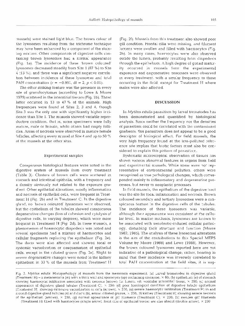
Fig. 1. Mytilus edulis. Histopathology of mussels from field sites: (a) Epithelial cells of digestive gland tubules (Site 2), containing numerous brown coloured lysosomes (arrow) - the other cellular components have a normal appearance; x 260; (b) large
granulocytomas [gr) surrounded by haemocytic infiltration (arrow) in a mussel from Site 3; g : gonad, s: stomach; x 50

104 GEEP WORKSHOP: CELLULAR- AND HISTOPATHOLOGY
Auffret: Histopathology of n~ussels 105
mussels) were stained light blue. The brown colour of the lysosomes resulting from the tnchrome technique may have been enhanced by a component of the stain- ing mixture. Other components of digestive cells con- taining brown lysosomes had a normal appearance (Fig. l a ) . The incidence of these 'brown coloured' lysosomes decreased steadily from Site 1 (87 %) to Site 4 (13 %), and there was a significant negative correla- tion between incidence of these lysosomes and total PAH concentration (r = -0.991, df = 2, p < 0.01).
The other striking feature was the presence in every site of granulocytomas (according to Lowe & Moore 1979) scattered in the interstitial tissues (Fig. l b ) . These latter occurred in 13 to 47 % of the animals. High frequencies were found at Sites 2, 3 and 4, though Site 3 was the only one with significantly higher inci- dence than Site 1. The mussels showed variable repro- ductive condition, that is, some specimens were fully mature, male or female, while others had empty folli- cles. Areas of necrosis were observed in mature female follicles, affecting every mussel at Site 4 and up to 50 O/O of the mussels at the other sites.
Experimental samples
Conspicuous histological features were noted in the digestive system of mussels from every treatment (Table 3). Clusters of brown cells were scattered in stomach and intestinal epithelia, with a frequency and a density obviously not related to the exposure gra- dient. Other epithelial alterations, mostly inflammation and necrosis of epithelial cells, were frequent in Treat- ment H (Fig. 2b) and in Treatment C. In the digestive gland, no 'brown coloured' lysosomes were observed, but the epithelium of the tubules showed conspicuous degenerative changes (loss of cohesion and cytolysis of digestive cells, to varying degrees), which were more frequent in Treatment H (Fig. 2d). In these mussels, a phenomenon of haemocytic diapedesis was noted and several specimens had a mixture of haemocytes and cellular fragments replacing the epithelium (Fig. 2e). The ducts were also affected and showed focal or systemic vacuolization or desquamation of epithelial cells, except in the ciliated groove (Fig. 2e). Slight to severe degenerative changes were noted in the kidney epithelium in 33 % of the mussels from Treatment H
(Fig. 2f). Mussels from this treatment also showed poor gill condition. Frontal cilia were missing, and filament lumens were swollen and filled with haemocytes (Fig. 2h). In many cases, haemocytes were also observed outide the lumen, probably resulting from diapedesis through the epithelium. A high degree of gonad matur- ity occurred in mussels from the experimental exposures and degenerative processes were observed in every treatment, with a similar frequency to those occurring in the field, except for Treatment H where males were also affected.
DISCUSSION
In Mytilus edulis parasitism by larval trematodes has been demonstrated and quantified by histological analysis. Since neither the frequency nor the densities of parasitism could be correlated with the contaminant gradients, this parasitism does not appear to be a good descriptor of biological effect. For field mussels, the very high frequency found at the less-polluted refer- ence site implies that biotic factors must also be con- sidered to explain this pattern of parasitism.
Systematic microscopical observation of tissues has shown various abnormal features in organs from field and experimental mussels. While some were not rep- resentative of environmental pollution, others were recognized as true pathological changes, which corres- ponded mainly to inflammatory and degenerative pro- cesses, but never to neoplastic processes.
In field mussels, the epithelium of the digestive tract was the site for focal inflammat~on and necrosis. Brown coloured secondary and tertiary lysosomes were a con- spicuous feature in the digestive cells of the tubules. The incidence of these lysosomes was variable, although their appearance was consistent at the cellu- lar level. In marine molluscs, lysosomes are known to be associated with xenobiotic-induced cellular pathol- ogy, disturbing their structure and function (Moore 1982, 1985). The analysis of these lysosomal alterations is the aim of the contributions to this Special MEPS Volume by Moore (1988) and Lowe (1988). However, the brown coloured lysosomes reported here are not indicative of a pathological change; rather, bearing in mind that their incidence was inversely correlated to total PAH concentration a t the field sites, it is sug-
Fig. 2. Mytilus edulis. Histopathology of mussels from the mesocosm experiment. (a) Larval trematodes in digestive gland (Treatment H) - a metacercar~a (m) with a thick wall and sporocysts (sp) containing cercariae, X 60; (b) epithelium (e) of stomach showing haemocytic infiltration associated with necrosis (arrow); lu: lumen, vit: vesicular interstitial tissue, X 200; (c) normal appearance of digestive gland tubules (Treatment C), X 250; (d) poor histological condition of digestwe tubule epithelium (Treatment H), showing extensive vacuolization in cells (arrows), X 250; (e) severe haemocytic infiltration (Treatment H) in and around digestive gland tubules (a) and ducts (b), arrow: ciliated groove, x 250; ( f ) kidney (Treatment H) showing severe necrosis of the epithelium (arrows), X 200; (g) normal appearance of gill filaments (Treatment C), X 230; (h) swollen gill filaments
(Treatment H) filled with haemocytes (single arrow), front cilia of epithelial border are also absent (double arrow), X 230
106 GEEP WORKSHOP: CELLUI -AR- AND HISTOPATHOLOGY
gested that a high incidence reflects the normal condi- tion. The most obvious pathological condition consisted of granulocytomas, which occurred throughout the interstitial tissues of numerous field mussels. Lowe & Moore (1979) have suggested a relationship between this particular type of inflammatory response and chronic pollution, while Rasmussen et al. (1983b) have induced a similar pathology in mussels after chronic chemical exposure. It seems probable therefore that the occurrence of granulocytomas in the Langesundfjord mussels is a consequence of chronic exposure to conta- minants.
In the experimental treatments, specimens exposed to the highest concentrations of hydrocarbons and cop- per had severe degenerative changes in the epithelia of the digestive gland. The appearance of such an ulti- mate cellular disturbance is in agreement with the hypothesis of an autolytic process, as a consequence of full lysosomal destabilization (Moore 1985). Necrosis and infiltration of severely damaged tubules by haemocytes have been reported by Rasmussen (1982) and Rasmussen et al. (1983a, b) in mussels challenged with N-nitroso compounds. Couch (1985) described atrophic epithelium sloughing of cells and necrosis as an ultimate tubular degeneration, in oysters from con- taminated estuaries. The findings of Rasmussen sug- gest that chemically-induced injuries are implicated in the formation of such lesions. The other striking tissue lesion noted in these mussels involved the gills, which exhibited morphological changes in their filaments and severe disturbance of the ciliated epithelia1 cells, when compared to mussels from control and low exposure treatments. Similar lesions have been reported in mussels exposed to sublethal thermal stress (Gonzales & Yevich 1976), to N-nitrosodimethylamine (Ras- mussen 1982) and to copper and cadmium exposure (Sunila 1986). This apparent non-specificity ~ndicates that the gills are particularly sensitive organs in mussels.
It is clearly important to assess the histopathological responses of Mytilus edulis in relation to the measured contaminant levels. Briefly, the chemical analysis of mussel tissues revealed an increasing content for PAHs and PCBs from Site 1 to Site 4, as well as for copper from Treatment C to Treatment H and for PAHs from Treatment C to Treatment M. In field mussels, the granulocytomas were very frequent in the most con- taminated sites. Their occurrence was significantly higher at Site 3 than the reference site, and the incidences at Sites 2, 3 and 4 were not significantly different from each other The level of quantification (presence or absence in n = 15 mussels) is insufficient to expect to demonstrate a detailed 'dose-response' relationship. Nonetheless, the increased prevalence of granulocytomas at the contaminated sites corroborates
the results of Lowe & Moore (1979) with mussels from British coasts, and lends support to the postulate that granulocytomas can be used as an index of haemocytic response to aquatic contamination. Conversely, pathological changes in epithelia of the digestive tract occurred infrequently and irregularly among the 4 sites, and this could be interpreted as a response to unidentified deleterious factors. In the experimental treatments, histopathological response could not be correlated to dosing levels of contaminants, with the exception of the H treatment, for which the mussels &splayed severe pathological changes involving the epithelia of the digestive system, gills and kidney. These obviously reflected a poor health condition, pre- sumably induced by the xenobiotics. Furthermore, the pathology observed in H mussels, together with that reported for experimental treatments by Rasmussen et al. (1983a,b, 1985), confirm that the digestive gland is a target tissue for xenobiotic effects (Sindermann 1980).
When comparing results from Mytilus edulis in the field and experimental studies, it appears that, although total PAH tissue concentrations span similar ranges, the response in terms of pathological changes is not the same, as no granulocytomas were detected in the experimental treatments. However, chemical analysis (Klungseryr et al. 1988) revealed that the com- position of the PAHs differed between field and experi- mental conditions, with higher molecular weight com- pounds dominating in the field and lower molecular weight compounds in the experimental exposures. This was probably the major contributory factor to the differences in the observed pathology, although the experimental results may have been affected by the starved condition of the mussels (Lowe 1988), and the field results by possible interactive effects of contami- nants.
In conclusion, this study has demonstrated that, in field mussels, non-parasitic tissue abnormalities were related to chemical contamination. Similar pathological effects were not generally induced in the experimental treatments, though Mytilus edulis from the highest dose treatment exhibited a clear response to contami- nants. In an attempt to select pathological indicators of pollution, this histological analysis of tissue modifica- tions may not entirely satisfy general criteria required in monitoring programmes, since it is difficult to define a smooth dose-response relation based only on pres- ence or absence of histopathological features. This underlines the need for standard histology to be under- taken parallel to other types of cellular investigations, such as quantitative histology and cell biochemistry. However, standard histology has shown its utility here for characterizing tissue effects of xenobiotic exposure that can be compared directly with other data sets.
Auffret: Histopathc Aogy of mussels 107
Acknowledgements. The author thanks the International Council for the Exploration of the Sea for funding h s parbcl- pation at the GEEP Workshop, and Dr M. N. Moore for critically reviewing the manuscript and providing the statisti- cal analysis of the data.
LITERATURE CITED
Abdullah, M. I., Steffenak, I. (1988). The GEEP Workshop: trace metal analyses. Mar. Ecol. Prog. Ser. 46: 27-30
Bakke, T., Follum, 0. A., Moe, K. A., Ssrensen, K. (1988). The GEEP Workshop: mesocosm exposures. Mar. Ecol. Prog. Ser. 46: 13-18
Balouet, G., Poder, M. (1981). Effets biologiques de la pollu- tion par les hydrocarbures de 1'Amoco-Cadiz sur l'os- treiculture en Bretagne Nord. In: Arnoco-Cadiz. Conse- quences d'une pollution accidentelle par les hydrocar- bures. CNEXO, Paris, p. 703-713
Cheng, T C. (1985). Evidence for molecular specificities involved in molluscan inflammation. In: Cheng, T.C. (ed.) Comparative pathobiology, Vol. 8. Plenum Press, New York, p. 129-142
Couch, J. A. (1985). Prospective study of infectious and non- infectious diseases in oysters and fishes in three Gulf of Mexico estuaries. Dis. aquat. Org. 1: 59-82
Follum, 0. A., Moe, K. A (1988). The GEEP Workshop: field sampling. Mar. Ecol. Prog. Ser. 46: 7-12
Gonzales, J. G., Yevich, P, (1976). Responses of an estuarine population of the blue mussel Mytilus edulis to heated water from a steam generatmg plant. Mar. Biol. 34. 177- 189
Klungsayr, J., Wilhelmsen, S., Westrheim, K., Saetvedt, E.. Palmork, K. H. (1988). The GEEP Workshop: organic chemical analyses. Mar. Ecol. Prog. Ser. 46: 19-26
Lowe, D. M. (1988). Alterations in cellular structure of Mytilus edulis resulting from exposure to environmental contami- nants under field and experimental conditions. Mar. Ecol. Prog. Ser. 46: 91-100
Lowe, D. M., Moore, M. N. (1979). The cytology and occur- rence of granulocytomas in mussels. Mar. Pollut. Bull. 10: 137-141
Moore, M. N. (1982). Lysosomes and environmental stress. Mar. Pollut. Bull. 13: 42-43
Moore, M. N. (1985). Cellular responses to pollutants. Mar Pollut. Bull. 16: 134-139
Moore, M. N. (1988). Cytochemical responses of the lysosomal system and NADPH-ferrihemoprotein reductase in mollus- can digestive cells to environmental and expenmental exposure to xenobiotics Mar Ecol. Prog. Ser. 46: 81-89
Pauley, G. B. (1967). A modification of Mallory's trichrome anilin blue collagen stain for oyster tissue. J. invert. Pathol. 9: 268-269
Pearse, A. G. E. (1972). Histochemistry, theoretical and applied, Vol. 2. Churchd Livingstone, London
Rasmussen, L. (1982). Light microscopical studies on the acute toxic effects of N-~trosodimethylamine on the marine mussel, Mytilus edulis. J. invert. Pathol. 39: 66-80
Rasmussen, L. P. D., Hage, E., Karlog, 0. (1983a). Light and electron microscopic studies on the acute and chronic toxic effects of N-nitroso compounds on the marine mussel, Mytilus eduhs (L.). I. N-nitrosodimethylamine. Aquat. Toxicol. 3: 285-299
Rasmussen, L. P. D., Hage, E., Karlog, 0 . (198313). Light and electron microscopic s tudes on the acute and chronic toxic effects of N-nitroso compounds on the marine mussel, Mytilus edulis (L.). 11. N-methyl-N-nitrosoguanine. Aquat. Toxicol. 3: 301-311
Rasmussen, L. P. D., Hage. E., Karlog, 0. (1985). Light and electron microscopic studies on the acute and long-term toxic effects of N-nitrosodipropylamine and N-methyl- nitrosurea on the marine mussel, Mytilus edulis. Mar. Biol. 85: 55-65
Sindermann, C. J . (1980). The use of pathological effects of pollutants in marine environmental monitoring programs. Rapp. P,-v. Reun. Cons. int. Explor. Mer 179: 129-134
Sindermann, C. J., Bang, F. B., Christensen, N. O., Dethlefsen, V., Harshbarger, J. C., Mitchell, J. R., Mulcahy. M. F. (1980). The role and value of pathology in pollution effects monitoring programmes, Pathology Panel report. Rapp. P: v. Reun. Cons. int. Explor. Mer 179: 135-151
Sunila, I. (1986). Chronic histopathological effects of short- term copper and cadmium exposure on the gill of the mussel, Mytilus edulis. J. invert. Pathol. 47: 125-142
Yevich, P. O., Barszcz, C. A. (1976). Histopathology as a monitor for marine polluhon; results of the histopathologic examination of the animals collected for the U.S. mussel watch program. Report of the U.S. Environmental Protec- tion Agency