imaging of pneumonia: an overview
TRANSCRIPT
THORACIC IMAGING (T BUXI, SECTION EDITOR)
Imaging of Pneumonia: An Overview
Mandeep Garg1 • Nidhi Prabhakar1 • P. Kiruthika1 • Ritesh Agarwal2 •
Ashutosh Aggarwal2 • Ajay Gulati1 • Niranjan Khandelwal1
Published online: 24 February 2017
� Springer Science+Business Media New York 2017
Abstract
Purpose of review Pneumonia is one of the common
causes of morbidity and mortality in general population.
Imaging plays an important role in the management of
pneumonia.
Recent findings In the current era, there has been an
increase in the patients with extremes of age, immuno-
compromised status, underlying lung pathology, post-
transplant status, and atypical infections. It is necessary to
use cross-sectional imaging modalities like computed
tomography (CT) due to atypical or non-specific chest
radiograph findings in such cases. CT narrows down the
differential diagnosis, for etiological agent. It helps in the
evaluation of the causes of non-resolving pneumonia,
pulmonary, and non-pulmonary complications of pneu-
monia. Pneumonia is classified into three main types as
community-acquired pneumonia, hospital-acquired pneu-
monia, and aspiration pneumonia. It is important to dif-
ferentiate these three types, since host factors and
etiological organisms differ, thus changing the course and
management in these patients.
Summary Knowing the clinical background and correla-
tion with imaging findings may help in the early detection
of pathogen and direct the physician toward appropriate
management. Imaging also helps in follow-up of patients to
look for response to therapy. Cross-sectional imaging can
help in ruling out diseases mimicking pneumonia.
Keywords Pneumonia � Lung � Infection � Tuberculosis �Radiograph � Chest
Introduction
Pneumonia is one of the common causes of morbidity and
mortality in general population. Imaging plays an important
role in the management of pneumonia. In a patient suffering
from fever, cough or sputum production, imaging helps in
confirming the diagnosis of pneumonia. However, identifi-
cation of specific etiological agent is not always possible,
since the imaging findings may be non-specific. Response of
lungs to any kind of inflammation or infection is limited,
most of thempresenting as alveolar opacities, and hence non-
infectious pathologies may also have an appearance of
pneumonias and are most often termed as pneumonia mim-
ics. Chest radiography is the most widely used radiological
investigation and in most cases may be the only investiga-
tion necessary in treating a patient with pneumonia. How-
ever, in the current era with an increase in the people with
extremes of age, immunocompromised status, underlying
lung pathology, post-transplant patients, and also infections
due to atypical organisms, it is necessary to use cross-sec-
tional imaging modalities like computed tomography (CT)
due to atypical or non-specific chest radiograph findings [1].
CT also helps in determining the causes of non-resolving
pneumonias, pulmonary and non-pulmonary complications
This article is part of the Topical Collection on Thoracic Imaging.
& Mandeep Garg
1 Department of Radiodiagnosis, Post Graduate Institute of
Medical Education and Research, Chandigarh 160012, India
2 Department of Pulmonary Medicine, Post Graduate Institute
of Medical Education and Research, Chandigarh 160012,
India
123
Curr Radiol Rep (2017) 5:16
DOI 10.1007/s40134-017-0209-9
of pneumonia and serves as a guide to intervention in
choosing the site for transbronchial lung biopsy or percuta-
neous biopsy and drainage of abscesses or pleural
collections [2]. This review article gives an overall view
about different types of pneumonias with special emphasis
on pneumonias in immunocompromised patients.
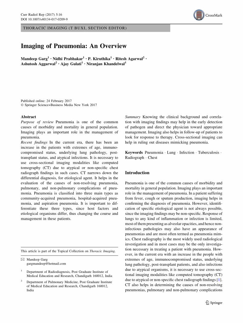
Fig. 1 Lobar Pneumonia due to
Streptococcus in different
patients. a Chest radiograph
shows the presence of
consolidation (asterisk) in the
left upper lobe with the presence
of air bronchogram (black
arrow). b Chest CT in another
patient showing consolidation
involving the right lower lobe
(asterisk). c CT in a patient with
lobar consolidation showing air
bronchogram sign (black arrow)
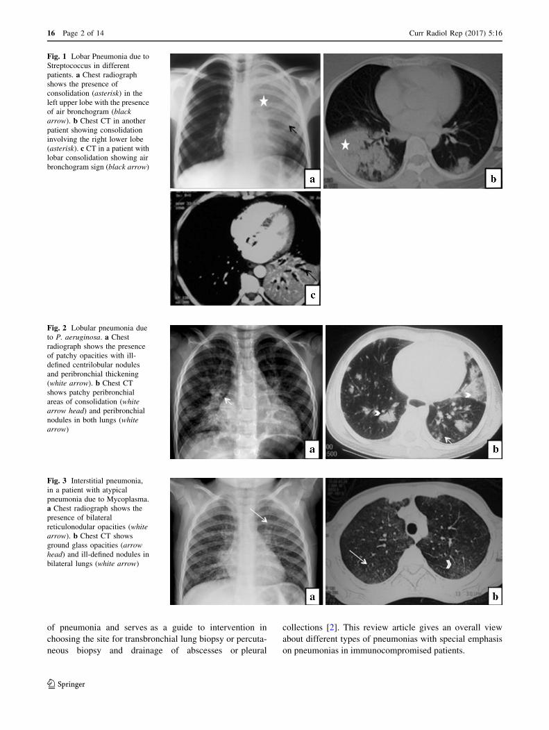
Fig. 2 Lobular pneumonia due
to P. aeruginosa. a Chest
radiograph shows the presence
of patchy opacities with ill-
defined centrilobular nodules
and peribronchial thickening
(white arrow). b Chest CT
shows patchy peribronchial
areas of consolidation (white
arrow head) and peribronchial
nodules in both lungs (white
arrow)
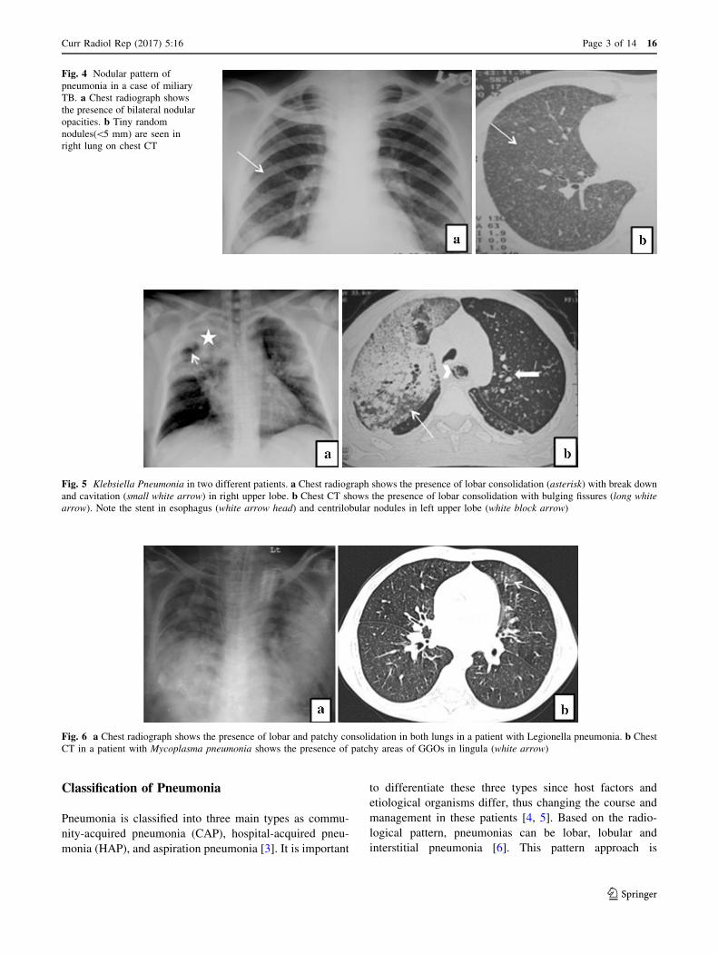
Fig. 3 Interstitial pneumonia,
in a patient with atypical
pneumonia due to Mycoplasma.
a Chest radiograph shows the
presence of bilateral
reticulonodular opacities (white
arrow). b Chest CT shows
ground glass opacities (arrow
head) and ill-defined nodules in
bilateral lungs (white arrow)
16 Page 2 of 14 Curr Radiol Rep (2017) 5:16
123
Classification of Pneumonia
Pneumonia is classified into three main types as commu-
nity-acquired pneumonia (CAP), hospital-acquired pneu-
monia (HAP), and aspiration pneumonia [3]. It is important
to differentiate these three types since host factors and
etiological organisms differ, thus changing the course and
management in these patients [4, 5]. Based on the radio-
logical pattern, pneumonias can be lobar, lobular and
interstitial pneumonia [6]. This pattern approach is
Fig. 4 Nodular pattern of
pneumonia in a case of miliary
TB. a Chest radiograph shows
the presence of bilateral nodular
opacities. b Tiny random
nodules(\5 mm) are seen in
right lung on chest CT
Fig. 5 Klebsiella Pneumonia in two different patients. a Chest radiograph shows the presence of lobar consolidation (asterisk) with break down
and cavitation (small white arrow) in right upper lobe. b Chest CT shows the presence of lobar consolidation with bulging fissures (long white
arrow). Note the stent in esophagus (white arrow head) and centrilobular nodules in left upper lobe (white block arrow)
Fig. 6 a Chest radiograph shows the presence of lobar and patchy consolidation in both lungs in a patient with Legionella pneumonia. b Chest
CT in a patient with Mycoplasma pneumonia shows the presence of patchy areas of GGOs in lingula (white arrow)
Curr Radiol Rep (2017) 5:16 Page 3 of 14 16
123
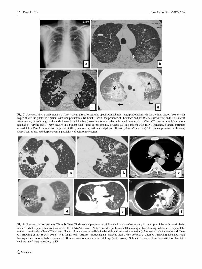
Fig. 8 Spectrum of post-primary TB. a, b Chest CT shows the presence of thick-walled cavity (black arrow) in right upper lobe with centrilobular
nodules in both upper lobes, with few areas of GGOs (white arrow). Note associated peribronchial thickeningwith coalescing nodules in left upper lobe
(white arrowhead).cChestCT in a caseofTuberculoma, showingwell-definednodulewith eccentric cavitation (white arrow) in left upper lobe.dChestCT showing cavity (black arrow) with fungal ball (asterisk) producing air crescent sign (white arrow). e Chest CT showing loculated right
hydropneumothorax with the presence of diffuse centrilobular nodules in both lungs (white arrow). f Chest CT shows volume loss with bronchiectatic
cavities in left lung secondary to TB
Fig. 7 Spectrum of viral pneumonias. aChest radiograph shows reticular opacities in bilateral lungs predominantly in the perihilar region (arrow) with
hyperinflated lung fields in a patient with viral pneumonia. b Chest CT shows the presence of ill-defined nodules (block white arrow) and GGOs (short
white arrow) in both lungs with subtle interstitial thickening (arrow head) in a patient with viral pneumonia. c Chest CT showing multiple random
nodules of varying sizes (white arrow) in a patient with Varicella pneumonia. d Chest CT in a patient with H1N1 influenza, bilateral perihilar
consolidation (black asterisk) with adjacent GGOs (white arrow) and bilateral pleural effusion (black block arrow). This patient presented with fever,
altered sensorium, and dyspnea with a possibility of pulmonary edema
16 Page 4 of 14 Curr Radiol Rep (2017) 5:16
123
sometimes useful in identifying the etiological agent.
However, the radiological pattern should be correlated with
clinical findings and should only be used as a guide to
diagnosis, as variation in imaging findings are common.
For example, single organism can manifest in wide variety
of ways like mycobacterium tuberculosis presenting as
consolidation, nodules, miliary pattern, etc. In addition,
patients with pre-existing lung pathologies and immuno-
compromised status may not have classical imaging find-
ings. Clinical suspicion and cross-sectional imaging can
help in identifying the type of organism in these patients,
even if chest radiograph is non-contributory.
Morphological Patterns of Pneumonia
Airspace Consolidation/Lobar Pneumonia
In air space consolidation, the microorganisms damage the
alveoli leading on to increase in secretion of fluid into the
alveoli that further spreads through collateral drift (termi-
nal airways and pores of Kohn) to involve a entire segment
or lobe. Consolidation of lung is caused by fluid, cellular
infiltration, and fibrinous exudates. Lobar pneumonia is
characterized by relatively sharply marginated homoge-
neous consolidation of lung parenchyma with patent air
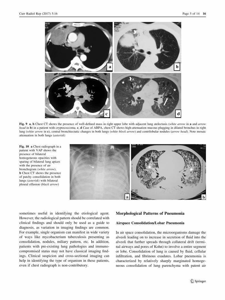
Fig. 10 a Chest radiograph in a
patient with VAP shows the
presence of bilateral
homogeneous opacities with
sparing of bilateral lung apices
with the presence of air
bronchogram (white arrow).
b Chest CT shows the presence
of patchy consolidation in both
lungs (asterisk) with bilateral
pleural effusion (black arrow)
Fig. 9 a, b Chest CT shows the presence of well-defined mass in right upper lobe with adjacent lung atelectasis (white arrow in a and arrow
head in b) in a patient with cryptococcoma. c, d Case of ABPA, chest CT shows high-attenuation mucous plugging in dilated bronchus in right
lung (white arrow in c), central bronchiectatic changes in both lungs (white block arrow) and centrilobular nodules (arrow head). Note mosaic
attenuation in both lungs (asterisk)
Curr Radiol Rep (2017) 5:16 Page 5 of 14 16
123
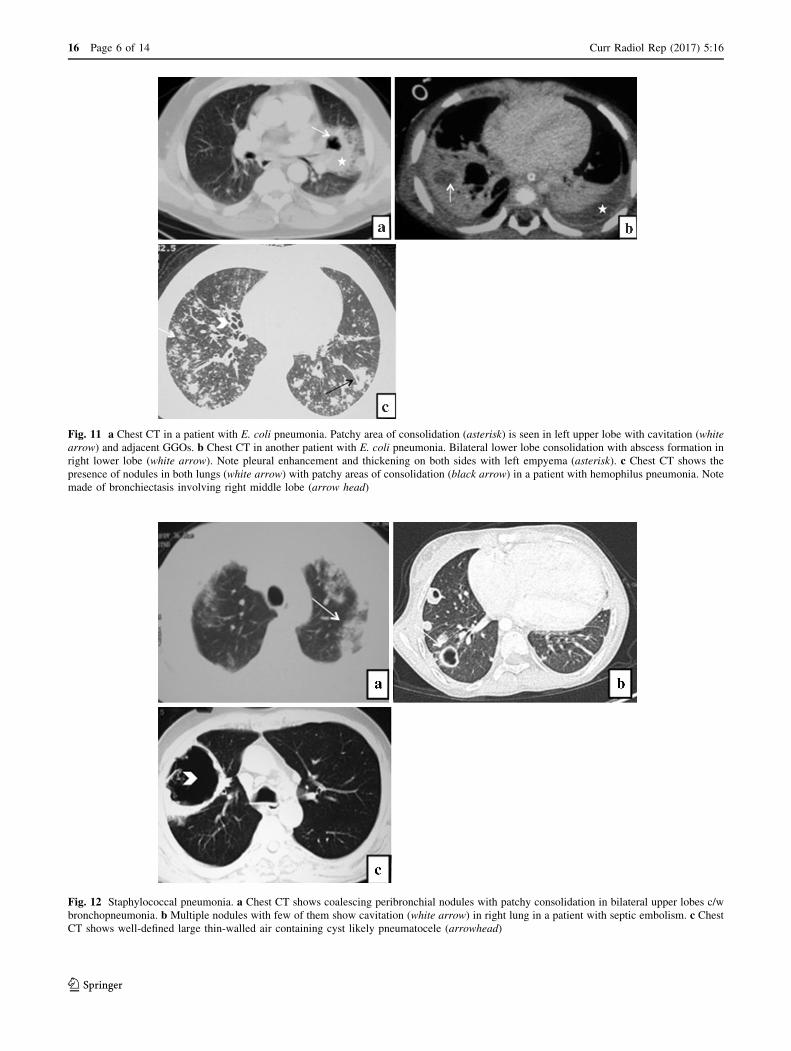
Fig. 11 a Chest CT in a patient with E. coli pneumonia. Patchy area of consolidation (asterisk) is seen in left upper lobe with cavitation (white
arrow) and adjacent GGOs. b Chest CT in another patient with E. coli pneumonia. Bilateral lower lobe consolidation with abscess formation in
right lower lobe (white arrow). Note pleural enhancement and thickening on both sides with left empyema (asterisk). c Chest CT shows the
presence of nodules in both lungs (white arrow) with patchy areas of consolidation (black arrow) in a patient with hemophilus pneumonia. Note
made of bronchiectasis involving right middle lobe (arrow head)
Fig. 12 Staphylococcal pneumonia. a Chest CT shows coalescing peribronchial nodules with patchy consolidation in bilateral upper lobes c/w
bronchopneumonia. b Multiple nodules with few of them show cavitation (white arrow) in right lung in a patient with septic embolism. c ChestCT shows well-defined large thin-walled air containing cyst likely pneumatocele (arrowhead)
16 Page 6 of 14 Curr Radiol Rep (2017) 5:16
123
ways thus producing air bronchogram sign (Fig. 1). Most
common causes of lobar pneumonia include Streptococcus
pneumonia, Chlamydia pneumophila, Mycoplasma pneu-
monia and Klebsiella pneumonia [6].
Bronchopneumonia/Lobular Pneumonia
In lobular pneumonia, the causative organism directly
attacks the peripheral airways damaging the walls of ter-
minal and respiratory bronchioles causing necrosis of walls
leading on to bronchiolitis and bronchitis which further
cause secretion of fluid and inflammatory cells and later on
involvement of parenchyma [6]. Radiologically, it is seen
as patchy centrilobular or peribronchial nodules which later
on cause dense consolidation (Fig. 2). Most common
causes of bronchopneumonia are Staphylococcus aureus
and Pseudomonas aeruginosa. Sometimes this pattern of
involvement can be seen with Hemophilus influenzae,
Mycoplasma pneumonia, and Mycobacterium tuberculosis.
Interstitial Pneumonia
Interstitial pneumonia is secondary to an infectious agent
that damages the ciliated epithelial cells and bronchial
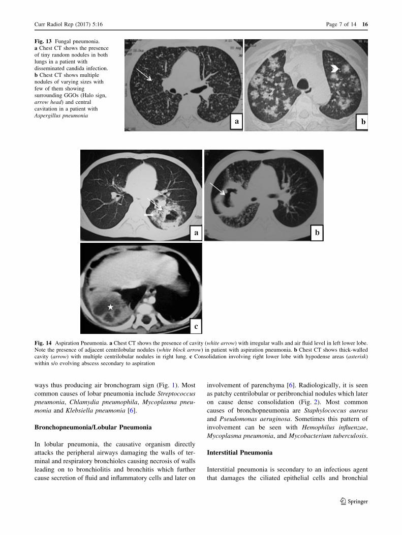
Fig. 14 Aspiration Pneumonia. a Chest CT shows the presence of cavity (white arrow) with irregular walls and air fluid level in left lower lobe.
Note the presence of adjacent centrilobular nodules (white block arrow) in patient with aspiration pneumonia. b Chest CT shows thick-walled
cavity (arrow) with multiple centrilobular nodules in right lung. c Consolidation involving right lower lobe with hypodense areas (asterisk)
within s/o evolving abscess secondary to aspiration
Fig. 13 Fungal pneumonia.
a Chest CT shows the presence
of tiny random nodules in both
lungs in a patient with
disseminated candida infection.
b Chest CT shows multiple
nodules of varying sizes with
few of them showing
surrounding GGOs (Halo sign,
arrow head) and central
cavitation in a patient with
Aspergillus pneumonia
Curr Radiol Rep (2017) 5:16 Page 7 of 14 16
123

mucous gland cells due to which edema and lymphocytic
cellular infiltration occurs. This results in alveolar infil-
trates and interstitial septal thickening. Imaging findings
include ground glass opacities (GGOs), linear reticular or
reticulonodular, and random nodules or patchy consolida-
tions (Fig. 3). In addition to viral pneumonias, Myco-
plasma pneumonia and Chlamydia are the most common
pathogens causing interstitial pneumonia, together they are
called as atypical pneumonias [6].
Nodular Predominant Pattern
This unique pattern is secondary to hematogenous spread
of pathogens or granulomata formation. Most
Fig. 15 Pneumocystis jiroveci
Pneumonia. a, b Chest CT
shows bilateral interstitial
pattern of pneumonia with
GGOs (arrow in a and b) andfew cysts (block white arrow in
a). c Chest CT in a different
patient shows diffuse GGO’s
(asterisk) in both lower lobes
with few ill-defined
centrilobular nodules (arrow) in
a post-renal transplant patient
(combined cytomegalovirus and
P. jiroveci infection)
Fig. 16 a Semi-invasive
aspergillosis in an
immumocompromised patient,
chest CT shows cavity with
eccentric soft tissue (asterisk)
and surrounding GGOs (black
arrow). GGOs represent
hemorrhage secondary to
vascular invasion. b Airway
Invasive Aspergillosis, chest CT
shows coalescing areas of
consolidation with GGOs in
right lung (black block arrow in
b). c Disseminated
Histoplasmosis in a post-renal
transplant patient. Chest CT
shows areas of consolidation in
right upper lobe (asterisk)
16 Page 8 of 14 Curr Radiol Rep (2017) 5:16
123
commonly encountered nodules are secondary to septic
embolism, tuberculosis, or fungal infections and rarely
viral infection (for example, Varicella zoster pneumonia).
Random nodules are seen which do not respect any
segmental boundaries or bronchovascular pattern [6, 7]
(Fig. 4).
Clinicoradiological Classification of Pneumonia
Community-Acquired Pneumonia
Organisms most commonly causing CAP pneumonia in
previously healthy patients include Gram-positive bacteria
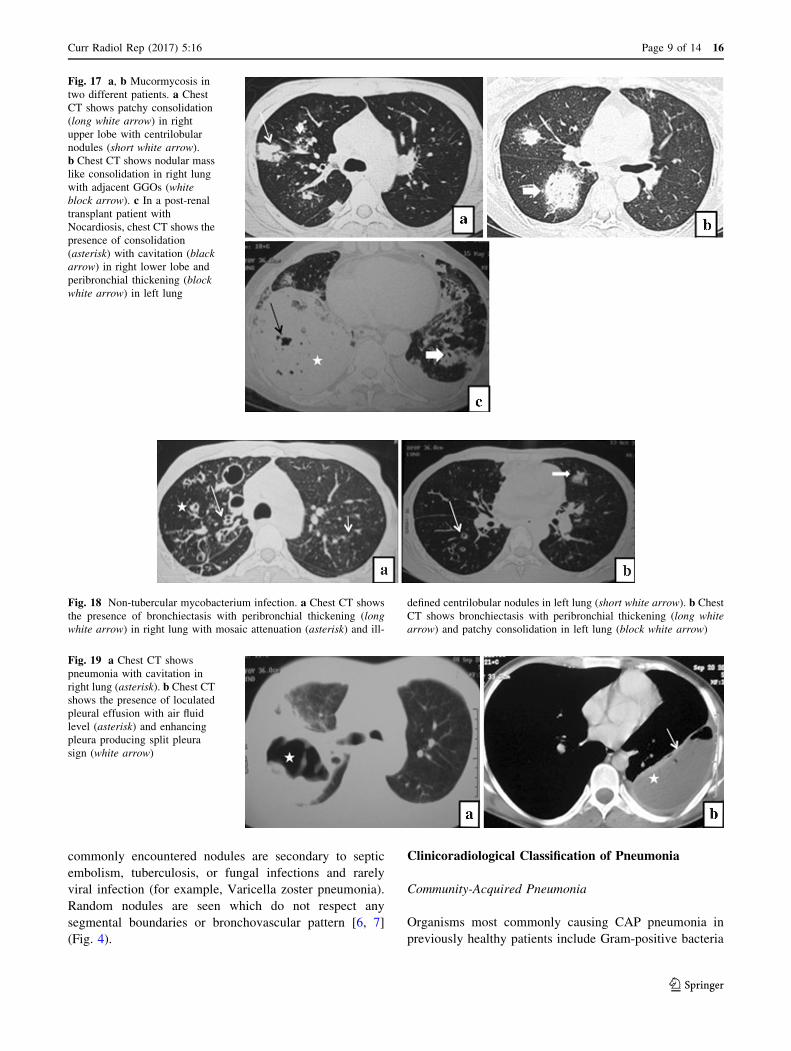
Fig. 19 a Chest CT shows
pneumonia with cavitation in
right lung (asterisk). b Chest CT
shows the presence of loculated
pleural effusion with air fluid
level (asterisk) and enhancing
pleura producing split pleura
sign (white arrow)
Fig. 17 a, b Mucormycosis in
two different patients. a Chest
CT shows patchy consolidation
(long white arrow) in right
upper lobe with centrilobular
nodules (short white arrow).
b Chest CT shows nodular mass
like consolidation in right lung
with adjacent GGOs (white
block arrow). c In a post-renal
transplant patient with
Nocardiosis, chest CT shows the
presence of consolidation
(asterisk) with cavitation (black
arrow) in right lower lobe and
peribronchial thickening (block
white arrow) in left lung
Fig. 18 Non-tubercular mycobacterium infection. a Chest CT shows
the presence of bronchiectasis with peribronchial thickening (long
white arrow) in right lung with mosaic attenuation (asterisk) and ill-
defined centrilobular nodules in left lung (short white arrow). b Chest
CT shows bronchiectasis with peribronchial thickening (long white
arrow) and patchy consolidation in left lung (block white arrow)
Curr Radiol Rep (2017) 5:16 Page 9 of 14 16
123
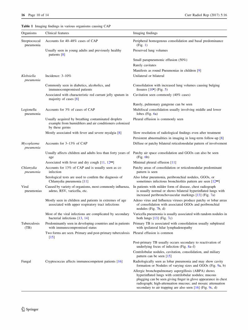
Table 1 Imaging findings in various organisms causing CAP
Organisms Clinical features Imaging findings
Streptococcal
pneumonia
Accounts for 40–48% cases of CAP Peripheral homogenous consolidation and basal predominance
(Fig. 1)
Usually seen in young adults and previously healthy
patients [8]
Preserved lung volumes
Small parapneumonic effusion (50%)
Rarely cavitates
Manifests as round Pneumonias in children [9]
Klebsiella
pneumonia
Incidence: 3–10% Unilateral or bilateral
Commonly seen in diabetics, alcoholics, and
immunocompromised patients
Consolidation with increased lung volumes causing bulging
fissures [10•] (Fig. 5)
Associated with characteristic red currant jelly sputum in
majority of cases [8]
Cavitation seen commonly (40% cases)
Rarely, pulmonary gangrene can be seen
Legionella
pneumonia
Accounts for 3% of cases of CAP Multifocal consolidation usually involving middle and lower
lobes (Fig. 6a)
Usually acquired by breathing contaminated droplets
example from humidifiers and air conditioners colonized
by these germs
Pleural effusion is commonly seen
Mostly associated with fever and severe myalgia [8] Slow resolution of radiological findings even after treatment
Persistent abnormalities in imaging in long-term follow-up [8]
Mycoplasma
pneumonia
Accounts for 3–13% of CAP Diffuse or patchy bilateral reticulonodular pattern of involvement
Usually affects children and adults less than forty years of
age
Patchy air space consolidation and GGOs can also be seen
(Fig. 6b)
Associated with fever and dry cough [11, 12••] Minimal pleural effusion [11]
Chlamydia
pneumonia
Accounts for 13% of CAP and is usually seen as co
infection
Patchy areas of consolidation or reticulonodular predominant
pattern is seen
Serological tests are used to confirm the diagnosis of
Chlamydia pneumonia [11]
Also lobar pneumonia, peribronchial nodules, GGOs, or
sometimes infectious bronchiolitis pattern are seen [12••]
Viral
pneumonias
Caused by variety of organisms, most commonly influenza,
adeno, RSV, varicella, etc.
In patients with milder form of disease, chest radiograph
is usually normal or shows bilateral hyperinflated lungs with
increased peribronchovascular markings [13] (Fig. 7a)
Mostly seen in children and patients in extremes of age
associated with upper respiratory tract infections
Adeno virus and Influenza viruses produce patchy or lobar areas
of consolidation with associated GGOs and peribronchial
nodules (Fig. 7b, d)
Most of the viral infections are complicated by secondary
bacterial infections [13, 14]
Varicella pneumonia is usually associated with random nodules in
both lungs [13] (Fig. 7c)
Tuberculosis
(TB)
Predominantly seen in developing countries and in patients
with immunocompromised status
Primary TB is associated with consolidation usually subpleural
with ipsilateral hilar lymphadenopathy
Two forms are seen. Primary and post-primary tuberculosis
[15]
Pleural effusion is common
Post-primary TB usually occurs secondary to reactivation of
underlying focus of infection (Fig. 8a–f)
Centrilobular nodules, cavitation, consolidation, and miliary
pattern can be seen [15]
Fungal Cryptococcus affects immunocompetent patients [16] Radiologically seen as lobar pneumonia and may show cavity
formation or Nodules of varying sizes and GGOs (Fig. 9a, b)
Allergic bronchopulmonary aspergillosis (ABPA) shows
hyperinflated lungs with centrilobular nodules; mucous
plugging can be seen giving finger in glove appearance in chest
radiograph; high-attenuation mucous; and mosaic attenuation
secondary to air trapping are also seen [16] (Fig. 9c, d)
16 Page 10 of 14 Curr Radiol Rep (2017) 5:16
123
such as Streptococcus pneumonia and atypical bacteria such
asMycoplasma pneumoniae andLegionella pneumophila. In
elderly patients with compromised immune status, Staphy-
lococcus, Gram-negative bacilli and Streptococcus are
responsible for majority of cases [4, 8]. Streptococcus
pneumonia is themost common cause of CAP accounting for
*40% of cases [9]. CAP is mostly associated with mild
parapneumonic effusion. Most commonly encountered
imaging findings in various organisms causing community-
acquired pneumonia are given in Table 1.
Nosocomial Pneumonia/Hospital-Acquired Pneumonia
Nosocomial pneumonia (NP) or hospital-acquired pneu-
monia is defined as pneumonia occurring 48 h after
hospital admission, excluding any infection that is incu-
bating at the time of hospital admission. NP also
includes pneumonia which occurs within 48 h after dis-
charge from the hospital [17]. It is divided into two types as
ventilator-associated pneumonia (VAP) and pneumonia in
non-ventilated patients. Patients on ventilator have
increased risk of acquiring pneumonia due to favorable
condition and also have higher mortality rates [18].
Immune status of the patient, extremes of age, severity of
comorbid conditions, and longer hospital stay are risk
factors for NP. Aerobic Gram-negative bacilli like
Escherichia coli and P. aeruginosa, Staphylococcus aur-
eus, and Streptococcus pneumonia are common etiological
organisms. Polymicrobial infections are common. In VAP,
if the initial period is within 5 days of ventilation,
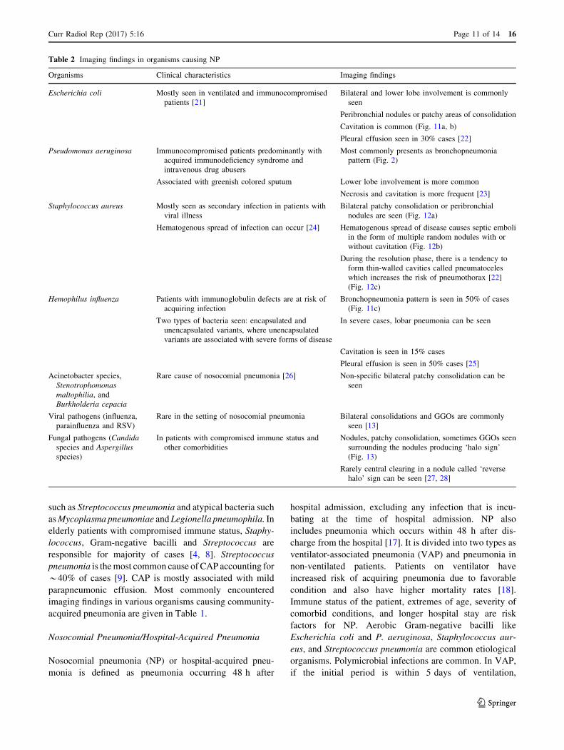
Table 2 Imaging findings in organisms causing NP
Organisms Clinical characteristics Imaging findings
Escherichia coli Mostly seen in ventilated and immunocompromised
patients [21]
Bilateral and lower lobe involvement is commonly
seen
Peribronchial nodules or patchy areas of consolidation
Cavitation is common (Fig. 11a, b)
Pleural effusion seen in 30% cases [22]
Pseudomonas aeruginosa Immunocompromised patients predominantly with
acquired immunodeficiency syndrome and
intravenous drug abusers
Most commonly presents as bronchopneumonia
pattern (Fig. 2)
Associated with greenish colored sputum Lower lobe involvement is more common
Necrosis and cavitation is more frequent [23]
Staphylococcus aureus Mostly seen as secondary infection in patients with
viral illness
Bilateral patchy consolidation or peribronchial
nodules are seen (Fig. 12a)
Hematogenous spread of infection can occur [24] Hematogenous spread of disease causes septic emboli
in the form of multiple random nodules with or
without cavitation (Fig. 12b)
During the resolution phase, there is a tendency to
form thin-walled cavities called pneumatoceles
which increases the risk of pneumothorax [22]
(Fig. 12c)
Hemophilus influenza Patients with immunoglobulin defects are at risk of
acquiring infection
Bronchopneumonia pattern is seen in 50% of cases
(Fig. 11c)
Two types of bacteria seen: encapsulated and
unencapsulated variants, where unencapsulated
variants are associated with severe forms of disease
In severe cases, lobar pneumonia can be seen
Cavitation is seen in 15% cases
Pleural effusion is seen in 50% cases [25]
Acinetobacter species,
Stenotrophomonas
maltophilia, and
Burkholderia cepacia
Rare cause of nosocomial pneumonia [26] Non-specific bilateral patchy consolidation can be
seen
Viral pathogens (influenza,
parainfluenza and RSV)
Rare in the setting of nosocomial pneumonia Bilateral consolidations and GGOs are commonly
seen [13]
Fungal pathogens (Candida
species and Aspergillus
species)
In patients with compromised immune status and
other comorbidities
Nodules, patchy consolidation, sometimes GGOs seen
surrounding the nodules producing ‘halo sign’
(Fig. 13)
Rarely central clearing in a nodule called ‘reverse
halo’ sign can be seen [27, 28]
Curr Radiol Rep (2017) 5:16 Page 11 of 14 16
123
etiological agents are Streptococcus pneumonia, He-
mophilus influenza, and Moraxella catarrhalis. Late onset
VAP (after 5 days) is usually due to aerobic Gram-negative
rods and methicillin-resistant Staphylococcus aureus [18].
Role of radiology in NP is to diagnose and in follow-up. It
is difficult to radiologically identify the etiological agent as
most causative organisms show multilobar consolidation as
predominant finding. Imaging findings may also mimic
acute respiratory distress syndrome [19, 20] (Fig. 10).
Imaging findings are given in Table 2.
Aspiration Pneumonia
Aspiration is defined as intake of solid or liquid materials
into airways and lungs. Aspiration pneumonia can either be
due to microorganisms or due to chemicals for example
gastric acidic contents [29]. Common pathogens causing
aspiration are organisms colonizing the oropharynx and
stomach. Gram-negative anaerobic organisms are most
common pathogens. Aspiration pneumonia can be either
acute or chronic. In acute aspiration, lobar or segmental
pneumonia, bronchopneumonia, lung abscess, and
empyema are seen (Fig. 14). Chronic aspiration pneumonia
is usually due to repeated aspiration and is seen as focal
centrilobular nodules or peribronchial thickening [30]. The
posterior segment of the upper lobes and the superior
segment of the lower lobes are commonly affected.
Infections in Immunocompromised Patients
In the current era, with the increase in prevalence of
patients with diabetes mellitus, post-transplant immuno-
suppression and patients with acquired and congenital
immune deficiency disorders, there is increase in the
infections with atypical organisms [31–33]. Most common
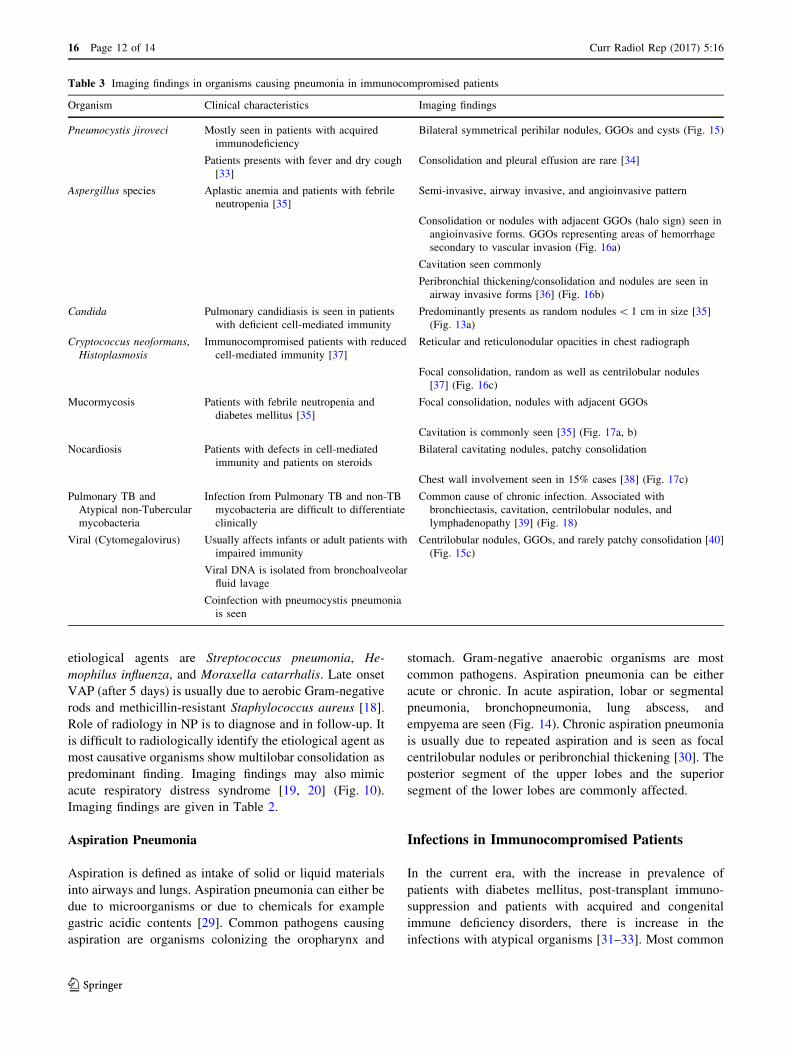
Table 3 Imaging findings in organisms causing pneumonia in immunocompromised patients
Organism Clinical characteristics Imaging findings
Pneumocystis jiroveci Mostly seen in patients with acquired
immunodeficiency
Bilateral symmetrical perihilar nodules, GGOs and cysts (Fig. 15)
Patients presents with fever and dry cough
[33]
Consolidation and pleural effusion are rare [34]
Aspergillus species Aplastic anemia and patients with febrile
neutropenia [35]
Semi-invasive, airway invasive, and angioinvasive pattern
Consolidation or nodules with adjacent GGOs (halo sign) seen in
angioinvasive forms. GGOs representing areas of hemorrhage
secondary to vascular invasion (Fig. 16a)
Cavitation seen commonly
Peribronchial thickening/consolidation and nodules are seen in
airway invasive forms [36] (Fig. 16b)
Candida Pulmonary candidiasis is seen in patients
with deficient cell-mediated immunity
Predominantly presents as random nodules\ 1 cm in size [35]
(Fig. 13a)
Cryptococcus neoformans,
Histoplasmosis
Immunocompromised patients with reduced
cell-mediated immunity [37]
Reticular and reticulonodular opacities in chest radiograph
Focal consolidation, random as well as centrilobular nodules
[37] (Fig. 16c)
Mucormycosis Patients with febrile neutropenia and
diabetes mellitus [35]
Focal consolidation, nodules with adjacent GGOs
Cavitation is commonly seen [35] (Fig. 17a, b)
Nocardiosis Patients with defects in cell-mediated
immunity and patients on steroids
Bilateral cavitating nodules, patchy consolidation
Chest wall involvement seen in 15% cases [38] (Fig. 17c)
Pulmonary TB and
Atypical non-Tubercular
mycobacteria
Infection from Pulmonary TB and non-TB
mycobacteria are difficult to differentiate
clinically
Common cause of chronic infection. Associated with
bronchiectasis, cavitation, centrilobular nodules, and
lymphadenopathy [39] (Fig. 18)
Viral (Cytomegalovirus) Usually affects infants or adult patients with
impaired immunity
Centrilobular nodules, GGOs, and rarely patchy consolidation [40]
(Fig. 15c)
Viral DNA is isolated from bronchoalveolar
fluid lavage
Coinfection with pneumocystis pneumonia
is seen
16 Page 12 of 14 Curr Radiol Rep (2017) 5:16
123
pathogens causing infection includes fungal (Pneumocystis
jiroveci, Aspergillus, Mucormycosis, Histoplasmosis,
Candida, and Cryptococcus), bacterial (Pseudomonas,
Streptococcal pneumonia, Staphylococcal, Nocardiosis,
Legionella, Rhodococcus etc.), and viral (Cytomegalovirus,
Herpes simplex, and influenza). Imaging findings in dif-
ferent types of infection is given in Table 3.
Complications of Pneumonia
Complications after pulmonary infections are common in
immunosuppresed patients. The most commonly encoun-
tered complications are pleural effusion, empyema, cavi-
tation, bronchopleural fistula, hydropneumothorax, and
chest wall involvement. Reactive pleural effusion is com-
monly associated with streptococcal and Gram-negative
pneumonias. Empyema is usually seen in pneumonia sec-
ondary to Gram-negative organisms and aspiration pneu-
monia (Fig. 19). Cavitation is commonly seen in anaerobic
infection, TB, and in fungal infections. Chest wall
involvement in the form of rib erosions and abscess for-
mation is seen in TB, Nocardiosis, and in actinomycosis
[38]. Pneumatoceles leading on to pneumothorax is com-
monly seen in staphylococcal pneumonia. Rarely pul-
monary gangrene can occur in severe cases of
Staphylococcal and Klebsiella pneumonia [41].
Conclusion
Imaging plays an important role in the management of
pneumonia. Knowing the clinical background and corre-
lation with imaging findings may help in early detection of
pathogen and direct the physician towards appropriate
management. Imaging also helps in follow-up of patients to
look for response to therapy. Imaging can identify the
complications of pneumonia. In addition, imaging partic-
ularly cross-sectional, helps in ruling out other lung dis-
eases which may mimic pneumonia.
Author Contribution Imaging: Mandeep Garg. Literature search
and Manuscript preparation: Nidhi Prabhakar,Kiruthika P. Manuscript
editing: Mandeep Garg, Ajay Gulati, Ritesh Agarwal, Ashutosh
Aggarwal. Final manuscript editing: Niranjan Khandelwal.
Compliance with Ethical Standards
Conflict of Interest Mandeep Garg, Nidhi Prabhakar, P. Kiruthika,
Ritesh Agarwal, Ashutosh Aggarwal, Ajay Gulati, and Niranjan
Khandelwal each declare no potential conflicts of interest.
Human and Animal Rights and Informed Consent This article
does not contain any studies with human or animal subjects per-
formed by any of the authors.
References
Recently published papers of particular interest have been
highlighted as:• Of importance•• Of major importance
1. Bhalla M, McLoud TC. Pulmonary infections in the normal host.
In: McLoud TC, editor. Thoracic radiology, the requisites. St
Louis: Mosby; 1998.
2. Thanos L, Galani P, Mylona S, Pomoni M, Mpatakis N. Percu-
taneous CT-guided core needle biopsy versus fine needle aspi-
ration in diagnosing pneumonia and mimics of pneumonia.
Cardiovasc Intervent Radiol. 2004;24:329–34.
3. Tarver R, Teague S, Heitkamp D, Conces D Jr. Radiology of
community-acquired pneumonia. Radiol Clin N Am.
2005;43:497–512.
4. American Thoracic Society. Guidelines for the management of
adults with community-acquired pneumonia. Am J Respir Crit
Care Med. 2001;163(7):1730–54.
5. Muller NL, Franquet T. Lee KS. In: McAllister L, editor. Imaging
of pulmonary infections. Philadelphia: Wolters Kluwer/Lippon-
cott Williams & Wilkins; 2007.
6. Heitzman ER. The radiological diagnosis of pneumonia in the
adult: a commentary. Semin Roentgenol. 1989;24(4):212–7.
7. Fraser RS, Pare JAP, Fraser RG, Pare PD. Infectious disease of
the lungs. Synopsis of diseases of the chest. 2nd ed. Philadelphia:
W.B. Saunders Company; 1994. p. 287–391.
8. Tanaka N, Matsumoto T, Kuramitsu T, Nakaki H, Ito K, Uchi-
sako H, Miura G, Matsunaga N, Yamakawa K. High resolution
CT findings in community-acquired pneumonia. J Comput Assist
Tomogr. 1996;20:600–8.
9. Kantror HG. The many radiologic faces of pneumococcal pneu-
monia. AJR Am J Roentgenol. 1981;137:1213–20.
10. • Walker CM, Abbott GF, Greene RE, Shepard JA, Vummidi D,
Digumarthy SR. Imaging pulmonary infection: classic signs and
patterns. AJR Am J Roentgenol. 2014;202(3):479–92. This arti-
cle is relevant as it explicitly explains the common and uncom-
mon signs of pneumonias.
11. Primary Bragg F, Pneumonia Atypical. Am J Public Health.
1944;34:347–57.
12. •• Nambu A, Ozawa K, Kobayashi N, Tago M. Imaging of
community-acquired pneumonia: roles of imaging examinations,
imaging diagnosis of specific pathogens and discrimination from
noninfectious diseases. World J Radiol. 2014;6(10):779–93. This
article is important as it is one of the few recent review articles
on the imaging of pulmonary infections. This article has reviewed
the imaging features of community acquired pneumonias and
included tips on how imaging of community acquired pneumonias
can help in the management of patient.
13. Kim EA, Lee KS, Primack SL, Suh GY, Kwon OJ, Han J. Viral
pneumonias in adults: radiologic and pathologic findings.
Radiographics. 2002;22:137–49.
14. Miller WT Jr, Barbosa E Jr, Mickus TJ, et al. Chest CT imaging
characteristics of viral acute lower respiratory illnesses: a case-
control study. J Comput Assist Tomogr. 2011;35:524–53.
15. Jeong YJ, Lee KS. Pulmonary tuberculosis: up-to-date imaging
and management. AJR Am J Roentgenol. 2008;191:834–44.
Curr Radiol Rep (2017) 5:16 Page 13 of 14 16
123
16. Fox DL, Muller NL. Pulmonary cryptococcosis in immunocom-
petent patients: CT findings in 12 patients. AJR Am J Roentgenol.
2005;185:622–6.
17. Lipchik RJ, Kuzo RS. Nosocomial pneumonia. Radiol Clin N
Am. 1996;34:47–58.
18. Chastre J, Fagon JY. Ventilator-associated pneumonia. Am J
Respir Crit Care Med. 2002;165(7):867–903.
19. Hoffken G, Niederman MS. Nosocomial pneumonia. Chest.
2002;122(6):2183–96.
20. Winer-Muram HT, Steiner RM, Gurney JW, et al. Ventilator
associated pneumonia in patients with adult respiratory distress
syndrome: CT evaluation. Radiology. 1998;208:193–9.
21. Zornoza J, Goldman AM, Wallace S, et al. Radiologic features of
gramnegative pneumonias in the neutropenic patient. Am J
Roentgenol. 1976;127:989–96.
22. Franquet T. Imaging of pneumonia: trends and algorithms. Eur
Respir J. 2001;18(1):196–208.
23. Shah RM, Wechsler R, Salazar AM, Spirn PW. Spectrum of CT
findings in nosocomial pseudomonas aeruginosa pneumonia.
J Thorac Imaging. 2002;17:53–7.
24. Macfarlane J, Rose D. Radiographic features of staphylococcal
pneumonia in adults and children. Thorax. 1996;51(5):539–40.
25. Gillis S, Dann EJ, Berkman N, et al. Fatal Haemophilus
influenzae septicemia following bronchoscopy in a splenec-
tomized patient. Chest. 1993;104(5):1607–9.
26. Hospital-acquired pneumonia in adults: diagnosis, assessment of
severity, initial antimicrobial therapy, and preventive strategies.
A consensus statement, American Thoracic Society, November
1995. Am J Respir Crit Care Med. 1996;153:1711–25.
27. Lee YR, Choi YW, Lee KJ, et al. CT halo sign: the spectrum of
pulmonary diseases. Br J Radiol. 2005;78:862–5.
28. Georgiadou SP, Sipsas NV, Marom EM, Kontoyiannis DP. The
diagnostic value of halo and reversed halo signs for invasive
mold infections in compromised hosts. Clin Infect Dis.
2011;52:1144–55.
29. Marik PE. Aspiration pneumonitis and aspiration pneumonia.
N Engl J Med. 2001;344(9):665–71.
30. Marom EM, McAdams HP, Erasmus JJ, Goodman PC. The many
faces of pulmonary aspiration. AJR Am J Roentgenol.
1999;172:121–8.
31. Baughman RP. The lung in the immunocompromised patient.
Infectious complications part 1. Respiration. 1999;66(2):95e109.
32. Allen CM, Al-Jahdali HH, Irion KL, et al. Imaging lung mani-
festations of HIV/AIDS. Ann Thorac Med. 2010;5(4):201–16.
33. Rano A, Agustı C, Sibila O, et al. Pulmonary infections in non-
HIV immunocompromised patients. Curr Opin Pulm Med.
2005;11(3):213–7.
34. Vogel MN, Vatlach M, Weissgerber P, Goeppert B, Claussen CD,
Hetzel J, Horger M. HRCT-features of Pneumocystis jiroveci
pneumonia and their evolution before and after treatment in non-
HIV immunocompromised patients. Eur J Radiol.
2011;81:1315–20.
35. Franquet T, Gimenez A, Hidalgo A. Imaging of opportunistic
fungal infections in immunocompromised patient. Eur J Radiol.
2004;51(2):130–8.
36. Logan PM, Primack SL, Miller RR, et al. Invasive aspergillosis of
the airways: radiographic, CT and pathologic findings. Radiol-
ogy. 1994;193:383–8.
37. Cameron ML, Barlett JA, Gallis HA, et al. Manifestations of
pulmonary cryptococcosis in patients with acquired immunode-
ficiency syndrome. Rev Infect Dis. 1991;13:64–7.
38. Husain S, McCurry K, Dauber J, et al. Nocardia infection in lung
transplant recipients. J Heart Lung Transpl. 2002;21:354.
39. Miller WT Jr. Spectrum of pulmonary nontuberculous
mycobacterial infection. Radiology. 1994;191:343–50.
40. Franquet T. Imaging of pulmonary viral pneumonia. Radiology.
2011;260:18–39.
41. Penner C, Maycher B, Long R. Pulmonary gangrene. A compli-
cation of bacterial pneumonia. Chest. 1994;105:567–73.
16 Page 14 of 14 Curr Radiol Rep (2017) 5:16
123