institute for engineering in medicine - iem iem annual report.pdf · the institute for engineering...
TRANSCRIPT

Institute for Engineering in Medicine
INSTITUTE FOR ENGINEERING IN MEDICINE providing engineering solutions to medicine providing engineering solutions to medicine
2013-2014 IEM ANNUAL REPORT

2
Table of Contents
Director’s Message . . . . . . . . . . . . . . . . . . . . 3
Institute for Engineering in Medicine (IEM)
About IEM/Organization . . . . . . . . . . . . . . . 4Mission and Vision . . . . . . . . . . . . . . . . . . . . . 5Executive Committee . . . . . . . . . . . . . . . . . . 6Scientific Advisory Board . . . . . . . . . . . . . . 8Research Themes . . . . . . . . . . . . . . . . . . . . . 9
IEM Members’ Research Highlights . . . . 11
IEM Members’ New Companies . . . . . . . .21
IEM Research Centers
Medical Devices Center . . . . . . . . . . . . . . . 22Center for Neuroengineering . . . . . . . . . . 23Biopreservation Core Resource . . . . . . . . 24Visible Heart® Laboratory . . . . . . . . . . . . . 25
IEM Award Recipients . . . . . . . . . . . . . . . . 26
Education and Outreach
Educational Initiatives . . . . . . . . . . . . . . . . . 28Annual Short Courses . . . . . . . . . . . . . . . . . 29Conferences and Symposia . . . . . . . . . . . . 30Fellowship Programs . . . . . . . . . . . . . . . . . . 34
IEM Funding
2013-2014 Funding Programs . . . . . . . . . . . 36Funding Analysis . . . . . . . . . . . . . . . . . . . . . . 37
IEM Members
Membership Breakdown . . . . . . . . . . . . . . 38
Contact Information . . . . . . . . . . . . . . . . . 39

3
It has been another exciting year for the Institute for Engineering in Medicine (IEM). IEM is an interdisciplinary research organization aimed at advancing engineering solutions to medical and health problems by fostering collaborations between engineering and biomedical faculty at the University of Minnesota, and between University faculty and industrial colleagues. Challenges to medicine and healthcare necessitate interdisciplinary and multidisciplinary collaborations. IEM’s membership consists of about 159 University faculty representing 44 academic departments. These IEM members are tackling challenges to biomedical research in thematic areas including Cardiovascular Engineering, Neuroengineering, Cellular and Molecular Bioengineering, Medical and Biological Imaging, and Medical Devices. Through organized interdisciplinary collaborative research, as well as outreach to industry and the public, IEM strives for excellence in interfacing engineering with medicine—seeking innovative engineering solutions to tomorrow’s medicine and healthcare.
IEM has organized a number of activities throughout 2013-2014 to accomplish its mission. IEM seed group grants have funded multidisciplinary faculty groups, many of which led to monthly group meetings discussing research collaborations, funded major federal grants or development of federal center/program grant proposals. IEM has also funded individual investigators from biomedical and engineering disciplines in order to explore collaborative research. These modest investment has resulted in significant returns to external funding representing a 10.5:1 ratio of investment. IEM members continuously publish cutting-edge biomedical research in leading journals. The IEM Annual Conference and Retreat has become a highly interactive platform to facilitate developing collaborations between engineers and biomedical scientists and clinicians. IEM has also organized or sponsored well attended conferences and symposia including the popular Design of Medical Devices Conference and Minnesota Neuromodulation Symposium, which attract a significant number of participants from academia, industry and government. In addition, IEM has co-sponsored events in Washington, D.C. to facilitate public debates at the national level on BRAIN Grand Challenges. Through IEM conferences, symposia, and short courses, there has been enhanced interactions and collaborations between IEM members and the medical device industry.
In this annual report, you will find information about the theme programs and affiliated research centers of IEM, highlights of IEM members’ research, IEM education and outreach activities. We look forward to your continued interest and support. Please contact me at [email protected] with ideas that can improve what we do. Together, we will make a difference in tomorrow’s medicine and healthcare through engineering innovation.
Sincerely,
Bin HeDirector, Institute for Engineering in Medicine
“The future of medicine and healthcare requires
collaborative eff orts that address grand challenges
in employing innovative engineering solutions to
eff ectively diagnose, treat, and prevent diseases.
The Institute for Engineering in Medicine plays a
signifi cant role to promote collaborations between
engineers and biomedical scientists.”
Director’s Message

4
Institute for Engineering in MedicineABOUT�IEM��The Institute for Engineering in Medicine (IEM) is an interdisciplinary research organization that strengthens collaborations between the disciplines of engineering and medicine at the University of Minnesota while providing solutions and partnership opportunities to the medical device industry. IEM members include about 159 affiliated researchers representing 44 departments across the campus. Utilizing this extensive base of knowledge not only allows us to engage the best talents at the University, but also drives innovation in finding solutions for challenges that are important to the researchers, clinicians, patients, and the industry.
IEM focuses around five core research themes, and features four affiliated research centers and laboratories to provide faculty, students, and industrial partners with world class resources for conducting interdisciplinary research. Additionally, we function as a gateway into the University of Minnesota and offer a contact point for members of the public sector looking for partnerships on engineering solutions to medical and health issues.
ORGANIZATION
ScientificAdvisory
Board
Institute for Engineering in Medicine
IndustryAdvisory
Board
AcademicHealthCenter
Cardiovascular Engineering
Medical & Biological Imaging
Medical Devices
Center for Neuroengineering
Visible Heart® Laboratory
EXTERNAL
ResearchCenters
ResearchThemes
Medical Devices Center
Biopreservation Core ResourceCellular & Molecular Bioengineering
Neuroengineering
UNIVERSITY OF MINNESOTA
College ofScience &
Engineering

55
MISSIONThe Institute for Engineering in Medicine strives to be a world leader in the creation and innovative application of engineering solutions to medical and health problems.
VISION The future of health care necessitates that patients be cared for in a high-quality, safe, and effective manner at a price that the nation can manage. As the costs of health care increase and the population ages, innovative and transformative approaches are needed. Important aspects for future health care will be the creation and application of engineering technologies that will provide these new approaches with the ability to manage various disorders in a faster, safer, and less expensive manner, and with fewer side effects. At present, it is crucial to explore comprehensive programs that combine engineering advances with disease and medical knowledge, and to apply these to the diagnosis, treatment, and prevention of major diseases that affect our society.
The University of Minnesota’s Institute for Engineering in Medicine (IEM) integrates expertise in engineering technology, basic sciences, biological and medical sciences, and the social aspects of health care. This exceptional organization addresses the future needs of health care and develops and applies technology to meet these needs and improve health for patients. The position of IEM within the University—along with the University’s strong relationship to the Minnesota state government, support of the public, and presence amidst the world’s major medical device industry—provides an outstanding environment for the application of innovation into clinical practice and patient care. IEM is positioned to identify engineering solutions to a wide variety of health issues and has the unique ability to take the innovations through the entire process from basic scientific discovery, to translational research, initial patient care research, commercialization, and widespread application.
The Institute for Engineering in Medicine is truly where medicine meets technology for tomorrow’s innovation.

6
Institute for Engineering in Medicine
JOHN�C��BISCHOF��PH�D��
IEM Associate Director for Development; Distinguished McKnight University Professor of Mechanical and Biomedical Engineering and Urology; Carl and Janet Kuhrmeyer Chair in Mechanical Engineering
“The Institute for Engineering in Medicine is a unique structure that brings together basic, translational, and
clinical biomedical engineers and scientists at the University of Minnesota. I have benefi ted intellectually from the
diverse and rich group of collaborators within IEM membership. IEM helps increase the societal impact of existing
and helps to build new biomedical research groups at the University of Minnesota.”
WEI�CHEN��PH�D��
Professor of Radiology
“The Institute for Engineering in Medicine enhances research opportunities and broadens collaborations with
other researchers with diff erent backgrounds.”
EXECUTIVE�COMMITTEEUnder the guidance of Director Bin He, Ph.D., the Institute for Engineering in Medicine’s Executive Committee is composed of eminent University of Minnesota faculty from both the Academic Health Center and College of Science and Engineering. Their passion for innovation and growth puts them in a unique position to advocate the mission of IEM while functioning in areas such as research and development, education, clinical and industry needs, grant funding, and technology transfer. The Executive Committee members attend monthly operational meetings, chair IEM research themes, promote collaborative efforts between disciplines, and champion the needs of special biomedical research challenges. Their dedication produces immeasurable benefits and allows the Institute for Engineering in Medicine to thrive.
PAUL�A��IAIZZO��PH�D��
IEM Associate Director for Education and Outreach; Director of the Visible Heart® Laboratory; Professor of Surgery, Integrative Biology, Physiology, and the Carlson School of Management; Director of the Malignant Hyperthermia Muscle Biopsy Center
“The Institute for Engineering in Medicine continues to promote device development through interfacing
clinicians, basic scientists, engineers, and industry which serves to carry on the rich tradition of innovative
collaborations at the University of Minnesota such as the one between C. Walton Lillehei and Earl Bakken.”
BIN�HE��PH�D�
IEM Director; Medtronic-Bakken Endowed Chair for Engineering in Medicine; Distinguished McKnight University Professor of Biomedical Engineering
“The future of medicine and healthcare requires collaborative eff orts that address grand challenges in employing
innovative engineering solutions to eff ectively diagnose, treat, and prevent diseases. The Institute for Engineering in
Medicine plays a signifi cant role to promote collaborations between engineers and biomedical scientists. I enjoy the
opportunity to collaborate with colleagues from other disciplines and to help enhance such opportunities for others.”
ARTHUR�G��ERDMAN��PH�D��
Director of the Medical Devices Center; Richard C. Jordan Professor and Morse Alumni Distinguished Teaching Professor of Mechanical Engineering
“The Institute for Engineering in Medicine provides a home base for the Medical Devices Center and a
connection to the Academic Health Center, particularly its Medical School. IEM’s Executive Commi� ee consists of
colleagues who strive to enrich the University’s contribution to improvement in healthcare.”

7
KELVIN�O��LIM��M�D��
Drs. T.J. and Ella M. Arneson Land-Grant Chair in Human Behavior; Professor and Vice Chair of Research, Department of Psychiatry
“Psychiatry is in need of new approaches for clinical assessment and interventions. The Institute for Engineering
in Medicine provides a critical avenue for advancing these approaches.”
DAVID�ODDE��PH�D��
Professor of Biomedical Engineering
“We need to connect genomic and molecular level information to the physical workings of cells in their
environment. The Institute for Engineering in Medicine provides the engineering perspective on the cell and connects
it to medically relevant problems.”
JIAN-PING�WANG��PH�D���
Distinguished McKnight University Professor of Electrical and Computer Engineering
“The Institute for Engineering in Medicine provides an extremely important cross-disciplinary platform for
interaction with other researchers from the Academic Health Center and medical device industry, especially to fully
understand the needs and criteria for disease early detection and drug discovery.”
JIANYI�ZHANG��M�D���PH�D��
Professor of Medicine (Cardiology)
“The Institute for Engineering in Medicine provides leadership in promoting research that crosses academic
disciplines via translation of regenerative biological science, tissue engineering to clinical medicine in order to be� er
solve the critical challenges in healthcare and life sciences.”
ALLISON�HUBEL��PH�D�
Director of the Biopreservation Core Resource; Professor of Mechanical Engineering
“The Institute for Engineering in Medicine facilitates groups of faculty ge� ing together to address areas of
common need/interest. This function results in novel approaches to critical problems in biomedical research.”
KALPNA�GUPTA��PH�D��
Professor of Medicine (Hematology, Oncology, and Transplantation)
“The current needs of research can only be met by interdisciplinary investigations involving the partnership
between basic biology and technology to enhance medically applicable approaches. My own translational research
has utilized this approach successfully. The Institute for Engineering in Medicine utilizes these eff orts to advance
interdisciplinary research between biomedical and bioengineering scientists.”
EXECUTIVE�COMMITTEE��CONT��

8
Institute for Engineering in MedicineSCIENTIFIC�ADVISORY�BOARD
SHU�CHIEN��PH�D��
Director of UCSD Institute of Engineering in Medicine; University Professor of Bioengineering and Medicine at UCSDMember, National Academy of EngineeringMember, National Academy of Science Member, Institute of Medicine, National Academies
Shu Chien is University Professor of Bioengineering & Medicine and Director of California Institute of Bioengineering in the University of California System. At UC San Diego, he is Y.C. Fung Professor of Bioengineering and Director of Institute of Engineering in Medicine at UCSD, where he was Founding Chair of Department of Bioengineering. He is a world leader in molecular, cellular and integrative studies on bioengineering and physiology in health and disease. His current research interests are mechanotransduction in endothelial cells in health and disease, and the role of microenvirnoment in modulating stem cell fate.
ARTHUR�F��KRAMER��PH�D�
Swanlund Chair and Professor of Psychology and Neuroscience at the University of Illinois at Urbana-Champaign; Director of the Beckman Institute for Advanced Science and Technology
Arthur Kramer is the Director of the Beckman Institute for Advanced Science & Technology and the Swanlund Chair and Professor of Psychology and Neuroscience at the University of Illinois. He holds appointments in the Department of Psychology, Neuroscience program, and the Beckman Institute. Professor Kramer’s research projects include topics in Cognitive Psychology, Cognitive Neuroscience, Aging, and Human Factors. A major focus of his lab's recent research is the understanding and enhancement of cognitive and neural plasticity across the lifespan.
RAPHAEL�LEE��M�D���SC�D�
Paul and Allene Russell Professor of Surgery (Plastic); Professor of Medicine (Dermatology) and Organismal Biology and Anatomy at the University of ChicagoMember, National Academy of Engineering
Raphael Lee, a Paul S. and Allene T. Russell Professor at the University of Chicago, holds appointments in Surgery (Plastic), Medicine (Dermatology), Molecular Medicine, Translational Medicine and Organismal Biology & Anatomy (Biomechanics). He directs the Laboratory for Molecular Regeneration at the University. He is also a founder and Chairman of the Board of Directors for Avocet Polymer Technologies, Inc., Renacyte BioMolecular Technologies, Inc., Electrokiinetic Signal Research, and Maroon Biotech, Inc. all of Chicago, Illinois.
P��HUNTER�PECKHAM��PH�D�
Donnell Institute Professor at the Case Western Reserve University, Director of the Functional Electrical Stimulation CenterMember, National Academy of Engineering
P. Hunter Peckham is the Donnell Institute Professor of Biomedical Engineering and Orthopaedics; Distinguished University Professor; Executive Director, Institute for Functional Restoration at Case Western Reserve University; Senior Career Research Scientist and Associate Director of Technology Transfer, Cleveland FES Center of Excellence, in the Department of Veterans Aff airs; and on the Bioscientifi c Staff at Metrohealth Medical Center.

9
CARDIOVASCULAR�ENGINEERINGTheme Co-Chairs: Paul Iaizzo, Ph.D. and Jianyi Zhang, M.D., Ph.D.The major research goal of the Cardiovascular Engineering Theme is to understand and seek solutions to manage cardiovascular diseases, which represent a major public health challenge in the US and developed countries. Multiscale approaches ranging from gene, cell, tissue, and organ levels are being utilized. Additionally, cellular and tissue engineering approaches are being developed to repair cardiovascular systems. Novel imaging and sensing modalities are also being developed to understand the mechanisms of cardiovascular diseases and provide engineering solutions to aid clinical diagnosis and management.
RESEARCH�THEMES Interdisciplinary collaborations are essential among physical science, engineering, and biomedicine. The Institute for Engineering in Medicine (IEM) promotes research collaborations between the Academic Health Center, particularly its Medical School, and College of Science and Engineering, and enhances overall biomedical engineering research at the University of Minnesota. This results in a broader base of knowledge to solve issues in biomedicine and allows us to utilize our best talents at the University.
Research at IEM is focused around five core themes: Cardiovascular Engineering, Neuroengineering, Cellular
and Molecular Bioengineering, Medical and Biological Imaging, and Medical Devices. These themes represent important biomedical engineering research aimed at advancing our understanding of the mechanisms of major diseases affecting human health and developing engineering solutions to various medical and health problems, ranging from diagnosis, treatment, and prevention of diseases through engineering innovation.
NEUROENGINEERINGTheme Co-Chairs: Bin He, Ph.D. and Kelvin Lim, M.D.The goal of the Neuroengineering Theme is to better understand mechanisms of neurological and psychiatric disorders and develop engineering solutions to sense, image, interface, and modulate disorders of the brain and nervous systems. One of the key challenges the theme group is addressing is to discover mechanisms of
and seek solutions to brain disorders at systems level. University of Minnesota faculty has been at the forefront of development of systems neuroengineering, with particular strengths in neural decoding and imaging, neural interfacing and neuromodulation.
Image: Effects of cell therapy in hearts with a severely abnormal heterogeneity of
myocardial bioenergetics. Cover page on Circulation Research, Volume 111, Issue
4, pp. 455-468, by Xiong et al.
Image: Illustration of a novel noninvasive brain-computer interface that can control
the flight of a virtual helicopter in 3-D virtual space of the University of Minnesota
campus from “thoughts” sensed over the scalp of human subjects. Work published
in PLoS ONE (Doud et al, 2011), and featured by ABC News.

10
MEDICAL�AND�BIOLOGICAL�IMAGINGTheme Chair: Wei Chen, Ph.D.The major research goal of the Medical and Biological Imaging theme is to strengthen the critical roles of imaging in support of biomedical research and clinical translation. This goal will be accomplished by several aims: 1) Integrate and share a large variety of state-of-the-art imaging approaches (including MRI, in vivo MRS, PET, CT, ultrasound, optical imaging, cellular and electrophysiological imaging), technology, expertise, facilities, and resource for addressing essential biomedical questions from the cellular to system level; 2) Foster interdisciplinary collaborations between basic science, bioengineering, and clinical researchers, and between academia and the biomedical industry; 3) Create innovative ideas for conducting challenging biomedical research and new imaging solutions aimed at improving health care and clinical diagnosis; and 4) Promote new opportunities of state and federal funding for supporting interdisciplinary research.
MEDICAL�DEVICESTheme Co-Chairs: Arthur Erdman, Ph.D. and Jian-Ping Wang, Ph.D. The goal of the Medical Devices theme is to develop devices and technologies that can aid in the diagnosis,
treatment, and management of various diseases. Emphasis is placed on innovation in engineering that addresses unmet medical needs. Research approaches include virtual prototyping-based optimization of medical device design, and optimizing devices and instruments that may be of immediate need for clinical applications.
Left image: Visual pathway and LGN ocular dominance layers (ODLs). Right image:
Functional MRI maps of ODLs (A, A1 and CM) in cat LGNs (A receiving inputs from
ipsilateral eyes, A1 and CM receiving inputs from contralateral eyes). Cover page
on NeuroImage, Volume 50, Issue 4, pp. 1456-1463, by Zhang et al.
Image: Researchers at the Medical Devices Center are innovating the approach for
medical device design through high-resolution 3D visualization and virtual prototyping.
CELLULAR�AND�MOLECULAR�BIOENGINEERINGTheme Co-Chairs: Kalpna Gupta, PhD and David Odde, Ph.D.The Cellular and Molecular Bioengineering Theme seeks to understand and control cells so we can design therapeutic strategies to halt disease and promote regenerative healing. As a result of systematic genome sequencing projects, such as the Human Genome Project, we now have essentially a molecular “parts list” for the cell. We now seek to apply physically-based modeling and advanced instrumentation to understand the cell, which can be treated as a microscale system where forces balance, and mass and energy are conserved. The key challenge the Theme Group
is now addressing includes understanding how molecular parts work together as functioning cellular-level systems to enable cells to execute specific disease-related tasks, and then using the models to design novel therapeutic strategies.
Image: Cellular traction force dynamics. A neuron (green) uses myosin motors to pull
on the deformable substrate to which it is adhered. Red fluorescent nanoparticles
embedded in the gel revealed load-and-fail dynamics as predicted by a stochastic
motor-clutch model. From “Traction dynamics of filopodia on compliant substrates.”
Science. Volume 322, pp. 1687-1691, by Chan CE and Odde DJ.
Institute for Engineering in Medicine

11
BRIDGING�SCALES�IN�TISSUE�MECHANICS
Soft tissues that bear load in body – tendons, ligaments, blood vessels, heart valves, skin, and so on – are hierarchical, containing a highly organized combination of cells and structural proteins. This hierarchical structure leads to complex interactions between the structural scale (cells and protein fibers, microns) and the functional scale (tissues and organs, millimeters to centimeters). As our understanding of tissue function and dysfunction continues to grow, it is imperative that we know how to link the two scales. Specifically, we must ask how small-scale events, such as cellular remodeling of the tissue, fiber failure, or fiber rearrangement, lead to different mechanical properties in the tissue as a whole; we must likewise ask how a tissue-level event, such as a stretch injury or change in the loading profile, leads to changes in the tissue architecture and to cellular microenvironment. If we think of the tissue as a large, interconnected population of cells and proteins, how do the behaviors of the members affect the population, and how do stimuli to the population affect the individual members?
Victor H. Barocas, Ph.D., has been studying this problem for the last two decades, starting when he was a graduate student in the lab of Prof. Robert Tranquillo, who has been an invaluable collaborator ever since. Barocas and Tranquillo, along with their collaborators at the Rensselaer Polytechnic Institute, recently received an extension on an NIH grant to study multiscale mechanical effects in bioengineered tissues. Their work on this project focuses on how changes in the structure of an artificial tissue (e.g., an artery), driven by cellular activity, and the actions of the engineer creating the tissue, or both, lead to predictable changes in the behavior of the tissue. Recent work has focused specifically on the question of how multiple components – different proteins and/or cells and/or other material in the tissue – interact to give the tissue its unique properties.
The same paradigm has been applied to work, in collaboration with Prof. Beth Winkelstein of the University of Pennsylvania, to study the mechanical behavior of the facet capsular ligament, an important spinal ligament associated with whiplash pain in the cervical (neck) region of the spine, and with some chronic pain in the lumbar (low-back) region. This team has received an NIH grant to develop computational models of the ligament during normal and pathological loading, and to use those models to predict the stresses that an individual nerve would experience under a given tissue-level load.
With Prof. Kevin Dorfman of Chemical Engineering and Materials Science, and Prof. Yoav Segal of Medicine, the team is exploring how structure determines function in the filtration apparatus of the kidneys, work that is now supported
by an NSF grant and was originally supported by an IEM seed grant. In all of these cases, the underlying questions are the same—how is the tissue put together, and how does that determine how it behaves?
“There is an explosion of informa-
tion from the imaging and biological
scientists, making it possible to develop
newer and better computer models of tis-
sue. It is very exciting.”
Victor Barocas, Ph.D.
Professor, Director of Graduate Studies
Biomedical Engineering
Multiscale Model of the Facet Capsular Ligament. (a) Spinal column, with lumbar region circled. (b) Blow-up of the second through fourth lumbar vertebrae, with one of the facet joints circled. (c) Finite-element model of the facet capsular ligament. Circle marks a small region within the model. For each element in the model, there are eight microsctural models. (d) Typical microstructural model with complex fi ber architecture. The organization and mechanical behavior of the thousands of tiny networks, taken in aggregate, determine the motion of the tissue as a whole.
(a) (b)
(c)
(d)
3
4
IEM Members’ Research Highlights

12
IEM Members’ Research HighlightsADVANCING�IN�VIVO�MRS�IMAGING�TECHNIQUES�FOR�BRAIN�RESEARCH�AT�HIGH�FIELD
Cerebral glucose and oxygen metabolisms play crucial roles in supporting ATP energy production in mitochondria through oxidative phosphorylation. These biochemical processes are essential for providing energy and supporting electrophysiological brain activities under resting and working states. They are tightly regulated by the NAD+/NADH redox state in the brain (see the cellular metabolism and pathway in the figure). Despite of the importance, it’s the lack of sophisticated neuroimaging methods for quantitatively measuring these energy metabolism processes in living brains. One of research projects in Dr. Wei Chen’s lab located at the Center for Magnetic Resonance Research (CMRR) aims to develop a variety of novel in vivo magnetic resonance spectroscopy (MRS) imaging techniques for noninvasive and quantitative assessments of brain metabolic and energetic states at high/ultrahigh field including:
i) Developing in vivo 2H (deuterium) MRS technique in combination with infusionof deuterium glucose for simultaneous measurements of the cerebral metabolic rate of glucose (CMRGlc) and the tricarboxylic acid (TCA) cycle rate (see bottom- left panel in figure)ii) Developing in vivo 17O MRS technique in combination with inhalation of 17O-nonradiactive isotope labeled oxygen for simultaneous and rapid imaging of the cerebral metabolic rate of oxygen (CMRO2), cerebral blood flow (CBF) and oxygen
extraction fraction (OEF) in the animal and human brains (see top-right panel)iii) Developing in vivo 31P MRS technique in combination with magnetization transfer preparation for quantitative imaging of the cerebral metabolic rates of ATP energy utilization (CMRATP). This advanced in vivo imaging technique enables the first time of direct measurement of the ATP energy utilization rate in the human visual cortex neurons (see left-top panel)iv) Developing a novel in vivo 31P MRS technique for noninvasive imaging of intracellular concentrations of NAD+ and NADH as well as the NAD+/NADH redox state in the animal and human brains (see right-low panel).
The novel imaging approaches developed by Dr. Chen’s lab have provided a quantitative matrix of brain metabolic fingerprints associated with brain function and dysfunction at a cellular level. His research has shown that the imaging measures are highly sensitive and specific to physiology and pathology changes associated with aging, stroke and neurodegenerative diseases. These advance in vivo
MRS imaging approaches can be integrated with other structure and functional MRI methods on the same scanner for providing complementary information, and are readily translational to clinical research.
“The essence roles of cerebral en-
ergy metabolism in brain function and
dysfunction inspires me for developing
a variety of noninvasive neuroimaging
technqiues in CMRR at the UMN.”
Wei Chen, Ph.D.
Professor
Radiology and Biomedical Engineering

13
Microstructures in consecutive cycles: a, Polished martensite surface of Au25 a� er 64 cycles. b, Austenite (inverse) microstructure of Au25 for three consec-utive cycles immediately a� er taking the micrograph in a. c, Polished martensite surface of Au27 a� er 64 cycles. d, Austenite (inverse) microstructure of Au27 for three consecutive cycles immediately a� er taking the micrograph in b. e, Polished austenite surface of Au30 a� er 64 cycles. f, Martensite microstructure of Au30 for six consecutive cycles immediately a� er taking the micrograph in e.
Richard James, Ph.D.
Professor
Aerospace Engineering & Mechanics
ENHANCED� REVERSIBILITY� &� UNUSUAL� MICROSTRUCTURE� OF� A�PHASE-TRANSFORMING�MATERIAL
Dr. Richard James, and his group have discovered a new strategy for the discov-ery of highly reversible shape memory alloys and have reported this strategy in "Enhanced Reversibility and Unusual Microstructure of a Phase-transforming material." The strategy involves the systematic tuning of lattice parameters – the distances between atoms – by changes of composition, so as to achieve perfect fitting together of the phases. Shape memory alloys, which are used in biomedical applications as stents, guidewires and dental arch wires, are currently only used in the pseudoelastic regime, that is, for their great elasticity.
This research opens the way to the use of these materials as microactuators in the body. These new alloys can be made to change shape by changing temperature, but, significantly, James’ group has also found examples that can be actuated by a remotely applied magnetic field, opening the way to applications in which an ac-tuator inside the body is made to change shape by an externally applied magnetic field. These findings were reported in Nature 502 (3 October 2013), 85-88.
Various austenite–martensite boundaries and special junctions: a, Planar phase boundary with transition layer. b, Planar phase boundary without transition layer. c, A triple junction formed by austenite and a type I twin pair, and its two-dimensional projection (d). e, A quad junc-tion formed by four variants, and its two-dimensional projection (f). In d and f, solid lines are austenite–martensite interfaces with normals mA and mB, whereas dashed and do� ed lines are type I and type II twin walls respectively, with normals given by nI and nII, with subscripts indicating the neighbouring variants. g, Curved phase boundary and riverine microstructure. In a–c, e and g, the red la� ice represents austenite, and other colours are variants of martensite.

14
VISUALIZING�THE�FUTURE��COUPLING�COMPUTATIONAL�TECHNIQUES�WITH�HUMAN�CAPACITIES
The ability to picture and interact with concepts in new ways has always been intrinsic
to the processes of discovery and creativity. Today, engineers, scientists, and artists
routinely rely upon physical models and 3D prototypes—often it is the physical act of
touching, rotating, and annotating these models that brings forth new insights. Imagine
if all these visual, physical, spatial human activities could take place in a virtual space,
where powerful computational techniques could be combined with natural human
interactions and visual communication.
Associate Professor Daniel Keefe, PhD is making this future possible. In recent years,
many powerful computing technologies have emerged, but the potential to closely couple
computational techniques with human capacities (e.g., sight, touch) remains largely untapped.
Dr. Keefe’s goal is to enable scientific discoveries, creative workflows, and new applications
of computer science by creating new opportunities for humans and computers to work
together in virtual spaces used for engineering, medicine, art, and more. To accomplish this,
his research focuses on: 3D computer graphics, human-computer interfaces, and real-world
applications to biomedical science and other data-intensive fields.
Dr. Keefe believes one of the most important enablers of the future of computing in biomedicine will be 3D display and touch
interface technologies. According to Dr. Keefe, "As we strive to analyze new datasets of ever-increasing complexity, we need to
recognize that the highest-bandwidth input into the human brain is through the visual system. So, data visualization is critical to data
understanding. If we can also interact with these data, reaching out to rotate, explore, query, design, and test new ideas displaying
using computers, then this would fundamentally transform our lives. What will
the biomedical researcher or clinician’s desktop of the future look like?"
As in the image to the left, it will potentially include a stereoscopic 3D
display, where clinicians will interact with their data using touch, gesture,
voice and other modes of natural human communication, not a keyboard
and a mouse. This leads to the question, how will researchers analyze
complex multidimensional biomedical datasets in the future? Dr. Keefe
believes we will use 2D and 3D interactive computer graphics to present
an overview of the data, then zoom and filter to narrow our analyses, and
finally bring up detailed data on demand. This is the strategy used to
explore surgical training data within the visualization tool pictured below.
“I envision a world where hu-
man-computer interactions are al-
ways designed mindfully and where
these interactions fundamentally
make people better.”
Daniel Keefe, Ph.D.
Associate Professor
Computer Science and Engineering
Top: Stereoscopic 3D exploratory visualization of fi ber directions captured via second harmonic generation microscopy. Biomedical researchers interact with the visualization using natural hand gestures and physical props found around one’s desk; a Microso� Kinect depth camera detects the hand gestures and custom computer graphics visualization so� ware interprets the user input relative to the underlying 3D dataset. Col-laborative work with B. Jackson, T. Lau, D. Schroeder, and K. Toussaint. Bo� om: Visualizations of laparoscop-ic surgical training data. For the fi rst time, surgeons can “see” their use of force, enabling new feedback and objective, data-driven evaluations of skill. Collaborative work with D. Schroeder, T. Kowalewski, T. White, J. Carl-is, E. Santos, R. Sweet, T. Lendvay, and T. Reihsen.
IEM Members’ Research Highlights

15
DEEP�BRAIN�STIMULATION�FOR�HEARING��FROM�ANIMAL�STUDIES�TO�CLINICAL�TRIALS
Hubert Lim, Ph.D., is an Assistant Professor of Biomedical Engineering, and an adjunct faculty of Otolaryngology, Head and Neck Surgery. He is also an Institute for Translational Neuroscience Scholar. He was recently awarded a $1.6 million 5-year U01 grant from the NIH NIDCD to perform a Phase I clinical study testing the safety and efficacy of a new deep brain stimulation array for hearing restoration, known as the auditory midbrain implant (AMI).
The cochlear implant (CI; Image A) is one of the most successful neural prostheses to date, implanted in over 200,000 individuals for hearing restoration. However, since the CI is fully implanted into the cochlea, it must send current through bony structure to reach the auditory nerve, causing broad and distorted activation patterns that have been linked to its poor performance in noisy environments, or for more complex stimuli, such as music. There are also many individuals with varying degrees of, or complete damage of, the auditory nerve. These individuals have been implanted with the auditory brainstem implant (ABI) or the penetrating ABI (PABI) directly in the brain (Image A). In over 1,500 deaf individuals implanted with brainstem devices, most have achieved limited hearing performance.
For the past 10 years, Dr. Lim has collaborated with Co-PI Thomas Lenarz (Chair of Otorhinolaryngology at Hannover Medical School) and Co-PI James Patrick (Chief Scientist/Senior Vice President at Cochlear Limited) to demonstrate the potential for hearing restoration by stimulating the inferior colliculus (Image A), which is more surgically accessible than the brainstem. A first generation AMI device (Image B and C) was implanted in five individuals in 2006-2008 with encouraging results in its ability to provide safe and useful hearing, but the performance levels were comparable to the brainstem devices. Through further research, a second generation AMI device was developed (Image C) and will be implanted in five deaf individuals in an upcoming U01 clinical trial. The AMI device has a large number of sites, fast processor, high stimulation rates, current steering options, multi-site neural recording capabilities, and a wireless interface. The success of this project will lead to a larger clinical study across multiple implant centers worldwide. The AMI technology will also be used to treat tinnitus. Demonstrating the safety and success of the AMI can potentially open up its use for other deep brain stimulation and clinical applications.
“My vision is to create new brain
technologies and encoding-decoding
algorithms to restore more natural
hearing to deaf individuals or treat
tinnitus. It is an exciting time in the
neural engineering field!”
Hubert H. Lim, Ph.D.
Assistant Professor
Biomedical Engineering
Image A: Simplifi ed schematic of the brain showing locations of neural prosthetic arrays implanted in humans for restoring hearing. CI, ABI, PABI, and AMI are example devices developed by Cochlear Limited (Australia).
Image B: AMI array next to typical DBS array by Medtronic (USA).
Image C: AMI has 20 ring sites (200 μm spacing, 200 μm thickness, 400 μm diam-eter) along a silicone carrier. Dacron mesh prevents over-insertion of the array into the auditory midbrain and tethers it to the brain. Stylet allows for array insertion into the brain and is removed a� er array placement.
Image D: A new two-shank array will be im-planted in 5 deaf individuals in an upcoming clinical trial. Each shank is similar to the sin-gle-shank array in Image C, except there are 11 sites per shank with a 300 μm site spacing (22 sites is the limit for the stimulator).

16
Top and side views of 3D protein and lipid dis-tributions for an embryoid body colony of stem cells.
Brenda Ogle, Ph.D.
Associate Professor
Biomedical Engineering
D�SPECTRAL�IMAGING�TO�UNRAVEL�THE�SECRETS�OF�STEM�CELLS
Dr. Brenda Ogle, and the authors of 3D Spectral Imaging to Unravel the Secrets
of Stem Cells, introduce Fourier transform infrared spectro-microtomography for nondestructive 3D imaging of the chemical composition of intact biological samples. The report provides several examples of imaging that might be accomplished and perhaps most striking is analysis of intact stem cell aggregates. They found cell bod-ies were easily identified from the IR absorption of peptide bonds in the proteins and isopeptide bonds in the protein side chains. In addition they found discontinuity in lipid-dominated morphology and suspect this might correspond to differentiating vs. pluripotent cell types in concordance with recent literature.
Thus this technique has promise for stem cell screening without the use of dyes or probes and also for enabling a better understanding of the biochemical structure of differentiating stem cells in their microenvironment. Research was conducted in collaboration with the laboratory of Carol Hirschmugl, Physics Department at the University of Wisconsin-Milwaukee. These findings were reported in Nature Meth-ods 10 (4 August 2013) 861-864.
IEM Members’ Research Highlights
Top, perspective view of a tomographic reconstruction of a Z. elegans tracheary element inside a polyimide microloop holder. The red color shows the reconstruction of the inte-grated intensity from the 2,356.7–2,376.0 cm-1 spectral region, where only the sharp edges of the microloop are visualized. The blue-green colors show the reconstruction of the 3,317–3,340 cm-1 spectral region associated with OH stretching, highlighting the parallel secondary cell wall thickenings. Reconstructions of this sample were performed at all 816 individual wavelengths measured. Bo� om, the intensity of one voxel in the stem of the microloop was reassembled to extract the spectrum of one 1.1 x 1.1 x 1.1 μm3 voxel showing a polyimide spectrum with quantitative absorbance matching the volume of the voxel (see Online Methods). Scale bar, 20 μm.
17
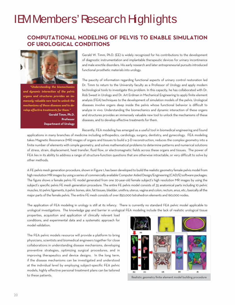
NEURAL� SIGNAL� PROCESSING�� BIOMARKERS� FOR� NEUROLOGICAL�AND�MENTAL�DISORDERSDr. Keshab Parhi's research is focused on developing signal processing methods to process brain
signals extracting biomarkers for identification of brain disorders. The processes he develops
incorporates electroencephalographic (EEG) signals, magnetoencephalographic (MEG) signals,
and structural and functional magnetic resonance imaging (MRI), and extract discriminating features
by using signal processing and machine learning. Machine learning and pattern recognition are
used for analyzing and classifying different classes of data. However, performance characteristics
of these classifiers depend on how discriminating the input features are. There is a need to
identify simple features that are computationally efficient and can act as biomarkers with high
sensitivity and specificity. Finding such features is non-trivial, and often non-intuitive. The goal of
this research is to find simple features or biomarkers using a combination of signal processing and
machine learning approaches. Dr. Parhi's work, in collaboration with colleagues at the University of
Minnesota from Neurology, Center for Magnetic Resonance (CMRR), Psychiatry, and Biomedical
Engineering, addresses signal processing for various neurological and mental disorders including
epilepsy, schizophrenia, borderline personality disorder (BPD), major depressive disorder (MDD),
and pediatric obsessive compulsive disorder (OCD). He is also researching brain connectivity to
better understand the differences of functional brain networks among control and patient groups
for various disorders.
One of the most debilitating features of epilepsy is the unpredictable nature of seizures. Epileptic seizures strike the patients
in an unpredictable fashion, causing embarrassing situations and often serious injury to the patients. Therefore, a seizure
prediction device would enable closed-loop therapy in the form of neuromodulation or delivery of anti-epileptic drugs, and
would dramatically improve the quality of life of the epileptics.
There are no quantitative biological patterns (biomarkers) to characterize Schizophrenia Identification using Language
Processing. Thus, exploring robust biomarkers for schizophrenia will benefit clinical diagnosis and treatment, and more
importantly, will increase our understanding of the physiological basis of the illness. Dr. Parhi's lab employs Power Spectral
Density Ratios (PSDRs) as features to characterize the brain activity during sentence processing.
Current work is also being directed towards extraction of features and biomarkers for borderline personality disorder,
major depressive disorder, and pediatric obsessive compulsive disorder using functional MRI images. These images are
also being analyzed with respect
to brain connectivity. Dr. Parhi's
efforts not only help in finding
biomarkers for these disorders, but
also help in understanding brain
connectivity for healthy subjects
and subjects with mental disorders.
(Le� top) Electrode locations in the brain, (le� bo� om) six EEG signals, (right top) raw EEG of one electrode for 24 hours where seizures are marked by vertical red lines, (right bo� om) features extracted from the same electrode can predict each seizure.
“The goal of this research is
to extract signal processing based
features from magnetoencephalo-
grams (MEG) and functional mag-
netic resonance imaging (fMRI) to
identify mental disorders.”
Keshab K. Parhi, Ph.D.
Professor
Electrical and Computer Engineering

18
HANDHELD�SENSORS�FOR�MEASUREMENT�OF�IN-VIVO�TISSUE�PROPERTIES�
Prof. Rajamani’s laboratory at the University of Minnesota is developing sensors that enable handheld measurement of in-vivo tissue properties such as stiffness, Young’s modulus, shear modulus of elasticity, tension in taut soft tissues, and pressure in muscle compartments. Collaborators in his research projects have included Dr. Art Erdman from Mechanical Engineering, Dr. Joan E. Bechtold from Orthopedic Surgery, and Dr. Rick Odland from Otolaryngology.
The research utilizes arrays of force sensors with appropriately configured properties in the array. Properties such as sensor size, distances between sensors, number of electrodes on each sensor, and stiffness of individual sensors in the array are configured so as to enable tailored measurement of particular tissue properties. For example, a sensor array on a handheld probe has been developed that is configured for measurement of tension in the soft tissues surrounding bone. This probe can be useful during a wide variety of orthopedic surgical procedures, including total knee replacement, ACL repair, shoulder stabilization, patella dislocation, tendon repair, and total hip replacement. In these surgeries, balancing the tensile forces in the soft tissues surrounding bone would lead to good restoration of overall limb function in the patient and a long lasting implant. During hip replacement surgery, the probe can be used to measure tension in the abductor muscles to ensure the hip implant is in balance and has the right amount of normal force to prevent both hip dislocation as
well as uneven gait. Likewise, during knee replacement surgery, the probe can be used to measure and balance tension in the collateral and cruciate ligaments, leading to good restoration of knee function and a long lasting knee implant. The team’s patent application from this research has been licensed to a start-up company, FocusStart, which has signed a contract with the University of Minnesota, and plans to make the product ready for FDA submission in 3 years.
Funds for the research activities described in this article have come from MIMTeC (a NSF Industry-University Cooperative Research Center), the National Science Foundation, the IEM Seed grant program, and a Doctoral Dissertation Fellowship.
“My research focuses on the devel-
opment of biomechanical sensors that
are useful either in improving medical
procedures or in enabling on-body moni-
toring applications.”
Rajesh Rajamani, Ph.D.
Professor
Mechanical Engineering
(b)
(a)
(c) The micro-fabricated fl exible polymer sensor array in Figure (a) has 64 sensing electrodes and provides si-multaneous measurement of Young’s modulus and shear modulus of elasticity. Figure (b) shows elasticity sensors being used in a handheld mode for in-vitro measure-ment of Young’s modulus of so� polymer specimens. Figure (c) shows a handheld sensor array developed for measurement of tension in taut so� tissues. This device is used to measure tension in muscles, ligaments, tendons and other so� tissues simply by pushing on the tissue during orthopedic surgical procedures.
IEM Members’ Research Highlights

19
MECHANISTIC UNDERSTANDING OF SYNAPTIC VESICLE FUSION BY α-SYNUCLEIN
Dr. Johnathan Sachs' laboratory studies the protein α-Synuclein, whose aggregation into insoluble Lewy bodies is commonly associated with Parkinson’s disease (PD). While the connection between α-Synuclein and PD has been recognized for some time, the normal function of αS remains a mystery. Although abnormally high levels of α-Synuclein may lead to Lewy bodies, high levels of αS have also been shown to disrupt normal vesicle trafficking and markedly inhibit neurotransmitter release without the formation of αS aggregates. It is also known that α-Synuclein binds to synaptic vesicles, and based on biophysical studies it is presumed that this binding is very strong. How and if this binding affects the vesicles remains unknown, but there is clear evidence that it can greatly diminish the propensity of vesicles to fuse (a key element in the synaptic vesicle trafficking process). Thus, there is growing consensus that at normal (non-pathological) levels in the neuron, α-Synuclein may act as a central regulatory component of synaptic vesicle trafficking.
The goal of his research is to develop a mechanistic understanding of the inhibition of synaptic vesicle regulation by α-Synuclein. A precise understanding of the native interactions between α-Synuclein and synaptic vesicle membranes will position them to evaluate the protein’s role in vesicle trafficking defects as they relate to PD. The lab's approach involves quantitative studies of the biophysical and mechanical properties of synaptic vesicle membranes. They will combine state-of-the-art computational molecular simulations (utilizing the highly powerful machines in the Minnesota Supercomputer Institute) with a panel of complementary biophysical experiments (including atomic force microscopy—to be performed at the University’s Characterization Facility). Led by a brilliant graduate student, Anthony Braun, Dr. Sachs' aim is to establish the foundation for new therapeutic strategies in the treatment of PD, namely to exploit the native function of the
protein (i.e., restoring proper vesicle trafficking).
"A seed grant from the IEM provided critical financial
backing during a time of extremely tight federal funding.
Like many researchers at the University and across the
country, internal support like that provided by the IEM
has played a critical role in stabilizing our research
program. The preliminary data we were able to collect
because of the IEM grant played an essential role in
securing our first RO1 grant from the National Institutes
of Neurological Disorders and Stroke."
“The preliminary data we were
able to collect because of the IEM
grant played an essential role in
securing our first RO1 grant from the
National Institutes of Neurological
Disorders and Stroke.”
Jonathan N. Sachs, Ph.D.
Associate Professor
Biomedical Engineering
Ph.D. candidate Anthony Braun (Le� ) discusses his computational models of how alph-Synuclein perturbs the mechanical properties of synaptic vesicles with Dr. Sachs
20
COMPUTATIONAL�MODELING�OF�PELVIS�TO�ENABLE�SIMULATION�OF�UROLOGICAL�CONDITIONS
Gerald W. Timm, Ph.D. (EE) is widely recognized for his contributions to the development of diagnostic instrumentation and implantable therapeutic devices for urinary incontinence and male erectile disorders. His early research and later entrepreneurial pursuits introduced functional prosthetic materials into urology.
The paucity of information regarding functional aspects of urinary control restoration led Dr. Timm to return to the University faculty as a Professor of Urology and apply modern technological tools to investigate this problem. In this capacity, he has collaborated with Dr. Rob Sweet in Urology and Dr. Art Erdman in Mechanical Engineering to apply finite element analysis (FEA) techniques to the development of simulation models of the pelvis. Urological diseases involve organs deep inside the pelvis whose functional behavior is difficult to analyze in vivo. Understanding the biomechanics and dynamic interaction of these organs and structures provides an immensely valuable new tool to unlock the mechanisms of these diseases, and to develop effective treatments for them.
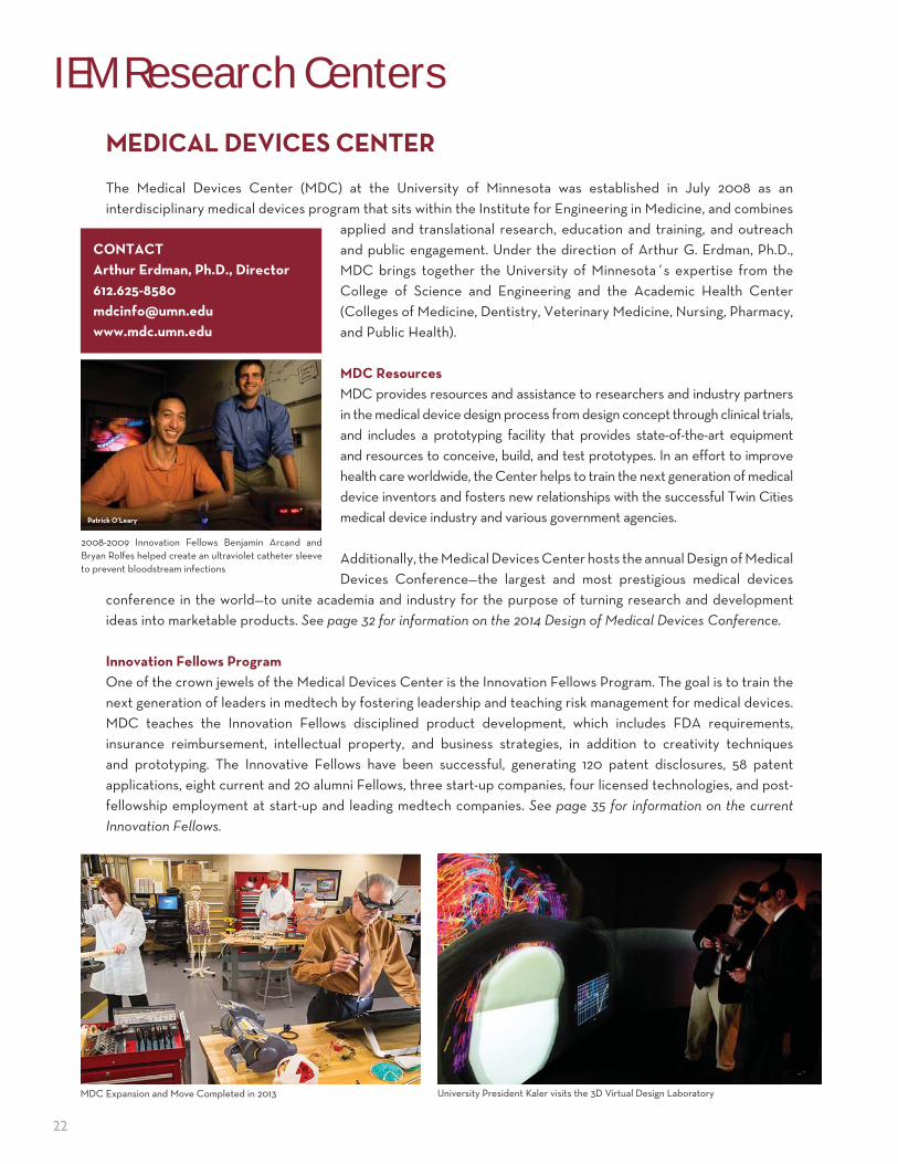
Recently, FEA modeling has emerged as a useful tool in biomedical engineering and found applications in many branches of medicine including orthopedics, cardiology, surgery, dentistry, and gynecology. FEA modeling takes Magnetic Resonance (MR) images of organs and tissues to build a 3-D reconstruction, reduces the complex geometry into a finite number of elements with simple geometry, and solves mathematical problems to determine patterns and numerical solutions of stress, strain, displacement, heat transfer, fluid flow, or electromagnetic fields across these organs and tissues. The power of FEA lies in its ability to address a range of structure-function questions that are otherwise intractable, or very difficult to solve by other methods.
A FE pelvis mesh generation procedure, shown in Figure 1, has been developed to build the realistic geometry female pelvis model from high resolution MR images by using a series of commercially available Computer Aided Design/Engineering (CAD/E) software packages. The figure shows a female pelvis FE model generated from one 20-year-old female subject’s high resolution MR images by using the subject’s specific pelvis FE mesh generation procedure. The entire FE pelvis model consists of 35 anatomical parts including 10 pelvic muscles, 10 pelvic ligaments, 6 pelvic bones, skin, fat tissues, bladder, urethra, uterus, vagina and colon, rectum, anus, etc.; basically all the major parts of the female pelvis. The entire FE mesh consists of over 850,000 tetrahedron elements and 160,000 nodes.
The application of FEA modeling in urology is still at its infancy. There is currently no standard FEA pelvic model applicable to urological investigations. The knowledge gap and barrier in urological FEA modeling include the lack of realistic urological tissue properties, acquisition and application of clinically relevant load conditions, and experimental data and a systematic approach for model validation.
The FEA pelvic models resource will provide a platform to bring physicians, scientists and biomedical engineers together for close collaborations in understanding disease mechanisms, developing preventive strategies, optimizing surgical procedures, and in improving therapeutics and device designs. In the long term, if the disease mechanisms can be investigated and understood at the individual level by employing subject-specific FEA pelvic models, highly effective personal treatment plans can be tailored to these patients.
“Understanding the biomechanics
and dynamic interaction of the pelvic
organs and structures provides an im-
mensely valuable new tool to unlock the
mechanisms of these diseases and to de-
velop effective treatments for them.”
Gerald Timm, Ph.D.
Professor
Department of Urology
Realistic geometry fi nite element model building procedure
IEM Members’ Research Highlights

21
IEM Members’ New Companies
MESOFLOW��THE�BIOPRESERVATION�CORE�RESOURCE��BIOCOR�
BioCoR, an Institute for Engineering in Medicine affiliated center, is a centralized resource for preservation protocol development, advancing preservation science through cutting-edge research, and educating individuals in the best practices for biospecimen preservation.
A small startup company, MesoFlow has been developed by BioCoR Director Allison Hubel, Ph.D. The company competed in the MN Cup (a small business plan competition) and placed second in the medical device division. Seed funding for MesoFlow has been obtained as well. As research progresses in different research projects in BioCoR, additional opportunities for small company development or licensing of intellectual property is anticipated.
Individual BioCoR faculty have had success in attracting new funding from both NSF and NIH and publications related to preservation. Preservation is a translational field and BioCoR is committed to translation of research into clinical use or commercial application. To that end, BioCoR faculty are involved in the commercialization of a microfluidic device for the removal of specialized solutions used in preservation.
Dr. Allison Hubel
22
IEM Research Centers
MEDICAL�DEVICES�CENTER
The Medical Devices Center (MDC) at the University of Minnesota was established in July 2008 as an interdisciplinary medical devices program that sits within the Institute for Engineering in Medicine, and combines
applied and translational research, education and training, and outreach and public engagement. Under the direction of Arthur G. Erdman, Ph.D., MDC brings together the University of Minnesota´s expertise from the College of Science and Engineering and the Academic Health Center (Colleges of Medicine, Dentistry, Veterinary Medicine, Nursing, Pharmacy, and Public Health).
MDC Resources
MDC provides resources and assistance to researchers and industry partners in the medical device design process from design concept through clinical trials, and includes a prototyping facility that provides state-of-the-art equipment and resources to conceive, build, and test prototypes. In an effort to improve health care worldwide, the Center helps to train the next generation of medical device inventors and fosters new relationships with the successful Twin Cities medical device industry and various government agencies.
Additionally, the Medical Devices Center hosts the annual Design of Medical Devices Conference—the largest and most prestigious medical devices
conference in the world—to unite academia and industry for the purpose of turning research and development ideas into marketable products. See page 32 for information on the 2014 Design of Medical Devices Conference.
Innovation Fellows Program
One of the crown jewels of the Medical Devices Center is the Innovation Fellows Program. The goal is to train the next generation of leaders in medtech by fostering leadership and teaching risk management for medical devices. MDC teaches the Innovation Fellows disciplined product development, which includes FDA requirements, insurance reimbursement, intellectual property, and business strategies, in addition to creativity techniques and prototyping. The Innovative Fellows have been successful, generating 120 patent disclosures, 58 patent applications, eight current and 20 alumni Fellows, three start-up companies, four licensed technologies, and post-fellowship employment at start-up and leading medtech companies. See page 35 for information on the current
Innovation Fellows.
University President Kaler visits the 3D Virtual Design Laboratory MDC Expansion and Move Completed in 2013
CONTACT
Arthur Erdman, Ph.D., Director
612.625-8580
www.mdc.umn.edu
Patrick O’Leary
2008-2009 Innovation Fellows Benjamin Arcand and Bryan Rolfes helped create an ultraviolet catheter sleeve to prevent bloodstream infections
23
CENTER�FOR�NEUROENGINEERING
The Center for Neuroengineering (CNE), an Institute for Engineering in Medicine affiliated research center, is actively advancing neuroengineering research, by fostering interdisciplinary collaborations between neuroscience and engineering faculty, clinical and industrial partners, and training the next generation of leaders in this dynamic field. CNE includes 27 faculty from engineering, neuroscience, and clinical departments. CNE has been the driving force in successfully winning an NSF IGERT Neuroengineering Training Grant, and hosts a biweekly neuroengineering seminar series.
Bridging the disciplines of neuroscience and engineering, neuroenigineering is an emerging field that translates research discoveries into neurotechnologies to provide innovative tools for basic and clinical neuroscience research leading to enhanced patient care. CNE research activities include the following thrust areas—neural decoding and imaging, neural sensing and interfacing, and neuromodulation. There are a number of collaborative research projects conducted by CNE faculty and their students, including:
• Brain-computer interface research to develop systems that assist in facilitating motor function by decoding the “motor intentions” of subjects
• Deep brain stimulation (DBS) to treat movement disorders using computational modeling, animal studies, and clinical studies
• Noninvasive neuromodulation including transcranial magnetic stimulation (TMS), transcranial direct current stimulation (tDCS), and transcutaneous electrical nerve stimulation (TENS)
• Auditory neuroprosthetics to replace absent or aberrant function through neural decoding and neuromodulation• High-field MRI and spectroscopy to identify anatomical and physiological biomarkers of the healthy and diseased brain• Mechanisms, detection, prediction, and localization of epileptic seizures in animal models and human patients using
research tools including optical imaging, electrophysiological sensing and mapping, signal processing, and imaging using EEG and fMRI
• Quantification of pain from electrophysiological measurements and imaging of pain networks using EEG source imaging and fMRI
• Visual neuroscience by fMRI, EEG, and behavioral studies to explore the mechanisms of rivalry, perception, attention, and plasticity
• Neural decoding of decision making using computational modeling and electrophysiology• Development of adaptive and closed-loop algorithms for optimizing neuromodulation treatments based on
perceptual feedback and physiological biomarkers• Development of flexible, wearable, and active electronics for sensing and stimulation of the nervous system for
real-time applications
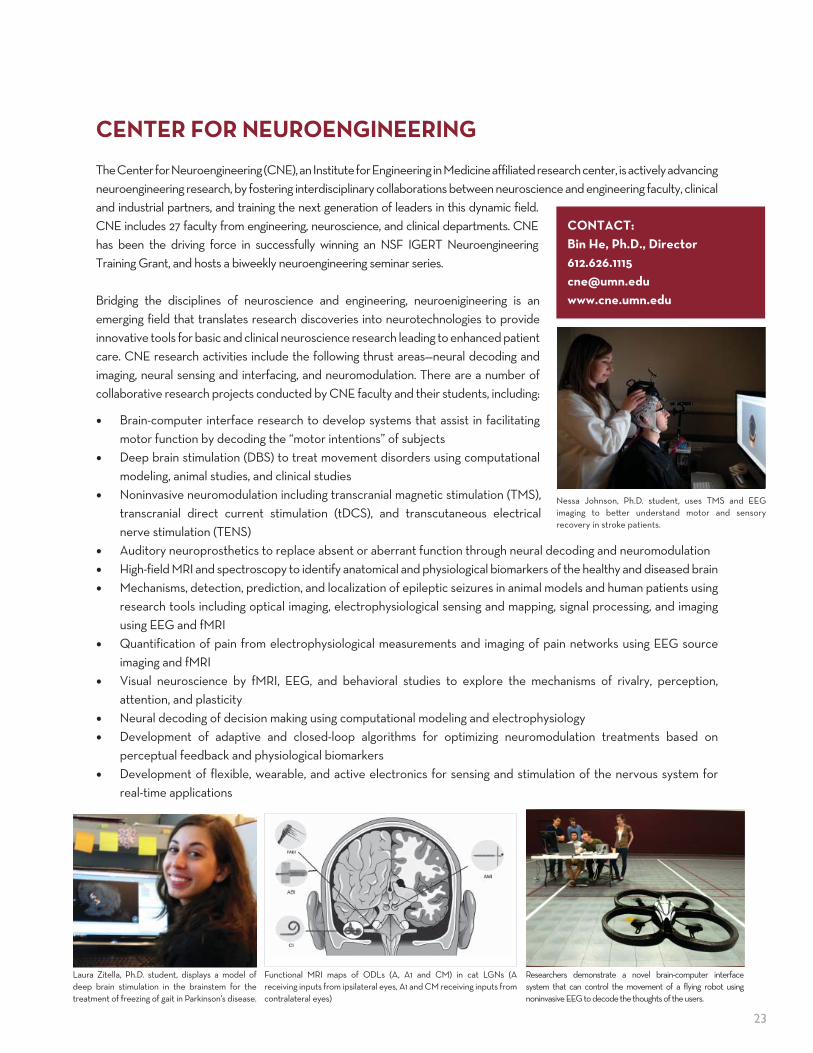
Nessa Johnson, Ph.D. student, uses TMS and EEG imaging to be� er understand motor and sensory recovery in stroke patients.
CONTACT:
Bin He, Ph.D., Director
612.626.1115
www.cne.umn.edu
Functional MRI maps of ODLs (A, A1 and CM) in cat LGNs (A receiving inputs from ipsilateral eyes, A1 and CM receiving inputs from contralateral eyes)
Laura Zitella, Ph.D. student, displays a model of deep brain stimulation in the brainstem for the treatment of freezing of gait in Parkinson's disease.
Researchers demonstrate a novel brain-computer interface system that can control the movement of a flying robot using noninvasive EEG to decode the thoughts of the users.

24
IEM Research Centers
BIOPRESERVATION�CORE�RESOURCEThe University of Minnesota’s Biopreservation Core Resource (BioCoR) was established in 2010 by a group of core faculty members through funding from the Institute of Engineering in Medicine, College of Science and Engineering,
and Academic Health Center. Under the direction of Allison Hubel, Ph.D., the mission of BioCoR is to advance the science, technology, and practice of biospecimen preservation by developing specific biopreservation protocols, improving preservation and storage technologies, establishing standards and guidelines, and training individuals and institutions in the science and technology of biopreservation.
Gap/Need
Effective methods of processing, preservation, and storage are critical to research on and clinical use of biospecimens. Biospecimen procurers and users are separated by gaps, which are typically both physical (different locations) and temporal (different times). The usefulness of a biospecimen is determined in large part by our ability to efficiently preserve the critical biological properties of the biospecimen (e.g. stabilization of the biomarkers) and its function (e.g. for therapeutic purposes). Conventional methods of preserving biospecimens were developed in the 1970s. There has been little evolution in the techniques used to
preserve biospecimens in the intervening decades. As a result, today we are in a situation where we have very limited knowledge, technology, and resources—and thus we fail—to successfully preserve the most promising diagnostic and therapeutic biospecimens we have discovered to-date.
BioCoR Resources
BioCoR aims at providing a unique national resource for the biospecimen community by serving to improve the quality of biospecimens available for biomedical research and clinical use. BioCoR is not a biorepository but a core resource in biopreservation. As a resource, BioCoR provides three basic functions for individual researchers and institutions (academic, industrial, and government): • Service resource for those who need assistance in developing standard biopreservation protocols and techniques.
BioCoR sends out monthly newsletters and maintains a healthy list of subscribers through the BioCoR website. • Research resource where new methods of preserving biospecimens and technology to improve biospecimen
quality are developed, directly feeding back into the service resource. BioCoR has attracted funding from both NSF and NIH. BioCoR faculty are currently involved in the commercialization of a microfluidic device for the removal of specialized solutions used in preservation.
• Education/training resource for instructing individuals and institutions on the scientific basis for preservation, teaching best practices, and supplying protocol-specific training as a service to the community. BioCoR has expanded its professional short course offerings to include, “Preservation of Molecular, Cellular, and Tissue Biospeciens” and “Presentation of Cellular Therapies: An Interactive Workshop”.
CONTACT:
Allison Hubel, Ph.D., Director
612.625.6808
www.biocor.umn.edu
25
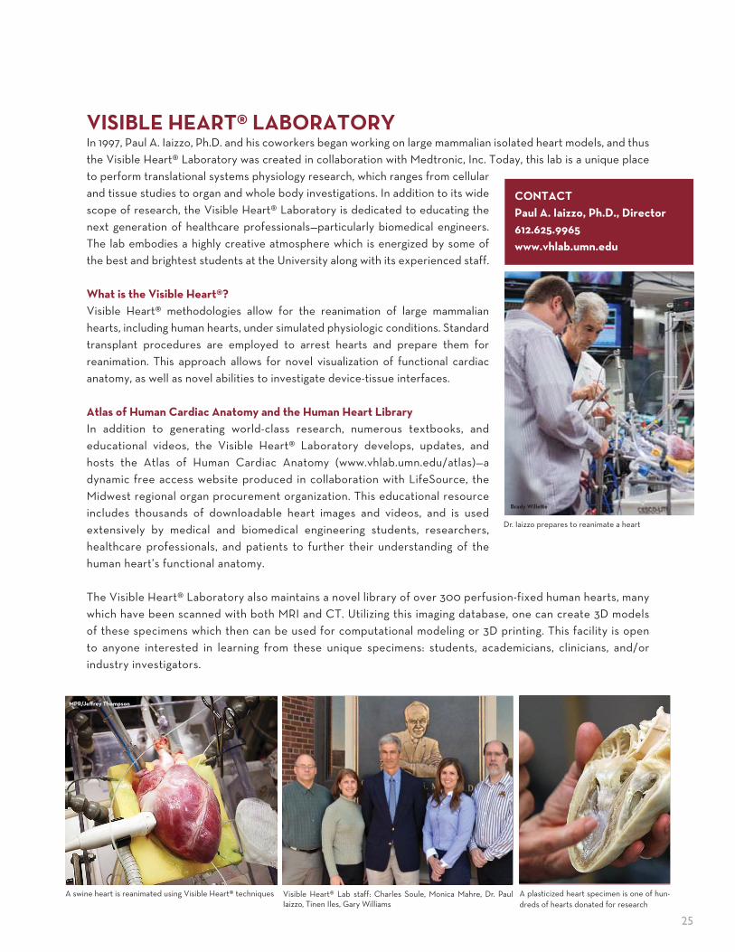
VISIBLE�HEART®�LABORATORYIn 1997, Paul A. Iaizzo, Ph.D. and his coworkers began working on large mammalian isolated heart models, and thus the Visible Heart® Laboratory was created in collaboration with Medtronic, Inc. Today, this lab is a unique place to perform translational systems physiology research, which ranges from cellular and tissue studies to organ and whole body investigations. In addition to its wide scope of research, the Visible Heart® Laboratory is dedicated to educating the next generation of healthcare professionals—particularly biomedical engineers. The lab embodies a highly creative atmosphere which is energized by some of the best and brightest students at the University along with its experienced staff.
What is the Visible Heart®?
Visible Heart® methodologies allow for the reanimation of large mammalian hearts, including human hearts, under simulated physiologic conditions. Standard transplant procedures are employed to arrest hearts and prepare them for reanimation. This approach allows for novel visualization of functional cardiac anatomy, as well as novel abilities to investigate device-tissue interfaces.
Atlas of Human Cardiac Anatomy and the Human Heart Library
In addition to generating world-class research, numerous textbooks, and educational videos, the Visible Heart® Laboratory develops, updates, and hosts the Atlas of Human Cardiac Anatomy (www.vhlab.umn.edu/atlas)—a dynamic free access website produced in collaboration with LifeSource, the Midwest regional organ procurement organization. This educational resource includes thousands of downloadable heart images and videos, and is used extensively by medical and biomedical engineering students, researchers, healthcare professionals, and patients to further their understanding of the human heart’s functional anatomy.
The Visible Heart® Laboratory also maintains a novel library of over 300 perfusion-fixed human hearts, many which have been scanned with both MRI and CT. Utilizing this imaging database, one can create 3D models of these specimens which then can be used for computational modeling or 3D printing. This facility is open to anyone interested in learning from these unique specimens: students, academicians, clinicians, and/or industry investigators.
MPR/Jeff rey Thompson
A swine heart is reanimated using Visible Heart® techniques Visible Heart® Lab staff: Charles Soule, Monica Mahre, Dr. Paul Iaizzo, Tinen Iles, Gary Williams
Dr. Iaizzo prepares to reanimate a heart
Brady Wille� e
CONTACT
Paul A. Iaizzo, Ph.D., Director
612.625.9965
www.vhlab.umn.edu
A plasticized heart specimen is one of hun-dreds of hearts donated for research

26
ACADEMY�OF�MEDICAL�DEVICE�INNOVATORSDr. Gerald Timm has been elected to the 2014 class of the Academy of Medical Device Innovators of the Institute for Engineering in Medicine, for his contributions to the development of diagnostic instrumentation and implantable therapeutic devices for urinary incontinence and male erectile disorders. Dr. Timm is an awardee of 23 patents as well as international counterparts for various Urological devices, author of 83 academic publications, and contributor to four textbooks. As a cornerstone of his work, Dr. Timm’s seminal paper on inflatable penile implants is one of the three most frequently cited articles in medical urology literature. In addition, his pioneering research on electrical bladder stimulation and fluid energy transfer systems has led directly to development of a variety of medical devices for diagnosing and treating urinary control and male erectile disorders. In the process, he and his associates essentially started an entire new industry in urological medical devices.
The IEM Academy of Medical Device Innovators was established in 2012 through the University of Minnesota’s Medical School and College of Science and Engineering, to honor and promote researchers who have had great impact on patients’ lives through their work on medical devices while at the University. Dr. Timm’s addition to the academy complements the existing class of world renowned researchers who have been a part of keeping us healthy, longer, and with a higher standard of living, through work performed at the University of Minnesota. The Institute for Engineering in Medicine is proud to extend congratulations to Dr. Timm, and welcome him into the academy.
MINNESOTA�INNOVATION�COLLABORATIONSThe University’s Medical Devices Center (MDC), an affilliated research center with the Institute for Engineering in Medicine, is becoming a more visible and valued resource to small business and the larger corporate med-tech community. With expanded capabilities and its new location in the heart of the University’s healthcare and biomedical activities, MDC is further improving the visibility and importance of medical devices for research and engineering faculty, staff, students and administrators.
A new initiative called Innovation Collaborations (IC) was initiated in September 2012 to increase the University’s outreach as well as create a valuable resource for medical technology companies. As part of this initiative, MDC has reached out to companies to forge new relationships allowing them to articulate their unmet needs. MDC brings together a wealth of expertise (faculty, staff, and students) spanning from the Veterinary School, Carlson School of Management, College of Science and Engineering, and the Academic Health Center, to develop an interdisciplinary team to collaborate with business on a managed project. Under this new initiative, MDC invited more than 30 companies to discuss their unmet needs, many of which previously did not see the U of MN as an approachable med-tech resource. The first company engagements, structured under domestic research agreements, have yielded both significant results for the company and valuable exposure for the UM IC teams and individual members.
MDC’s IC initiative is made possible under the new University of Minnesota’s Minnesota Innovation Partnership (MN-IP) policy, managed under the Office of the Vice President for Research, which has been designed to streamline agreements with industrial partners. Once explained to company leadership, MN-IP terms established by the policy have been favorably received and have allowed collaborative research and engineering teams to initiate and work through projects with great efficiency.
More information on MN-IP can be found on the Office of the Vice President for Research Website: http://www.research.umn.edu/
IEM Member Recognition
27
DR��ROBERT�SWEET
In 2013, Dr. Robert Sweet was granted the William L. Anderson Endowed Chair for Research and Development of Military Medical Stimulation Training and Technology. Dr. Sweet was also named President of the Society of Laparoendoscopic Surgeons (SLS).
DR��MICHAEL�LOUSHIN
Dr. Michael Loushin cofounded the company Preceptis Medical, Inc., a medical device manufactuer developing surgical tools which allow children’s ear tube procedures to be less painful and with reduced surgical time. Preceptis Medical, Inc. was named the Best Breakthrough Business Idea by the 2013 Minnesota Cup.
DR��JAKUB�TOLAR
Dr. Jakub Tolar was selected for the 2013 Leadership in Healthcare Change Agent Award by Minnesota Business Magazine. Dr. Tolar was also inducted into the 2013 Rare Disease Research Hall of Fame by the National Organization for Rare Disorders.
IEM�MEMBERS’�EXTERNAL�HONORS�AND�AWARDS
DR��JIANYI�"JAY"�ZHANG
The American Heart Associate named Dr. Jianyi “Jay” Zhang the George E. Brown Memorial Lecturer for 2014. This lecture was established in 1935 to honor the memory of Dr. George E. Brown, general practitioner, researcher, and chief of a section in the Division of Medicine of the Mayo Clinic.
DR��JOAN�BECHTOLD
Dr. Joan Bechtold was named the 2013 Orthopaedic Research Society (ORS) President. Founded in 1954, the Orthopaedic Research Society strives to be the world’s leading forum for the dissemination of new musculoskeletal research fi ndings.
DR��WILLIAM�DURFEE
In 2014, Dr. William Durfee was inducted into the American Institute for Medical and Biological Engineering (AIMBE) College of Fellows. AIMBE recognizes outstanding biomedical and biological engineers in academia, industry, and government who have distinguished themselves through contributions in research, industrial practice and education.
DR��ARTHUR�ERDMAN
Dr. Arthur Erdman was named the Freudenstein Distinguished Lecturer of 2013. In addition, he was awarded a 2014 Technology Advocate Award by The Minneapolis-St. Paul Business Journal as recognition of his outstanding leadership in assisting, advancing, and accelerating the performance of the technology community.
DR��MELISSA�GARDNER
In 2014, Dr. Melissa Gardner received a National Science Foundation Faculty Early Career Award in support of research on the origin and role of internally generated mitotic spindle forces during cell division.Dr. Gardner’s research will boost understanding of biophysics of cell division and provide data on potential cancer therapies.
DR��CHRISTY�HAYNES
Dr. Christy Haynes was chosen to present the 2013 Kavli Foundation Emerging Leader in Chemistry Lecture. This American Chemical Society (ACS) lecture series is sponsored by The Kavli Foundation and shines the spotlight on scientists under 40 years of age who have made exceptional achievements in scientifi c or engineering research.
DR��BIN�HE
Dr. Bin He was honored by being named a Theme Keynote Speaker in neuroengineering at the 2014 International Conference of IEEE Engineering in Medicine and Biology Society (EMBS). In addition, Dr. He received the Distinguished Service Award from IEEE EMBS.
DR��JIAN-PING�WANG
Dr. Jian-Ping Wang was recognized as one of the 2014 Titans of Technology by the Minneapolis - St. Paul Business Journal. Through this program he was named one of three Technology Inventors in recognition for his accomplishments creating breakthrough ideas, processes, and products impacting the technology industry.

28
Education and OutreachEDUCATIONAL�INITIATIVES��
One of the primary goals of the Institute for Engineering in Medicine (IEM) is to serve the educational needs of both the University of Minnesota and corporate biomedical communities in the area of engineering in medicine. To do so, IEM supports the development of numerous short courses, symposia, lectures, and conferences as well as fellowship programs.
IEM sponsored/co-sponsored educational programs and activities in the 2013/2014 fiscal year included:
• Advanced Cardiac Physiology and Anatomy Short Course • Preservation of Molecular, Cellular, and Tissue Biospecimens Short Course • Scott D. and Susan D. Augustine Biomedical Engineering Research Fellowship• Interdisciplinary Doctoral Fellowship (IDF)• Medical Devices Center Innovation Fellows Program
The overall objectives of these activities is to increase the visibility of medically-related engineering activities at the University and provide general support to local industry while strengthening University-industry relationships, through increased collaboration, contract research, licensing of intellectual property, philanthropy, and involvement of industry personnel in University teaching activities. In addition, IEM provides current information on available degree programs relevant to biomedicine at the University of Minnesota. Through collaboration, IEM helps the University of Minnesota aim to be one of the world’s leaders in educating the next generation of biomedical scientist and engineers as well as provide optimal life-long learning for current ones.
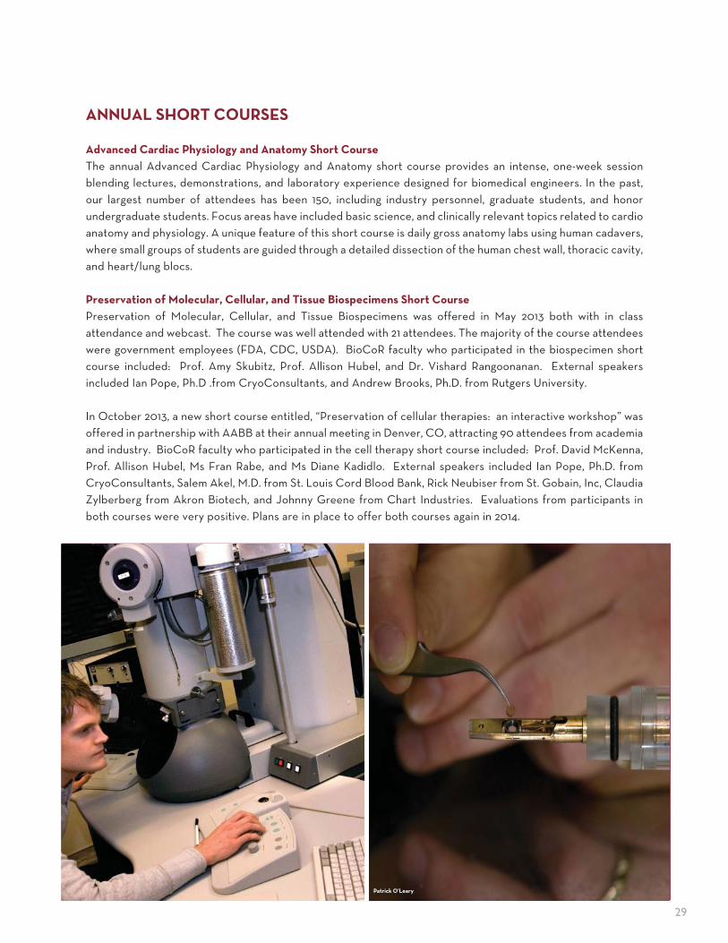
29
ANNUAL SHORT COURSES
Advanced Cardiac Physiology and Anatomy Short Course
The annual Advanced Cardiac Physiology and Anatomy short course provides an intense, one-week session blending lectures, demonstrations, and laboratory experience designed for biomedical engineers. In the past, our largest number of attendees has been 150, including industry personnel, graduate students, and honor undergraduate students. Focus areas have included basic science, and clinically relevant topics related to cardio anatomy and physiology. A unique feature of this short course is daily gross anatomy labs using human cadavers, where small groups of students are guided through a detailed dissection of the human chest wall, thoracic cavity, and heart/lung blocs.
Preservation of Molecular, Cellular, and Tissue Biospecimens Short Course
Preservation of Molecular, Cellular, and Tissue Biospecimens was offered in May 2013 both with in class attendance and webcast. The course was well attended with 21 attendees. The majority of the course attendees were government employees (FDA, CDC, USDA). BioCoR faculty who participated in the biospecimen short course included: Prof. Amy Skubitz, Prof. Allison Hubel, and Dr. Vishard Rangoonanan. External speakers included Ian Pope, Ph.D .from CryoConsultants, and Andrew Brooks, Ph.D. from Rutgers University.
In October 2013, a new short course entitled, “Preservation of cellular therapies: an interactive workshop” was offered in partnership with AABB at their annual meeting in Denver, CO, attracting 90 attendees from academia and industry. BioCoR faculty who participated in the cell therapy short course included: Prof. David McKenna, Prof. Allison Hubel, Ms Fran Rabe, and Ms Diane Kadidlo. External speakers included Ian Pope, Ph.D. from CryoConsultants, Salem Akel, M.D. from St. Louis Cord Blood Bank, Rick Neubiser from St. Gobain, Inc, Claudia Zylberberg from Akron Biotech, and Johnny Greene from Chart Industries. Evaluations from participants in both courses were very positive. Plans are in place to offer both courses again in 2014.
Patrick O’Leary

30
CONFERENCES�AND�SYMPOSIAIEM Annual Conference and Retreat
The First Annual Institute for Engineering in Medicine Conference and Retreat took place on September 23rd, 2013 at the McNamara Alumni Center on the University of Minnesota’s Twin Cities Campus. This conference is a yearly public event for the University of Minnesota community to exchange ideas on engineering solutions to medical and health problems. Featuring keynote talks from world recognized leaders in medical and engineer-ing research, this year’s conference attracted a sold out crowd of over 350 participants from the community, including students from a few local high schools looking to learn more about medical and biological engineering research. Following plenary talks, there were five breakout sessions where faculty groups and other interested parties engaged in strategic discussions on how to further enhance research collaboration opportunities relating to Cardiovascular Engineer-ing, Neuroengineering, Cellular and Molecular Bioengineering, Medical and Biological Imaging, and Medical Devices.
Starting mid afternoon, there was a highly competitive poster session, featuring work of 115 University of Minnesota faculty, research centers, and students. Students involved in the session were judged by a panel of University of Minnesota faculty on the quality of their presentations. Posters were divided into one of IEM’s five research themes – Cardiovascular Engineering, Cellular and Mollecular Bioengineering, Medical and Biological Imaging, Medical De-vices, and Neuroengineering. The top students in each session received a certificate and $100 award for their work.
2013 Poster Awardees:
Cardiovascular Engineering Theme• Joseph Ippolito, et al.• Jacqueline Wendel, et al.
Neuroengineering Theme• Allison Connolly, et al.• Abbey Holt, et al.• Sara Offut, et al.
Cellular and Molecular Bioengineering Theme• Anthony Braun, et al. • Rebecca Klank, et al.
Medical and Biological Imaging Theme• Sohini Roychowdhury, et al. • Qi Shao, et al. • Jennifer Taylor, et al. • Hui Wang, et al.
Medical Devices Theme• Yulong Li, et al. • Zhenpeng Qin, et al.
Education and Outreach

31
CONFERENCES�AND�SYMPOSIAOtto Schmitt Distinguished Lecture Series
In 2013, The Institute for Engineering in Medicine announced the launch of the Otto Schmitt Distinguished Lecture Series. This series is open to the University of Minnesota community to build collaborative projects between the disciplines of engineering and medicine, as well as provide an opportunity to hear presentations from national opinion leaders in research, industry, and government.
When the name of Otto Schmitt is mentioned by engineers, comments are usually made about his invention of the Schmitt trigger, a threshold detecting circuit with hysteresis that is commonly used in digital circuits. In reality, he made significant contributions in a wide range of fields including submarine detection, electro cardiography, and quality of life measures. He was also an early organizer of professional societies focusing on biomedical engineering and biophysics. Compared to many of his colleagues who tended to focus their research in a single field, Otto made significant contributions in many areas. Although very diverse, when answering a scientific question he was very thorough. He had said “Some people are in it for the money, others for pleasure, still other for power. Me, I’m into ideas”. He talked frequently about doing mental jogging as an exercise. He had over 60 patents and 200 publications, although he did not patent the Schmitt trigger. Dr. O� o H. Schmi�
William Hawkins, President, American Institute for Medical & Biological Engineering
Dr. Mehmet Toner, Professor of Surgery at Massachussetts General Hospital & Harvard Medical School

32
CONFERENCES�AND�SYMPOSIADesign of Medical Devices Conference
The University of Minnesota's annual Design of Medical Devices Conference—the world’s largest premier medical devices conference—creates opportunities to learn from world-renowned experts in a wide variety of fields. The conference seeks to: provide a national forum to bring together world-class medical device designers, researchers, manufacturers, and the public sector to share perspectives and innovations in medical device design; showcase the University of Minnesota as a leader in the medical device community; and raise funds from corporate sponsorships to support medical device education at the University of Minnesota.
The 13th Annual Design of Medical Devices Conference was held at The Commons Hotel on April 7-10, 2014. It featured an International Student Design Showcase, 3-in-5 Competition, scientific poster session, innovation workshop, 5K run/walk, networking opportunities, award luncheons, University laboratory and research center tours, technical sessions, and keynote and plenary speakers.
2014 Keynote Speakers (alphabetical order):
• Just Herder: Professor of Interactive Mechanisms and Mechatronics, Delft University of Technology• Martin S. Kohn, M.S., M.D., FACEP, FACPE: Chief Medical Scientist at Jointly Health, former Chief
Medical Scientist, Care Delivery Systems, IBM Research• Steve Levine: Senior Director of Product Strategy, Dassault Systèmes SIMULIA Corp.• Chaim Lotan: Heart Institute, Hadassah-Hebrew University Medical Center; President, Israel Heart Society• Michelle McMurry-Health, M.D., Ph.D.: Associate Director for Science, Office of the Center Director,
Center for Devices and Radiological Health, Food and Drug Administration• Tina Morrison, Ph.D.: Advisor of Computational Modeling, Center for Devices and Radiological Health,
Food and Drug Administration• Bill Murray: President and CEO, Medical Device Innovation Consortium (MDIC)• Kathryn M. O’Callaghan: Senior Science Health Adivsor, Office of the Center Director, Center for
Devices and Radiological Health, Food and Drug Administration• Ellen R. Strahlman, M.D., MHSc: Senior Vice President, Research and Development, Chief Medical Officer, BD
Ellen Strahlman, Dawn Bardot, Art Erdman, and Anita Bestelmeyer discuss med-ical devices a� er Thursday's Keynote Lunch Presentation during the conference.
Students presenting work to Just Herder, 2014 DMD Conference Award Winner, during the International Student Design Showcase
Chaim Lotan speaking during a Luncheon Keynote Presentation on Tuesday.
Education and Outreach

33
CONFERENCES�AND�SYMPOSIAMinnesota Neuromodulation Symposium
The Second Annual Minnesota Neuromodulation Symposium took place April 10-11, 2014, immediately following the Design of Medical Devices Conference in Minneapolis, Minnesota. The program was organized and sponsored by the Institute for Engineering in Medicine, co-sponsored by Medtronic Neuromodulation, MnDrive Brain Conditions, Center for Neuroengineering, and NSF IGERT Training Program, with technical co-sponsor IEEE Engineering in Medicine and Biology Society, and endorsed by International Federation of Medical and Biological Engineering. The Symposium brought together scientists, engineers, clinicians, industrial practitioners, and entrepreneurs to discuss challenges and opportunities in neuromodulation. The agenda consisted of plenary presentations and panel discussions by neuromodulation experts from academia, industry, and government. Additionally, a poster and networking session was offered to help participants exchange ideas in this exciting field.
Plenary Speakers (alphabetical order):
• Anthony T. Barker, Ph.D.: Consultant Clinical Scientist, Royal Hallamshire Hospital, U.K.• Brian Herman, Ph.D.: Vice President of Research, University of Minnesota• Andres Lozano, M.D., Ph.D.: RR Tasker Professor and Chairman of Neurosurgery, University of Toronto• Helen Mayberg, M.D.: Professor, Psychiatry and Radiology and Dorothy C. Fuqua Chair in Psychiatric
Neuroimaging and Therapeutics, Emory University School of Medicine
Additional Speakers and Panelists (alphabetical order):
• Marom Bikson, Ph.D.: Associate Professor of Biomedical Engineering, The City College of The City University of New York• Rafael Carbunaru, Ph.D.: Vice President, Research & Development, Boston Scientific• Tim Denison, Ph.D.: Director of Core Technology for the Neuromodulation Division, Medtronic Inc.• Dominique M. Durand, Ph.D.: Linsedth Professor of Biomedical Engineering and Neurosciences, Director of
the Neural Engineering Center, Case Western Reserve University• Kendall H. Lee, M.D., Ph.D.: Associate Professor, Neurosurgery, Physiology, and Biomedical Engineering, Mayo Clinic• Kip Ludwig, Ph.D.: Program Director, Neural Engineering, NINDS, NIH• Gregory F. Molnar, Ph.D.: Director of Neuromodulation Research, Medtronic Neuromodulation• Randy Nelson, Ph.D.: Founder and President, Evergreen Medical Technologies• Annie Saha: Acting Director, External Expertise and Partnerships, FDA/CDRH,OCD• Dirk Smith: Chief Technical Officer and Chief Human Resource Officer, Minnetronix• Douglas Weber, Ph.D.: Program Manager, DARPA Microsystems Technology Office

34
FELLOWSHIP�PROGRAMSSco� D. and Susan D. Augustine Biomedical Engineering Research Fellowship
The MD/MS dual degree program in biomedical engineering at the University of Minnesota provides top-ranked medical students with graduate training in biomedical engineering. To help strengthen this program in 2004, Uni-versity of Minnesota alumni Dr. Scott and Susan Augustine contributed $1 million for the establishment of the Scott D. and Susan D. Augustine Biomedical Engineering Research Fellowship fund. This fund supports annual fellowships for students pursuing an MS degree in biomedical engineering and who already have earned an MD degree, or are enrolled in the University of Minnesota Medical School. The payout from this fund is matched on a dollar-for-dollar basis by the University’s 21st Century Graduate Fellowship Endowment.
The 2013-2014 Scott D. and Susan D. Augustine Biomedical Engineering Research Fellowship recipient is:
SALAH�EL�HADDAI
Mr. El Haddi has degrees in Biochemistry and Physiology from the University of Minnesota. His research focuses on optimization of isolated cardiac tissue function in addition to enhancing clinical ablation techniques. In addition to his research endeavors, he has worked on educational outreach and contributions to materials on the visible heart atlas of cardiac anatomy.
Interdisciplinary Doctoral Fellowship (IDF)
The Interdisciplinary Doctoral Fellowship (IDF) awards outstanding graduate students with interdisciplinary dissertation topics who would benefit from interaction with faculty at one of the University of Minnesota’s interdisciplinary research centers or institutes. The fellowship provides a unique study opportunity for students with research and scholarly interests that complement those of the host center or institute and its faculty. The Institute for Engineering in Medicine (IEM) is pleased to be one of the host sites for the Graduate School’s IDF. In addition to the stipend awards through the Graduate School, IEM offers additional funds to support professional development as well as space to accommodate the needs of research projects.
The IDF recipients for 2014 are:
GARRET�NELSON
“Enabling Auscultation in High Noise Enviroments”Mr. Nelson has a Bachelor’s Degree in Mechanical Engineering from the University of St. Thomas, and is currently pursuing a Ph.D. in Mechanical Engineering from the University of Minnesota. His research addresses improving the usability of stethoscopes by use of active noise cancellation techniques.
TINGTING�XU
“Biomarkers for Borderline Personality Disorder (BPD) from fMRI”Ms. Xu has a Master of Engineering in Signal Processing from Nanjing University of Posts and Telecommunications, and is currently pursuing a Ph.D. in Electrical Engineering at the University of Minnesota. Her research is focused on identifying biomarkers for borderline personality disorder (BPD) and pediatric obsessive compulsive disorder (OCD)
Education and Outreach
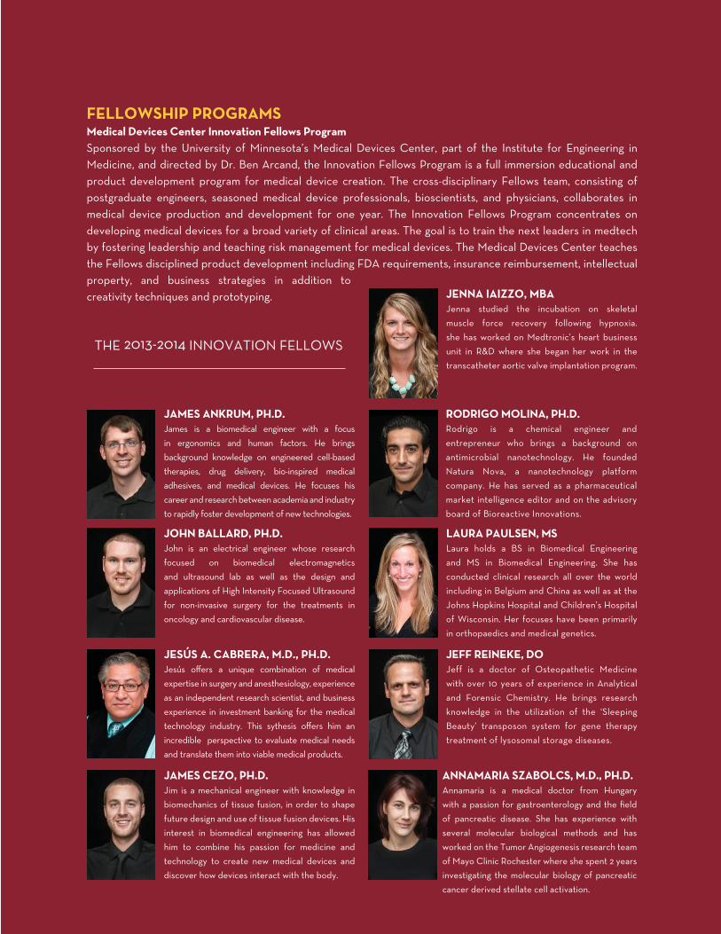
35
FELLOWSHIP�PROGRAMSMedical Devices Center Innovation Fellows Program
Sponsored by the University of Minnesota’s Medical Devices Center, part of the Institute for Engineering in Medicine, and directed by Dr. Ben Arcand, the Innovation Fellows Program is a full immersion educational and product development program for medical device creation. The cross-disciplinary Fellows team, consisting of postgraduate engineers, seasoned medical device professionals, bioscientists, and physicians, collaborates in medical device production and development for one year. The Innovation Fellows Program concentrates on developing medical devices for a broad variety of clinical areas. The goal is to train the next leaders in medtech by fostering leadership and teaching risk management for medical devices. The Medical Devices Center teaches the Fellows disciplined product development including FDA requirements, insurance reimbursement, intellectual property, and business strategies in addition to creativity techniques and prototyping.
THE 2013-2014 INNOVATION FELLOWS
JAMES�ANKRUM��PH�D�
James is a biomedical engineer with a focus in ergonomics and human factors. He brings background knowledge on engineered cell-based therapies, drug delivery, bio-inspired medical adhesives, and medical devices. He focuses his career and research between academia and industry to rapidly foster development of new technologies.
JENNA�IAIZZO��MBA
Jenna studied the incubation on skeletal muscle force recovery following hypnoxia. she has worked on Medtronic’s heart business unit in R&D where she began her work in the transcatheter aortic valve implantation program.
JOHN�BALLARD��PH�D�
John is an electrical engineer whose research focused on biomedical electromagnetics and ultrasound lab as well as the design and applications of High Intensity Focused Ultrasound for non-invasive surgery for the treatments in oncology and cardiovascular disease.
RODRIGO�MOLINA��PH�D�
Rodrigo is a chemical engineer and entrepreneur who brings a background on antimicrobial nanotechnology. He founded Natura Nova, a nanotechnology platform company. He has served as a pharmaceutical market intelligence editor and on the advisory board of Bioreactive Innovations.
JESÚS�A��CABRERA��M�D���PH�D�
Jesús off ers a unique combination of medical expertise in surgery and anesthesiology, experience as an independent research scientist, and business experience in investment banking for the medical technology industry. This sythesis off ers him an incredible perspective to evaluate medical needs and translate them into viable medical products.
LAURA�PAULSEN��MS
Laura holds a BS in Biomedical Engineering and MS in Biomedical Engineering. She has conducted clinical research all over the world including in Belgium and China as well as at the Johns Hopkins Hospital and Children’s Hospital of Wisconsin. Her focuses have been primarily in orthopaedics and medical genetics.
JAMES�CEZO��PH�D�
Jim is a mechanical engineer with knowledge in biomechanics of tissue fusion, in order to shape future design and use of tissue fusion devices. His interest in biomedical engineering has allowed him to combine his passion for medicine and technology to create new medical devices and discover how devices interact with the body.
JEFF�REINEKE��DO
Jeff is a doctor of Osteopathetic Medicine with over 10 years of experience in Analytical and Forensic Chemistry. He brings research knowledge in the utilization of the ‘Sleeping Beauty’ transposon system for gene therapy treatment of lysosomal storage diseases.
ANNAMARIA�SZABOLCS��M�D���PH�D�
Annamaria is a medical doctor from Hungary with a passion for gastroenterology and the fi eld of pancreatic disease. She has experience with several molecular biological methods and has worked on the Tumor Angiogenesis research team of Mayo Clinic Rochester where she spent 2 years investigating the molecular biology of pancreatic cancer derived stellate cell activation.

36
IEM FundingFUNDING�PROGRAMS
The objective of the Institute for Engineering in Medicine (IEM) seed grant program is to enhance biomedical and health related engineering research at the University of Minnesota while fostering interdisciplinary collaborations between the Academic Health Center and the College of Science and Engineering. Since Fiscal Year 2013 (FY13), IEM launched two new seed grant programs: the Group Grant and the Exploratory Grant.
Group Grant projects are aimed at supporting a cohort of investigators that work collaboratively to tackle significant biomedical and health problems with a multidisciplinary approach including innovative engineering solutions. Exploratory Grant projects are aimed at supporting the exploration of highly innovative ideas by 2-3 collaborating faculty, and are expected to generate preliminary data in order to support external funding proposals.
The following projects were funded by IEM in FY14:
Funding Type Title PIs and Co-PIs
Exploratory Grant Depth-resolving tissue oxygen needle sensor Shai Ashkenazi, Alan Hirsch
Exploratory Grant Computer-Aided Engineering of Antibiotic Cellbots
Yiannis Kaznessis, Garry Dunny
Exploratory Grant Biorelevant 3D ECM Microenvironments for Stem Cell Diff erentiation and Delivery to the Heart
Brenda Ogle, Jay Zhang
Exploratory Grant Automated Screening for Hypertensive Retinopathy and Stroke from Fundus Images
Kashab Parhi, Dara Koozekanani
Exploratory Grant Creating an Endothelium from Autologous Mesenchymal Stem Cells
Robert Tranquillo, Jakub Tolar
Group Grant IEM Cancer Animal Core Lab John Bischof
Group Grant Engineered biomaterials for preventing peripheral nerve injury and healing chronic wounds
Klapna Gupta, Chun Wang, Wei Shen
Group Grant Cardiovascular Engineering Research Theme Group
Paul Iaizzo, Jay Zhang, Robert Tranquillo
Group Grant Enhancing non-invasive neuromodulation in neurological and psychiatric disorders using a pairing paradigm
Kelvin Lim, Hubert Lim
Group Grant Organ, Tissue, and Cellular Engineering Walter Low, Wei-Shou Hu
Group Grant Discovering Systems Biomarkers for Neural Disorders
Theoden Netoff , Wei Chen
Group Grant Cell Engineering Approach to Dynamic Biomarkers for Poor Prognosis Cancers
David Odde, Amy Skubitz

37
FUNDING�ANALYSIS�� �-� ���FY���RESULTS�OF�FUNDING�PROGRAMS
Every year the Institute for Engineering in Medicine collects data on financial and non-financial returns to the University of Minnesota which can be directly attributed to work performed on IEM research grants. These results showcase the impact seed grant funding has on giving freedom to PIs to generate intellectual proper-ty, leading to the development of grant proposals in order to capture funding from external agencies like the National Institutes of Health (NIH), the National Science Foundation (NSF), industrial sponsorships, and private foundations. These funds not only offset costs to the university for things like student support, they also allow us to strengthen our collective body of research, ultimately leading to the development of products and proce-dures alleviating medical conditions for millions of patients worldwide. Over the last 9 years our program has been highly successful. To highlight this success, as of today, IEM has been able to measure $67M returns on $6M in investment, as well as being able to financially support the generation of 238 publications.
Δ Publications: 238Δ Students Supported: 185Δ Disclosures: 35Δ Patent Applications: 25Δ Patents Awarded: 16Δ Proposals for Additional Funding: 175
NIH NSF Other Total Return Investment ROI
Group Grants $11,764,950 $0 $0 $11,764,950 $557,620 2010%
Major Programs $10,682,012 $4,382,412 $11,186,237 $26,250,661 $1,792,000 1365%
Interest Groups $12,901,413 $1,441,920 $3,893,794 $18,237,127 $1,360,222 1241%
Seed Grants $5,337,980 $2,175,940 $715,000 $8,228,920 $786,695 946%
MDC Grants $2,111,835 $1,231,000 $0 $3,342,835 $975,354 243%
What If Grants $0 $100,00 $879,708 $979,708 $321,360 205%
Exploratory Grants $0 $223,963 $280,000 $503,963 $229,739 119%
Total $42,798,190 $9,555,235 $16,954,739 $69,308,164 $6,022,990 1051%
*Return on Investment (ROI) based on reported returns and verified for accuracy.
0
10M
20M
30M
40M
50M
60M
70M
$80M
Investment Return to the University

38
IEM Members
MEMBERS BY ACADEMIC DEPARTMENT
Aerospace Engineering . . . . . . . . . . . . . . . . . . . 1Anesthesia . . . . . . . . . . . . . . . . . . . . . . . . . . . 1Biomedical Engineering . . . . . . . . . . . . . . . . . . 18Biostatistics . . . . . . . . . . . . . . . . . . . . . . . . . . 1Chemical Engineering and Material Science . . . . . . .3Chemistry . . . . . . . . . . . . . . . . . . . . . . . . . . .6Civil Engineering . . . . . . . . . . . . . . . . . . . . . . . 1Computer Science and Engineering . . . . . . . . . . . .4Dermatology . . . . . . . . . . . . . . . . . . . . . . . . . . 1Diagnostic and Biological Sciences . . . . . . . . . . . . 1Electrical and Computer Engineering . . . . . . . . . . 12Enviromental Health . . . . . . . . . . . . . . . . . . . . . 1Epidemiology and Community Health . . . . . . . . . . . 1Experimental and Clinical Pharmacology . . . . . . . . . 1Family Medicine and Community Health . . . . . . . . . 1Genetics, Cell Biology, and Development . . . . . . . .3Institute for Molecular Virology . . . . . . . . . . . . . . 1Laboratory Medicine and Pathology . . . . . . . . . . . .5Law . . . . . . . . . . . . . . . . . . . . . . . . . . . . . . . 2Mechanical Engineering . . . . . . . . . . . . . . . . . . .6Medical Chemistry . . . . . . . . . . . . . . . . . . . . . . 1Medicine . . . . . . . . . . . . . . . . . . . . . . . . . . . 13
Neurology . . . . . . . . . . . . . . . . . . . . . . . . . . .9Neuroscience . . . . . . . . . . . . . . . . . . . . . . . . .4Neurosurgery . . . . . . . . . . . . . . . . . . . . . . . . .5Oncology . . . . . . . . . . . . . . . . . . . . . . . . . . . . 1Ophthalmology . . . . . . . . . . . . . . . . . . . . . . . . 1Orthopaedic Surgery . . . . . . . . . . . . . . . . . . . . . 1Otolaryngology . . . . . . . . . . . . . . . . . . . . . . . .5Pediatrics . . . . . . . . . . . . . . . . . . . . . . . . . . . 2Pharmaceutics. . . . . . . . . . . . . . . . . . . . . . . . . 2Physical Medicine and Rehabilitation . . . . . . . . . . .4Physics . . . . . . . . . . . . . . . . . . . . . . . . . . . . . 1Psychiatry . . . . . . . . . . . . . . . . . . . . . . . . . . . 2Psychology . . . . . . . . . . . . . . . . . . . . . . . . . . . 2Radiology . . . . . . . . . . . . . . . . . . . . . . . . . . 14Restorative Sciences . . . . . . . . . . . . . . . . . . . . . 2Stem Cell Institute . . . . . . . . . . . . . . . . . . . . . . 2Surgery . . . . . . . . . . . . . . . . . . . . . . . . . . . . . 7Therapeutic Radiology . . . . . . . . . . . . . . . . . . . . 1Urologic Surgery . . . . . . . . . . . . . . . . . . . . . . .4Urology . . . . . . . . . . . . . . . . . . . . . . . . . . . . . 2Veterinary Clinical Sciences . . . . . . . . . . . . . . . .3Veterinary Population Medicine . . . . . . . . . . . . . . 1TOTAL . . . . . . . . . . . . . . . . . . . . . . . . . . .159
MEMBERSHIP�BREAKDOWN
The Institute for Engineering in Medicine (IEM) is comprised of 159 faculty members who serve in 44 academic departments and represent nine University of Minnesota colleges: the Medical School, College of Science and Engineering, College of Veterinary Medicine, School of Dentistry, College of Biological Sciences, School of Public Health, College of Pharmacy, College of Liberal Arts, and School of Law. Benefits of IEM membership include: access to research funding provided by IEM, participation in organized research groups, assistance in developing contacts or collaborations within the University and with local industry, reduced costs for attendance at IEM-sponsored conferences and activities, and reduced costs for use of IEM-sponsored research centers as well as potential support for presenting at conferences or symposia, use of IEM equipment or facilities, sponsorship of visiting scientists or lecturers, and support for graduate student research.
MEMBERS BY COLLEGE AFFILIATION
College of Biological Sciences . . . . . . . . . . . . . . .3College of Liberal Arts . . . . . . . . . . . . . . . . . . . . 2College of Pharmacy . . . . . . . . . . . . . . . . . . . . .3College of Science and Engineering . . . . . . . . . . . 53College of Veterinary Medicine . . . . . . . . . . . . . .4Medical School . . . . . . . . . . . . . . . . . . . . . . . 85School of Dentistry . . . . . . . . . . . . . . . . . . . . . .4School of Law . . . . . . . . . . . . . . . . . . . . . . . . . 2School of Public Health . . . . . . . . . . . . . . . . . . .3
MEMBERS BY RESEARCH THEME
Cardiovascular Engineering . . . . . . . . . . . . . . . . 35
Cellular and Molecular Bioengineering . . . . . . . . . 59
Medical and Biological Imaging . . . . . . . . . . . . . . 74
Medical Devices . . . . . . . . . . . . . . . . . . . . . . 76
Neuroengineering . . . . . . . . . . . . . . . . . . . . . 48
39
IEM�STAFFAdam Klein, Senior Administrative Director
Stephanie Scott, Executive Administrative AssistantBrittany Long, Annual Report Editor
Angeliki Beyko, Principle Lab TechnicianKai Yu, Webmaster
CONTACTInstitute for Engineering in Medicine
University of Minnesota420 Delaware Street Southeast
725 Mayo Memorial Building, MMC 609Minneapolis, Minnesota 55455
Telephone: 612-626-5493Facsimile: 612-625-5633Email: [email protected]
Website: www.iem.umn.edu
SUPPORT�IEMThe Institute for Engineering in Medicine supports multiple projects yearly through our funding programs. Programs like the IEM Exploratory Grant and Group Grant projects have a proven record assisting researchers in developing major research initiatives, and making fundamental discoveries. Please help make a difference and bring engineering solutions to medical problems. Your tax-deductible gift can be only a click away. Or, if you would like to support the Institute in a different way, we would love to hear from you.
Tax deductable donations can be made to the Institute for Engineering in Medicine website. Funds will be used to advance IEM programs, research, and staff support. If you would like to make a donation to a specific cause, please contact our staff so we may speak to you.
Correspondence:
IEM Office, 612-624-8483, [email protected]
Institute for Engineering in Medicine

40
Institute for Engineering in Medicine
Published by the University of Minnesota’s Institute for Engineering in Medicine. Edited by Brittany Long, Adam Klein, and Stephanie Scott.
© 2014 Regents of the University of Minnesota. All rights reserved. The University of Minnesota is an equal opportunity educator and employer.