interleukin-10 in the control of tumor immunity and autoimmunity
TRANSCRIPT
Interleukin-10 in the Control of Tumor Immunity and Autoimmunity
by
Cailin Moira Wilke
A dissertation submitted in partial fulfillment of the requirements for the degree of
Doctor of Philosophy (Immunology)
in the University of Michigan 2011
Doctoral Committee:
Professor Weiping Zou, Chair Professor David A. Fox Professor Gabriel Nuñez Professor Lloyd M. Stoolman
Associate Professor Bethany B. Moore Research Investigator Ilona E. Kryczek
“It is better to light a candle than to curse the darkness.”
(Ancient Chinese Proverb)
“Make of yourself a light.” (Buddha)

© Cailin Moira Wilke
2011

ii
This is dedicated to my grandmother,
Alice Marie Dewhirst Olsen,
in honor of her indefatigable spirit,
her easy laughter,
and her belief in the infinite possibility of things.
iii
Acknowledgements
I would like first and foremost to thank my family for their unwavering love,
support, and confidence in me, even when I lost faith in myself.
I am deeply grateful to Weiping and Inka, who synergized like IL-7 and IL-15 to
guide and support my doctoral education. From them I learned true dedication,
careful planning, and how to ask the necessary questions.
Thank you also to the members of the Zou laboratory, for their patience, humor,
and encouragement. I am so lucky to have spent my last five years in your
company.
I owe a debt of gratitude to my thesis committee members, collaborators, and
mentors in the Immunology program, who never hesitated to share their
expertise or reagents.
It would be a transgression of epic proportions if I did not express my profound
thanks to the tireless Zarinah Aquil, without whom we would all be terribly lost.
My dear friends Molly, Karthik, Laura, and Karlyn: I could not have done this
without you. Thank you for the steady reminders that no grad student is an
island and that I was not alone.
A special thank you goes to my ninth-grade Biology teacher, Cynthia Ottesen,
who persuaded me to explore bench science with a closer lens than those
provided in textbooks and lab manuals.
I am additionally grateful to my extended circle of friends who have served as
safe havens, solid supports, and enthusiastic advocates throughout this journey.
Lastly, I acknowledge the many mice that participated in my research and whose
sacrifice served to expand upon our collective immunological knowledge.
iv
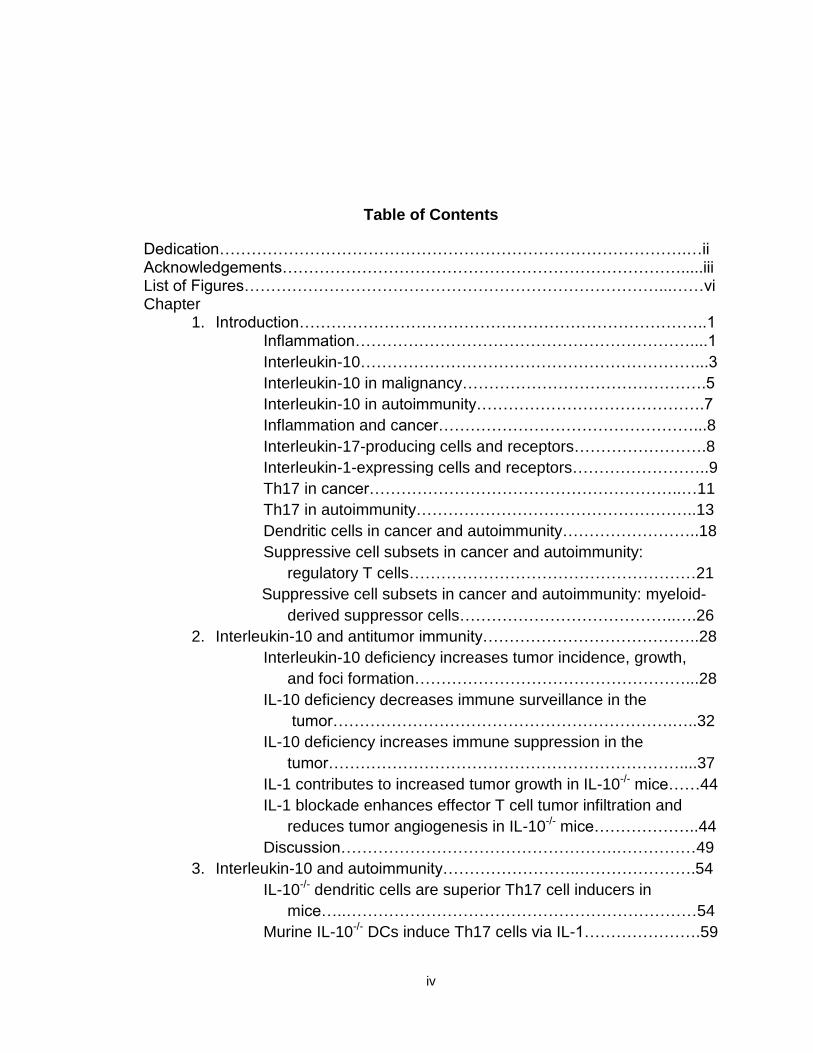
Table of Contents
Dedication…………………………………………………………………………….…ii Acknowledgements………………………………………………………………….....iii List of Figures……………………………………………………………………...……vi Chapter
1. Introduction…………………………………………………………………..1 Inflammation………………………………………………………....1
Interleukin-10………………………………………………………...3
Interleukin-10 in malignancy……………………………………….5
Interleukin-10 in autoimmunity…………………………………….7
Inflammation and cancer…………………………………………...8
Interleukin-17-producing cells and receptors…………………….8
Interleukin-1-expressing cells and receptors……………………..9
Th17 in cancer…………………………………………………..…11
Th17 in autoimmunity……………………………………………..13
Dendritic cells in cancer and autoimmunity……………………..18
Suppressive cell subsets in cancer and autoimmunity:
regulatory T cells………………………………………………21
Suppressive cell subsets in cancer and autoimmunity: myeloid-
derived suppressor cells…………………………………..….26
2. Interleukin-10 and antitumor immunity…………………………………..28
Interleukin-10 deficiency increases tumor incidence, growth,
and foci formation……………………………………………...28
IL-10 deficiency decreases immune surveillance in the
tumor……………………………………………………….…..32
IL-10 deficiency increases immune suppression in the
tumor…………………………………………………………....37
IL-1 contributes to increased tumor growth in IL-10-/- mice……44
IL-1 blockade enhances effector T cell tumor infiltration and
reduces tumor angiogenesis in IL-10-/- mice………………..44
Discussion…………………………………………….……………49
3. Interleukin-10 and autoimmunity……………………..………………….54
IL-10-/- dendritic cells are superior Th17 cell inducers in
mice…..…………………………………………………………54
Murine IL-10-/- DCs induce Th17 cells via IL-1………………….59
v
Increased Th17 cells and IL-17 in the intestine of patients with
Crohn’s disease……………………………………….……….64
Th17 cells are associated with decreased IL-10 and increased
IL-1 in patients with Crohn’s disease………………….….…66
Th17 cells promote inflammation in patients with Crohn’s
disease………………………………………………………….68
Discussion………………………………………………………….71
4. Conclusions………………………………………………………..……….75 Appendix………………………………………………………………………………..82 Materials and Methods………………………………………………………..82 References……………………………………………………………………………..87
vi
List of Figures
Figure 2.1: IL-10 deficiency increased tumor incidence and growth…………..…30
Figure 2.2: IL-10 deficiency increased tumor foci formation………………………31
Figure 2.3: MHC I expression in APC subsets in IL-10-/- and IL-10+/+ mice……..33
Figure 2.4: IL-10 deficiency reduced tumor surveillance by NK cells……………34
Figure 2.5: IL-10 deficiency reduced tumor surveillance by CD8+ T cells…….…35
Figure 2.6: IL-10 stimulates effector cytokine expression in CD4+ and CD8+ T cells……………………………………………………………………………...36
Figure 2.7: MDSC percentages are comparable in IL-10+/+ and IL-10-/- mice…..38
Figure 2.8: IL-10 deficiency increases absolute numbers of MDSC…………......39
Figure 2.9: IL-10 deficiency increases immune suppression……………………..40
Figure 2.10: Tumor-derived IL-10-/- MDSCs are superior to IL-10+/+ MDSCs in Treg induction………………………………………………………………….41
Figure 2.11: IL-10-/- MDSC suppress T cell activation and impede antitumor immunity…………………………………………………..…………………….43
Figure 2.12: IL-1 contributes to increased tumor growth in IL-10-/- mice………...45
Figure 2.13: Tumor growth is reduced in IL-1R-/- mice…………………………….46
Figure 2.14: IL-1 blockade altered immune phenotype in IL-10-/- mice………….47
Figure 2.15: Anakinra treatment reduces vascularization in IL-10-/- mice…….....48
Figure 2.16: Model of IL-10 in antitumor immunity……………………….………..50
Figure 3.1: IL-17+ cells are increased in IL-10-/- mice……………………………...56
Figure 3.2: IL-10-/- DCs are more powerful inducers of Th17 cells……………....58
Figure 3.3: IL-10-/- DC produce more IL-1 than IL-10+/+ DC…………………….…60
Figure 3.4: Blockade of IL-1R signaling decreases Th17 populations in vitro…..62
Figure 3.5: In vivo IL-1Ra administration reduces endogenous Th17 in IL-10-/- mice…………………………………………………………………………..…63
Figure 3.6: Th17 cells and IL-17 are increased in Crohn’s disease patient tissue…………………………………………………………………………....65
vii
Figure 3.7: IL-17 in Crohn’s disease patients is associated with increased IL-1 and decreased IL-10…………………………………………………………..67
Figure 3.8: T cell-derived IL-17 stimulates production of proinflammatory cytokines in Crohn’s disease tissue……………………………………….…69
Figure 3.9: Model of IL-10 in autoimmunity………………………………………....72
1
Chapter 1
Introduction
Inflammation.
Inflammation is the term given to a mixture of biological processes set in
motion by vascular tissues to combat invading pathogens or physiological insult.
These processes include increased blood flow, accelerated cellular metabolism,
vasodilatation, fluid and cell extravasation, infiltration of cells into the damaged
tissue, and release of soluble mediators such as cytokines, chemokines,
prostaglandins, leukotrienes, and various other proteins, peptides and enzymes.
The purpose of inflammation is to protect the host, eliminate trespassing
organisms or objects, and begin the healing process. Classical signs of
inflammation include rubor (redness), tumor (swelling), calor (heat), dolor (pain),
and functio laesa (loss or disturbance of function)[1]. Based upon pathological
progression, there are two types of inflammation: acute and chronic. Acute
inflammation is most typically experienced as a result of tissue injury or infection
and resolves after the pathogen has been removed or the wound healed.
Inflammation is typically self-limiting: it requires constant stimulation to be
maintained. Cellular infiltrates in acute inflammation generally include
neutrophils and macrophages, whose presence allows for the elimination of
cellular detritus via phagocytosis. Macrophages and neutrophils also secrete
proinflammatory cytokines (notably tumor necrosis factor-alpha (TNFα) and
interleukin(IL)-1) and chemokines that increase leukocyte trafficking to the
affected area, as well as proteins responsible for collagen deposition, fibroblast
proliferation, and tissue remodeling [2]. In situations of incomplete healing or
where the offending agent persists, high numbers of macrophages and
neutrophils remain in the affected area, secreting proinflammatory mediators and
other tissue-remodeling agents, leading to bystander tissue damage and in some
2
cases, fibrosis. Monocytes, macrophages, and dendritic cells (DC), functioning
as antigen-presenting cells (APCs), serve as the bridge between the innate and
adaptive immune systems, and play a central role in polarization of T-helper-cell
(Th; CD4+ T cell) subsets: macrophages can control skewing away from a T
helper-1(Th1)-mediated environment to a T helper-17(Th17)-controlled setting.
Th1 cells, whose development is controlled by the cytokines interleukin(IL)-12
and interferon-gamma (IFNγ) and the transcription factor T-box expressed in T
cells (Tbet/Tbx21), are useful in combating infection. Th17 cells, first discovered
in 2005 [3-4], are controlled by the transcription factor RAR-related orphan
receptor gamma T (RORγt) and the cytokines transforming growth factor-beta
(TGFβ), IL-6, IL-1, and IL-23. Although beneficial in certain cases of bacterial
invasion, this cell population is more often associated with the development of
chronic inflammation and autoimmunity.
Our laboratory has examined some of the cellular and molecular events
that might happen in a paradigm shift from acute to chronic inflammation. IFNγ is
a signature pro-inflammatory cytokine produced predominantly by immune cells
in conditions of viral or bacterial infection. IFNγ can inhibit viral replication
directly, activate the lysosome, and increase antibacterial activity in
macrophages [5]. It serves as a chemoattractant or repellant for leukocytes [6],
controls isotype switching [7] and antibody production in B cells, and directs the
growth and differentiation of several cell types [8-9]. IFNγ is a well-known
product of T-helper-1 (Th1) cells and plays a key role in skewing of naïve T cells
to a Th1 phenotype, both through inhibition of IL-4 production [10] and induction
of IL-12 secretion [11]. We found that Th1-derived IFNγ could rapidly induce
elevated B7-H1 expression on APCs and stimulate their production of IL-1 and
IL-23. B7-H1 signaling resulted in abrogation of the Th1-polarizing capacity of
APCs, while APC secretion of IL-1 and IL-23 directed T cells towards a memory
Th17-expanding phenotype [12]. In the course of inflammation, then, we believe
that the acute Th1-mediated response is attenuated by IFNγ-induced B7-H1 on
APCs and is subsequently evolved toward Th17-mediated chronic inflammation
by APC-derived IL-1 and IL-23. In addition to challenging the dogma that IFNγ
3
suppresses Th17 and enhances Th1 development, this data reinforces the notion
that T cell subset kinetics depend strongly on the context of the ongoing immune
response and the constituents of the cytokine milieu, both of which are influenced
by disease progression.
The human body possesses multiple control systems whereby situations
of chronic inflammation are averted. These include immunosuppressive cell
subsets, like regulatory T cells (Tregs), and the major immunoregulatory
cytokines, transforming growth factor-beta (TGFβ) and interleukin(IL)-10, that
serve to control immune responses and inhibit the development of autoimmunity.
IL-10, the object of this thesis work, has been studied for more than two decades.
Interleukin-10.
IL-10 was first identified by Fiorentino, et al in 1988 as a product of T-
helper-type 2 (Th2) cells that inhibited cytokine production from Th1 cells [13].
Subsequent early reports investigated effects of IL-10 on T cell development,
while later studies established IL-10’s reputation as a modulator of APC capacity.
In more recent years, several groups have explored the immune-stimulating
potential of IL-10.
Much of what we know about the regulatory roles of IL-10 we have
learned from experiments investigating anti-viral responses. IL-10 is produced
by many types of cells, including macrophages and dendritic cells (DCs), B cells
[14], keratinocytes, mast cells [15-16], and several subsets of T cells, including
Tregs, Th2 cells, and interestingly, a population of Th17 cells [17]. It has recently
been shown that IL-10 has direct effects on Tregs, which secrete IL-10 as one of
their many immunosuppressive activities. Murai, et al demonstrated that
myeloid-derived IL-10 maintains Forkhead box P3 (FoxP3) expression and
suppressive function in mice with colitis [18]. Several studies in the last decade
have confirmed the importance of IL-10 in mediating intestinal homeostasis [19-
23]. In addition to effects upon and via Tregs, IL-10 is a key modulator of
antigen presentation.
The IL-10 receptor (IL-10R) is a heterodimer, made up of two subunits
termed α and β. IL-10Rα exists on most hematopoetic cells. Activation of T cells
4
leads to a decrease in T cell expression levels of IL-10Rα, while the receptor is
upregulated on monocytes after their activation. IL-10Rα expression can be
induced in fibroblasts, epidermal cells, and keratinocytes via various stimuli.
Interestingly, but perhaps not surprisingly, this receptor is constitutively
expressed on the colonic epithelium, where IL-10 signaling plays a key role in
maintaining an immunosuppressive environment. IL-10Rβ, however, is
constitutively expressed on most cells and tissues, but its expression does not
appear to be regulated by cell activation. It is hypothesized, therefore, that
presence of the IL-10Rα should render a given cell responsive to environmental
IL-10 [24].
IL-10 enacts most of its immunosuppressive activity indirectly, via effects
on APCs such as monocytes, DCs, and macrophages. IL-10 downregulates
major histocompatibility molecule (MHC) and B7 co-stimulatory molecule
expression on APCs. It limits proinflammatory cytokine and chemokine
expression in APCs, but can also directly affect CD4+ T cells by limiting their
activation, proliferation, and antigen-dependent production of cytokines such as
IFNγ, IL-2, IL-4, IL-5, IL-13, and TNFα [24-27]. Interestingly, a study with
mycobacterium showed that autocrine IL-10 signaling in DCs can prevent their
trafficking to lymph nodes [28]. This impedes the recruitment of naïve T cells to
draining lymph nodes, as well as polarization of these same T cells to a Th1
phenotype.
Splenic B cells from mice upregulate their expression of MHC II upon
treatment with either human or mouse recombinant (r)IL-10 [29]. IL-10 also
serves as a survival factor for B cells and increases their antibody production.
Early experiments on human B cells demonstrated that IL-10 served as a co-
stimulatory factor for B cell proliferation and synergized with IL-4 to expand B cell
cultures even further [30]. Additionally, IL-10 treatment stimulated B cell
production of immunoglobulin (Ig)M, IgG, and IgA. Because IL-10 augmented
the number of antibody-producing B cells in culture, it is of course possible that
this increased Ig resulted from increased cell viability and not from a direct effect
of IL-10 on Ig production itself. A subsequent study showed that IL-10 supported
5
viability of human germinal center B cells and induced synthesis of B-cell
lymphoma 2(bcl-2) [31], a protein which was already known to play a key role in
the rescue of germinal center B cells from apoptosis [32].
MacNeil, et al first showed in 1990 that IL-10, along with IL-2 and/or IL-4,
supported the growth and proliferation of mature and immature T cells [33].
Shortly thereafter, more specific effects of IL-10 on CD8+ T cells were explored.
Interestingly, IL-10 was found to be capable of inducing cytotoxic lymphocyte
(CTL) precursors (in cell populations cultured in the presence of IL-2) and
augmenting cytotoxic function [34]. IL-10 is therefore a growth and differentiation
factor for CD8+ T cells.
As we mentioned above, much of our current knowledge regarding the
functions of IL-10 arises from studies of viral immunity and in vitro studies. Many
reports present evidence for IL-10’s involvement in systemic or chronic/ non-
healing leishmaniasis [35]. Investigation of a mouse model of influenza infection
showed that recovered mice were much more susceptible to secondary infection
by pneumococcal pneumonia. Part of this susceptibility was due to excessive IL-
10 production in the lungs, quite possibly a consequence of immune response
resolution [36]. The laboratory of Joshua Fierer has demonstrated that IL-10
levels correlate directly to murine susceptibility to Coccidioides immitis peritonitis
at least in part because IL-10 downregulates nitric oxide synthesis [37-38]. Other
groups have shown that IL-10 abolishes host resistance to Listeria
monocytogenes [39]. One study investigated the effects of increased IL-10 in
autoimmune disease, viral infection, and tumor immunity. As might be
hypothesized, mice expressing higher levels of IL-10 had impaired immune
responses to transferred tumors [40].
Interleukin-10 in malignancy.
In the past few years, a few laboratories have documented new roles for
IL-10, especially in the context of tumor immunity. Some of the perhaps
surprisingly contradictory effects of this cytokine in a malignant setting are
reviewed in [41]. Suzuki, et al showed that tumor cells transfected with murine
IL-10 grew more slowly in vivo and were frequently rejected by the host animals
6
[42]. The following year, IL-10 was discovered to inhibit tumor metastasis in both
experimental and spontaneous tumor models via effects on natural killer (NK)
cells [43]. Although the precise mechanisms involved were not explored in this
paper, a subsequent study from the Fulton group demonstrated that IL-10
downregulated MHC I expression on tumor cells and in doing so, supported NK
cell-mediated tumor cell lysis [44]; without expression of self-MHC on target cells,
the inhibitory signal to NK cells is abolished and lysis ensues [45]. A more recent
paper examining the effect of IL-10 on antitumor CTLs found that recombinant
human (rh)IL-10 treatment in immunized mice after tumor challenge significantly
enhanced antitumor immunity and vaccine efficacy. Three weeks after IL-10
administration, the investigators found that splenic CD8+CD44hiCD122+
(activated memory) T cell numbers had increased and antigen-specific
proliferation in vitro was enhanced. Additionally, antigen-specific IFNγ production
at the single-cell level was increased in animals challenged with tumor and then
treated with IL-10 when compared to animals only given tumor challenge.
Interestingly, IL-10’s effect on CTL function could be enhanced by CD4+ T cell
depletion, supporting the notion that IL-10 can have opposing effects on CD8+
and CD4+ T cells, at least in a tumor model [46]. In the first part of this thesis, we
more closely examine the mechanisms whereby IL-10 supports the antitumor
immune response.
Of course, numerous publications have also documented functions of IL-
10 that interrupt or suppress immunity in tumor-bearing hosts. In several
preclinical models, blockade of IL-10 signaling by tumor or T cells via several
mechanisms has been shown to enhance antitumor immunity [47-49]. In 2002,
Seo, et al observed that γδ T cells and intermediate αβ T cells could produce
TGFβ and IL-10, which immediately suppresses NK and Natural Killer T (NKT)
cells, and ultimately impedes Th1 cell and CTL activation [50]. Other studies
have demonstrated that IL-10 (from tumor cells themselves or elsewhere) can
downregulate MHC molecule expression on tumor cells in vitro, thus hindering
CTL-mediated killing [51-52]. Tumor cell-derived IL-10 has been associated with
increased expression of the nonclassical human leukocyte antigen ( HLA) class
7
Ib molecule HLA-G; this also obstructs host lymphocyte cytolytic activity [53].
The tumor microenvironment has been characterized as IL-10-rich. Tumor-
infiltrating DC are capable of producing large amounts of IL-10 [54], which can
anergize CTL towards melanoma-associated antigens [55]. Perhaps not
surprisingly, T cell proliferation-inhibiting IL-10-producing monocytes have been
isolated from ascites of ovarian cancer patients [56]. Cytotoxic T-Lymphocyte
Antigen 4 (CTLA-4), another molecule of interest in the study of tumor immunity,
seems to act downstream of IL-10 signaling: blockade of IL-10 abrogates IFNγ
production induced by CTLA-4 signaling, and interruption of either CTLA-4 or IL-
10 induces anti-tumor responses of comparable efficacy [57]. It has also been
established that some of the immunosuppressive effects of tumor cell-derived
cyclooxygenase(COX)-2 activity are due to upregulation of IL-10 [58-60]. It is of
note, then, to recognize that the functions of IL-10 are many and context-
dependent. As investigators, we may see simply the net result of IL-10’s
individual effects on several molecules and cell types in a given system.
Interleukin-10 in autoimmunity.
IL-10 is an interesting cytokine in the context of autoimmunity. Primarily, it
is well-known for limiting the inflammatory response and preventing unneeded
tissue damage. Conversely, because of its capability to temper the responses of
innate and adaptive immune cell subsets, it can interfere with pathogen
clearance and contribute to sustained infection [25,61] [and discussed above].
As a product of regulatory T cells, IL-10 has been implicated in the susceptibility
to and development of low-level chronic infection by some parasites and fungal
pathogens [62-63]. IL-10 serves as the master regulator of homeostasis and
maintainer of tolerance to resident flora in the gut. Because of its strong
immunomodulatory functions, IL-10 has often been explored as a means of
treatment in several autoimmune diseases, such as psoriasis and inflammatory
bowel disease [64]. Many of these treatments have experienced limited success,
so it appears necessary to further investigate the molecules and cells under the
control of IL-10. Perhaps one or more of these targets will be a more appropriate
candidate for treatment of autoimmune responses. In this dissertation, we have
8
investigated the relationship between IL-10 and Th17 cells in IL-10-deficient mice
and patients with Crohn’s Disease (CD).
Inflammation and cancer.
Perhaps not surprisingly, inflammation serves multiple functions in settings
of malignancy and can initiate or facilitate conditions of autoimmunity. For more
than a century, physicians and researchers have noted that patients with chronic
inflammatory conditions were more likely to develop tumors in the affected
organs than healthy patients. Recent findings have substantiated this
relationship in a number of ways. Ongoing cell proliferation in an area already
subject to chronic inflammation exposes the cells to growth and survival factors,
various inflammatory mediators, activated stroma, and agents that are capable of
damaging DNA, such as reactive oxygen species. Additionally, in an
environment where cancer is already growing, the process of “smoldering
inflammation” contributes to the longevity of the tumor—certain pro-inflammatory
cytokines are also pro-angiogenic and support the growth of blood vessels or
lymphatics that serve as delivery systems to feed malignancies. Additionally,
many cellular subsets, traditionally categorized as “pro-inflammatory,” such as
immature myeloid cells and fibroblasts, secrete chemokines and cytokines that
serve as mitogens for neoplastic cells [65]. In the current view, then,
inflammation and tumor growth are quite intricately linked: in select pathological
instances, one cannot examine the causality of malignancy without postulating
an inflamed environment or preceding infection (such as in hepatocellular
carcinoma), and cannot treat a chronic inflammatory condition without evaluating
the likelihood of future tumor development (colitis, etc.). It is with this in mind that
our laboratory has further explored the relationship between an immune cell
subset now classically linked with chronic inflammation—Th17—and its role in
cancer development.
Interleukin-17-producing cells and receptors.
IL-17 is a proinflammatory cytokine which can profoundly induce the
recruitment of neutrophils [66] and the production of other proinflammatory
cytokines, chemokines, and prostaglandins. It has six family members (IL-17A
9
through F) which are expressed by a variety of innate and adaptive immune cell
types, including mast cells, epithelial cells, smooth muscle cells, invariant natural
killer T (iNKT) cells, NK cells, paneth cells, lymphoid-tissue inducer (LTi)-like
cells, neutrophils, and finally, gamma-delta (γδ) and alpha-beta (αβ) T cells (both
CD4+ and CD8+) [67]. The most well-studied of these cytokines are IL-17A and
IL-17F; this dissertation focuses on IL-17A. As for the IL-17 receptor, there are
at least three variants, termed IL-17 receptor (IL-17R) A, B, and C. Earlier
studies demonstrated the ubiquity of IL-17R expression (on hematopoetic and
non-hematopoetic tissue, including tumor cell lines and primary tumors [68-71]),
but current knowledge suggests that these reports be re-examined to determine
which receptor subunits are involved. Fibroblasts, epithelial cells, macrophages,
and endothelial cells express both IL-17RA and IL-17RC, whereas T cells
express only IL-17RA homodimers [72]. The IL-17RA-IL-17RC heterodimer has
a much higher affinity for IL-17 than the IL-17RA homodimer. T cells may thus
have a lower affinity for IL-17 than those cells that express heterodimeric
receptor complexes [72-73]. Functional IL-17RA has also been found on glial
cells in the central nervous system [74].
Interleukin-1-expressing cells and receptors.
There are two IL-1 agonists, termed IL-1α and IL-1β, and an antagonist,
termed IL-1 receptor antagonist (IL-1Ra). While both agonists bind the same
receptors and induce the same cellular effects, they are active in predominantly
different ways. The rarely-secreted agonist IL-1α is active in both cytoplasmic
and membrane-bound forms, more well-known as an autocrine regulator of cell
homeostasis. Its expression is upregulated in conditions of inflammation. In
contrast, IL-1β is secreted only upon cellular reception of inflammatory signals;
its secreted form is its only active form. IL-1β is present within the cell as an
inactive precursor protein, pro-IL-1β. Cleavage of pro-IL-1β to its active form
requires the enzymatic activity of caspase-1 (also termed Interleukin-1β-
converting enzyme, or ICE) on an inflammasome scaffold. The NALP3
inflammasome is well-studied in this context. Signals such as endotoxin
exposure induce the activation of caspase-1 and subsequent cleavage of pro-IL-
10
1β, while a second signal, like exposure to ATP, maximizes IL-1β secretion [75].
The primary sources of IL-1 are antigen-presenting cells, such as monocytes,
macrophages, and myeloid dendritic cells. IL-1 can also be produced by
endothelium, stromal and epidermal cells, fibroblasts, granulocytes, mast cells,
platelets, and various lymphocytes. Interestingly, necrotizing cells can also
release IL-1 [76].
There are multiple receptors for IL-1. IL-1 receptor (IL-1R) I is the
receptor that propagates cellular signals upon IL-1α or IL-1β binding. IL-1RII
serves as a decoy receptor, and binding to it induces no signal. IL-1RII likely
serves as a scavenger for surplus IL-1 and protects the host from excessive
inflammation. Binding of IL-1RI by its ligand induces the recruitment of the IL-1R
accessory protein (IL-1RAcP), which forms a heterodimer with IL-1RI. This
receptor transmits the IL-1 signal to the nucleus, where it effects the production
of many other proinflammatory mediators, often via activation of the transcription
factor nuclear factor kappa beta (NFκβ). Both the binding of IL-1 agonists to IL-
1RII and IL-Ra binding to IL-1RI fail to recruit IL-1RAcP, so no signal is
propagated [76-77]. The IL-1RI is present on a variety of cells, including B and T
lymphocytes, monocytes and macrophages, endothelial and epithelial cells, and
mesenchymal cells [78].
It is now commonly accepted that IL-1 signaling plays a crucial role in
Th17 lineage commitment and expansion. As Chung and colleagues
demonstrated in 2009, IL-1 is required for early programming of the Th17
lineage, and IL-1R expression on T cells is induced by IL-6. IL-1 signaling is also
required for dendritic cell-mediated Th17 differentiation from naïve or regulatory
T cell precursors. Cytokine expression in Th17 cells is maintained by IL-1, IL-6,
and IL-23. Moreover, IL-1 regulated the expression of transcription factors IRF4
and RORγt during Th17 cell differentiation [79]. Interestingly, Gulen et al recently
observed that single Ig IL-1R-related molecule (SIGIRR) negatively regulates the
expression of IRF4 and RORγt; SIGIRR is induced during Th17 lineage
commitment and governs differentiation. T cells lacking SIGIRR can more easily
be polarized to a Th17 phenotype, and this polarization is even stronger in the
11
presence of exogenous IL-1. SIGIRR controls the IL-1-mediated phosphorylation
of JNK and mTOR kinase. In mTOR-deficient Th17 cells, IL-1 cannot induce
expansion as it normally does, demonstrating an essential role for mTOR
activation in Th17 proliferation [80].
Th17 and cancer.
Since their discovery only five years ago, Th17 cells have risen to
prominence in studies of virology, autoimmune disease, inflammation, and
immune responses to various parasites and fungi. While their role in the
pathogenesis of many of these conditions is rather well-defined, their function(s)
in the context of tumor immunology remains controversial. These cells have
been examined in cancer patients by a few laboratories, including our own. We
have shown that human tumor-associated Th17 cells express minimal levels of
HLA-DR, CD25, and granzyme B, suggesting that they are not a “conventional”
effector cell population. Moreover, these cells also do not express programmed
cell death 1 (PD-1) or FoxP3, making it unlikely that they enact immune
suppression through either pathway. As for cytokine products, Th17 cells in
cancer patients produce high levels of granulocyte-macrophage colony
stimulating factor (GM-CSF), TNFα, IL-2, and IFNγ, but no IL-10 [81]. Tumor-
associated Th17 cytokine products mimic those found in some instances of viral
infection [82-83]; we believe that tumor-associated Th17 cells have the ability to
influence immune responses through the action of these proteins.
Many laboratories have studied Th17 populations in the blood and
(occasionally) tissues of patients with various cancers. Our group has
extensively examined Th17 distribution and function in ovarian cancer patients.
We have made several key observations: firstly, that the prevalence of Th17 in
the tumor-draining lymph nodes (TDLN) and blood of these patients is
comparable to that of healthy donors. Secondly, although Th17 cells constitute a
small population within the tumor microenvironment, they are found in
proportionally higher numbers here in comparison to other immune cell subsets.
Tumor-associated Th17 levels correlate positively with microenvironmental Th1
cells, cytotoxic CD8+ T cells, and NK cells, and inversely with Tregs [81,84]. Su,
12
et al also found significantly higher numbers of Th17 cells expanded or induced
from TIL populations in cancer patients than in lymphocyte populations from non-
tumor tissue [85]. In ovarian cancer patients, Th17 cells were the sole source of
IL-17 in ascites, and the level of IL-17 in this fluid correlated positively with
patient survival. Even after controlling for surgical debulking and other
parameters, tumor-associated IL-17 was a negative predictor of death hazard. In
the tumor microenvironment, IL-17 synergized with IFNγ to induce CXCL9 and
CXCL10 production. These Th1-type chemokines recruit effector populations to
the tumor itself: we found that ascites levels of CXCL9 and CXCL10 correlated
directly with tumor-infiltrating NK and CD8+ T cells [81]. In agreement with our
finding that Th17 cells are protective, Sfanos, et al found an inverse correlation
between the differentiation stage of Th17 cells in prostate glands of cancer
patients and their tumor progression [86]. However, in another study examining
patients with hormone-resistant prostate cancer, Derhovanessian, et al
demonstrated an inverse correlation between pre-treatment circulating levels of
Th17 cells and time to disease progression[87]. Recall that the levels of Th17
cells are usually limited in cancer patients [81,84]. A larger population of Th17 in
the blood may indicate an underlying infection or inflammatory state, which would
influence the efficacy of immunotherapy and speed of tumor development. It
would be interesting to further evaluate these patient samples and try to
determine the initial cause of the expanded blood Th17 populations. Finally, Ye
and colleagues investigated Th17 cells from 30 patients with lung
adenocarcinoma or squamous cell carcinoma. Malignant pleural effusion (MPE)
from these patients was chemotactic for Th17 cells, and this activity was partially
abrogated by CCL20 and/or CCL22 blockade. Interestingly, higher accumulation
of Th17 cells in MPE predicted improved patient survival [88].
Our laboratory has also studied Th17 cells in murine cancer. Similar to
humans, Th17 populations are limited in healthy mice, but relatively expanded in
the blood, bone marrow, and spleens of mice bearing the aggressive B16
melanoma. Interestingly, the largest populations of Th17 cells occurred within
the tumor. We also observed expanded Th17 populations in mouse melanoma,

13
prostate cancer, fibrosarcoma, and advanced head and neck cancer [89]. The
laboratory of Nicholas Restifo published a study in 2008 investigating the effect
of a tumor antigen-specific T cell clone on the eradication of murine melanoma.
Interestingly, Th17-polarized (via IL-6 and TGFβ) T cell clones were better than
Th1-polarized clones in destroying advanced B16 tumors, although their effect
seemed to depend largely on their production of IFNγ. Soon after these
experiments were published, Sharma, et al treated B16-bearing mice with an
indoleamine 2,3-dioxygenase (IDO) inhibitor and antitumor vaccine which
increased the frequency of IL-6 production by plasmacytoid DCs (pDC). This
treatment also caused a conversion of many Tregs in TDLNs to Th17 cells, and
the investigators observed an increase in activated CD8+ T cells along with
augmented antitumor efficacy [90]. Several other groups have investigated Th17
cells in murine cancer, with controversial results [91-94].
In the more exhaustive studies of patients with established epithelial
cancer, Th17 presence and function have correlated with reduced tumor
progression and improved patient survival. In mice with established tumors,
studies have documented potent antitumor efficacy for both Th17 and IL-
17+CD8+ T cell (Tc17) populations. However, it is possible that Th17 function
may vary according to cancer cause, type, and location [95], as well as stage of
disease. Although human studies are technically challenging, it is now essential
to investigate the roles of Th17 cells and IL-17 in the very early phases of human
tumor growth to better understand how these roles may change during disease
progression.
Th17 in autoimmunity.
Autoimmunity develops when the body turns against itself: when cells of
the host immune system begin attacking host tissues. In tumor-bearing patients,
induction of immunity of this sort—to “altered” host tissues—is desirable.
Autoreactive immune responses that occur under homeostatic conditions are
both necessary and regulatory. However, when pathological autoimmunity
develops it is quite harmful. Over the past several years, it has been established
that Th17 cells play significant roles in the development and pathogenesis of
14
many autoimmune diseases, where once Th1 cells were thought to be key
mediators. Several of these conditions include psoriasis, rheumatoid arthritis
(RA), multiple sclerosis (MS), and the family of inflammatory bowel diseases
(IBD).
Psoriasis is a chronic inflammatory disease of the skin involving epidermal
infiltration of T cells, DC, and monocytes. The condition is characterized by
epidermal hyperplasia and angiogenesis in the dermis. Both Th1-type and Th17-
type cytokines are over-expressed in lesional skin and serum of patients, but
more convincing proof that Th17 cells have a key role is the up-regulation of
RAR-related orphan receptor C (RORC), IL-1β, IL-6 and IL-23 in psoriatic skin
when compared with healthy skin samples [96-97]. Our laboratory has recently
documented expanded populations of both Tc17 cells and Th17 cells in psoriatic
lesions, and that myeloid APC from psoriasis samples support induction of these
populations. We also found that IFNγ, which is increased in psoriatic blood and
skin, programs myeloid APCs to induce human IL-17+ T cells via IL-1 and IL-23
[98]. IFNγ also stimulates APC production of CCL20, a chemokine which
supports IL-17+ T cell migration. It is possible that Th1 cells and IL-17+ T cells
may collaborate in the pathogenesis of human psoriasis. Interestingly,
treatments that target the p40 subunit of IL-12 and IL-23 have been shown to be
effective in psoriasis patients [99], and that patient improvement is associated
with a decrease in multiple proinflammatory cytokines and chemokines that may
further mediate disease pathogenesis [100]. A recent study investigating
etanercept (the soluble TNF receptor) showed that treatment downregulated
many Th17-polarizing cytokines, as well as CCL20 and certain anti-microbial
peptides [101]. Some of the same investigators then found that psoriatic lesions
are characterized by an accumulation of immature CD11c+blood dendritic cell
antigen (BDCA)- DC that secrete inflammatory cytokines [102]. These “psoriatic
dermal DCs” induced a population of activated T cells that produced both IL-17
and IFNγ: a population of T cells not induced by normal (BDCA+) dermal DCs. It
remains to be determined if these IL-17+ IFNγ+ double-positive cells are
15
pathogenic. It appears that treatments targeting Th17 cells or factors that
support Th17 development are promising in the management of psoriasis.
Another rheumatic autoimmune disease mediated by Th17 cells is RA.
RA patients suffer from chronic inflammation in multiple joints, and this is
associated with bone and cartilage destruction [96]. Mouse studies of collagen-
induced arthritis (CIA) have demonstrated that IL-23, a cytokine crucial in Th17
polarization, is necessary for disease; mice deficient in the p19-subunit of IL-23
do not develop arthritis [103]. IL-12p35-deficient mice actually develop more
severe disease, which suggests a protective role for IL-12 and/or IFNγ. Mice
lacking IL-17 develop less severe arthritis, and joint inflammation in wild-type
mice has been ameliorated via administration of anti-IL-17 antibody or soluble IL-
17 receptor (IL-17R) [104-106]. IL-17 also has demonstrated involvement in
human RA. Multiple studies have shown increases in IL-17 in sera and synovial
fluid of RA patients when compared to healthy controls, and there is also
evidence for IL-17 in the T cell-rich areas of the joint [107-110]. Recent research
has established that the development of a cytokine environment favoring Th17
cell development is an early event in RA pathogenesis [111]. Studies of disease
severity in RA patients have linked higher amounts of IL-17 and TNFα in the
synovium with more severe joint damage over time [112]. Investigators recently
showed that inhibition of IL-17 by an anti-IL-17 ribonucleic acid (RNA) aptamer
slowed onset of arthritic and neurological symptoms in mouse models of RA and
MS, respectively [113]. Apart from IL-17’s role as the signature cytokine of Th17
cells, it can induce the production of a host of other proinflammatory mediators
from myeloid cells and synovial fibroblasts, such as IL-1β, TNFα, IL-6 and IL-23,
therefore perpetuating the existence of an inflammatory environment and
positively feeding back into Th17 development and maintenance [114-115].
Studies from David Fox’s lab have shown that cytokine-activated T cells can
adhere to RA synovial fibroblasts and induce production of the prototypical
inflammatory cytokines IL-6 and IL-8; this production was increased upon
addition of IL-17 [116]. Interestingly, blockade of membrane-bound TNFα
abrogated this cytokine production. Not surprisingly, then, a very recent report
16
showed that Th17 presence in the joints of arthritis patients correlated positively
with several other synovial and systemic markers of inflammation [117]. Th17
cells are also capable of upregulating receptor activator of nuclear factor κβ
(RANK) ligand and effecting downstream bone destruction [118-119]. It seems
that IL-17, through both direct and indirect means, contributes to the
maintenance of the chronically inflamed environment observed in joints affected
by RA. Regulation of IL-17 expression by multiple factors [120] will no doubt
serve as a likely mechanism for future treatment options.
Th17 cells do not only play central roles in chronic rheumatic diseases;
they also serve to initiate and support various other conditions of autoimmunity.
Two of these are MS and IBD. MS, and its induced mouse model, experimental
autoimmune encephalomyelitis (EAE), are characterized by damage to the
myelin sheaths surrounding the axons of the nerves in the brain and spinal cord.
In 1999, it was discovered that there were increased levels of IL-17 message in
MS patients’ blood and cerebrospinal fluid (CSF) [121]. Higher levels of IL-17
and IL-8 were present in the CSF of Asian patients with the more severe
opticospinal form of disease when compared to patients with conventional MS
[96,122]. Du and colleagues reported that expression of mir-326—a Th17-
associated micro RNA—correlated significantly with disease severity in MS
patients and mice with EAE [123]. Abrogation of miR-326 expression resulted in
fewer Th17 cells and mild EAE, and its upregulation increased Th17 cell
numbers and was associated with severe EAE. Although EAE was once thought
to be a Th1-mediated condition, subsequent experiments with IL-12 receptor β2-
deficient and IL-23p19-deficient mice proved that theory incorrect. Whereas IL-
12Rβ2-/- mice developed more severe disease, disease in IL-23p19-/- mice was
completely abolished [4,124]. The CD4+ T cells that infiltrated the central
nervous system (CNS) in the IL-23p19-/- mice lacked IL-17, TNFα, and IL-6
expression. Investigators found that antigen-activated CD4+ T cells displayed
increased IL-17 production upon the addition of exogenous IL-23, and that
adoptive transfer of these Th17 cells was sufficient to induce EAE in mice
predisposed to disease [4]. Moreover, Kebir and colleagues demonstrated in
17
2007 that in vitro-polarized Th17 cells could more easily invade a layer of blood
brain barrier endothelial cells (BBB-EC) than Th1-polarized cells. They also
showed that treatment of BBB-EC with IL-17 or IL-22 made it easier for human
PBMC CD4+ T cells to travel through the monolayer [125]. It is possible then,
that Th17 in MS serve to weaken the blood brain barrier, facilitating the influx of
cells into the CNS. More recently, McGeachy et al found that stimulation of
myelin-reactive T cells with TGFβ and IL-6 eliminated their pathogenic function,
even though they up-regulated expression of IL-17 [17]. These cells failed to
express the chemokines crucial for CNS inflammation but instead produced IL-
10. In contrast, stimulation of these same myelin-reactive T cells with IL-23
induced IL-17 and proinflammatory chemokine expression. It seems that TGFβ
and IL-6 are required for Th17 lineage commitment but are instrumental in
curbing the pathogenic functions of these cells; rather, IL-23 exposure stimulates
pathogenicity. Intriguingly, CNS-resident NK cells may play a role in Th17
control and therefore extent of disease in MS models, since NK enrichment in
mice with EAE resulted in disease amelioration, whereas disease worsened
when NK cells were prevented from migrating to the CNS [126]. CNS-resident
NK cells interacted with microglia and enacted functional suppression of myelin-
reactive Th17 cells. Melton and colleagues recently demonstrated that the
integrin αvβ8, which can activate TGFβ, plays a critical role in Th17 cell
development [127]. Th17 cells were nearly absent in the colons of mice lacking
αvβ8 expression on DCs, and cells from these mice were defective in Th17
induction. Strikingly, these mice were almost completely refractory to induction
of EAE. In the future, DC αvβ8 may serve as a therapeutic target for the
treatment of Th17-driven autoimmune disease.
An additional family of chronic inflammatory diseases in which Th17 play a
role is the group of inflammatory bowel diseases, which includes colitis and
Crohn’s Disease (CD). Multiple studies of murine intestinal inflammation have
established that IL-23 is requisite in both spontaneous and infection-induced
disease [128-130]. Perhaps not surprisingly, then, antibodies and inhibitors that
target the p40 subunit shared by IL-12 and IL-23 have demonstrated clinical
18
efficacy in ameliorating CD patient symptoms [131-132]. Human lamina propria
monocyte and macrophage-derived IL-1β and IL-6, cytokines required for early T
cell commitment to the Th17 lineage, have long been regarded as key mediators
of intestinal inflammation [133-134]. Recent experiments by Huff et al have
established that IL-1β and IL-6 in CD stromal-conditioned media promoted T cell
proliferation, and that IL-1β alone could promote IL-17 and IFNγ expression.
Examination of tissues from colitis and CD patients revealed that IL-17 was
expressed in the inflamed colon, and that Th17 cells were clustered in the lamina
propria [135-137]. In multiple studies, restimulated T cells isolated from CD
patients were capable of producing high levels of IL-17 [138-140]. Interestingly,
IL-17 has also been found to inhibit the proliferation of intestinal epithelial cells, a
phenomenon that may contribute to the maintenance of the chronic inflammatory
environment in IBD by preventing damaged tissue from healing [141]. It seems
then that IL-17 itself, the mediators that induce its expression, and the
downstream targets of IL-17 are all functionally relevant in IBD. Animal and early
clinical trials targeting many of these molecules are ongoing [142-144], and it is
possible that several will prove useful for patient management of these
multifaceted diseases. We have accordingly explored the relationship of IL-10
and IL-1 signaling to Th17 development in an inflammatory setting in the second
part of this dissertation.
Dendritic cells in cancer and autoimmunity.
Myeloid dendritic cells (mDC) may be the most often-studied of the
antigen presenting cell subsets. They play a key role in the adaptive arm of the
immune system by stimulating the activation of naïve T cells [145]. Pulsing of
DC with killed ovarian tumor cells has been shown to effectively stimulate tumor-
specific blood-derived T cells, and these MHC-I-restricted T cells can produce
IFNγ upon encountering autologous tumor cells [146]. However, the tumor or
tumor environment often produces factors that suppress the development and
stimulatory function of DC [147-148], which in turn undermines antitumor
immunity and leads to accelerated tumor growth. Our laboratory’s studies of
ovarian cancer have demonstrated this suppression in numerous ways. In 2003,

19
we documented low expression levels of the inhibitory molecule B7-H1 on blood-
and lymph node(LN)-derived mDC in healthy individuals, but observed a striking
upregulation of B7-H1 on mDC from tumor-draining lymph nodes (TDLN) and
tumors [149] from ovarian cancer patients. In this study, B7-H1 expression on
these cells was upregulated via interleukin(IL)-10 and vascular endothelial
growth factor (VEGF). Interestingly, IL-10 had previously been shown to
decrease costimulatory molecule expression on DC [150], while VEGF could
inhibit DC differentiation from hematopoetic precursors [147]. B7-H1 blockade
enhanced mDC-mediated T-cell activation, and was associated with
downregulation of IL-10 and upregulation of IL-2 and IFNγ production by T cells.
Interestingly, this treatment also downregulated IL-10 in mDC and stimulated an
increase in IL-12 expression. Finally, T cells conditioned with the B7-H1−blocked
mDC were more potent inhibitors of autologous human ovarian carcinoma growth
in non-obese diabetic−severe combined immunodeficient (NOD-SCID) mice. In
2008, Huarte, et al demonstrated that CD11c+DEC205+ DCs coexpressing alpha-
smooth muscle actin and VE-cadherin migrate to perivascular areas in ovarian
carcinoma and are essential in maintaining intratumoral tumor vasculature [151].
Perhaps not surprisingly, subsequent experiments involving DC depletion in mice
bearing various established ovarian cancers delayed tumor growth and
enhanced chemotherapeutic effects. Altogether, mDC are thought to be the
major functional DC subsets in tumor environments. However, in patients with
ovarian cancer, functional mature mDC exist in limited numbers within the tumor.
This fact, along with data demonstrating that mDC are phenotypically and
functionally altered by tumor environments and are either dysfunctional or
mediate immune suppression, support the heretofore unsatisfying clinical
outcomes of DC vaccine trials.
DC in Crohn’s Disease have been studied extensively. There are
imbalances in population distribution and cytokine expression in diseased
intestinal tissue. DCs coexpressing CD11c+CD83+DC-SIGN+ are significantly
reduced in inflamed lamina propria and submucosa. Interestingly, myeloid DC
(mDC) levels are elevated in the omentum of CD patients, while there is a
20
significant decrease of mDC and plasmacytoid (pDC) in CD blood. Myeloid DC
expression of CD1a is increased in the lamina propria and ileum of CD patients;
this surface molecule both designates “conventional” DC that can skew T cells to
a Th1 phenotype and is a receptor for self or foreign lipid antigens; when lipids
are bound to CD1a, it can stimulate T cell activation [152-153]. The higher
incidence of CD1a+ DC in CD tissues may indicate an increased sensitivity to
immune activation by foreign or self antigens. A high percentage of DC isolated
from CD patients produce IL-12 and IL-6, in contrast to a very low percentage of
DC isolated from healthy donors [154]. Toll-like receptor (TLR) distribution is
also radically changed on DC from CD patients. A subset of those with CD have
mutations in their NOD2 receptors. Certain mutations prevent TLR2 signaling
from downregulating NFκβ, which, while active, induces proinflammatory cytokine
production. Patients who are homozygous for the NOD2fs mutation (which
predisposes the carrier to CD) have normal DC TLR receptor expression but fail
to upregulate CD80/86 in response to muramyl dipeptide (MDP; a NOD2 ligand).
MDP is also capable of inducing TNFα, IL-12 and IL-10 from normal DC, but
cannot elicit the same cytokine response in DC with the NOD2fs mutation.
These DC therefore have a loss-of-function phenotype, and may contribute to the
reduced IL-10 seen in some CD patients [155]. Interestingly, Correa and
colleagues documented that monocyte-derived DC from patients with severe CD
produce significantly less IL-10 upon LPS stimulation than those from healthy
donors [156]. Although mutations in the IL-10 promoter region have been
observed, this phenomenon was not associated with them. A possible link
between the decreased IL-10 production and mutations in the IL-10RA and B
genes previously seen in some patients with early-onset colitis [157] was not
investigated.
Other irregularities in immune cell subsets and their products have been
observed in settings of IBD: there is increased recruitment and retention of
macrophages, neutrophils, and T cells in the gut of affected patients. This
phenomenon contributes to the increased levels of proinflammatory cytokines
(IL-12, IL-17, IL-21, IL-23, and IL-27 are selectively upregulated in CD, while IL-
21
1β, IL-6, IL-8, IL-22, and TNFα are broadly observed), chemokines, and
adhesion (ICAM1) and costimulatory molecules (CD40, CD80/86, ICOS).
Increased endothelial expression of VCAM1, VLA4, and ICAM1 causes higher
percentages of circulating monocytes and neutrophils to adhere to inflamed
vessel walls in and near the gut. B cell responses to enteric flora are enhanced,
while intestinal T cells both execute inappropriately aggressive responses to
enteric flora and appear to be less susceptible to apoptosis [158-161].
Additionally, constitutive activation of STAT3 (required in Th17 differentiation
[162]) and STAT4 has been observed in intestinal T cells from CD patients [163].
Taken together, it is not hard to comprehend the massively proinflammatory,
activated milieu of cells and intercellular mediators present in tissues affected by
IBD.
Suppressive cell subsets in cancer and autoimmunity: regulatory T cells.
T regulatory (Treg) cells are a subpopulation of CD4+ T cells with
suppressive functionality. In healthy individuals, perhaps the most important role
of T regulatory cells is to maintain immune tolerance to self-antigens, which
prevents development of autoimmune disease. Treg cells are also responsible
for limiting tissue damage during ongoing and resolving immune responses,
maintaining oral and feto-maternal tolerance, and restraining asthma and allergy.
In settings of organ transplant and cancer, the suppressive function of Treg cells
is currently being manipulated in order to improve patient health and survival.
Investigators of transplantation biology are exploring ways to increase the
number of alloantigen-reactive T regulatory cells in transplant recipients to
minimize grafted tissue damage and prevent organ rejection[164]. In cancer
patients, where T regulatory cells contribute to the dampening of the anti-tumor
immune response, combination therapies that include the inhibition of T
regulatory cell function have been explored. Although few stage III trials of Treg
inhibition have reached their clinical endpoints, analysis of Tregs in tumor
environments can still yield useful information about patient prognosis and tumor
growth, and may eventually lead to new, more successful treatment regimes.
T regulatory cells, originally termed suppressive T cells, were first
22
described by Gershon et al. [165-166] in the early 1970s as thymus-derived
lymphocytes that tolerized bone marrow-derived lymphocytes to antigenic
challenge. Research in the laboratory of R. J. North subsequently demonstrated
that T cells expressing CD4 and CD25 from tumor-bearing mice abrogated tumor
rejection; this suggested the existence of a tumor-suppressor T cell population
[167-169] . Many years later, after more than a decade of intense skepticism
regarding the suppressive cells’ existence, Sakaguchi, et al ascertained that the
interleukin-2 (IL-2) receptor α-chain (also called CD25) could be used to identify
them [170]. Later studies in the same laboratory, as well as studies from
Rudensky et al, established the transcription factor forkhead box P3 (FoxP3) as
both a key intracellular marker of CD4+CD25+ T regulatory cells and necessary
factor for development and proper function of these cells [171-173], which was
described early on as prevention of autoimmune conditions (e.g. colitis [21]) and
suppression of CD8+ T cell homeostatic proliferation [174]. Beginning with these
reports, the field of T regulatory cells has expanded and progressed rapidly. In
fact, several distinct regulatory T cell populations have been proposed, including
CD8+ subsets. These include thymically-derived CD8+CD25+ T cells that utilize
cytotoxic T-lymphocyte-associated antigen-4 (CTLA4) and transforming growth
factor β (TGFβ) to suppress cell proliferation and activation [175], as well as a
CD8+CD28- T cell population from the periphery that targets immunoglobulin-like
transcripts 3 (ILT3) and 4 (ILT4) on dendritic cells (DCs) [176]. Our group has
identified CD8+ T cells [148,177] in human ovarian cancer that secrete the
suppressive cytokine interleukin-10 (IL-10). Interestingly, a CD8+ regulatory T
cell population specific for heme oxygenase-1 (HO1) has recently been identified
[178]. This population, isolated from the peripheral blood of cancer patients,
inhibited proliferation, cytotoxicity, and cytokine production of other cell immune
cells. Groux, et al identified a FoxP3- CD4+ population (termed TR1 cells) which
may also suppress through IL-10 in vitro [179]. Weiner characterized a CD4+
TGFβ+ population (TH3) that exerts suppressive action in vivo through TGFβ
[180]. Both aforementioned populations are likely derived from the periphery.
Classic T regulatory cells (Treg), CD4+CD25+FoxP3+ T cells, differentiate in the
23
thymus and migrate to the periphery [181-182]. They constitutively express
leukocyte common antigen isoform RO (CD45RO), glucocorticoid-induced
tumour-necrosis factor receptor-related protein (GITR), and CTLA-4 [183-187].
Finally, an excellent recent paper from the laboratory of Shimon Sakaguchi
presents the possibility of further categorizing naturally-occurring TRegs into three
subgroups: CD45RA+ FoxP3lo resting Treg, termed “rTreg” by the authors,
CD45RA- FoxP3hi activated Treg (aTreg) cells, and cytokine-secreting CD45RA-
FoxP3lo non-suppressive T cells [188]. Ongoing investigations into phenotype,
function, and associations with disease states will likely contribute to knowledge
of an even wider range of regulatory T cell populations in the future. Regardless,
it is important to emphasize that regulatory T cells must be defined not only by
phenotypic markers, but also by their suppressive activity in vivo.
In healthy mice and humans, Treg cells are found primarily in the thymus,
peripheral blood, lymph nodes, and spleen. They constitute 5-10% of the
resident CD4+ T cells in each of these organs [189-191]. In bone marrow,
however, Treg cells account for a remarkable 25% of CD4+ T cells [192]. Bone
marrow is the preferential site of metastasis for some cancers (such as breast,
lung, and prostate), suggesting that the suppressive environment here is
conducive to tumor growth. In tumors themselves, however, there are a number
of ways that Treg cells might accumulate: trafficking to the tumor under the
influence of chemokine ligand 22 (CCL22) [193], differentiation [148,177,194-
197] or expansion [198-200] within the tumor stroma, and conversion from
normal T cells [201-204]. Many tumors express tumor-associated antigens
(TAAs), molecules found on tumor cells but also on certain populations of normal
cells. The work of several groups has identified multiple mechanisms of
suppression by TAA-specific Treg cells. These may include induction of IL-10,
which can drastically suppress APC and T cell function [205], induction of TGFβ,
which may suppress natural killer (NK) cell function [206], competitive
consumption of interleukin-2 (IL-2), which is a survival factor for conventional T
cells [189,207-208], perforin and granzyme-dependent killing of T cells and APCs
[209-210], CTLA-4 induction of indolamine 2,3-dioxygenase (IDO)-expressing
24
APCs, which suppress T cell activation and promote tolerance [211-212], and
finally, induction of B7-H4 expression on APCs, which renders them
immunosuppressive [213-214]. Thus, Treg cells target both T cells and APCs to
create a generally tolerant tumor microenvironment.
Tumor-associated Treg cells have been studied largely with reagents that
target them in tumor-bearing mice. Treatment with CD25-specific antibody
(PC61) in vivo suppressed growth of several tumor types [215-216]. These early
studies demonstrated a correlation between reduced Treg numbers and reduced
tumor volume. Interestingly, depletion of total CD4+ T cells corroborated these
data and lead to improved tumor immunity and rejection of tumors [217-219].
Several groups confirmed these data with CD25-depletion alone or in concert
with other treatments, such as anti-CTLA4 antibody [218], anti-B7H1 antibodies
(WZ et al, unpublished observations), exogenous interferon-α (IFNα) [220] or
interleukin-12 (IL-12) [221], adoptive transfer of DCs [220,222], and irradiated
tumor cells [223]. Adoptive transfer of human [193] or mouse [224-225] Treg
cells into mice have also provided a direct functional connection between Treg
cell presence and reduced tumor immunity. One study examined B16
melanoma-bearing mice that received tumor-specific CD8+ T cells with either
classic Treg cells or with CD25-CD4+ T cells [225]. CD8+ T-cell-mediated tumor
immunity was abrogated in mice receiving classic Treg cells, but not CD25-CD4+
T cells. These studies demonstrate that Treg cells inhibit murine TAA-specific
immunity.
As for human cancer, June, et al observed increased numbers of Treg
cells in patients with non-small-cell lung cancer and ovarian carcinoma when
compared to healthy patients [226]. Since this study in 2001, several other
groups have made similar observations in the peripheral blood of patients with
various types of cancer, including pancreatic and breast cancer [227], colorectal
cancer [228-229], gastric and esophageal cancer [230-231], leukemia and
lymphoma [232-233], melanoma [234-235], lung and ovarian cancer [226,229],
and hepatocellular carcinoma [236]. In many cancers, increased Treg
populations correlate inversely with patient disease stage and survival. However,
25
this is not always the case [237]. In studies of gastric, colorectal, and anal
cancer, increased Treg populations within the tumor tissue seem to be beneficial.
It is interesting to note that all of these cancers are localized to the
gastrointestinal tract, portions of which are sites of the most rapid cell turnover in
the human body. It is possible that Treg cells in this environment are more
crucial for restraining inflammation (and thus preventing angiogenesis and other
developments beneficial to tumor growth and survival) than for shutting off the
host’s response to the tumor [65,238].
Interestingly, Tregs have also been found in significant numbers in mice
and humans with IBD. We have already mentioned that IL-10 is essential for
their suppressor function [21]. In patients with active Crohn’s Disease. Treg
populations are expanded in areas affected by inflammation—namely, in the
mucosal lymphoid tissues, including the lamina propria and mesenteric lymph
nodes [239-240]. In these same patients, Treg numbers in the blood are
decreased. This observation begs the question of how many of these cells are
needed and what functionalities are required to actively control a chronic
inflammatory response. Fantini and colleagues have demonstrated that effector
T cells in CD are more resistant to Treg-mediated suppression in patients with
CD, and that this phenomenon is Smad7-dependent [241]. Intriguingly, Tregs
isolated from IBD patients can suppress T cell proliferation equally as well as
those from healthy donors. Takahashi et al have established that higher
numbers of blood Tregs indicate less severe ulcerative colitis (UC) in patients,
and vice versa [242]. Recall that certain polymorphisms in the NOD2 gene are
associated with CD; patients with these mutations have significantly fewer Tregs
than those who do not. The NOD2 ligand, muramyl dipeptide (MDP), can
activate NFκβ in human Tregs. Interestingly, MDP-stimulated Tregs are
protected from apoptosis mediated by Fas; this protection was not observed in
Tregs isolated from Crohn’s Disease patients possessing the disease-associated
polymorphisms in NOD2. Because the Crohn’s Disease environment is rich in
Fas ligand, it is possible that Tregs in these patients may not live long enough to
mediate suppression of some of the inflammatory mediators in the intestine
26
[243]. Making the situation even more intriguing is the recent observation of
populations of T cells demonstrating characteristics specific to both Tregs and
Th17 cells in settings of chronic inflammation. These “double feature” cells can
suppress T cell activation and proliferation, but also stimulate proinflammatory
cytokines UC tissue [244]. These cells may contribute to UC pathogenesis via
inflammatory cytokine induction and inhibition of local T cell immunity. Further
research may reveal the existence and functionality of these cells in other
pathologies.
Suppressive cell subsets in cancer and autoimmunity: myeloid-derived
suppressor cells.
Myeloid-derived suppressor cells (MDSC) are a heterogeous population of
immature myeloid cells that are expanded in a number of pathologies, including
inflammation and cancer. Although their existence has been noted for decades,
the functionalities of MDSC have only recently begun to be elucidated. MDSC
can be divided into two main subpopulations, granulocytic (polymorphonuclear)
and monocytic (mononuclear, and in mice definable by their expression of the
surface marker CD115). The MDSC population as a whole expresses CD11b
and GR-1, but is predominantly characterized by its suppressor activity in vitro
and in vivo. During homeostasis, immature myeloid cells reside in the bone
marrow and are not suppressive; in pathological situations, however, they are
found in lymphoid organs and certain tissues (including tumor). Activated MDSC
display increased production of reactive oxygen and nitrogen species, and
arginase. Via these molecules, MDSC can strongly suppress T cell activation,
proliferation, and other functions [245].
In the blood of patients with various cancers, MDSC are expanded [246-
249]. In tumor-bearing mice, MDSC populations are increased in the spleen,
tumor-draining lymph nodes, and are present in tumor tissue itself. In addition to
negatively regulating the actions of T helper cells and CTLs in tumor-bearing
hosts, MDSC are also capable of Treg induction from bystander CD4+ T cells
[250-251]. Research is ongoing to determine whether MDSC execute their
suppressive effects in an antigen-specific or broad, non-specific manner (or
27
both). MDSC traffic to the tumor environment, where their suppressive activities
are more profound than in the periphery. Upon entering the tumor
microenvironment, they upregulate arginase and nitric oxide species,
downregulate their expression of reactive oxygen species, and can differentiate
into tumor-associated macrophages (TAMs), which nonspecifically suppress
antitumor T cell responses [252-253].
MDSC have also been observed in autoimmune disorders. The laboratory
of Tim Greten has explored this cell population in both murine and human
settings of IBD. In a mouse model of colitis induced by hemagglutinin (HA)-
specific T cells, investigators found an increase in NOS2+Arg+ CD11b+ GR-1+
myeloid cells in the spleen and intestines. These cells demonstrated ex vivo
suppressive capacity. Additionally, concurrent adoptive transfer of the myeloid
cells with HA-specific CD8+ T cells into naïve VILLIN-HA mice abrogated disease
development, demonstrating that MDSC have an immunoregulatory effect on T
cell induction of IBD. In the same report, Haile and colleagues observed an
increase in suppressive MDSC in the blood of patients with IBD [254]. Beatty et
al also documented increases in splenic MDSC in another mouse model of colitis
[255]. MDSC are present in settings of IBD, but it may be that they are not
powerful enough or do not exist in great enough numbers to inhibit ongoing, self-
perpetuating inflammation.
28
Chapter 2
Interleukin-10 and antitumor immunity
Because our current knowledge of IL-10 biology arises largely from
infectious disease studies, the role of IL-10 in cancer is often thought of as
analogous to that observed in chronic infectious diseases. Although the
underlying mechanisms are poorly understood, early studies have documented
an immune stimulatory role for exogenous IL-10 in the tumor microenvironment.
Transfection of tumor cells with IL-10 or systemic IL-10 administration
significantly suppressed tumor growth and led to tumor rejection [42-43,46,256].
These data suggest that the biological activities of IL-10 (particularly endogenous
IL-10) in tumor immunity may be highly context-dependent.
Over the past few years, we and others have achieved important insights
into tumor immunopathogenesis in patients with cancer: the tumor
microenvironment contains Treg cells [257-258], MDSCs [195,251,253,259-264]
and dysfunctional APCs [149,213,265-266] that form a suppressive network to
defeat tumor-specific immunity and efficaciously promote tumor. Because IL-10
is functionally linked to this immunosuppressive network, and Treg cells and
MDSCs have not been examined in the previous studies which investigated the
role of endogenous IL-10 in tumor immunity [43,46,256], we revisited the role of
IL-10 in tumor immunity in this study and examined how endogenous IL-10 is
involved in regulating MDSCs, Tregs, and effector T cells in the tumor
microenvironment.
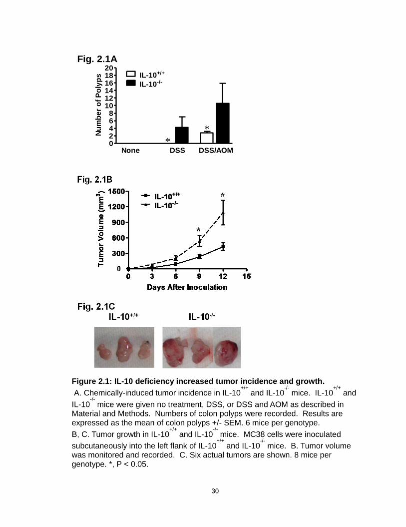
IL-10 deficiency increases tumor incidence, growth, and foci formation.
The immune-inhibitory role of IL-10 has been well defined in numerous
experimental settings. However, the in vivo effects of endogenous IL-10 on
tumorigenesis and tumor immunity are poorly understood. We compared tumor
incidence, growth, and foci formation in IL-10-deficient (IL-10-/-) and wild-type (IL-
29
10+/+) mice. The mice were subjected to administration of dextran sodium sulfate
(DSS) and/or azoxymethane (AOM) as previously described [267]. IL-10-/- mice—
but not IL-10+/+ mice—treated with DSS developed numerous colon polyps. In
the presence of DSS and AOM, colon polyps developed in both IL-10-/- and IL-
10+/+ mice. However, there were more polyps in IL-10-/- mice than IL-10+/+ mice
(Figure 2.1A). We next subcutaneously injected a colon cancer cell line, MC38,
into mice and monitored tumor growth over a period of two weeks. MC38 had
accelerated growth in IL-10-/- mice as compared to IL-10+/+ mice (Figure 2.1B, C).
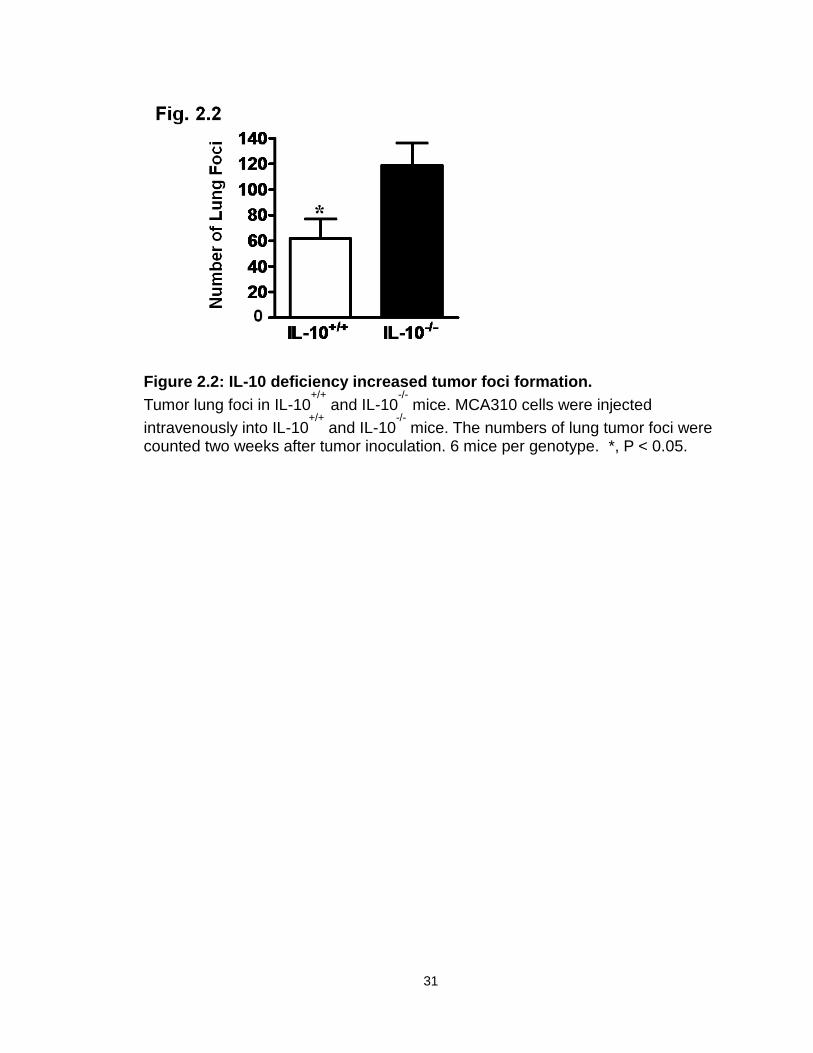
We further examined the effect of endogenous IL-10 on the development of
mouse lung foci. To this end, MCA310, a methylcholanthrene-induced sarcoma,
was intravenously injected into IL-10+/+ and IL-10-/- mice. IL-10-/- mice had more
tumor foci in the lungs than IL-10+/+ mice (Figure 2.2). Thus, IL-10 deficiency
increases tumor incidence, growth, and foci formation.
30
02468
101214161820
IL-10+/+
IL-10-/-
None DSS DSS/AOM*
*
Fig. 2.1A
Nu
mb
er
of
Po
lyp
s
Figure 2.1: IL-10 deficiency increased tumor incidence and growth.
A. Chemically-induced tumor incidence in IL-10+/+
and IL-10-/-
mice. IL-10+/+
and
IL-10-/-
mice were given no treatment, DSS, or DSS and AOM as described in Material and Methods. Numbers of colon polyps were recorded. Results are expressed as the mean of colon polyps +/- SEM. 6 mice per genotype.
B, C. Tumor growth in IL-10+/+
and IL-10-/-
mice. MC38 cells were inoculated
subcutaneously into the left flank of IL-10+/+
and IL-10-/-
mice. B. Tumor volume was monitored and recorded. C. Six actual tumors are shown. 8 mice per genotype. *, P < 0.05.
31
Figure 2.2: IL-10 deficiency increased tumor foci formation.
Tumor lung foci in IL-10+/+
and IL-10-/-
mice. MCA310 cells were injected
intravenously into IL-10+/+
and IL-10-/-
mice. The numbers of lung tumor foci were counted two weeks after tumor inoculation. 6 mice per genotype. *, P < 0.05.

32
IL-10 deficiency decreases immune surveillance in the tumor.
We then investigated the phenotype and cytokine profile of key innate and
adaptive immune cells in the tumor and tumor-draining lymph nodes (TDLN) in
these mice. Not surprisingly, MHC I expression was increased on both DCs
(Figure 2.3A) and MDSCs (Figure 2.3B) in IL-10-/- mice. Interestingly, the
percentage (Figure 2.4A, B) and absolute numbers (Figure 2.4 C) of NK cells
(both NK1.1+CD49b+ and NK1.1-CD49b+ populations) were lower in tumors and
TDLNs in IL-10-/- mice than in IL-10+/+ mice bearing MC38. We further quantified
the numbers of tumor-infiltrating CD8+ T cells. Intriguingly, there were also fewer
tumor-infiltrating CD8+ T cells in IL-10-/- mice than in IL-10+/+ mice (Figure 2.5A).
Furthermore, the expression levels of effector cytokines, including IFNγ and
TNFα, were reduced in CD4+ and CD8+ T cells in the tumors and TDLNs of IL-10-
/- mice (Figure 2.5B). This suggests that IL-10 may influence either the
development or function of effector T cells. In support of this possibility, we
demonstrated that addition of IL-10 directly stimulated basal expression of IFNγ,
TNFα, and IL-2 in CD4+ and CD8+ T cells from IL-10-/- mice (Figure 2.6A), but not
from IL-10+/+ mice (not shown). This suggests that biological levels of
endogenous IL-10 may support effector T cell function. To further support this
possibility, we stimulated IL-10+/+ CD4+ and CD8+ T cells with optimal
concentrations of anti-CD3 and anti-CD28 in culture, and blocked endogenous
levels of IL-10. We observed that blockade of IL-10 reduced IFNγ expression in
CD4+ and CD8+ T cells (Figure 2.6B). These data provide strong evidence that
endogenous IL-10 can support T cell function, and that IL-10 deficiency
decreases immune surveillance in the tumor.
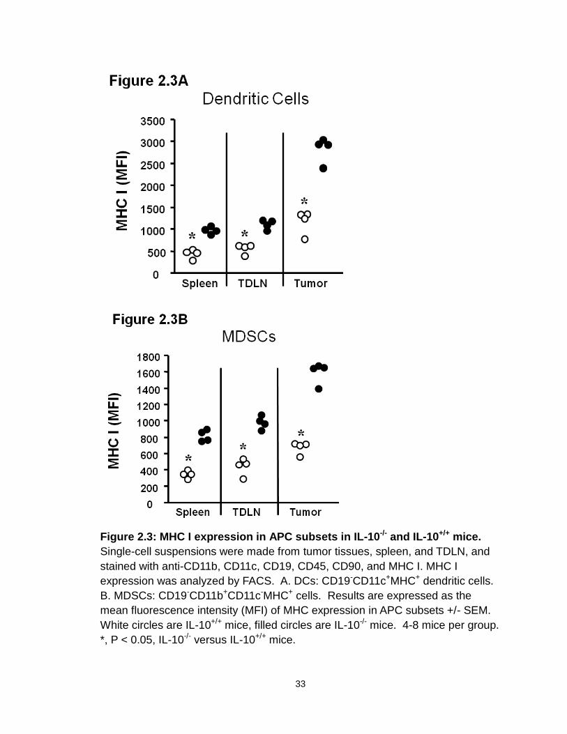
33
Figure 2.3: MHC I expression in APC subsets in IL-10-/- and IL-10+/+ mice.
Single-cell suspensions were made from tumor tissues, spleen, and TDLN, and
stained with anti-CD11b, CD11c, CD19, CD45, CD90, and MHC I. MHC I
expression was analyzed by FACS. A. DCs: CD19-CD11c+MHC+ dendritic cells.
B. MDSCs: CD19-CD11b+CD11c-MHC+ cells. Results are expressed as the
mean fluorescence intensity (MFI) of MHC expression in APC subsets +/- SEM.
White circles are IL-10+/+ mice, filled circles are IL-10-/- mice. 4-8 mice per group.
*, P < 0.05, IL-10-/- versus IL-10+/+ mice.
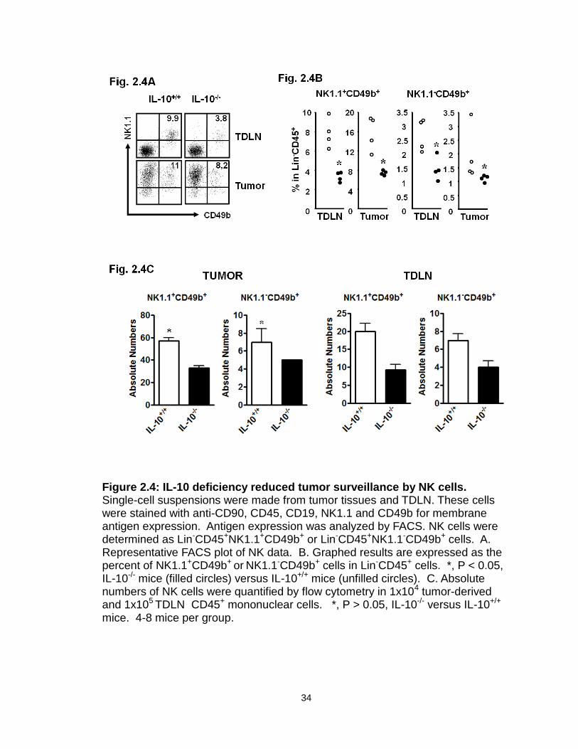
34
Figure 2.4: IL-10 deficiency reduced tumor surveillance by NK cells. Single-cell suspensions were made from tumor tissues and TDLN. These cells were stained with anti-CD90, CD45, CD19, NK1.1 and CD49b for membrane antigen expression. Antigen expression was analyzed by FACS. NK cells were determined as Lin-CD45+NK1.1+CD49b+ or Lin-CD45+NK1.1-CD49b+ cells. A. Representative FACS plot of NK data. B. Graphed results are expressed as the percent of NK1.1+CD49b+ or NK1.1-CD49b+ cells in Lin-CD45+ cells. *, P < 0.05, IL-10-/- mice (filled circles) versus IL-10+/+ mice (unfilled circles). C. Absolute numbers of NK cells were quantified by flow cytometry in 1x10⁴ tumor-derived and 1x10⁵ TDLN CD45+ mononuclear cells. *, P > 0.05, IL-10-/- versus IL-10+/+ mice. 4-8 mice per group.
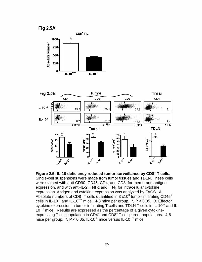
35
Figure 2.5: IL-10 deficiency reduced tumor surveillance by CD8+ T cells. Single-cell suspensions were made from tumor tissues and TDLN. These cells were stained with anti-CD90, CD45, CD4, and CD8, for membrane antigen expression, and with anti-IL-2, TNFα and IFNγ for intracellular cytokine expression. Antigen and cytokine expression was analyzed by FACS. A. Absolute numbers of CD8+ T cells quantified in 3 x10³ tumor-infiltrating CD45+ cells in IL-10-/- and IL-10+/+ mice. 4-8 mice per group. *, P < 0.05. B. Effector cytokine expression in tumor-infiltrating T cells and TDLN T cells in IL-10-/- and IL-10+/+ mice. Results are expressed as the percentage of a given cytokine-expressing T cell population in CD4+ and CD8+ T cell parent populations. 4-8 mice per group. *, P < 0.05, IL-10-/- mice versus IL-10+/+ mice.
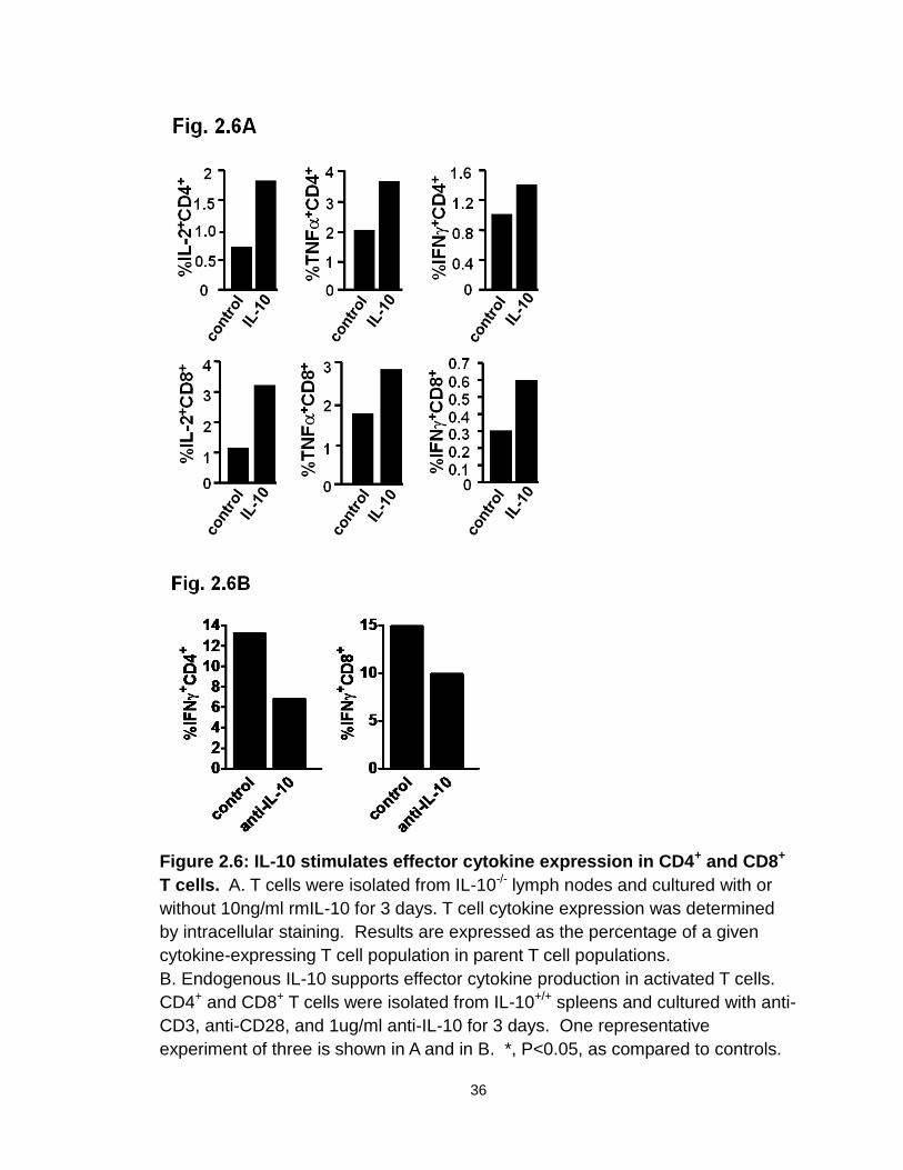
36
Figure 2.6: IL-10 stimulates effector cytokine expression in CD4+ and CD8+
T cells. A. T cells were isolated from IL-10-/- lymph nodes and cultured with or
without 10ng/ml rmIL-10 for 3 days. T cell cytokine expression was determined
by intracellular staining. Results are expressed as the percentage of a given
cytokine-expressing T cell population in parent T cell populations.
B. Endogenous IL-10 supports effector cytokine production in activated T cells.
CD4+ and CD8+ T cells were isolated from IL-10+/+ spleens and cultured with anti-
CD3, anti-CD28, and 1ug/ml anti-IL-10 for 3 days. One representative
experiment of three is shown in A and in B. *, P<0.05, as compared to controls.
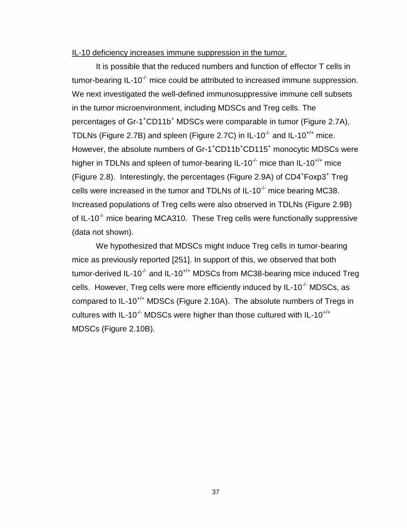
37
IL-10 deficiency increases immune suppression in the tumor.
It is possible that the reduced numbers and function of effector T cells in
tumor-bearing IL-10-/- mice could be attributed to increased immune suppression.
We next investigated the well-defined immunosuppressive immune cell subsets
in the tumor microenvironment, including MDSCs and Treg cells. The
percentages of Gr-1+CD11b+ MDSCs were comparable in tumor (Figure 2.7A),
TDLNs (Figure 2.7B) and spleen (Figure 2.7C) in IL-10-/- and IL-10+/+ mice.
However, the absolute numbers of Gr-1+CD11b+CD115+ monocytic MDSCs were
higher in TDLNs and spleen of tumor-bearing IL-10-/- mice than IL-10+/+ mice
(Figure 2.8). Interestingly, the percentages (Figure 2.9A) of CD4+Foxp3+ Treg
cells were increased in the tumor and TDLNs of IL-10-/- mice bearing MC38.
Increased populations of Treg cells were also observed in TDLNs (Figure 2.9B)
of IL-10-/- mice bearing MCA310. These Treg cells were functionally suppressive
(data not shown).
We hypothesized that MDSCs might induce Treg cells in tumor-bearing
mice as previously reported [251]. In support of this, we observed that both
tumor-derived IL-10-/- and IL-10+/+ MDSCs from MC38-bearing mice induced Treg
cells. However, Treg cells were more efficiently induced by IL-10-/- MDSCs, as
compared to IL-10+/+ MDSCs (Figure 2.10A). The absolute numbers of Tregs in
cultures with IL-10-/- MDSCs were higher than those cultured with IL-10+/+
MDSCs (Figure 2.10B).
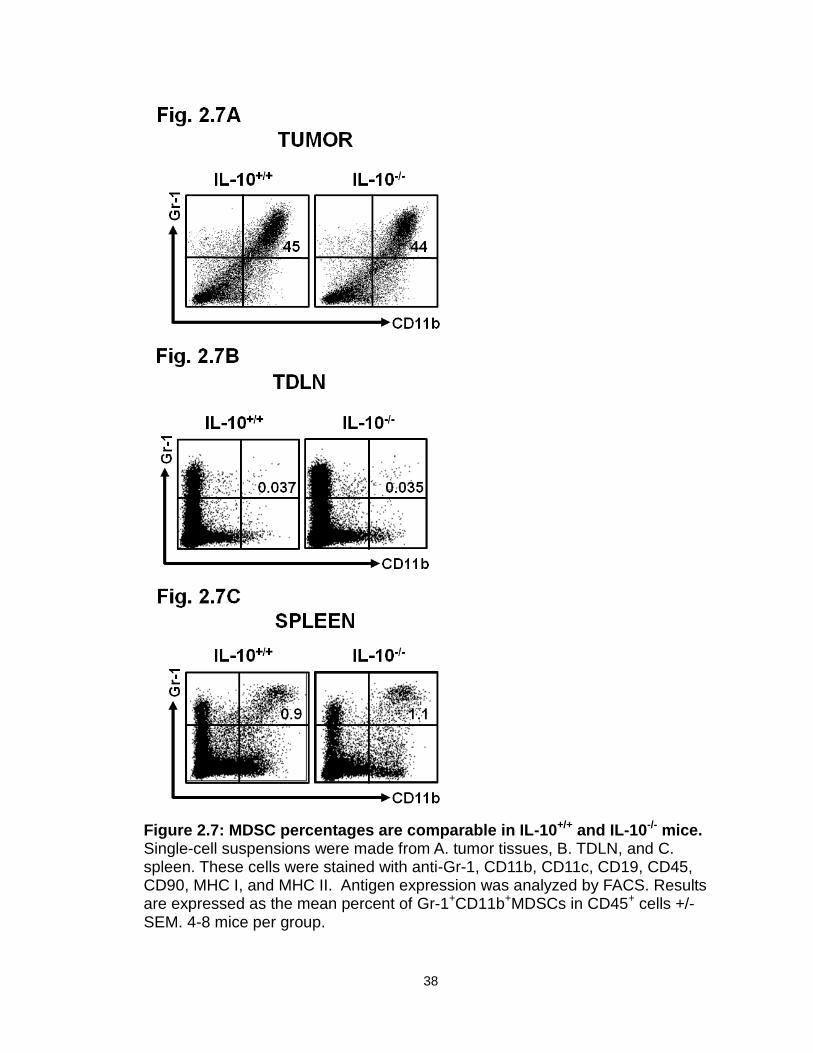
38
Figure 2.7: MDSC percentages are comparable in IL-10+/+ and IL-10-/- mice. Single-cell suspensions were made from A. tumor tissues, B. TDLN, and C. spleen. These cells were stained with anti-Gr-1, CD11b, CD11c, CD19, CD45, CD90, MHC I, and MHC II. Antigen expression was analyzed by FACS. Results are expressed as the mean percent of Gr-1+CD11b+MDSCs in CD45+ cells +/- SEM. 4-8 mice per group.
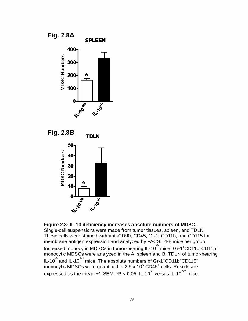
39
Figure 2.8: IL-10 deficiency increases absolute numbers of MDSC. Single-cell suspensions were made from tumor tissues, spleen, and TDLN. These cells were stained with anti-CD90, CD45, Gr-1, CD11b, and CD115 for membrane antigen expression and analyzed by FACS. 4-8 mice per group.
Increased monocytic MDSCs in tumor-bearing IL-10-/-
mice. Gr-1+CD11b+CD115+ monocytic MDSCs were analyzed in the A. spleen and B. TDLN of tumor-bearing
IL-10-/-
and IL-10+/+
mice. The absolute numbers of Gr-1+CD11b+CD115+ monocytic MDSCs were quantified in 2.5 x 10⁵ CD45+ cells. Results are
expressed as the mean +/- SEM. *P < 0.05, IL-10-/-
versus IL-10+/+
mice.
40
Figure 2.9: IL-10 deficiency increases immune suppression.
Increased Treg cells in tumors and TDLNs in IL-10-/-
mice. A. Foxp3+CD4+ Treg
cells were determined in MC38 and MC38-associated TDLNs in IL-10-/-
and IL-
10+/+
mice. B. Foxp3+CD4+ Treg cells were determined in MCA310-associated
TDLNs in IL-10-/-
and IL-10+/+
mice. Results are expressed as the percentage of CD4+Foxp3+ T cells in CD4+ T cells or the mean values +/- SEM. *, P < 0.05, IL-
10-/-
versus IL-10+/+
mice.
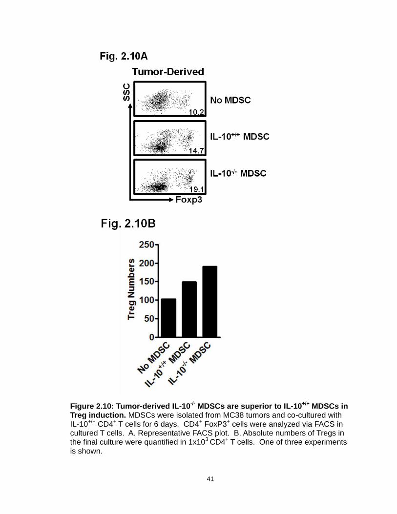
41
Figure 2.10: Tumor-derived IL-10-/- MDSCs are superior to IL-10+/+ MDSCs in Treg induction. MDSCs were isolated from MC38 tumors and co-cultured with IL-10+/+ CD4+ T cells for 6 days. CD4+ FoxP3+ cells were analyzed via FACS in cultured T cells. A. Representative FACS plot. B. Absolute numbers of Tregs in the final culture were quantified in 1x10³ CD4+ T cells. One of three experiments is shown.
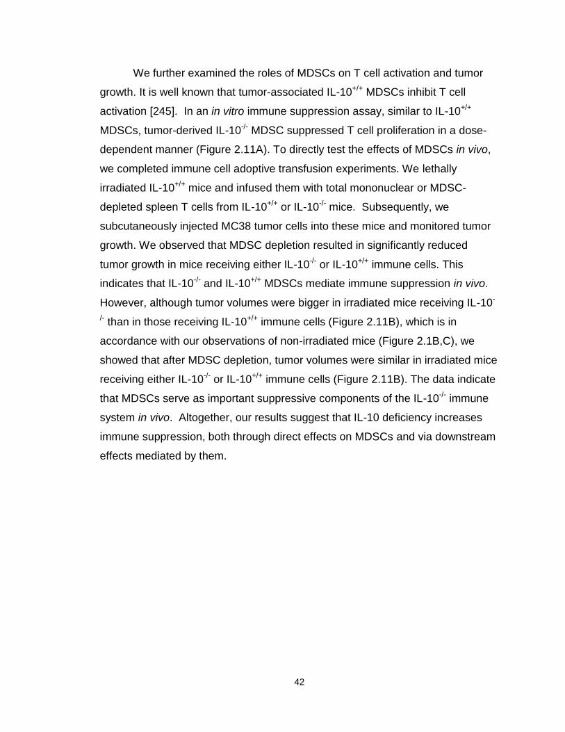
42
We further examined the roles of MDSCs on T cell activation and tumor
growth. It is well known that tumor-associated IL-10+/+ MDSCs inhibit T cell
activation [245]. In an in vitro immune suppression assay, similar to IL-10+/+
MDSCs, tumor-derived IL-10-/- MDSC suppressed T cell proliferation in a dose-
dependent manner (Figure 2.11A). To directly test the effects of MDSCs in vivo,
we completed immune cell adoptive transfusion experiments. We lethally
irradiated IL-10+/+ mice and infused them with total mononuclear or MDSC-
depleted spleen T cells from IL-10+/+ or IL-10-/- mice. Subsequently, we
subcutaneously injected MC38 tumor cells into these mice and monitored tumor
growth. We observed that MDSC depletion resulted in significantly reduced
tumor growth in mice receiving either IL-10-/- or IL-10+/+ immune cells. This
indicates that IL-10-/- and IL-10+/+ MDSCs mediate immune suppression in vivo.
However, although tumor volumes were bigger in irradiated mice receiving IL-10-
/- than in those receiving IL-10+/+ immune cells (Figure 2.11B), which is in
accordance with our observations of non-irradiated mice (Figure 2.1B,C), we
showed that after MDSC depletion, tumor volumes were similar in irradiated mice
receiving either IL-10-/- or IL-10+/+ immune cells (Figure 2.11B). The data indicate
that MDSCs serve as important suppressive components of the IL-10-/- immune
system in vivo. Altogether, our results suggest that IL-10 deficiency increases
immune suppression, both through direct effects on MDSCs and via downstream
effects mediated by them.
43
Figure 2.11: IL-10-/- MDSC suppress T cell activation and impede antitumor immunity. A. Tumor-associated IL-10-/- MDSCs mediate immune suppression in vitro. MDSCs were sorted from MC38 tumors in IL-10-/- mice and cultured with naïve T cells in different ratios as detailed in Materials and Methods. T cell proliferation was determined by thymidine incorporation. Results are expressed as the mean of counts per minute (CPM) +/- SEM in triplicates. Similar results were observed with IL-10+/+ MDSCs. One of three experiments is shown. **P < 0.01 compared to control (ratio 1:0). B. MDSCs suppressed antitumor immunity in vivo. IL-10+/+ mice were lethally irradiated and given an infusion of IL-10+/+ or IL-10-/- whole spleen cells or spleen cells depleted of MDSCs. On the same day, mice were injected subcutaneously with MC38. Tumor growth was measured over two weeks. 3-4 mice per group. *P < 0.05, Day 9-15: IL-10-/- vs. IL-10+/+, IL-10-/- vs. MDSC-depleted IL-10-/- ; and Day 12-15 : IL-10+/+ vs. MDSC-depleted IL-10+/+.
44
IL-1 contributes to increased tumor growth in IL-10-/- mice.
After demonstrating the cellular mechanisms which may contribute to
increased tumor incidence, growth and foci formation in IL-10-/- mice, we
examined the relevant molecular patterns associated with such phenomena.
Interestingly, myeloid cells—including MDSCs—expressed higher levels of IL-1α
and IL-1β in IL-10-/- than IL-10+/+ mice (Figure 2.12A). We then investigated the
role of IL-1 in tumor growth in IL-10-/- mice. Anakinra, the recombinant human IL-
1 receptor antagonist (IL-1Ra), has demonstrated biological efficacy in mice
[268]. We treated tumor-bearing IL-10-/- and IL-10+/+ mice with Anakinra or
vehicle over a period of three weeks. We observed that IL-10-/- mice treated with
Anakinra had reduced tumor burden (Figure 2.12B). Interestingly, tumor growth
was comparable in IL-10+/+ mice treated with Anakinra and those treated with
PBS vehicle (Figure 2.12C). In further support of the relevance of IL-1, we
showed that tumor volume was decreased in IL-1 receptor-deficient (IL-1R-/-)
mice as compared to IL-1R+/+ mice (Figure 2.13). It is likely that IL-1 contributes
to the increased tumor growth observed in IL-10-/- mice.
IL-1 blockade enhances effector T cell tumor infiltration and reduces tumor
angiogenesis in IL-10-/- mice.
We next examined tumor-infiltrating T cells in IL-10-/- mice treated with
Anakinra. Immunofluorescent staining revealed that there were more tumor-
infiltrating CD8+ T cells in IL-10-/- mice treated with Anakinra as compared to
control (Figure 2.14A). The levels of intratumoral IFNγ+CD8+ T cells were also
increased in mice treated with Anakinra (Figure 2.14B). Although the levels of
MDSCs were comparable in the two groups (not shown), the numbers of tumor
infiltrating Treg cells were reduced in mice treated with Anakinra (Figure 2.14C).
Furthermore, tumor microvessel intensity and size (Figure 2.15) were reduced by
Anakinra treatment. Altogether, the data suggest that IL-1 blockade promotes
tumor immune surveillance and reduces tumor angiogenesis.
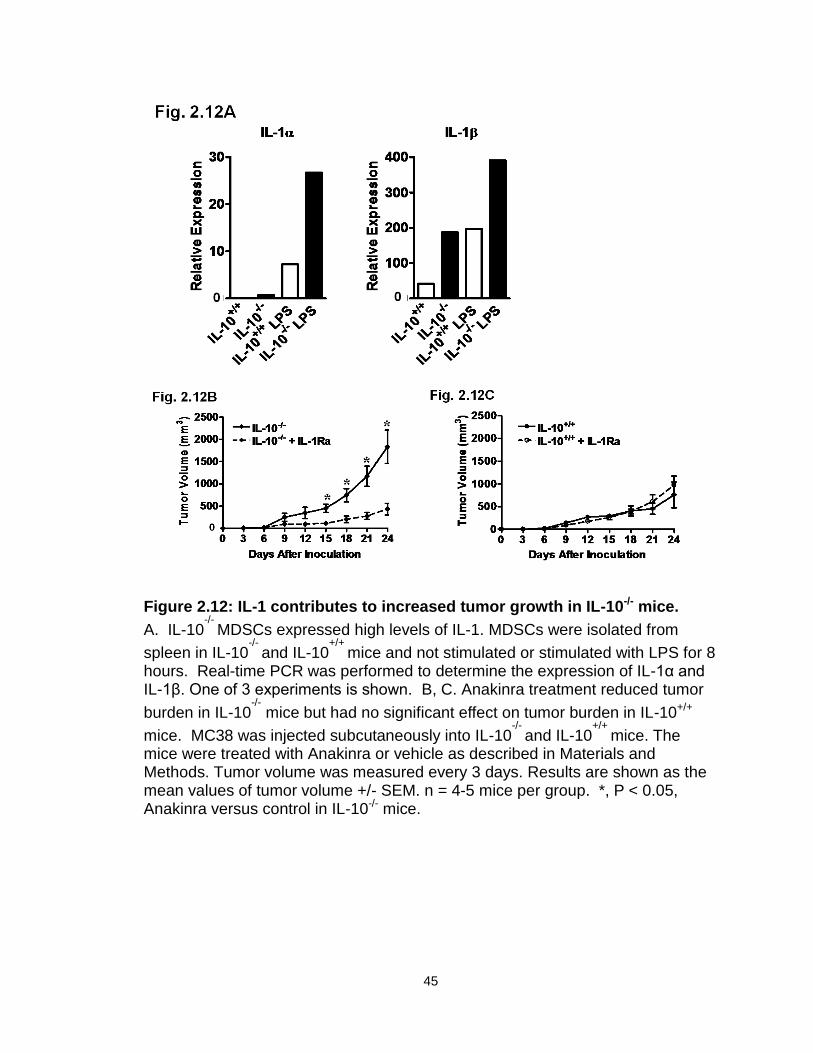
45
Figure 2.12: IL-1 contributes to increased tumor growth in IL-10-/- mice.
A. IL-10-/-
MDSCs expressed high levels of IL-1. MDSCs were isolated from
spleen in IL-10-/-
and IL-10+/+
mice and not stimulated or stimulated with LPS for 8 hours. Real-time PCR was performed to determine the expression of IL-1α and IL-1β. One of 3 experiments is shown. B, C. Anakinra treatment reduced tumor
burden in IL-10-/-
mice but had no significant effect on tumor burden in IL-10+/+
mice. MC38 was injected subcutaneously into IL-10-/-
and IL-10+/+
mice. The mice were treated with Anakinra or vehicle as described in Materials and Methods. Tumor volume was measured every 3 days. Results are shown as the mean values of tumor volume +/- SEM. n = 4-5 mice per group. *, P < 0.05, Anakinra versus control in IL-10-/- mice.
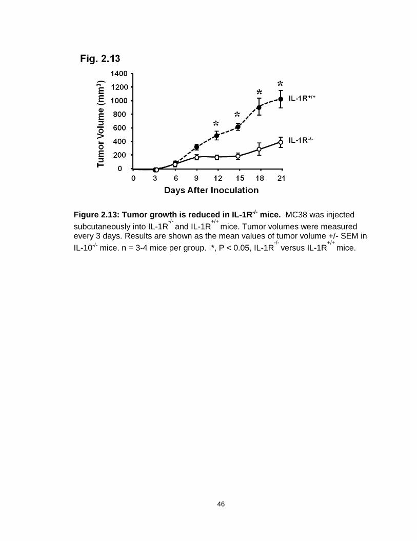
46
Figure 2.13: Tumor growth is reduced in IL-1R-/- mice. MC38 was injected
subcutaneously into IL-1R-/-
and IL-1R+/+
mice. Tumor volumes were measured every 3 days. Results are shown as the mean values of tumor volume +/- SEM in
IL-10-/- mice. n = 3-4 mice per group. *, P < 0.05, IL-1R-/-
versus IL-1R+/+
mice.
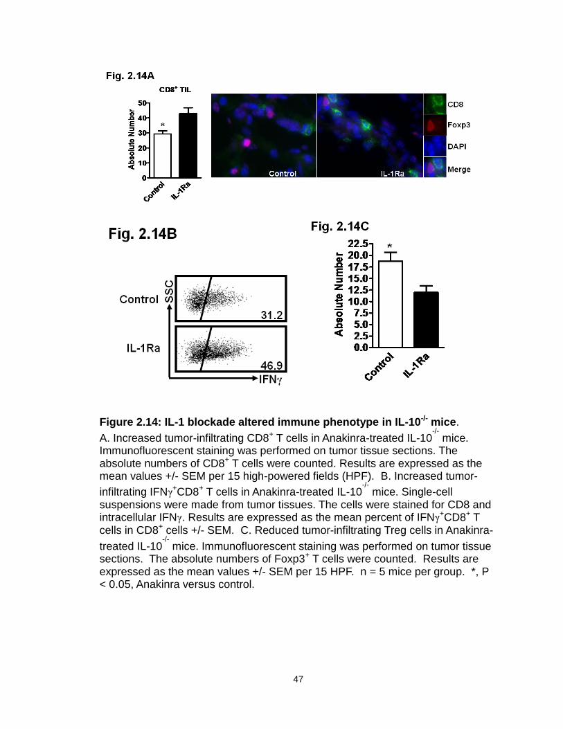
47
Figure 2.14: IL-1 blockade altered immune phenotype in IL-10-/- mice.
A. Increased tumor-infiltrating CD8+ T cells in Anakinra-treated IL-10-/-
mice. Immunofluorescent staining was performed on tumor tissue sections. The absolute numbers of CD8+ T cells were counted. Results are expressed as the mean values +/- SEM per 15 high-powered fields (HPF). B. Increased tumor-
infiltrating IFNγ+CD8+ T cells in Anakinra-treated IL-10
-/- mice. Single-cell
suspensions were made from tumor tissues. The cells were stained for CD8 and intracellular IFNγ. Results are expressed as the mean percent of IFNγ
+CD8+ T cells in CD8+ cells +/- SEM. C. Reduced tumor-infiltrating Treg cells in Anakinra-
treated IL-10-/-
mice. Immunofluorescent staining was performed on tumor tissue sections. The absolute numbers of Foxp3+ T cells were counted. Results are expressed as the mean values +/- SEM per 15 HPF. n = 5 mice per group. *, P < 0.05, Anakinra versus control.
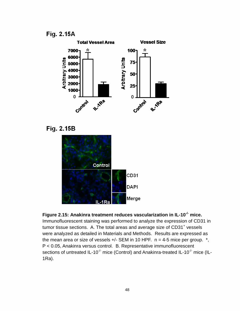
48
Figure 2.15: Anakinra treatment reduces vascularization in IL-10-/- mice.
Immunofluorescent staining was performed to analyze the expression of CD31 in
tumor tissue sections. A. The total areas and average size of CD31+ vessels
were analyzed as detailed in Materials and Methods. Results are expressed as
the mean area or size of vessels +/- SEM in 10 HPF. n = 4-5 mice per group. *,
P < 0.05, Anakinra versus control. B. Representative immunofluorescent
sections of untreated IL-10-/- mice (Control) and Anakinra-treated IL-10-/- mice (IL-
1Ra).
49
Discussion.
Here we present a series of experiments demonstrating that endogenous
IL-10 plays a stimulatory role in antitumor immunity. Our data challenge
commonly-held beliefs about the immune regulatory role of IL-10 and suggest a
more complex IL-10 biology, at least in the context of tumor immunology (Figure
2.16).
IL-10 has been thought to be broadly immune suppressive. To our
surprise, chemically-induced tumor incidence, transplanted tumor growth, and
lung foci formation are increased in IL-10-/- mice. This is associated with a
number of immune phenotypic and functional signatures.
We have observed reduced NK cells, CD8+ effector T cells and effector T
cell cytokine expression in tumor and TDLN of tumor-bearing IL-10-/- mice. This
indicates that endogenous IL-10 is crucial for tumor immune surveillance.
Consistent with our observations, earlier studies in mice have shown that
exogenous IL-10 promoted tumor immunity and led to reduced tumor growth or
tumor rejection [43,46,256]. However, the underlying mechanisms by which IL-10
mediates suppression of tumor growth despite its defined immune-inhibitory
functions remained elusive. Furthermore, it remained unknown if endogenous IL-
10 could mediate protective tumor immunity. Our studies suggest that the
biological activities of IL-10 may be highly context-dependent, and vary in the
presence of different cellular targets, phases of immune responses and disease
model systems.
We show that IL-10 directly targets and stimulates T cell effector cytokine
production. The levels of effector cytokines, including TNFα and IFNγ, are
reduced in IL-10-/- tumor-bearing mice and may be recovered by addition of IL-
10. Additionally, IL-10 blockade reduces effector cytokine production by IL-10+/+ T
cells. The data indicate that IL-10 may target effector T cells to maintain and
enhance their functionalities. In support of this possibility, it has been reported
that IL-10 may promote CD8 differentiation and expansion [33-34,269]. In fact,
humans treated with IL-10 showed a reduction of IL-1 and increased IFNγ [270-
271].
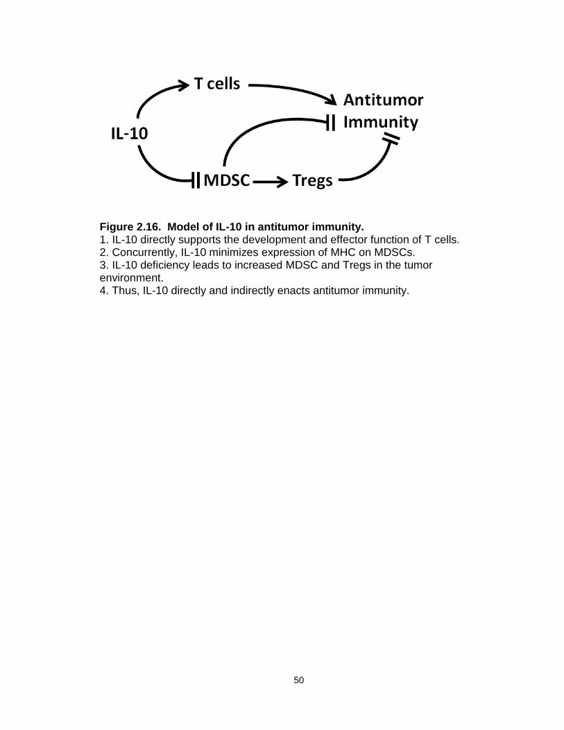
50
Figure 2.16. Model of IL-10 in antitumor immunity. 1. IL-10 directly supports the development and effector function of T cells. 2. Concurrently, IL-10 minimizes expression of MHC on MDSCs. 3. IL-10 deficiency leads to increased MDSC and Tregs in the tumor environment. 4. Thus, IL-10 directly and indirectly enacts antitumor immunity.
51
Given that IL-10 can be immune-stimulatory and inhibitory, the balance of
these effects may determine whether IL-10 is beneficial or detrimental in a given
model system and/or disease stage. The immunoregulatory roles of IL-10 are
prominently defined in infectious disease models. In these scenarios, APCs are
the immediate and predominant targets of IL-10 [269]. Infectious pathogens
rapidly activate Toll-like receptors on APCs, and induce antigen-specific T cell
priming. Exogenous or induced endogenous IL-10 inhibits the maturation and
function of APCs via reduced MHC expression and IL-12 production, and
subsequently suppresses T cell priming and Th1-type responses [272-273]. In a
tumor-bearing host, however, when there is no obvious acute phase of immune
response [266], tumor-associated antigen (TAA)-specific priming may slowly
occur, and IL-10 may be gradually induced. Thus, IL-10 may preferentially target
and promote effector T cells during the effector phase of immune responses in
tumor-bearing hosts [269]. This may partially explain the discrepancies
concerning IL-10 biology in the literature.
It has been reported that IL-1 supports the development of MDSCs in
tumor-bearing mice [261-262,274]. In line with this possibility, IL-10-/- myeloid
cells express high levels of IL-1, and there are more monocytic MDSCs in IL-10-/-
tumor-bearing mice. MDSCs, particularly CD115+ monocytic MDSCs, promote
Treg cell development [251]. Consistent with this, we have observed that IL-10-/-
MDSCs efficiently suppress T cell expansion and are stronger inducers of Treg
cells than IL-10+/+ MDSCs. It has been suggested that loss of MHC or mutations
in MHC expression machinery may result in limited tumor antigen recognition and
presentation [275]. Interestingly, IL-10-/- MDSCs express high levels of MHC
molecules, and may efficiently present self-antigens to Treg cells, thus activating
and expanding Treg cells as we have observed. This may explain why IL-10-/-
MDSCs are superior to IL-10+/+ MDSCs in Treg cell induction. Perhaps most
tellingly, depletion of MDSCs in our immune cell-transfer model profoundly
reduces tumor volume, demonstrating that the presence of MDSCs has a
negative impact on host immunity in vivo. Additionally, tumor burden in mice
receiving IL-10-/- cells depleted of MDSCs was reduced by a larger volume than
52
in those mice receiving IL-10+/+ cells depleted of MDSCs. This suggests that
MDSCs that develop in the absence of IL-10 may mediate more powerful
suppressive activities in tumor-bearing mice. However, although IL-1 promotes
the development of MDSCs in tumor-bearing mice [261-262,274], intratumoral
and tumor-draining lymph node MDSC number did not change after IL-1Ra
administration in IL-10-/- mice. We reason that the effects of IL-1 on MDSCs may
be more long-lasting than can be reversed with temporary IL-1 signaling
blockade.
IL-10 suppresses the expression of inflammatory cytokines in chronic
inflammation models [272-273]. IL-10-/- mice develop chronic colitis in
conventional conditions [19]. Although there is no obvious inflammation
(including colitis) in IL-10-/- mice when housed in specific pathogen-free facilities,
IL-10-/- myeloid cells express high levels of IL-1, which may suggest the presence
of microscopic inflammation in these mice. This inflammation may be increased
after tumor inoculation or induction. Inflammation is often linked to increased
tumor vascularization, and IL-1 has long been known as a pro-angiogenic
cytokine [276] and promoter of tumorigenesis [277]. In line with this, IL-1
blockade reduces tumor microvessel density and size in IL-10-/- mice. However, it
is also possible that the reduced density of tumor microvessels may be due to
the resulting enhanced T cell immunity that includes IFNγ production. IFNγ is a
potent anti-angiogeneic factor [278]. Finally, we have demonstrated that IL-1
blockade reduces tumor growth in IL-10-/- mice. Accordingly, the levels of
intratumoral Treg cells are reduced, and the numbers of tumor-infiltrating effector
T cells and effector cytokines are enhanced. Based upon our observations, IL-10
both directly and indirectly supports T cell immunity by inhibiting IL-1, and
reduces IL-1-mediated MDSCs and Treg cells in tumor-bearing hosts.
In summary, in contrast to the established views, endogenous IL-10 is
negatively linked to the development of immunosuppressive MDSCs and Treg
cells, subverts tumor immune suppression, induces the activation of tumor-
infiltrating effector T cells, and in turn contributes to anti-tumor immunity. Our
53
data indicate that the biological activities of IL-10 can be highly context-
dependent.
54
Chapter 3
Interleukin-10 and autoimmunity
IL-10-/- mice housed in conventional conditions develop chronic
enterocolitis and then colon adenocarcinoma [279]. IL-10-/- colitis shares many
architectural signatures with human CD [19], and this disease progression partly
mimics IBD-associated cancer in humans. Colitic mice display some
histopathological signatures associated with human IBD, including increased
numbers of focal ulcerations and transmural lesions, like CD patients [279].
Perhaps not surprisingly, patients with CD or colitis have a highly increased risk
of cancer in comparison to healthy individuals [280], and the IL-10-/- mouse now
serves as a useful model of spontaneous inflammation-induced cancer. It was
also noted early on that a lymphocyte population in colitic mice stimulated by IL-
23 produced IL-6 and IL-17 [128]. A subsequent report in 2009 demonstrated an
increase in the IL-17 levels of mesenteric lymph node cultures of IL-10-/- mice
[281]. We set out to analyze the prevalence and distribution of Th17 cells in IL-
10-/- mice, investigate the molecular causes of increased Th17 populations (if this
phenomenon is systemic), and examine whether a similar physiological
relationship existed in human conditions of disrupted IL-10 production.
IL-10-/- dendritic cells are superior Th17 cell inducers in mice.
First, we examined the cellular and molecular link between IL-10 and Th17
cells in IL-10-/- and IL-10+/+ mice. We observed increased levels of Th17 cells in
the lymph nodes, spleen, blood, and intestines (also specifically in the colon) of
IL-10-/- mice as compared to WT mice (Fig. 3A, B). Interestingly, we also found
increased levels of IL-17+CD8+ (Tc17) cells in multiple IL-10-/- organs (Fig. 3C).
There were no differences in other immune cell subsets, including B cells,
macrophages, dendritic cells, granulocytes, and natural killer (NK) cells between
IL-10-/- and IL-10+/+ mice (not shown). It is of note that there was a significant
55
decrease in the intestinal regulatory T cell population of IL-10-/- mice, an
observation in line with a recent publication documenting the importance of IL-10
in maintaining FoxP3 expression [18]. Thus, Th17 cells are spontaneously and
systemically increased in IL-10-/- mice.

56
Figure 3.1: IL-17+ cells are increased in IL-10-/- mice. Total mononuclear cells from unchallenged Il-10+/+ and IL-10-/- mouse organs were analyzed via flow cytometry. A. Th17 cell percentages in CD4+ T cell parent population. 6 mice per group, *p<0.05. B. Colons from three IL-10+/+ and three IL-10-/- mice were pooled, processed and analyzed for Th17 cells via flow cytometry. One experiment of two with three mice per group each. C. IL-17+ cell percentages in CD8+ T cell parent population. 6 mice per group, *p<0.05.
57
We next studied the potential underlying mechanisms causing the spontaneous
increase of Th17 cells in IL-10-/- mice. To this end, IL-10+/+ and IL-10-/-
splenocytes were cultured under Th17-polarizing conditions. We observed that
there were more Th17 cells in the IL-10-/- spleen cultures and more IL-17 in the
IL-10-/- culture supernatant when compared to IL-10+/+ cultures (Fig. 3.2A, B).
We then investigated the role of IL-10-/- DCs in Th17 cell induction. We co-
cultured IL-10+/+ T cells with IL-10+/+ or IL-10-/- DCs, and examined the resulting
cellular phenotypes. We found increased Th17 cells in co-cultures with IL-10-/-
DCs as compared to those with IL-10+/+ DCs (Fig. 3.2C). Thus, IL-10-/- DCs are
superior inducers of Th17 cells.
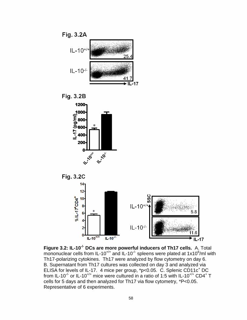
58
Figure 3.2: IL-10-/- DCs are more powerful inducers of Th17 cells. A. Total mononuclear cells from IL-10+/+ and IL-10-/- spleens were plated at 1x10⁶/ml with Th17-polarizing cytokines. Th17 were analyzed by flow cytometry on day 6. B. Supernatant from Th17 cultures was collected on day 3 and analyzed via ELISA for levels of IL-17. 4 mice per group, *p<0.05. C. Splenic CD11c+ DC from IL-10-/- or IL-10+/+ mice were cultured in a ratio of 1:5 with IL-10+/+ CD4+ T cells for 5 days and then analyzed for Th17 via flow cytometry, *P<0.05. Representative of 6 experiments.
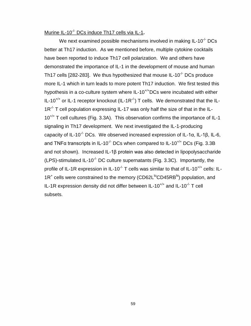
59
Murine IL-10-/- DCs induce Th17 cells via IL-1.
We next examined possible mechanisms involved in making IL-10-/- DCs
better at Th17 induction. As we mentioned before, multiple cytokine cocktails
have been reported to induce Th17 cell polarization. We and others have
demonstrated the importance of IL-1 in the development of mouse and human
Th17 cells [282-283]. We thus hypothesized that mouse IL-10-/- DCs produce
more IL-1 which in turn leads to more potent Th17 induction. We first tested this
hypothesis in a co-culture system where IL-10+/+DCs were incubated with either
IL-10+/+ or IL-1 receptor knockout (IL-1R-/-) T cells. We demonstrated that the IL-
1R-/- T cell population expressing IL-17 was only half the size of that in the IL-
10+/+ T cell cultures (Fig. 3.3A). This observation confirms the importance of IL-1
signaling in Th17 development. We next investigated the IL-1-producing
capacity of IL-10-/- DCs. We observed increased expression of IL-1α, IL-1β, IL-6,
and TNFα transcripts in IL-10-/- DCs when compared to IL-10+/+ DCs (Fig. 3.3B
and not shown). Increased IL-1β protein was also detected in lipopolysaccharide
(LPS)-stimulated IL-10-/- DC culture supernatants (Fig. 3.3C). Importantly, the
profile of IL-1R expression in IL-10-/- T cells was similar to that of IL-10+/+ cells: IL-
1R+ cells were constrained to the memory (CD62LloCD45RBhi) population, and
IL-1R expression density did not differ between IL-10+/+ and IL-10-/- T cell
subsets.
60
Figure 3.3: IL-10-/- DC produce more IL-1 than IL-10+/+ DC. A. Splenic CD11c+ DC from IL-1R+/+ mice were cultured in a ratio of 1:5 with CD4+ T cells from IL-1R+/+ or IL-1R-/- mice for 5 days and then analyzed for Th17 via flow cytometry,*p<0.05. Representative of 3 experiments. B. DC from IL-10+/+ or IL-10-/- mice were cultured for 8 hours with or without LPS. IL-1α and IL-1β message was quantified via RT-PCR. Representative of 3 experiments. C. DC from IL-10+/+ or IL-10-/- mice were cultured for 48 hours with or without LPS. Supernatant was analyzed for IL-1β via ELISA. *p<0.05, average of 3 experiments.
61
In order to determine whether this increased IL-1 was involved in the stronger
Th17 induction documented in our experiments with IL-10-/- DCs, we added anti-
IL-1R monoclonal antibody to the co-cultures of T cells with IL-10+/+ or IL-10-/-
DCs. Blockade of IL-1R, but not IL-6 or TNFα, resulted in significantly decreased
Th17 cells and IL-17 levels in both culture supernatants (Fig. 3.4A, B, and not
shown). Finally, we investigated whether blockade of IL-1 signaling had any
impact on Th17 in vivo. We administered the human recombinant IL-1R
antagonist (IL-1Ra) Anakinra (Kineret ®), already shown to have efficacy in mice
[268], to IL-10-/- mice and analyzed Th17 cells in different organs. As expected,
in vivo IL-1 blockade decreased Th17 cells (Fig. 3.5 A, B) and Tc17 cells (Fig.
3.5C) in most IL-10-/- organs evaluated. Interestingly, treatment did not affect
Th17 population size in the intestine in general or, upon closer examination, in
the colon particularly. We speculate that because the gastrointestinal tract is one
of the sites of the most rapid cell turnover in the body, and because potential
microscopic inflammation in the gastrointestinal tract may induce rapid Th17
recruitment, Th17 cells may be able to repopulate rather quickly in this organ. It
is also possible that IL-1Ra cannot penetrate or reach the GI tissue efficiently
(recall that IL-1R distribution is widespread, and IL-1Ra may saturate receptors in
other organs before reaching the gut). These data indicate that increased IL-1
production by IL-10-/- DCs is at least partly responsible for the stronger Th17
induction in IL-10-/- mice.
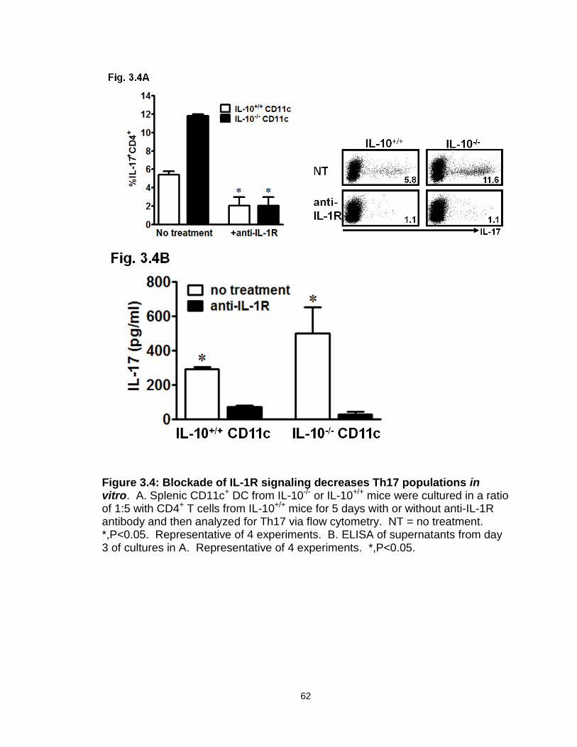
62
Figure 3.4: Blockade of IL-1R signaling decreases Th17 populations in vitro. A. Splenic CD11c+ DC from IL-10-/- or IL-10+/+ mice were cultured in a ratio of 1:5 with CD4+ T cells from IL-10+/+ mice for 5 days with or without anti-IL-1R antibody and then analyzed for Th17 via flow cytometry. NT = no treatment. *,P<0.05. Representative of 4 experiments. B. ELISA of supernatants from day 3 of cultures in A. Representative of 4 experiments. *,P<0.05.
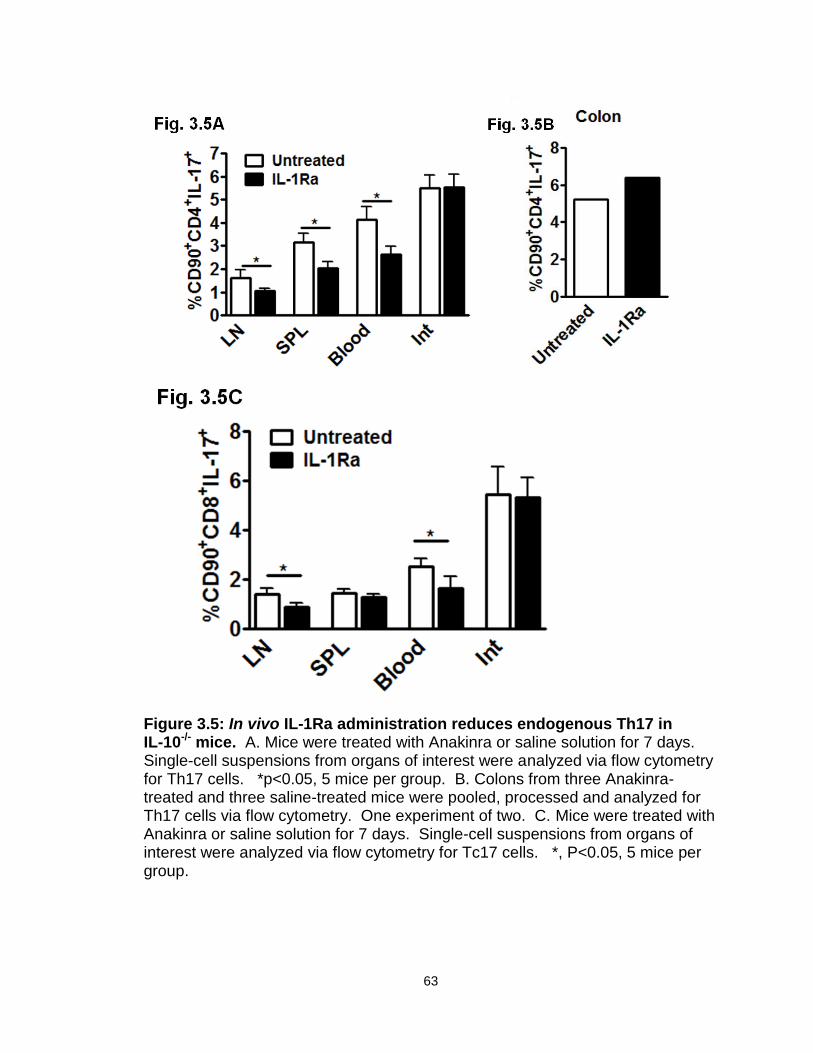
63
Figure 3.5: In vivo IL-1Ra administration reduces endogenous Th17 in IL-10-/- mice. A. Mice were treated with Anakinra or saline solution for 7 days. Single-cell suspensions from organs of interest were analyzed via flow cytometry for Th17 cells. *p<0.05, 5 mice per group. B. Colons from three Anakinra-treated and three saline-treated mice were pooled, processed and analyzed for Th17 cells via flow cytometry. One experiment of two. C. Mice were treated with Anakinra or saline solution for 7 days. Single-cell suspensions from organs of interest were analyzed via flow cytometry for Tc17 cells. *, P<0.05, 5 mice per group.
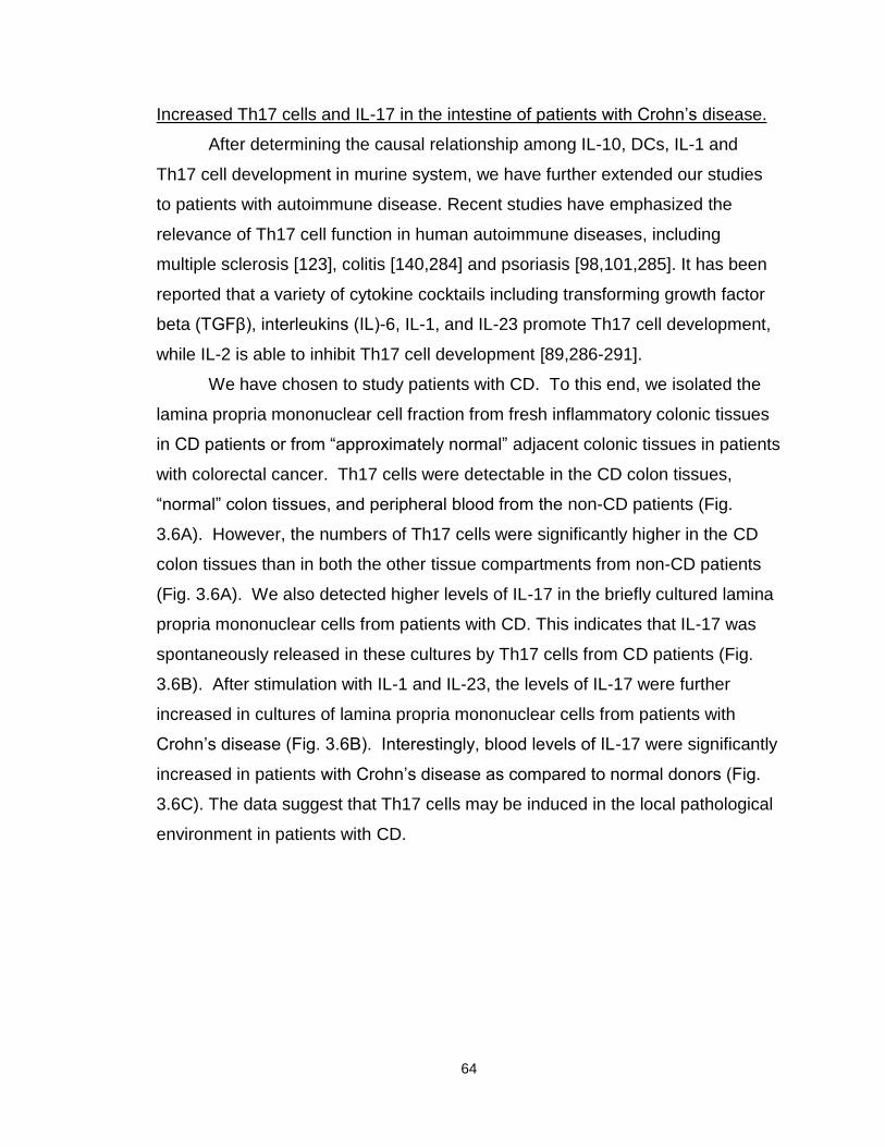
64
Increased Th17 cells and IL-17 in the intestine of patients with Crohn’s disease.
After determining the causal relationship among IL-10, DCs, IL-1 and
Th17 cell development in murine system, we have further extended our studies
to patients with autoimmune disease. Recent studies have emphasized the
relevance of Th17 cell function in human autoimmune diseases, including
multiple sclerosis [123], colitis [140,284] and psoriasis [98,101,285]. It has been
reported that a variety of cytokine cocktails including transforming growth factor
beta (TGFβ), interleukins (IL)-6, IL-1, and IL-23 promote Th17 cell development,
while IL-2 is able to inhibit Th17 cell development [89,286-291].
We have chosen to study patients with CD. To this end, we isolated the
lamina propria mononuclear cell fraction from fresh inflammatory colonic tissues
in CD patients or from “approximately normal” adjacent colonic tissues in patients
with colorectal cancer. Th17 cells were detectable in the CD colon tissues,
“normal” colon tissues, and peripheral blood from the non-CD patients (Fig.
3.6A). However, the numbers of Th17 cells were significantly higher in the CD
colon tissues than in both the other tissue compartments from non-CD patients
(Fig. 3.6A). We also detected higher levels of IL-17 in the briefly cultured lamina
propria mononuclear cells from patients with CD. This indicates that IL-17 was
spontaneously released in these cultures by Th17 cells from CD patients (Fig.
3.6B). After stimulation with IL-1 and IL-23, the levels of IL-17 were further
increased in cultures of lamina propria mononuclear cells from patients with
Crohn’s disease (Fig. 3.6B). Interestingly, blood levels of IL-17 were significantly
increased in patients with Crohn’s disease as compared to normal donors (Fig.
3.6C). The data suggest that Th17 cells may be induced in the local pathological
environment in patients with CD.
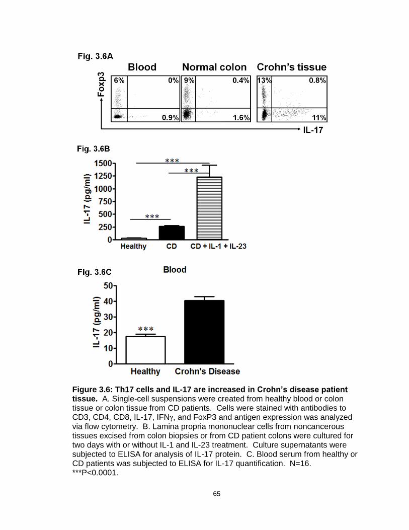
65
Figure 3.6: Th17 cells and IL-17 are increased in Crohn’s disease patient tissue. A. Single-cell suspensions were created from healthy blood or colon tissue or colon tissue from CD patients. Cells were stained with antibodies to CD3, CD4, CD8, IL-17, IFNγ, and FoxP3 and antigen expression was analyzed via flow cytometry. B. Lamina propria mononuclear cells from noncancerous tissues excised from colon biopsies or from CD patient colons were cultured for two days with or without IL-1 and IL-23 treatment. Culture supernatants were subjected to ELISA for analysis of IL-17 protein. C. Blood serum from healthy or CD patients was subjected to ELISA for IL-17 quantification. N=16. ***P<0.0001.
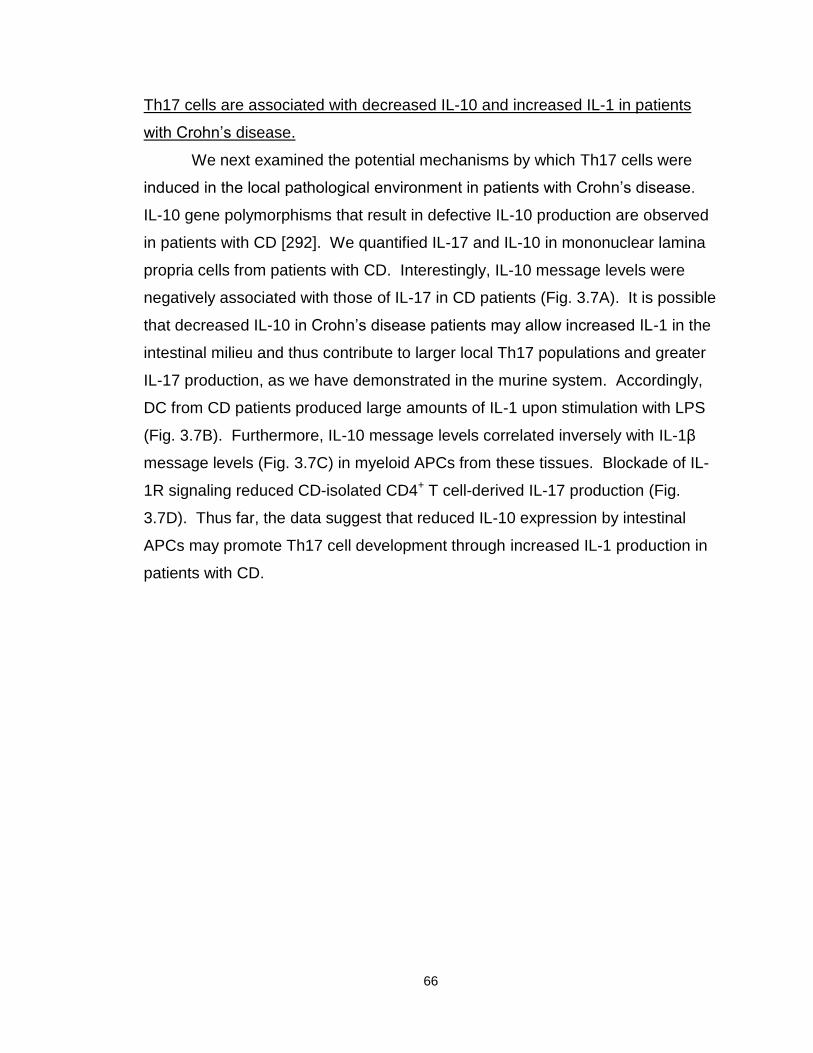
66
Th17 cells are associated with decreased IL-10 and increased IL-1 in patients
with Crohn’s disease.
We next examined the potential mechanisms by which Th17 cells were
induced in the local pathological environment in patients with Crohn’s disease.
IL-10 gene polymorphisms that result in defective IL-10 production are observed
in patients with CD [292]. We quantified IL-17 and IL-10 in mononuclear lamina
propria cells from patients with CD. Interestingly, IL-10 message levels were
negatively associated with those of IL-17 in CD patients (Fig. 3.7A). It is possible
that decreased IL-10 in Crohn’s disease patients may allow increased IL-1 in the
intestinal milieu and thus contribute to larger local Th17 populations and greater
IL-17 production, as we have demonstrated in the murine system. Accordingly,
DC from CD patients produced large amounts of IL-1 upon stimulation with LPS
(Fig. 3.7B). Furthermore, IL-10 message levels correlated inversely with IL-1β
message levels (Fig. 3.7C) in myeloid APCs from these tissues. Blockade of IL-
1R signaling reduced CD-isolated CD4+ T cell-derived IL-17 production (Fig.
3.7D). Thus far, the data suggest that reduced IL-10 expression by intestinal
APCs may promote Th17 cell development through increased IL-1 production in
patients with CD.

67
Figure 3.7: IL-17 in Crohn’s disease patients is associated with increased
IL-1 and decreased IL-10. A. IL-17 and IL-10 message from fresh CD
mononuclear lamina propria cells were quantified via RT-PCR. ***, P<0.0001,
Chi-squared (χ2) test. B. IL-1 protein was quantified in supernatant of colon DC
isolated from CD patients cultured for two days. N = 6. **, P<0.005. C. IL-1 and
IL-10 message from fresh CD mononuclear lamina propria cells were quantified
via RT-PCR. ***, P<0.0001, Chi-squared (χ2) test. D. IL-17 protein in
supernatant from two-day co-cultures of CD colon CD4+ T cells and myeloid DC
with or without the addition of anti-IL-1R antibody was analyzed via ELISA. N =
6. **, P<0.005.
68
Th17 cells promote inflammation in patients with Crohn’s disease.
To define the inflammatory functionality of intestinal Th17 cells, we
isolated and activated intestinal CD4+ T cells from patients with CD. Autologous
fresh colon tissue cells were exposed to supernatants from Crohn’s T cells for a
short time. In the absence of CD T cells, colon tissue cells produced minimal
amounts of IL-1β, IL-6, and IL-8 (Fig. 3.8A, B, C). Interestingly, the levels of IL-
1β, IL-6 and IL-8 protein were dramatically increased in the presence of CD T cell
supernatants (Fig. 3.8A, B, C). Blockade of IL-17 receptor (IL-17R) reduced the
production of IL-1β, IL-6, and IL-8 stimulated by CD T cells (Fig. 3.8A, B, C).
Crohn’s Th17 cells may thus play an active pro-inflammatory role in the local
disease environment. Taken together, these data demonstrate a potent cellular
and molecular link between IL-10 and Th17 cells in mice and in humans with CD.
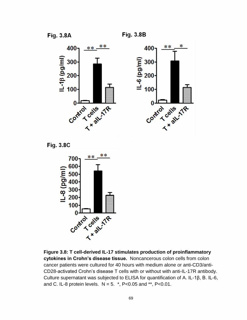
69
Figure 3.8: T cell-derived IL-17 stimulates production of proinflammatory
cytokines in Crohn’s disease tissue. Noncancerous colon cells from colon
cancer patients were cultured for 40 hours with medium alone or anti-CD3/anti-
CD28-activated Crohn’s disease T cells with or without with anti-IL-17R antibody.
Culture supernatant was subjected to ELISA for quantification of A. IL-1β, B. IL-6,
and C. IL-8 protein levels. N = 5. *, P<0.05 and **, P<0.01.
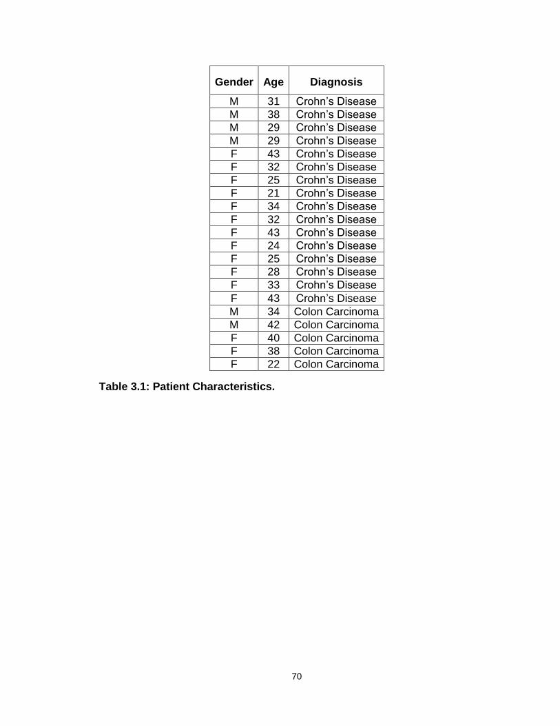
70
Gender Age Diagnosis
M 31 Crohn’s Disease
M 38 Crohn’s Disease
M 29 Crohn’s Disease
M 29 Crohn’s Disease
F 43 Crohn’s Disease
F 32 Crohn’s Disease
F 25 Crohn’s Disease
F 21 Crohn’s Disease
F 34 Crohn’s Disease
F 32 Crohn’s Disease
F 43 Crohn’s Disease
F 24 Crohn’s Disease
F 25 Crohn’s Disease
F 28 Crohn’s Disease
F 33 Crohn’s Disease
F 43 Crohn’s Disease
M 34 Colon Carcinoma
M 42 Colon Carcinoma
F 40 Colon Carcinoma
F 38 Colon Carcinoma
F 22 Colon Carcinoma
Table 3.1: Patient Characteristics.
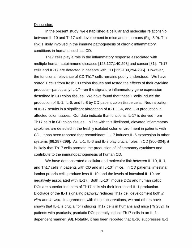
71
Discussion.
In the present study, we established a cellular and molecular relationship
between IL-10 and Th17 cell development in mice and in humans (Fig. 3.9). This
link is likely involved in the immune pathogenesis of chronic inflammatory
conditions in humans, such as CD.
Th17 cells play a role in the inflammatory response associated with
multiple human autoimmune diseases [125,127,140,293] and cancer [81]. Th17
cells and IL-17 are detected in patients with CD [135-139,294-296]. However,
the functional relevance of CD Th17 cells remains poorly understood. We have
sorted T cells from fresh CD colon tissues and tested the effects of their cytokine
products—particularly IL-17—on the signature inflammatory gene expression
described in CD colon tissues. We have found that these T cells induce the
production of IL-1, IL-6, and IL-8 by CD patient colon tissue cells. Neutralization
of IL-17 results in a significant abrogation of IL-1, IL-6, and IL-8 production in
affected colon tissues. Our data indicate that functional IL-17 is derived from
Th17 cells in CD colon tissues. In line with this likelihood, elevated inflammatory
cytokines are detected in the freshly isolated colon environment in patients with
CD. It has been reported that recombinant IL-17 induces IL-6 expression in other
systems [66,297-299]. As IL-1, IL-6 and IL-8 play crucial roles in CD [300-304], it
is likely that Th17 cells promote the production of inflammatory cytokines and
contribute to the immunopathogenesis of human CD.
We have demonstrated a cellular and molecular link between IL-10, IL-1,
and Th17 cells in patients with CD and in IL-10-/- mice. In CD patients, intestinal
lamina propria cells produce less IL-10, and the levels of intestinal IL-10 are
negatively associated with IL-17. Both IL-10-/- mouse DCs and human colitic
DCs are superior inducers of Th17 cells via their increased IL-1 production.
Blockade of the IL-1 signaling pathway reduces Th17 cell development both in
vitro and in vivo. In agreement with these observations, we and others have
shown that IL-1 is crucial for inducing Th17 cells in humans and mice [79,282]. In
patients with psoriasis, psoriatic DCs potently induce Th17 cells in an IL-1-
dependent manner [98]. Notably, it has been reported that IL-10 suppresses IL-1
72
Figure 3.9. Model of IL-10 in autoimmunity. 1. IL-10 downregulates DC expression of IL-1, which directly abrogates Th17 development. 2. This in turn decreases Th17-mediated upregulation of the proinflammatory cytokines IL-1, IL-6, and IL-8. 3. Lower inflammatory mediator levels minimize Th17 self-perpetuation, curtail inflammation, and reduce the potential of developing autoimmunity.
73
production [305-306] and that IL-1 is involved in controlling Th17 cells in the
mouse model of EAE [283]. Exogenous IL-10 can suppress the in vitro
development of Th17 cells from CD4+ T cells in patients with RA [307]. However,
our study is the first to demonstrate a role for IL-10 in Th17 development through
the control of IL-1 expression by DCs in both mouse and human systems. Our
data indicate that IL-1 plays a key role in Th17 cell development in human
autoimmune diseases, and support the notion that IL-1 signaling blockade is a
meaningful strategy to treat patients with autoimmune diseases. IL-10, via
downregulation of IL-1, is thus able to limit development of Th17 cells in mice
and humans, and in doing so executes some of its anti-inflammatory effects. In
future experiments, it would be interesting to investigate the cytokines that these
mouse and human Th17 cells produce in addition to IL-17. Th17 cells are
capable of producing multiple proinflammatory cytokines, and IL-17+IFNγ+
double-positive cells have been identified in numerous inflammatory settings
[81,308-309]. Further characterization of these Th17 populations might yield new
insights into their contributions to the development of pathology.
The next logical step is to investigate how IL-10 controls IL-1 production
by APCs. It has recently been shown that IL-10 dampens MyD88-dependent
signaling in DCs and leads to LPS hyporesponsiveness [310]. Because IL-1
signaling can be mediated by MyD88, this may explain how IL-10 controls
endotoxin-induced IL-1 production. It is also possible that IL-10 controls IL-1
expression machinery, such as IL-1 converting enzyme (ICE) and components of
the inflammasome [311-312]. However, it remains to be determined if IL-10
suppresses IL-1 production induced by other stimuli, including the necrotic tissue
often found in a chronic inflammatory environment.
One key question is why IL-10 production is reduced in patients with
Crohn’s disease. Recent genetic studies have shed some light on the
explanation. It has been reported that a nucleotide-binding oligomerization
domain containing 2 (NOD2) mutation commonly observed in patients with
Crohn’s disease leads to inhibition of IL-10 transcription [292]. However, we
have not examined the gene profile of NOD expression in our patient
74
populations. Since 30% of patients with Crohn’s disease have NOD mutations, it
is likely that alterations in NOD2 transcription may at least partially contribute to
the reduced IL-10 production in our patient population. Further genetic studies
are required to confirm this possibility.
In summary, we have demonstrated that IL-10 suppresses Th17 cell
development in mice and humans through modulation of IL-1 production by DCs.
The data document a cellular and molecular link between IL-10, IL-1, and Th17
cells, and suggest that IL-10 may inhibit inflammation via control of Th17 cell
development.
75
Chapter 4
Conclusions
Perhaps the broadest conclusion that may be drawn from this work is one
that is already commonly accepted: IL-10 is a pluripotent cytokine that controls
the development of several immune cell populations and the expression of
multiple proteins crucial in immune signaling. In fact, both of the experimental
series contained in this work explore effects that IL-10 mediates through the
classic pro-inflammatory cytokine IL-1. In one series, IL-10 is shown to affect
Th17 cell population development and size through the control of IL-1, and the
relationship between IL-10 and Th17 cells is inversely correlated. Thus, IL-10
down-regulates IL-1 production in myeloid cells, and in doing so hinders
expansion of Th17 populations by decreasing bioavailability of IL-1. The
increased Th17 populations in the IL-10-/- mouse, then, result at least in part from
an increase in IL-1 expression. This finding is not far from the generally
accepted role for IL-10 as an immunosuppressive molecule: IL-10 keeps pro-
inflammatory cytokine levels in check, and its absence leads to a stimulatory
environment that supports the development or expansion of certain immune cell
subsets typically associated with inflammation. This data serves to reinforce the
commonly-held view of IL-10 as a regulator and restrainer of immune responses.
In this context, our novel observation is that IL-10 suppresses Th17 cells through
inhibition of DC-derived IL-1, and in turn negatively controls autoimmunity in both
mice and humans.
However, from the second set of experiments in this work, we find fairly
convincing evidence that IL-10 displays immune-supporting functions, especially
in the context of tumor immunity. Although IL-10 has been shown to serve as a
development factor for CD8+ T cells [34,313], we find that it can also support
effector cytokine production in both CD8+ and CD4+ T cells. Its absence causes

76
an increase in MDSC accumulation in tumor-bearing animals (whether this effect
is direct or occurs through other mediators remains to be determined). These
MDSC are potent suppressors of T cell proliferation and also more powerful
inducers of regulatory T cells in vitro and likely in vivo. Here, it is likely that some
phenotypic signatures in the tumor-bearing mice result from IL-10’s control of IL-
1 expression, especially in the context of MDSC recruitment and function in the
tumor environment [261-262,274]. Upon IL-1 signal blockade, effector cytokine
production was partially rescued, and Treg numbers within the tumor
environment were decreased. Although the role of IL-1 in support of T cell
effector cytokine expression must still be explored, its impact on intratumoral
Treg cell populations may be easier to elucidate. Because IL-1 contributes to
MDSC recruitment to and retention in the tumor microenvironment, and because
a functional signature of MDSC is the induction of Treg cells from parent CD4+ T
cell populations [251], it is logical to surmise that these interactions occur
sequentially and culminate in expanded Treg cell populations within the tumor.
Interestingly, blockade of IL-1 signaling did not decrease MDSC populations in
tumor-bearing animals. This suggests either that IL-1’s effects on MDSC are
more durable than can be reversed with temporary abrogation of signaling, or
that a factor (or factors) distinct from IL-1 may contribute to MDSC recruitment.
Future research will be required to determine which scenario is correct. For this
set of data, it is clear that IL-10 supports antitumor immunity both through
maintenance of effector T cell populations and their effector cytokine production,
and by moderation of pro-tumor immune suppression mediated by MDSC and
Tregs.
As with any set of experiments that challenges the commonly-held beliefs
about certain cells or mediators, this work raises as many questions as it
answers. One of the most important is: how precisely does IL-10 regulate IL-1
expression in myeloid cells? As we mentioned above, it is possible that this
occurs through IL-10’s regulation of MyD88 [310], but this may not be the only
mechanism. The process whereby IL-1 is activated and released from the cell is
rather complicated: IL-1 exists as pro-IL-1β before it is cleaved by ICE/ caspase

77
1, and caspase 1 requires the scaffolding of the NLRP3 inflammasome in order
to cleave pro-IL-1β. Caspase 1 itself is produced as a zymogen and must be
cleaved in order to become active. Active Caspase 1 interacts with a protein
called Apoptosis-associated speck-like protein containing a CARD (PYCARD)
[314]. It is possible that IL-10 controls one or more factors involved in IL-1
processing and release from cells. The most straightforward experiment may be
the direct analysis of the expression levels and activation of these factors within
IL-10-deficient myeloid cells (both stimulated and unstimulated) and wild-type
cells. This could be accomplished by examination via RT-PCR and protein
analysis by Western Blot. Alternatively, IL-10 may contribute to the regulation of
the autophagy pathway. Autophagy is a catabolic process normally active in
homeostasis, involving the sequestration and degradation of cytosolic
macromolecules, surplus or damaged organelles, and certain intracellular
pathogens. Under normal physiological conditions, cellular autophagy can be
induced by nutrient deprivation, exposure to inhibitors of mTOR, or stimulation by
IFNγ or TNFα. It is inhibited by the Th2 cytokines IL-13 and IL-4. In settings of
infection, autophagy can be initiated by the recognition of pattern-associated
molecular patterns (PAMPs) by TLRs on myeloid cells. Signaling through TLRs
induces the sequestration of pro-IL-1β into autophagosomes, where it is
degraded. Inhibition of autophagy leads to an increase in the processing and
secretion of IL-1β from antigen-presenting cells; this is dependent on the NLRP3
inflammasome and TRIF [315]. In order to investigate whether IL-10 can, in fact,
regulate IL-1β expression via control of autophagy, the process should be
examined in IL-10-sufficient and deficient myeloid cells. Stimulation of cells by
cytokines or inhibitors of mTOR could be used to induce or abrogate autophagy,
and then cellular release of IL-1β could be measured by ELISA. Additionally, to
more closely examine the process, pro-IL-1β could be localized and quantified in
cellular compartments (via fluorescence microscopy) after cell stimulation.
An overarching question remains for both sets of experiments. Given that
the phenotypes we discovered and discussed were observed in IL-10-/- mice,
they could be the result of a global lack of IL-10 expression during birth,
78
development and maturation of these animals. In order to determine whether the
molecular signatures we observe result from or are complicated by the complete
absence of one protein, it will be essential to examine the same parameters in
wild-type, C57Bl/6 mice that have developed normally but undergo IL-10
blockade. This blockade may be genetic (global or tissue-specific) or biological
(administration of anti-IL-10 or anti-IL-10R antibodies). Using one or more of
these methods to abrogate IL-10 signaling for a few weeks before tumor
inoculation, colitis induction, or systemic examination of Th17 populations would
be ideal. In this way, one could more concretely identify the cellular and
molecular phenotypes as the result of a (temporary) lack of IL-10 in a more
physiological setting.
Finally, because IL-10-deficient mice are known for their gastrointestinal
phenotypes, and because more and more often, investigators are observing
profound effects of intestinal flora on disease development, it is crucial to
investigate the role of commensals in dissecting the phenotypes we observe.
Two very recent papers have examined the induction of IL-10 production in
adaptive immune cells by commensal bacteria. Pils and colleagues documented
an increase in T cell IL-10 mRNA levels after DSS exposure of wild-type mice
housed in SPF but not germ-free conditions. Intriguingly, germ-free mice
developed more severe intestinal disease, and some experiments had to be
terminated early because of mouse mortality [316]. This suggests that the
bacteria present in mice living in SPF conditions—in these experiments, Charles
River altered Schaedler flora (CRASF)—have significant roles in regulating
immune responses. In the same year, Round and Mazmanian observed that
regulatory T cell development could be influenced by commensal flora.
Monocolonization of germ-free mice with Bacteriodes fragilis increased both
suppressive activity and anti-inflammatory cytokine production by intestinal
regulatory T cells [317]. Interestingly, polysaccharide A (PSA) from B. fragilis
was determined to be the molecule responsible for converting CD4+ T cells to IL-
10+ FoxP3+ regulatory T cells during commensal colonization of the gut. For this
induction, signaling through TLR2 was required, once again emphasizing the
79
complex relationship between innate immune cues and adaptive immune
development in the gastrointestinal tract. Interestingly, exposure of mice to PSA
could cure TNBS-induced colitis. A thorough investigation of the commensal
contribution to the phenotypes we have observed in IL-10-/- mice would take
years. At baseline, it would be necessary to establish the differences in gut
colonization of wild-type and IL-10-/- mice, using microarrays or high-throughput
PCR techniques specific for certain species of bacteria. It would perhaps be
easier to establish how the differences in flora contributed to increased Th17
populations in IL-10-/- mice and their higher susceptibility to tumor development
than to the individual adaptive immune signatures we observe in tumor-bearing
IL-10-/- mice. A comparison of immune cell populations in IL-10-/- and wild-type
mice housed in germ-free conditions might also prove informative. Mono- or
oligocolonization of IL-10-/- mice kept in germ-free conditions could be
undertaken to observe the possible kinetic induction of Th17 populations; again,
these experiments are tedious and would need to be carried out with extreme
caution to minimize accidental colonization of mice with unknown commensals or
pathogens.
From an immunological perspective, the biological effects of given cell
populations, cytokines, and other mediators are all context-dependent. In this
case, we are exploring the downstream effects of IL-10 in two very different
contexts: tumor immunity and autoimmunity. It is well established that Tregs and
MDSC constitute major suppressive populations in the tumor microenvironment.
Settings of malignancy (and especially advanced malignancy) are generally
suppressive. In addition to MDSC and Treg expansion, immune populations
responsible for T cell activation and stimulation are dysregulated. Myeloid and
plasmacytoid DC, as well as tumor-associated macrophages, lose expression of
costimulatory molecules and upregulate inhibitory molecules, including B7-H1
and B7-H4 [149,213,318]. Their cytokine profiles also change dramatically [319].
In tumor microenvironments, IL-10 limits IL-1 production and MHC expression in
myeloid cells. Lower MHC expression could effect a decrease in presentation of
tumor peptides by myeloid cells to regulatory T cells, and thus a decrease in

80
tumor tolerance. Lower IL-1 production decreases MDSC exposure to this
crucial cytokine, and thus minimizes their development and function in the tumor
site. Fewer tumor-associated MDSC results in abrogated induction of Tregs from
local CD4+ T cells, and perhaps then prevents or lowers suppression of local
antitumor T cell populations. At the same time, we have demonstrated that IL-10
directly supports the development and effector function of T cells. Therefore, in
our experimental setting, IL-10 affects both innate and adaptive immunity to the
benefit of the host.
Multiple groups have reported decreased Treg numbers and dysregulated
Treg function in conditions of autoimmunity [320-321][and others]. As we have
already discussed, the myeloid cell populations in the gut of mice and humans
affected with IBD are massively dysregulated. Their numbers, locations, and
molecular phenotype are changed, making them more susceptible to certain
activatory signals [158-159]. Their molecular signatures are different (whether
they possess NOD2 mutations or not), and it is likely that their increase in
proinflammatory cytokine production and decrease in suppressive cytokine
production contributes greatly to the maintenance of the local inflammatory
milieu. In this context, the predominant effect of IL-10 may be on APCs, where
IL-10 suppresses IL-1 expression. Additionally, because T cells in IBD are more
strongly retained in the gut, and some of them have demonstrated higher
resistance to apoptosis, mDC-derived IL-1 may more easily reach T cell targets
and expand populations of memory Th17 cells. More Th17 cells means higher
levels of IL-17, IL-23 and other Th17 signature cytokines in the gut, and signaling
via these cytokines leads to more profound induction of other proinflammatory
cytokines from stromal and epithelial tissue. In this way, it is straightforward to
hypothesize the impact of IL-10 on multiple downstream populations as mediated
through its direct effects on myeloid cells. In both tumor immunity and
autoimmunity, we see the net effect of IL-10’s interactions with its environment
and on its target cells, and this can be profoundly different.
Taken together, the data presented herein expand upon previously-held
conceptions regarding IL-10. We have presented an additional manner in which
81
this cytokine moderates pro-inflammatory responses: through constraint of Th17
development. Yet, we have also demonstrated that IL-10 stimulates and
supports host immunity in the context of malignancy. It is crucial, then, to
carefully evaluate the type of immune response in question before assigning a
role (or roles) to the cytokine of interest—in this case, IL-10. The inquisitive
investigator will no doubt explore other ways or elucidate further mechanisms
whereby this cytokine may support immune responses. In light of the current
evidence, however, we must remind ourselves that, especially in the case of IL-
10, “context is everything.”
82
Appendix
Materials and Methods
Mice and Tumor models
6-12 week old female C57BL/6 mice were purchased from Charles River. IL-10-/-
mice (strain #2251) and IL-1R-/- mice (strain #3245) were purchased from the
Jackson Laboratory (Jackson Laboratory, Maine) and bred in-house. The mice
were maintained in specific pathogen-free conditions. This research was
approved by the committee on Use and Care of Animals at the University of
Michigan. 1×10⁶ MC38 mouse colon carcinoma cells were inoculated
subcutaneously into the left flank of IL-10-/-, IL-10+/+, or IL-1R-/- C57/BL6 mice.
Tumor size was measured each three days using calipers fitted with a Vernier
scale. Tumor volume was calculated based on three perpendicular
measurements. For other experiments, 2x10⁵ MCA310 cells were inoculated
intravenously into the tail vein of IL-10+/+ and IL-10-/- mice. At 2 weeks post-
inoculation, mice were sacrificed and their lungs were harvested, perfused with
India Ink, and fixed in paraformaldehyde. The numbers of lung foci were
quantified with the aid of a magnifier.
Patients
Blood was collected from patients with Crohn’s Disease and healthy volunteers.
Fresh colon tissues were collected from patients with CD who underwent colonic
resections or diagnostic biopsies. Fresh “approximately normal” colon tissues
adjacent to colorectal carcinoma were also collected as control tissues. All
patients with CD were in remission and were not treated with steroid drugs or
antibiological therapy during the 2 months before the study. Patient information is
presented in Table 3.1. This research was approved by local Institutional Review
Boards.
83
AOM/DSS treatment
Mice were given 10mg/kg azoxymethane (Sigma) via intraperitoneal injection.
Five days later they were allowed free access to water containing 2% dextran
sodium sulfate (DSS, MP Biomedicals/ Fisher) for five days, followed by 16 days
of regular water. This cycle was repeated twice and mice were sacrificed 2
weeks after the end of the last DSS cycle or at the end of 9 weeks. Colons were
harvested, flushed of feces, and slit open longitudinally to count tumors with the
aid of a magnifier.
IL-1Ra treatment
The human recombinant IL-1 receptor antagonist Anakinra (Kineret®) was
administered at 150mg/kg to mice intraperitoneally for 5 days before tumor
inoculation and each day thereafter for three weeks. In experiments not involving
tumor inoculation, Anakinra (Kineret®) was administered at 150mg/kg to mice
intraperitoneally for 8 days. On the eighth day, mice were sacrificed and their
organs were harvested for phenotyping. Mice not receiving Anakinra were
injected with PBS vehicle.
Flow cytometry analysis
Single-cell suspensions were made from human colon tissue or blood, mouse
spleen, lymph nodes, tumor-draining lymph nodes and/or tumor. Cells were
labeled with fluorescence-conjugated antibodies to CD45, CD11c (both
Invitrogen), CD4, CD8, Gr-1, CD11b, CD90, CD115, IL-17, and/or FoxP3 (all
eBioscience), CD3, IFNγ, TNFα, IL-2, MHC I, NK1.1, CD49b, CD19 (all BD
Pharmingen), and/or Foxp3 (eBioscience). For cytokine profiles, the cells were
stimulated with 50ng/mL PMA (Sigma) and 1 µM Ionomycin (Sigma-Aldrich, St.
Louis, MO) for 4 hours in the presence of GolgiPlug and GolgiStop (BD
Biosciences, San Jose, CA). Cells were first stained extracellularly with specific
antibodies, then fixed and permeabilized with Fix/Perm solution (eBioscience),
and finally stained intracellularly with specific antibodies. Samples were acquired
on a special order LSR II flow cytometer (BD Biosciences), and data were
84
analyzed as previously published with DIVA software (BD Biosciences)
[213,257].
Immunofluorescence analysis
Immunofluorescence analysis was performed as previously described [213,257].
Briefly, harvested tissues were frozen in OCT and then fixed with
paraformaldehyde. Permeabilized tissues were stained with rat anti-mouse CD8
(1:50, BD Pharmingen), rat anti-mouse CD31 (1:00, BD Pharmingen), and/or
rabbit anti-mouse Foxp3 (1:500, Abcam) followed by goat anti-rat and goat anti-
rabbit secondaries conjugated to Alexa Fluor 488 and Alexa Fluor 568,
respectively (both 1:2000; Molecular Probes/Invitrogen). Nuclei were stained with
DAPI (Molecular Probes/ Invitrogen). Fluorescent images were acquired on a
fluorescence microscope (Leica) and analyzed by ImagePro Plus software.
MDSC Suppression and Treg Induction
For the immunosuppressive assay, MDSCs were isolated from tumor or spleen in
tumor-bearing mice and cultured with irradiated spleen cells from wild-type (IL-
10+/+) mice. These cells were stimulated for 3 days with anti-CD3 (2.5μg/ml) and
anti-CD28 (1.25μg/ml) in the presence of different concentrations of MDSCs.
Thymidine was added in the last 16 hours. T cell proliferation was determined by
thymidine incorporation. In Treg induction experiments, tumor-associated
MDSCs were cultured with IL-10+/+ CD4+ T cells for 6 days. On day 6 the cells
were removed from culture, permeabilized, and stained with antibodies to FoxP3,
CD4, CD90, and CD11b. FoxP3 expression in total CD4+ T cells was analyzed
via FACS.
Cytokine expression
T cells were isolated from spleen with Mouse T Cell or CD4+ or CD8+ Enrichment
Kit (StemCell Technologies). The enriched T cells were cultured with 10 ng/ml IL-
10 (R&D Systems) or 1ug/ml anti-IL-10 for 3 days. IL-2, TNFα, and IFNγ were
analyzed on a special order LSR II flow cytometer (BD Biosciences).

85
Real-time reverse-transcriptase polymerase chain reaction (RT-PCR)
CD11b+ cells were isolated from IL-10+/+ or IL-10-/- splenic single-cell
suspensions with CD11b positive selection microbeads (Miltenyi), and CD11c-
CD11b+ cells were further sorted with FACSAria. CD11c+ cells were isolated
from IL-10+/+ or IL-10-/- splenic single-cell suspensions with a CD11c+ positive
selection kit (StemCell Technologies) and cultured for 8 hours with or without
LPS stimulation. In other experiments, fresh DC from Crohn’s Disease patients
were isolated and evaluated directly. mRNA was isolated with Trizol (Gibco
BRL). Cytokine transcripts were detected by real-time RT-PCR as previously
described [81]. Gene-specific primer pairs and Fast SYBR Green Master Mix
(Applied Biosystems) were used in a Multiplex instrument (Eppendorf). Data
analysis is based on the Ct method with normalization of raw data to a
housekeeping gene (HPRT).
ELISA Cytokine Detection
Supernatant was collected from 3-day cultures of mouse T and CD11c+ cells, or
after 48-hour LPS stimulation of CD11c+ cells, or after two-day cultures of fresh
human colon lamina propria or CD4+ T cells, or co-cultures of colon cells with or
without T cells from Crohn’s Disease patients. In other experiments, fresh serum
from the blood of healthy volunteers or Crohn’s Disease patients was directly
subjected to ELISA. IL-1β, IL-17, IL-6 and IL-8 were detected using murine or
human DuoSet kits (R&D Systems, Minneapolis, MN) with standard curves
ranging from 8-1000pg/ml and a detection limit of 8pg/ml.
DC and T cell culture
Mouse spleens were harvested, processed, and subjected to Ficoll density-
gradient centrifugation, rinsed, and resuspended as single-cell suspensions.
CD11c+ DC and CD4+ T cells were isolated using positive (CD11c+) and negative
(CD4+ T) selection kits (Stemcell Technologies). Cells were co-cultured in a ratio
of 1:5 for 5-6 days with anti-CD3 and anti-CD28 antibodies. For single cultures,
T or CD11c+ cells were plated at 1x10⁶ cells/ml, with anti-CD3 and anti-CD28
antibodies added for T cells and 1ug/ml LPS or no stimulation for CD11c+ DC.
86
Colon and T cell Cultures
Noncancerous colon cells (2 x 10⁶/ ml) from colon cancer patients were cultured
for 40 hours with medium alone, anti-CD3/anti-CD28-activated T cells isolated
from Crohn’s disease patients, or anti-CD3/anti-CD28-activated T cells isolated
from Crohn’s disease patients plus anti-IL-17R antibody (R&D, clone 133617).
The ratio of colon cells to T cells was 1:5.
Statistics
Most experiments were evaluated using the Mann-Whitney test, with P < 0.05
considered significant. Some cases were evaluated with Student’s T test, also
with P < 0.05 considered significant. Statistics were performed in the GraphPad
Prism program suite (GraphPad Software, Inc., La Jolla, CA) and Statistica
program suite (StatSoft, Tulsa, OK).
87
References
1. Ferrero-Miliani, L., Nielsen, O.H., Andersen, P.S. and Girardin, S.E. (2007) Chronic inflammation: importance of NOD2 and NALP3 in interleukin-1beta generation. Clin Exp Immunol, 147, 227-35.
2. Chen, G.Y. and Nunez, G. (2010) Sterile inflammation: sensing and reacting to damage. Nat Rev Immunol, 10, 826-37.
3. Harrington, L.E., Hatton, R.D., Mangan, P.R., Turner, H., Murphy, T.L., Murphy, K.M. and Weaver, C.T. (2005) Interleukin 17-producing CD4+ effector T cells develop via a lineage distinct from the T helper type 1 and 2 lineages. Nat Immunol, 6, 1123-32.
4. Langrish, C.L., Chen, Y., Blumenschein, W.M., Mattson, J., Basham, B., Sedgwick, J.D., McClanahan, T., Kastelein, R.A. and Cua, D.J. (2005) IL-23 drives a pathogenic T cell population that induces autoimmune inflammation. J Exp Med, 201, 233-40.
5. Nathan, C.F., Murray, H.W., Wiebe, M.E. and Rubin, B.Y. (1983) Identification of interferon-gamma as the lymphokine that activates human macrophage oxidative metabolism and antimicrobial activity. J Exp Med, 158, 670-89.
6. Flaishon, L., Hershkoviz, R., Lantner, F., Lider, O., Alon, R., Levo, Y., Flavell, R.A. and Shachar, I. (2000) Autocrine secretion of interferon gamma negatively regulates homing of immature B cells. J Exp Med, 192, 1381-8.
7. Finkelman, F.D., Katona, I.M., Mosmann, T.R. and Coffman, R.L. (1988) IFN-gamma regulates the isotypes of Ig secreted during in vivo humoral immune responses. J Immunol, 140, 1022-7.
8. Balkwill, F. and Taylor-Papadimitriou, J. (1978) Interferon affects both G1 and S+G2 in cells stimulated from quiescence to growth. Nature, 274, 798-800.
88
9. Xaus, J., Cardo, M., Valledor, A.F., Soler, C., Lloberas, J. and Celada, A. (1999) Interferon gamma induces the expression of p21waf-1 and arrests macrophage cell cycle, preventing induction of apoptosis. Immunity, 11, 103-13.
10. Gajewski, T.F. and Fitch, F.W. (1988) Anti-proliferative effect of IFN-gamma in immune regulation. I. IFN-gamma inhibits the proliferation of Th2 but not Th1 murine helper T lymphocyte clones. J Immunol, 140, 4245-52.
11. Yoshida, A., Koide, Y., Uchijima, M. and Yoshida, T.O. (1994) IFN-gamma induces IL-12 mRNA expression by a murine macrophage cell line, J774. Biochem Biophys Res Commun, 198, 857-61.
12. Kryczek, I., Wei, S., Gong, W., Shu, X., Szeliga, W., Vatan, L., Chen, L., Wang, G. and Zou, W. (2008) Cutting edge: IFN-gamma enables APC to promote memory Th17 and abate Th1 cell development. J Immunol, 181, 5842-6.
13. Fiorentino, D.F., Bond, M.W. and Mosmann, T.R. (1989) Two types of mouse T helper cell. IV. Th2 clones secrete a factor that inhibits cytokine production by Th1 clones. J Exp Med, 170, 2081-95.
14. O'Garra, A., Stapleton, G., Dhar, V., Pearce, M., Schumacher, J., Rugo, H., Barbis, D., Stall, A., Cupp, J., Moore, K. and et al. (1990) Production of cytokines by mouse B cells: B lymphomas and normal B cells produce interleukin 10. Int Immunol, 2, 821-32.
15. Galli, S.J., Kalesnikoff, J., Grimbaldeston, M.A., Piliponsky, A.M., Williams, C.M. and Tsai, M. (2005) Mast cells as "tunable" effector and immunoregulatory cells: recent advances. Annu Rev Immunol, 23, 749-86.
16. Grimbaldeston, M.A., Nakae, S., Kalesnikoff, J., Tsai, M. and Galli, S.J. (2007) Mast cell-derived interleukin 10 limits skin pathology in contact dermatitis and chronic irradiation with ultraviolet B. Nat Immunol, 8, 1095-104.
17. McGeachy, M.J., Bak-Jensen, K.S., Chen, Y., Tato, C.M., Blumenschein, W., McClanahan, T. and Cua, D.J. (2007) TGF-beta and IL-6 drive the production of IL-17 and IL-10 by T cells and restrain T(H)-17 cell-mediated pathology. Nat Immunol, 8, 1390-7.
18. Murai, M., Turovskaya, O., Kim, G., Madan, R., Karp, C.L., Cheroutre, H. and Kronenberg, M. (2009) Interleukin 10 acts on regulatory T cells to

89
maintain expression of the transcription factor Foxp3 and suppressive function in mice with colitis. Nat Immunol, 10, 1178-84.
19. Kuhn, R., Lohler, J., Rennick, D., Rajewsky, K. and Muller, W. (1993) Interleukin-10-deficient mice develop chronic enterocolitis. Cell, 75, 263-74.
20. Davidson, N.J., Leach, M.W., Fort, M.M., Thompson-Snipes, L., Kuhn, R., Muller, W., Berg, D.J. and Rennick, D.M. (1996) T helper cell 1-type CD4+ T cells, but not B cells, mediate colitis in interleukin 10-deficient mice. J Exp Med, 184, 241-51.
21. Asseman, C., Mauze, S., Leach, M.W., Coffman, R.L. and Powrie, F. (1999) An essential role for interleukin 10 in the function of regulatory T cells that inhibit intestinal inflammation. J Exp Med, 190, 995-1004.
22. Zhou, P., Streutker, C., Borojevic, R., Wang, Y. and Croitoru, K. (2004) IL-10 modulates intestinal damage and epithelial cell apoptosis in T cell-mediated enteropathy. Am J Physiol Gastrointest Liver Physiol, 287, G599-604.
23. Jarry, A., Bossard, C., Bou-Hanna, C., Masson, D., Espaze, E., Denis, M.G. and Laboisse, C.L. (2008) Mucosal IL-10 and TGF-beta play crucial roles in preventing LPS-driven, IFN-gamma-mediated epithelial damage in human colon explants. J Clin Invest, 118, 1132-42.
24. Moore, K.W., de Waal Malefyt, R., Coffman, R.L. and O'Garra, A. (2001) Interleukin-10 and the interleukin-10 receptor. Annu Rev Immunol, 19, 683-765.
25. Couper, K.N., Blount, D.G. and Riley, E.M. (2008) IL-10: the master regulator of immunity to infection. J Immunol, 180, 5771-7.
26. Schandene, L., Alonso-Vega, C., Willems, F., Gerard, C., Delvaux, A., Velu, T., Devos, R., de Boer, M. and Goldman, M. (1994) B7/CD28-dependent IL-5 production by human resting T cells is inhibited by IL-10. J Immunol, 152, 4368-74.
27. Joss, A., Akdis, M., Faith, A., Blaser, K. and Akdis, C.A. (2000) IL-10 directly acts on T cells by specifically altering the CD28 co-stimulation pathway. Eur J Immunol, 30, 1683-90.
90
28. Demangel, C., Bertolino, P. and Britton, W.J. (2002) Autocrine IL-10 impairs dendritic cell (DC)-derived immune responses to mycobacterial infection by suppressing DC trafficking to draining lymph nodes and local IL-12 production. Eur J Immunol, 32, 994-1002.
29. Go, N.F., Castle, B.E., Barrett, R., Kastelein, R., Dang, W., Mosmann, T.R., Moore, K.W. and Howard, M. (1990) Interleukin 10, a novel B cell stimulatory factor: unresponsiveness of X chromosome-linked immunodeficiency B cells. J Exp Med, 172, 1625-31.
30. Rousset, F., Garcia, E., Defrance, T., Peronne, C., Vezzio, N., Hsu, D.H., Kastelein, R., Moore, K.W. and Banchereau, J. (1992) Interleukin 10 is a potent growth and differentiation factor for activated human B lymphocytes. Proc Natl Acad Sci U S A, 89, 1890-3.
31. Levy, Y. and Brouet, J.C. (1994) Interleukin-10 prevents spontaneous death of germinal center B cells by induction of the bcl-2 protein. J Clin Invest, 93, 424-8.
32. Liu, Y.J., Mason, D.Y., Johnson, G.D., Abbot, S., Gregory, C.D., Hardie, D.L., Gordon, J. and MacLennan, I.C. (1991) Germinal center cells express bcl-2 protein after activation by signals which prevent their entry into apoptosis. Eur J Immunol, 21, 1905-10.
33. MacNeil, I.A., Suda, T., Moore, K.W., Mosmann, T.R. and Zlotnik, A. (1990) IL-10, a novel growth cofactor for mature and immature T cells. J Immunol, 145, 4167-73.
34. Chen, W.F. and Zlotnik, A. (1991) IL-10: a novel cytotoxic T cell differentiation factor. J Immunol, 147, 528-34.
35. Peters, N. and Sacks, D. (2006) Immune privilege in sites of chronic infection: Leishmania and regulatory T cells. Immunol Rev, 213, 159-79.
36. van der Sluijs, K.F., van Elden, L.J., Nijhuis, M., Schuurman, R., Pater, J.M., Florquin, S., Goldman, M., Jansen, H.M., Lutter, R. and van der Poll, T. (2004) IL-10 is an important mediator of the enhanced susceptibility to pneumococcal pneumonia after influenza infection. J Immunol, 172, 7603-9.
37. Jimenez Mdel, P., Walls, L. and Fierer, J. (2006) High levels of interleukin-10 impair resistance to pulmonary coccidioidomycosis in mice in part through control of nitric oxide synthase 2 expression. Infect Immun, 74, 3387-95.
91
38. Fierer, J., Walls, L., Eckmann, L., Yamamoto, T. and Kirkland, T.N. (1998) Importance of interleukin-10 in genetic susceptibility of mice to Coccidioides immitis. Infect Immun, 66, 4397-402.
39. Kelly, J.P. and Bancroft, G.J. (1996) Administration of interleukin-10 abolishes innate resistance to Listeria monocytogenes. Eur J Immunol, 26, 356-64.
40. Hagenbaugh, A., Sharma, S., Dubinett, S.M., Wei, S.H., Aranda, R., Cheroutre, H., Fowell, D.J., Binder, S., Tsao, B., Locksley, R.M., Moore, K.W. and Kronenberg, M. (1997) Altered immune responses in interleukin 10 transgenic mice. J Exp Med, 185, 2101-10.
41. Mocellin, S., Marincola, F.M. and Young, H.A. (2005) Interleukin-10 and the immune response against cancer: a counterpoint. J Leukoc Biol, 78, 1043-51.
42. Suzuki, T., Tahara, H., Narula, S., Moore, K.W., Robbins, P.D. and Lotze, M.T. (1995) Viral interleukin 10 (IL-10), the human herpes virus 4 cellular IL-10 homologue, induces local anergy to allogeneic and syngeneic tumors. J Exp Med, 182, 477-86.
43. Zheng, L.M., Ojcius, D.M., Garaud, F., Roth, C., Maxwell, E., Li, Z., Rong, H., Chen, J., Wang, X.Y., Catino, J.J. and King, I. (1996) Interleukin-10 inhibits tumor metastasis through an NK cell-dependent mechanism. J Exp Med, 184, 579-84.
44. Kundu, N. and Fulton, A.M. (1997) Interleukin-10 inhibits tumor metastasis, downregulates MHC class I, and enhances NK lysis. Cell Immunol, 180, 55-61.
45. Karre, K. (1993) Natural killer cells and the MHC class I pathway of peptide presentation. Semin Immunol, 5, 127-45.
46. Fujii, S., Shimizu, K., Shimizu, T. and Lotze, M.T. (2001) Interleukin-10 promotes the maintenance of antitumor CD8(+) T-cell effector function in situ. Blood, 98, 2143-51.
47. Kim, B.G., Joo, H.G., Chung, I.S., Chung, H.Y., Woo, H.J. and Yun, Y.S. (2000) Inhibition of interleukin-10 (IL-10) production from MOPC 315 tumor cells by IL-10 antisense oligodeoxynucleotides enhances cell-mediated immune responses. Cancer Immunol Immunother, 49, 433-40.
92
48. Matar, P., Rozados, V.R., Gervasoni, S.I. and Scharovsky, O.G. (2001) Down regulation of T-cell-derived IL-10 production by low-dose cyclophosphamide treatment in tumor-bearing rats restores in vitro normal lymphoproliferative response. Int Immunopharmacol, 1, 307-19.
49. Vicari, A.P., Chiodoni, C., Vaure, C., Ait-Yahia, S., Dercamp, C., Matsos, F., Reynard, O., Taverne, C., Merle, P., Colombo, M.P., O'Garra, A., Trinchieri, G. and Caux, C. (2002) Reversal of tumor-induced dendritic cell paralysis by CpG immunostimulatory oligonucleotide and anti-interleukin 10 receptor antibody. J Exp Med, 196, 541-9.
50. Seo, N., Hayakawa, S. and Tokura, Y. (2002) Mechanisms of immune privilege for tumor cells by regulatory cytokines produced by innate and acquired immune cells. Semin Cancer Biol, 12, 291-300.
51. Kim, J., Modlin, R.L., Moy, R.L., Dubinett, S.M., McHugh, T., Nickoloff, B.J. and Uyemura, K. (1995) IL-10 production in cutaneous basal and squamous cell carcinomas. A mechanism for evading the local T cell immune response. J Immunol, 155, 2240-7.
52. Kurte, M., Lopez, M., Aguirre, A., Escobar, A., Aguillon, J.C., Charo, J., Larsen, C.G., Kiessling, R. and Salazar-Onfray, F. (2004) A synthetic peptide homologous to functional domain of human IL-10 down-regulates expression of MHC class I and Transporter associated with Antigen Processing 1/2 in human melanoma cells. J Immunol, 173, 1731-7.
53. Urosevic, M. and Dummer, R. (2003) HLA-G and IL-10 expression in human cancer--different stories with the same message. Semin Cancer Biol, 13, 337-42.
54. Enk, A.H., Jonuleit, H., Saloga, J. and Knop, J. (1997) Dendritic cells as mediators of tumor-induced tolerance in metastatic melanoma. Int J Cancer, 73, 309-16.
55. Steinbrink, K., Jonuleit, H., Muller, G., Schuler, G., Knop, J. and Enk, A.H. (1999) Interleukin-10-treated human dendritic cells induce a melanoma-antigen-specific anergy in CD8(+) T cells resulting in a failure to lyse tumor cells. Blood, 93, 1634-42.
56. Loercher, A.E., Nash, M.A., Kavanagh, J.J., Platsoucas, C.D. and Freedman, R.S. (1999) Identification of an IL-10-producing HLA-DR-negative monocyte subset in the malignant ascites of patients with ovarian carcinoma that inhibits cytokine protein expression and proliferation of autologous T cells. J Immunol, 163, 6251-60.
93
57. Jovasevic, V.M., Gorelik, L., Bluestone, J.A. and Mokyr, M.B. (2004) Importance of IL-10 for CTLA-4-mediated inhibition of tumor-eradicating immunity. J Immunol, 172, 1449-54.
58. Huang, M., Stolina, M., Sharma, S., Mao, J.T., Zhu, L., Miller, P.W., Wollman, J., Herschman, H. and Dubinett, S.M. (1998) Non-small cell lung cancer cyclooxygenase-2-dependent regulation of cytokine balance in lymphocytes and macrophages: up-regulation of interleukin 10 and down-regulation of interleukin 12 production. Cancer Res, 58, 1208-16.
59. Stolina, M., Sharma, S., Lin, Y., Dohadwala, M., Gardner, B., Luo, J., Zhu, L., Kronenberg, M., Miller, P.W., Portanova, J., Lee, J.C. and Dubinett, S.M. (2000) Specific inhibition of cyclooxygenase 2 restores antitumor reactivity by altering the balance of IL-10 and IL-12 synthesis. J Immunol, 164, 361-70.
60. Sharma, S., Stolina, M., Yang, S.C., Baratelli, F., Lin, J.F., Atianzar, K., Luo, J., Zhu, L., Lin, Y., Huang, M., Dohadwala, M., Batra, R.K. and Dubinett, S.M. (2003) Tumor cyclooxygenase 2-dependent suppression of dendritic cell function. Clin Cancer Res, 9, 961-8.
61. Sanjabi, S., Zenewicz, L.A., Kamanaka, M. and Flavell, R.A. (2009) Anti-inflammatory and pro-inflammatory roles of TGF-beta, IL-10, and IL-22 in immunity and autoimmunity. Curr Opin Pharmacol, 9, 447-53.
62. Netea, M.G., Sutmuller, R., Hermann, C., Van der Graaf, C.A., Van der Meer, J.W., van Krieken, J.H., Hartung, T., Adema, G. and Kullberg, B.J. (2004) Toll-like receptor 2 suppresses immunity against Candida albicans through induction of IL-10 and regulatory T cells. J Immunol, 172, 3712-8.
63. Belkaid, Y., Piccirillo, C.A., Mendez, S., Shevach, E.M. and Sacks, D.L. (2002) CD4+CD25+ regulatory T cells control Leishmania major persistence and immunity. Nature, 420, 502-7.
64. O'Garra, A., Barrat, F.J., Castro, A.G., Vicari, A. and Hawrylowicz, C. (2008) Strategies for use of IL-10 or its antagonists in human disease. Immunol Rev, 223, 114-31.
65. Coussens, L.M. and Werb, Z. (2002) Inflammation and cancer. Nature, 420, 860-7.
66. Laan, M., Cui, Z.H., Hoshino, H., Lotvall, J., Sjostrand, M., Gruenert, D.C., Skoogh, B.E. and Linden, A. (1999) Neutrophil recruitment by human IL-17 via C-X-C chemokine release in the airways. J Immunol, 162, 2347-52.
94
67. Cua, D.J. and Tato, C.M. (2010) Innate IL-17-producing cells: the sentinels of the immune system. Nat Rev Immunol, 10, 479-89.
68. Honorati, M.C., Cattini, L. and Facchini, A. (2007) Possible prognostic role of IL-17R in osteosarcoma. J Cancer Res Clin Oncol, 133, 1017-21.
69. Wang, L., Yi, T., Kortylewski, M., Pardoll, D.M., Zeng, D. and Yu, H. (2009) IL-17 can promote tumor growth through an IL-6-Stat3 signaling pathway. J Exp Med, 206, 1457-64.
70. Prabhala, R.H., Pelluru, D., Fulciniti, M., Prabhala, H.K., Nanjappa, P., Song, W., Pai, C., Amin, S., Tai, Y.T., Richardson, P.G., Ghobrial, I.M., Treon, S.P., Daley, J.F., Anderson, K.C., Kutok, J.L. and Munshi, N.C. (2010) Elevated IL-17 produced by TH17 cells promotes myeloma cell growth and inhibits immune function in multiple myeloma. Blood, 115, 5385-92.
71. Gaffen, S.L. (2009) Structure and signalling in the IL-17 receptor family. Nat Rev Immunol, 9, 556-67.
72. Ishigame, H., Kakuta, S., Nagai, T., Kadoki, M., Nambu, A., Komiyama, Y., Fujikado, N., Tanahashi, Y., Akitsu, A., Kotaki, H., Sudo, K., Nakae, S., Sasakawa, C. and Iwakura, Y. (2009) Differential roles of interleukin-17A and -17F in host defense against mucoepithelial bacterial infection and allergic responses. Immunity, 30, 108-19.
73. Ely, L.K., Fischer, S. and Garcia, K.C. (2009) Structural basis of receptor sharing by interleukin 17 cytokines. Nat Immunol, 10, 1245-51.
74. Das Sarma, J., Ciric, B., Marek, R., Sadhukhan, S., Caruso, M.L., Shafagh, J., Fitzgerald, D.C., Shindler, K.S. and Rostami, A. (2009) Functional interleukin-17 receptor A is expressed in central nervous system glia and upregulated in experimental autoimmune encephalomyelitis. J Neuroinflammation, 6, 14.
75. Franchi, L., Eigenbrod, T., Munoz-Planillo, R. and Nunez, G. (2009) The inflammasome: a caspase-1-activation platform that regulates immune responses and disease pathogenesis. Nat Immunol, 10, 241-7.
76. Apte, R.N. and Voronov, E. (2008) Is interleukin-1 a good or bad 'guy' in tumor immunobiology and immunotherapy? Immunol Rev, 222, 222-41.
95
77. Kuno, K. and Matsushima, K. (1994) The IL-1 receptor signaling pathway. J Leukoc Biol, 56, 542-7.
78. Bienenstock J, M.J., Lamm ME, Strober W, McGhee JR (ed.) (2005) Mucosal Immunology. Elsevier.
79. Chung, Y., Chang, S.H., Martinez, G.J., Yang, X.O., Nurieva, R., Kang, H.S., Ma, L., Watowich, S.S., Jetten, A.M., Tian, Q. and Dong, C. (2009) Critical regulation of early Th17 cell differentiation by interleukin-1 signaling. Immunity, 30, 576-87.
80. Gulen, M.F., Kang, Z., Bulek, K., Youzhong, W., Kim, T.W., Chen, Y., Altuntas, C.Z., Sass Bak-Jensen, K., McGeachy, M.J., Do, J.S., Xiao, H., Delgoffe, G.M., Min, B., Powell, J.D., Tuohy, V.K., Cua, D.J. and Li, X. (2010) The receptor SIGIRR suppresses Th17 cell proliferation via inhibition of the interleukin-1 receptor pathway and mTOR kinase activation. Immunity, 32, 54-66.
81. Kryczek, I., Banerjee, M., Cheng, P., Vatan, L., Szeliga, W., Wei, S., Huang, E., Finlayson, E., Simeone, D., Welling, T.H., Chang, A., Coukos, G., Liu, R. and Zou, W. (2009) Phenotype, distribution, generation, and functional and clinical relevance of Th17 cells in the human tumor environments. Blood, 114, 1141-9.
82. Precopio, M.L., Betts, M.R., Parrino, J., Price, D.A., Gostick, E., Ambrozak, D.R., Asher, T.E., Douek, D.C., Harari, A., Pantaleo, G., Bailer, R., Graham, B.S., Roederer, M. and Koup, R.A. (2007) Immunization with vaccinia virus induces polyfunctional and phenotypically distinctive CD8(+) T cell responses. J Exp Med, 204, 1405-16.
83. Almeida, J.R., Price, D.A., Papagno, L., Arkoub, Z.A., Sauce, D., Bornstein, E., Asher, T.E., Samri, A., Schnuriger, A., Theodorou, I., Costagliola, D., Rouzioux, C., Agut, H., Marcelin, A.G., Douek, D., Autran, B. and Appay, V. (2007) Superior control of HIV-1 replication by CD8+ T cells is reflected by their avidity, polyfunctionality, and clonal turnover. J Exp Med, 204, 2473-85.
84. Curiel, T.J., Coukos, G., Zou, L., Alvarez, X., Cheng, P., Mottram, P., Evdemon-Hogan, M., Conejo-Garcia, J.R., Zhang, L., Burow, M., Zhu, Y., Wei, S., Kryczek, I., Daniel, B., Gordon, A., Myers, L., Lackner, A., Disis, M.L., Knutson, K.L., Chen, L. and Zou, W. (2004) Specific recruitment of regulatory T cells in ovarian carcinoma fosters immune privilege and predicts reduced survival. Nat Med, 10, 942-9.
96
85. Su, X., Ye, J., Hsueh, E.C., Zhang, Y., Hoft, D.F. and Peng, G. (2010) Tumor microenvironments direct the recruitment and expansion of human Th17 cells. J Immunol, 184, 1630-41.
86. Sfanos, K.S., Bruno, T.C., Maris, C.H., Xu, L., Thoburn, C.J., DeMarzo, A.M., Meeker, A.K., Isaacs, W.B. and Drake, C.G. (2008) Phenotypic analysis of prostate-infiltrating lymphocytes reveals TH17 and Treg skewing. Clin Cancer Res, 14, 3254-61.
87. Derhovanessian, E., Adams, V., Hahnel, K., Groeger, A., Pandha, H., Ward, S. and Pawelec, G. (2009) Pretreatment frequency of circulating IL-17+ CD4+ T-cells, but not Tregs, correlates with clinical response to whole-cell vaccination in prostate cancer patients. Int J Cancer, 125, 1372-9.
88. Ye, Z.-J., Zhou, Q., Gu, Y.-Y., Qin, S.-M., Ma, W.-L., Xin, J.-B., Tao, X.-N. and Shi, H.-Z. (2010) Generation and Differentiation of Interleukin-17-Producing CD4+ T Cells in Malignant Pleural Effusion. J Immunol.
89. Kryczek, I., Wei, S., Zou, L., Altuwaijri, S., Szeliga, W., Kolls, J., Chang, A. and Zou, W. (2007) Cutting edge: Th17 and regulatory T cell dynamics and the regulation by IL-2 in the tumor microenvironment. J Immunol, 178, 6730-3.
90. Sharma, M.D., Hou, D.Y., Liu, Y., Koni, P.A., Metz, R., Chandler, P., Mellor, A.L., He, Y. and Munn, D.H. (2009) Indoleamine 2,3-dioxygenase controls conversion of Foxp3+ Tregs to TH17-like cells in tumor-draining lymph nodes. Blood, 113, 6102-11.
91. Kuang, D.M., Peng, C., Zhao, Q., Wu, Y., Chen, M.S. and Zheng, L. Activated monocytes in peritumoral stroma of hepatocellular carcinoma promote expansion of memory T helper 17 cells. Hepatology, 51, 154-64.
92. Charles, K.A., Kulbe, H., Soper, R., Escorcio-Correia, M., Lawrence, T., Schultheis, A., Chakravarty, P., Thompson, R.G., Kollias, G., Smyth, J.F., Balkwill, F.R. and Hagemann, T. (2009) The tumor-promoting actions of TNF-alpha involve TNFR1 and IL-17 in ovarian cancer in mice and humans. J Clin Invest, 119, 3011-23.
93. Gnerlich, J.L., Mitchem, J.B., Weir, J.S., Sankpal, N.V., Kashiwagi, H., Belt, B.A., Herndon, J.M., Eberlein, T.J., Goedegebuure, P. and Linehan, D.C. (2010) Induction of Th17 Cells in the Tumor Microenvironment Improves Survival in a Murine Model of Pancreatic Cancer. J Immunol.
97
94. Wilke, C.M., Kryczek, I., Wei, S., Zhao, E., Wu, K., Wang, G. and Zou, W. (2011) Th17 Cells in Cancer: Help or Hindrance? Carcinogenesis.
95. Zou, W. and Restifo, N.P. (2010) T(H)17 cells in tumour immunity and immunotherapy. Nat Rev Immunol, 10, 248-56.
96. Tesmer, L.A., Lundy, S.K., Sarkar, S. and Fox, D.A. (2008) Th17 cells in human disease. Immunol Rev, 223, 87-113.
97. Wilson, N.J., Boniface, K., Chan, J.R., McKenzie, B.S., Blumenschein, W.M., Mattson, J.D., Basham, B., Smith, K., Chen, T., Morel, F., Lecron, J.C., Kastelein, R.A., Cua, D.J., McClanahan, T.K., Bowman, E.P. and de Waal Malefyt, R. (2007) Development, cytokine profile and function of human interleukin 17-producing helper T cells. Nat Immunol, 8, 950-7.
98. Kryczek, I., Bruce, A.T., Gudjonsson, J.E., Johnston, A., Aphale, A., Vatan, L., Szeliga, W., Wang, Y., Liu, Y., Welling, T.H., Elder, J.T. and Zou, W. (2008) Induction of IL-17+ T cell trafficking and development by IFN-gamma: mechanism and pathological relevance in psoriasis. J Immunol, 181, 4733-41.
99. Krueger, G.G., Langley, R.G., Leonardi, C., Yeilding, N., Guzzo, C., Wang, Y., Dooley, L.T. and Lebwohl, M. (2007) A human interleukin-12/23 monoclonal antibody for the treatment of psoriasis. N Engl J Med, 356, 580-92.
100. Toichi, E., Torres, G., McCormick, T.S., Chang, T., Mascelli, M.A., Kauffman, C.L., Aria, N., Gottlieb, A.B., Everitt, D.E., Frederick, B., Pendley, C.E. and Cooper, K.D. (2006) An anti-IL-12p40 antibody down-regulates type 1 cytokines, chemokines, and IL-12/IL-23 in psoriasis. J Immunol, 177, 4917-26.
101. Zaba, L.C., Cardinale, I., Gilleaudeau, P., Sullivan-Whalen, M., Suarez-Farinas, M., Fuentes-Duculan, J., Novitskaya, I., Khatcherian, A., Bluth, M.J., Lowes, M.A. and Krueger, J.G. (2007) Amelioration of epidermal hyperplasia by TNF inhibition is associated with reduced Th17 responses. J Exp Med, 204, 3183-94.
102. Zaba, L.C., Fuentes-Duculan, J., Eungdamrong, N.J., Abello, M.V., Novitskaya, I., Pierson, K.C., Gonzalez, J., Krueger, J.G. and Lowes, M.A. (2009) Psoriasis is characterized by accumulation of immunostimulatory and Th1/Th17 cell-polarizing myeloid dendritic cells. J Invest Dermatol, 129, 79-88.
98
103. Murphy, C.A., Langrish, C.L., Chen, Y., Blumenschein, W., McClanahan, T., Kastelein, R.A., Sedgwick, J.D. and Cua, D.J. (2003) Divergent pro- and antiinflammatory roles for IL-23 and IL-12 in joint autoimmune inflammation. J Exp Med, 198, 1951-7.
104. Bush, K.A., Farmer, K.M., Walker, J.S. and Kirkham, B.W. (2002) Reduction of joint inflammation and bone erosion in rat adjuvant arthritis by treatment with interleukin-17 receptor IgG1 Fc fusion protein. Arthritis Rheum, 46, 802-5.
105. Nakae, S., Nambu, A., Sudo, K. and Iwakura, Y. (2003) Suppression of immune induction of collagen-induced arthritis in IL-17-deficient mice. J Immunol, 171, 6173-7.
106. Lubberts, E., Koenders, M.I., Oppers-Walgreen, B., van den Bersselaar, L., Coenen-de Roo, C.J., Joosten, L.A. and van den Berg, W.B. (2004) Treatment with a neutralizing anti-murine interleukin-17 antibody after the onset of collagen-induced arthritis reduces joint inflammation, cartilage destruction, and bone erosion. Arthritis Rheum, 50, 650-9.
107. Chabaud, M., Durand, J.M., Buchs, N., Fossiez, F., Page, G., Frappart, L. and Miossec, P. (1999) Human interleukin-17: A T cell-derived proinflammatory cytokine produced by the rheumatoid synovium. Arthritis Rheum, 42, 963-70.
108. Ziolkowska, M., Koc, A., Luszczykiewicz, G., Ksiezopolska-Pietrzak, K., Klimczak, E., Chwalinska-Sadowska, H. and Maslinski, W. (2000) High levels of IL-17 in rheumatoid arthritis patients: IL-15 triggers in vitro IL-17 production via cyclosporin A-sensitive mechanism. J Immunol, 164, 2832-8.
109. Hwang, S.Y. and Kim, H.Y. (2005) Expression of IL-17 homologs and their receptors in the synovial cells of rheumatoid arthritis patients. Mol Cells, 19, 180-4.
110. Leipe, J., Grunke, M., Dechant, C., Reindl, C., Kerzendorf, U., Schulze-Koops, H. and Skapenko, A. (2010) Role of Th17 cells in human autoimmune arthritis. Arthritis Rheum, 62, 2876-85.
111. Cascao, R., Moura, R.A., Perpetuo, I., Canhao, H., Vieira-Sousa, E., Mourao, A.F., Rodrigues, A.M., Polido-Pereira, J., Queiroz, M.V., Rosario, H.S., Souto-Carneiro, M.M., Graca, L. and Fonseca, J.E. (2010) Identification of a cytokine network sustaining neutrophil and Th17

99
activation in untreated early rheumatoid arthritis. Arthritis Res Ther, 12, R196.
112. Kirkham, B.W., Lassere, M.N., Edmonds, J.P., Juhasz, K.M., Bird, P.A., Lee, C.S., Shnier, R. and Portek, I.J. (2006) Synovial membrane cytokine expression is predictive of joint damage progression in rheumatoid arthritis: a two-year prospective study (the DAMAGE study cohort). Arthritis Rheum, 54, 1122-31.
113. Ishiguro, A., Akiyama, T., Adachi, H., Inoue, J.I. and Nakamura, Y. (2010) Therapeutic potential of anti-interleukin-17a aptamer: Suppression of IL-17A signaling and attenuation of autoimmunity in mouse models. Arthritis Rheum.
114. Lundy, S.K., Sarkar, S., Tesmer, L.A. and Fox, D.A. (2007) Cells of the synovium in rheumatoid arthritis. T lymphocytes. Arthritis Res Ther, 9, 202.
115. van Hamburg, J.P., Asmawidjaja, P.S., Davelaar, N., Mus, A.M., Colin, E.M., Hazes, J.M., Dolhain, R.J. and Lubberts, E. (2011) Th17 cells, but not Th1 cells, from patients with early rheumatoid arthritis are potent inducers of matrix metalloproteinases and proinflammatory cytokines upon synovial fibroblast interaction, including autocrine interleukin-17A production. Arthritis Rheum, 63, 73-83.
116. Tran, C.N., Lundy, S.K., White, P.T., Endres, J.L., Motyl, C.D., Gupta, R., Wilke, C.M., Shelden, E.A., Chung, K.C., Urquhart, A.G. and Fox, D.A. (2007) Molecular interactions between T cells and fibroblast-like synoviocytes: role of membrane tumor necrosis factor-alpha on cytokine-activated T cells. Am J Pathol, 171, 1588-98.
117. Zizzo, G., De Santis, M., Bosello, S.L., Fedele, A.L., Peluso, G., Gremese, E., Tolusso, B. and Ferraccioli, G. (2011) Synovial fluid-derived T helper 17 cells correlate with inflammatory activity in arthritis, irrespectively of diagnosis. Clin Immunol, 138, 107-16.
118. Sato, K., Suematsu, A., Okamoto, K., Yamaguchi, A., Morishita, Y., Kadono, Y., Tanaka, S., Kodama, T., Akira, S., Iwakura, Y., Cua, D.J. and Takayanagi, H. (2006) Th17 functions as an osteoclastogenic helper T cell subset that links T cell activation and bone destruction. J Exp Med, 203, 2673-82.
119. Pollinger, B., Junt, T., Metzler, B., Walker, U.A., Tyndall, A., Allard, C., Bay, S., Keller, R., Raulf, F., Di Padova, F., O'Reilly, T., Horwood, N.J.,
100
Patel, D.D. and Littlewood-Evans, A. (2011) Th17 Cells, Not IL-17+ {gamma}{delta} T Cells, Drive Arthritic Bone Destruction in Mice and Humans. J Immunol.
120. Cooney, L.A., Lundy, S.K., Sarkar, S. and Fox, D.A. (2011) Sensitivity and resistance to regulation by IL-4 during Th17 maturation. Journal of Immunology.
121. Matusevicius, D., Kivisakk, P., He, B., Kostulas, N., Ozenci, V., Fredrikson, S. and Link, H. (1999) Interleukin-17 mRNA expression in blood and CSF mononuclear cells is augmented in multiple sclerosis. Mult Scler, 5, 101-4.
122. Ishizu, T., Osoegawa, M., Mei, F.J., Kikuchi, H., Tanaka, M., Takakura, Y., Minohara, M., Murai, H., Mihara, F., Taniwaki, T. and Kira, J. (2005) Intrathecal activation of the IL-17/IL-8 axis in opticospinal multiple sclerosis. Brain, 128, 988-1002.
123. Du, C., Liu, C., Kang, J., Zhao, G., Ye, Z., Huang, S., Li, Z., Wu, Z. and Pei, G. (2009) MicroRNA miR-326 regulates TH-17 differentiation and is associated with the pathogenesis of multiple sclerosis. Nat Immunol, 10, 1252-9.
124. Zhang, G.X., Gran, B., Yu, S., Li, J., Siglienti, I., Chen, X., Kamoun, M. and Rostami, A. (2003) Induction of experimental autoimmune encephalomyelitis in IL-12 receptor-beta 2-deficient mice: IL-12 responsiveness is not required in the pathogenesis of inflammatory demyelination in the central nervous system. J Immunol, 170, 2153-60.
125. Kebir, H., Kreymborg, K., Ifergan, I., Dodelet-Devillers, A., Cayrol, R., Bernard, M., Giuliani, F., Arbour, N., Becher, B. and Prat, A. (2007) Human TH17 lymphocytes promote blood-brain barrier disruption and central nervous system inflammation. Nat Med, 13, 1173-5.
126. Hao, J., Liu, R., Piao, W., Zhou, Q., Vollmer, T.L., Campagnolo, D.I., Xiang, R., La Cava, A., Van Kaer, L. and Shi, F.D. (2010) Central nervous system (CNS)-resident natural killer cells suppress Th17 responses and CNS autoimmune pathology. J Exp Med, 207, 1907-21.
127. Melton, A.C., Bailey-Bucktrout, S.L., Travis, M.A., Fife, B.T., Bluestone, J.A. and Sheppard, D. (2010) Expression of alphavbeta8 integrin on dendritic cells regulates Th17 cell development and experimental autoimmune encephalomyelitis in mice. J Clin Invest, 120, 4436-44.
101
128. Yen, D., Cheung, J., Scheerens, H., Poulet, F., McClanahan, T., McKenzie, B., Kleinschek, M.A., Owyang, A., Mattson, J., Blumenschein, W., Murphy, E., Sathe, M., Cua, D.J., Kastelein, R.A. and Rennick, D. (2006) IL-23 is essential for T cell-mediated colitis and promotes inflammation via IL-17 and IL-6. J Clin Invest, 116, 1310-6.
129. Hue, S., Ahern, P., Buonocore, S., Kullberg, M.C., Cua, D.J., McKenzie, B.S., Powrie, F. and Maloy, K.J. (2006) Interleukin-23 drives innate and T cell-mediated intestinal inflammation. J Exp Med, 203, 2473-83.
130. Kullberg, M.C., Jankovic, D., Feng, C.G., Hue, S., Gorelick, P.L., McKenzie, B.S., Cua, D.J., Powrie, F., Cheever, A.W., Maloy, K.J. and Sher, A. (2006) IL-23 plays a key role in Helicobacter hepaticus-induced T cell-dependent colitis. J Exp Med, 203, 2485-94.
131. Mannon, P.J., Fuss, I.J., Mayer, L., Elson, C.O., Sandborn, W.J., Present, D., Dolin, B., Goodman, N., Groden, C., Hornung, R.L., Quezado, M., Yang, Z., Neurath, M.F., Salfeld, J., Veldman, G.M., Schwertschlag, U. and Strober, W. (2004) Anti-interleukin-12 antibody for active Crohn's disease. N Engl J Med, 351, 2069-79.
132. Burakoff, R., Barish, C.F., Riff, D., Pruitt, R., Chey, W.Y., Farraye, F.A., Shafran, I., Katz, S., Krone, C.L., Vander Vliet, M., Stevens, C., Sherman, M.L., Jacobson, E. and Bleday, R. (2006) A phase 1/2A trial of STA 5326, an oral interleukin-12/23 inhibitor, in patients with active moderate to severe Crohn's disease. Inflamm Bowel Dis, 12, 558-65.
133. Stevens, C., Walz, G., Singaram, C., Lipman, M.L., Zanker, B., Muggia, A., Antonioli, D., Peppercorn, M.A. and Strom, T.B. (1992) Tumor necrosis factor-alpha, interleukin-1 beta, and interleukin-6 expression in inflammatory bowel disease. Dig Dis Sci, 37, 818-26.
134. Reinecker, H.C., Steffen, M., Witthoeft, T., Pflueger, I., Schreiber, S., MacDermott, R.P. and Raedler, A. (1993) Enhanced secretion of tumour necrosis factor-alpha, IL-6, and IL-1 beta by isolated lamina propria mononuclear cells from patients with ulcerative colitis and Crohn's disease. Clin Exp Immunol, 94, 174-81.
135. Nielsen, O.H., Kirman, I., Rudiger, N., Hendel, J. and Vainer, B. (2003) Upregulation of interleukin-12 and -17 in active inflammatory bowel disease. Scand J Gastroenterol, 38, 180-5.
136. Rovedatti, L., Kudo, T., Biancheri, P., Sarra, M., Knowles, C.H., Rampton, D.S., Corazza, G.R., Monteleone, G., Di Sabatino, A. and Macdonald, T.T.
102
(2009) Differential regulation of interleukin 17 and interferon gamma production in inflammatory bowel disease. Gut, 58, 1629-36.
137. Bogaert, S., Laukens, D., Peeters, H., Melis, L., Olievier, K., Boon, N., Verbruggen, G., Vandesompele, J., Elewaut, D. and De Vos, M. (2010) Differential mucosal expression of Th17-related genes between the inflamed colon and ileum of patients with inflammatory bowel disease. BMC Immunol, 11, 61.
138. Annunziato, F., Cosmi, L., Santarlasci, V., Maggi, L., Liotta, F., Mazzinghi, B., Parente, E., Fili, L., Ferri, S., Frosali, F., Giudici, F., Romagnani, P., Parronchi, P., Tonelli, F., Maggi, E. and Romagnani, S. (2007) Phenotypic and functional features of human Th17 cells. J Exp Med, 204, 1849-61.
139. Saruta, M., Yu, Q.T., Avanesyan, A., Fleshner, P.R., Targan, S.R. and Papadakis, K.A. (2007) Phenotype and effector function of CC chemokine receptor 9-expressing lymphocytes in small intestinal Crohn's disease. J Immunol, 178, 3293-300.
140. Kleinschek, M.A., Boniface, K., Sadekova, S., Grein, J., Murphy, E.E., Turner, S.P., Raskin, L., Desai, B., Faubion, W.A., de Waal Malefyt, R., Pierce, R.H., McClanahan, T. and Kastelein, R.A. (2009) Circulating and gut-resident human Th17 cells express CD161 and promote intestinal inflammation. J Exp Med, 206, 525-34.
141. Schwartz, S., Beaulieu, J.F. and Ruemmele, F.M. (2005) Interleukin-17 is a potent immuno-modulator and regulator of normal human intestinal epithelial cell growth. Biochem Biophys Res Commun, 337, 505-9.
142. Caviglia, R., Ribolsi, M., Rizzi, M., Emerenziani, S., Annunziata, M.L. and Cicala, M. (2007) Maintenance of remission with infliximab in inflammatory bowel disease: efficacy and safety long-term follow-up. World J Gastroenterol, 13, 5238-44.
143. Li, Y., Yu, C., Zhu, W.M., Xie, Y., Qi, X., Li, N. and Li, J.S. (2010) Triptolide ameliorates IL-10-deficient mice colitis by mechanisms involving suppression of IL-6/STAT3 signaling pathway and down-regulation of IL-17. Mol Immunol, 47, 2467-74.
144. Sasaoka, T., Ito, M., Yamashita, J., Nakajima, K., Tanaka, I., Narita, M., Hara, Y., Hada, K., Takahashi, M., Ohno, Y., Matsuo, T., Kaneshiro, Y., Tanaka, H. and Kaneko, K. (2010) Treatment with IL-27 attenuates experimental colitis through the suppression of the development of IL-17-producing T helper cells. Am J Physiol Gastrointest Liver Physiol.
103
145. Banchereau, J. and Steinman, R.M. (1998) Dendritic cells and the control of immunity. Nature, 392, 245-52.
146. Schlienger, K., Chu, C.S., Woo, E.Y., Rivers, P.M., Toll, A.J., Hudson, B., Maus, M.V., Riley, J.L., Choi, Y., Coukos, G., Kaiser, L.R., Rubin, S.C., Levine, B.L., Carroll, R.G. and June, C.H. (2003) TRANCE- and CD40 ligand-matured dendritic cells reveal MHC class I-restricted T cells specific for autologous tumor in late-stage ovarian cancer patients. Clin Cancer Res, 9, 1517-27.
147. Gabrilovich, D.I., Chen, H.L., Girgis, K.R., Cunningham, H.T., Meny, G.M., Nadaf, S., Kavanaugh, D. and Carbone, D.P. (1996) Production of vascular endothelial growth factor by human tumors inhibits the functional maturation of dendritic cells. Nat Med, 2, 1096-103.
148. Zou, W., Machelon, V., Coulomb-L'Hermin, A., Borvak, J., Nome, F., Isaeva, T., Wei, S., Krzysiek, R., Durand-Gasselin, I., Gordon, A., Pustilnik, T., Curiel, D.T., Galanaud, P., Capron, F., Emilie, D. and Curiel, T.J. (2001) Stromal-derived factor-1 in human tumors recruits and alters the function of plasmacytoid precursor dendritic cells. Nat Med, 7, 1339-46.
149. Curiel, T.J., Wei, S., Dong, H., Alvarez, X., Cheng, P., Mottram, P., Krzysiek, R., Knutson, K.L., Daniel, B., Zimmermann, M.C., David, O., Burow, M., Gordon, A., Dhurandhar, N., Myers, L., Berggren, R., Hemminki, A., Alvarez, R.D., Emilie, D., Curiel, D.T., Chen, L. and Zou, W. (2003) Blockade of B7-H1 improves myeloid dendritic cell-mediated antitumor immunity. Nat Med, 9, 562-7.
150. Steinbrink, K., Wolfl, M., Jonuleit, H., Knop, J. and Enk, A.H. (1997) Induction of tolerance by IL-10-treated dendritic cells. J Immunol, 159, 4772-80.
151. Huarte, E., Cubillos-Ruiz, J.R., Nesbeth, Y.C., Scarlett, U.K., Martinez, D.G., Buckanovich, R.J., Benencia, F., Stan, R.V., Keler, T., Sarobe, P., Sentman, C.L. and Conejo-Garcia, J.R. (2008) Depletion of dendritic cells delays ovarian cancer progression by boosting antitumor immunity. Cancer Res, 68, 7684-91.
152. Chang, C.C., Wright, A. and Punnonen, J. (2000) Monocyte-derived CD1a+ and CD1a- dendritic cell subsets differ in their cytokine production profiles, susceptibilities to transfection, and capacities to direct Th cell differentiation. J Immunol, 165, 3584-91.
104
153. Moody, D.B. (2006) TLR gateways to CD1 function. Nat Immunol, 7, 811-7.
154. Silva, M.A. (2009) Intestinal dendritic cells and epithelial barrier dysfunction in Crohn's disease. Inflamm Bowel Dis, 15, 436-53.
155. Kramer, M., Netea, M.G., de Jong, D.J., Kullberg, B.J. and Adema, G.J. (2006) Impaired dendritic cell function in Crohn's disease patients with NOD2 3020insC mutation. J Leukoc Biol, 79, 860-6.
156. Correa, I., Veny, M., Esteller, M., Pique, J.M., Yague, J., Panes, J. and Salas, A. (2009) Defective IL-10 production in severe phenotypes of Crohn's disease. J Leukoc Biol, 85, 896-903.
157. Glocker, E.O., Kotlarz, D., Boztug, K., Gertz, E.M., Schaffer, A.A., Noyan, F., Perro, M., Diestelhorst, J., Allroth, A., Murugan, D., Hatscher, N., Pfeifer, D., Sykora, K.W., Sauer, M., Kreipe, H., Lacher, M., Nustede, R., Woellner, C., Baumann, U., Salzer, U., Koletzko, S., Shah, N., Segal, A.W., Sauerbrey, A., Buderus, S., Snapper, S.B., Grimbacher, B. and Klein, C. (2009) Inflammatory bowel disease and mutations affecting the interleukin-10 receptor. N Engl J Med, 361, 2033-45.
158. Sartor, R.B. (2006) Mechanisms of disease: pathogenesis of Crohn's disease and ulcerative colitis. Nat Clin Pract Gastroenterol Hepatol, 3, 390-407.
159. Macdonald, T.T. (2010) Inside the microbial and immune labyrinth: totally gutted. Nat Med, 16, 1194-5.
160. Brand, S., Beigel, F., Olszak, T., Zitzmann, K., Eichhorst, S.T., Otte, J.M., Diepolder, H., Marquardt, A., Jagla, W., Popp, A., Leclair, S., Herrmann, K., Seiderer, J., Ochsenkuhn, T., Goke, B., Auernhammer, C.J. and Dambacher, J. (2006) IL-22 is increased in active Crohn's disease and promotes proinflammatory gene expression and intestinal epithelial cell migration. Am J Physiol Gastrointest Liver Physiol, 290, G827-38.
161. Yamamoto-Furusho, J.K., Miranda-Perez, E., Fonseca-Camarillo, G., Sanchez-Munoz, F., Dominguez-Lopez, A. and Barreto-Zuniga, R. (2010) Colonic epithelial upregulation of interleukin 22 (IL-22) in patients with ulcerative colitis. Inflamm Bowel Dis, 16, 1823.
162. Yang, X.O., Panopoulos, A.D., Nurieva, R., Chang, S.H., Wang, D., Watowich, S.S. and Dong, C. (2007) STAT3 regulates cytokine-mediated generation of inflammatory helper T cells. J Biol Chem, 282, 9358-63.
105
163. Lovato, P., Brender, C., Agnholt, J., Kelsen, J., Kaltoft, K., Svejgaard, A., Eriksen, K.W., Woetmann, A. and Odum, N. (2003) Constitutive STAT3 activation in intestinal T cells from patients with Crohn's disease. J Biol Chem, 278, 16777-81.
164. Long, E. and Wood, K.J. (2009) Regulatory T cells in transplantation: transferring mouse studies to the clinic. Transplantation, 88, 1050-6.
165. Gershon, R.K. and Kondo, K. (1971) Infectious immunological tolerance. Immunology, 21, 903-14.
166. Gershon, R.K. and Kondo, K. (1970) Cell interactions in the induction of tolerance: the role of thymic lymphocytes. Immunology, 18, 723-37.
167. Berendt, M.J. and North, R.J. (1980) T-cell-mediated suppression of anti-tumor immunity. An explanation for progressive growth of an immunogenic tumor. J Exp Med, 151, 69-80.
168. Bursuker, I. and North, R.J. (1984) Generation and decay of the immune response to a progressive fibrosarcoma. II. Failure to demonstrate postexcision immunity after the onset of T cell-mediated suppression of immunity. J Exp Med, 159, 1312-21.
169. North, R.J. and Bursuker, I. (1984) Generation and decay of the immune response to a progressive fibrosarcoma. I. Ly-1+2- suppressor T cells down-regulate the generation of Ly-1-2+ effector T cells. J Exp Med, 159, 1295-311.
170. Sakaguchi, S., Sakaguchi, N., Asano, M., Itoh, M. and Toda, M. (1995) Immunologic self-tolerance maintained by activated T cells expressing IL-2 receptor alpha-chains (CD25). Breakdown of a single mechanism of self-tolerance causes various autoimmune diseases. J Immunol, 155, 1151-64.
171. Hori, S., Nomura, T. and Sakaguchi, S. (2003) Control of regulatory T cell development by the transcription factor Foxp3. Science, 299, 1057-61.
172. Fontenot, J.D., Gavin, M.A. and Rudensky, A.Y. (2003) Foxp3 programs the development and function of CD4+CD25+ regulatory T cells. Nat Immunol, 4, 330-6.
173. Khattri, R., Cox, T., Yasayko, S.A. and Ramsdell, F. (2003) An essential role for Scurfin in CD4+CD25+ T regulatory cells. Nat Immunol, 4, 337-42.
106
174. Murakami, M., Sakamoto, A., Bender, J., Kappler, J. and Marrack, P. (2002) CD25+CD4+ T cells contribute to the control of memory CD8+ T cells. Proc Natl Acad Sci U S A, 99, 8832-7.
175. Cosmi, L., Liotta, F., Lazzeri, E., Francalanci, M., Angeli, R., Mazzinghi, B., Santarlasci, V., Manetti, R., Vanini, V., Romagnani, P., Maggi, E., Romagnani, S. and Annunziato, F. (2003) Human CD8+CD25+ thymocytes share phenotypic and functional features with CD4+CD25+ regulatory thymocytes. Blood, 102, 4107-14.
176. Chang, C.C., Ciubotariu, R., Manavalan, J.S., Yuan, J., Colovai, A.I., Piazza, F., Lederman, S., Colonna, M., Cortesini, R., Dalla-Favera, R. and Suciu-Foca, N. (2002) Tolerization of dendritic cells by T(S) cells: the crucial role of inhibitory receptors ILT3 and ILT4. Nat Immunol, 3, 237-43.
177. Wei, S., Kryczek, I., Zou, L., Daniel, B., Cheng, P., Mottram, P., Curiel, T., Lange, A. and Zou, W. (2005) Plasmacytoid dendritic cells induce CD8+ regulatory T cells in human ovarian carcinoma. Cancer Research, 65, 5020-6.
178. Andersen, M.H., Sorensen, R.B., Brimnes, M.K., Svane, I.M., Becker, J.C. and thor Straten, P. (2009) Identification of heme oxygenase-1-specific regulatory CD8+ T cells in cancer patients. J Clin Invest, 119, 2245-56.
179. Groux, H., O'Garra, A., Bigler, M., Rouleau, M., Antonenko, S., de Vries, J.E. and Roncarolo, M.G. (1997) A CD4+ T-cell subset inhibits antigen-specific T-cell responses and prevents colitis. Nature, 389, 737-42.
180. Weiner, H.L. (2001) Induction and mechanism of action of transforming growth factor-beta-secreting Th3 regulatory cells. Immunol Rev, 182, 207-14.
181. Wood, K.J. and Sakaguchi, S. (2003) Regulatory T cells in transplantation tolerance. Nat Rev Immunol, 3, 199-210.
182. Bach, J.F. (2003) Regulatory T cells under scrutiny. Nat Rev Immunol, 3, 189-98.
183. Takahashi, T., Tagami, T., Yamazaki, S., Uede, T., Shimizu, J., Sakaguchi, N., Mak, T.W. and Sakaguchi, S. (2000) Immunologic self-tolerance maintained by CD25(+)CD4(+) regulatory T cells constitutively expressing cytotoxic T lymphocyte-associated antigen 4. J Exp Med, 192, 303-10.
107
184. Dieckmann, D., Plottner, H., Berchtold, S., Berger, T. and Schuler, G. (2001) Ex vivo isolation and characterization of CD4(+)CD25(+) T cells with regulatory properties from human blood. J Exp Med, 193, 1303-10.
185. Read, S., Malmstrom, V. and Powrie, F. (2000) Cytotoxic T lymphocyte-associated antigen 4 plays an essential role in the function of CD25(+)CD4(+) regulatory cells that control intestinal inflammation. J Exp Med, 192, 295-302.
186. McHugh, R.S., Whitters, M.J., Piccirillo, C.A., Young, D.A., Shevach, E.M., Collins, M. and Byrne, M.C. (2002) CD4(+)CD25(+) immunoregulatory T cells: gene expression analysis reveals a functional role for the glucocorticoid-induced TNF receptor. Immunity, 16, 311-23.
187. Shimizu, J., Yamazaki, S., Takahashi, T., Ishida, Y. and Sakaguchi, S. (2002) Stimulation of CD25(+)CD4(+) regulatory T cells through GITR breaks immunological self-tolerance. Nat Immunol, 3, 135-42.
188. Miyara, M., Yoshioka, Y., Kitoh, A., Shima, T., Wing, K., Niwa, A., Parizot, C., Taflin, C., Heike, T., Valeyre, D., Mathian, A., Nakahata, T., Yamaguchi, T., Nomura, T., Ono, M., Amoura, Z., Gorochov, G. and Sakaguchi, S. (2009) Functional delineation and differentiation dynamics of human CD4+ T cells expressing the FoxP3 transcription factor. Immunity, 30, 899-911.
189. Shevach, E.M. (2002) CD4+ CD25+ suppressor T cells: more questions than answers. Nat Rev Immunol, 2, 389-400.
190. Sakaguchi, S. (2005) Naturally arising Foxp3-expressing CD25+CD4+ regulatory T cells in immunological tolerance to self and non-self. Nat Immunol, 6, 345-52.
191. Stephens, L.A., Mottet, C., Mason, D. and Powrie, F. (2001) Human CD4(+)CD25(+) thymocytes and peripheral T cells have immune suppressive activity in vitro. Eur J Immunol, 31, 1247-54.
192. Zou, L., Barnett, B., Safah, H., Larussa, V.F., Evdemon-Hogan, M., Mottram, P., Wei, S., David, O., Curiel, T.J. and Zou, W. (2004) Bone marrow is a reservoir for CD4+CD25+ regulatory T cells that traffic through CXCL12/CXCR4 signals. Cancer Res, 64, 8451-5.
193. Curiel, T.J., Coukos, G., Zou, L., Alvarez, X., Cheng, P., Mottram, P., Evdemon-Hogan, M., Conejo-Garcia, J.R., Zhang, L., Burow, M., Zhu, Y., Wei, S., Kryczek, I., Daniel, B., Gordon, A., Myers, L., Lackner, A., Disis,

108
M.L., Knutson, K.L., Chen, L. and Zou, W. (2004) Specific recruitment of regulatory T cells in ovarian carcinoma fosters immune privilege and predicts reduced survival.[see comment]. Nature Medicine, 10, 942-9.
194. Zou, W. (2005) Immunosuppressive networks in the tumour environment and their therapeutic relevance. Nature Reviews. Cancer, 5, 263-74.
195. Gabrilovich, D. (2004) Mechanisms and functional significance of tumour-induced dendritic-cell defects. Nat Rev Immunol, 4, 941-52.
196. Dhodapkar, M.V., Steinman, R.M., Krasovsky, J., Munz, C. and Bhardwaj, N. (2001) Antigen-specific inhibition of effector T cell function in humans after injection of immature dendritic cells. J Exp Med, 193, 233-8.
197. Chakraborty, N.G., Chattopadhyay, S., Mehrotra, S., Chhabra, A. and Mukherji, B. (2004) Regulatory T-cell response and tumor vaccine-induced cytotoxic T lymphocytes in human melanoma. Hum Immunol, 65, 794-802.
198. Ghiringhelli, F., Puig, P.E., Roux, S., Parcellier, A., Schmitt, E., Solary, E., Kroemer, G., Martin, F., Chauffert, B. and Zitvogel, L. (2005) Tumor cells convert immature myeloid dendritic cells into TGF-beta-secreting cells inducing CD4+CD25+ regulatory T cell proliferation. J Exp Med, 202, 919-29.
199. Yamazaki, S., Iyoda, T., Tarbell, K., Olson, K., Velinzon, K., Inaba, K. and Steinman, R.M. (2003) Direct expansion of functional CD25+ CD4+ regulatory T cells by antigen-processing dendritic cells. J Exp Med, 198, 235-47.
200. Tarbell, K.V., Yamazaki, S., Olson, K., Toy, P. and Steinman, R.M. (2004) CD25+ CD4+ T cells, expanded with dendritic cells presenting a single autoantigenic peptide, suppress autoimmune diabetes. J Exp Med, 199, 1467-77.
201. Chen, W., Jin, W., Hardegen, N., Lei, K.J., Li, L., Marinos, N., McGrady, G. and Wahl, S.M. (2003) Conversion of peripheral CD4+CD25- naive T cells to CD4+CD25+ regulatory T cells by TGF-beta induction of transcription factor Foxp3. J Exp Med, 198, 1875-86.
202. Curotto de Lafaille, M.A., Lino, A.C., Kutchukhidze, N. and Lafaille, J.J. (2004) CD25- T cells generate CD25+Foxp3+ regulatory T cells by peripheral expansion. J Immunol, 173, 7259-68.
109
203. Fantini, M.C., Becker, C., Monteleone, G., Pallone, F., Galle, P.R. and Neurath, M.F. (2004) Cutting edge: TGF-beta induces a regulatory phenotype in CD4+CD25- T cells through Foxp3 induction and down-regulation of Smad7. J Immunol, 172, 5149-53.
204. Liang, S., Alard, P., Zhao, Y., Parnell, S., Clark, S.L. and Kosiewicz, M.M. (2005) Conversion of CD4+ CD25- cells into CD4+ CD25+ regulatory T cells in vivo requires B7 costimulation, but not the thymus. J Exp Med, 201, 127-37.
205. Hawrylowicz, C.M. and O'Garra, A. (2005) Potential role of interleukin-10-secreting regulatory T cells in allergy and asthma. Nat Rev Immunol, 5, 271-83.
206. Ghiringhelli, F., Menard, C., Terme, M., Flament, C., Taieb, J., Chaput, N., Puig, P.E., Novault, S., Escudier, B., Vivier, E., Lecesne, A., Robert, C., Blay, J.Y., Bernard, J., Caillat-Zucman, S., Freitas, A., Tursz, T., Wagner-Ballon, O., Capron, C., Vainchencker, W., Martin, F. and Zitvogel, L. (2005) CD4+CD25+ regulatory T cells inhibit natural killer cell functions in a transforming growth factor-beta-dependent manner. J Exp Med, 202, 1075-85.
207. von Boehmer, H. (2005) Mechanisms of suppression by suppressor T cells. Nat Immunol, 6, 338-44.
208. de la Rosa, M., Rutz, S., Dorninger, H. and Scheffold, A. (2004) Interleukin-2 is essential for CD4+CD25+ regulatory T cell function. Eur J Immunol, 34, 2480-8.
209. Grossman, W.J., Verbsky, J.W., Barchet, W., Colonna, M., Atkinson, J.P. and Ley, T.J. (2004) Human T regulatory cells can use the perforin pathway to cause autologous target cell death. Immunity, 21, 589-601.
210. Gondek, D.C., Lu, L.F., Quezada, S.A., Sakaguchi, S. and Noelle, R.J. (2005) Cutting edge: contact-mediated suppression by CD4+CD25+ regulatory cells involves a granzyme B-dependent, perforin-independent mechanism. J Immunol, 174, 1783-6.
211. Mellor, A.L. and Munn, D.H. (2004) IDO expression by dendritic cells: tolerance and tryptophan catabolism. Nat Rev Immunol, 4, 762-74.
212. Fallarino, F., Grohmann, U., Hwang, K.W., Orabona, C., Vacca, C., Bianchi, R., Belladonna, M.L., Fioretti, M.C., Alegre, M.L. and Puccetti, P.
110
(2003) Modulation of tryptophan catabolism by regulatory T cells. Nat Immunol, 4, 1206-12.
213. Kryczek, I., Zou, L., Rodriguez, P., Zhu, G., Wei, S., Mottram, P., Brumlik, M., Cheng, P., Curiel, T., Myers, L., Lackner, A., Alvarez, X., Ochoa, A., Chen, L. and Zou, W. (2006) B7-H4 expression identifies a novel suppressive macrophage population in human ovarian carcinoma. J Exp Med, 203, 871-81.
214. Kryczek, I., Wei, S., Zou, L., Zhu, G., Mottram, P., Xu, H., Chen, L. and Zou, W. (2006) Cutting edge: induction of B7-H4 on APCs through IL-10: novel suppressive mode for regulatory T cells.[see comment]. Journal of Immunology, 177, 40-4.
215. Onizuka, S., Tawara, I., Shimizu, J., Sakaguchi, S., Fujita, T. and Nakayama, E. (1999) Tumor rejection by in vivo administration of anti-CD25 (interleukin-2 receptor alpha) monoclonal antibody. Cancer Res, 59, 3128-33.
216. Shimizu, J., Yamazaki, S. and Sakaguchi, S. (1999) Induction of tumor immunity by removing CD25+CD4+ T cells: a common basis between tumor immunity and autoimmunity. J Immunol, 163, 5211-8.
217. van Elsas, A., Sutmuller, R.P., Hurwitz, A.A., Ziskin, J., Villasenor, J., Medema, J.P., Overwijk, W.W., Restifo, N.P., Melief, C.J., Offringa, R. and Allison, J.P. (2001) Elucidating the autoimmune and antitumor effector mechanisms of a treatment based on cytotoxic T lymphocyte antigen-4 blockade in combination with a B16 melanoma vaccine: comparison of prophylaxis and therapy. J Exp Med, 194, 481-9.
218. Sutmuller, R.P., van Duivenvoorde, L.M., van Elsas, A., Schumacher, T.N., Wildenberg, M.E., Allison, J.P., Toes, R.E., Offringa, R. and Melief, C.J. (2001) Synergism of cytotoxic T lymphocyte-associated antigen 4 blockade and depletion of CD25(+) regulatory T cells in antitumor therapy reveals alternative pathways for suppression of autoreactive cytotoxic T lymphocyte responses. J Exp Med, 194, 823-32.
219. Yu, P., Lee, Y., Liu, W., Krausz, T., Chong, A., Schreiber, H. and Fu, Y.X. (2005) Intratumor depletion of CD4+ cells unmasks tumor immunogenicity leading to the rejection of late-stage tumors. J Exp Med, 201, 779-91.
220. Steitz, J., Bruck, J., Lenz, J., Knop, J. and Tuting, T. (2001) Depletion of CD25(+) CD4(+) T cells and treatment with tyrosinase-related protein 2-transduced dendritic cells enhance the interferon alpha-induced, CD8(+)
111
T-cell-dependent immune defense of B16 melanoma. Cancer Res, 61, 8643-6.
221. Nagai, H., Horikawa, T., Hara, I., Fukunaga, A., Oniki, S., Oka, M., Nishigori, C. and Ichihashi, M. (2004) In vivo elimination of CD25+ regulatory T cells leads to tumor rejection of B16F10 melanoma, when combined with interleukin-12 gene transfer. Exp Dermatol, 13, 613-20.
222. Prasad, S.J., Farrand, K.J., Matthews, S.A., Chang, J.H., McHugh, R.S. and Ronchese, F. (2005) Dendritic cells loaded with stressed tumor cells elicit long-lasting protective tumor immunity in mice depleted of CD4+CD25+ regulatory T cells. J Immunol, 174, 90-8.
223. Golgher, D., Jones, E., Powrie, F., Elliott, T. and Gallimore, A. (2002) Depletion of CD25+ regulatory cells uncovers immune responses to shared murine tumor rejection antigens. Eur J Immunol, 32, 3267-75.
224. Turk, M.J., Guevara-Patino, J.A., Rizzuto, G.A., Engelhorn, M.E., Sakaguchi, S. and Houghton, A.N. (2004) Concomitant tumor immunity to a poorly immunogenic melanoma is prevented by regulatory T cells. J Exp Med, 200, 771-82.
225. Antony, P.A., Piccirillo, C.A., Akpinarli, A., Finkelstein, S.E., Speiss, P.J., Surman, D.R., Palmer, D.C., Chan, C.C., Klebanoff, C.A., Overwijk, W.W., Rosenberg, S.A. and Restifo, N.P. (2005) CD8+ T cell immunity against a tumor/self-antigen is augmented by CD4+ T helper cells and hindered by naturally occurring T regulatory cells. J Immunol, 174, 2591-601.
226. Woo, E.Y., Chu, C.S., Goletz, T.J., Schlienger, K., Yeh, H., Coukos, G., Rubin, S.C., Kaiser, L.R. and June, C.H. (2001) Regulatory CD4(+)CD25(+) T cells in tumors from patients with early-stage non-small cell lung cancer and late-stage ovarian cancer. Cancer Res, 61, 4766-72.
227. Liyanage, U.K., Moore, T.T., Joo, H.G., Tanaka, Y., Herrmann, V., Doherty, G., Drebin, J.A., Strasberg, S.M., Eberlein, T.J., Goedegebuure, P.S. and Linehan, D.C. (2002) Prevalence of regulatory T cells is increased in peripheral blood and tumor microenvironment of patients with pancreas or breast adenocarcinoma. J Immunol, 169, 2756-61.
228. Somasundaram, R., Jacob, L., Swoboda, R., Caputo, L., Song, H., Basak, S., Monos, D., Peritt, D., Marincola, F., Cai, D., Birebent, B., Bloome, E., Kim, J., Berencsi, K., Mastrangelo, M. and Herlyn, D. (2002) Inhibition of cytolytic T lymphocyte proliferation by autologous CD4+/CD25+ regulatory
112
T cells in a colorectal carcinoma patient is mediated by transforming growth factor-beta. Cancer Res, 62, 5267-72.
229. Wolf, A.M., Wolf, D., Steurer, M., Gastl, G., Gunsilius, E. and Grubeck-Loebenstein, B. (2003) Increase of regulatory T cells in the peripheral blood of cancer patients. Clin Cancer Res, 9, 606-12.
230. Sasada, T., Kimura, M., Yoshida, Y., Kanai, M. and Takabayashi, A. (2003) CD4+CD25+ regulatory T cells in patients with gastrointestinal malignancies: possible involvement of regulatory T cells in disease progression. Cancer, 98, 1089-99.
231. Ichihara, F., Kono, K., Takahashi, A., Kawaida, H., Sugai, H. and Fujii, H. (2003) Increased populations of regulatory T cells in peripheral blood and tumor-infiltrating lymphocytes in patients with gastric and esophageal cancers. Clin Cancer Res, 9, 4404-8.
232. Karube, K., Ohshima, K., Tsuchiya, T., Yamaguchi, T., Kawano, R., Suzumiya, J., Utsunomiya, A., Harada, M. and Kikuchi, M. (2004) Expression of FoxP3, a key molecule in CD4CD25 regulatory T cells, in adult T-cell leukaemia/lymphoma cells. Br J Haematol, 126, 81-4.
233. Marshall, N.A., Christie, L.E., Munro, L.R., Culligan, D.J., Johnston, P.W., Barker, R.N. and Vickers, M.A. (2004) Immunosuppressive regulatory T cells are abundant in the reactive lymphocytes of Hodgkin lymphoma. Blood, 103, 1755-62.
234. Viguier, M., Lemaitre, F., Verola, O., Cho, M.S., Gorochov, G., Dubertret, L., Bachelez, H., Kourilsky, P. and Ferradini, L. (2004) Foxp3 expressing CD4+CD25(high) regulatory T cells are overrepresented in human metastatic melanoma lymph nodes and inhibit the function of infiltrating T cells. J Immunol, 173, 1444-53.
235. Gray, C.P., Arosio, P. and Hersey, P. (2003) Association of increased levels of heavy-chain ferritin with increased CD4+ CD25+ regulatory T-cell levels in patients with melanoma. Clin Cancer Res, 9, 2551-9.
236. Ormandy, L.A., Hillemann, T., Wedemeyer, H., Manns, M.P., Greten, T.F. and Korangy, F. (2005) Increased populations of regulatory T cells in peripheral blood of patients with hepatocellular carcinoma. Cancer Res, 65, 2457-64.
237. Wilke, C.M., Wu, K., Zhao, E., Wang, G. and Zou, W. (2010) Prognostic significance of regulatory T cells in tumor. Int J Cancer, 127, 748-58.
113
238. Mantovani, A., Allavena, P., Sica, A. and Balkwill, F. (2008) Cancer-related inflammation. Nature, 454, 436-44.
239. Saruta, M., Yu, Q.T., Fleshner, P.R., Mantel, P.Y., Schmidt-Weber, C.B., Banham, A.H. and Papadakis, K.A. (2007) Characterization of FOXP3+CD4+ regulatory T cells in Crohn's disease. Clin Immunol, 125, 281-90.
240. Buckner, J.H. (2010) Mechanisms of impaired regulation by CD4(+)CD25(+)FOXP3(+) regulatory T cells in human autoimmune diseases. Nat Rev Immunol, 10, 849-59.
241. Fantini, M.C., Rizzo, A., Fina, D., Caruso, R., Sarra, M., Stolfi, C., Becker, C., Macdonald, T.T., Pallone, F., Neurath, M.F. and Monteleone, G. (2009) Smad7 controls resistance of colitogenic T cells to regulatory T cell-mediated suppression. Gastroenterology, 136, 1308-16, e1-3.
242. Takahashi, M., Nakamura, K., Honda, K., Kitamura, Y., Mizutani, T., Araki, Y., Kabemura, T., Chijiiwa, Y., Harada, N. and Nawata, H. (2006) An inverse correlation of human peripheral blood regulatory T cell frequency with the disease activity of ulcerative colitis. Dig Dis Sci, 51, 677-86.
243. Rahman, M.K., Midtling, E.H., Svingen, P.A., Xiong, Y., Bell, M.P., Tung, J., Smyrk, T., Egan, L.J. and Faubion, W.A., Jr. (2010) The pathogen recognition receptor NOD2 regulates human FOXP3+ T cell survival. J Immunol, 184, 7247-56.
244. Kryczek I, W.K., Zhao E, Wei S, Vatan L, Szeliga W, Huang E, Greenson J, Chang A, Roliński J, Radwan P, Fang J, Wang G and Zou W (2011) IL-17+ Regulatory T Cells in the Microenvironments of Chronic Inflammation and Cancer. Journal of Immunology.
245. Gabrilovich, D.I. and Nagaraj, S. (2009) Myeloid-derived suppressor cells as regulators of the immune system. Nat Rev Immunol, 9, 162-74.
246. Almand, B., Clark, J.I., Nikitina, E., van Beynen, J., English, N.R., Knight, S.C., Carbone, D.P. and Gabrilovich, D.I. (2001) Increased production of immature myeloid cells in cancer patients: a mechanism of immunosuppression in cancer. J Immunol, 166, 678-89.
247. Mirza, N., Fishman, M., Fricke, I., Dunn, M., Neuger, A.M., Frost, T.J., Lush, R.M., Antonia, S. and Gabrilovich, D.I. (2006) All-trans-retinoic acid improves differentiation of myeloid cells and immune response in cancer patients. Cancer Res, 66, 9299-307.

114
248. Ochoa, A.C., Zea, A.H., Hernandez, C. and Rodriguez, P.C. (2007) Arginase, prostaglandins, and myeloid-derived suppressor cells in renal cell carcinoma. Clin Cancer Res, 13, 721s-726s.
249. Diaz-Montero, C.M., Salem, M.L., Nishimura, M.I., Garrett-Mayer, E., Cole, D.J. and Montero, A.J. (2009) Increased circulating myeloid-derived suppressor cells correlate with clinical cancer stage, metastatic tumor burden, and doxorubicin-cyclophosphamide chemotherapy. Cancer Immunol Immunother, 58, 49-59.
250. Yang, R., Cai, Z., Zhang, Y., Yutzy, W.H.t., Roby, K.F. and Roden, R.B. (2006) CD80 in immune suppression by mouse ovarian carcinoma-associated Gr-1+CD11b+ myeloid cells. Cancer Res, 66, 6807-15.
251. Huang, B., Pan, P.Y., Li, Q., Sato, A.I., Levy, D.E., Bromberg, J., Divino, C.M. and Chen, S.H. (2006) Gr-1+CD115+ immature myeloid suppressor cells mediate the development of tumor-induced T regulatory cells and T-cell anergy in tumor-bearing host. Cancer Res, 66, 1123-31.
252. Kusmartsev, S., Nefedova, Y., Yoder, D. and Gabrilovich, D.I. (2004) Antigen-specific inhibition of CD8+ T cell response by immature myeloid cells in cancer is mediated by reactive oxygen species. J Immunol, 172, 989-99.
253. Kusmartsev, S.A., Li, Y. and Chen, S.H. (2000) Gr-1+ myeloid cells derived from tumor-bearing mice inhibit primary T cell activation induced through CD3/CD28 costimulation. J Immunol, 165, 779-85.
254. Haile, L.A., von Wasielewski, R., Gamrekelashvili, J., Kruger, C., Bachmann, O., Westendorf, A.M., Buer, J., Liblau, R., Manns, M.P., Korangy, F. and Greten, T.F. (2008) Myeloid-derived suppressor cells in inflammatory bowel disease: a new immunoregulatory pathway. Gastroenterology, 135, 871-81, 881 e1-5.
255. Beatty, P.L., Narayanan, S., Gariepy, J., Ranganathan, S. and Finn, O.J. (2010) Vaccine against MUC1 antigen expressed in inflammatory bowel disease and cancer lessens colonic inflammation and prevents progression to colitis-associated colon cancer. Cancer Prev Res (Phila), 3, 438-46.
256. Berman, R.M., Suzuki, T., Tahara, H., Robbins, P.D., Narula, S.K. and Lotze, M.T. (1996) Systemic administration of cellular IL-10 induces an effective, specific, and long-lived immune response against established tumors in mice. J Immunol, 157, 231-8.
115
257. Curiel, T.J., Coukos, G., Zou, L., Alvarez, X., Cheng, P., Mottram, P., Evdemon-Hogan, M., Conejo-Garcia, J.R., Zhang, L., Burow, M., Zhu, Y., Wei, S., Kryczek, I., Daniel, B., Gordon, A., Myers, L., Lackner, A., Disis, M.L., Knutson, K.L., Chen, L. and Zou, W. (2004) Specific recruitment of regulatory T cells in ovarian carcinoma fosters immune privilege and predicts reduced survival. Nat Med, 10, 942-949.
258. Zou, W. (2006) Regulatory T cells, tumour immunity and immunotherapy. Nat Rev Immunol, 6, 295-307.
259. Bronte, V. and Zanovello, P. (2005) Regulation of immune responses by L-arginine metabolism. Nat Rev Immunol, 5, 641-54.
260. Sinha, P., Clements, V.K. and Ostrand-Rosenberg, S. (2005) Reduction of myeloid-derived suppressor cells and induction of M1 macrophages facilitate the rejection of established metastatic disease. J Immunol, 174, 636-45.
261. Bunt, S.K., Sinha, P., Clements, V.K., Leips, J. and Ostrand-Rosenberg, S. (2006) Inflammation induces myeloid-derived suppressor cells that facilitate tumor progression. J Immunol, 176, 284-90.
262. Bunt, S.K., Yang, L., Sinha, P., Clements, V.K., Leips, J. and Ostrand-Rosenberg, S. (2007) Reduced inflammation in the tumor microenvironment delays the accumulation of myeloid-derived suppressor cells and limits tumor progression. Cancer Res, 67, 10019-26.
263. Rodriguez, P.C., Hernandez, C.P., Quiceno, D., Dubinett, S.M., Zabaleta, J., Ochoa, J.B., Gilbert, J. and Ochoa, A.C. (2005) Arginase I in myeloid suppressor cells is induced by COX-2 in lung carcinoma. J Exp Med, 202, 931-9.
264. Zea, A.H., Rodriguez, P.C., Atkins, M.B., Hernandez, C., Signoretti, S., Zabaleta, J., McDermott, D., Quiceno, D., Youmans, A., O'Neill, A., Mier, J. and Ochoa, A.C. (2005) Arginase-producing myeloid suppressor cells in renal cell carcinoma patients: a mechanism of tumor evasion. Cancer Res, 65, 3044-8.
265. Zou, W., Machelon, V., Coulomb-L'Hermin, A., Borvak, J., Nome, F., Isaeva, T., Wei, S., Krzysiek, R., Durand-Gasselin, I., Gordon, A., Pustilnik, T., Curiel, D.T., Galanaud, P., Capron, F., Emilie, D. and Curiel, T.J. (2001) Stromal-derived factor-1 in human tumors recruits and alters the function of plasmacytoid precursor dendritic cells. Nat Med, 7, 1339-46.
116
266. Zou, W. (2005) Immunosuppressive networks in the tumour environment and their therapeutic relevance. Nat Rev Cancer, 5, 263-74.
267. Chen, G.Y., Shaw, M.H., Redondo, G. and Nunez, G. (2008) The innate immune receptor Nod1 protects the intestine from inflammation-induced tumorigenesis. Cancer Res, 68, 10060-7.
268. Surguladze, D., Deevi, D., Claros, N., Corcoran, E., Wang, S., Plym, M.J., Wu, Y., Doody, J., Mauro, D.J., Witte, L., Busam, K.J., Pytowski, B., Rodeck, U. and Tonra, J.R. (2009) Tumor necrosis factor-alpha and interleukin-1 antagonists alleviate inflammatory skin changes associated with epidermal growth factor receptor antibody therapy in mice. Cancer Res, 69, 5643-7.
269. Groux, H., Bigler, M., de Vries, J.E. and Roncarolo, M.G. (1998) Inhibitory and stimulatory effects of IL-10 on human CD8+ T cells. J Immunol, 160, 3188-93.
270. Lauw, F.N., Pajkrt, D., Hack, C.E., Kurimoto, M., van Deventer, S.J. and van der Poll, T. (2000) Proinflammatory effects of IL-10 during human endotoxemia. J Immunol, 165, 2783-9.
271. Tilg, H., van Montfrans, C., van den Ende, A., Kaser, A., van Deventer, S.J., Schreiber, S., Gregor, M., Ludwiczek, O., Rutgeerts, P., Gasche, C., Koningsberger, J.C., Abreu, L., Kuhn, I., Cohard, M., LeBeaut, A., Grint, P. and Weiss, G. (2002) Treatment of Crohn's disease with recombinant human interleukin 10 induces the proinflammatory cytokine interferon gamma. Gut, 50, 191-5.
272. Vicari, A.P. and Trinchieri, G. (2004) Interleukin-10 in viral diseases and cancer: exiting the labyrinth? Immunol Rev, 202, 223-36.
273. O'Garra, A. and Murphy, K.M. (2009) From IL-10 to IL-12: how pathogens and their products stimulate APCs to induce T(H)1 development. Nat Immunol, 10, 929-32.
274. Tu, S., Bhagat, G., Cui, G., Takaishi, S., Kurt-Jones, E.A., Rickman, B., Betz, K.S., Penz-Oesterreicher, M., Bjorkdahl, O., Fox, J.G. and Wang, T.C. (2008) Overexpression of interleukin-1beta induces gastric inflammation and cancer and mobilizes myeloid-derived suppressor cells in mice. Cancer Cell, 14, 408-19.
117
275. Ferrone, S. and Marincola, F.M. (1995) Loss of HLA class I antigens by melanoma cells: molecular mechanisms, functional significance and clinical relevance. Immunol Today, 16, 487-94.
276. Voronov, E., Carmi, Y. and Apte, R.N. (2007) Role of IL-1-mediated inflammation in tumor angiogenesis. Adv Exp Med Biol, 601, 265-70.
277. Krelin, Y., Voronov, E., Dotan, S., Elkabets, M., Reich, E., Fogel, M., Huszar, M., Iwakura, Y., Segal, S., Dinarello, C.A. and Apte, R.N. (2007) Interleukin-1beta-driven inflammation promotes the development and invasiveness of chemical carcinogen-induced tumors. Cancer Res, 67, 1062-71.
278. Carmeliet, P. and Jain, R.K. (2000) Angiogenesis in cancer and other diseases. Nature, 407, 249-57.
279. Berg, D.J., Davidson, N., Kuhn, R., Muller, W., Menon, S., Holland, G., Thompson-Snipes, L., Leach, M.W. and Rennick, D. (1996) Enterocolitis and colon cancer in interleukin-10-deficient mice are associated with aberrant cytokine production and CD4(+) TH1-like responses. J Clin Invest, 98, 1010-20.
280. Popivanova, B.K., Kitamura, K., Wu, Y., Kondo, T., Kagaya, T., Kaneko, S., Oshima, M., Fujii, C. and Mukaida, N. (2008) Blocking TNF-alpha in mice reduces colorectal carcinogenesis associated with chronic colitis. J Clin Invest, 118, 560-70.
281. Pini, M., Gove, M.E., Fayad, R., Cabay, R.J. and Fantuzzi, G. (2009) Adiponectin deficiency does not affect development and progression of spontaneous colitis in IL-10 knockout mice. Am J Physiol Gastrointest Liver Physiol, 296, G382-7.
282. Kryczek, I., Wei, S., Vatan, L., Escara-Wilke, J., Szeliga, W., Keller, E.T. and Zou, W. (2007) Cutting edge: opposite effects of IL-1 and IL-2 on the regulation of IL-17+ T cell pool IL-1 subverts IL-2-mediated suppression. J Immunol, 179, 1423-6.
283. Sutton, C., Brereton, C., Keogh, B., Mills, K.H. and Lavelle, E.C. (2006) A crucial role for interleukin (IL)-1 in the induction of IL-17-producing T cells that mediate autoimmune encephalomyelitis. J Exp Med, 203, 1685-91.
284. Cosmi, L., De Palma, R., Santarlasci, V., Maggi, L., Capone, M., Frosali, F., Rodolico, G., Querci, V., Abbate, G., Angeli, R., Berrino, L., Fambrini, M., Caproni, M., Tonelli, F., Lazzeri, E., Parronchi, P., Liotta, F., Maggi, E.,
118
Romagnani, S. and Annunziato, F. (2008) Human interleukin 17-producing cells originate from a CD161+CD4+ T cell precursor. J Exp Med, 205, 1903-16.
285. Lowes, M.A., Kikuchi, T., Fuentes-Duculan, J., Cardinale, I., Zaba, L.C., Haider, A.S., Bowman, E.P. and Krueger, J.G. (2008) Psoriasis vulgaris lesions contain discrete populations of Th1 and Th17 T cells. J Invest Dermatol, 128, 1207-11.
286. Veldhoen, M., Hocking, R.J., Atkins, C.J., Locksley, R.M. and Stockinger, B. (2006) TGFbeta in the context of an inflammatory cytokine milieu supports de novo differentiation of IL-17-producing T cells. Immunity, 24, 179-89.
287. Bettelli, E., Carrier, Y., Gao, W., Korn, T., Strom, T.B., Oukka, M., Weiner, H.L. and Kuchroo, V.K. (2006) Reciprocal developmental pathways for the generation of pathogenic effector TH17 and regulatory T cells. Nature, 441, 235-8.
288. Mangan, P.R., Harrington, L.E., O'Quinn, D.B., Helms, W.S., Bullard, D.C., Elson, C.O., Hatton, R.D., Wahl, S.M., Schoeb, T.R. and Weaver, C.T. (2006) Transforming growth factor-beta induces development of the T(H)17 lineage. Nature, 441, 231-4.
289. Nakae, S., Iwakura, Y., Suto, H. and Galli, S.J. (2007) Phenotypic differences between Th1 and Th17 cells and negative regulation of Th1 cell differentiation by IL-17. J Leukoc Biol, 81, 1258-68.
290. McGeachy, M.J., Chen, Y., Tato, C.M., Laurence, A., Joyce-Shaikh, B., Blumenschein, W.M., McClanahan, T.K., O'Shea, J.J. and Cua, D.J. (2009) The interleukin 23 receptor is essential for the terminal differentiation of interleukin 17-producing effector T helper cells in vivo. Nat Immunol, 10, 314-24.
291. Laurence, A., Tato, C.M., Davidson, T.S., Kanno, Y., Chen, Z., Yao, Z., Blank, R.B., Meylan, F., Siegel, R., Hennighausen, L., Shevach, E.M. and O'Shea J, J. (2007) Interleukin-2 signaling via STAT5 constrains T helper 17 cell generation. Immunity, 26, 371-81.
292. Noguchi, E., Homma, Y., Kang, X., Netea, M.G. and Ma, X. (2009) A Crohn's disease-associated NOD2 mutation suppresses transcription of human IL10 by inhibiting activity of the nuclear ribonucleoprotein hnRNP-A1. Nat Immunol, 10, 471-9.
119
293. Annunziato, F., Cosmi, L., Liotta, F., Maggi, E. and Romagnani, S. (2009) Type 17 T helper cells-origins, features and possible roles in rheumatic disease. Nat Rev Rheumatol, 5, 325-31.
294. Beltran, C.J., Candia, E., Erranz, B., Figueroa, C., Gonzalez, M.J., Quera, R. and Hermoso, M.A. (2009) Peripheral cytokine profile in Chilean patients with Crohn's disease and ulcerative colitis. Eur Cytokine Netw, 20, 33-8.
295. Seiderer, J., Elben, I., Diegelmann, J., Glas, J., Stallhofer, J., Tillack, C., Pfennig, S., Jurgens, M., Schmechel, S., Konrad, A., Goke, B., Ochsenkuhn, T., Muller-Myhsok, B., Lohse, P. and Brand, S. (2008) Role of the novel Th17 cytokine IL-17F in inflammatory bowel disease (IBD): upregulated colonic IL-17F expression in active Crohn's disease and analysis of the IL17F p.His161Arg polymorphism in IBD. Inflamm Bowel Dis, 14, 437-45.
296. Hovhannisyan, Z., Treatman, J., Littman, D.R. and Mayer, L. (2010) Characterization of IL-17-producing regulatory T cells in inflamed intestinal mucosa from patients with inflammatory bowel diseases. Gastroenterology.
297. Hata, K., Andoh, A., Shimada, M., Fujino, S., Bamba, S., Araki, Y., Okuno, T., Fujiyama, Y. and Bamba, T. (2002) IL-17 stimulates inflammatory responses via NF-kappaB and MAP kinase pathways in human colonic myofibroblasts. Am J Physiol Gastrointest Liver Physiol, 282, G1035-44.
298. Shimada, M., Andoh, A., Hata, K., Tasaki, K., Araki, Y., Fujiyama, Y. and Bamba, T. (2002) IL-6 secretion by human pancreatic periacinar myofibroblasts in response to inflammatory mediators. J Immunol, 168, 861-8.
299. Andoh, A., Takaya, H., Makino, J., Sato, H., Bamba, S., Araki, Y., Hata, K., Shimada, M., Okuno, T., Fujiyama, Y. and Bamba, T. (2001) Cooperation of interleukin-17 and interferon-gamma on chemokine secretion in human fetal intestinal epithelial cells. Clin Exp Immunol, 125, 56-63.
300. Pallone, F. and Monteleone, G. (2001) Mechanisms of tissue damage in inflammatory bowel disease. Curr Opin Gastroenterol, 17, 307-12.
301. McCormack, G., Moriarty, D., O'Donoghue, D.P., McCormick, P.A., Sheahan, K. and Baird, A.W. (2001) Tissue cytokine and chemokine expression in inflammatory bowel disease. Inflamm Res, 50, 491-5.
120
302. Murata, Y., Ishiguro, Y., Itoh, J., Munakata, A. and Yoshida, Y. (1995) The role of proinflammatory and immunoregulatory cytokines in the pathogenesis of ulcerative colitis. J Gastroenterol, 30 Suppl 8, 56-60.
303. Baumgart, D.C. and Carding, S.R. (2007) Inflammatory bowel disease: cause and immunobiology. Lancet, 369, 1627-40.
304. Baumgart, D.C. and Sandborn, W.J. (2007) Inflammatory bowel disease: clinical aspects and established and evolving therapies. Lancet, 369, 1641-57.
305. Kawachi, S., Jennings, S., Panes, J., Cockrell, A., Laroux, F.S., Gray, L., Perry, M., van der Heyde, H., Balish, E., Granger, D.N., Specian, R.A. and Grisham, M.B. (2000) Cytokine and endothelial cell adhesion molecule expression in interleukin-10-deficient mice. Am J Physiol Gastrointest Liver Physiol, 278, G734-43.
306. Toth, L.A. and Opp, M.R. (2001) Cytokine- and microbially induced sleep responses of interleukin-10 deficient mice. Am J Physiol Regul Integr Comp Physiol, 280, R1806-14.
307. Heo, Y.J., Joo, Y.B., Oh, H.J., Park, M.K., Heo, Y.M., Cho, M.L., Kwok, S.K., Ju, J.H., Park, K.S., Cho, S.G., Park, S.H., Kim, H.Y. and Min, J.K. (2010) IL-10 suppresses Th17 cells and promotes regulatory T cells in the CD4+ T cell population of rheumatoid arthritis patients. Immunol Lett, 127, 150-6.
308. Momcilovic, M., Miljkovic, Z., Popadic, D., Miljkovic, D. and Mostarica-Stojkovic, M. (2008) Kinetics of IFN-gamma and IL-17 expression and production in active experimental autoimmune encephalomyelitis in Dark Agouti rats. Neurosci Lett, 447, 148-52.
309. Boniface, K., Blumenschein, W.M., Brovont-Porth, K., McGeachy, M.J., Basham, B., Desai, B., Pierce, R., McClanahan, T.K., Sadekova, S. and de Waal Malefyt, R. (2010) Human Th17 cells comprise heterogeneous subsets including IFN-gamma-producing cells with distinct properties from the Th1 lineage. J Immunol, 185, 679-87.
310. Chang, J., Kunkel, S.L. and Chang, C.H. (2009) Negative regulation of MyD88-dependent signaling by IL-10 in dendritic cells. Proc Natl Acad Sci U S A, 106, 18327-32.
121
311. Fantuzzi, G. and Dinarello, C.A. (1999) Interleukin-18 and interleukin-1 beta: two cytokine substrates for ICE (caspase-1). J Clin Immunol, 19, 1-11.
312. Martinon, F., Burns, K. and Tschopp, J. (2002) The inflammasome: a molecular platform triggering activation of inflammatory caspases and processing of proIL-beta. Mol Cell, 10, 417-26.
313. Rowbottom, A.W., Lepper, M.A., Garland, R.J., Cox, C.V. and Corley, E.G. (1999) Interleukin-10-induced CD8 cell proliferation. Immunology, 98, 80-9.
314. Mariathasan, S., Newton, K., Monack, D.M., Vucic, D., French, D.M., Lee, W.P., Roose-Girma, M., Erickson, S. and Dixit, V.M. (2004) Differential activation of the inflammasome by caspase-1 adaptors ASC and Ipaf. Nature, 430, 213-8.
315. Harris, J., Hartman, M., Roche, C., Zeng, S.G., O'Shea, A., Sharp, F.A., Lambe, E.M., Creagh, E.M., Golenbock, D.T., Tschopp, J., Kornfeld, H., Fitzgerald, K.A. and Lavelle, E.C. (2011) Autophagy controls IL-1{beta} secretion by targeting pro-IL-1{beta} for degradation. J Biol Chem.
316. Pils, M.C., Bleich, A., Prinz, I., Fasnacht, N., Bollati-Fogolin, M., Schippers, A., Rozell, B. and Muller, W. (2010) Commensal gut flora reduces susceptibility to experimentally induced colitis via T-cell-derived interleukin-10. Inflamm Bowel Dis.
317. Round, J.L. and Mazmanian, S.K. (2010) Inducible Foxp3+ regulatory T-cell development by a commensal bacterium of the intestinal microbiota. Proc Natl Acad Sci U S A, 107, 12204-9.
318. Kryczek, I., Wei, S., Zhu, G., Myers, L., Mottram, P., Cheng, P., Chen, L., Coukos, G. and Zou, W. (2007) Relationship between B7-H4, regulatory T cells, and patient outcome in human ovarian carcinoma. Cancer Research, 67, 8900-5.
319. Curiel, T.J., Cheng, P., Mottram, P., Alvarez, X., Moons, L., Evdemon-Hogan, M., Wei, S., Zou, L., Kryczek, I., Hoyle, G., Lackner, A., Carmeliet, P. and Zou, W. (2004) Dendritic cell subsets differentially regulate angiogenesis in human ovarian cancer. Cancer Res, 64, 5535-8.
320. Haas, J., Fritzsching, B., Trubswetter, P., Korporal, M., Milkova, L., Fritz, B., Vobis, D., Krammer, P.H., Suri-Payer, E. and Wildemann, B. (2007) Prevalence of newly generated naive regulatory T cells (Treg) is critical for
122
Treg suppressive function and determines Treg dysfunction in multiple sclerosis. J Immunol, 179, 1322-30.
321. Dejaco, C., Duftner, C., Grubeck-Loebenstein, B. and Schirmer, M. (2006) Imbalance of regulatory T cells in human autoimmune diseases. Immunology, 117, 289-300.